Embed Size (px)
Citation preview
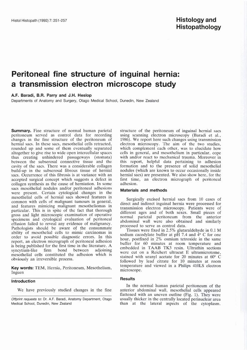
Histol Histopath (1 992) 7: 251 -257 Histology and Histopathology
Peritoneal fine structure of inguinal hernia: a transmission electron microscope study A.F. Baradi, B.R. Parry and J.H. Heslop Departments of Anatomy and Surgery, Otago Medical School, Dunedin, New Zealand
Sumrnary. Fine structure of normal human parietal peritoneum served as control data for recording changes in the fine structure of the peritoneum of hernial sacs. In these sacs, mesothelial cells retracted, rounded up and some of them eventually separated altogether to give rise to wide open intercellular spaces thus creating unhindered passageways (stomata) between the subserosal connective tissue and the cavity of the sacs. There was a considerable collagen build-up in the subserosal fibrous tissue of hernial sacs. Occurrence of this fibrosis is at variance with an accepted surgical concept which suggests a defect in collagen synthesis as the cause of herniation. In some sacs mesothelial nodules andior peritoneal adhesions were present. Certain cytological changes in the mesothelial cells of hernial sacs showed features in common with cells of malignant tumours in general, and features mimicing malignant mesotheliomas in particular. This is in spite of the fact that thorough gross and light microscopic examination of operative specimens and cytological evaluation of peritoneal effusion failed to reveal any evidence of malignancy. Pathologists should be aware of the consummate ability of mesothelial cells to mimic carcinomas in order to avoid possible diagnostic errors. In this report, an electron micrograph of peritoneal adhesion is being published for the first time in the literature. A syncytium-like firm bond between adjoining mesothelial cells constituted the adhesion which is obviously an irreversible process.
Key words: TEM, Hernia, Peritoneum, Mesothelium, Inguen
lntroduction
We have previously studied changes in the fine
Offprint requests to: Dr. A.F. Baradi, Anatomy Department, Otago Medical School, Dunedin, New Zealand
structure of the peritoneum of inguinal hernial sacs using scanning electron microscopy (Baradi et al., 1986). We report here such changes using transmission electron microscopy. The aim of the two studies, which complement each other, was to elucidate how cells in general, and mesothelium in particular, cope with andlor react to mechanical trauma. Moreover in this report, helpful data pertaining to adhesion formation and to the presence of solid mesothelial nodules (which are known to occur occasionally inside hernial sacs) are presented. We also show here, for the first time, an electron micrograph of peritoneal adhesion.
Materials and methods
Surgically excised hernial sacs from 10 cases of direct and indirect inguinal hernia were processed for transmission electron microscopy. Patients were of different ages and of both sexes. Small pieces of normal parietal peritoneum from the anterior abdominal wall were also obtained and similarly processed to serve as control data.
Tissues were fixed in 2.5% glutaraldehyde in 0.1 M sodium cacodylate buffer at pH 7.4 and 4O C for one hour, postfied in 2% osmium tetroxide in the same buffer for 60 minutes at room temperature and embedded in TAAB TK3 resin. Ultrathin sections were cut on a Reichert ultracut E ultramicrotome, stained with uranyl acetate for 20 minutes at 60° C followed by lead citrate for 10 minutes at room temperature and viewed in a Philips 410LS electron microscope.
Results
In the normal human parietal peritoneum of the anterior abdominal wall, mesothelial cells appeared flattened with an uneven outline (Fig. 1). They were usually thicker in the centrally located perinuclear area than at the lateral aspects of the cytoplasm.
TEM study of hernia1 peritoneum
TEM study of hernia1 peritoneum
,F,? :$,,<,i .$. !iC*~.,d.)J sr, .. . .,i
..-;.,:$i.@@.:; ;$.e .*-- .' Tf ,:. P
254 TEM study of hernia1 peritoneum
TEM study of hernial peritoneum
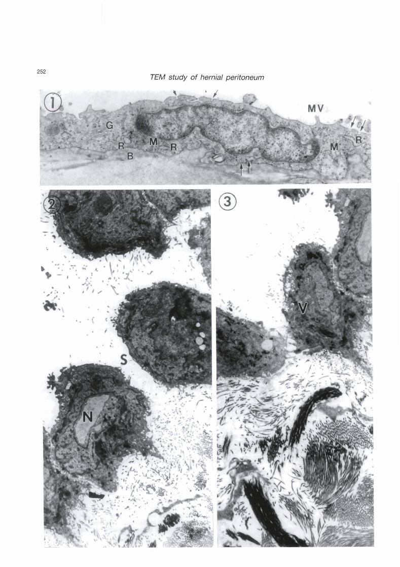
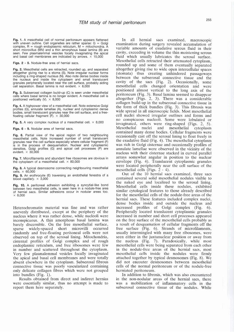
Fig. l. A mesothelial cell of normal peritoneum appears flattened with uneven outline. Cell organelles are rather sparse: G = Golgi complex, R = rough endoplasmic reticulum, M = mitochondria. A short microvillus (MV) and a thin amorphous basal lamina (B) are seen. Few plasmalemmal vesicles focally invaginating the apical and basal cell membranes are indicated by arrows. x 15,000
ngs. 2 - 5. Nodule-free area of hemial sacs.
Fig. 2. Mesothelial cells are retracted, rounded up, and separated altogether giving rise to a stoma (S). Note irregular nuclear forms including a ring-shaped nucleus (N). Also note dense bodies inside the nucleus and inside the cytoplasm and small translucent granules peripherally located near the cell surface, probably aiding cell separation. Basal lamina is not evident. x 8,000
Fig. 3. Subserosal collagen build-up (C) is seen under mesothelial cells where basal lamina is no longer evident. A mesothelial cell is positioned vertically O/). x 8,000
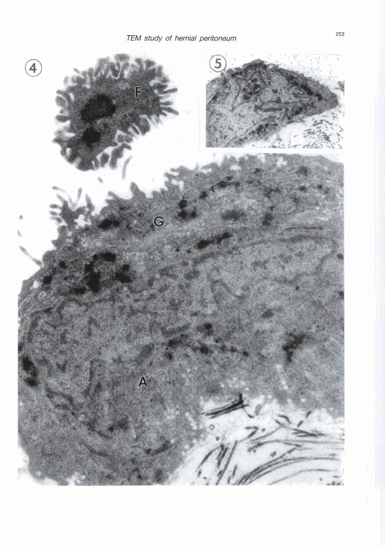
Fig. 4. A highpower view of a mesothelial cell. Note extensive Golgi profiles (G), annulate lamellae (A), nuclear and cytoplasmic dense bodies, small translucent granules near the cell surface, and a free- floating cellular fragment (F). x 30,000
Fig. 5. A very complex nucleus of a mesothelial cell. x 8,000
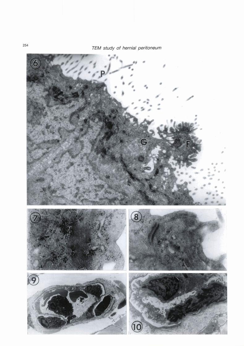
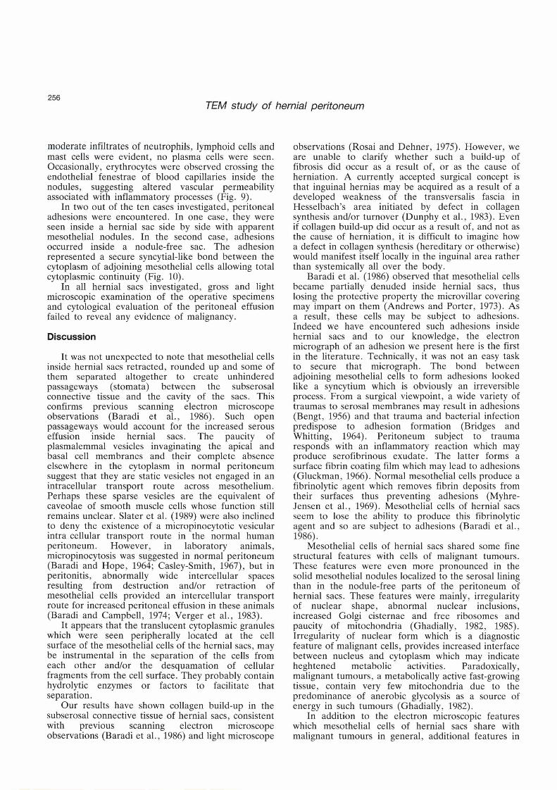
Figs. 6 - 9. Nodular area of hemial sacs.
Fig. 6. Partial view of the apical region of two neighbouring mesothelial cells. Note increased number of small translucent granules near the cell surface especially where cellular fragment (F) is in the process of desquamation. Nuclear and cytoplasmic densities, Golgi profiles (G) and apical cell processes (P) are evident. x 30,000
Fig. 7. Microfilaments and abundant free nbosomes are obvious in the cytoplasm of a rnesothelial cell. x 80,000
Fig. 8. A typical desmosome connecting neighbouring mesothelial cells. x 60,000 Fig. 9. An etythrocyte (E) traversing an endothelial fenestra of a blwd capillary. x 3,000
Fig. 10. A peritoneal adhesion exhibiting a syncytial-like bond between two mesothelial cells, is seen here in a nodule-free area of hemial sacs. Similar adhesions occur in nodular areas too. X 8,000
Heterochromatin material was fine and was rather unevenly distributed, except at the periphery of the nucleus where it was rather dense, while nucleoli were inconspicuous. A thin amorphous basal lamina was barely discernible. On the free mesothelial surface, sparse widely-spaced short microvilli occurred randomly and free-floating pentoneal cells were not obsemed on top of the serosal lining. Mitochondria, cisterna1 profiles of Golgi complex and of rough endoplasmic reticulum, and free ribosomes were few in number and scattered throughout the cytoplasm. Very few plasmalemmal vesicles focally invaginated the apical and basal cell membranes and were totally absent elsewhere in the cytoplasm. Subserosal fibrous connective tissue was poorly vascularized containing only delicate collagen fibres which were not grouped into bundles (Fig. 1).
Results obtained from direct and indirect hernias were essentially similar, thus no attempt is made to report them here separately.
In al1 hernial sacs examined, macroscopic examination during surgery revealed accumulation of variable amounts of exudative serous fluid in their cavity, exceeding in volume the thin moistening serous fluid which usually lubricates the serosal surface. Mesothelial cells retracted their attenuated cytoplasm, rounded up and some of them eventually separated altogether giving rise to wide open intercellular spaces (stomata) thus creating unhindered passageways between the subserosal connective tissue and the cavity of the sacs (Fig. 2). Occasionally, some mesothelial cells changed orientation and were positioned almost vertical to the long axis of the peritoneum (Fig. 3). Basal lamina seemed to disappear altogether (Figs. 2, 3). There was a considerable collagen build-up in the subserosal connective tissue in the form of thick bundles (Fig. 3). This fibrosis was wide spread in al1 microscopic fields. Most mesothelial cell nuclei showed irregular outlines and forms and no conspicuous nucleoli. Some were lobulated or invaginated, others were ring-shaped (Figs. 2, 5). Mesothelial nuclei and mesothelial cytoplasm contained many dense bodies. Gellular fragments were occasionally cast off the serosal lining to float freely in the exudative fluid (Fig. 4). The mesothelial cytoplasm was rich in Golgi cisternae and occasionally profiles of annulate lamellae were obsemed in the vicinity of the nucleus with their cisternae stacked in curved parallel arrays somewhat angular in position to the nuclear envelope (Fig. 4). Translucent cytoplasmic granules were located peripherally near the cell surface of the mesothelial cells (Figs. 2 - 4).
Out of the 10 hernial sacs examined, three sacs contained several solid mesothelial nodules visible to the naked eye and localized to the serosal lining. Mesothelial cells inside these nodules, exhibited similar cytological features to those already described for the mesothelial cells of the nodule-free areas of the hernial sacs. These features included complex nuclei, dense bodies inside and outside the nucleus and increased profiles of Golgi complex (Fig. 6). Peripherally located translucent cytoplasmic granules increased in number and short cell processes appeared on the free surface of the mesothelial cells probably as a result of desquamation of cellular fragments off the free surface (Fig. 6). Strands of microfilaments, usually intermingled with many free ribosomes, were seen either in the juxtanuclear position or away from the nucleus (Fig. 7). Paradoxically, while most mesothelial cells were being separated from each other in the nodule-free areas of the hernial sacs, most mesothelial cells inside the nodules were firmly attached together by typical desmosomes (Fig. 8). We did not encouter desmosomes between mesothelial cells of the normal peritoneum or of the nodule-free herniated peritoneum.
In addition to fibrosis, which was also encountered in the non-nodular areas of the hernial sacs, there was a mobilization of idammatory cells in the subserosal connective tissue of the nodules. While
TEM study of hernial peritoneum
moderate infiltrates of neutrophils, lymphoid ceils and mast cells were evident, no plasma cells were seen. Occasionally, erythrocytes were observed crossing the endothelial fenestrae of blood capillaries inside the nodules, suggesting altered vascular permeabiiity associated with inflamrnatory processes (Fig. 9).
In two out of the ten cases investigated, peritoneal adhesions were encountered. In one case, they were seen inside a hemial sac side by side with apparent mesothelial nodules. In the second case, adhesions occurred inside a nodule-free sac. The adhesion represented a secure syncytial-like bond between the cytoplasm of adjoining mesothelial cells allowing total cytoplasmic continuity (Fig. 10).
In al1 hernial sacs investigated, gross and light microscopic examination of the operative specimens and cytological evaluation of the peritoneal effusion failed to reveal any evidence of malignancy.
Discussion
It was not unexpected to note that mesotheiial cells inside hernial sacs retracted, rounded up and some of them separated altogether to create unhindered passageways (stomata) between the subserosal connective tissue and the cavity of the sacs. This confirms previous scanning electron microscope observations (Baradi et al., 1986). Such open passageways would account for the increased serous effusion inside hemial sacs. The paucity of plasmalemmal vesicles invaginating the apical and basa1 cell membranes and their complete absence elsewhere in the cytoplasm in normal peritoneum suggest that they are static vesicles not engaged in an intracellular transport route across mesothelium. Perhaps these sparse vesicles are the equivalent of caveolae of smooth muscle cells whose function still remains unclear. Slater et al. (1989) were also inclined to deny the existence of a micropinocytotic vesicular intra cellular transport route in the normal human peritoneum. However, in laboratory animals, micropinocytosis was suggested in normal peritoneum (Baradi and Hope, 1964; Casley-Smith, 1967), but in peritonitis, abnormaily wide intercellular spaces resulting from destruction andor retraction of mesothelial cells provided an intercellular transport route for increased peritoneal effusion in these animals (Baradi and Campbell, 1974; Verger et al., 1983).
It appears that the translucent cytoplasmic granules which were seen peripherally located at the cell surface of the mesothelial cells of the hernial sacs, rnay be instrumental in the separation of the cells from each other andlor the desquamation of cellular fragments from the cell surface. They probably contain hydrolytic enzymes or factors to facilitate that separation.
Our results have shown collagen build-up in the subserosal connective tissue of hernial sacs, consistent with previous scanning electron microscope observations (Baradi et al., 1986) and light microscope
observations (Rosai and Dehner, 1975). However, we are unable to clarify whether such a build-up of fibrosis did occur as a result of, or as the cause of herniation. A currently accepted surgical concept is that inguinal hernias rnay be acquired as a result of a developed weakness of the transversalis fascia in Hesselbach's area initiated by defect in collagen synthesis andor turnover (Dunphy et al., 1983). Even if collagen build-up did occur as a result of, and not as the cause of herniation, it is difficult to imagine how a defect in collagen synthesis (hereditary or otherwise) would manifest itself locally in the inguinal area rather than systemically al1 over the body.
Baradi et al. (1986) observed that mesothelial cells became partially denuded inside hernial sacs, thus losing the protective property the microvillar covering rnay impart on them (Andrews and Porter, 1973). As a result, these cells rnay be subject to adhesions. Indeed we have encountered such adhesions inside hernial sacs and to our knowledge, the electron micrograph of an adhesion we present here is the first in the literature. Technically, it was not an easy task to secure that micrograph. The bond between adjoining mesothelial cells to form adhesions looked like a syncytium which is obviously an irreversible process. From a surgical viewpoint, a wide variety of traumas to serosal membranes rnay result in adhesions (Bengt, 1956) and that trauma and bacteria1 infection predispose to adhesion formation (Bridges and Whitting, 1964). Peritoneum subject to trauma responds with an inflammatory reaction which rnay produce serofibrinous exudate. The latter forms a surface fibrin coating film which rnay lead to adhesions (Gluckman, 1966). Normal mesothelial cells produce a fibrinolytic agent which removes fibrin deposits from their surfaces thus preventing adhesions (Myhre- Jensen et al., 1969). Mesothelial cells of hernial sacs seem to lose the ability to produce this fibrinolytic agent and so are subject to adhesions (Baradi et al., 1986).
Mesothelial cells of hernial sacs shared some fine structural features with cells of malignant tumours. These features were even more pronounced in the solid mesothelial nodules localized to the serosal lining than in the nodule-free parts of the peritoneum of hernial sacs. These features were mainly, irregularity of nuclear shape, abnormal nuclear inclusions, increased Golgi cistemae and free ribosomes and paucity of mitochondria (Ghadially, 1982, 1985). Irregularity of nuclear form which is a diagnostic feature of malignant cells, provides increased interface between nucleus and cytoplasm which rnay indicate heghtened metabolic activities. Paradoxically, malignant tumours, a metabolically active fast-growing tissue, contain very few mitochondna due to the predominance of anerobic glycolysis as a source of energy in such tumours (Ghadially, 1982).
In addition to the electron microscopic features which mesothelial cells of hemial sacs share with malignant tumours in general, additional features in
TEM study of hernial peritoneum
our report were similar to those described in the literature for malignant mesothelioma in particular. In mesotheliomas, there is an abundance of annulate lamellae, desmosomes, translucent cytoplasmic granules and cytoplasmic microfilaments (Ghadially, 1982, 1985). Such electron microscopic features were particularly prominent in the mesothelial nodules of hernial sacs in our preparations. If the cytoplasmic microfilaments in our preparations are fibrin, then according to Parry and Ghadially (1967), this may be due to the escape of plasma proteins from leaky blood vessels, a process known as insudation, followed by the conversion of fibrinogen into fibrin by metabolic factors released from injured cells during inflammation. It is noteworthy that malignant mesotheliomas usually lack an inflammatory component (Rosai and Dehner, 1975). This is further evidence that mesothelial nodules of hernial sacs are not malignant as they usually involve inflammation among their features which we already discussed.
Ackerman (1954) drew attention to the consummate ability of mesothelial cells to stimulate adenocarcinomas. Rosai and Dehner (1975) reviewed 13 cases of hernial sacs that exhibited benign mesothelial nodules, similar to those we report here, and simulated tumours of malignant mesothelioma. Their light microscopic findings are remarkably similar to our electron microscopic data. Thorough gross anatomical and light microscopic examination of the operative specimens in their report, however, failed to reveal any evidence of malignant tumours, such as atypical mitotic figures and prominent nucleoli. Nistal and Santamaria (1981) also reported similar observations. We join these authors in suggesting that if a diagnostic difficulty arises over a suspected malignancy in the peritoneal cavity, cytological examination of the peritoneal effusion should be mandatory to reinforce the light and electron microscopic evaluation of surgical biopsy. Furthermore, the role of electron microscopy in pathological investigation should remain only supplemental to other means of investigation.
In conclusion, awareness by pathologists that benign mesothelial nodules found in hernial sacs do mimic malignant mesotheliomas, should hopefully eliminate the possibility of diagnostic errors. Though mesotheliomas are extremely rare as primary malignant tumors of the peritoneum, yet they are very common as metastases of different types of primary malignant tumours found elsewhere in the body (Thomson and Cotton, 1983).
Acknowledgements. Thanks to Professor Dr. Gareth Jones for reading the manuscript, and to Mr Allan Mitchell for technical assistance.
References
Ackerman L.V. (1954). Tumors of the retroperitoneum,
mesentery and peritoneum. Atlas of Tumor Pathology. Sect. 6. Armed Forces lnstitute of Pathology, Washington DC. 134-1 35.
Andrews P.M. and Porter K.R. (1973). The ultrastructural morphology and possible functional significance of mesothelial microvilli. Anat. Rec. 177, 409-426.
Baradi A.F. and Campbell W.G. (1974). Exudative peritonitis induced in mice by bovine serum albumin: an ultramicroscopic study. Arch. Pathol. 97, 2-12.
Baradi A.F., Heslop J.H. and Rao N.S. (1986). Peritoneal fine structure of inguinal hernia: a scanning electron microscope study. Histol. Histopath. 1, 89-92.
Baradi A.F. and Hope J. (1964). Observations on the ultrastructure of rabbit mesothelium. Exp. Cell Res. 34, 33-44.
Bengt F. (1956). Polyphloretin phosphate - a hyaluronidase inhibitor - and hyaluronidase in prevention of intraperitoneal adhesions. Acta Chir. Scand. (Suppl.) 21 7, 1-97.
Bridges J.B. and Whitting H.W. (1964). Parietal peritoneal healing in the rat. J. Pathol. Bacteriol. 87, 123-130.
Casley-Smith J.R. (1967). An electron microscopical study of the passage of ions through the endothelium of lymphatic and blood capillaries and through the mesothelium. Quart. J. Exp. Physiol. 52, 105-1 13.
Dunphy J.E., Hall A.D. and Linder H.H. (1983). Hernias and other lesions of the abdominal wall. In: Current surgical diagnosis and treatment. 6th ed. Way L.E. (ed). Lange Medical Publications, Los Altos, California. pp 667-682.
Ghadially F.N. (1985). Ultrastructural pathology of the cell and matrix. 2nd ed. Butterworths. London. pp 6, 228, 354, 428 and 810.
Ghadially F.N. (1985). Diagnostic electron microscopy of tumours. 2nd ed. Butterworths. London. pp 40, 59, 97 and 102.
Gluckman D.L. (1966). Serosal integrity and intestinal adhesions. Surgery 60, 1009-1 01 1.
Myhre-Jensen O., Larsen S.B. and Astrup T. (1969). Fibrinolytic activity in serosal and synovial membranes. Arch. Pathol. 88, 623-630.
Nistal M. and Santamaria L. (1981). Hiperplasia mesotelial en sacos herniarios. Med. Esp. 80, 43-46.
Parry E.W. and Ghadially F.N. (1967). Fibrin in hepatocytes. Naturwissenchaften 20, 541 -555.
Rosai J. and Dehner L.P. (1975). Nodular mesothelial hyperplasia in hernia sacs: a benign reactive condition simulating a neoplastic process. Cancer 35, 165-1 75.
Slater N.J., Raítery A.T. and Cope G.H. (1989). The ultrastructure of human abdominal mesothelium. J. Anat. 167, 47-56.
Thomson A.D. and Cotton R.E. (1983). Lecture notes on pathology, 3rd ed. Blackwell Scientific Publications. Oxford. pp 221.
Verger C., Luger A., More H.L. and Nolph K.D. (1983). Acute changes in peritoneal morphology and transport properties with infectious peritonitis and mechanical injury. Kidney Internat. 23, 823-831.
Accepted October 25, 1991