Embed Size (px)
Citation preview
FALL 2015 SUPPLEMENT TO ENDOVASCULAR TODAY 1
There are multiple endovascular options for treat-ment of infrainguinal disease, but treatment of severe calcific disease of the superficial femoral
artery (SFA), popliteal artery, and tibial vessels remains a challenge. Peripheral atherectomy is a unique treat-ment modality because it allows debulking of plaque with luminal gain and minimal barotrauma. This results in less injury to the vessel during initial treatment and theoretically reduces hyperplastic reaction to the initial treatment. In severely calcific vessels, calcium debulking changes the vessel wall compliance with the removal of calcium. It can then be treated with low-pressure balloon inflation with minimal injury to the vessel wall. This is now a particularly attractive concept with the availability of drug-coated balloons and drug-eluting stents, as the vessel can be prepared with atherectomy before delivery of these devices. This may ensure ade-quate drug delivery to the tissue, thereby reducing inti-mal hyperplastic reaction and increasing durability of
the procedures. Prevailing concerns with atherectomy (ie, dissection, perforation, clinically significant emboli-zation, and durability) have prevented the widespread use of atherectomy.1
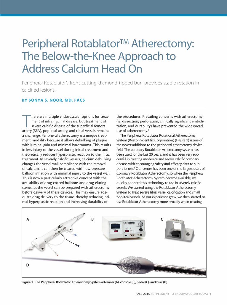
The Peripheral Rotablator Rotational Atherectomy System (Boston Scientific Corporation) (Figure 1) is one of the newer additions to the peripheral atherectomy device field. The coronary Rotablator Atherectomy system has been used for the last 20 years, and it has been very suc-cessful in treating moderate and severe calcific coronary disease, with encouraging safety and efficacy data to sup-port its use.2 Our center has been one of the largest users of Coronary Rotablator Atherectomy, so when the Peripheral Rotablator Atherectomy System became available, we quickly adopted this technology to use in severely calcific vessels. We started using the Rotablator Atherectomy System to treat severe tibial vessel calcification and small popliteal vessels. As our experience grew, we then started to use Rotablator Atherectomy more broadly when treating
Peripheral Rotablator’s front-cutting, diamond-tipped burr provides stable rotation in
calcified lesions.
Peripheral Rotablator™ Atherectomy: The Below-the-Knee Approach to Address Calcium Head On
BY SONYA S. NOOR, MD, FACS
Figure 1. The Peripheral Rotablator Atherectomy System advancer (A), console (B), pedal (C), and burr (D).
A B C
D
06_peripheral.indd 1 1/27/16 2:24 PM
2 SUPPLEMENT TO ENDOVASCULAR TODAY FALL 2015
infrainguinal calcific disease, and found a reduction in the use of stents (or only focal stenting was required).
ROTABLATOR FEATURES AND MECHANISM OF ACTION
The Peripheral Rotablator Atherectomy System is fairly simple to use and requires a connection to the console, a power source, a compressed nitrogen tank, and an IV fluid mix to start using the device. The foot pedal starts the rotational atherectomy. Usually, 20- to 30-second runs are done under live fluoroscopy. This device uses a front-cutting diamond-coated burr to ablate the cal-cium particles, while rotating at 160,000 to 180,000 RPM in the lumen of the vessel. The coronary literature has studied the ablation particles over the last 20 years, and when proper technique is employed, the ablation par-ticles generated measured about 5 µm, which is smaller than a red blood cell. These particles are then washed downstream during the treatment and picked up by the reticular endothelial system. For this reason, embolic pro-tection devices are of no use, as the pore size of embolic protection devices are generally in the range of 100 µm and would not catch the ablation particles. Rotablator Atherectomy can only be performed with a 0.009-inch wire, which does not support the use of embolic protec-tion devices, either.
The Rotablator Atherectomy System’s front-cutting dia-mond burr is very useful in moderately and severe calcific stenotic lesions, as it can ablate its way through the calcium and create a channel that is smooth and has a predictable concentric lumen.
Other devices have a leading edge, which has to be introduced first through the lumen before the device can be introduced and treatment can be performed, requiring predilatation or dottering, and causing barotrauma to the vessel prior to treatment. We have found this front-cutting feature particularly helpful in moderate and severe calcific disease and when negoti-ating even a predilatation balloon catheter can prove difficult.
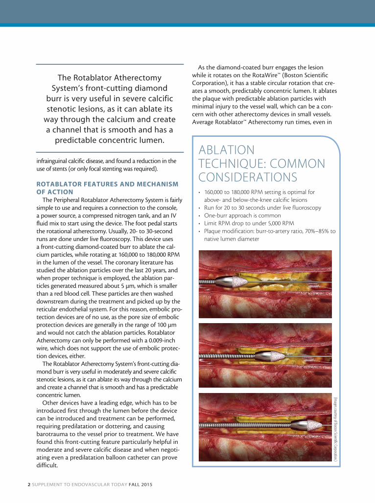
As the diamond-coated burr engages the lesion while it rotates on the RotaWire™ (Boston Scientific Corporation), it has a stable circular rotation that cre-ates a smooth, predictably concentric lumen. It ablates the plaque with predictable ablation particles with minimal injury to the vessel wall, which can be a con-cern with other atherectomy devices in small vessels. Average Rotablator™ Atherectomy run times, even in
ABLATION TECHNIQUE: COMMON CONSIDERATIONS• 160,000 to 180,000 RPM setting is optimal for
above- and below-the-knee calcific lesions• Run for 20 to 30 seconds under live fluoroscopy • One-burr approach is common• Limit RPM drop to under 5,000 RPM• Plaque modification: burr-to-artery ratio, 70%–85% to
native lumen diameter
The Rotablator Atherectomy System’s front-cutting diamond
burr is very useful in severe calcific stenotic lesions, as it can ablate its
way through the calcium and create a channel that is smooth and has a
predictable concentric lumen.
(Images courtesy of Boston Scientific Corporation.)
06_peripheral.indd 2 1/27/16 2:24 PM
FALL 2015 SUPPLEMENT TO ENDOVASCULAR TODAY 3
long, diffuse lesions, are typically 3 to 4 minutes per vessel, making this an efficient treatment modality.
ROTABLATOR BEST PRACTICES As with other atherectomy devices, the Rotablator
Atherectomy System has its own learning curve and per-forms well when proper technique is employed. We have used the Rotablator™ Atherectomy System in severe cal-cific disease as the first line of treatment for these lesions. We currently use it in the SFA, popliteal, and tibial vessels as the first line of treatment. Rotablator Atherectomy proves to be an effective tool for calcific ablation requir-ing low atmospheric balloon postdilatation and only focal stenting, if at all necessary. We are also starting to use Rotablator Atherectomy for vessel preparation before drug-coated balloon usage in order to potentially improve drug uptake in the vessel wall.
We have found that the 160,000 to 180,000 RPM set-ting is optimal for both above- and below-knee calcific lesions. Twenty- to 30-second runs under live fluoros-copy and a slow, deliberate pecking action with the burr engaging the lesion for a second or two, followed by a gentle pullback, results in successful luminal gain. It is important to engage the lesion with the burr, but also pull back for 1 to 2 seconds, which allows dissipation of frictional heat and flushing of microparticles into the distal circulation. Overzealous advancement of the burr can lead to the device stalling within calcific disease and distal embolization, which should be avoided.
CASE 1*A 71-year-old African American woman with end-
stage renal disease, a previous cerebrovascular accident, coronary artery disease, and coronary artery bypass
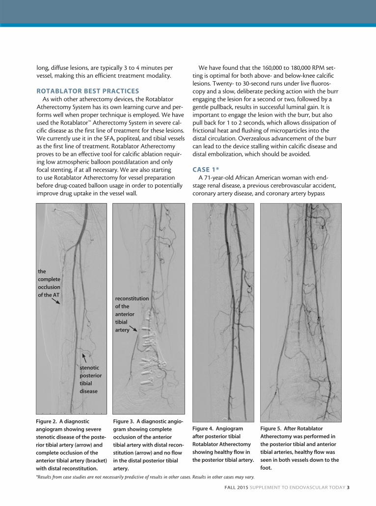
Figure 2. A diagnostic
angiogram showing severe
stenotic disease of the poste-
rior tibial artery (arrow) and
complete occlusion of the
anterior tibial artery (bracket)
with distal reconstitution.
Figure 3. A diagnostic angio-
gram showing complete
occlusion of the anterior
tibial artery with distal recon-
stitution (arrow) and no flow
in the distal posterior tibial
artery.
Figure 4. Angiogram
after posterior tibial
Rotablator Atherectomy
showing healthy flow in
the posterior tibial artery.
Figure 5. After Rotablator
Atherectomy was performed in
the posterior tibial and anterior
tibial arteries, healthy flow was
seen in both vessels down to the
foot.
reconstitution
of the
anterior
tibial
artery
the
complete
occlusion
of the AT
stenotic
posterior
tibial
disease
*Results from case studies are not necessarily predictive of results in other cases. Results in other cases may vary.
06_peripheral.indd 3 1/27/16 2:24 PM
4 SUPPLEMENT TO ENDOVASCULAR TODAY FALL 2015
graft surgery presented with a left heel ulcer that was not healing despite treatment for many months. The patient’s arterial Doppler showed noncompressible ves-sels and a toe-brachial index of 0.1, with reduced wave-forms at all levels.
Procedural DetailsLeft common femoral artery ultrasound and puncture
was performed with placement of a right side sheath, and a diagnostic angiogram was obtained, which clearly showed complete occlusion of the posterior tibial and anterior tibial artery (Figures 2 and 3). The patient under-went placement of a 5-F, 70-cm sheath and was hepa-rinized before successful crossing of the posterior tibial artery occlusion and subsequently, the anterior tibial artery. The patient underwent Rotablator Atherectomy with a 1.75-mm burr of the posterior tibial artery first (Figure 4), followed by the anterior tibial artery. The total run time was 4 minutes, and the 180,000 RPM setting
was used in both arteries. After atherectomy was com-pleted, low-pressure balloon angioplasty was performed using a 2.5-mm X 220-mm balloon, for a total of 2 min-utes for each inflation.
Completion angiography showed no evidence of dis-section, perforation, or distal embolization (Figure 5).
At 5-month follow-up, the patient was found to have almost completely healed ulcers, and arterial Dopplers showed improved waveforms at all levels, noncompressible ankle-brachial indices (ABIs), and a toe-brachial index of 0.5.
CASE 2*A 65-year-old man presented with severe claudication
and ischemic ulceration of the right second toe. Arterial Doppler exam showed an ABI of 0.58 on the right and 0.90 on the left. Pulmonary vascular resistance wave-forms indicated distal SFA and popliteal artery disease.
Procedural DetailsThe patient underwent left common femoral artery
access with placement of a 5-F sheath. Diagnostic angi-ography confirmed complete occlusion of the right SFA and popliteal artery at the adductor canal (Figure 6), with reconstitution of a popliteal artery at the knee joint and some mild diffuse tibial vessel disease. The patient underwent placement of a 7-F, 70-cm sheath, after which the patient was heparinized. A stiff Glidewire (Terumo Interventional Systems) and a 0.035-inch Quick-Cross catheter (Spectranetics Corporation) was used to cross the total occlusion, and reentry into the popliteal artery was confirmed. The RotaWire was placed into the tibial vessels, and Rotablator Atherectomy was performed using a 2.5-mm burr. A setting of 170,000 to 180,000 RPM was used for a total of 5 minutes. Then a 5- X 220-mm-long Sterling™ Balloon (Boston Scientific Corporation) was used for low-pressure angioplasty of 4 atm, for a total of 3 min-utes. A follow-up angiogram (Figure 7) revealed excellent
Figure 6. A diagnostic
angiogram showing
occlusion of the distal SFA
and popliteal artery.
Figure 7. Angiogram after
Rotablator Atherectomy and
low-pressure angioplasty.
Restoration of flow is noted
in the SFA and popliteal
artery.
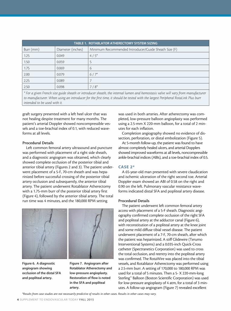
TABLE 1. ROTABLATOR ATHERECTOMY SYSTEM SIZING
Burr (mm) Diameter (inches) Minimum Recommended Introducer/Guide Sheath Size (F)
1.25 0.049 4 / 5*
1.50 0.059 5
1.75 0.069 6
2.00 0.079 6 / 7*
2.25 0.089 7
2.50 0.098 7 / 8*
* For a given French size guide sheath or introducer sheath, the internal lumen and hemostasis valve will vary from manufacturer to manufacturer. When using an introducer for the first time, it should be tested with the largest Peripheral RotaLink Plus burr intended to be used with it.
*Results from case studies are not necessarily predictive of results in other cases. Results in other cases may vary.
06_peripheral.indd 4 1/27/16 2:24 PM

FALL 2015 SUPPLEMENT TO ENDOVASCULAR TODAY 5
flow, good luminal gain, and no evidence of perforation, dissection, or significant embolization.
The patient was seen in follow-up 1 month after the procedure with no complaints of claudication, near com-plete ulceration healing, and arterial Doppler exams that showed an ABI of 0.84 on the right and 0.77 on the left, with good waveforms at all levels.
CONCLUSIONThe Rotablator Atherectomy System has been used
to treat moderate and severe calcific disease safely and efficiently for over 20 years in the coronary vasculature, and we started to use Rotablator Atherectomy to treat similar calcific disease in the periphery. At our center, we now use Rotablator Atherectomy as the first line of treatment whenever we encounter moderate or severe calcific disease. We have found the Rotablator Atherectomy System to be easy to set up and use, and it is efficient in ablating and treating calcium with short procedure times. There have been minimal dissections, perforation, or clinically significant embolization. As with all atherectomy devices, it is important to use proper technique while handling the device to minimize compli-cations. The benefit of the front-cutting diamond burr is
especially useful in negotiating tight stenotic or occlusive lesions (where no predilatation is necessary), thereby minimizing barotrauma to the vessel before treatment. The stable rotation of the burr engages the calcium and ablates it, leaving a predictable concentric lumen. We have been successful in changing vessel compliance with calcium ablation, allowing minimal adjunctive therapy (such as low-pressure angioplasty, no stenting, or only focal stenting). We are also starting to use the Rotablator Atherectomy System to remove the calcium plaque bur-den and prep the vessel before use of drug-coated bal-loons. It remains to be seen if this strategy enhances drug uptake into to the vessel wall, and therefore increase the durability of this procedure. n
Sonya S. Noor, MD, FACS, is a clinical associate professor with the University at Buffalo Gates Vascular Institute in Buffalo, New York. She has disclosed that she is a consul-tant for Boston Scientific Corporation. Dr. Noor may be reached at [email protected].
1. Zeller T, Frank U, Bürgelin K, et al. Initial clinical experience with percutaneous atherectomy in the infragenicular
arteries. J Endovasc Ther. 2003;10:987-993.
2. Reisman MD. Guide to Rotational Atherectomy. Birmingham, MI: Physicians Press; 1997.
06_peripheral.indd 5 1/27/16 2:24 PM

ABBREVIATED STATEMENTS
ROTABLATOR PERIPHERAL ROTALINK PLUS ROTABLATOR PERIPHERAL ROTAWIRE GUIDEWIRE AND WIRECLIP TORQUER ROTABLATOR ROTATIONAL ATHERECTOMY SYSTEM CONSOLE ROTAGLIDE LUBRICANTCAUTION: Federal law (USA) restricts this device to sale by or on the order of a physician. Rx only. Prior to use, please see the complete “Directions for Use” for more information on Indications, Contraindications, Warnings, Precautions, Adverse Events, and Operator’s Instructions.Rotalink Plus INTENDED USE/INDICATIONS FOR USEThe Rotablator Rotational Atherectomy System is intended for percutaneous use in the peripheral vessels in patients with occlusive atherosclerotic disease who are acceptable candidates for endovascular procedures.RotaWire: INDICATIONS FOR USE/INTENDED USEThese guidewires are intended for use with the Rotablator Rotational Atherectomy System.Lubricant INDICATIONS FOR USE Rotaglide lubricant is intended for use with the Rotablator atherectomy system, for the purpose of increas-ing the lubricity of the system. CONTRAINDICATIONS AND RESTRICTIONSContraindications1. Occlusions through which a guidewire will not pass.2. Use in coronary arteries.3. Long (≥ 20 cm) total occlusions.4. Angiographic evidence of thrombus prior to treatment with the Rotablator Rotational Atherectomy System. Such patients may be treated with thrombolytics (e.g., Urokinase). When the thrombus has been resolved for two to four weeks, the lesion may be treated with the Rotablator Rotational Atherectomy System.5. Angiographic evidence of significant dissection at the treatment site. The patient may be treated conser-vatively for approximately four weeks to permit the dissection to heal before treating the lesion with the Rotablator Rotational Atherectomy System.Lubricant CONTRAINDICATIONS Rotaglide™ lubricant is contraindicated in patients with known allergies to the lubricant ingredients: olive oil, egg yolk phospholipids, glycerin, sodium deoxycholate, L-histidine, disodium EDTA, sodium hydroxide, and water. Restrictions• Federal (USA) law restricts the use of this system to physicians who are credentialed in peripheral angio-plasty and who have attended the Rotablator System Physician Training Program.WARNINGS• The risks of Rotational Atherectomy can be reduced if the device and associated accessories are used in
the appropriate patient population by a physician who has had adequate training.• If the Peripheral RotaLink Plus shows evidence of mechanical failure at any time prior to or during the
angioplasty procedure, immediately discontinue use of the device and return it to Customer Service for evaluation. Do NOT attempt to use a damaged Peripheral RotaLink Plus; use may result in device malfunc-tion and/or patient injury.
• Never operate the Peripheral RotaLink Plus without saline infusion. Flowing saline is essential for cooling and lubricating the working parts of the advancer. Operation of the advancer without proper saline infu-sion may result in permanent damage to the advancer.
• Never operate the Peripheral RotaLink Plus with the Rotablator Rotational Atherectomy System in Dynaglide™ mode or operate the guidewire brake defeat button unless you have a firm grip on the guide-wire using the wireClip™ Torquer. The wireClip Torquer may be held with the fingers or inserted com-pletely into the docking port after the brake button is depressed. Defeating the brake, or operating the Peripheral RotaLink Plus with the Rotablator Rotational Atherectomy System in Dynaglide mode, without securing the guidewire may result in rotation and entanglement of the guidewire.
• During setup of the Peripheral RotaLink Plus never grip or pull on the flexible shaft.• The burr at the distal tip of the Peripheral RotaLink Plus is capable of rotating at very high speeds. Do
NOT allow parts of the body or clothing to come in contact with the burr. Contact may result in physical injury or entanglement.
• Never advance the rotating burr to the point of contact with the guidewire spring tip. Such contact could result in distal detachment and embolization of the tip.
• If the Peripheral RotaLink Plus stops and the red STALL light on the console illuminates, retract the burr and immediately discontinue treatment. Check the advancer for proper connection to the console. If the connections are correct, use fluoroscopy to analyze the situation. Never force the system when rotational or translational resistance occurs, as vessel perforation may occur.
• Never advance the rotating burr by advancing the sheath. Guidewire buckling may occur and perforation or vascular trauma may result. Always advance the rotating burr by using the advancer knob.
• If resistance is encountered, retract the burr and stop treatment immediately. Use fluoroscopy to analyze the situation. Never force the Peripheral RotaLink Plus when rotational or translational resistance occurs, as vessel perforation, vessel trauma or embolism due to burr detachment or fractured wire may occur and in rare instances may result in surgical intervention and death.
• The use of Rotablator Rotational Atherectomy System for in-stent restenosis might lead to damage of stent components and/or Peripheral RotaLink Plus, which may lead to patient injury.
• Always keep the burr advancing or retracting while it is rotating. Maintaining the burr in one location while it is rotating may lead to excessive tissue removal or damage to the Peripheral RotaLink Plus or entrapment of the Peripheral RotaLink Plus. It is best to advance and retreat the burr no more than 3 cm at a time in a smooth pecking motion, being careful to engage the lesion only minimally when resistance is met. Do not allow the individual burr run time to exceed 30 seconds with total rotational procedure time not to exceed five minutes.
RotaWire WARNINGSUse extreme caution and careful judgment in patients for whom anticoagulation is not indicated. Console WARNINGS• Never use oxygen as the propellant for the Rotablator Rotational Atherectomy System.• The use of accessories, transducers and cables other than those specified, with the exception of transduc-
ers and cables sold by the manufacturer of the Rotablator System as replacement parts for internal com-ponents, may result in increased emissions or decreased immunity of the Rotablator System.
• This device is not to be used in the presence of flammable anesthetics.• Do NOT operate the Rotablator Console with gas pressures in excess of 758.4 kPa (110 psi). • Do not modify or repair. Lubricant WARNINGS Discard vial if there are particulates in the emulsion or if an oiling-out of emulsion has occurred. PRECAUTIONS• Percutaneous rotational angioplasty with the Rotablator Rotational Atherectomy System should only be carried out at hospitals where emergency bypass surgery can be immediately performed in the event of a potentially injurious or lifethreatening complication.• Appropriate drug therapy including (but not limited to) anticoagulant and vasodilator therapy must be provided to the patient during all phases of patient care.• When the Peripheral RotaWire™ Guidewires and/or Peripheral RotaLink Plus are in the body, they should only be manipulated while they are under fluoroscopic observation with radiographic equipment that pro-vides high resolution images.• Use only normal saline as the infusate. Never inject contrast agent, or any other substance that is not approved as part of the Rotablator Rotational Atherectomy System, into the infusion port or saline infusion bag as this may cause permanent damage to the Peripheral RotaLink Plus.Console PRECAUTIONS• User should take precautions when using the console in conjunction with other medical electrical equip-ment.• The Rotablator Console needs special precautions regarding EMC and needs to be installed and put into service according to the EMC information provided in Appendix D in the DFU.ADVERSE EVENTSPotential adverse reactions which may result from the use of this device include but are not limited to:• Additional intervention • Allergic reaction • Amputation • Death • Embolism • Hematoma/Hemorrhage• Hemodynamic changes • Hemoglobinuria • Infection • Restenosis • Stroke • Slow, no flow, abrupt vessel closure • Surgery including arterial bypass • Thrombosis and vessel occlusion • Vessel trauma (dissection, perforation, psudoaneurysm, arteriovenous fistula)There may also be complications associated with distortion, kinks, and fracture of the guidewire and physi-cal deterioration or malfunction of the device, which can lead to patient injury or death.
STERLING MR STERLING OTW CAUTION: Federal law (USA) restricts this device to sale by or on the order of a physician. Rx only. Prior to use, please see the complete “Instructions for Use” for more information on Indications, Contraindications, Warnings, Precautions, Adverse Events, and Operator’s Instructions.INTENDED USE/INDICATIONS FOR USEThe Sterling PTA Balloon Dilatation Catheters are indicated for Percutaneous Transluminal Angioplasty in the peripheral vasculature, including iliac, femoral, popliteal, infra-popliteal, renal, and for the treatment of obstructive lesions of native or synthetic arteriovenous dialysis fistulae. These devices are also indicated for post-dilatation of balloon expandable and self-expanding stents in the peripheral vasculature. The Sterling Monorail PTA Balloon Dilatation Catheter (only) is also indicated for the carotid arteries.CONTRAINDICATIONS None Known.GENERAL PRECAUTIONSThe Sterling PTA Balloon Dilatation Catheter should be used with caution for procedures involving calcified lesions or synthetic vascular grafts due to the abrasive nature of these lesions.The Sterling PTA Balloon Dilatation Catheters are not intended for injection of contrast medium.The Sterling™ PTA Balloon Dilatation Catheter shall only be used by physicians trained in the performance of percutaneous transluminal angioplasty.Precautions to prevent or reduce clotting should be taken when any catheter is used:• Consider systemic heparinization.• Flush or rinse all products entering the vascular system with sterile isotonic saline or a similar solution
prior to use.ADVERSE EVENTSThe complications that may result from a balloon dilatation procedure include, but are not limited to:• Allergic reaction to contrast medium • Arrhythmias • Arteriovenous fistula • Cerebrovascular accidents (specific to MR) • Death • Hematoma • Hemodynamic instability • Hemorrhage • Pseudoaneurysm • Pyrogenic reaction • Sepsis/infection • Thromboembolic episodes • Vascular thrombosis • Vessel injury, e.g. dissection, perforation, rupture • Vessel occlusion • Vessel spasm
All Trademarks are the property of their respective owners. © 2015 Boston Scientific Corporation or its affiliates. All rights reserved. PI-360611-AA DEC2015
06_peripheral.indd 6 1/27/16 2:24 PM