Embed Size (px)
Citation preview
Peripheral Nervous System
(nervous Tissue)
September 23, 2009
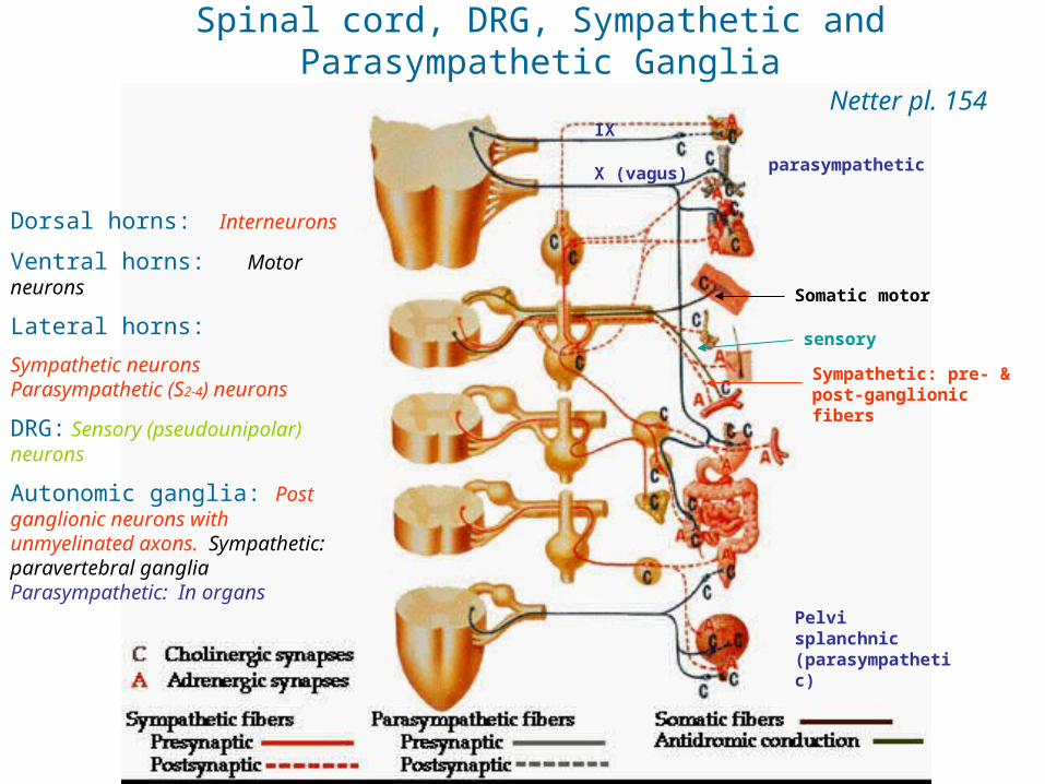
Spinal cord, DRG, Sympathetic and Parasympathetic Ganglia
Netter pl. 154
Dorsal horns: Interneurons
Ventral horns: Motor neurons
Lateral horns:
Sympathetic neurons Parasympathetic (S2-4) neurons
DRG: Sensory (pseudounipolar) neurons
Autonomic ganglia: Post ganglionic neurons with unmyelinated axons. Sympathetic: paravertebral ganglia Parasympathetic: In organs
X (vagus)
IX
parasympathetic
Somatic motor
sensory
Sympathetic: pre- & post-ganglionic fibers
Pelvi splanchnic (parasympathetic)

#65 Spinal cord and DRG
Ventral horn
Lateral horn
Dorsal horn
Dorsal root ganglion (DRG)
65-1N, Trichrome65-2, H&E65-1, Trichrome
Ventral median fissure
Dorsal median sulcus
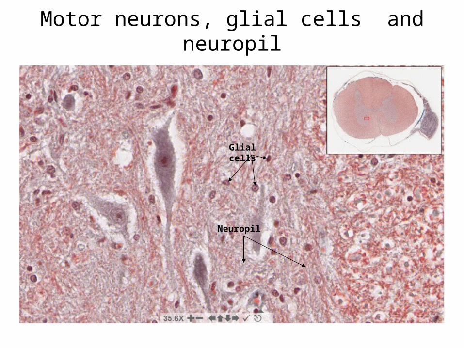
Motor neurons, glial cells and neuropil
Glial cells
Neuropil

Dorsal root ganglion
65-165-1N65-2
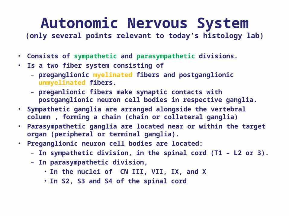
Autonomic Nervous System(only several points relevant to today’s histology lab)
• Consists of sympathetic and parasympathetic divisions.• Is a two fiber system consisting of
– preganglionic myelinated fibers and postganglionic unmyelinated fibers.
– preganlionic fibers make synaptic contacts with postganglionic neuron cell bodies in respective ganglia.
• Sympathetic ganglia are arranged alongside the vertebral column , forming a chain (chain or collateral ganglia)
• Parasympathetic ganglia are located near or within the target organ (peripheral or terminal ganglia).
• Preganglionic neuron cell bodies are located:– In sympathetic division, in the spinal cord (T1 – L2 or 3).– In parasympathetic division,
• In the nuclei of CN III, VII, IX, and X• In S2, S3 and S4 of the spinal cord

Spinal cord, DRG, Sympathetic and Parasympathetic Ganglia
Netter pl. 154
Dorsal horns: Interneurons
Ventral horns: Motor neurons
Lateral horns:
Sympathetic neurons Parasympathetic (S2-4) neurons
DRG: Sensory (pseudounipolar) neurons
Autonomic ganglia: Post ganglionic neurons with unmyelinated axons. Sympathetic: paravertebral ganglia Parasympathetic: In organs
X (vagus)
IX
parasympathetic
Somatic motor
sensory
Sympathetic: pre- & post-ganglionic fibers
Pelvi splanchnic (parasympathetic)
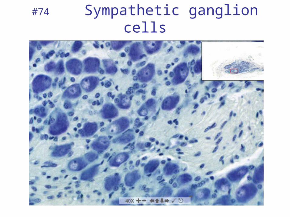
#74 Sympathetic ganglion cells
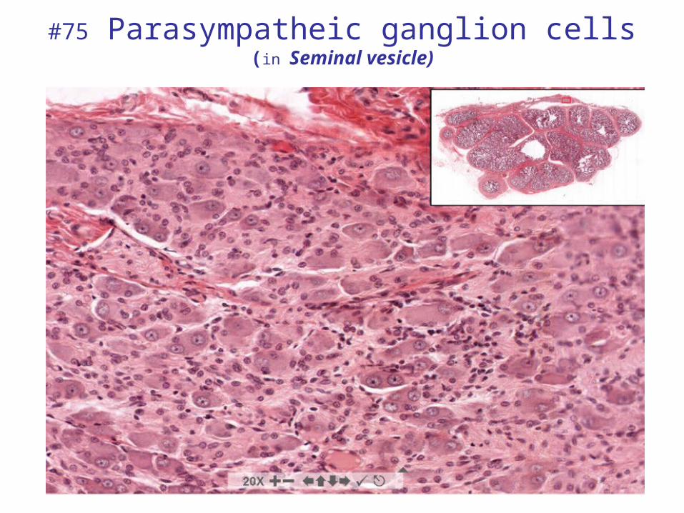
#75 Parasympatheic ganglion cells(in Seminal vesicle)
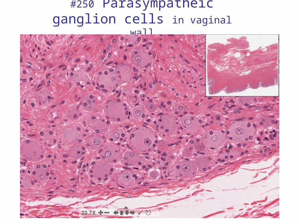
#250 Parasympatheic ganglion cells in vaginal wall
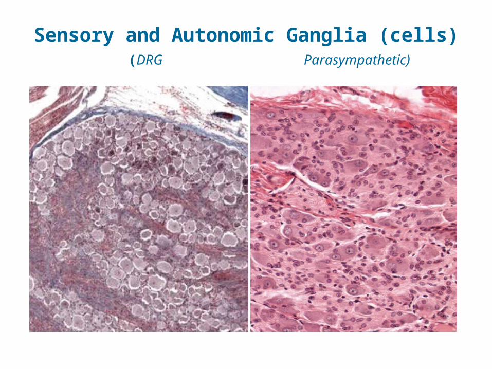
Sensory and Autonomic Ganglia (cells) (DRG Parasympathetic)
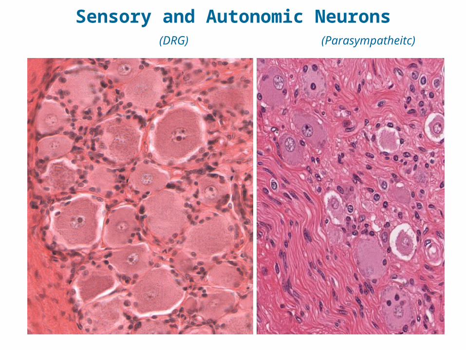
Sensory and Autonomic Neurons (DRG) (Parasympatheitc)
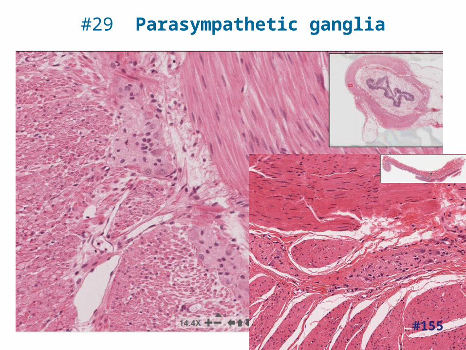
#29 Parasympathetic ganglia
#155
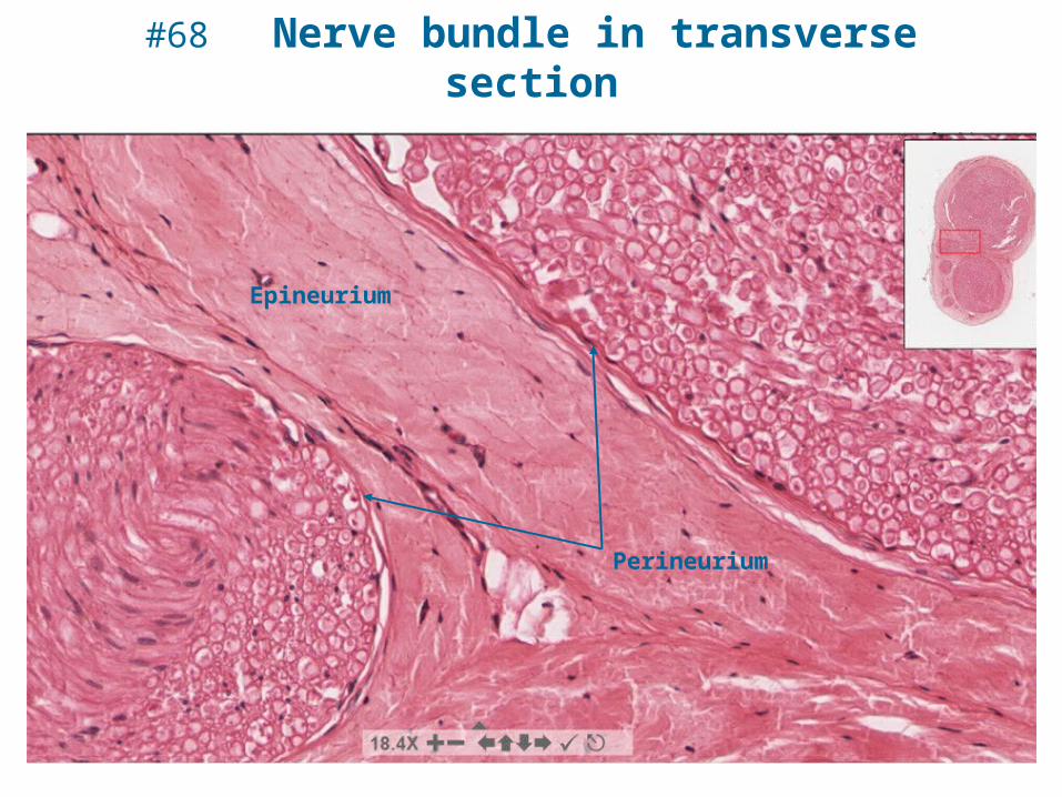
#68 Nerve bundle in transverse section
Epineurium
Perineurium
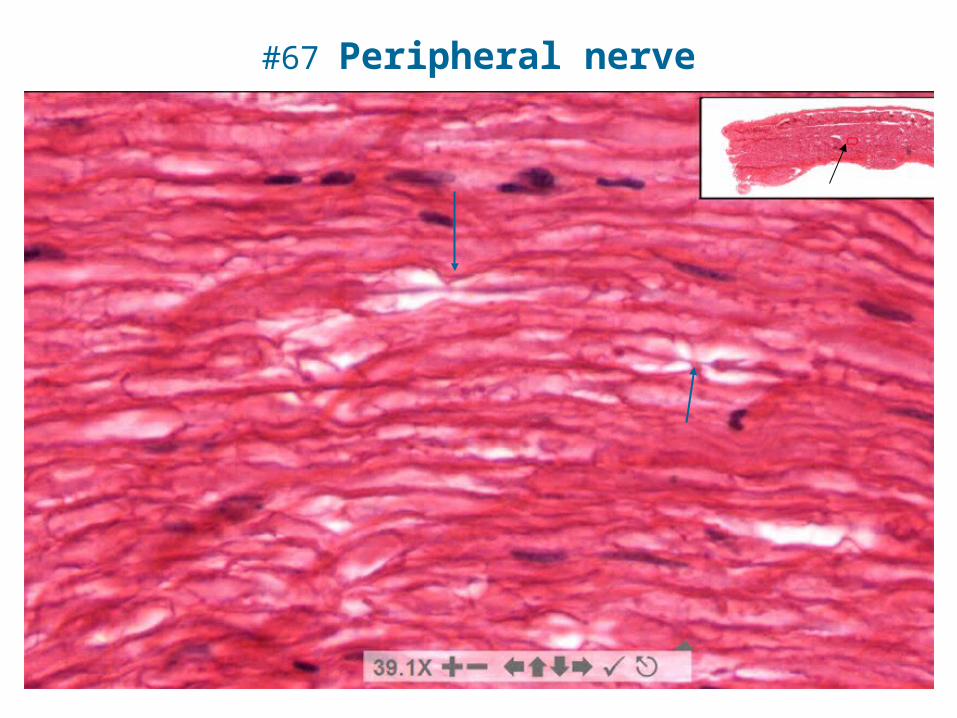
#67 Peripheral nerve
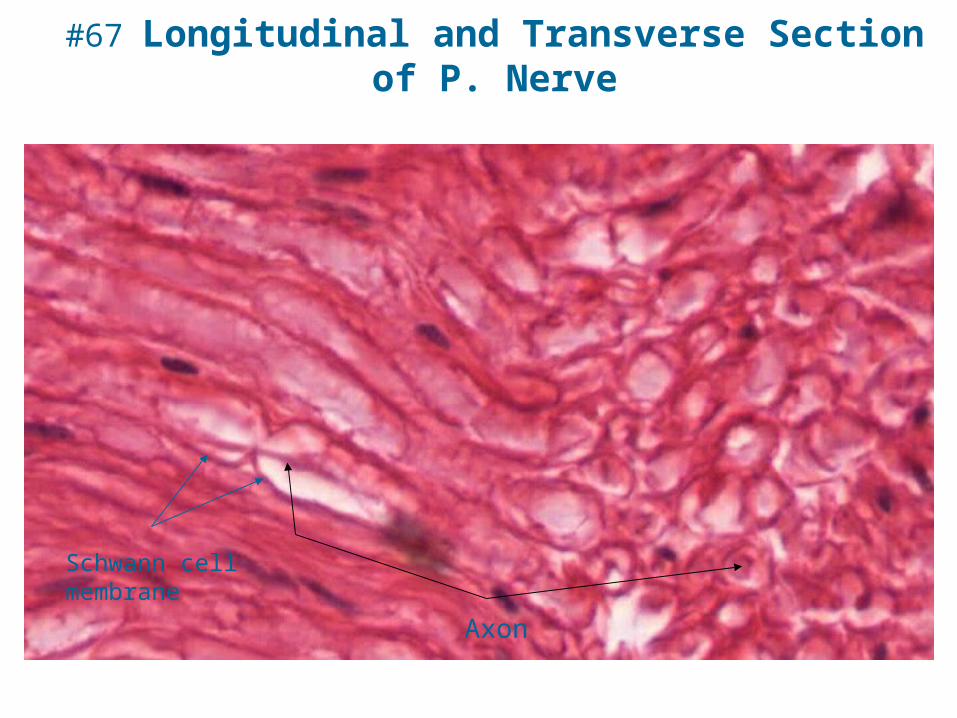
#67 Longitudinal and Transverse Section of P. Nerve
Axon
Schwann cell membrane
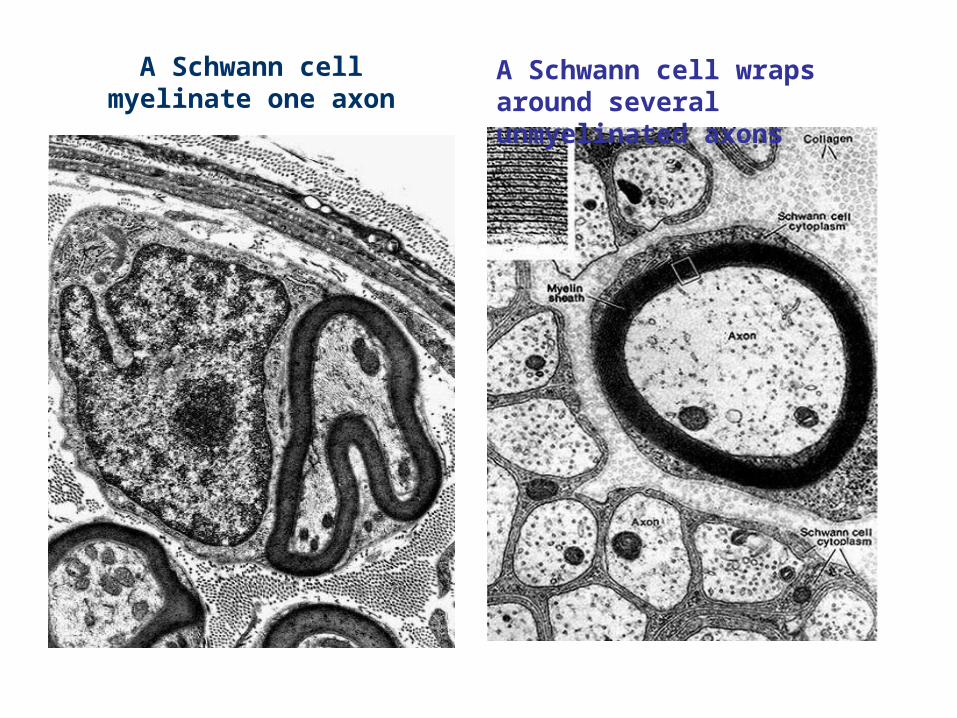
A Schwann cell myelinate one axon
A Schwann cell wraps around several unmyelinated axons
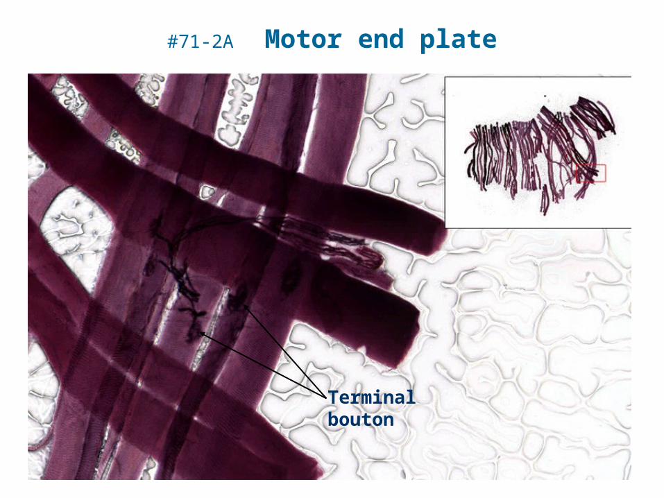
#71-2A Motor end plate
Terminal bouton
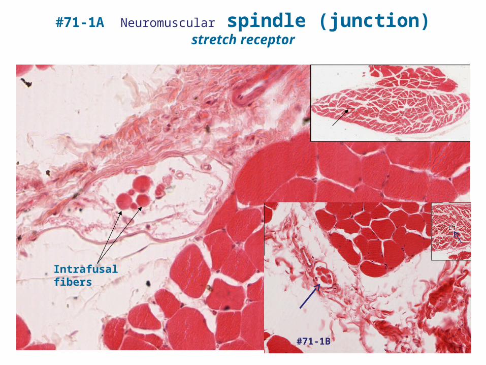
#71-1A Neuromuscular spindle (junction)stretch receptor
Intrafusal fibers
#71-1B