Embed Size (px)
Citation preview
Peripheral Nerve Injuriesin Baseball Players
Craig A. Cummins, MD*, David S. Schneider, DOLake Cook Orthopedic Associates, 27401 West Highway 22, #125,
Barrington, IL 60010, USA
Baseball is a sport requiring speed, coordination, strength and timingthat is enjoyed by millions of people around the world. From a biomechan-ical perspective, the throwing motion, and in particular the pitching motion,places extreme stress on the throwing arm. Baseball pitchers are required tothrow frequently, complicating the trauma placed across the pitcher’sthrowing arm. This combination of high stress placed on the shoulder andelbow during the throwing motion, coupled with the cumulative micro-trauma caused from repetitive overuse, places baseball pitchers at increasedrisk for peripheral nerve injuries in the upper extremity. Neuropathies of theshoulder comprise less than 2% of all causes of pain and weakness for pa-tients in this region [1]. Because of its uncommon nature, it is not unusualfor the diagnosis of a neuropathy of the shoulder to be delayed. Signsand symptoms of a neuropathy typically overlap with more common causesof shoulder pain or weakness, further complicating diagnosis. Affected base-ball players often have coexisting shoulder diagnoses such as rotator cuffpathology, labral tears, instability, and internal impingement, which maybe the more dominant clinical finding. For example, patients who havesuprascapular neuropathy may present with a more dominant impingementsyndrome caused by to the weakness in the rotator cuff muscles as a result ofthe peripheral nerve injury. Baseball players are susceptible to nerve injuriesseen in other sports (cervical radiculopathy, lumbar radiculopathy,‘‘burners’’ or ‘‘stinger,’’ carpal tunnel syndrome). The focus of this articleis on reviewing forms of peripheral neuropathies more common to baseballplayers and resulting from their unique throwing motion.
Neurol Clin 26 (2008) 195–215
* Corresponding author.
E-mail address: [email protected] (C.A. Cummins).
0733-8619/08/$ - see front matter � 2008 Elsevier Inc. All rights reserved.
doi:10.1016/j.ncl.2007.11.014 neurologic.theclinics.com
196 CUMMINS & SCHNEIDER
Biomechanics
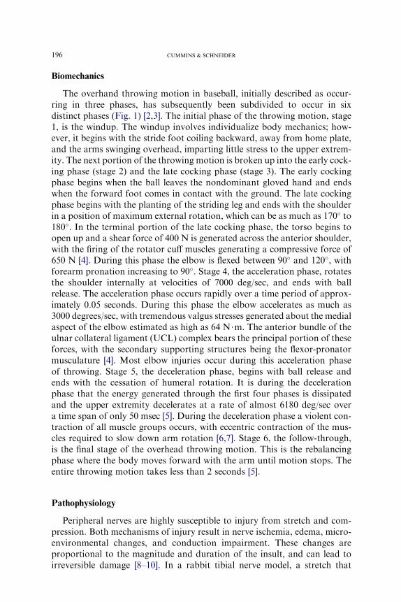
The overhand throwing motion in baseball, initially described as occur-ring in three phases, has subsequently been subdivided to occur in sixdistinct phases (Fig. 1) [2,3]. The initial phase of the throwing motion, stage1, is the windup. The windup involves individualize body mechanics; how-ever, it begins with the stride foot coiling backward, away from home plate,and the arms swinging overhead, imparting little stress to the upper extrem-ity. The next portion of the throwing motion is broken up into the early cock-ing phase (stage 2) and the late cocking phase (stage 3). The early cockingphase begins when the ball leaves the nondominant gloved hand and endswhen the forward foot comes in contact with the ground. The late cockingphase begins with the planting of the striding leg and ends with the shoulderin a position of maximum external rotation, which can be as much as 170� to180�. In the terminal portion of the late cocking phase, the torso begins toopen up and a shear force of 400 N is generated across the anterior shoulder,with the firing of the rotator cuff muscles generating a compressive force of650 N [4]. During this phase the elbow is flexed between 90� and 120�, withforearm pronation increasing to 90�. Stage 4, the acceleration phase, rotatesthe shoulder internally at velocities of 7000 deg/sec, and ends with ballrelease. The acceleration phase occurs rapidly over a time period of approx-imately 0.05 seconds. During this phase the elbow accelerates as much as3000 degrees/sec, with tremendous valgus stresses generated about the medialaspect of the elbow estimated as high as 64 N$m. The anterior bundle of theulnar collateral ligament (UCL) complex bears the principal portion of theseforces, with the secondary supporting structures being the flexor-pronatormusculature [4]. Most elbow injuries occur during this acceleration phaseof throwing. Stage 5, the deceleration phase, begins with ball release andends with the cessation of humeral rotation. It is during the decelerationphase that the energy generated through the first four phases is dissipatedand the upper extremity decelerates at a rate of almost 6180 deg/sec overa time span of only 50 msec [5]. During the deceleration phase a violent con-traction of all muscle groups occurs, with eccentric contraction of the mus-cles required to slow down arm rotation [6,7]. Stage 6, the follow-through,is the final stage of the overhead throwing motion. This is the rebalancingphase where the body moves forward with the arm until motion stops. Theentire throwing motion takes less than 2 seconds [5].
Pathophysiology
Peripheral nerves are highly susceptible to injury from stretch and com-pression. Both mechanisms of injury result in nerve ischemia, edema, micro-environmental changes, and conduction impairment. These changes areproportional to the magnitude and duration of the insult, and can lead toirreversible damage [8–10]. In a rabbit tibial nerve model, a stretch that
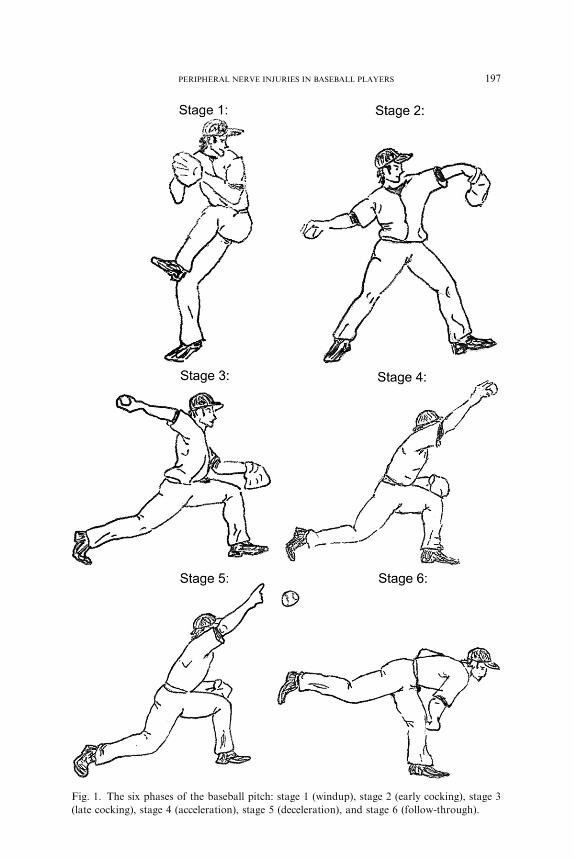
Fig. 1. The six phases of the baseball pitch: stage 1 (windup), stage 2 (early cocking), stage 3
(late cocking), stage 4 (acceleration), stage 5 (deceleration), and stage 6 (follow-through).
197PERIPHERAL NERVE INJURIES IN BASEBALL PLAYERS
198 CUMMINS & SCHNEIDER
increased in situ length as little as 6% resulted in conduction abnormalities[8]. In this samemodel, a stretch of 15% leads to irreversible neural functionaldeficits. Intraneural vascular supply to a rabbit tibial nerve was compromisedwhen the nerve was elongated by 8%, with total occlusion at 15% [9]. Similarchanges have been reported to occur with compression. The unique stressesand arm positions occurring during the baseball throwing motion likely placethe peripheral nerves of the thrower’s dominant upper extremity at risk forsimilar and significant mechanical insults. If these nerve injuries are recog-nized early, cessation of throwing and an appropriate rehabilitation programmay be of benefit and preventative of more permanent injury.
Suprascapular neuropathy
Anatomy
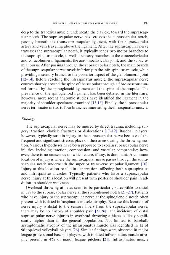
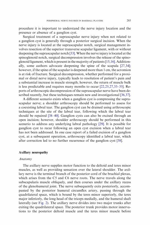
The suprascapular nerve is a mixed motor and sensory nerve that arisesfrom the upper trunk of the brachial plexus (C5 and C6 nerve roots) and sup-plies motor innervations to the supraspinatus and infraspinatus muscles, andsensory innervations to the coracohumeral and coracoacromial ligaments,subacromial bursa, and acromioclavicular and glenohumeral joint (Fig. 2).Al-though most anatomic studies have not identified any cutaneous innervation,cutaneous innervations of the proximal-lateral arm has been reported [11].
After the suprascapular nerve originates from the brachial plexus, it ini-tially courses through the posterior triangle of the neck before traveling
Fig. 2. The posterior aspect of the right shoulder. The supraspinatus, infraspinatus, and deltoid
muscles have been partially removed. The suprascapular nerve (black arrow) can be identified
passing under the superior transverse scapular ligament and around the spinoglenoid notch.
The axillary nerve (gray arrow) can be identified exiting through the quadrilateral space.
199PERIPHERAL NERVE INJURIES IN BASEBALL PLAYERS
deep to the trapezius muscle, underneath the clavicle, toward the suprascap-ular notch. The suprascapular nerve next crosses the suprascapular notch,passing beneath the transverse scapular ligament, with the suprascapularartery and vein traveling above the ligament. After the suprascapular nervetraverses the suprascapular notch, it typically sends two motor branches tothe supraspinatus muscle, as well as sensory branches to the coracoclavicularand coracohumeral ligaments, the acromioclavicular joint, and the subacro-mial bursa. After passing through the suprascapular notch, the main branchof the suprascapular nerve travels inferiorly to the infraspinatus muscle, whileproviding a sensory branch to the posterior aspect of the glenohumeral joint[12–14]. Before reaching the infraspinatus muscle, the suprascapular nervecourses sharply around the spine of the scapular through a fibro-osseous tun-nel formed by the spinoglenoid ligament and the spine of the scapula. Theprevalence of the spinoglenoid ligament has been debated in the literature;however, more recent anatomic studies have identified the ligament in themajority of shoulder specimens examined [15,16]. Finally, the suprascapularnerve terminates in two to four branches innervating the infraspinatusmuscle.
Etiology
The suprascapular nerve may be injured by direct trauma, including sur-gery, traction, clavicle fractures or dislocations [17–19]. Baseball players,however, typically sustain injury to the suprascapular nerve because of thefrequent and significant stresses place on their arms during the throwing mo-tion. Various hypotheses have been proposed to explain suprascapular nerveinjuries, including traction, compression, and vascular compromise; how-ever, there is no consensus on which cause, if any, is dominant. A commonlocation of injury is where the suprascapular nerve passes through the supra-scapular notch underneath the superior transverse scapular ligament [20].Injury at this location results in denervation, affecting both supraspinatusand infraspinatus muscles. Typically patients who have a suprascapularnerve injury at this location will present with posterior shoulder pain in ad-dition to shoulder weakness.
Overhead throwing athletes seem to be particularly susceptible to distalinjury to the suprascapular nerve at the spinoglenoid notch [21–27]. Patientswho have injury to the suprascapular nerve at the spinoglenoid notch oftenpresent with isolated infraspinatus muscle atrophy. Because this location ofnerve injury is distal to the sensory fibers from the suprascapular nerve,there may be no history of shoulder pain [21,26]. The incidence of distalsuprascapular nerve injuries in overhead throwing athletes is likely signifi-cantly higher than in the general population. Not limited to baseball,asymptomatic atrophy of the infraspinatus muscle was identified in 12 of96 top-level volleyball players [26]. Similar findings were observed in majorleague professional baseball players, with isolated infraspinatus muscle atro-phy present in 4% of major league pitchers [21]. Infraspinatus muscle
200 CUMMINS & SCHNEIDER
atrophy was more common among starting pitchers, more experiencedpitchers, and pitchers who had thrown for more innings at the major leaguelevel, so amount of throwing appears to play a significant role [21].
An additional cause of suprascapular nerve injury is compression bya ganglion cyst [28,29]. Baseball players can develop superior and posteriorlabral tears as a result of the stresses placed on the shoulder during thethrowing motion. Ganglion cysts may develop because of labral tear actingas a one-way valve , permitting joint fluid to escape into the cyst cavity butnot allowing the return of the synovial fluid back into the joint. The mostcommon location for labral tears is the superior posterior labrum, whichis probably why the most common location for a shoulder ganglion cystis the spinoglenoid notch. This location of a ganglion cyst can result in com-pression to the suprascapular nerve once the ganglion cyst has reached suf-ficient size, leading to isolated denervation of the infraspinatus muscle.
Clinical evaluation
History and examination
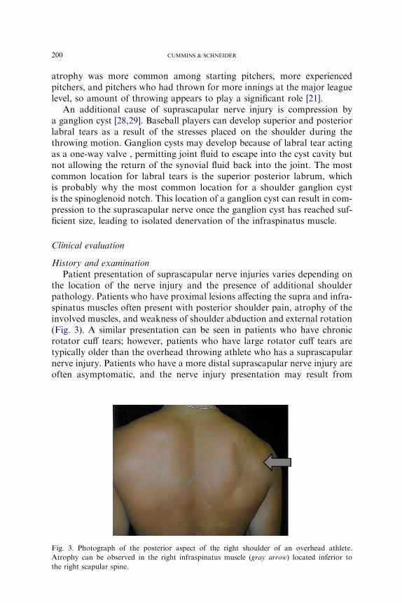
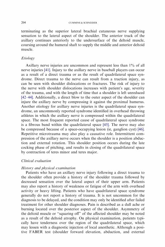
Patient presentation of suprascapular nerve injuries varies depending onthe location of the nerve injury and the presence of additional shoulderpathology. Patients who have proximal lesions affecting the supra and infra-spinatus muscles often present with posterior shoulder pain, atrophy of theinvolved muscles, and weakness of shoulder abduction and external rotation(Fig. 3). A similar presentation can be seen in patients who have chronicrotator cuff tears; however, patients who have large rotator cuff tears aretypically older than the overhead throwing athlete who has a suprascapularnerve injury. Patients who have a more distal suprascapular nerve injury areoften asymptomatic, and the nerve injury presentation may result from
Fig. 3. Photograph of the posterior aspect of the right shoulder of an overhead athlete.
Atrophy can be observed in the right infraspinatus muscle (gray arrow) located inferior to
the right scapular spine.
201PERIPHERAL NERVE INJURIES IN BASEBALL PLAYERS
casual observation of the scapular muscle atrophy only. A small percentageof patients who have a distal suprascapular nerve injury may have posteriorshoulder pain, however. This pain may be caused by dysfunction in therelationship of the posterior capsules sensory branch to the spinoglenoidligament. Finally, it is common for a patient’s initial presentation to bethe result of other shoulder pathology. An example is patients who havea suprascapular nerve injury caused by a ganglion cyst, in which the labraltear associated with the ganglion cyst causes the shoulder pain leading toclinical attention.
Diagnostic studies
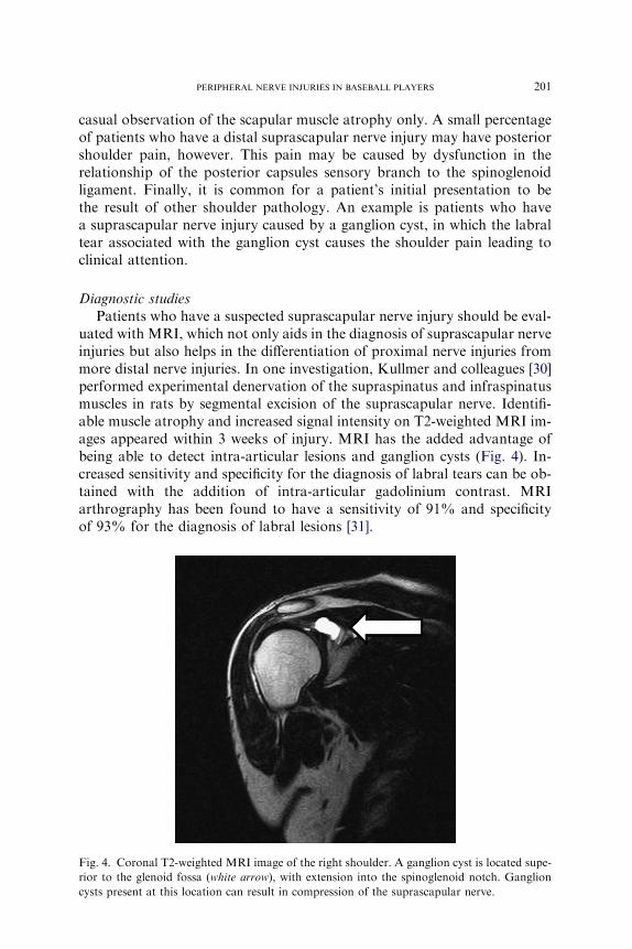
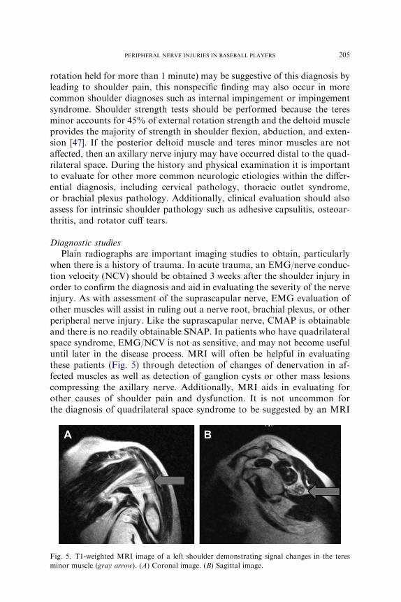
Patients who have a suspected suprascapular nerve injury should be eval-uated with MRI, which not only aids in the diagnosis of suprascapular nerveinjuries but also helps in the differentiation of proximal nerve injuries frommore distal nerve injuries. In one investigation, Kullmer and colleagues [30]performed experimental denervation of the supraspinatus and infraspinatusmuscles in rats by segmental excision of the suprascapular nerve. Identifi-able muscle atrophy and increased signal intensity on T2-weighted MRI im-ages appeared within 3 weeks of injury. MRI has the added advantage ofbeing able to detect intra-articular lesions and ganglion cysts (Fig. 4). In-creased sensitivity and specificity for the diagnosis of labral tears can be ob-tained with the addition of intra-articular gadolinium contrast. MRIarthrography has been found to have a sensitivity of 91% and specificityof 93% for the diagnosis of labral lesions [31].
Fig. 4. Coronal T2-weighted MRI image of the right shoulder. A ganglion cyst is located supe-
rior to the glenoid fossa (white arrow), with extension into the spinoglenoid notch. Ganglion
cysts present at this location can result in compression of the suprascapular nerve.
202 CUMMINS & SCHNEIDER
Electrodiagnositic studies are also important in confirming the diagnosisof a suprascapular nerve injury, localizing the site of the nerve injury, andby excluding other etiologies in the differential diagnosis, such as cervical rad-iculopathy and brachial plexopathy. An increased latency when performingthe suprascapular compound motor action potentials (CMAP) indicates de-myelination or a conduction block. Although there are sensory innervationsfrom the suprascapular nerve, there are no cutaneous nerve branches capableof generating a sensory nerve action potential (SNAP). Electromyography(EMG) is also a useful method for evaluating axonal loss from a suprascapu-lar nerve injury by the demonstration of increased involuntary spontaneousactivity with fibrillations and positive sharp waves. These changes tend tooccur 2 to 3 weeks after the injury. Disadvantages of these electrodiagnositicstudies include their invasiveness and user dependence; however, EMG hasa high sensitivity and specificity for diagnosing the suprascapular neuropa-thy, and may have prognostic value. For example, identification of polypha-sic potentials with large-amplitude and long-duration motor unit actionpotentials (MUAPS) with disappearance of involuntary activity indicates col-lateral sprouting, reinnervation, and the absence of ongoing denervation.
Treatment
Nonoperative
Nonoperative treatment of suprascapular nerve injuries should be consid-ered as the initial treatment option, unless nerve compression is caused bya mass lesion. Nonoperative treatment includes the avoidance of activitiesfelt to be causative, obviously including cessation of throwing. This treat-ment should be combined with a rehabilitation program. The initial focusof a rehabilitation program is on shoulder flexibility, with particular atten-tion given to improving internal rotation contractures, postural training,and strengthening of the periscapular and deltoid muscles. Strengtheningof the rotator cuff muscles can be started once symptoms of nerve impinge-ment have subsided. The advancement of the rehabilitation program shouldbe monitored closely to avoid reinjury to the nerve while it is healing. In gen-eral, success rates with nonoperative treatment are less satisfactory when thenerve injury results from a mass lesion such as a ganglion cyst. One concernwith nonoperative treatment is that a delay in decompression of the supra-scapular nerve may result in incomplete restoration of muscle function. Ithas been demonstrated that fatty atrophy in the rotator cuff muscle is notreversible following repair of large rotator cuff tears [32]. A similar situationlikely occurs when portions of rotator cuff muscles (supraspinatus and infra-spinatus) are denervated following nerve injury.
Operative treatment
The operative treatment of suprascapular nerve injuries depends on theetiology and location of the nerve lesion. When planning the surgical
203PERIPHERAL NERVE INJURIES IN BASEBALL PLAYERS
procedure it is important to understand the nerve injury location and thepresence or absence of a ganglion cyst.
Surgical treatment of a suprascapular nerve injury when not related toa ganglion cyst is generally through a posterior surgical incision. When thenerve injury is located at the suprascapular notch, surgical management in-volves resection of the superior transverse scapular ligament, with or withoutdeepening the suprascapular notch [33].When the nerve injury is located at thespinoglenoid notch, surgical decompression involves the release of the spino-glenoid ligament,which is present in themajority of patients [15,16].Addition-ally, some authors advocate deepening the spine of the scapula [27,34];however, if the spine of the scapular is deepenedmore than 1 cm, the acromionis at risk of fracture. Surgical decompression, whether performed for a proxi-mal or distal nerve injury, typically leads to resolution of patient’s pain anda substantial increase in muscle strength; however, the return of muscle bulkis less predictable and requires many months to occur [22,25,27,33–35]. Re-ports of arthroscopic decompression of the suprascapular nerve have been de-scribed recently, but these techniques remain new and investigational [36,37].
A different scenario exists when a ganglion cyst is compressing the supra-scapular nerve; a shoulder arthroscopy should be performed to assess fora coexisting labral tear. The ganglion cyst can be drained using arthroscopictechniques at the site of the labral tear, following which the labral tearshould be repaired [38–40]. Ganglion cysts can also be excised through anopen incision; however, shoulder arthroscopy should be performed in thisscenerio to address any underlying labral pathology [29]. It is possible forganglion cyst to recur following an open cyst excision when a labral tearhas not been addressed. In one case report of a failed excision of a ganglioncyst, at a subsequent operation, arthroscopy identified a labral tear, whichafter correction led to no further recurrence of the ganglion cyst [38].
Axillary neuropathy
Anatomy
The axillary nerve supplies motor function to the deltoid and teres minormuscles, as well as providing sensation over the lateral shoulder. The axil-lary nerve is the terminal branch of the posterior cord of the brachial plexus,which arises from the C5 and C6 nerve roots. The nerve travels along thesubscapularis muscle obliquely, and then courses under the axillary recessof the glenohumeral joint. The nerve subsequently exits posteriorly, accom-panied by the posterior humeral circumflex artery, passing through thequadrilateral space, which is bound by the teres minor superiorly, the teresmajor inferiorly, the long head of the triceps medially, and the humeral shaftlaterally (see Fig. 2). The axillary nerve divides into two major trunks afterexiting the quadrilateral space. The posterior trunk provides motor innerva-tions to the posterior deltoid muscle and the teres minor muscle before
204 CUMMINS & SCHNEIDER
terminating as the superior lateral brachial cutaneous nerve supplyingsensation to the lateral aspect of the shoulder. The anterior truck of theaxillary continues anteriorly to the undersurface of the deltoid muscle,coursing around the humeral shaft to supply the middle and anterior deltoidmuscle.
Etiology
Axillary nerve injuries are uncommon and represent less than 1% of allnerve injuries [41]. Injury to the axillary nerve in baseball players can occuras a result of a direct trauma or as the result of quadrilateral space syn-drome. Direct trauma to the nerve can result from a traction injury, ascan be seen with shoulder dislocations or fractures. The risk of injury tothe nerve with shoulder dislocations increases with patient’s age, severityof the trauma, and with the length of time that a shoulder is left unreduced[42–44]. Additionally, a direct blow to the outer aspect of the shoulder caninjure the axillary nerve by compressing it against the proximal humerus.Another etiology for axillary nerve injuries is the quadrilateral space syn-drome, an uncommonly reported syndrome identified in overhead throwingathletes in which the axillary nerve is compressed within the quadrilateralspace. The most frequent reported cause of quadrilateral space syndromeis a fibrous band within the quadrilateral space [45]. The nerve may alsobe compressed because of a space-occupying lesion (ie, ganglion cyst) [46].Repetitive microtrauma may also play a causative role. Intermittent com-pression of the axillary nerve occurs when the shoulder is a position abduc-tion and external rotation. This shoulder position occurs during the latecocking phase of pitching, and results in closing of the quadrilateral spaceby contraction of teres minor and teres major.
Clinical evaluation
History and physical examination
Patients who have an axillary nerve injury following a direct trauma tothe shoulder often provide a history of the shoulder trauma followed bydecreased sensation over the lateral aspect of their upper arm. Patientsmay also report a history of weakness or fatigue of the arm with overheadactivity or heavy lifting. Patients who have quadrilateral space syndromegenerally do not report a history of trauma. It is not uncommon for thediagnosis to be delayed, and the condition may only be identified after failedtreatment for other shoulder diagnoses. Pain is described as a dull ache orburning located over the posterior aspect of the shoulder. Asymmetry ofthe deltoid muscle or ‘‘squaring off’’ of the affected shoulder may be notedas a result of the deltoid atrophy. On physical examination, patients typi-cally have tenderness over the region of the quadrilateral space, whichmay lessen with a diagnostic injection of local anesthetic. Although a posi-tive FABER test (shoulder forward elevation, abduction, and external
205PERIPHERAL NERVE INJURIES IN BASEBALL PLAYERS
rotation held for more than 1 minute) may be suggestive of this diagnosis byleading to shoulder pain, this nonspecific finding may also occur in morecommon shoulder diagnoses such as internal impingement or impingementsyndrome. Shoulder strength tests should be performed because the teresminor accounts for 45% of external rotation strength and the deltoid muscleprovides the majority of strength in shoulder flexion, abduction, and exten-sion [47]. If the posterior deltoid muscle and teres minor muscles are notaffected, then an axillary nerve injury may have occurred distal to the quad-rilateral space. During the history and physical examination it is importantto evaluate for other more common neurologic etiologies within the differ-ential diagnosis, including cervical pathology, thoracic outlet syndrome,or brachial plexus pathology. Additionally, clinical evaluation should alsoassess for intrinsic shoulder pathology such as adhesive capsulitis, osteoar-thritis, and rotator cuff tears.
Diagnostic studies
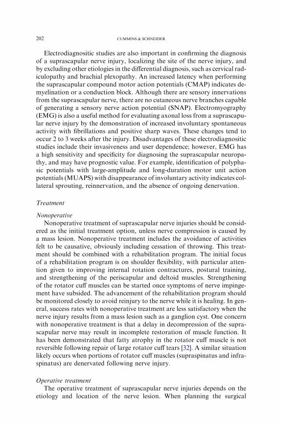
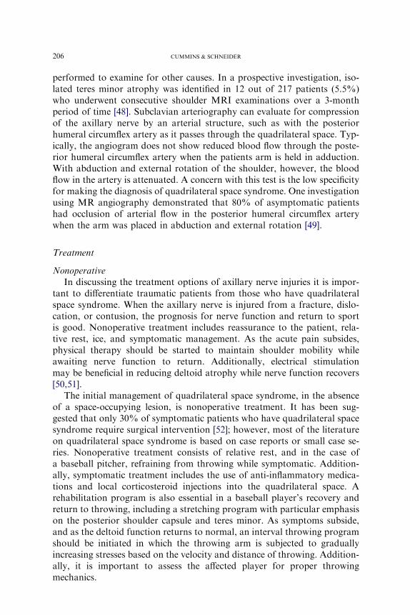
Plain radiographs are important imaging studies to obtain, particularlywhen there is a history of trauma. In acute trauma, an EMG/nerve conduc-tion velocity (NCV) should be obtained 3 weeks after the shoulder injury inorder to confirm the diagnosis and aid in evaluating the severity of the nerveinjury. As with assessment of the suprascapular nerve, EMG evaluation ofother muscles will assist in ruling out a nerve root, brachial plexus, or otherperipheral nerve injury. Like the suprascapular nerve, CMAP is obtainableand there is no readily obtainable SNAP. In patients who have quadrilateralspace syndrome, EMG/NCV is not as sensitive, and may not become usefuluntil later in the disease process. MRI will often be helpful in evaluatingthese patients (Fig. 5) through detection of changes of denervation in af-fected muscles as well as detection of ganglion cysts or other mass lesionscompressing the axillary nerve. Additionally, MRI aids in evaluating forother causes of shoulder pain and dysfunction. It is not uncommon forthe diagnosis of quadrilateral space syndrome to be suggested by an MRI
Fig. 5. T1-weighted MRI image of a left shoulder demonstrating signal changes in the teres
minor muscle (gray arrow). (A) Coronal image. (B) Sagittal image.
206 CUMMINS & SCHNEIDER
performed to examine for other causes. In a prospective investigation, iso-lated teres minor atrophy was identified in 12 out of 217 patients (5.5%)who underwent consecutive shoulder MRI examinations over a 3-monthperiod of time [48]. Subclavian arteriography can evaluate for compressionof the axillary nerve by an arterial structure, such as with the posteriorhumeral circumflex artery as it passes through the quadrilateral space. Typ-ically, the angiogram does not show reduced blood flow through the poste-rior humeral circumflex artery when the patients arm is held in adduction.With abduction and external rotation of the shoulder, however, the bloodflow in the artery is attenuated. A concern with this test is the low specificityfor making the diagnosis of quadrilateral space syndrome. One investigationusing MR angiography demonstrated that 80% of asymptomatic patientshad occlusion of arterial flow in the posterior humeral circumflex arterywhen the arm was placed in abduction and external rotation [49].
Treatment
Nonoperative
In discussing the treatment options of axillary nerve injuries it is impor-tant to differentiate traumatic patients from those who have quadrilateralspace syndrome. When the axillary nerve is injured from a fracture, dislo-cation, or contusion, the prognosis for nerve function and return to sportis good. Nonoperative treatment includes reassurance to the patient, rela-tive rest, ice, and symptomatic management. As the acute pain subsides,physical therapy should be started to maintain shoulder mobility whileawaiting nerve function to return. Additionally, electrical stimulationmay be beneficial in reducing deltoid atrophy while nerve function recovers[50,51].
The initial management of quadrilateral space syndrome, in the absenceof a space-occupying lesion, is nonoperative treatment. It has been sug-gested that only 30% of symptomatic patients who have quadrilateral spacesyndrome require surgical intervention [52]; however, most of the literatureon quadrilateral space syndrome is based on case reports or small case se-ries. Nonoperative treatment consists of relative rest, and in the case ofa baseball pitcher, refraining from throwing while symptomatic. Addition-ally, symptomatic treatment includes the use of anti-inflammatory medica-tions and local corticosteroid injections into the quadrilateral space. Arehabilitation program is also essential in a baseball player’s recovery andreturn to throwing, including a stretching program with particular emphasison the posterior shoulder capsule and teres minor. As symptoms subside,and as the deltoid function returns to normal, an interval throwing programshould be initiated in which the throwing arm is subjected to graduallyincreasing stresses based on the velocity and distance of throwing. Addition-ally, it is important to assess the affected player for proper throwingmechanics.
207PERIPHERAL NERVE INJURIES IN BASEBALL PLAYERS
Operative treatment
Surgical indications for treating an axillary nerve injury include surgicallyderived iatrogenic injuries, penetrating trauma, and a symptomatic patientwho has no clinical or EMG/NCV evidence of nerve recovery by 3 to 6months. The best surgical results are seen when surgery is performed within6 months from the injury; however, functional improvement can still occurwhen surgery is performed up to 1 year after the injury occurred [53–55]. Sur-gical techniques involved include such procedures as neurolysis, neurorrha-phy, nerve grafting, nerve transfer, and neurotization. In general, theseoperations can be performed through both an anterior and posterior incision.
Surgical intervention for quadrilateral space syndrome should be consid-ered when a space-occupying lesion is present, and when symptoms have per-sisted despite nonoperative interventions for a 3- to 6-month period of time.
Surgery for quadrilateral space syndrome can be performed througha muscle-sparing technique or with detachment of the deltoid muscle fromthe acromion. The axillary nerve and its branches should be identified asthey exit the quadrilateral space, and any fibrous bands or space-occupyinglesions that are compressing the nerve should be excised. Confirmation of anadequate decompression may involve placing the arm in an abducted andexternally rotated position and palpating a pulse in the posterior humeralcircumflex artery. Information regarding patient outcomes following surgi-cal decompression of the axillary nerve at the quadrilateral space is limited,because most of the data are derived from small case series [45,46,52,56–59].Of 23 patients reported in one study, 10 patients had dramatic relief ofsymptoms, 11 had improvement of symptoms, and 2 patients had noimprovement [59].
Ulnar neuropathy
Anatomy
The ulnar nerve is a mixed sensorimotor nerve derived from the medialcord of the brachial plexus (C8 and T1). The motor innervations from theulnar nerve are to the majority of the intrinsic muscles of the hand, theflexor carpi ulnaris muscle, and medial aspect of the flexor digitorumprofundus. The sensory innervations are the dorsal and palmar surfacesof the fourth and fifth fingers and the ulnar border of the hand.
The ulnar nerve travels down the upper arm through the anterior com-partment adjacent to the brachial artery. No significant branches of theulnar nerve are present in the upper arm; however, occasionally there isa variant supracondylar motor branch to the flexor carpi ulnaris. In the mid-dle third of the upper arm, the nerve pierces the medial intermuscular sep-tum as it courses from the anterior to the posterior compartment, passingthrough the arcade of Struthers, a musculofascial band locatedapproximately 8 cm proximal to the medial epicondyle. The ulnar nerve
208 CUMMINS & SCHNEIDER
subsequently travels behind the medial epicondyle in a fibro-osseous groovethat is bound by the medial epicondyle anteriorly, the olecranon and ulno-humeral ligament laterally, and a fibroaponeurotic band medially. Laxity ofthe fibroaponeurotic band can result in subluxation or dislocation of theulnar nerve out of the epicondylar groove. This occurs during elbow flexion,with reduction during elbow extension. Asymptomatic hypermobility of thenerve is found in approximately 16% of the population, and is often bilat-eral [60]. Next the nerve passes between the two heads of the flexor carpiulnaris, under a fascial band called Osborne’s ligament. Elbow flexioncauses stretching of Osborne’s ligament, resulting in the cross-sectionalshape of the tunnel changing from an oval to a flattened ellipse [61]. Theulnar collateral ligament relaxes and bulges medially, further narrowingthe space available for the ulnar nerve. During elbow flexion, pressurewithin the tunnel increases sevenfold, and can increase more than 20-foldwith contraction of the flexor carpi ulnaris [62].
The diagnosis and treatment of ulnar nerve injuries in baseball playersrequires an understanding of the relationship between the unique anatomyof the elbow and the stresses placed across those structures during thethrowing motion. The stability of the elbow joint is primarily supplied bythe bony anatomy when the elbow is within 20�of extension or flexedbeyond 120�. The primary provider of elbow stability between 20� and120� of motion is the surrounding soft tissue [63–65]. In particular, duringthe throwing motion the anterior bundle of the ulnar collateral ligamentis the primary restraint to valgus forces. This ligament encounters repetitivenear-failure tensile stresses during the pitching motion [4,64,66–69]. Thepathophysiology behind most elbow injuries in baseball players has beentermed the ‘‘valgus extension overload syndrome’’ [48]. The combinationof large valgus forces and rapid extension of the elbow places tensile stressas high as 64 Nm along the medial side of the elbow [4]. Structures affectedby these forces include the ulnar nerve, ulnar collateral ligament, flexor-pronator mass, and medial epicondyle apophysis in adolescents.
Etiology
Ulnar neuropathy at the elbow is the second most common compressionneuropathy after carpal tunnel syndrome. The ulnar nerve travels in a super-ficial location behind the medial epicondyle, and can be injured as a result ofa direct insult. More commonly, the ulnar nerve is injured because of com-pression or traction where it travels in the cubital tunnel between the upperarm and forearm. Injury to the nerve can occur at several locations withinthe cubital tunnel, including at the intermuscular septum, the arcade ofStruthers, the fibro-osseous groove at the medial epicondyle, and betweenthe two heads of the flexor carpi ulnaris origin. Baseball players are partic-ularly prone to ulnar neuropathy because of the repetitive valgus stressesplaced on the nerve during the throwing motion. Symptoms involving the
209PERIPHERAL NERVE INJURIES IN BASEBALL PLAYERS
ulnar nerve are very common in throwing athletes, and occur in more than40% of athletes who have valgus instability and 60% of throwers who havemedial epicondylitis [70,71]. The pressure within the ulnar nerve in the flexedelbow and extended wrist has been shown to be elevated more than threetimes the resting level [72]. When this elbow position is combined withshoulder abduction, the intraneural pressure may be elevated as much assix times the resting level. This is the arm position that occurs during theacceleration phase of throwing. As a result of the valgus extension overloadsyndrome, other changes occur in throwers’ elbows, resulting in tethering ofthe ulnar nerve and increased intraneural pressures, such as with degenera-tive changes and osteophyte formation impinging on the ulnar groove.Other potential factors affecting the ulnar nerve include loose bodies, trac-tion spurs, hypertrophy of the triceps and anconeus epitrochlearis muscles,synovitis, thickened retinaculum, scar tissue, and calcification of the UCL.
Clinical evaluation
History and physical examination
Ulnar neuropathy is a diagnosis that is typically made via history andphysical examination. Baseball pitchers typically present with a history ofmedial elbow pain, with or without radiation to the hand. The player mayalso report clumsiness of the hand and parasthesias in the little and ringfingers. These symptoms initially occur during times of throwing andsubside with rest; however, as the disease progresses, symptoms may alsooccur at rest. Motor symptoms affecting the intrinsic hand muscles andextrinsic forearm muscles are often not observed in the initial stages,when most players present.
Physical examination is important in making the diagnosis of ulnar neu-ropathy and excluding other potential sources of the patient’s neurologicsymptoms. Evaluation for a cervical radiculopathy, brachial plexus injury,or coexisting nerve compression should be performed. In evaluating thepatient who has ulnar nerve symptoms, the physician needs to assess forcompression at more than one site [73]. Examination of the elbow shouldassess for areas of tenderness, range of motion, strength, and stability. Ten-derness over the flexor pronator tendon origin reproduced with resistedwrist flexion and forearm pronation suggests a diagnosis of medial epicon-dylitis. Pain over the UCL reproduced with a valgus stress to the elbow isconsistent with an injury to the UCL. The ulnar nerve should be evaluatedfor subluxation during elbow flexion. Pain over the ulnar nerve, a positiveTinel’s sign posterior to the medial epicondyle, and a positive elbow flexiontest all may suggest involvement of the ulnar nerve. As with the Phalen’stest, the elbow flexion test is more sensitive than specific, with false-positiveresults reported in 10% of normal individuals. When motor weaknessdevelops, involvement of the intrinsic muscles of the hand occurs beforethat of extrinsic forearm muscles, likely because of to a more superficial
210 CUMMINS & SCHNEIDER
location of these nerve fascicles at the elbow [74]. One of the first signs ofintrinsic motor weakness is an inability to adduct the little finger (Warten-berg’s sign). When intrinsic weakness is severe and associated with musclewasting, a claw hand deformity may be seen as a flexion of the fourthand fifth proximal and distal interphalyngeal joints, with extension of thefourth and fifth metacarpal phalyngeal joints.
Diagnostic studies
Radiographic imaging of the elbow is important when patients presentwith a history of trauma. Radiographic views should include an anteriorposterior, lateral, and cubital tunnel view. If there is concern of elbow insta-bility, then stress radiographs can also be obtained. Medial joint openinggreater than 3 mm is consistent with instability [65,75]. MRI will allowassessment of soft tissue masses, but is not essential for diagnosis or plan-ning appropriate treatment in most patients. If the patient’s history andphysical examination suggest an injury to the ulnar collateral ligament, how-ever, then an MRI may be useful for evaluating for ligament avulsions,partial and complete ulnar collateral ligament tears, and for assessing theflexor pronator tendon origin [75,76].
Electrodiagnositic studies can be an important adjunct in confirming thediagnosis of ulnar neuropathy and assessing the severity of the nerve injurywhen the diagnosis is not obvious based on clinical evaluation [77]. False-negatives can occur if the electrodiagnositic testing is performed too earlyin the disease process. In focal ulnar nerve compression, sensory fibers areaffected before motor fibers. It is important to obtain the dorsal ulnar cuta-neous SNAP because an absent response may indicate a moderate to severelesion of the ulnar nerve at the elbow [78]. The most common technique forassessing proximal ulnar nerve function is evaluating the ulnar CMAPbelow and above the elbow. A nerve conduction velocity of less than49 m/sec across the elbow [79] or a difference of 10 m/sec or greater betweenthe arm and above the elbow to below elbow segment is considered abnor-mal [80]. When using this technique, it is important to perform the test withthe elbow fully flexed, otherwise the true length of the nerve can be under-estimated, leading to false slowing of conduction times [79]. The segmentacross the elbow should be greater than 10 cm to increase sensitivity.When conduction velocity is slowed, it is occasionally accompanied bya drop in CMAP amplitude of 20% to 30% at the site of the lesion at theelbow. An alternative method of finding a focal lesion at the elbow is to per-form multiple short segmental stimulations of 1 cm, both proximal and dis-tal to the medial epicondyle [81], called an ‘‘inching technique.’’ An increasein the latency of 0.4 msec or more is considered significant, and identifiesa focal lesion at that segment [82]. EMG is helpful in confirming denervationin ulnar-innervated muscles as well as evaluating other forms of nerveinjuries such as a C8/T1 radiculopathy or lower trunk/medial cordplexopathy.
211PERIPHERAL NERVE INJURIES IN BASEBALL PLAYERS
Treatment
Nonoperative
Nonoperative management of ulnar nerve injuries at the elbow involvesrest, avoidance of aggravating activities, and nonsteroidal anti-inflamma-tory medication. Local corticosteroid injections are not recommended. Ifthe patient’s symptoms occur at night or are more severe, splinting withthe arm in slight flexion can be helpful. This can be particularly beneficialin patients who have ulnar nerve subluxation or dislocation. Players whosesymptoms occur predominantly during throwing should not be allowed tothrow until they are asymptomatic. Once the elbow is asymptomatic, theplayer should be started on a strengthening program focusing on thedynamic stabilizers of the elbow. The final stage of rehabilitation includesan interval throwing program, with the goal of having the player returnto competitive throwing. Unfortunately, unlike the general population, ath-letes who have valgus instability of the elbow often have recurrence of theirulnar nerve symptoms with resumption of throwing.
Operative treatment
Indications for surgical intervention include failed nonoperative treat-ment or coexisting elbow pathology that requires surgical treatment. Addi-tionally, patients who have more severe compression of the ulnar nerve, toinclude those patients who have axonal loss, profound sensory deficits, andmotor weakness, may be considered for earlier surgical intervention. Surgi-cal procedures include simple decompression, medial epicondylectomy,anterior subcutaneous transposition, and submuscular transposition. Simpledecompression of the ulnar nerve and medial epicondylectomy in the throw-ing athlete are contraindicated. Both anterior subcutaneous transpositionand submuscular transposition have demonstrated favorable outcomes inthe overhead throwing athlete [70,83–88]. When this procedure is comparedwith submuscular transpositions, it has the advantage of less surgical dissec-tion and faster rehabilitation. Potential disadvantages include the nervebeing in a more superficial location and therefore vulnerable to directtrauma. Additionally, the ulnar nerve follows a less direct course, and canbe iatrogenically compressed by the subcutaneous fascial-dermal sling. Sub-muscular transposition protects the transposed nerve from both direct andindirect trauma by placing the nerve deep to the flexor muscle mass [85].Disadvantages of the submuscular transposition include increased surgicaldissection, the need for temporary immobilization and protection of therepaired flexor tendon, and a longer rehabilitation period. There are surgicaladvocates for both procedures; however, there are no prospective random-ized controlled trials or meta-analyses available to guide treatment. Animportant issue in the operative management of the overhead athlete is toaddress all of the pathology that is contributing to the athlete’s elbow symp-toms. Specifically, if valgus instability of the elbow is present, an ulnar
212 CUMMINS & SCHNEIDER
collateral ligament reconstruction should be performed at the time of theulnar nerve decompression. Additionally, significant tendinopathy or tear-ing of the flexor pronator tendon origin should be debrided and repaired.Failure to address the underlying pathology in the elbow can lead to recur-rent elbow problems [86].
Summary
The throwing motion in baseball places significant and repetitive stressacross the shoulder and elbow. These forces result in unique patterns ofinjuries seen in the overhead athlete. Specific nerve injuries identified inthe overhead athlete include suprascapular neuropathy, quadrilateral spacesyndrome, and cubital tunnel syndrome. The principles of nonoperativemanagement include cessation of throwing and symptomatic treatment.As the athletes’ symptoms subside, they should be started in supervisedrehabilitation that progresses to an interval throwing program. The goalof treatment is the resolution of nerve symptoms and the return to compet-itive throwing. In those overhead athletes who fail to improve with nonop-erative treatment, surgery can result in a positive outcome in particularpatients. Additional indications for surgery include more profound neurop-athy and mass-lesion–induced nerve compression.
References
[1] Narakas A. Compression and traction neuropathies about the shoulder and arm. In: RHG,
editor. Operative nerve repair and reconstruction. Philadelphia: JB Lippincott; 1991.
[2] Meister K. Injuries to the shoulder in the throwing athlete: part one: biomechanics/
pathophysiology/classification of injury. Am J Sports Med 2000;28:265–75.
[3] Tullos H, King J. Throwing mechanism in sports. Orthop Clin North Am 1973;4:709–20.
[4] Fleisig G, Andrews J, Dillman C, et al. Kinetics of baseball pitching with implications about
injury mechanism. Am J Sports Med 1995;23:233–9.
[5] Pappas A, Zawacki R, Sullivan T. Biomechanics of baseball pitching. A preliminary report.
Am J Sports Med 1985;13:216–22.
[6] Jobe F, Moynes D, Tibone J, et al. An EMG analysis of the shoulder in pitching: a second
report. Am J Sports Med 1984;12:218–20.
[7] Jobe F, Tibone J, Perry J, et al. An EMG analysis of the shoulder in throwing and pitching:
a preliminary report. Am J Sports Med 1983;11:3–5.
[8] Kwan M, Wall E, Massie J, et al. Strain, stress and stretch of peripheral nerve. Rabbit
experiment in vitro and in vivo. Acta Orthop Scand 1992;63:267–72.
[9] Lundborg G, Rydevik B. Effects of stretching the tibial nerve of the rabbit. A preliminary
study of the intraneural circulation and the barrier function of the perineurium. J Bone Joint
Surg Br 1973;55:390–401.
[10] Lundborg G. Structure and function of the intraneural microvessels as related to trauma,
edema formation and nerve function. J Bone Joint Surg Am 1975;57:938–48.
[11] Ajmani M. The cutaneous branch of the human suprascapular nerve. J Anat 1994;185:
439–42.
[12] Bigliani L, Dalsey R, McCann P, et al. An anatomical study of the suprascapular nerve.
Arthroscopy 1990;6:301–5.
213PERIPHERAL NERVE INJURIES IN BASEBALL PLAYERS
[13] Mestdagh H, Drizenko A, Ghestem P. Anatomical basis of suprascapular nerve syndrome.
Anat Clin 1981;3:67–71.
[14] Ozer Y, Grossman J, Gilbert A. Anatomical observations on the suprascapular nerve. Hand
Clin 1995;11:539–44.
[15] Cummins C, Anderson K, Bowen M, et al. Anatomy and histological characteristics of the
spinoglenoid ligament. J Bone Joint Surg Am 1998;80:1622–5.
[16] Plancher K, Peterson R, Johnston J, et al. The spinoglenoid ligament: anatomy, morphol-
ogy, and histological findings. J Bone Joint Surg Am 2005;87:361–5.
[17] Edeland H, Zachrisson B. Fracture of the scapular notch associated with lesion of the
suprascapular nerve. Acta Orthop Scand 1975;46:758–63.
[18] BerryH,KongK,HudsonA, et al. Isolated suprascapular nerve palsy: a review of nine cases.
Can J Neurol Sci 1995;22:301–4.
[19] Travlos J, Goldberg I, Boome R. Brachial plexus lesions associated with dislocated
shoulders. J Bone Joint Surg Br 1990;72:68–71.
[20] Rengachary SS, Neff JP, Singer PA, et al. Suprascapular entrapment neuropathy: a clinical,
anatomical and comparative study. Part 1: clinical study. Neurosurgery 1979;5:441–6.
[21] Cummins C, Messer T, Schafer M. Infraspinatus muscle atrophy in professional baseball
players. Am J Sports Med 2004;32(1):116–20.
[22] Cummins C, BowenM, Anderson K, et al. Suprascapular nerve entrapment at the spinogle-
noid notch in a professional baseball pitcher. Am J Sports Med 1999;27:810–2.
[23] Ringel S, TreihaftM, CarrM, et al. Suprascapular neuropathy in pitchers. Am J SportsMed
1990;18:80–6.
[24] BryanW,Wild J. Isolated infraspinatus atrophy: a common cause of posterior shoulder pain
and weakness in throwing athletes? Am J Sports Med 1989;17:130–1.
[25] Ferretti A, De Carli A, Fontana M. Injury of the suprascapular nerve at the spinoglenoid
notch: the natural history of infraspinatus atrophy in volleyball players. Am J Sports Med
1998;26:759–63.
[26] Ferretti A, Cerullo G, Russo G. Suprascapular neuropathy in volleyball players. J Bone
Joint Surg Am 1987;69:260–3.
[27] Sandow M, Ilic J. Suprascapular nerve rotator cuff compression syndrome in volleyball
players. J Shoulder Elbow Surg 1998;7:516–21.
[28] Chochole M, Senker W, Meznik C, et al. Glenoid-labral cyst entrapping the suprascapular
nerve: dissolution after arthroscopic debridement of an extended SLAP lesion. Arthroscopy
1997;13:753–5.
[29] Fehrman D, Orwin J, Jennings R. Suprascapular nerve entrapment by a ganglion cyst:
a report of six cases with arthroscopic finding and review of the literature. Arthroscopy
1995;11:727–34.
[30] Kullmer K, Sievers K, Reimers C, et al. Changes of sonographic, magnetic resonance
tomographic, electromyographic and histopathologic findings within a 2-month period of
examinations after experimental muscle denervation. Arch Orthop Trauma Surg 1998;
117:228–34.
[31] Palmer W, Brown J, Rosenthal D. Labral-ligamentous complex of the shoulder: evaluation
with MR arthrography. Radiology 1994;190:645–51.
[32] Gladstone J, Bishop J, Lo IK, et al. Fatty infiltration and atrophy of the rotator cuff do not
improve after rotator cuff repair and correlate with poor functional outcome. Am J Sports
Med 2007;35:719–28.
[33] Post M, Mayer J. Suprascapular nerve entrapment. Diagnosis and treatment. Clin Orthop
Relat Res 1987;223:126–36.
[34] Hama H, Ueba Y, Morinaga T, et al. A new stategy for treatment of suprascapular entrap-
ment neuropathy in athletes: shaving of the base of the scapular spine. J Shoulder Elbow
Surg 1992;1:253–60.
[35] Hagert C, Linder L. Entrapment neuropathy of the suprascapular nerve. ActaOrthop Scand
1984;55:107.
214 CUMMINS & SCHNEIDER
[36] Lafosse L, Tomasi A, Corbett S, et al. Arthroscopic release of the suprascapular nerve
entrapment at the suprascapular notch: technique and preliminary results. Arthroscopy
2007;23:34–42.
[37] Barwood S, Burkhart S, Lo I. Arthroscopic suprascapular nerve release at the suprascapular
notch in a cadaveric model: an anatomic approach. Arthroscopy 2007;23:221–5.
[38] Moore T, Fritts H, Quick D, et al. Suprascapular nerve entrapment caused by supraglenoid
cyst compression. J Shoulder Elbow Surg 1997;6:455–62.
[39] Westerheide K, Dopirak R, Karzel R, et al. Suprascapular nerve palsy secondary to
spinoglenoid cysts: results of arthroscopic treatment. Arthroscopy 2006;22:721–7.
[40] Iannotti J, RamseyM.Arthroscopic decompression of a ganglion cyst causing suprascapular
nerve compression. Arthroscopy 1996;12:739–45.
[41] Pollack L, Davis L. Peripheral nerve injuries. Am J Surg 1932;17:462–71.
[42] Gumina S, Postacchini F. Anterior dislocation of the shoulder in elderly patients. J Bone
Joint Surg Br 1972;79:540–3.
[43] Perlmutter G, Apruzzese W. Axillary nerve injury in contact sports; recommendations for
treatment and rehabilitation. Sports Med 1998;26:351–61.
[44] Toolanen G, Hildingsson T, Hedlund T, et al. Early complications after anterior dislocation
of the shoulder in patients over 40 years; an ultrasonographic and electromyographic study.
Acta Orthop Scand 1993;64:549–52.
[45] Cahill B, Palmer R. Quadrilateral space syndrome. J Hand Surg [Am] 1983;8:65–9.
[46] Sanders T, Tirman P. Paralabral cyst: an unusual case of quadrilateral space syndrome-
Arthroscopy 1999;15:632–7.
[47] Saha A. Surgery of the paralyzed and flail shoulder. Acta Orthop Scand 1967;97(Suppl):
5–90.
[48] Wilson L, Sundaram M, Piraino D, et al. Isolated teres minor atrophy: manifestation of
quadrilateral space syndrome or traction injury to the axillary nerve? Orthopedics 2006;
29(5):447–50.
[49] Mochizuki T, Isoda H,Masui T, et al. Occlusion of the posterior circumflex humeral artery;
detection with MR angiography in healthy volunteers and in a patient with quadrilateral
space syndrome. AJR Am J Roentgenol 1994;163:625–7.
[50] Crameri R,WestonA, Rutkowski S, et al. Effects of electrical stimulation leg training during
the acute phase of spinal cord injury: a pilot study. Eur J Appl Physiol 2000;83:409–15.
[51] Dupont Salter A, Richmond F, Leob G. Prevention of muscle disuse atrophy by low-
frequency electrical stimulation in rats. IEEE Trans Neural Syst Rehabil Eng 2003;11:
218–26.
[52] Lester B, JeongG,WeilandA, et al. Quadrilateral space syndrome: diagnosis, pathology and
treatment. Am J Orthop 1999;28:718–25.
[53] Alnot J, Valenti P. Surgical reconstruction of the axillary nerve. In: Post M, MB, Hawkins
RJ, editors. Surgery of the shoulder. St Louis (MO): Mosby; 1990. p. 330–3.
[54] Petrucci F, Morelli A, Raimondi P. Axillary nerve injuries: 21 cases treated by nerve graft
and neurolysis. J Hand Surg [Am] 1982;7:271–8.
[55] Rochwerger A, Benaim L, Toledano E, et al. Surgical repair of the axillary nerve: results of
a five-year follow up. Chir Main 2000;19:31–5.
[56] Chen D, Cai P, Lao G, et al. Quadrilateral space syndrome. Chin Med J (Engl) 1995;108:
109–12.
[57] Kuang Y, Hou C. The diagnosis and tratment of quadrilateral space syndrome. Zhongguo
Xiu Fu Chong Jian Wai Ke Za Zhi 2001;15:199–201 [In Chinese].
[58] Francel T, Dellon A, Campbell J. Quadrilateral space syndrome: diagnosis and operative
decompression technique. Plast Reconstr Surg 1991;87:911–6.
[59] Safran M. Nerve injuries about the shoulder in athletes, part 1: suprascapular nerve and
axillary nerve. Am J Sports Med 2004;32:803–19.
[60] Childress H. Recurrent ulnar-nerve dislocation at the elbow. Clin Orthop Relat Res 1975;
108:168–73.
215PERIPHERAL NERVE INJURIES IN BASEBALL PLAYERS
[61] Apfelberg D, Larson S. Dynamic anatomy of the ulnar nerve at the elbow. Plast Reconstr
Surg 1973;51:79–81.
[62] Werner C, Ohlin P, Elmqvist D. Pressures recorded in ulnar neuropathy. ActaOrthop Scand
1985;56:404–6.
[63] Miller R. The cubital tunnel syndrome: diagnosis and precise localization. AnnNeurol 1979;
6:56–9.
[64] Morrey B, Tanaka S, An K. Valgus stability of the elbow: a definition of primary and
secondary constraints. Clin Orthop Relat Res 1991;265:187–95.
[65] Schwab G, Bennett J, Woods G, et al. Biomechanics of elbow instability: the role of the
medial collateral ligament. Clin Orthop Relat Res 1980;146:42–52.
[66] Hotchkiss R, Weiland A. Valgus stability of the elbow. J Orthop Res 1987;5:372–7.
[67] Morrey B, AnK. Articular and ligamentous contributions to the stability of the elbow joint.
Am J Sports Med 1983;11:315–9.
[68] Morrey B, An K. Functional anatomy of the ligaments of the elbow. Clin Orthop Relat Res
1985;201:84–90.
[69] Regan W, Korinek S, Morrey B, et al. Biomechanical study of ligaments around the elbow
joint. Clin Orthop Relat Res 1991;271:170–9.
[70] Andrews J, Timmerman L. Outcome of elbow surgery in professional baseball players. Am J
Sports Med 1995;23:407–13.
[71] Bennett G. Elbow and shoulder lesions of the professional baseball pitcher. Am J Surg 1959;
98:484–92.
[72] Pechan J, Julis I. The pressure measurement in the ulnar nerve: a contribution to the
pathophysiology of the cubital tunnel syndrome. J Biomech 1975;8:75–9.
[73] Upton A, McComas A. The double crush in nerve entrapment syndromes. Lancet 1973;2:
359–62.
[74] Sunderland S, editor. Nerve and nerve injuries. 2nd edition. New York: Churchill Living-
stone; 1978.
[75] Jobe F, Kvitne R. Elbow instability in the athlete. Instr Course Lect 1991;40:17–23.
[76] Timmerman L, Andrews J. Undersurface tear of the ulnar collateral ligament in baseball
players: a newly recognized lesion. Am J Sports Med 1994;22:33–6.
[77] Posner M. Compressive ulnar neuropathies at the elbow: I. Etiology and diagnosis. J Am
Acad Orthop Surg 1998;6:282–8.
[78] Jabre J. Ulnar nerve lesions at the wrist: new techniques for recording from the sensory
dorsal branch of the ulnar nerve. Neurology 1980;30:873–6.
[79] Kincaid J. AAEE minimonograph #31: the electrodiagnosis of ulnar neuropathy at the
elbow. Muscle Nerve 1988;11:1005–15.
[80] Eisen A. Early diagnosis of ulnar palsy. Neurology 1974;24:256–62.
[81] Miller C, Savoie FI. Valgus extension injuries of the elbow in the throwing athlete. J Am
Acad Orthop Surg 1994;2:261–9.
[82] CampbellW, PridgeonR, Sahni K. Short segment incremental studies in evaluation of ulnar
neuropathy at the elbow. Muscle Nerve 1992;15:1050–4.
[83] Aoki M, Kanaya K, Aiki H, et al. Cubital tunnel syndrome in adolescent baseball players:
a report of six cases with 3- to 5-year follow-up. Arthroscopy 2005;21(6):758.
[84] Boatright J, D’AlessandroD, et al. Nerve entrapment syndromes at the elbow. In: Jobe FW,
PM, Glousman RE, editors. Operative techniques in upper extremity sports injuries.
St Louis (MO): Mosby-Year Book; 1996.
[85] Del Pizzo W, Jobe F, Norwood L. Ulnar nerve entrapment syndrome in baseball players.
Am J Sports Med 1977;5(5):182–5.
[86] Glousman R. Ulnar nerve problems in the athlete’s elbow. Clin Sports Med 1990;9:365–77.
[87] Rettig A, Ebben J. Anterior subcutaneous transfer of the ulnar nerve in the athlete. Am J
Sports Med 1993;21:836–40.
[88] Rokito A, McMahon P, Jobe F. Cubital tunnel syndrome. Oper Tech Sports Med 1996;4:
15–20.