Embed Size (px)
Citation preview
Scheer, Katz / Peripheral Metal Complexes: Chlorophyll “Isomers” 56 1
Peripheral Metal Complexes: Chlorophyll “Isomers” with Magnesium Bound to the Ring E @-Keto Ester System
Hugo Scheer and Joseph J. Katz* Contribution from the Chemistry Division, Argonne National Laboratory, Argonne, Illinois 60439. Received June 6. 1977
Abstract: Peripheral magnesium complexes of methyl pheophorbides a and b, bacteriomethyl pheophorbide a, bacteriopheo- phytin a, and protopheophytin a have been prepared and characterized by UV-vis, IR, ‘H N M R , and luminescence spectros- copy. In these compounds, Mg is bound to the peripheral p-keto ester system present in most chlorophylls rather than to the central tetrapyrrole cavity as in the chlorophyll proper. The ligand exchange reactions at Mg have been characterized, and the equilibrium between the peripheral complexes and their free ligands has been studied over the concentration range lo-’- M. AGO for complex formation has been determined by ’H N M R spectroscopy.
Introduction The chlorophylls have a primary function in photosynthesis
both in light collection and in light conversion processes. Considerable progress has been made in the past 2 decades in the understanding of the in vitro properties of chlorophyll and this has contributed to a better understanding of the role of chlorophyll on the molecular level in photosynthesis.’ The salient features of chlorophyll aggregates or species present in vivo can be related to and can be studied in detail by suitable in vitro systemse2 Similarly models have been developed for the redox chemistry of metal loch lor in^.^^^ The aggregation be- havior of the chlorophylls is largely due to the presence of both ligand acceptors (the central magnesium atom) and donors (predominantly the 9-ketocarbonyl group) within the same molecule.1b,2 The easy (photo)oxidation of chlorins and bac- teriochlorins to 7r-cation can be related to the electronegativity of the central magnesium atom.3
Little is known thus far about the functional role in chlo- rophyll of the enolizable P-keto ester system at ring E. It is present in all chlorophylls (except for the chlorobium chloro- phylls6), and in particular in all chlorophylls present in pho- toreaction centers, including bacteriochlorophyll b.’ From the chemical point of view it is probably the most reactive func- tional group within the molecule. Indeed, most alteration products and side reactions observed in the chlorophylls are related to the &keto ester system. Reactions involving the P-keto ester system are responsible for the easy epimeriza- tion4a,s and oxidation at C-10,9 for the ready cleavage of ring E with amines,gc and for the loss of the 10-carbomethoxy group at elevated temperatures.1° A test for the presence of the intact enolizable a-keto ester system was developed as early as 1896;” this reaction involves formation of the enolate anion upon treatment with strong bases.Ia In neutral solution, only the diketo form of the P-keto ester system can be detected spectroscopically,2as12 but related porphyrins with an open chain P-keto ester substituent are present as diketo-enol mixture^.'^^'^ Recently, more readily enolizable chlorophyll derivatives have been prepared in which a C- 10 carbonyl group is covalently linked to the 7 substituent.I5 An early attempt to relate the enolizable @-keto ester system to the photosynthetic process goes back to Franck,’6a and a model for oxygen pro- duction in photosynthesis has been proposed by Mauzerall and Chiwis’6b in which the oxygen evolved by the water-splitting reaction of photosystem I1 is derived from the &keto ester system.
We noticed during studies on the metalation of the pheo- phorbides of the a series5d that the substrates fall into two classes:” (a) pheophorbides that are easily metalated to chlorophyllides by anhydrous magnesium salts in refluxing pyridine,18 and (b) pheophorbides of the second class that give
0002-7863/78/1500-0561 $Ol.OO/O
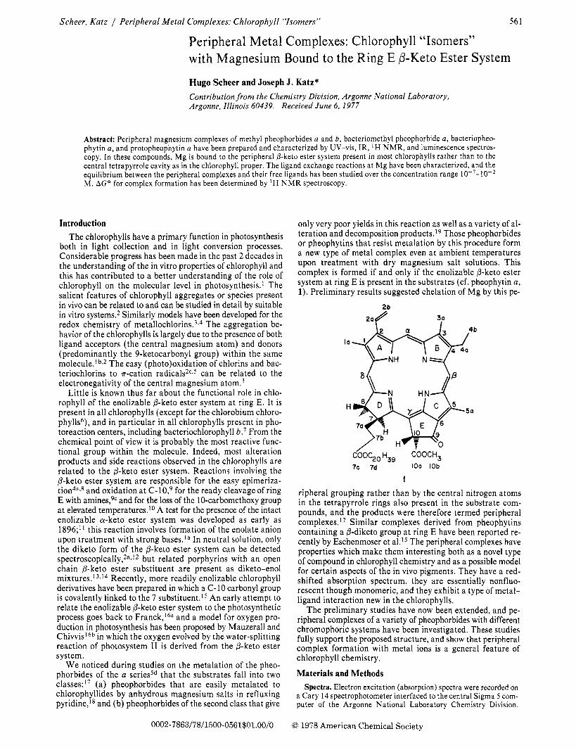
only very poor yields in this reaction as well as a variety of al- teration and decomposition products.19 Those pheophorbides or pheophytins that resist metalation by this procedure form a new type of metal complex even at ambient temperatures upon treatment with dry magnesium salt solutions. This complex is formed if and only if the enolizable @-keto ester system at ring E is present in the substrates (cf. pheophytin a , 1). Preliminary results suggested chelation of Mg by this pe-
la+-+ N-
5a H
C60C20 H,, COOCH, 7c 7d IOa IOb
1 ripheral grouping rather than by the central nitrogen atoms in the tetrapyrrole rings also present in the substrate com- pounds, and the products were therefore termed peripheral complexes. l 7 Similar complexes derived from pheophytins containing a P-diketo group at ring E have been reported re- cently by Eschenmoser et al.15 The peripheral complexes have properties which make them interesting both as a novel type of compound in chlorophyll chemistry and as a possible model for certain aspects of the in vivo pigments. They have a red- shifted absorption spectrum, they are essentially nonfluo- rescent though monomeric, and they exhibit a type of metal- ligand interaction new in the chlorophylls.
The preliminary studies have now been extended, and pe- ripheral complexes of a variety of pheophorbides with different chromophoric systems have been investigated. These studies fully support the proposed structure, and show that peripheral complex formation with metal ions is a general feature of chlorophyll chemistry.
Materials and Methods Spectra. Electron excitation (absorption) spectra were recorded on
a Cary 14 spectrophotometer interfaced to the central Sigma 5 com- puter of the Argonne National Laboratory Chemistry Division.
0 1978 American Chemical Society
562 Journal of the American Chemical Society / 100:2 / January 18,1978
Computer deconvolutions of the UV-vis spectra were carried out by the variable metric minimization procedure of Davidon,20 modified and adapted to the Sigma 5 by A. Zielen. This program allows for the resolution of up to ten peaks with different individual line shapes within the same spectrum. Fluorescence spectra were obtained on pyridine solutions with an Argonne-built laser-excited fluorimeter described elsewhere.2i Phosphorescence experiments were carried out in glasses at cryogenic temperatures in the same apparatus. Samples for phosphorescence were prepared in a 1 : 1 mixture of toluene and a saturated solution of Mg(C10& in pyridine, degassed by repeated freeze-thawing, and then sealed off under high vacuum. For fluo- rescence titration experiments, an Hitachi Perkin-Elmer MPF-2A spectrofluorimeter was used. ‘H NMR spectra were measured in pyridine-ds on a Varian HR 220 NMR spectrometer operated in pulse FT mode and interfaced to the Sigma 5. IR spectra were obtained with a Beckman IR 7 spectrometer. Pyridine-d5 was used as solvent, and titrations were carried out with 2H10, if not stated otherwise.
Solvents and Reagents. All solvents were reagent grade and were dried over molecular sieves prior to use. Anhydrous Mg(C104)~ (G. F. Smith) was driedZZ under a high vacuum at slowly increasing temperatures and then kept at 225 OC for at least 6 h. Anhydrous metal chlorides and bromides were obtained from the hydrates in a stream of hydrogen chloride or bromide, respectively. All dry metal salts were handled in glove boxes kept dry with anhydrous Mg(C104)~ or P205.
Compounds. Chlorophylls were obtained by standard procedures and demetalated with dilute HC1 to the pheophytin~.~~ For conversion of bacteriochlorophyll b into bacteriopheophytin b, the time of contact with acid was less than 1 min to avoid extensive rearrangement.
Methyl pheophorbide a ( 2 ) and b (3) and bacteriomethyl pheo- phorbide a (4) were prepared from the respective pheophytins by transesterification with 5% methanolic sulfuric acid. Protopheophytin a ( 6 ) was obtained from pheophytin a (1) by dehydrogenation with freshly sublimed 2,3-dichlor0-5,6-dicyanobenzoquinone.~~~~~ Pyro- methyl pheophorbide a (7),Io 10-methoxymethyl pheophorbide a (8),9d and 9-hydroxy-9-deoxomethyl pheophorbide a (9)25 were ob- tained by standard procedures.
All anhydrous metal salt solutions and solutions of the peripheral complexes were handled in nitrogen dryboxes or glovebags (I2R). For the preparation of the peripheral complexes, the pheophorbide was usually dissolved in a freshly prepared anhydrous saturated solution of Mg(C104) (or other anhydrous salt) in pyridine. The saturated solutions were obtained by adding an excess of the anhydrous salt to pyridine or other solvent, and the undissolved salt was subsequently removed by centrifugation. For the mixed solvent systems, methanol was saturated with the metal salt; in the case of Mg(C104)2, 5% pyridine was added for safety reasons. The pheo- phorbide, dissolved separately in the other component, was then added to this solution.
The Anhydrous Magnesium Salt Solutions. Anhydrous magnesium salts are among the most powerful
desiccants, and the perchlorate surpasses even PzOj. As the peripheral complexes are sensitive to water (see below), all salts must be dried extensively prior to use and handled with the rigorous exclusion of water. Dissolution of the anhydrous salts in pyridine is strongly exothermic, and the salt must always be added to the solvent to prevent extensive and undesirable caking. If an excess of the metal salt is used, the remaining solid is an effective drying agent. Thus, pyridine containing 1% water gave results similar to carefully dried pyridine when treated with excess anhydrous salts. The saturated solutions are extremely hygroscopic once they are separated from the precipitate by centrifugation, and even in a good drybox may take up enough water to prevent complex formation after a few hours of exposure. Storage for extended periods is possible, however, under high vacuum. The actual concentration of Mg(C104)z in a saturated pyridine solution prepared as de- scribed may show considerable variation. For a series of solu- tions prepared under similar conditions, a range of values from 0.12 to 0.24 mol/L have been found by atomic absorption spectroscopy.26 We ascribe this variation in concentration to varying amounts of residual water, which is present in molar
amounts comparable to the Mg salt even with all the precau- tions taken.
The original water content and the ligand exchange
[MgPy6l2+ 4- xH2O il xpy + [MgPy6-x(H20)x]2+ ( x < 6)
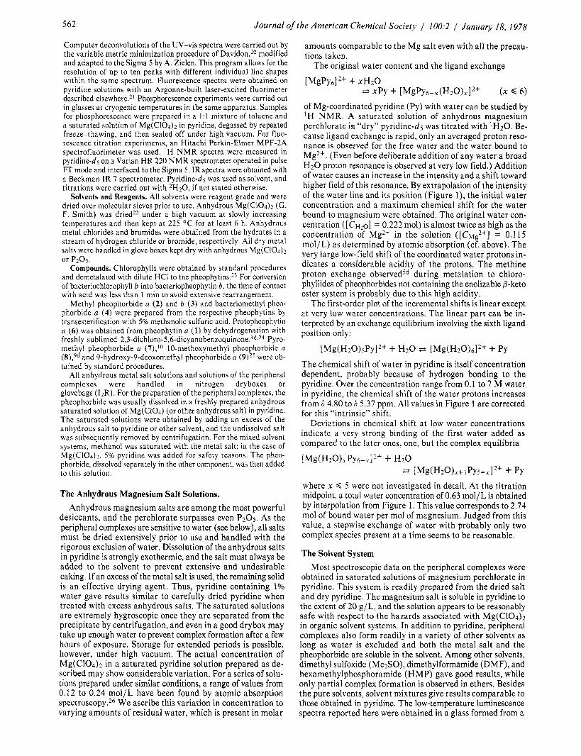
of Mg-coordinated pyridine (Py) with water can be studied by ‘H N M R . A saturated solution of anhydrous magnesium perchlorate in “dry” pyridine-dj was titrated with ‘H20. Be- cause ligand exchange is rapid, only an averaged proton reso- nance is observed for the free water and the water bound to Mg2+. (Even before deliberate addition of any water a broad H2O proton resonance is observed a t very low field.) Addition of water causes an increase in the intensity and a shift toward higher field of this resonance. By extrapolation of the intensity of the water line and its position (Figure l ) , the initial water concentration and a maximum chemical shift for the water bound to magnesium were obtained. The original water con- centration ( [ C H ~ ~ ] = 0.222 mol) is almost twice as high as the concentration of Mg2+ in the solution ([Ch.lg2+] = 0.1 15 mol/L) as determined by atomic absorption (cf. above). The very large low-field shift of the coordinated water protons in- dicates a considerable acidity of the protons. The methine proton exchange observed5d during metalation to chloro- phyllides of pheophorbides not containing the enolizable P-keto ester system is probably due to this high acidity.
The first-order plot of the incremental shifts is linear except a t very low water concentrations. The linear part can be in- terpreted by an exchange equilibrium involving the sixth ligand position only:
The chemical shift of water in pyridine is itself concentration dependent, probably because of hydrogen bonding to the pyridine. Over the concentration range from 0.1 to 7 M water in pyridine, the chemical shift of the water protons increases from 6 4.80 to 6 5.37 ppm. All values in Figure 1 are corrected for this “intrinsic” shift.
Deviations in chemical shift a t low water concentrations indicate a very strong binding of the first water added as compared to the later ones, one, but the complex equilibria
[Mg(HzO),Py6-.71’+ i- Hz0
where x d 5 were not investigated in detail. At the titration midpoint, a total water concentration of 0.63 mol/L is obtained by interpolation from Figure 1. This value corresponds to 2.74 mol of bound water per mol of magnesium. Judged from this value, a stepwise exchange of water with probably only two complex species present a t a time seems to be reasonable.
The Solvent System Most spectroscopic data on the peripheral complexes were
obtained in saturated solutions of magnesium perchlorate in pyridine. This system is readily prepared from the dried salt and dry pyridine. The magnesium salt is soluble in pyridine to the extent of 20 g/L, and the solution appears to be reasonably safe with respect to the hazards associated with Mg(C104)2 in organic solvent systems, In addition to pyridine, peripheral complexes also form readily in a variety of other solvents as long as water is excluded and both the metal salt and the pheophorbide are soluble in the solvent. Among other solvents, dimethyl sulfoxide (MezSO), dimethylformamide (DMF), and hexamethylphosphoramide (HMP) gave good results, while only partial complex formation is observed in ethers. Besides the pure solvents, solvent mixtures give results comparable to those obtained in pyridine. The low-temperature luminescence spectra reported here were obtained in a glass formed from a
* [ M ~ ( H ~ O ) ~ + I P Y S - ~ I ~ + + PY
Scheer, Katz / Peripheral Metal Complexes: Chlorophyll ‘
r - 7 7 ‘Isomers” 563
Figure 1. Concentration dependence of the ‘H N M R chemical shift (6, ppm) of water ( ‘H02H) in a saturated solution of Mg(C104)2 in pyri- dine-ds. See text for details.
saturated Mg(ClO4)z/pyridine solution diluted 1:l with tol- uene. If a concentrated solution of Mg(C104)2 in methanol (containing 5% pyridine) is used as one component, peripheral complexes are observed to be formed with methyl pheophor- bide a dissolved in practically any hydrocarbon or halocarbon solvent.
Peripheral Complex Electronic Excitation Spectra Formation of the peripheral complexes from the free
pheophorbide is always accompanied by a pronounced color change. A solution of methyl pheophorbide a (2) is brown,
2
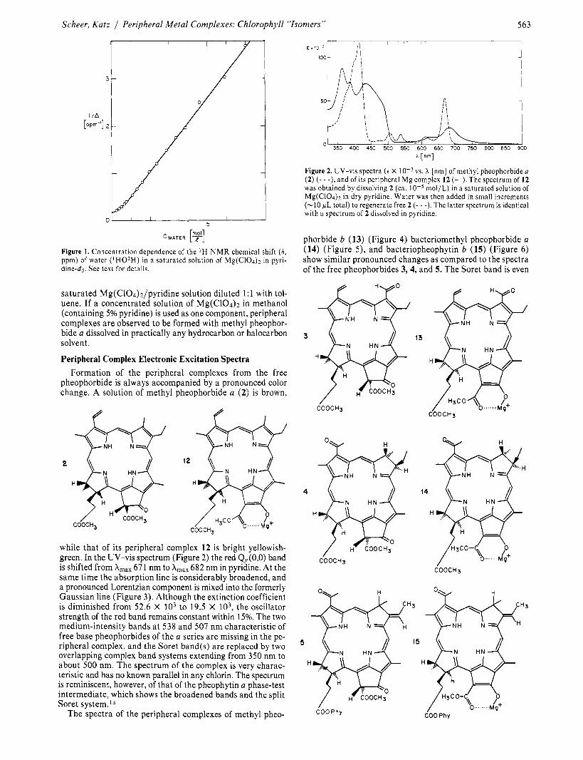
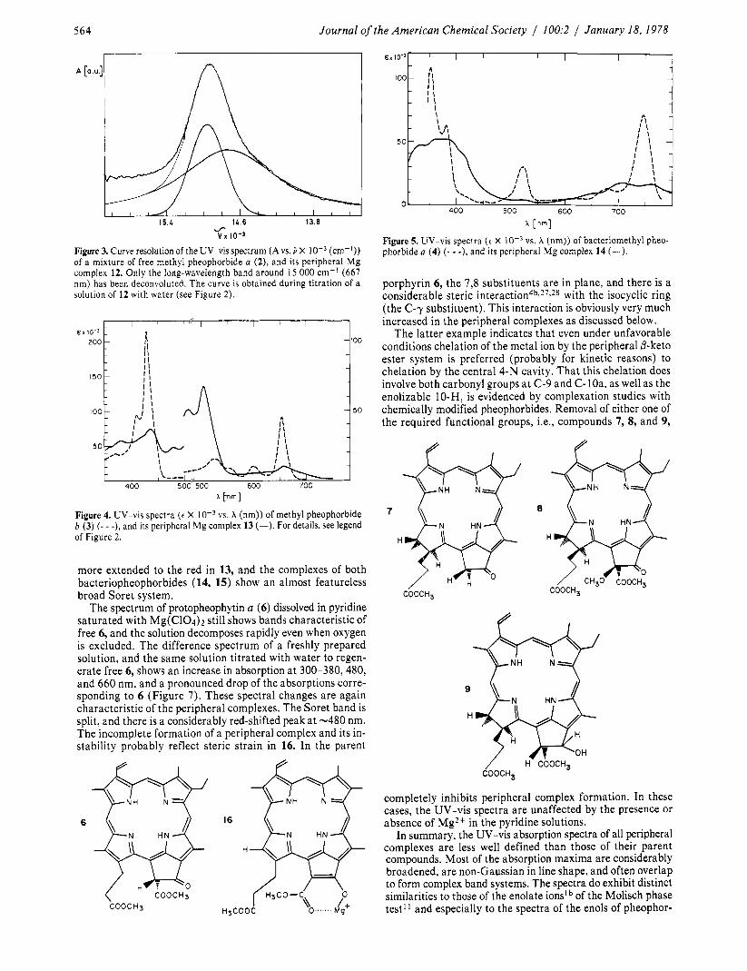
while that of its peripheral complex 12 is bright yellowish- green. In the UV-vis spectrum (Figure 2) the red Qy(O,O) band is shifted from A,,, 67 1 nm to A,,, 682 nm in pyridine. At the same time the absorption line is considerably broadened, and a pronounced Lorentzian component is mixed into the formerly Gaussian line (Figure 3). Although the extinction coefficient is diminished from 52.6 X lo3 to 19.5 X lo3, the oscillator strength of the red band remains constant within 15%. The two medium-intensity bands at 538 and 507 nm characteristic of free base pheophorbides of the a series are missing in the pe- ripheral complex, and the Soret band(s) are replaced by two overlapping complex band systems extending from 350 nm to about 500 nm. The spectrum of the complex is very charac- teristic and has no known parallel in any chlorin. The spectrum is reminiscent, however, of that of the pheophytin a phase-test intermediate, which shows the broadened bands and the split Soret system.’,
The spectra of the peripheral complexes of methyl pheo-
Figure 2. UV-vis spectra ( e X vs. X [nm] of methyl pheophorbide a (2) ( 7 - -), and of its peripheral Mg complex 12 (-). The spectrum of 12 was obtained by dissolving 2 (ca. mol/L) in a saturated solution of Mg(C104)~ in dry pyridine. Water was then added in small increments (-10 pL total) to regenerate free 2 (- - -). The latter spectrum is identical with a spectrum of 2 dissolved in pyridine.
phorbide b (13) (Figure 4) bacteriomethyl pheophorbide a (14) (Figure 5 ) , and bacteriopheophytin b (15) (Figure 6) show similar pronounced changes as compared to the spectra of the free pheophorbides 3,4, and 5. The Soret band is even
COOCH3
4
H
C 0 0 C H 3
14
H
COOCH3
6 0 0 ~ 1 - 1 ~
COOPhy
564 Journal of
I I I I I ~ ~ 1 0 - 3
200 - 1 - rl
‘the American Chemical Society / 100:2 / January 18, 1978
100
A [a.u] n
I , , I I I I , , I , , 1 5 . 4 14.6 13.8
fl v I 10-3
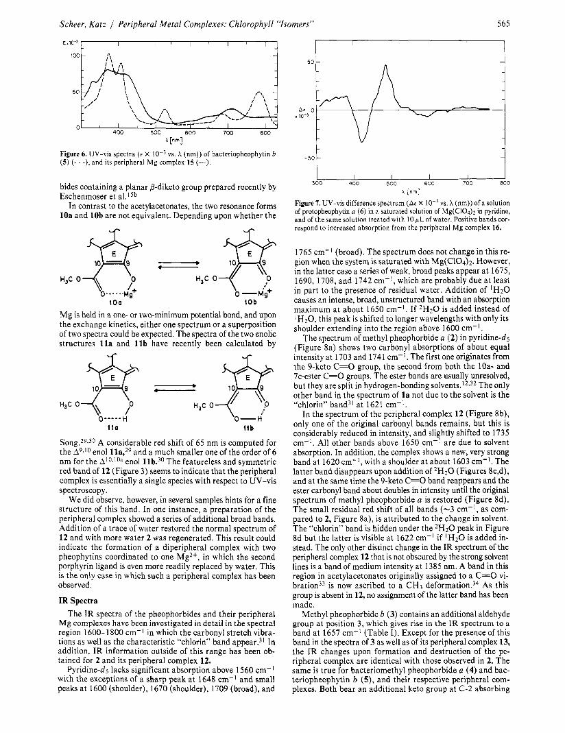
Figure 3. Curve resolution of the UV-vis spectrum (A vs. 3 X (cm-I)) of a mixture of free methyl pheophorbide a ( 2 ) , and its peripheral Mg complex 12. Only the long-wavelength band around 15 000 cm-’ (667 nm) has been deconvoluted. The curve is obtained during titration of a solution of 12 with water (see Figure 2).
more extended to the red in 13, and the complexes of both bacteriopheophorbides (14, 15) show an almost featureless broad Soret system.
The spectrum of protopheophytin a (6) dissolved in pyridine saturated with Mg(C104)2 still shows bands characteristic of free 6, and the solution decomposes rapidly even when oxygen is excluded. The difference spectrum of a freshly prepared solution, and the same solution titrated with water to regen- erate free 6, shows an increase in absorption at 300-380,480, and 660 nm, and a pronounced drop of the absorptions corre- sponding to 6 (Figure 7). These spectral changes are again characteristic of the peripheral complexes. The Soret band is split, and there is a considerably red-shifted peak at -480 nm. The incomplete formation of a peripheral complex and its in- stability probably reflect steric strain in 16. In the parent
COOCH3
[ n m l
Figure 5. UV-vis spectra (c X phorbide a (4) (- - -), and its peripheral Mg complex 14 (-).
vs. A (nm)) of bacteriomethyl pheo-
porphyrin 6, the 7,s substituents are in plane, and there is a considerable steric i n t e r a c t i ~ n ~ ~ , ~ ~ . * ~ with the isocyclic ring (the C-y substituent). This interaction is obviously very much increased in the peripheral complexes as discussed below.
The latter example indicates that even under unfavorable conditions chelation of the metal ion by the peripheral @-keto ester system is preferred (probably for kinetic reasons) to chelation by the central 4-N cavity. That this chelation does involve both carbonyl groups a t C-9 and C-loa, as well as the enolizable lO-H, is evidenced by complexation studies with chemically modified pheophorbides. Removal of either one of the required functional groups, Le., compounds 7, 8, and 9,
7
H
COOCH,
8
9
H
COOCHS
completely inhibits peripheral complex formation. In these cases, the UV-vis spectra are unaffected by the presence or absence of Mg2+ in the pyridine solutions.
In summary, the UV-vis absorption spectra of all peripheral complexes are less well defined than those of their parent compounds. Most of the absorption maxima are considerably broadened, are non-Gaussian in line shape, and often overlap to form complex band systems. The spectra do exhibit distinct similarities to those of the enolate ionslb of the Molisch phase test” and especially to the spectra of the enols of pheophor-
Scheer, Katz / Peripheral Metal Complexes: Chlorophyll “Isomers” 565
A [nm]
vs. X (nm)) of bacteriopheophytin b Figure 6. UV-vis spectra ( e X ( 5 ) (- - -), and its peripheral Mg complex 15 (-).
bides containing a planar P-diketo group prepared recently by Eschenmoser et al.ISb
In contrast to the acetylacetonates, the two resonance forms 10a and 10b are not equivalent. Depending upon whether the
t o o (Ob
Mg is held in a one- or two-minimum potential bond, and upon the exchange kinetics, either one spectrum or a superposition of two spectra could be expected. The spectra of the two enolic structures l l a and l l b have recently been calculated by
’0-H t t b
Song.29$30 A considerable red shift of 65 nm is computed for the A9,I0 enol lla,29 and a much smaller one of the order of 6 nm for the Al0,loa enol llb.30 The featureless and symmetric red band of 12 (Figure 3) seems to indicate that the peripheral complex is essentially a single species with respect to UV-vis spectroscopy.
We did observe, however, in several samples hints for a fine structure of this band. In one instance, a preparation of the peripheral complex showed a series of additional broad bands. Addition of a trace of water restored the normal spectrum of 12 and with more water 2 was regenerated. This result could indicate the formation of a diperipheral complex with two pheophytins coordinated to one Mg2+, in which the second porphyrin ligand is even more readily replaced by water. This is the only case in which such a peripheral complex has been observed.
IR Spectra The IR spectra of the pheophorbides and their peripheral
Mg complexes have been investigated in detail in the spectral region 1600-1800 cm-I in which the carbonyl stretch vibra- tions as well as the characteristic “chlorin” band appear.31 In addition, IR information outside of this range has been ob- tained for 2 and its peripheral complex 12.
Pyridine-ds lacks significant absorption above 1560 cm-’ with the exceptions of a sharp peak a t 1648 cm-I and small peaks a t 1600 (shoulder), 1670 (shoulder), 1709 (broad), and
I I I I I 1
300 400 500 600 700 800
Figure 7. UV-vis difference spectrum (A6 X vs. X (nm)) of a solution of protopheophytin a (6) in a saturated solution of Mg(C104)Z in pyridine, and of the same solution treated with I O pL of water. Positive bands cor- respond to increased absorption from the peripheral Mg complex 16.
[ n m l
1765 cm-’ (broad). The spectrum does not change in this re- gion when the system is saturated with Mg(C104)2. However, in the latter case a series of weak, broad peaks appear a t 1675, 1690, 1708, and 1742 cm-’, which are probably due a t lease in part to the presence of residual water. Addition of ‘HzO causes an intense, broad, unstructured band with an absorption maximum a t about 1650 cm-l. If 2H20 is added instead of lH20, this peak is shifted to longer wavelengths with only its shoulder extending into the region above 1600 cm-l.
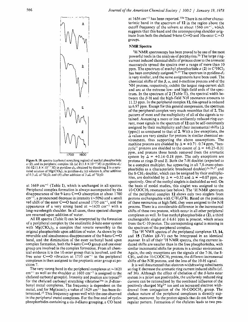
The spectrum of methyl pheophorbide a (2) in pyridine-d5 (Figure 8a) shows two carbonyl absorptions of about equal intensity a t 1703 and 1741 cm-I. The first one originates from the 9-keto C=O group, the second from both the loa- and 7c-ester C=O groups. The ester bands are usually unresolved, but they are split in hydrogen-bonding solvent^.'^*^^ The only other band in the spectrum of la not due to the solvent is the “chlorin” band31 a t 1621 cm-l.
In the spectrum of the peripheral complex 12 (Figure 8b), only one of the original carbonyl bands remains, but this is considerably reduced in intensity, and slightly shifted to 1735 cm-I. All other bands above 1650 cm-’ are due to solvent absorption. In addition, the complex shows a new, very strong band at 1620 cm-I, with a shoulder a t about 1603 cm-I. The latter band disappears upon addition of 2H20 (Figures 8c,d), and at the same time the 9-keto C=O band reappears and the ester carbonyl band about doubles in intensity until the original spectrum of methyl pheophorbide a is restored (Figure 8d). The small residual red shift of all bands (-3 cm-I, as com- pared to 2, Figure 8a), is attributed to the change in solvent. The “chlorin” band is hidden under the 2H20 peak in Figure 8d but the latter is visible a t 1622 cm-l if IH20 is added in- stead. The only other distinct change in the IR spectrum of the peripheral complex 12 that is not obscured by the strong solvent lines is a band of medium intensity a t 1385 nm. A band in this region in acetylacetonates originally assigned to a C=O vi- b r a t i ~ n ~ ~ is now ascribed to a CH3 d e f ~ r m a t i o n . ~ ~ As this group is absent in 12, no assignment of the latter band has been made.
Methyl pheophorbide b (3) contains an additional aldehyde group a t position 3, which gives rise in the IR spectrum to a band a t 1657 cm-I (Table I). Except for the presence of this band in the spectra of 3 as well as of its peripheral complex 13, the IR changes upon formation and destruction of the pe- ripheral complex are identical with those observed in 2. The same is true for bacteriomethyl pheophorbide a (4) and bac- teriopheophytin b (S), and their respective peripheral com- plexes. Both bear an additional keto group a t C-2 absorbing
566 Journal of the American Chemical Society / 100:2 / January 18, 1978
I
I
I 1700 1600
Figure 8. IR spectra (carbonyl stretching region) of methyl pheophorbide a (2), and its peripheral complex 12. (a) 2 (1.8 X M) in pyridine-ds; (b) 12 (1.8 X M) in pyridine-ds, obtained by dissolving 2 i n a satu- rated solution of Mg(C104)~ in pyridine-ds; (c) solution b, after addition of 0.3 fiL of 2H20; and (d) after addition of 3 gL of 2H20.
a t 1669 cm-I (Table I), which is unchanged in all spectra. Peripheral complex formation is always accompanied by the disappearance of the 9-keto C=O absorption at about 1700 cm-I, a pronounced decrease in intensity (-50%) and a small red shift of the ester C=O band around 1735 cm-I, and the appearance of a very strong band at -1620 cm-I with a long-wavelength shoulder. In all cases, these spectral changes are reversed upon addition of water.
All IR spectra (Table I) can be interpreted by the formation of a peripheral complex by the enolizabie P-keto ester system with Mg(C104)2, a complex that reverts reversibly to the original pheophorbide upon addition of water. As shown by the reversible and simultaneous disappearance of the 9-keto C 4 band, and the diminuition of the ester carbonyl band upon complex formation, both the 9-keto C=O group and one ester group are involved in the complex formation. From all chem- ical evidence it is the 10-ester group that is involved, and the free ester C=O vibration at 1735 cm-’ in the peripheral complexes is then assigned to the propionic ester group a t po- sition 7.
The very strong band in the peripheral complexes at -1620 cm-’ as well as the shoulder at 1603 cm-I is assigned to the chelated carbonyl group(s). Intensity and position are typical35 for the chelated carbonyl stretching vibratiod4a in P-dicar- bony1 metal complexes. The frequency is dependent on the metal, and for Mg(acac)2 a value of 1629 cm-’ has been de- t e r m i r ~ e d . ~ ~ This frequency agrees well with the one observed in the peripheral metal complexes. For the free enol of cyclo- pheophorbides containing a cis @-diketo grouping, a C O band
a t 1656 cm-I has been r e ~ 0 r t e d . I ~ ~ There is no other charac- teristic band in the spectrum of 12 in the region above the cutoff frequency of the solvent at about 1560 cm-I, which suggests that this band and the accompanying shoulder orig- inate from both the chelated 9-keto C=O and loa-ester C=O groups.
NMR Spectra ‘H NMR spectroscopy has been proved to be one of the most
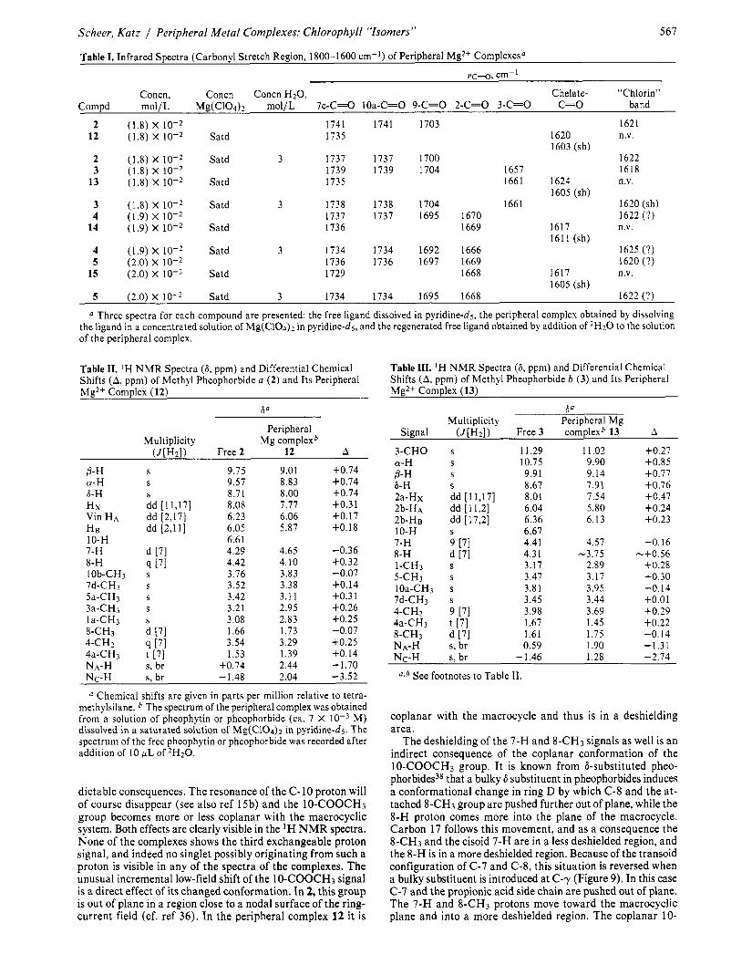
powerful tools in the analysis of porphyrin^.^^ The large ring- current induced chemical shifts of protons close to the aromatic macrocycle spread the spectra over a range of more than 10 ppm. The spectrum of methyl pheophorbide a (2) in C2HC13 has been completely a~s igned .*~,~’ The spectrum in pyridine-ds is very similar, and the same assignments have been used. The chemical shifts of the 6, a, and b-methine protons and of the N H protons, respectively, exhibit the largest ring-current shift and are a t the extreme low- and high-field ends of the spec- trum. In the spectrum of 2 (Table 11), the spectral width be- tween the @-H and the high-field NH resonance amounts to 1 1.23 ppm. In the peripheral complex 12, this spread is reduced to 6.97 ppm. Except for this general compression, the spectrum of the peripheral complex very much resembles that of 2. The pattern of most and the multiplicity of all of the signals is re- tained. Assuming a more or less uniformly reduced ring cur- rent, most signals in the spectrum of 12 can be self-consistently assigned by their multiplicity and their incremental shifts (A (ppm)) as compared to that of 2. With a few exceptions, the A values are very similar for protons in similar chemical en- vironment, thus supporting the above assumptions. The methine protons are shielded by A = +0.71-0.74 ppm, “ben- zylic” protons are shielded to the extent of A = +0.25-0.31 ppm, and protons three bonds removed from the aromatic system by A = +0.14-0.18 ppm. The only exceptions are protons a t rings D and E. Both the 7-H doublet (expected to be a complex multiplet, but appearing in all 7,8-trans-pheo- phorbides as a characteristic broadened d o ~ b l e t ) * ~ , ~ ~ , ~ ’ and the 8-CH3 doublet, which can be assigned by their multiplic- ities, are deshielded by A = -0.32 and A = -0.07 ppm, re- spectively. One of the methyl singlets is deshielded as well. On the basis of model studies, this singlet was assigned to the 10-COOCH3 resonance (see below). The ‘ H NMR spectrum of the peripheral complex 12 shows only two singlets from protons exchangeable with C2H302H. Based on the position of these resonances at high field, they were assigned to the NH protons. There is a considerable difference in the incremental shifts of these two protons, which occur in all other peripheral complexes as well. In free methyl pheophorbide a (2), a third exchangeable singlet at 6 6.61 ppm is present, which arises from the C-10 proton. The corresponding signal is missing in the spectrum of the peripheral complex.
The ‘H NMR spectra of the peripheral complexes 13,14, and 15 (Tables 111-V) can be interpreted in an identical manner. In all of their ‘H NMR spectra, the ring current in- duced shifts are smaller than in the free pheophorbides, with similar incremental shifts for protons in a similar environment. Again, the only exceptions are the signals of the 7-H, the 8- CH3, and the 10-COOCH3 protons, the different incremental shifts of the N H protons, and the loss of the 10-H signal.
It is well documented that electron-withdrawing substituents at ring E decrease the aromatic ring current induced shifts (cf. ref 36). Although the effect of chelation of the &keto ester system is a priori not predictable, the uniformly reduced ring current can be rationalized by the combined influence of the positively charged Mg2+ ion and an increased electron with- drawal from conjugation of the 10-COOCH3 group. The chelate nature of the peripheral complexes is directly sup- ported, moreover, by the proton signals that do not follow the regular pattern. Formation of the chelates leads to two pre-
Scheer, Katz / Peripheral Metal Complexes: Chlorophyll “Isomers”
Table I. Infrared Spectra (Carbonyl Stretch Region, 1800-1600 cm-I) of Peripheral Mg2+ Complexesa
567
vc-0, cm-l Concn, Concn Concn H20 , Chelate- “Chlorin”
Compd mol/L Mg(C104)2 mol/L 7c-C=0 lOa-C=O 9-C=0 2-C=0 3-C=0 C=O band
2 (1.8) X 1741 1741 1703 1621 12 (1.8) X Satd 1735 1620 n.v.
1603 (sh) 2 (1.8) X Satd 3 1737 1737 1700 1622 3 (1.8) x 10-2 1739 1739 1704 1657 1618
13 (1.8) X Satd 1735 1661 1624 n.v. 1605 (sh)
3 (1.8) X Satd 3 1738 1738 1704 1661 1620 (sh) 4 (1.9) X 1737 1737 1695 1670 1622 (?)
14 (1.9) X Satd 1736 1669 1617 n.v. 1611 (sh)
4 (1.9) X Satd 3 1734 1734 1692 1666 1625 (?) 5 (2.0) x 10-2 1736 1736 1697 1669 1620 (?)
15 (2.0) X Satd 1729 1668 1617 n.v. 1605 (sh)
5 (2.0) X Satd 3 1734 1734 1695 1668 1622 (?)
a Three spectra for each compound are presented: the free ligand dissolved in pyridine-d5, the peripheral complex obtained by dissolving the ligand in a concentrated solution of Mg(C104)2 in pyridine-d5, and the regenerated free ligand obtained by addition of 2H20 to the solution of the peripheral complex.
Table 11. ’ H NMR Spectra (6, ppm) and Differential Chemical Shifts (A, ppm) of Methyl Pheophorbide a (2) and Its Peripheral Mg2+ Complex (12)
Table 111. ‘H NMR Spectra (6, ppm) and Differential Chemical Shifts (A, ppm) of Methyl Pheophorbide b (3) and Its Peripheral Mg2+ Complex (13)
Peripheral Multiplicity Mg complexb
(J[H2]) Free 2 12 A
0- H S 9.75 9.01 +0.74 CY- H S 9.57 8.83 +0.74 6-H S 8.7 1 8.00 +0.74 Hx dd [11,17] 8.08 7.77 +0.31 Vin HA dd [2,17] 6.23 6.06 +0.17 H B dd [2,11] 6.05 5.87 +O. 18 10-H 6.61 7-H d [71 4.29 4.65 -0.36
+0.32 8-H [71 4.42 4.10 10b-CH3 s 3.76 3.83 -0.07 7d-CH3 s 3.52 3.38 +O. 14 5a-CH3 s 3.42 3.1 1 +0.31 3a-CH3 s 3.21 2.95 +0.26 la-CH3 s 3.08 2.83 +0.25 8-CH3 d 171 1.66 1.73 -0.07
+0.25 4-CH2 4 [71 3.54 3.29 4a-CH3 t [7] 1.53 1.39 +0.14 NA-H s, br +0.74 2.44 - 1.70 Nc-H s, br - 1.48 2.04 -3.52
a Chemical shifts are given in parts per million relative to tetra- methylsilane. The spectrum of the peripheral complex was obtained from a solution of pheophytin or pheophorbide (ca. 7 X M) dissolved in a saturated solution of Mg(C10& in pyridine-d5. The spectrum of the free pheophytin or pheophorbide was recorded after addition of I O WL of 2H20.
dictable consequences. The resonance of the (2-10 proton will of course disappear (see also ref 15b) and the 10-COOCH3 group becomes more or less coplanar with the macrocyclic system. Both effects are clearly visible in the ‘H NMR spectra. None of the complexes shows the third exchangeable proton signal, and indeed no singlet possibly originating from such a proton is visible in any of the spectra of the complexes. The unusual incremental low-field shift of the 10-COOCH3 signal is a direct effect of its changed conformation. In 2, this group is out of plane in a region close to a nodal surface of the ring- current field (cf. ref 36). In the peripheral complex 12 it is
l3a Multiplicity Peripheral Mg
3-CHO s 1 1.29 11.02 +0.27 CY-H S 10.75 9.90 +0.85 P- H S 9.91 9.14 +0.77 6-H S 8.67 7.91 $0.76 2a-Hx dd [11,17] 8.01 7.54 +0.47 2b-HA dd [ 11,2] 6.04 5.80 +0.24 2b-HB dd [17,2] 6.36 6.13 +0.23 10-H S 6.67
4.41 4.57 -0.16 4.3 1 -3.75 -+0.56 3.17 2.89 +0.28
5-CH3 s 3.47 3.17 +0.30 loa-CH3 s 3.81 3.95 -0.14 7d-CH3 s 3.45 3.44 f 0 . 0 1 4-CH2 9 [7] 3.98 3.69 +0.29 4a-CH3 t [7] 1.67 1.45 +0.22
Signal (J[H2]) Free 3 complexb 13 A
7-H 9 [71 8-H d [71 1-CH3 s
8-CH3 d [7] 1.61 1.75 -0. I4 NA-H s, br 0.59 1.90 -1.31 Nc-H s. br - 1.46 1.28 -2.74
a,b See footnotes to Table 11.
coplanar with the macrocycle and thus is in a deshielding area.
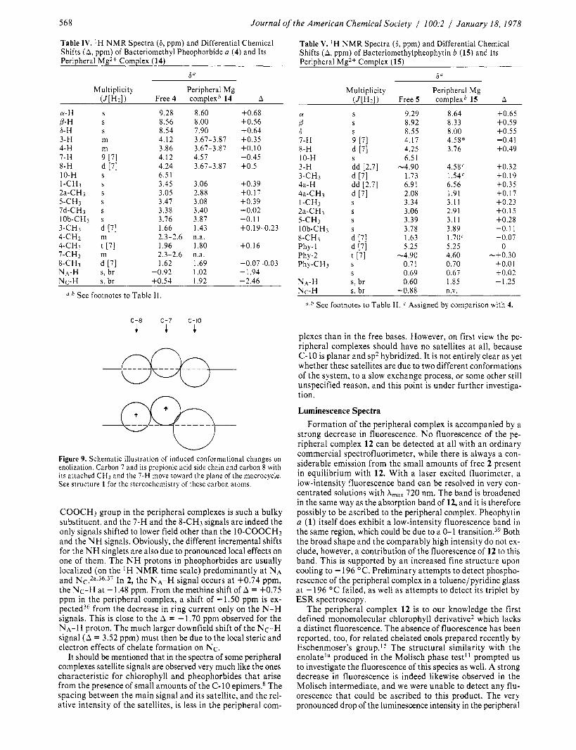
The deshielding of the 7-H and 8-CH3 signals as well is an indirect consequence of the coplanar conformation of the 10-COOCH3 group. It is known from &substituted pheo- phorbides3* that a bulky 6 substituent in pheophorbides induces a conformational change in ring D by which C-8 and the at- tached 8-CH3 group are pushed further out of plane, while the 8-H proton comes more into the plane of the macrocycle. Carbon 17 follows this movement, and as a consequence the 8-CH3 and the cisoid 7-H are in a less deshielded region, and the 8-H is in a more deshielded region. Because of the transoid configuration of C-7 and C-8, this situation is reversed when a bulky substituent is introduced at C-y (Figure 9). In this case C-7 and the propionic acid side chain are pushed out of plane. The 7-H and 8-CH3 protons move toward the macrocyclic plane and into a more deshielded region. The coplanar 10-
568 Journal of the American Chemical Society / 100:2 1 January 18, 1978
Table IV. ' H NMR Spectra (a, ppm) and Differential Chemical Shifts (A , ppm) of Bacteriomethyl Pheophorbide a (4) and Its PeriDheral Me2+ Comdex (14)
Multiplicity Peripheral Mg (JW21) Free 4 complexb 14 A
CY- H P- €4 6-H 3-H 4-H 7-H 8-H 10-H I-CH3
5-CH3 7d-CH3 lob-CH, 3-CH3 4-CHz 4-CH3 7-CHz 8-CH3 N A - H
2a-CH3
S
S
S m m 9 [71 d [71 S
S
S S S
S
d [71
t [71
d [71
m
m
s, br
9.28 8.56 8.54 4.12 3.86 4.12 4.24 6.51 3.45 3.05 3.47 3.38 3.76 1.66
1.96
1.62
2.3-2.6
2.3-2.6
-0.92
8.60 8.00 7.90 3.67-3.87 3.67-3.87 4.57 3.67-3.87
3.06 2.88 3.08 3.40 3.87 1.43 n.a. 1 .80 n.a. 1.69 1.02
+0.68 +0.56 +0.64 +0.35 +0.10 -0.45 +0.5
f 0 . 3 9 +O. 17 +0.39 -0.02 -0.1 1 +0.19-0.23
+O. 16
-0.07-0.03 -1.94
N c - H s , br +0.54 1.92 -2.46
a , b See footnotes to Table 11.
c-8 c-7 c-IO 1 c c
Figure 9. Schematic illustration of induced conformational changes on enolization. Carbon 7 and its propionic acid side chain and carbon 8 with its attached C H j and the 7-H move toward the plane of the macrocycle. See structure 1 for the stereochemistry of these carbon atoms.
COOCH3 group in the peripheral complexes is such a bulky substituent, and the 7-H and the 8-CH3 signals are indeed the only signals shifted to lower field other than the 10-COOCH3 and the N H signals. Obviously, the different incremental shifts for the N H singlets are also due to pronounced local effects on one of them. The N H protons in pheophorbides are usually localized (on the IH N M R time scale) predominantly a t N A and N c . ~ ~ , ~ ~ , ~ ~ In 2, the NA-H signal occurs a t +0.74 ppm, the Nc-H at - 1.48 ppm. From the methine shift of A = +0.75 ppm in the peripheral complex, a shift of -1.50 ppm is ex- p e ~ t e d ~ ~ from the decrease in ring current only on the N-H signals. This is close to the A = -1.70 ppm observed for the NA-H proton. The much larger downfield shift of the Nc-H signal (A = 3.52 ppm) must then be due to the local steric and electron effects of chelate formation on N c .
It should be mentioned that in the spectra of some peripheral complexes satellite signals are observed very much like the ones characteristic for chlorophyll and pheophorbides that arise from the presence of small amounts of the C-10 epimers.8 The spacing between the main signal and its satellite, and the rel- ative intensity of the satellites, is less in the peripheral com-
Table V. IH N M R Spectra (6, ppm) and Differential Chemical Shifts (A , ppm) of Bacteriomethylpheophytin b (15) and Its Peripheral Mg2+ Complex (15)
80
Multiplicity Peripheral Mg (J[H2]) Free 5 complexb 15 A
CY
P 6 7-H 8-H IO-H 3-H 3-CH3 4a-H 4a-CH3
2a-CH3 I-CH3
5-CH3 lob-CH3 8-CH3 Phy- 1 Phy-2 Phy-CHj
N a - H
9.29 8.92 8.55 4.17 4.25 6.5 1
-4.90 1.73 6.9 1 2.08 3.34 3.06 3.39 3.78 1.63 5.25
-4.90 0.7 1 0.69 0.60
8.64 8.33 8.00 4.58* 3.76
4.5aC 1 .54c 6.56 1.91 3.1 1 2.91 3.11 3.89 1.70' 5.25 4.60 0.70 0.67 1.85
+0.65 f 0 . 5 9 +0.55
+0.49
+0.32 +0.19 f 0 . 3 5 +O. 17 +0.23 +0.15 +0.28 -0.1 1 -0.07
0 -+0.30
$0.01 +0.02 -1.25
-0.41
N c - H s, br -0.88 n.v.
See footnotes to Table 11. Assigned by comparison with 4.
plexes than in the free bases. However, on first view the pe- ripheral complexes should have no satellites a t all, because C-10 is planar and sp2 hybridized. It is not entirely clear as yet whether these satellites are due to two different conformations of the system, to a slow exchange process, or some other still unspecified reason, and this point is under further investiga- tion.
Luminescence Spectra Formation of the peripheral complex is accompanied by a
strong decrease in fluorescence. No fluorescence of the pe- ripheral complex 12 can be detected a t all with an ordinary commercial spectrofluorimeter, while there is always a con- siderable emission from the small amounts of free 2 present in equilibrium with 12. With a laser excited fluorimeter, a low-intensity fluorescence band can be resolved in very con- centrated solutions with A,,, 720 nm. The band is broadened in the same way as the absorption band of 12, and it is therefore possibly to be ascribed to the peripheral complex. Pheophytin a (1) itself does exhibit a low-intensity fluorescence band in the same region, which could be due to a 0-1 transition.39 Both the broad shape and the comparably high intensity do not ex- clude, however, a contribution of the fluorescence of 12 to this band. This is supported by an increased fine structure upon cooling to -196 OC. Preliminary attempts to detect phospho- rescence of the peripheral complex in a toluene/pyridine glass a t -196 O C failed, as well as attempts to detect its triplet by ESR spectroscopy.
The peripheral complex 12 is to our knowledge the first defined monomolecular chlorophyll derivative2 which lacks a distinct fluorescence. The absence of fluorescence has been reported, too, for related chelated enols prepared recently by Eschenmoser's group.I5 The structural similarity with the enolateIa produced in the Molisch phase test]' prompted us to investigate the fluorescence of this species as well. A strong decrease in fluorescence is indeed likewise observed in the Molisch intermediate, and we were unable to detect any flu- orescence that could be ascribed to this product. The very pronounced drop of the luminescence intensity in the peripheral
Scheer, Katz / Peripheral Metal Complexes: Chlorophyll “Isomers” 569
complexes as well as in the enolate ions indicates an efficient radiationless decay mechanism. Similar findings in hydroxy- benza1dehydes4O and benzophenone^^^ have been interpreted to arise from internal conversion induced by a tautomerization mechanism. The luminescence loss in the peripheral complexes may then be due to a similar process. Titration Experiments
The peripheral complexes are unstable against water and enolizable @-dicarbonyl compounds such as acetylacetone, hexafluoroacetylacetone, or 2-carbethoxycyclopentanone. The free pheophorbide is in all cases (except for the protopheo- phytin a, see above) recovered unchanged. The reaction is a ligand exchange process a t the peripheral Mg2+. By the use of fluorescence, UV-vis, IR, and ‘H N M R spectroscopy the ligand exchange can be studied over the large concentration range from lo-’ to lod2 M. In the lowest concentration range, the amount of free pheophytin can be determined from its fluorescence. Titrations of the peripheral complex 12 with water gave a sigmoidal curve for the fluorescence intensity as a function of the water concentration. The shape of the titration curve is interpreted as a displacement first of pyridine coor- dinated to the Mg2+, and the subsequent displacement of the chelated pheophytin.
mol/L) the ti- tration can be followed by UV-vis spectroscopy. Up to water concentrations of about 2 M, series of isosbestic points are observed over the entire spectral range. The overlapping red bands of the complex 12 and free 2 have been deconvoluted for a series of water concentrations (cf. Figure 3). During the entire titration, the envelope can be deconvoluted into two components of varying intensity. One of these corresponds to the free 2. This component has a Gaussian line shape, is posi- tioned at 669 nm, and has a half-width at half-height of 8.5 nm (165 cm-I). The other component originates from the pe- ripheral complex 12, and is best fitted with a Gaussian-Lo- rentzian combination line shape centered a t 685 nm. In the pure complex the Gaussian line width is 3650 cm-I, the Lo- rentzian line width 673 cm-l (because the lines are so broad, a fit of the energy (cm-I) spectrum rather than the frequency spectrum (nm) is desirable if deconvolution with symmetric bands is used). Although the absorption maximum remains constant, the line width of the two components changes during the titration.
The ratio of the intensity differences of the bands of 2 and 12 is constant during the titration, and is equal to the ratio of the extinction coefficients of 2 and 12. All the UV-vis data thus support the presence of only the two species 2 and 12 over the course of the entire titration. The only irregularity occurring is the change in the line shape of 12 during the titration, which might possibly be due to ligand exchange at Mg2+.
M, the equilibrium can be studied by ‘H N M R and by I R spectroscopy. In a mixture of methyl pheophorbide a (2) with its peripheral Mg complex 12, the chlorin ligand exchange reaction is slow enough to give rise to two separate IH N M R spectra, even a t elevated temperatures. Starting mixtures were obtained by dissociating part of the complex with a small amount of water. The resulting complex equilibrium mixture contains three competing ligands for the Mg2+ ion: water, pheophorbide, and pyridine. The relative amounts of 2 and 12 can be monitored by ‘H N M R from the intensity of their respective spectra. The amount of bound water can be judged from the position of the water resonance. Although the chemical shift of water is de- pendent on the temperature itself, the major contribution to its chemical shift seems to arise from coordination to Mg2+ (see above). At elevated temperatures, the concentration of the complex increases, while the concentration of bound water decreases.
At slightly higher concentrations
At concentrations higher than 5 X
In the temperature range between 20 and 70 OC, the reaction can be treated from the ’H N M R data as an equilibrium be- tween the peripheral complex of methyl pheophorbide a (12) and free methyl pheophorbide a (2), as indicated by the linear fit between In C ~ ~ / C Z and 1/T (van’t-Hoffs equation, corre- lation coefficient 0.997). After addition of 2 FL of water to a 0.007 M solution of 12 (0.5 mL), the (apparent) reaction en- thalpy and entropy for complex formation are AHo = 9.4 kcal/mol and ASo = 31 eu/mol, respectively. Both values decrease with increasing water concentration, and after ad- dition of 3 FL of water, values of AHo = 4.4 kcal/mol and ASo = 10.8 eu/mol were found.
The titration experiments indicate an increase in coordi- nation binding energy for the three ligands present in the sys- tem with pyridine < pheophorbide < water. That pyridine must be a weaker ligand for Mg2+ than the pheophorbide is obvious from the fact that peripheral complex formation would otherwise not be possible. The sigmoid shape of the titration curves with water is then interpreted as the subsequent re- placement of first the loosely bound pyridine and second the more strongly bound pheophorbide.
Conclusions The data demonstrate that the peripheral P-keto ester sys-
tem present in Mg-free chlorophyll derivatives with an intact isocyclic ring E binds Mg2+ more strongly than the central cavity of the m a c r o ~ y c l e . ~ ~ Complexes of chlorophyll with a variety of metal chlorides have been reported earlier by Dilung et al.43 These complexes are likewise sensitive to water, but do not show the spectral characteristics of the peripheral com- plexes, and it has been suggested that they are formed by x interactions with the N atoms. In the peripheral complexes, all spectroscopic evidence supports a chelate-type binding that is characteristic of enolizable 0-dicarbonyl derivatives. The potential of this functional group in chlorophylls for metal binding and enolization has been explored recently in a dif- ferent approach by the group of E~chenmoser , ’~ with the aid of P-diketones derived from chlorophylls (“cyclochlorophylls”) in which a loa-keto C=O group is held essentially coplanar with the 9-keto C=O group. Preliminary spectroscopic data on metal complexes of these diketones are very similar to those obtained for the peripheral metal complexes, Le., broadened and red-shifted UV-vis spectra, loss of fluorescence, and de- creased ring current induced ‘H NMR shifts.’5b
The peripheral metal complexes are thermodynamically less stable than the central complexes with respect to demetalation and metal exchange. This is best evidenced by Zn2+, which has a moderate affinity for both binding sites. Methyl pheophor- bide a , when treated with dry ZnClz, first forms the peripheral complex in a kinetically controlled reaction, which then transforms within a few hours to the thermodynamically fa- vored central complex. In the case of magnesium the central insertion requires carefully controlled conditions,15a and the complexes are thermodynamically unstable in aqueous solu- t i o n ~ . ~ ~ The peripheral Mg2+ complexes are thermodynami- cally even less stable; they are dissociated easily by small amounts of water and by other chelating agents that compete for the binding of Mg2+. However, in the absence of competing ligands formation of the peripheral Mgz+ complexes is favored kinetically as compared to formation of the central complexes (cf. ref 45). A similar situation exists in linear tetrapyrroles. Formation of bile pigment metal complexes (e.g., with Zn2+) in methanol is favored kinetically, as compared to complex formation by the macrocyclic porphyrins, but is not favored thermodynamically (cf. ref 46). For metal ions such as Cu2+ which bind readily and strongly to the central four-nitrogen site, no peripheral complexes have been observed, but central complexes form directly. The thermodynamics are controlled by the peripheral @-keto ester being a “harder” ligand than is
510 Journal of the American Chemical Society / 100:2 / January 18, 1978
the central 4-N site. Thus, the hard Mg2+ ion forms more stable peripheral complexes than the softer ions Zn2+ or Cu2+. On the other hand, the kinetics of insertion can be considered to be controlled by the flexibility of the two ligand sites, which is particularly unfavorable for the central 4-N site. The in- fluence of the solvent system, and thus of the ligands at the metal ions in solution, has not been studied.
There seems to be a mutual exclusion of the two binding sites. Thus, attempts to prepare peripheral Mg2+ complexes of the chlorophylls themselves were unsuccessful. There is no UV-vis evidence for a dimetallic intermediate during the conversion of Zn-peripheral complex to Zn-central complex, and the final spectrum is that of the pure central Zn pheo- phorbide ("Zn chlorophyllide"). This mutual exclusion is probably due both to steric hindrance of the C-7 and C-10 substituents and to electrostatic repulsion of the two proximate (4 A) positively charged ions. Similarly, peripheral as well as central complexes have been reported for the less hindered "cyclopheophorbides" prepared by Falk et al.,15 but none have been reported that bear two metal ions simultaneously at both the center and the periphery.
A possible physiological role for the peripheral metal com- plexes is yet to be defined. Self-aggregation of chlorophylls involves interaction of ring E with the central Mg2+ of the neighboring molecule, and such aggregates have been proposed as models for antenna chlorophyll.1b%2b However, studies with chemically modified chlorophyll^^^ show a predominant binding to the 9-keto C=O and indeed pyrochloro- phyll a (the central Mg complex of 8) shows much stronger aggregation behavior than does chlorophyll a.47 These results argue against a significant role for peripheral complexation in these aggregates. This view is supported by the UV-vis spectral data on chlorophyll a oligomers, and by model studies indicating unfavorable steric interactions with the 7-propionic ester side chain. Chelation is more likely with metals carrying less bulky additional ligands, especially in a hydrophobic en- vironment. Such conditions are present in large areas of the photosynthetic membrane system. It may be noted that the absorption maximum for photosystem I1 reaction centers49 is very close to that of the peripheral complex 12. While a role for peripheral complexes in photosynthesis cannot be assigned on the basis of current information, the possible participation of peripheral complexes of the pheophytins must be considered, especially in the light of the characteristic red shifts in the visible absorption spectrum caused by peripheral complex formation.
Acknowledgments. Dr. Hugo Scheer (Institut fur Botanik der Universitat, 8 Miinchen, West Germany) acknowledges with gratitude a grant from the Deutsche Forschungsge- meinschaft, Bonn-Bad Godesberg, West Germany. We thank A. Zielen for his assistance with the spectral deconvolution program, J. C. Hindman for the measurement of the low- temperature luminescence spectra, and P. R. Edwards and V. A. Heintz for their enthusiastic help as participants in the Undergraduate Research Participation Program administered by The Argonne Center for Educational Affairs. This work was performed under the auspices of the Division of Physical Re- search of the U S . Energy Research and Development Ad- ministration.
References and Notes (1) For reviews, see (a) G. R. Seely in "The Chlorophylls", L. P. Vernon and
G. R. Seeiy, Ed., Academic Press, New York, N.Y., 1966, p 67; (b) J. J. Katz in "Bioinorganic Chemistry", G. L. Eichhorn, Ed., Elsevier, Amsterdam, 1972, p 1022; (c) J. T. Warden and J. R. Bolton, Acc. Chem. Res., 7, 189 (1974); (d) W. W. Parson and R. J. Cogdell, Biochim. Biophys. Acta, 418, 105 (1974); (e) K. Sauer in "Bioenergetics of Photosynthesis", Govindjee, Ed., Academic Press, New York, N.Y., 1974, pp 115-191.
(2) (a) G. L. Closs, J. J. Katz, F. C. Pennington, M. R. Thomas, and H. H. Strain,
J. Am. Chem. Soc., 85, 3809 (1963); (b) T. M. Cotton, A. D. Trifunac, K. Ballschmiter. and J. J. Katz, Biochim. Biophys. Acta, 388, 181-198 (1974): (c) J. R. Norris, H. Scheer, and J. J. Katz, Ann. N.Y. Acad. Sci., 244, 260-280 (1974); (d) C. Houssier and K. Sauer, J. Am. Chem. SOC., 92,779 (1970).
(3) J. H. Fuhrhop, Z. Naturforsch. 8, 25, 255 (1970). (4) (a) H. Scheer in "The Porphyrins", D. Dolphin, Ed., Vol. 11, Academic Press,
New York, N.Y., in press, Chapter 1; (b) H. Scheer and H. H. lnhoffen in ref 4a, Chapter 2.
(5) (a) B. Commoner, J. Townsend, and G. Pake, Nature (London), 174, 689-691 (1954); (b) P. Sogo, N. Pon, and M. Calvin, Proc. Natl. Acad. Sci. U.S.A., 43, 387-393 (1957); (c) G. Feher, A. J. Hoff, R. A. Isaacson, and L. C. Ackerson, Ann. N. Y. Acad. Sci., 244, 239-259 (1973); (d) H. Scheer, J. R. Norris, and J. J. Katz, J. Am. Chem. SOC., 99, 1372 (1977); (e) D. C. Borg, A. Forman, and J. Fajer, ibid., 98, 6889 (1976).
(6) (a) A. S. Holt in ref la, p 11 1; (b) A. Gloe, N. Pfennig, H. Brockmann, Jr., and W. Trowitzsch, Arch. Mikrobiol., 102, 103 (1975); (c) H. Brockmann, Jr., Philos. Trans. R. SOC. London, Ser. B, 273, 277 (1976).
(7) H. Scheer, W. A. Svec, B. T. Cope, M. H. Studier, R. G. Scott, and J. J. Katz, J. Am. Chem. SOC., 98,3714 (1974).
(8) J. J. Katz, G. D. Norman, and W. A. Svec, J. Am. Chem. SOC., 86, 1418 (1964); (b) H. Wolf and H. Scheer, Justus Liebigs Ann. Chem., 1710 (1973).
(9) (a) H. Fischer and H. Pfeiffer, Justus Liebigs Ann. Chem., 555,94 (1944); (b) F. C. Pennington, H. H. Strain, W. A. Svec, and J. J. Katz, J. Am. Chem. soc., 89, 3875 (1967); (c) F. C. Pennington, S. D. Boyd, H. Horton, S. W. Taylor, D. G. Wuif, J. J. Katz, and H. H. Strain, ibid., 89, 3871 (1967); (d) H. Wolf, H. Brockmann, Jr., H. Biere, and H. H. Inhoffen, Justus Liebigs Ann. Chem., 704, 208 (1967): (e) H. Wolf, I. Richter, and H. H. Inhoffen, ibid., 725, 177 (1969).
(IO) H. Fischer and H. Orth, "Die Chemie des Pyrrols", Vol. II 12, Akademische Verlagsanstalt, 1940, p 73; (b) F. C. Pennington, H. H. Strain, W. A. Svec, and J. J. Katz, J. Am. Chem. Soc., 90, 6841 (1968).
(11) (a) H. Molisch, Ber., 14, 18 (1896); (b) R. A. Willstatter and A. Stoli, "Un- tersuchungen uber Chlorophyll", Springer, West Berlin 1918. p 28.
(12) (a) J. J. Katz, G. L. Closs. F. C. Pennington, M. R. Thomas, and H. H. Strain, J. Am. Chem. Soc., 85, 3801 (1963): (b) K. Ballschmiter and J. J. Katz, ibid., 91, 2661 (1969).
(13) H. Scheer and H. Wolf, Justus Liebigs Ann. Chem., 1741 (1973). (14) (a) M. T. Cox, T. T. Howarth, A. H. Jackson, and G. W. Kenner, J. Am. Chem.
Soc., 91, 1232 (1969); (b) M. T. Cox, A. H. Jackson, G. W. Kenner, S. W. McCombie. and K. M. Smith, J. Chem. SOC., Perkin Trans. 7, 516 (1974).
(15) (a) H. P. Isenring, E. Zass, K. Smith, H. Falk, J. L. Luisier, and A. Eschen- moser, Helv. Chim. Acta, 56, 2357 (1975); (b) H. Falk, G. Hoornaert, H. P. Isenring, and A. Eschenmoser, ibid., 58, 2347 (1975).
(16) (a) J. Franck in "Light and Life", W. D. McElroy and G. Blass, Ed., Johns Hopkins Press, Baltimore, Md., 1961, p 386; (b) D. Mauzerall and A. Chiwis, J. Theor. Biol., 42, 387 (1973).
(17) H. Scheer and J. J. Katz, J. Am. Chem. SOC., 97, 3273 (1975). (18) S. J. Baum, B. F. Burnham, andR. A. Plane, Proc. Natl. Acad. Sci. U.S.A.,
52, 1439 (1964). (19) A very useful method for the insertion of magnesium into the macrocycle
of pheophorbides that contain an enolizable P-keto ester system has been reported recently.15
(20) W. C. Davidon, "Variable Metric Method for Minimization", ANL-5990. Rev. 2, Argonne National Laboratory, 1966.
(21) J. C. Hindman, R. Kugel, A. Svirmickas, and J. J. Katz, Proc. Natl. Acad. Sci. U.S.A., 74, 5 (1977).
(22) G. F. Smith and E. G. Koch, Z. Anorg. Allg. Chem., 223, 19 (1935). (23) H. H. Strain, and W. Svec, in ref la , p 21. (24) H. Biere, Ph.D. Thesis, Technische Hochschule Braunschweig, 1966. (25) H. Wolf and H. Scheer, Justus Liebigs Ann. Chem., 745, 87 (1971). (26) Analyses by atomic absorption spectroscopy were carried out by R. Bane
(27) R. C. Pettersen, J. Am. Chem. SOC., 93, 5629 (1971). (28) R. B. Woodward, lnd. Chem. Belge, 1293 (1962). (29) P. S. Song, T. A. Moore, and M. Sun in "The Chemistry of Plant Pigments",
0. Chichester, Ed., Academic Press, New York, N.Y., 1972. (30) P. S. Song, private communication, 1974. (31) J. H. Golden, R. R. Linstead, andG. H. Whitham, J. Chem. Soc., 1725(1956);
H. R. Wetherell, M. J. Hendrichson. and A. R. Mclntyre, J. Am. Chem. SOC., 81. 4715 11959).
of the Argonne Analytical Division.
(32) K. H. Ballschmiter and J. J. Katz, J. Am. Chem. SOC., 91, 2661 (1969). (33) J. Lecomte, Discuss. faraday SOC., 9, 125 (1950). (34) (a) R. Mecke and E. Funck, Z. Nektrochem., 60, 1124 (1956); (b) K. Nak-
(35) K. E. Lawson. Spectrochim. Acta, 17, 248-258 (1961). (36) H. Scheer and J. J. Katz in "Porphyrins and Metalloporphyrins", K. M. Smith,
amoto, and A. E. Martell, J. Chem. Phys., 32, 588 (1960).
Ed., Elsevier. Amsterdam, 1976, D 399. (a) S. G. Boxer, G. L. Closs, and J. J. Katz, J. Am. Chem. SOC., 96, 7058 (1974); (b) W. Trowitzsch, Org. Magn. Reson., 59 (1976). G. Brockmann and H. Brockmann, llTNMR Newslett., 117-162 (1968). J. C. Goedheer in ref la, p 147. A. Beckett and G. Porter, Trans. faraday Soc., 59,2051 (1963); A. A. La- mola and L. J. Sharp, J. Phys. Chem., 70, 2634 (1966). G. S. Hammond, M. J. Turro, and P. A. Leermakers, J. Phys. Chem., 86,
Another type of peripheral complexation (by ?r interaction with benzene rings) has been proposed for Cr(C0)3 complexes of meso-tetraphenyl- porphyrins by N. J. Logan and 2. U. Siddiqui, Can. J. Chem., 50, 720 (1972). (a) I. I. Dilung and S. S. Butsko, Dokl. Akad. Nauk SSSR, 131,312 (1960); K&/. Chem., Engl. Trans/., 223 (1960); (b) I. I. Dilung and B. Ya. Dah. Russ. J. Phys. Chem. (Engl. Trans/.), 33, 605 (1959). H. Scheer and J. J. Katz, to be published.
1144(1962),
UskokoviC et al. / Synthesis of Meroquinene 57 1
(45) P. Hambright in ref 36, p 233; J. W. Buchler in ref 36, p 157. (46) H. Scheer, U. Linsenmeier. and C. Krauss, Hoppe-Seyler’s Z. Physiol.
(47) H. Scheer and J. J. Katz, to be published.
(48) L. L. Shipman, T. R. Janson, G. J. Ray, and J. J. Kat& Roc. Natl. Acad. Sci. U.S.A., 72, 2873 (1975).
(49) G. Doring, G. Stiehl, and H. T. Witt, Z. Naturforsch. G, 22, 639 (1967); G. Doring, G. Renger, J. Vater. and H. T. Witt, bid., 24, 1139 (1987).
Chem., 358, 185 (1977).
Total Synthesis of Cinchona Alkaloids. 1. Synthesis of Meroquinene
Milan R. Uskokovik,* Thomas Henderson, Charles Reese, Hsi Lin Lee, Guenter Grethe, and Jurg Gutzwiller Contribution from the Chemical Research Department, Hoffmann-La Roche Inc., Nutley, New Jersey 071 10. Received June 13, 1977
Abstract: Meroquinene (28), the key intermediate in several total syntheses of Cinchona alkaloids, has been synthesized by three methods. Starting from cis-2-benzoyloctahydro-6(2H)-isoquinolone (l), the acetic acid and the vinyl side chains of 28 were formed by either Baeyer-Villiger oxidation, opening of the lactone 2 to the hydroxy ester 4, and elimination, or by Schmidt rearrangement, nitrosation of the lactam 7, and pyrolysis. A completely stereospecific synthesis of meroquinene (28) was effected by catalytic hydrogenation of 3-ethyl-4-pyridineacetic acid methyl ester (21), followed by conversion of the ethyl group of 23 into the vinyl group by Loffler-Freytag rearrangement and elimination.
The medically important alkaloids quinine and quinidine have long been subjects of one of the most intensive structural and synthetic investigations in classical organic chemistry.’ The original and quite elegant syntheses of these are unfortunately not amenable to large-scale preparation of various analogues. With such an aim in mind, we have inves- tigated new synthetic routes to these alkaloids in the last few years. These investigations have led to several practical solu- tions which are reported in this paper and in the accompanying publications.
The quinuclidine ring with three chiral centers is the char- acteristic feature of the Cinchona alkaloids and elaboration of this ring system is the key for a successful total s y n t h e s i ~ . ’ ~ ~ The two contiguous chiral centers a t C-3 and C-4 have always controlled the selection of the synthetic precursors. In the classical synthesis2 of these alkaloids, the quinuclidine ring was derived from 3(R)-vinyl-4(S)-piperidinepropionic acid (ho- g - N’ H H ” H
H3CO , , ..@ \ *
C inc honam ine H*G Quinine
H
Meroquinene (2) momeroquinene), obtained by the degradation of cinchonine.6 The first total synthesis of these alkaloids was formally achieved with the synthesis of homomeroquinene All recent syntheses of Cinchona alkaloids,*-’’ however, utilize the corresponding nor analogue, 3(R)-vinyl-4(S)-piperi- dineacetic acid (meroquinene), which is also a degradation product of cinchonine.l2 In this paper, we describe in full detail the synthesis of meroquinene in its racemic form as well as in both enantiomeric modifications.
In the designing stage of the meroquinene synthesis, we were primarily concerned with the cis configuration of the two side chains. Two approaches were explored, one starting from the preformed cis-isoquinolone (1),13 and the other in which the cis configuration was to be achieved by the hydrogenation of
0002-7863/78/1500-0571$01.00/0
Scheme I
I e
, C O O C H 3
O A C s H 5
4 5 I 6 ..,
a pyridine precursor. In the first case, the formation of mero- quinene required an oxidative fragmentation of the cyclo- hexanone ring of 1 to produce the acetic acid and the vinyl side chains. Baeyer-Villiger oxidation was first examined for this purpose (Scheme I) . Treatment of 1 with m-chloroperbenzoic acid yielded the desired lactone 2 and its isomer 3 as a mixture, which could not be separated. When stirred a t room temper- ature in dilute methanolic hydrogen chloride, the lactones opened to give the hydroxy esters 4 and 5, which could be separated by repeated column chromatography. However, the overall yield of the desired hydroxy ester 4 from 1 was only 35%, while the isomeric 5 was obtained in 57% yield. The ester 4 was converted in high yield to the corresponding chloro an- alogue 6, which was then transformed into racemic N-ben- zoylmeroquinene (13) (Scheme V).
Since an opposite regioselectivity in the ring fragmentation of 1 was to be achieved, we turned our attention to the Schmidt rearrangementI4 (Scheme 11). On exposure to sodium azide in polyphosphoric acid, the ketone 1 was transformed quanti- tatively to a 1:l mixture of lactams 7 and 8, which could be separated from each other by fractional crystallization. A further improvement in the desired regioselectivity was ob- served in the Schmidt rearrangement of the corresponding
0 1978 American Chemical Society


![Isomers [compatibility mode]](https://img.pdfslide.us/doc/110x75/5590c68a1a28ab90718b4739/isomers-compatibility-mode.jpg)