Embed Size (px)
Citation preview
PERIPHERAL BLOOD PERIPHERAL BLOOD SMEARSMEAR
introductionintroduction
A properly prepared blood smear is A properly prepared blood smear is essential to accurate assessment of essential to accurate assessment of cellular morphologycellular morphology
The wedge smear is the most convenient The wedge smear is the most convenient and commonly used technique for making and commonly used technique for making PBSPBS
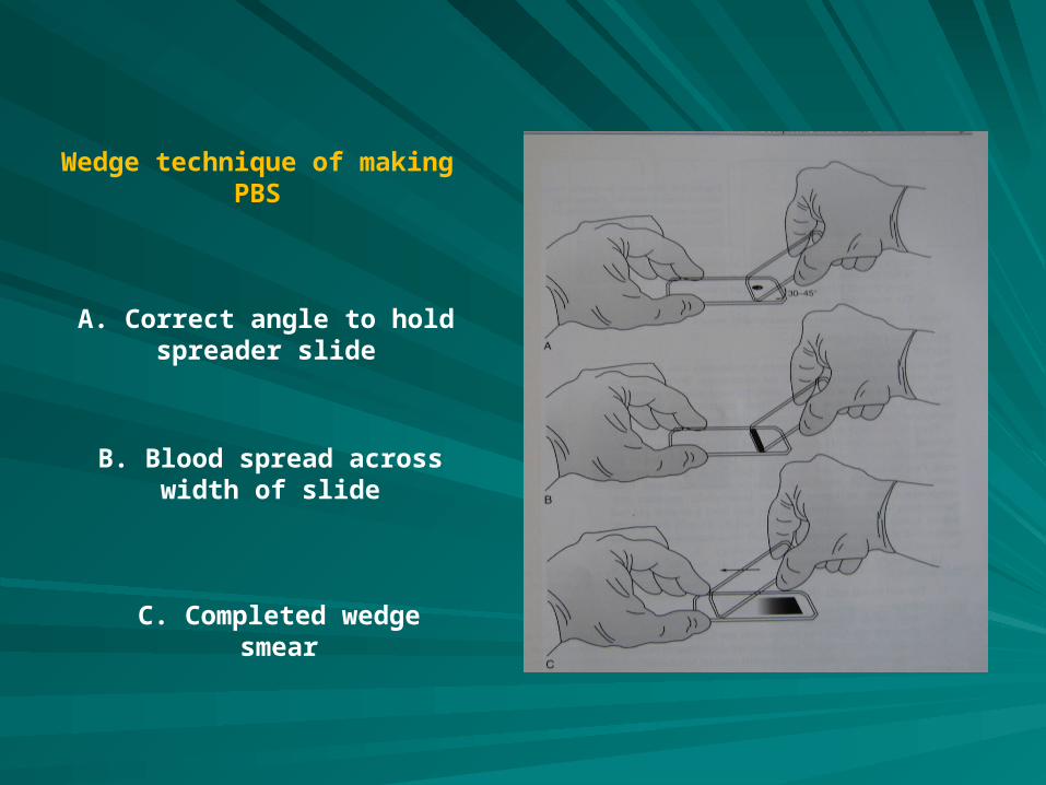
Wedge technique of making PBS
A. Correct angle to hold spreader slide
B. Blood spread across width of slide
C. Completed wedge smear
Characteristics are: Characteristics are:
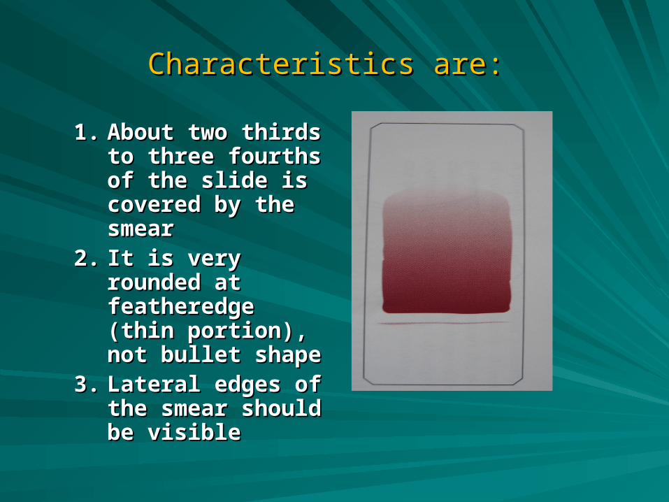
1.1. About two thirds to About two thirds to three fourths of the three fourths of the slide is covered by slide is covered by the smearthe smear
2.2. It is very rounded at It is very rounded at featheredge (thin featheredge (thin portion), not bullet portion), not bullet shapeshape
3.3. Lateral edges of the Lateral edges of the smear should be smear should be visiblevisible
Characteristics:Characteristics:
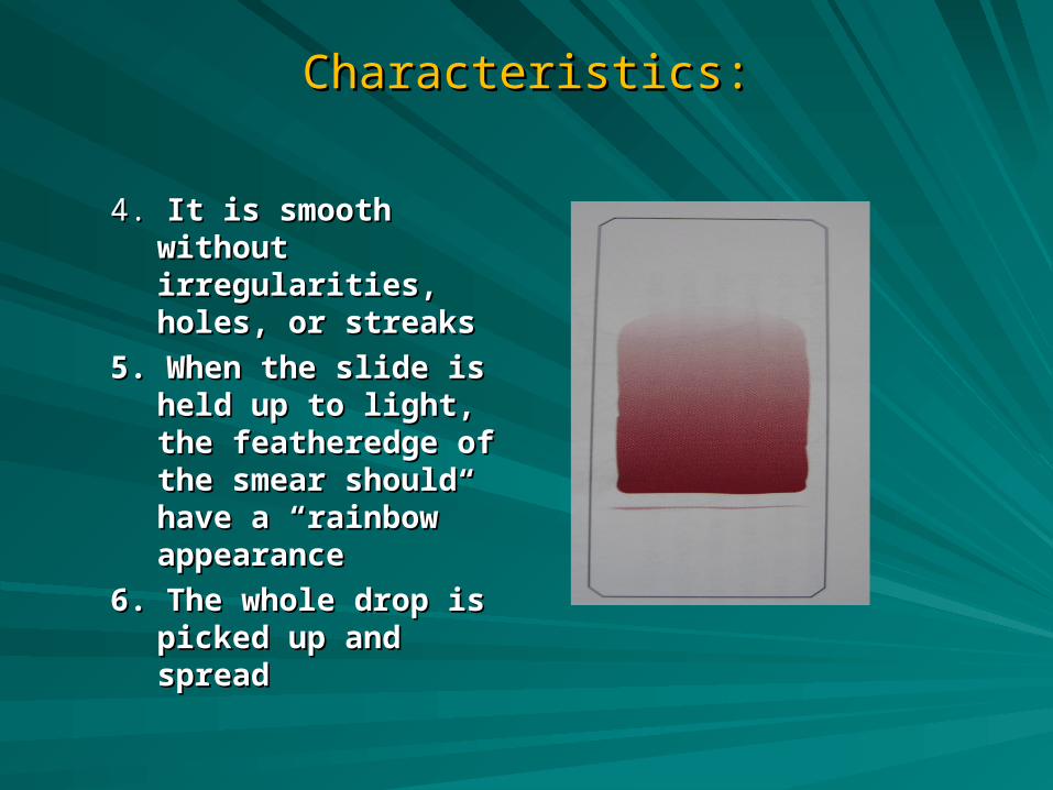
4. 4. It is smooth without It is smooth without irregularities, holes, irregularities, holes, or streaksor streaks
5. When the slide is 5. When the slide is held up to light, the held up to light, the featheredge of the featheredge of the smear should have smear should have a “rainbow” a “rainbow” appearanceappearance
6. The whole drop is 6. The whole drop is picked up and picked up and spreadspread
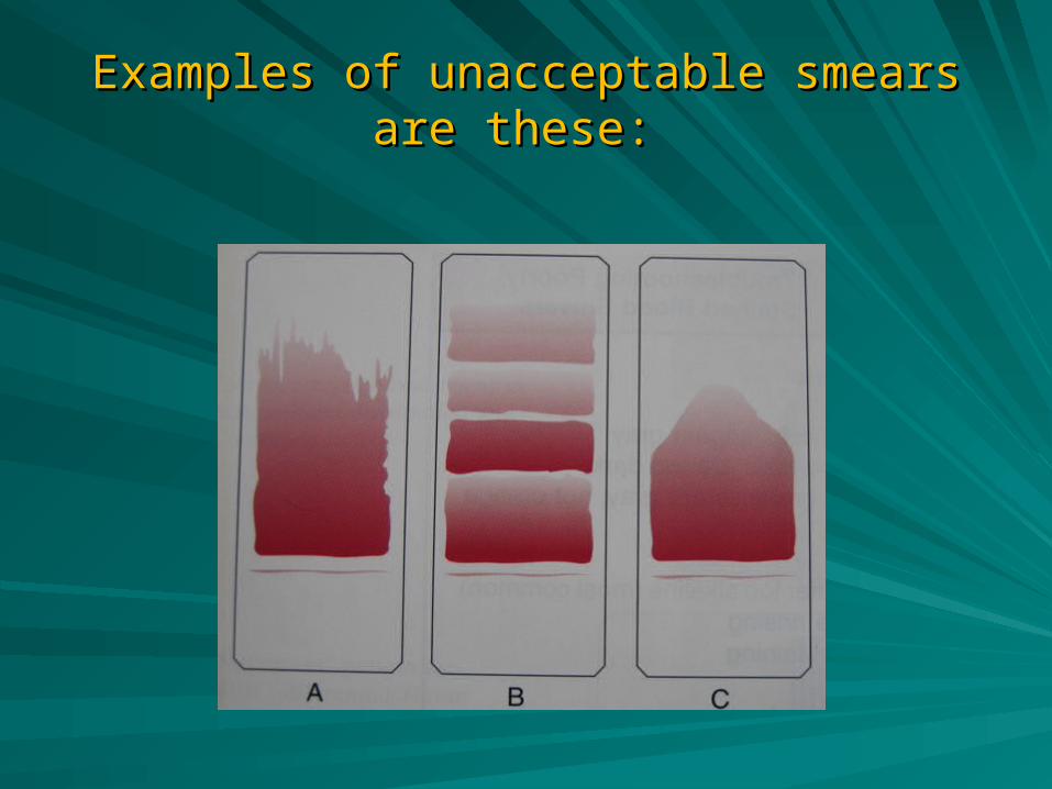
Examples of unacceptable smearsExamples of unacceptable smearsare these: are these:
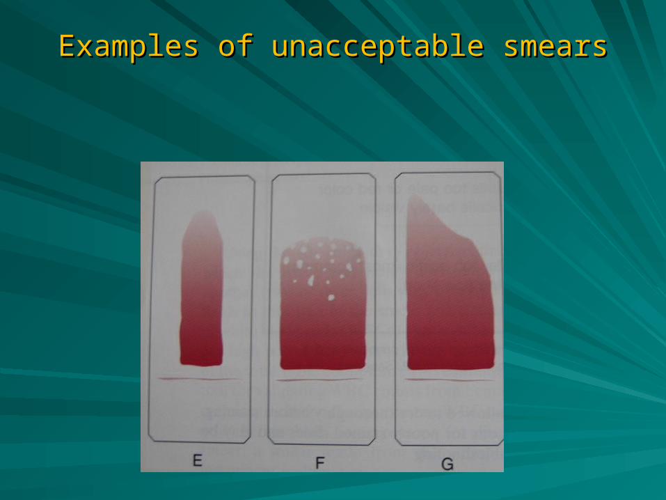
Examples of unacceptable smearsExamples of unacceptable smears
Staining of PBSStaining of PBS
Purpose of staining is to identify cells and Purpose of staining is to identify cells and recognize morphology easily through the recognize morphology easily through the microscopemicroscope
Uses Wright stain or Wright-Giemsa stain Uses Wright stain or Wright-Giemsa stain which contain both eosin and methylene blue which contain both eosin and methylene blue polychrome stains polychrome stains
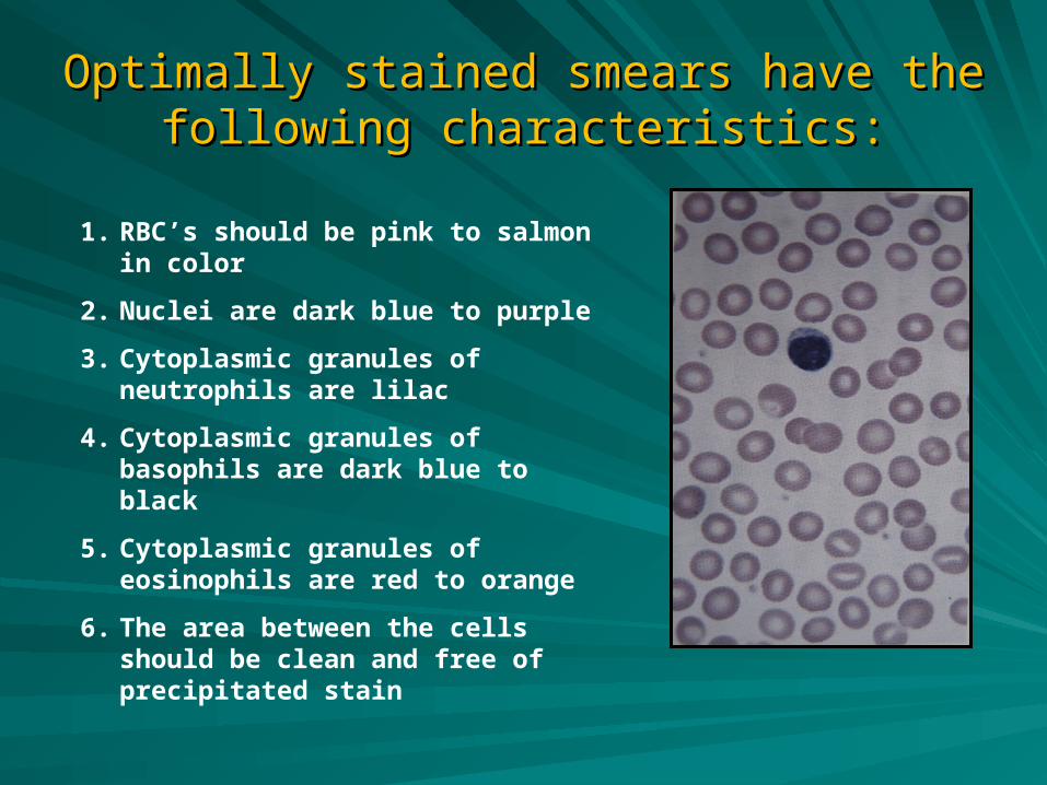
Optimally stained smears have the following Optimally stained smears have the following characteristics:characteristics:
1. RBC’s should be pink to salmon in color
2. Nuclei are dark blue to purple
3. Cytoplasmic granules of neutrophils are lilac
4. Cytoplasmic granules of basophils are dark blue to black
5. Cytoplasmic granules of eosinophils are red to orange
6. The area between the cells should be clean and free of precipitated stain
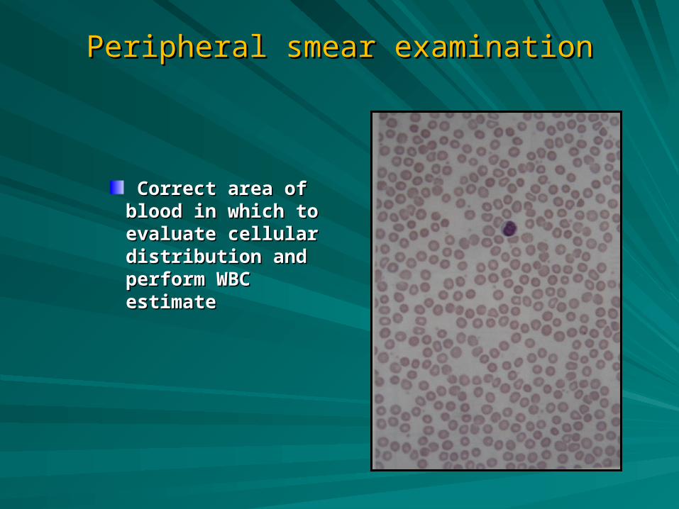
Peripheral smear examinationPeripheral smear examination
Correct area of blood Correct area of blood in which to evaluate in which to evaluate cellular distribution cellular distribution and perform WBC and perform WBC estimateestimate
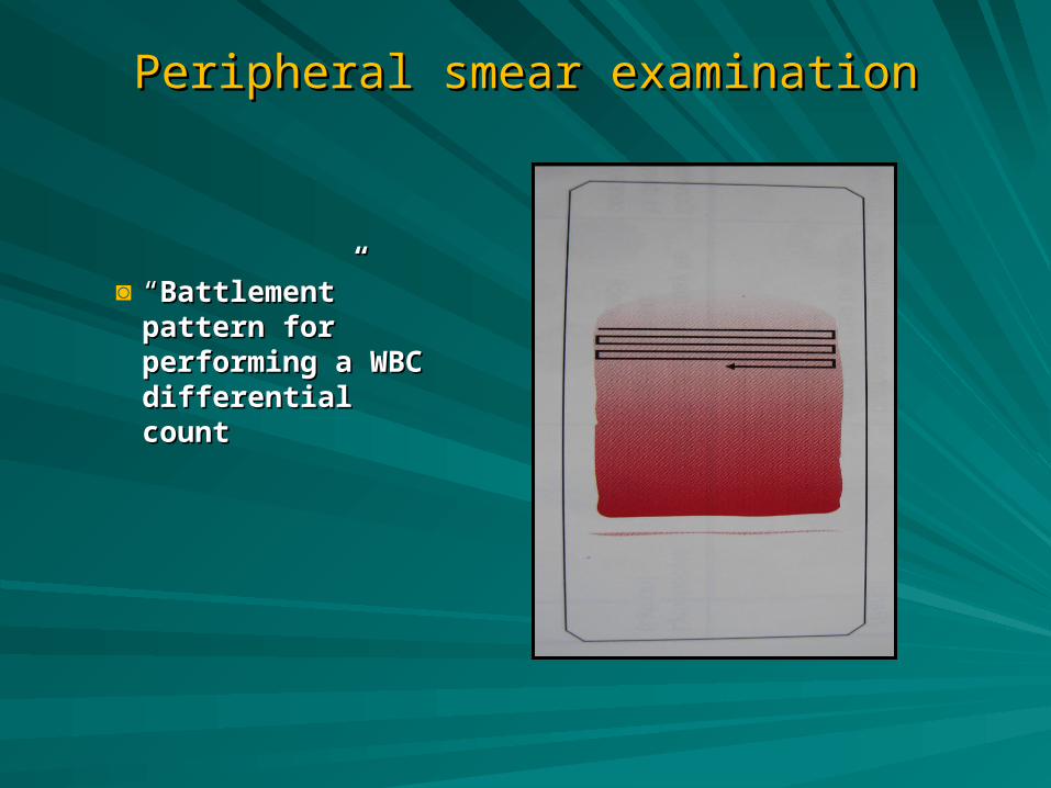
Peripheral smear examinationPeripheral smear examination
◙ ““Battlement” pattern Battlement” pattern for performing a WBC for performing a WBC differential countdifferential count