Embed Size (px)
Citation preview
HAL Id: tel-00675247https://tel.archives-ouvertes.fr/tel-00675247
Submitted on 29 Feb 2012
HAL is a multi-disciplinary open accessarchive for the deposit and dissemination of sci-entific research documents, whether they are pub-lished or not. The documents may come fromteaching and research institutions in France orabroad, or from public or private research centers.
L’archive ouverte pluridisciplinaire HAL, estdestinée au dépôt et à la diffusion de documentsscientifiques de niveau recherche, publiés ou non,émanant des établissements d’enseignement et derecherche français ou étrangers, des laboratoirespublics ou privés.
Peripersonal space : a multisensory interface forbody-objects interactions
Claudio Brozzoli
To cite this version:Claudio Brozzoli. Peripersonal space : a multisensory interface for body-objects interactions. Humanhealth and pathology. Université Claude Bernard - Lyon I, 2009. English. <NNT : 2009LYO10233>.<tel-00675247>
N° d’ordre 233-2009 Année 2009
THESE DE L‘UNIVERSITE DE LYON
Délivrée par
L’UNIVERSITE CLAUDE BERNARD LYON 1
ECOLE DOCTORALE
Neurosciences et Cognition
DIPLOME DE DOCTORAT(arrêté du 7 août 2006)
soutenue publiquement le 20/11/2009
par
M. Claudio BROZZOLI
PERIPERSONAL SPACE :A MULTISENSORY INTERFACE FOR BODY-OBJECTS INTERACTIONS
Directeur de thèse : Dr. FARNÈ Alessandro, Ph.D.
JURY: Prof. Y. Rossetti M.D., Ph.D. Dr. A. Farnè Ph.D., D.R. Prof. S. Soto-Faraco Ph.D. Prof. C. Spence Ph.D. Prof. O. Blanke M.D., Ph.D. Prof. F. Pavani, Ph.D. Dr. J.-R. Duhamel Ph.D., D.R.


A Fede,perché questo spazio
mi ha preso nel tempoche a volte era suo

AKNOWLEDGEMENTS
It has been amazingly exciting. I started something I didn�t know what it could be. Then the
trip kept on, impossible to be stopped, terrible sometime, but always exciting. And if during
this long ride I loved everything I did, it is also because without doubt I�m in debt to many
people who have contributed to my work, both directly and indirectly. Or just because they
were present. Or they would have liked to be. Thus, in random order, thank you all.
� Dopo avermi iniziato come laureando al pungente piacere della stimolazione elettrica,
all�importanza dei catch trials e alla conoscenza dell�estinzione, come se non bastasse, mi
ha anche introdotto allo “spazio peripersonale”. Solo per questo direi già: grazie Ale! Poi c�è
tutto il resto, le neuroscienze che ha continuato a farmi scoprire in questi anni con lo stesso
piacere d�un gioco, la fiducia che mi ha dato, la libertà che mi ha lasciato di spaziare dove
più avevo voglia di giocare. E ancora, i dischi di Luigi Tenco, il prosecco e l�affetto. Per tutto
quello che in questi anni ho potuto realizzare e per tutto quello che questi anni hanno fatto di
me, grazie Ale, il Professore che mi ha insegnato, il Supervisore che mi ha guidato e
incoraggiato, l�Amico che sei stato fin dall�inizio.
� Mes sincères remerciements vont à Alice, dont les mots directes mes fascinent et
m�ont fait du bien plus de ce que j�ai pu montrer. Merci pour le support que tu m�a donné et
qui va au delà des neurosciences et du français; pour le bourguignon qui a pas encore
trouvé des égales. Avec Ale et Giulio vous avez été mon repère familiale à Lyon.
� Per chiudere questa famiglia scientifica, ringrazio di tutto cuore Lucilla, la sorella
scientifica, ma soprattutto l�amica che ha ascoltato, risposto, considerato, capito, cercato e
trovato il mio affetto. L�amica che mi mancherà. La ringrazio fortemente anche per il lavoro
che abbiamo realizzato insieme e che senza di lei sarebbe stato niente affatto lo stesso e
sicuramente meno divertente.
� Je remercie vivement tous les chercheurs et les composents de l�Unité INSERM U864.
Travailler à l�unité a été un plaisir et l�atmosphère toujours tellement agréable que j�espère de
pouvoir encore avoir l�occasion de vous retrouver.
� Un grand et chaleureux merci à tous les étudiants de l�Unité INSERM 864: Laurence,
Muriel, Alessia et tous les autres qui m�ont taquiné et incouragé quand necéssaire.
� A special thanks goes to Nicholas Holmes and Tamar Makin with whom collaborating
has been interesting and not less amazing. They introduced me efficiently to the TMS
technique. And lot of TV series.
� A Alfredo e Pina, che instancabili, mi hanno seguito in tutti i miei tentativi e sostenuto
lungo diverse vie, va il mio grazie più constante e longevo.
� Pour le K factor que m�a fait aimé Lyon, les amis qui ont contribué à mon bien être, en
trouvant toujours la façon pour remplir mes pauses de la rédaction de thèse, entre le 203 et
le Broc, Eva, JB, Virginie et tous les autres: merci!

v

vi
CONTENTS
RESUME’ 1
ABSTRACT 3
INTRODUCTION 5
Multisensory perception 8
Multisensory perception in human behaviour 10
Multisensory attention 12
The spatial organization of numbers 14
CROSS-MODAL SHIFT OF ATTENTION 17
Discussion 29
Multisensory integration through anatomical convergence 29
Multisensory interactions through feedback on unisensory areas and inter-
connections among unisensory areas 31
Back-projections from multisensory higher-level to unisensory lower-level
areas 32
Early interactions between unisensory areas 34
Conclusion 35
vii
Multisensory and motor representations of peripersonal space 36
Multisensory features of peripersonal space: Visuo-tactile interaction around the
body 37
Parietal Visuo-tactile interactions 37
Premotor visuo-tactile interactions 42
Subcortical visuo-tactile interactions 46
Dynamic feature of peripersonal space representation 46
A visuo-tactile network 50
Motor features of peripersonal space: Visuo-tactile interactions around the
acting body 51
Inferior premotor cortex 52
Mirror neurons: a particular class of visuo-motor neuron 54
Parietal areas 55
Lesion studies 57
Conclusion: A multisensory-motor network for peripersonal space 58
Multisensory based peripersonal space in humans 60
Modularity of space through cross-modal extinction 61
EXTINCTION 63
Peripersonal space in humans: neuropsychological evidence 81
Peripersonal space in humans: evidence from healthy behaviour 84
viii
Cross-modal congruency effect: a perceptual paradigm for the investigation of the peripersonal space representation in healthy humans 86
Spatial modulation of CCE 88
Body-part-centred reference frame for the CCE 88
Tool-use dependent modulations of CCE 89
Peripersonal space in humans: neuroimaging studies 91
Conclusions 91
Peripersonal space: a multisensory interface for body-object interactions? 93
What kind of body-object interactions can the body-centered PpS representation
subserve? 95
CCE in action 96
The multisensori-motor task 97
The kinematic recording of movements: a real-time method for linking
multisensory perception and action 99
The kinematics of grasping 100
Neural network for grasping 102
Conclusion: kinematic-perceptual co-recording 104
RESULTS 105
STUDY 1 110
STUDY 2 143
ix
STUDY 3 153
DISCUSSION 185
Main results 187
Peripersonal space: an interface for avoidance reactions 189
A comparison with non-human primates studies 190
Peripersonal space: an interface for voluntary actions 192
A comparison with non-human primates studies 193
CONCLUSIONS AND PERSPECTIVES 197
REFERENCES 203
x
RESUME’
L’ESPACE PERIPERSONNEL:
UNE INTERFACE MULTISENSORIELLE POUR LES
INTERACTIONS ENTRE LE CORPS ET LES OBJETS
Notre habilité à interagir avec les objets du monde nécessite l’intégration d’informations
provenant de différents canaux sensoriels, dans le cadre de la construction d’une
représentation de l’espace en particulier des informations visuelles et tactiles. L’espace
péripersonnel et l’intégration visuo-tactile ont été l’objet d’importantes recherche récemment.
Des études neurophysiologiques chez le primate non-humain ont montré l’existence de
neurones bimodaux activés à la fois par des stimulations tactiles et par des stimulations
visuelles si ces dernières étaient présentées près d’une partie du corps (par exemple la main).
Il a été proposé que ces neurones bi-modaux constituent le substrat neuronal de la
représentation de l’espace péripersonnel. Les études neuropsychologiques menées chez des
patients présentant une extinction cross-modale consécutive à une lésion pariétale droite ont
permis de suggérer l’existence du même type de représentation de l’espace péripersonnel chez
l’homme. Les données issues des études en neuroimagerie fonctionnelle sont venues par la
suite conforter cette idée. Plus récemment, à travers l’utilisation d’outils, des données
acquises chez le primate humain et non humain ont révélé les propriétés dynamiques de cette
représentation spatiale.
Selon notre hypothèse la représentation de l’espace péripersonnel est une interface
présidant aux interactions du corps avec les objets du monde externe.
Nous avons donc évalué le rôle et l’état de l’espace péripersonnel lors de l’exécution
de mouvements volontaires vers des objets (comme une simple saisie) et lors de mouvements
involontaires d’évitement. Lors d’une première série d’expériences nous avons étudié les
coordonnées spatiales du codage des objets qui soudainement se rapprochent du corps grâce à
la mesure des potentiels évoqués moteurs. Cette étude a révélé que l’espace péripersonnel
joue un rôle dans la représentation des objets approchant le corps et dans la sélection des
mouvements appropriés en réponse. Lors d’une seconde série d’expériences nous avons
utilisé un paradigme d’interférence visuo-tactile couplé à l’enregistrement cinématique des
mouvements de saisie afin d’examiner la représentation de l’espace péripersonnel lors de
� 1� �
l’exécution d’actions volontaires. Cette approche novatrice nous a permis de mettre en
évidence que l’action volontaire induit un recodage en ligne de l’interaction visuo-tactile dans
l’espace de préhension. Ce recodage de l’action s’effectue en coordonnées centrées sur la
partie du corps qui exécute l’action.
En conclusion nos études expérimentales démontrent que l’espace péripersonnel est
une interface multisensorielle qui a été sélectionnée à travers l’évolution non seulement pour
la gestion des mouvements d’évitement et de défense mais également pour l’exécution
d’actions volontaires.
2
ABSTRACT
PERIPERSONAL SPACE:A MULTISENSORY INTERFACE
FOR BODY-OBJECTS INTERACTIONS
Our ability to interact with the environment requires the integration of multisensory
information for the construction of spatial representations. The peripersonal space (i.e., the
sector of space closely surrounding one’s body) and the integrative processes between visual
and tactile inputs originating from this sector of space have been at the center of recent years
investigations. Neurophysiological studies provided evidence for the presence in the monkey
brain of bimodal neurons, which are activated by tactile as well as visual information
delivered near to a specific body part (e.g., the hand). Neuropsychological studies on right
brain-damaged patients who present extinction and functional neuroimaging findings suggest
the presence of similar bimodal systems in the human brain. Studies on the effects of tool-use
on visual-tactile interaction revealed similar dynamic properties of the peripersonal space in
monkeys and humans.
The functional role of the multisensory coding of peripersonal space is, in our
hypothesis, that of providing the brain with a sensori-motor interface for body-objects
interactions. Thus, not only it could be involved in driving involuntary defensive movements
in response to objects approaching the body, but could be also dynamically maintained and
updated as a function of manual voluntary actions performed towards objects in the reaching
space.
We tested the hypothesis of an involvement of peripersonal space in executing both
voluntary and defensive actions. To these aims, we joined a well known cross-modal
congruency effect between visual and tactile information to a kinematic approach to
demonstrate that voluntary grasping actions induce an on-line re-weighting of multisensory
interactions in the peripersonal space. We additionally show that this modulation is hand-
centred. We also used a motor evoked potentials approach to investigate which coordinates
system is used to code the peripersonal space during motor preparation if real objects rapidly
approach the body. Our findings provide direct evidence for automatic hand-centred coding of
visual space and suggest that peripersonal space may also serve to represent rapidly
� 3� �
approaching and potentially noxious objects, thus enabling the rapid selection of appropriate
motor responses.
These results clearly show that peripersonal space is a multisensori-motor interface
that might have been selected through evolution for optimising the interactions between the
body and the objects in the external world.
4

INTRODUCTION

”Space is not a sort of ether in which all things float...
The points in space mark, in our vicinity, the varying range of our aims and our gestures"
(Merleau-Ponty)
Research in the last four decades has brought a considerable advance in our understanding of
how the brain synthesises perceptual information arising from different sensory modalities.
Indeed, many cortical and subcortical areas, also beyond those traditionally considered to be
‘associative’, have been shown to be involved in multisensory interaction and integration
(Ghazanfar and Schroeder 2006). Visuo-tactile interaction is of particular interest because of
the prominent role played by vision in guiding our actions and anticipating their tactile
consequences in everyday life. In this thesis, we focus on the functional role that visuo-tactile
processing may play in driving two types of body-object interactions: avoidance and
approach. We will first review some basic features of visuo-tactile interactions, as revealed by
single units recording studies in monkeys. These will prove to be relevant for interpreting the
subsequent human evidence. A crucial point that will be stressed is that these neuronal
populations have not only sensory, but also motor-related activity that qualifies them as
multisensory-motor interfaces. Evidence will then be presented for the existence of
functionally homologous processing in the human brain, both from neuropsychological
research in brain-damaged patients and in healthy people. The original experimental
contribution of this dissertation is focussed on healthy humans and supports the idea that the
human motor system is provided with a multisensory interface that allows for continuous
monitoring of the space near the body (i.e., peripersonal space). We will provide evidence of
the involvement of the peripersonal space representation in rapid reaction to approaching
objects. We further demonstrate that multisensory processing can be modulated on-line as a
consequence of acting voluntarily on objects. This indicates that, far from being passive, the
monitoring of peripersonal space is an active process subserving actions between our body
and objects located in the space around us.
7
Chapter I
Multisensory perception
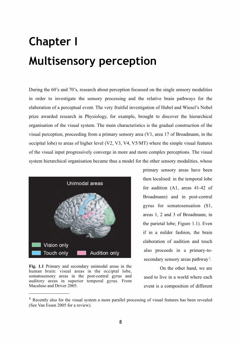
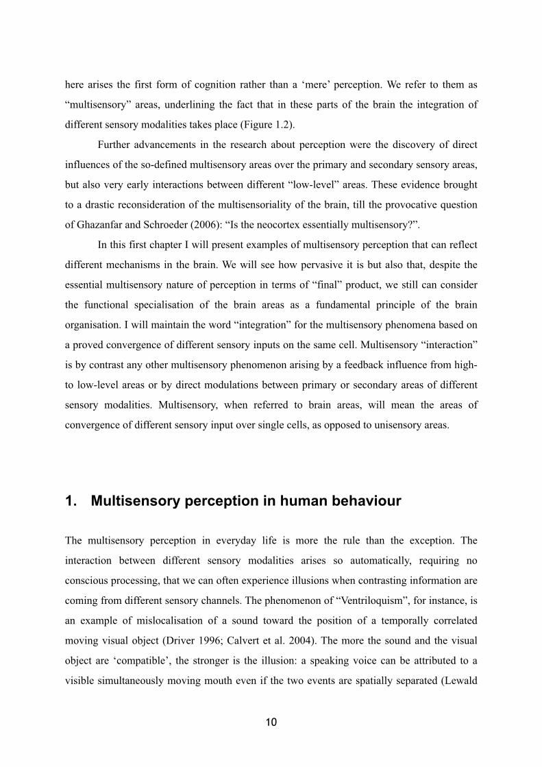
During the 60’s and 70’s, research about perception focussed on the single sensory modalities
in order to investigate the sensory processing and the relative brain pathways for the
elaboration of a perceptual event. The very fruitful investigation of Hubel and Wiesel’s Nobel
prize awarded research in Physiology, for example, brought to discover the hierarchical
organisation of the visual system. The main characteristics is the gradual construction of the
visual perception, proceeding from a primary sensory area (V1, area 17 of Broadmann, in the
occipital lobe) to areas of higher level (V2, V3, V4, V5/MT) where the simple visual features
of the visual input progressively converge in more and more complex perceptions. The visual
system hierarchical organisation became thus a model for the other sensory modalities, whose
primary sensory areas have been
then localised: in the temporal lobe
for audition (A1, areas 41-42 of
Broadmann) and in post-central
gyrus for somatosensation (S1,
areas 1, 2 and 3 of Broadmann, in
the parietal lobe; Figure 1.1). Even
if in a milder fashion, the brain
elaboration of audition and touch
also proceeds in a primary-to-
secondary sensory areas pathway 1.
On the other hand, we are
used to live in a world where each
event is a composition of different
8
1 Recently also for the visual system a more parallel processing of visual features has been revealed (See Van Essen 2005 for a review).
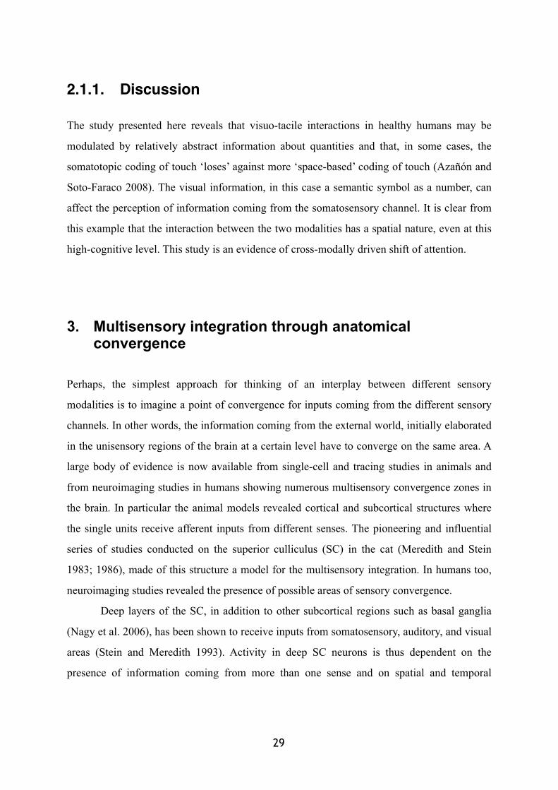
Fig. 1.1 Primary and secondary unimodal areas in the human brain: visual areas in the occiptal lobe, somatosensory areas in the post-central gyrus and auditory areas in superior temporal gyrus. From Macaluso and Driver 2005.
sensory inputs and where the multisensoriality is the rule. When we are talking with a friend
or just watching a movie or looking for our ringing cell-phone in order to grasp it, we are
simply receiving and automatically integrating information coming from different sensory
modalities. The visible mouth movements of my friend talking bring visual information that
can interact with its related audible counterpart; similarly the visual information of the ringing
cell-phone is integrated without any effort to the sound coming from it, sometime possibly
helping to find its location. Moreover, the form of the telephone I can be aware of through
vision, creates automatically expectations about the tactile feedback I will receive when my
hand will enter in contact with it. These everyday life’s examples clearly point out the fact
that the peripherally separated perceptual channels have to converge at a certain level of the
sensory elaboration in order to give the unitary perception of world events we are used to
experience.
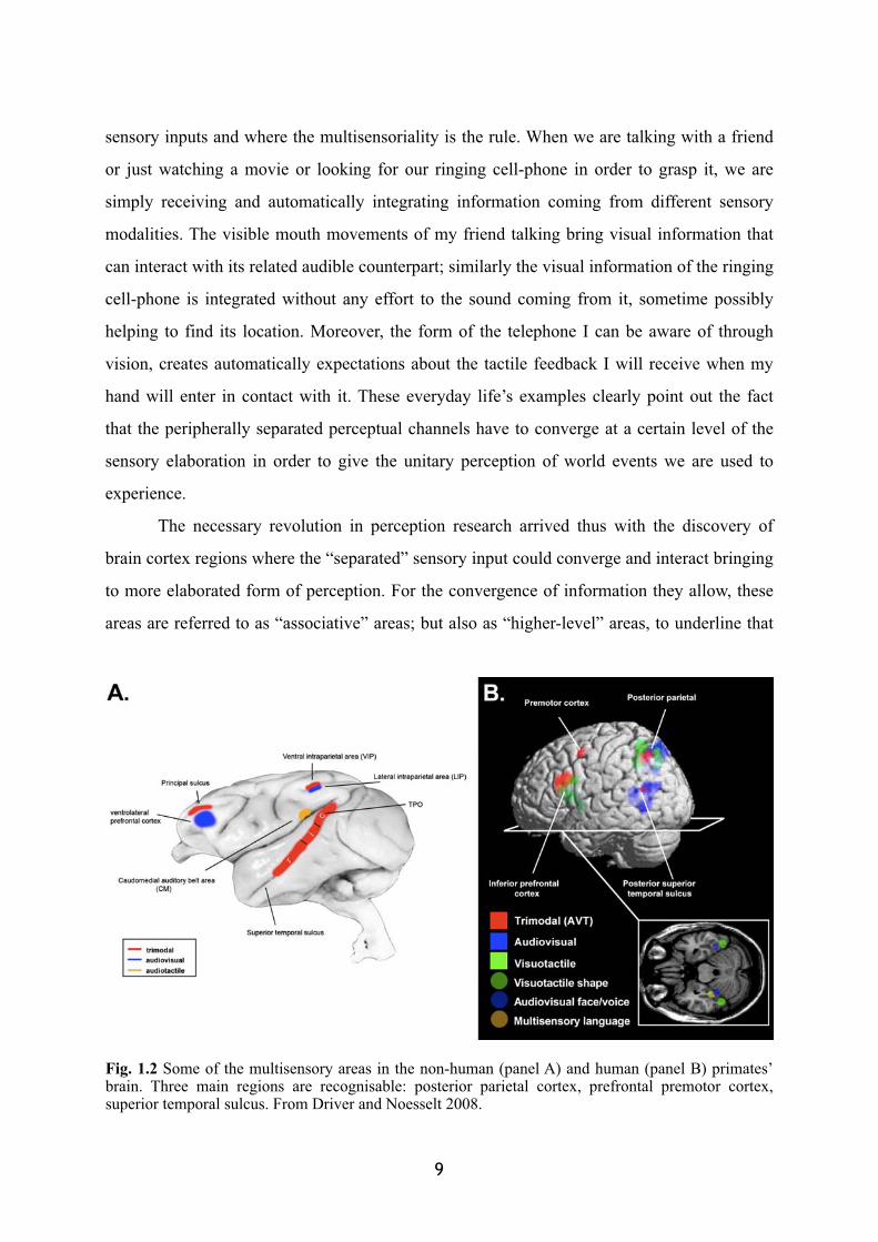
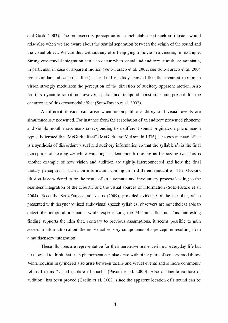
The necessary revolution in perception research arrived thus with the discovery of
brain cortex regions where the “separated” sensory input could converge and interact bringing
to more elaborated form of perception. For the convergence of information they allow, these
areas are referred to as “associative” areas; but also as “higher-level” areas, to underline that
9
Fig. 1.2 Some of the multisensory areas in the non-human (panel A) and human (panel B) primates’ brain. Three main regions are recognisable: posterior parietal cortex, prefrontal premotor cortex, superior temporal sulcus. From Driver and Noesselt 2008.
here arises the first form of cognition rather than a ‘mere’ perception. We refer to them as
“multisensory” areas, underlining the fact that in these parts of the brain the integration of
different sensory modalities takes place (Figure 1.2).
Further advancements in the research about perception were the discovery of direct
influences of the so-defined multisensory areas over the primary and secondary sensory areas,
but also very early interactions between different “low-level” areas. These evidence brought
to a drastic reconsideration of the multisensoriality of the brain, till the provocative question
of Ghazanfar and Schroeder (2006): “Is the neocortex essentially multisensory?”.
In this first chapter I will present examples of multisensory perception that can reflect
different mechanisms in the brain. We will see how pervasive it is but also that, despite the
essential multisensory nature of perception in terms of “final” product, we still can consider
the functional specialisation of the brain areas as a fundamental principle of the brain
organisation. I will maintain the word “integration” for the multisensory phenomena based on
a proved convergence of different sensory inputs on the same cell. Multisensory “interaction”
is by contrast any other multisensory phenomenon arising by a feedback influence from high-
to low-level areas or by direct modulations between primary or secondary areas of different
sensory modalities. Multisensory, when referred to brain areas, will mean the areas of
convergence of different sensory input over single cells, as opposed to unisensory areas.
1. Multisensory perception in human behaviour
The multisensory perception in everyday life is more the rule than the exception. The
interaction between different sensory modalities arises so automatically, requiring no
conscious processing, that we can often experience illusions when contrasting information are
coming from different sensory channels. The phenomenon of “Ventriloquism”, for instance, is
an example of mislocalisation of a sound toward the position of a temporally correlated
moving visual object (Driver 1996; Calvert et al. 2004). The more the sound and the visual
object are ‘compatible’, the stronger is the illusion: a speaking voice can be attributed to a
visible simultaneously moving mouth even if the two events are spatially separated (Lewald
10
and Guski 2003). The multisensory perception is so ineluctable that such an illusion would
arise also when we are aware about the spatial separation between the origin of the sound and
the visual object. We can thus without any effort enjoying a movie in a cinema, for example.
Strong crossmodal integration can also occur when visual and auditory stimuli are not static,
in particular, in case of apparent motion (Soto-Faraco et al. 2002; see Soto-Faraco et al. 2004
for a similar audio-tactile effect). This kind of study showed that the apparent motion in
vision strongly modulates the perception of the direction of auditory apparent motion. Also
for this dynamic situation however, spatial and temporal constraints are present for the
occurrence of this crossmodal effect (Soto-Faraco et al. 2002).
A different illusion can arise when incompatible auditory and visual events are
simultaneously presented. For instance from the association of an auditory presented phoneme
and visible mouth movements corresponding to a different sound originates a phenomenon
typically termed the “McGurk effect” (McGurk and McDonald 1976). The experienced effect
is a synthesis of discordant visual and auditory information so that the syllable da is the final
perception of hearing ba while watching a silent mouth moving as for saying ga. This is
another example of how vision and audition are tightly interconnected and how the final
unitary perception is based on information coming from different modalities. The McGurk
illusion is considered to be the result of an automatic and involuntary process leading to the
seamless integration of the acoustic and the visual sources of information (Soto-Faraco et al.
2004). Recently, Soto-Faraco and Alsius (2009), provided evidence of the fact that, when
presented with desynchronised audiovisual speech syllables, observers are nonetheless able to
detect the temporal mismatch while experiencing the McGurk illusion. This interesting
finding supports the idea that, contrary to previous assumptions, it seems possible to gain
access to information about the individual sensory components of a perception resulting from
a multisensory integration.
These illusions are representative for their pervasive presence in our everyday life but
it is logical to think that such phenomena can also arise with other pairs of sensory modalities.
Ventriloquism may indeed also arise between tactile and visual events and is more commonly
referred to as “visual capture of touch” (Pavani et al. 2000). Also a “tactile capture of
audition” has been proved (Caclin et al. 2002) since the apparent location of a sound can be
11
biased toward the location of a concurrent tactile stimulation. The synchrony between the
stimuli appears a fundamental parameter. Similarly in the “parchement-skin
illusion” (Jousmäki and Hari 1998), perturbing the sound as hands are rubbed together can
affect the perception of skin texture and moistness. Also in these cases, illusions appear to be
dependent on the spatial and temporal association between the two sensory inputs.
In the presented multisensory illusions, the brain is confronted with concurrent
information coming from two modalities but providing information about the same external
property. Often indeed, the auditory input jointly with the visual ones, both bring the spatial
information necessary, for example, to the localisation of a multisensory event. It appears thus
extremely adaptive that the brain might synthesise the different sensory information. By
contrast, recent examples of multisensory influences in perception show different types of
phenomena. Rather than different modalities providing independent information about the
same external sensory property, multisensory research also showed that stimulation in one
modality can affect the judgement of a property logically pertinent only to another modality.
Touch at a given location for instance, can improve judgements about the colour of a visual
stimulus delivered nearby, although touch itself does not convey any information about this
visual property (Spence et al. 2004). Other example of such a multisensory effect in behaviour
is the case of single flashes that can be illusory perceived as being double whenever
associated with two auditory signals (Shipley 1964; Shams et al. 2000). Again, a visuo-tactile
form of the “double-flash illusion” exists (Violentyev et al. 2005), where perceiving two
touches delivered simultaneously with a single flash induces the illusion to perceive a double
flash.
2. Multisensory attention
The presence of a sensory event can produce a shift of attention toward its location. This shift
could be either voluntary, “endogenous”, or “exogenous”, as it is driven by the sudden
appearance of a stimulus in the environment, in an involuntary fashion (Posner 1980). A part
of the multisensory research focussed on how the presence of multisensory information can
12
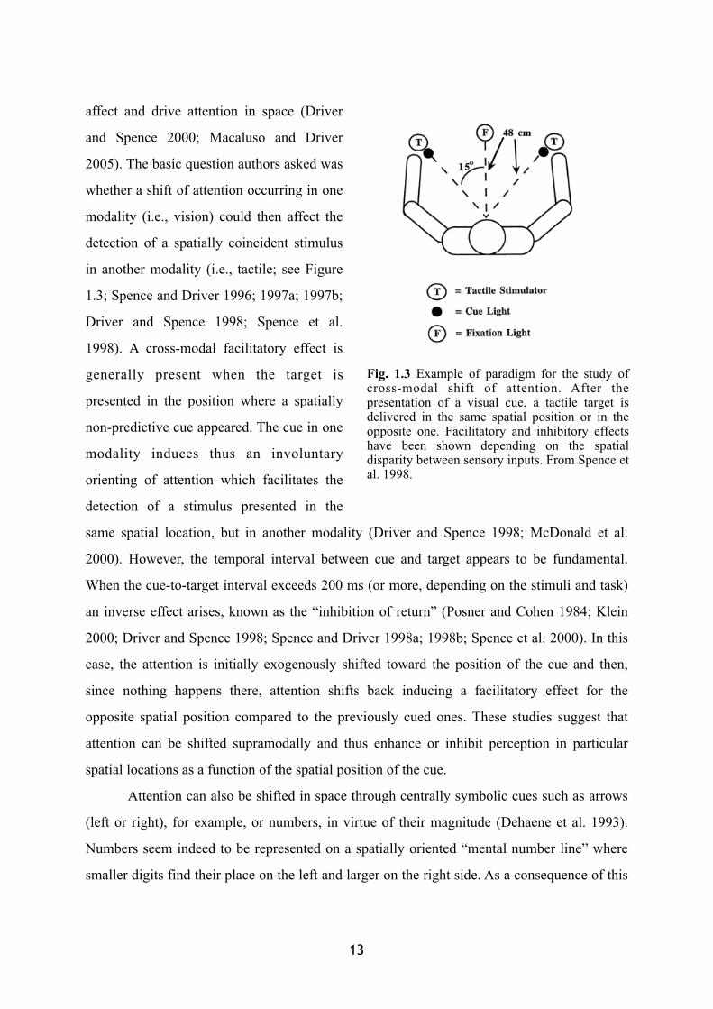
affect and drive attention in space (Driver
and Spence 2000; Macaluso and Driver
2005). The basic question authors asked was
whether a shift of attention occurring in one
modality (i.e., vision) could then affect the
detection of a spatially coincident stimulus
in another modality (i.e., tactile; see Figure
1.3; Spence and Driver 1996; 1997a; 1997b;
Driver and Spence 1998; Spence et al.
1998). A cross-modal facilitatory effect is
generally present when the target is
presented in the position where a spatially
non-predictive cue appeared. The cue in one
modality induces thus an involuntary
orienting of attention which facilitates the
detection of a stimulus presented in the
same spatial location, but in another modality (Driver and Spence 1998; McDonald et al.
2000). However, the temporal interval between cue and target appears to be fundamental.
When the cue-to-target interval exceeds 200 ms (or more, depending on the stimuli and task)
an inverse effect arises, known as the “inhibition of return” (Posner and Cohen 1984; Klein
2000; Driver and Spence 1998; Spence and Driver 1998a; 1998b; Spence et al. 2000). In this
case, the attention is initially exogenously shifted toward the position of the cue and then,
since nothing happens there, attention shifts back inducing a facilitatory effect for the
opposite spatial position compared to the previously cued ones. These studies suggest that
attention can be shifted supramodally and thus enhance or inhibit perception in particular
spatial locations as a function of the spatial position of the cue.
Attention can also be shifted in space through centrally symbolic cues such as arrows
(left or right), for example, or numbers, in virtue of their magnitude (Dehaene et al. 1993).
Numbers seem indeed to be represented on a spatially oriented “mental number line” where
smaller digits find their place on the left and larger on the right side. As a consequence of this
13
Fig. 1.3 Example of paradigm for the study of cross-modal shift of attention. After the presentation of a visual cue, a tactile target is delivered in the same spatial position or in the opposite one. Facilitatory and inhibitory effects have been shown depending on the spatial disparity between sensory inputs. From Spence et al. 1998.
spatial arrangement, the vision of a task irrelevant digit has been proved to shift attention in a
visual detection task (Fischer et al. 2005) in a Posner-like fashion, even though evidence has
been provided against the automaticity of this effect (Galfano et al. 2006). The case is
possible that the vision of a digit might shift attention also cross-modally, for instance,
modulating tactile perception. We have investigated this hypothesis and the study is presented
hereafter as an example of this thesis contribution to multisensory shifts of attention.
2.1.The spatial organization of numbers
My research experience, though mainly focussed on the investigation of multisensory
properties of peripersonal space, has brought me to study also a more general kind of visuo-
tactile interaction. In my first experimental contribution presented here, I investigated the
effects of a visual shift of attention on the tactile modality. In particular I investigated a shift
of attention induced by a visually presented numerical cue.
We are used to learn counting on our fingers and the digital representation of numbers
we develop is still present in adulthood in our counting motor behaviour. By virtue of such an
association between anatomy and digit magnitude we establish tight functional
correspondences between fingers and numbers. However, it has long been known that
numerical information is also spatially arranged along an oriented mental number line, where
digits are organised from left to right as a function of their magnitude.
In the following study, I investigated touch perception in order to disambiguate
whether number representation is embodied on the hand (“1” thumb; “5” little finger) or
disembodied in the extrapersonal space (“1” left; “5” right). In two experiments, the number
spatial representations have been directly contrasted each other using a single centrally
located effector (the foot) and a simple postural manipulation of the hand (palm-up vs. palm-
down). Results show that visual presentation of a number (‘‘1’’ or ‘‘5’’) shifts attention cross-
modally, modulating the detection of tactile stimuli delivered on the little finger or thumb.
When the hand rests palm-down, subjects perform better in reporting tactile stimuli delivered
to the little finger after presentation of number ‘‘5’’ than number ‘‘1.’’ Crucially, this pattern
reverses (better performance after number ‘‘1’’ than ‘‘5’’) when the hand is in a palm-up
14
posture, in which the position of the fingers in external space, but not their relative anatomical
position, is reversed. The human brain can thus use either space- or body-based representation
of numbers, but in case of competition, the former dominates the latter, showing the stronger
role played by the mental number line organisation.
15

CROSS-MODAL SHIFT OF ATTENTION
Touch perception reveals the dominance of spatial over digital representation of numbers
P.N.A.S. (2008)

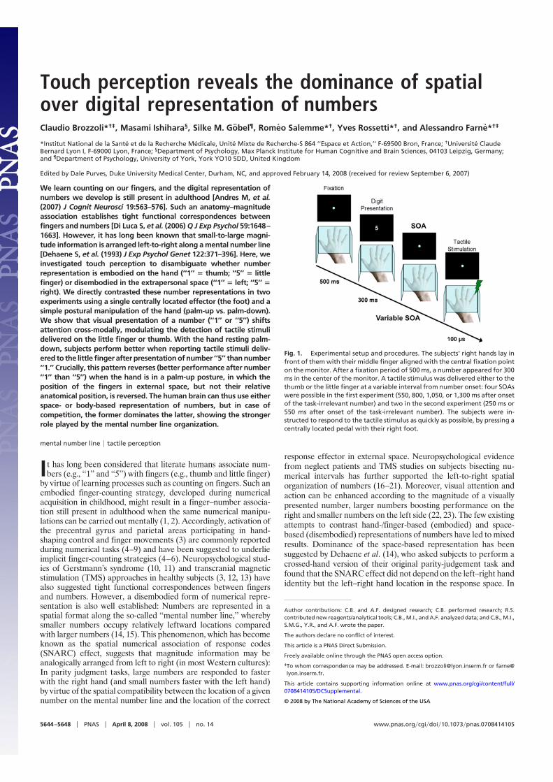
Touch perception reveals the dominance of spatialover digital representation of numbersClaudio Brozzoli*†‡, Masami Ishihara§, Silke M. Gobel¶, Romeo Salemme*†, Yves Rossetti*†, and Alessandro Farne*†‡
*Institut National de la Sante et de la Recherche Medicale, Unite Mixte de Recherche-S 864 ‘‘Espace et Action,’’ F-69500 Bron, France; †Universite ClaudeBernard Lyon I, F-69000 Lyon, France; §Department of Psychology, Max Planck Institute for Human Cognitive and Brain Sciences, 04103 Leipzig, Germany;and ¶Department of Psychology, University of York, York YO10 5DD, United Kingdom
Edited by Dale Purves, Duke University Medical Center, Durham, NC, and approved February 14, 2008 (received for review September 6, 2007)
We learn counting on our fingers, and the digital representation ofnumbers we develop is still present in adulthood [Andres M, et al.(2007) J Cognit Neurosci 19:563–576]. Such an anatomy–magnitudeassociation establishes tight functional correspondences betweenfingers and numbers [Di Luca S, et al. (2006) Q J Exp Psychol 59:1648–1663]. However, it has long been known that small-to-large magni-tude information is arranged left-to-right along a mental number line[Dehaene S, et al. (1993) J Exp Psychol Genet 122:371–396]. Here, weinvestigated touch perception to disambiguate whether numberrepresentation is embodied on the hand (‘‘1’’ � thumb; ‘‘5’’ � littlefinger) or disembodied in the extrapersonal space (‘‘1’’ � left; ‘‘5’’ �
right). We directly contrasted these number representations in twoexperiments using a single centrally located effector (the foot) and asimple postural manipulation of the hand (palm-up vs. palm-down).We show that visual presentation of a number (‘‘1’’ or ‘‘5’’) shiftsattention cross-modally, modulating the detection of tactile stimulidelivered on the little finger or thumb. With the hand resting palm-down, subjects perform better when reporting tactile stimuli deliv-ered to the little finger after presentation of number ‘‘5’’ than number‘‘1.’’ Crucially, this pattern reverses (better performance after number‘‘1’’ than ‘‘5’’) when the hand is in a palm-up posture, in which theposition of the fingers in external space, but not their relativeanatomical position, is reversed. The human brain can thus use eitherspace- or body-based representation of numbers, but in case ofcompetition, the former dominates the latter, showing the strongerrole played by the mental number line organization.
mental number line � tactile perception
It has long been considered that literate humans associate num-bers (e.g., ‘‘1’’ and ‘‘5’’) with fingers (e.g., thumb and little finger)
by virtue of learning processes such as counting on fingers. Such anembodied finger-counting strategy, developed during numericalacquisition in childhood, might result in a finger–number associa-tion still present in adulthood when the same numerical manipu-lations can be carried out mentally (1, 2). Accordingly, activation ofthe precentral gyrus and parietal areas participating in hand-shaping control and finger movements (3) are commonly reportedduring numerical tasks (4–9) and have been suggested to underlieimplicit finger-counting strategies (4–6). Neuropsychological stud-ies of Gerstmann’s syndrome (10, 11) and transcranial magneticstimulation (TMS) approaches in healthy subjects (3, 12, 13) havealso suggested tight functional correspondences between fingersand numbers. However, a disembodied form of numerical repre-sentation is also well established: Numbers are represented in aspatial format along the so-called ‘‘mental number line,’’ wherebysmaller numbers occupy relatively leftward locations comparedwith larger numbers (14, 15). This phenomenon, which has becomeknown as the spatial numerical association of response codes(SNARC) effect, suggests that magnitude information may beanalogically arranged from left to right (in most Western cultures):In parity judgment tasks, large numbers are responded to fasterwith the right hand (and small numbers faster with the left hand)by virtue of the spatial compatibility between the location of a givennumber on the mental number line and the location of the correct
response effector in external space. Neuropsychological evidencefrom neglect patients and TMS studies on subjects bisecting nu-merical intervals has further supported the left-to-right spatialorganization of numbers (16–21). Moreover, visual attention andaction can be enhanced according to the magnitude of a visuallypresented number, larger numbers boosting performance on theright and smaller numbers on the left side (22, 23). The few existingattempts to contrast hand-/finger-based (embodied) and space-based (disembodied) representations of numbers have led to mixedresults. Dominance of the space-based representation has beensuggested by Dehaene et al. (14), who asked subjects to perform acrossed-hand version of their original parity-judgement task andfound that the SNARC effect did not depend on the left–right handidentity but the left–right hand location in the response space. In
Author contributions: C.B. and A.F. designed research; C.B. performed research; R.S.contributed new reagents/analytical tools; C.B., M.I., and A.F. analyzed data; and C.B., M.I.,S.M.G., Y.R., and A.F. wrote the paper.
The authors declare no conflict of interest.
This article is a PNAS Direct Submission.
Freely available online through the PNAS open access option.
‡To whom correspondence may be addressed. E-mail: [email protected] or [email protected].
This article contains supporting information online at www.pnas.org/cgi/content/full/0708414105/DCSupplemental.
© 2008 by The National Academy of Sciences of the USA
Fig. 1. Experimental setup and procedures. The subjects’ right hands lay infront of them with their middle finger aligned with the central fixation pointon the monitor. After a fixation period of 500 ms, a number appeared for 300ms in the center of the monitor. A tactile stimulus was delivered either to thethumb or the little finger at a variable interval from number onset: four SOAswere possible in the first experiment (550, 800, 1,050, or 1,300 ms after onsetof the task-irrelevant number) and two in the second experiment (250 ms or550 ms after onset of the task-irrelevant number). The subjects were in-structed to respond to the tactile stimulus as quickly as possible, by pressing acentrally located pedal with their right foot.
5644–5648 � PNAS � April 8, 2008 � vol. 105 � no. 14 www.pnas.org�cgi�doi�10.1073�pnas.0708414105
contrast, finger-based dominance has been suggested by Di Luca etal. (24), who asked subjects to perform a visuomotor finger-numbercompatibility task and found better performance when the mappingwas congruent with the prototypical finger-counting strategy. Inaddition, a certain degree of flexibility in number representationhas been recently suggested (25–28), because the mapping betweennumbers and space can vary to some extent with instructionalcontext (25) and task demands (17).
Previous findings are thus not definitive with regard to numberrepresentation, because both the embodied and the disembodiedhypotheses have received empirical support. In this study, we useda previously undescribed approach to disambiguate between suchrepresentations within a corporeal modality, by investigating theattentional effects induced by numbers on the perception of touchesdelivered to the fingers. A postural manipulation of the hand(palm-up vs. -down) allowed us to directly contrast the embodiedand disembodied representations of numbers. A further manipu-lation was critically introduced to avoid any left–right arrangementin the response space, potentially favoring a space-based represen-tation, and any motor bias in the response effector, potentiallyfavoring a finger-based representation: Subjects had to respond totactile stimulation by pressing a centrally located pedal with thefoot.
Results and DiscussionParticipants performed a simple tactile detection task by makingspeeded foot-pedal responses to a tactile stimulus delivered toeither the thumb or little finger of their right (preferred andcounting) hand. Tactile intensity was set in a previous session toobtain an equal detection probability for the two fingers [seesupporting information (SI) Experiment 1, Supporting Proceduresand Supporting Results, Table S1, and Fig. S1]. In the first experi-ment, the task instructions were given as to emphasize the fingers(i.e., ‘‘you will feel a touch on either your thumb or little finger’’).At a variable stimulus onset asynchrony (SOA), an electrocutane-ous stimulus followed the presentation of a task-irrelevant number(‘‘1,’’ ‘‘2,’’ ‘‘4,’’ or ‘‘5’’) on a screen in front of their hand (Fig. 1). Thetactile task was performed with the unseen hand passively restingeither in a palm-down or -up posture.
Two main results were found: First, visual presentation of anumber cross-modally affects tactile performance. Second, thisnumerical cueing of touch does not follow a number–finger asso-ciation, but a number–space association, akin to the mental number
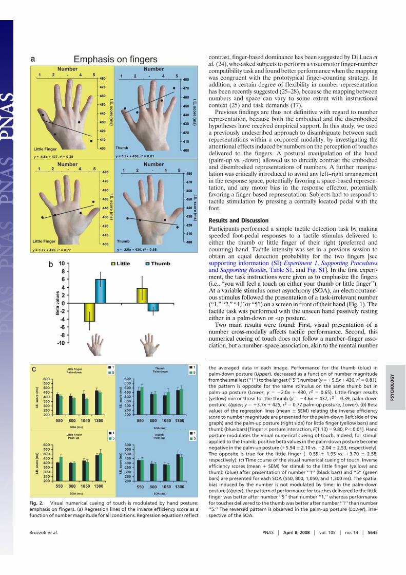
Fig. 2. Visual numerical cueing of touch is modulated by hand posture:emphasis on fingers. (a) Regression lines of the inverse efficiency score as afunction of number magnitude for all conditions. Regression equations reflect
the averaged data in each image. Performance for the thumb (blue) inpalm-down posture (Upper), decreased as a function of number magnitudefrom the smallest (‘‘1’’) to the largest (‘‘5’’) number (y � �5.9x � 436, r2 � 0.81);the pattern is opposite for the same stimulus on the same thumb but inpalm-up posture (Lower, y � �2.0x � 430, r2 � 0.65). Little-finger results(yellow) mirror those for the thumb (y � �4.6x � 437, r2 � 0.39, palm-downposture, Upper; y � �3.7x � 425, r2 � 0.77 palm-up posture, Lower). (b) Betavalues of the regression lines (mean � SEM) relating the inverse efficiencyscore to number magnitude are presented for the palm-down (left side of thegraph) and the palm-up posture (right side) for little finger (yellow bars) andthumb (blue bars) [finger � posture interaction, F(1,13) � 9.80, P � 0.01]. Handposture modulates the visual numerical cueing of touch. Indeed, for stimuliapplied to the thumb, positive beta values in the palm-down posture becomenegative in the palm-up posture (�5.94 � 2.10 vs. �2.04 � 2.53, respectively).The opposite is true for the little finger (�0.55 � 1.95 vs. �3.70 � 2.58,respectively). (c) Time course of the visual numerical cueing of touch. Inverseefficiency scores (mean � SEM) for stimuli to the little finger (yellow) andthumb (blue) after presentation of number ‘‘1’’ (black bars) and ‘‘5’’ (greenbars) are presented for each SOA (550, 800, 1,050, and 1,300 ms). The spatialbias induced by the number is not modulated by time: in the palm-downposture (Upper), the pattern of performance for touches delivered to the littlefinger was better after number ‘‘5’’ than number ‘‘1,’’ whereas performancefor touches delivered to the thumb was better after number ‘‘1’’ than number‘‘5.’’ The reversed pattern is observed in the palm-up posture (Lower), irre-spective of the SOA.
Brozzoli et al. PNAS � April 8, 2008 � vol. 105 � no. 14 � 5645
PSYC
HO
LOG
Y
line (14). A descriptive illustration of the results for all experimentalconditions including all of the numbers (‘‘1,’’ ‘‘2,’’ ‘‘4,’’ and ‘‘5’’) isprovided by Fig. 2a. When the right hand was in the palm-downposture, placed centrally with the middle finger aligned with thevisually presented number, subjects’ detection of brief tactile stimuliapplied to the little finger improved as a function of the precedingnumber magnitude. The larger the number, the better the perfor-mance in terms of inverse efficiency (IE) score, jointly indexingaccuracy, and response latency. The opposite pattern of results wasfound when the same little finger was stimulated with the hand inthe palm-up posture. In this condition, subjects’ tactile performanceactually decreased as the preceding number increased. The statis-tical comparison showed a significant finger � posture interaction[F(1,13) � 9.80; P � 0.01]: Fig. 2b shows that for stimuli applied onthe little finger, a difference was present between the slopes of IEregression lines in the palm-down and -up position (�0.55 vs. �3.70,respectively; P � 0.05; Fig. 2b, yellow bars). Results for the thumbmirrored those for the little finger (Fig. 2b, blue bars). When thehand was in the palm-down posture, subjects’ detection improvedas a function of the number’s magnitude. For the thumb, the smallerthe preceding number, the better the performance, because theregression line has a positive slope. On the contrary, when the handwas in the palm-up position, subjects’ detection of brief stimuli onthe thumb tended to worsen with decreasing magnitude of thepresented number (�5.94 vs. �2.04 for the palm-down and -uppostures, respectively; P � 0.053, Fig. 2b).
To further establish the dominant role played by the space-basedorganization of numbers, an additional analysis of tactile perfor-mance was run by focusing on those conditions with presentationof numbers ‘‘1’’ and ‘‘5’’ (i.e., excluding conditions ‘‘2’’ and ‘‘4’’). Thefour-way ANOVA revealed a significant main effect of SOA ontactile performance [F(3,39) � 15.35; P � 0.01]. Newman–Keulsposthoc test revealed that subjects’ performance was worst in thelonger SOA (1,300 ms), compared with shorter ones (550, 800, and1,050 ms; P � 0.01 for all comparisons). However, the variable SOAwas not involved in any significant interaction (Fig. 2c). Thehypothesis of an embodied representation of numbers predicts thatthe thumb is more closely associated with, and thus would be moreefficiently primed by, number ‘‘1’’ than number ‘‘5,’’ independentlyof the hand’s posture, with the opposite association for the littlefinger. Contrary to these predictions, a significant posture �finger � number interaction [F(1,13) � 14.43; P � 0.01] confirmedthat the numerical cueing of touch is mapped in extrapersonal
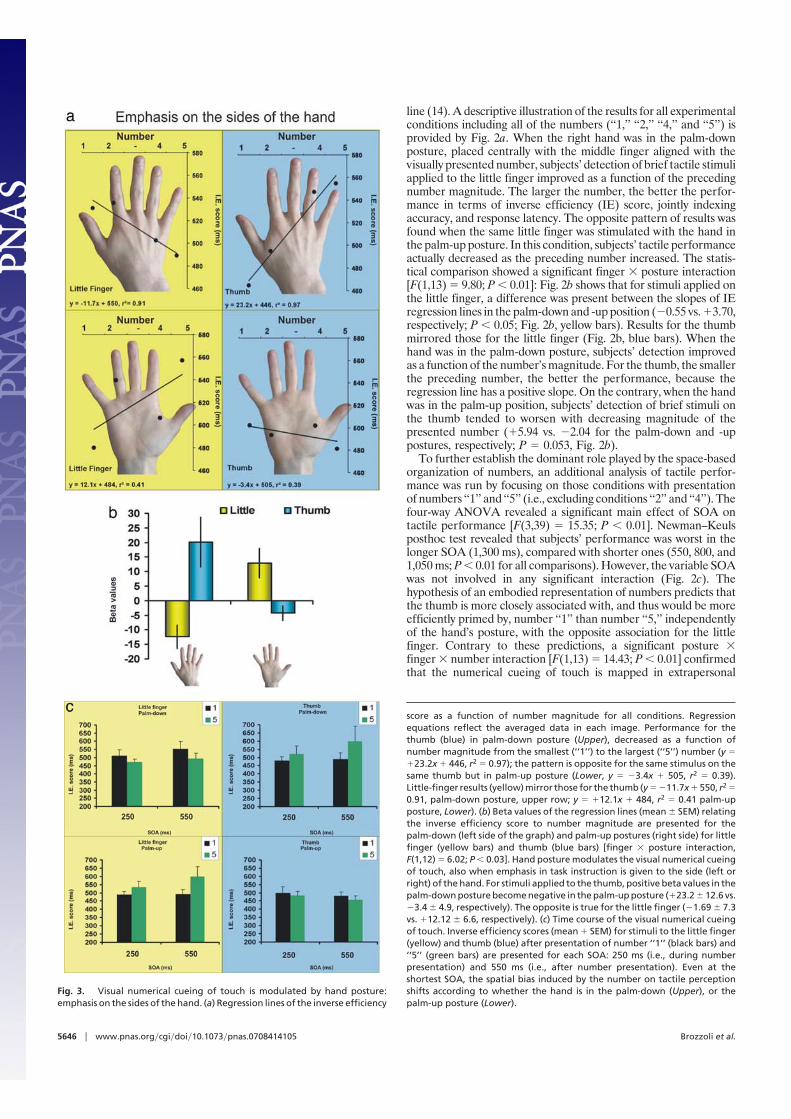
Fig. 3. Visual numerical cueing of touch is modulated by hand posture:emphasis on the sides of the hand. (a) Regression lines of the inverse efficiency
score as a function of number magnitude for all conditions. Regressionequations reflect the averaged data in each image. Performance for thethumb (blue) in palm-down posture (Upper), decreased as a function ofnumber magnitude from the smallest (‘‘1’’) to the largest (‘‘5’’) number (y ��23.2x � 446, r2 � 0.97); the pattern is opposite for the same stimulus on thesame thumb but in palm-up posture (Lower, y � �3.4x � 505, r2 � 0.39).Little-finger results (yellow) mirror those for the thumb (y � �11.7x � 550, r2 �0.91, palm-down posture, upper row; y � �12.1x � 484, r2 � 0.41 palm-upposture, Lower). (b) Beta values of the regression lines (mean � SEM) relatingthe inverse efficiency score to number magnitude are presented for thepalm-down (left side of the graph) and palm-up postures (right side) for littlefinger (yellow bars) and thumb (blue bars) [finger � posture interaction,F(1,12) � 6.02; P � 0.03]. Hand posture modulates the visual numerical cueingof touch, also when emphasis in task instruction is given to the side (left orright) of the hand. For stimuli applied to the thumb, positive beta values in thepalm-down posture become negative in the palm-up posture (�23.2 � 12.6 vs.�3.4 � 4.9, respectively). The opposite is true for the little finger (�1.69 � 7.3vs. �12.12 � 6.6, respectively). (c) Time course of the visual numerical cueingof touch. Inverse efficiency scores (mean � SEM) for stimuli to the little finger(yellow) and thumb (blue) after presentation of number ‘‘1’’ (black bars) and‘‘5’’ (green bars) are presented for each SOA: 250 ms (i.e., during numberpresentation) and 550 ms (i.e., after number presentation). Even at theshortest SOA, the spatial bias induced by the number on tactile perceptionshifts according to whether the hand is in the palm-down (Upper), or thepalm-up posture (Lower).
5646 � www.pnas.org�cgi�doi�10.1073�pnas.0708414105 Brozzoli et al.
space. Subjects’ performance was better in perceiving a touch onthe thumb after number ‘‘1’’ than ‘‘5’’ in the palm-down posture (IEscore: 447 vs. 470 ms, respectively; P � 0.05), but the oppositetendency was obtained when the hand posture was reversed (IEscore: 428 vs. 417 ms, respectively). Similarly, when considering thelittle finger, subjects’ performance mirrored that of the thumb: Inthe palm-down posture, stimuli on the little finger were detectedmore efficiently when preceded by number ‘‘5’’ than ‘‘1’’ (408 vs. 439ms, respectively; P � 0.05), but the opposite was true in the palm-upposture, in which performance was better when touches werepreceded by number ‘‘1’’ than ‘‘5’’ (429 vs. 447 ms, respectively; P �0.05). The same significant pattern of results was also obtainedwhen subjects’ accuracy was separately tested, and response laten-cies showed the same tendency. In other words, the same touchdelivered to the same little finger was better perceived if precededby number ‘‘5’’ than ‘‘1’’ in the palm-down posture but was betterperceived if preceded by number ‘‘1’’ than ‘‘5’’ in the palm-upposture.
To further explore the potential role played by instructionaland task-setting variables, we performed a second experimentwhereby tactile stimuli were always delivered on the thumb orlittle finger, but the side of the hand was stressed (i.e., ‘‘you willfeel a touch on either the left or right side of your hand’’).Moreover, to provide a finer description of the time course of theeffect of numerical cueing of touch, a shorter SOA was tested:tactile stimuli were delivered either 550 ms (i.e., as the shortestSOA in the first experiment) or 250 ms after number onset (i.e.,when the task-irrelevant number was still present on the screen;see Methods for details).
Results replicated the findings of the previous experiment. Asshown in Fig. 3b, tactile performance was cross-modally affectedby the visual presentation of a number, and numerical cueing oftouch again followed a number–space association, as revealed bythe significant finger � posture interaction [F(1,12) � 6.02; P �0.03]. In the palm-down posture, subjects’ tactile detection at thelittle finger improved with increasing number magnitude; theopposite pattern was observed in the palm-up posture. Forstimuli applied on the little finger, the slopes of IE regressionlines in the palm-down and -up position differed (�1.69 vs.�12.12, respectively; P � 0.04; Fig. 3b, yellow bars). Again,results for the thumb mirrored those for the little finger (Fig. 3b,blue bars). When the hand was in the palm-down posture,subjects’ detection improved with decreasing number magni-tude; the opposite tendency was present when the hand was inthe palm-up position (�23.22 vs. �3.35 for the palm-down and-up postures, respectively; P � 0.07; Fig. 3b). When consideringonly the numbers ‘‘1’’ and ‘‘5,’’ the ANOVA revealed a signif-icant posture � finger � number interaction [F(1,12) � 8.20; P �0.01], which further confirmed that the numerical cueing oftouch was mapped in extrapersonal space. Fig. 3c illustrates thatthis effect was also present at the shortest SOA, because neitherwas this variable significant nor was it involved in any interaction(Fig. 3c), thus suggesting a rather early space-based mapping ofnumbers.
The findings of both experiments clearly demonstrate that thehuman brain takes into account magnitude information pre-sented in the visual modality when processing tactile stimuli atthe fingers, but in so doing, it refers to an extrapersonal spatialrepresentation of numbers. Indeed, very similar and consistentresults were observed both when task instructions emphasizedthe (left or right) sides of the hand (second experiment), and the(little finger or thumb) fingers of the hand (first experiment), asfurther confirmed by the omnibus ANOVA run on data from thecommon SOA (550 ms from number onset), whereby the be-tween-subject variable emphasis was not involved in any inter-action. Therefore, even when emphasis was given to fingers andmight have in principle favored a finger-based numerical repre-sentation, the results were clear in showing a space-based
dominance in number representation. When compared withprevious studies, it is noteworthy that the present findings wereobtained within a best-suited approach to disambiguate betweennumber representations: First, number magnitude was totallytask-irrelevant, at odds with previous visuomotor number-fingermapping task (24); second, a single centrally located effector wasused, at variance with SNARC tasks whereby two left–righthorizontally aligned effectors are typical used (14, 17); finally,the foot was used as response effector, i.e., a body part that is notused to learn counting.
Here, the case for a connection between space and numbers (29)was studied in direct reference to the body. Our manipulation ofhand posture (30) was effective in distinguishing between the spatialreference frames in which tactile perception is biased by numericalcueing. By using an embodied approach based on tactile perception,we not only show that number-based attentional cueing crossessensory modalities but also demonstrate that number-based tactilepriming is early mapped according to an extrapersonal spatialrepresentation, thus providing a compelling support for the dom-inant role played by the spatial representation of numbers knownas the ‘‘mental number line.’’
MethodsSubjects. The first experiment was run on 14 (7 female, mean age 30.9; SD 10.1,range 20–51 years) neurologically healthy subjects. Thirteen (7 female, mean age29.3; SD 8.1, range 21–51 years) healthy subjects participated in the secondexperiment. Three subjects took part in both experiments. All participants gavetheir informed consent to take part in this study, which was approved by the localethics committee. They were asked to show how they usually count with theirfingers, without specifying in the request which hand to use first. However, toinduce subjects to use both hands, they were asked to count up to ‘‘8.’’ Onlysubjects who used the conventional (for Italian and French subjects) countingsystem(1, thumb;2, index;3,middle;4, ring;5, littlefinger) startingfromtherightthumb were admitted to the experimental session. Subjects were all right-handed according to the Edinburgh Handedness Inventory. They had normal orcorrected visual acuity, reported no somatosensory problems, and were naıve asto the purpose of the study.
Apparatus and Procedure. Both experiments were run with the same setup andprocedures were identical, unless otherwise stated. A personal computer (Dell,Optiplex GX270, Intel Pentium 4) equipped with a visual stimulus generator(ViSaGe, Cambridge Research Systems) was used to control stimulus presentationand response collection. Arabic numerals (‘‘1,’’ ‘‘2,’’ ‘‘4,’’ or ‘‘5’’) were presentedsingly at the center of a cathode ray tube monitor (Eizo FlexScan T931; resolution,800 � 600 pixels; refresh rate, 160 Hz), located 57 cm from the subjects’ eyes,subtending 1 � 1° of visual angle. Subjects’ right hidden hands lay in front ofthem, the middle finger aligned with the vertical meridian of the monitor, wherea fixation point appeared. Thumb and little finger were thus to the right or to theleft with respect to the middle finger. Two different postures could be assumed:Hand pronation (palm-down posture) or supination (palm-up posture). Subject’sfixation and eye movements were constantly monitored throughout each trialvia an eye-tracking system (Cambridge Research Systems; 250 Hz). After thesubject succeeded in keeping the fixation within a (nonvisible) circular windowcentered on the fixation point (2.5° side by side) for 500 ms, one of the fourequiprobable numbers (‘‘1,’’ ‘‘2,’’ ‘‘4,’’ or ‘‘5’’) appeared (300 ms). In the firstexperiment, a brief (100-�s) electrocutaneous stimulus was equiprobably deliv-ered via self-adhesive disposable electrodes (Neuroline 700-K, Ambu) to thethumb or little finger at one of four possible SOAs (550, 800, 1,050, or 1,300 ms).In the second experiment, the electrocutaneous stimulus was equiprobably de-livered to the thumb or little finger at one of two possible SOAs: 550 ms (i.e., sameas the shortest SOA in the first experiment) or 250 ms (i.e., 300 ms earlier than thefirst SOA, when the number was still present on the screen). In both experiments,subjects had to respond as fast as possible to the tactile stimulation by pressing acentral footpedalwiththeir rightfoot.Eyemovementsweremonitoreduptothefoot-pedal response. If central fixation was broken at any time during the trial,thetrialwasabortedandrandomlyreintroducedtoensurethatthesamenumberof trials was recorded for each condition. The tactile stimulus intensity was set toobtain �80% correct detections for both fingers with a titration procedure thatwas run in a preexperimental session (see SI Experiment 1 and SI Experiment 2).Each stimulator (DS7A, Digitimer) current was varied independently for eachfinger so that detection performance was comparable between the two fingers.Subjects were told that the number was totally irrelevant for the tactile detection
Brozzoli et al. PNAS � April 8, 2008 � vol. 105 � no. 14 � 5647
PSYC
HO
LOG
Y
task. To ensure that number magnitude was processed (see SI Experiment 1 andSI Experiment 2, Number Magnitude, and Table S2), they were also told theycould be asked without warning which number appeared in the immediatelypreceding trial.
Accuracy and reaction time (RT) were combined in the IE score, a standard wayto combine RT and accuracy data into a single performance measure, computedas the median RT divided by the proportion of correct trials for a given condition;a higher IE value indicates worse performance, just as for RT and error measures.The IE score was submitted to a four-way ANOVA with SOA, posture, finger, andnumber (‘‘1’’ vs. ‘‘5’’) as variables. Each posture was further analyzed by a three-
wayANOVA.Regression linebetavaluesbetweenIEscoreandnumberswerealsocalculated and submitted to a three-way ANOVA with SOA, posture, and fingeras within-subject variables. Significant sources of variance were explored byNewman–Keuls posthoc tests and planned comparisons.
ACKNOWLEDGMENTS. We thank F. Frassinetti, N. Holmes, F. Pavani, and A.Roy for thoughtful discussions and comments on an earlier version of themanuscript. This work was supported by the European Mobility Fellowship,AVENIR Grant no. R05265CS, and the Agence National de Recherche Grant no.JCJC06�133960.
1. Butterworth B (1999) A head for figures. Science 284:928–929.2. Simon TJ (1999) The foundations of numerical thinking in a brain without numbers.
Trends Cognit Sci 3:363–365.3. Binkofski F, et al. (1999) A fronto-parietal circuit for object manipulation in man:
evidence from an fMRI-study. Eur J Neurosci 11:3276–3286.4. Pesenti M, Thioux M, Seron X, De Volder A (2000) Neuroanatomical substrates of Arabic
number processing, numerical comparison, and simple addition: A PET study. J CognitNeurosci 12:461–479.
5. Stanescu-Cosson R, et al. (2000) Understanding dissociations in dyscalculia: a brainimaging study of the impact of number size on the cerebral networks for exact andapproximate calculation. Brain 123:2240–2255.
6. Zago L, et al. (2001) Neural correlates of simple and complex mental calculation.NeuroImage 13:314–327.
7. Rueckert L, et al. (1996) Visualizing cortical activation during mental calculation withfunctional MRI. NeuroImage 3:97–103.
8. Pinel P, Piazza M, Le Bihan D, Dehaene S (2004) Distributed and overlapping cerebralrepresentations of number, size, and luminance during comparative judgments. Neu-ron 41:983–993.
9. Piazza M, Pinel P, Le Bihan D, Dehaene S (2007) A magnitude code common tonumerosities and number symbols in human intraparietal cortex. Neuron 53:293–305.
10. Martory MD, et al. (2003) Pure global acalculia following a left subangular lesion.Neurocase 9:319–328.
11. Gerstmann J (1930) Zur Symptomatologie der Hirnlasionen im Ubergangsgebiet derunteren parietal und mittleren Occipitalwindung. Nervenarzt 3:691–695.
12. Rusconi E, Walsh V, Butterworth B (2005) Dexterity with numbers: rTMS over leftangular gyrus disrupts finger gnosis and number processing. Neuropsychologia43:1609–1624.
13. Sato M, Cattaneo L, Rizzolatti G, Gallese V (2007) Numbers within our hands: Modu-lation of corticospinal excitability of hand muscles during numerical judgment. JCognit Neurosci 19:684–693.
14. Dehaene S, Bossini S, Giraux P (1993) The mental representation of parity and numbermagnitude. J Exp Psychol Gen 122:371–396.
15. Hubbard EM, Piazza M, Pinel P, Dehaene S (2005) Interactions between number andspace in parietal cortex. Nat Rev Neurosci 6:435–448.
16. Doricchi F, Guariglia P, Gasparini M, Tomaiuolo F (2005) Dissociation between physicaland mental number line bisection in right hemisphere brain damage. Nat Neurosci8:1663–1665.
17. Priftis K, Zorzi M, Meneghello F, Marenzi R, Umilta C (2006) Explicit versus implicitprocessing of representational space in neglect: Dissociations in accessing the mentalnumber line. J Cognit Neurosci 18:680–688.
18. Rossetti Y, Jacquin-Courtois S, Rode G, Ota H, Michel C, Boisson D (2004) Does actionmake the link between number and space representation? Visuo-manual adaptationimproves number bisection in unilateral neglect. Psychol Sci 15:426–430.
19. Vuilleumier P, Ortigue S, Brugger P (2004) The number space and neglect. Cortex40:399–410.
20. Zorzi M, Priftis K, Umilta C (2002) Brain damage: Neglect disrupts the mental numberline. Nature 417:138–139.
21. Gobel SM, Calabria M, Farne A, Rossetti Y (2006) Parietal rTMS distorts the mentalnumber line: Simulating ‘spatial’ neglect in healthy subjects. Neuropsychologia44:860–868.
22. Fischer MH, Castel AD, Dodd MD, Pratt J (2003) Perceiving numbers causes spatial shiftsof attention. Nat Neurosci 6:555–556.
23. Ishihara M, Jacquin-Courtois S, Flory V, Salemme R, Imanaka K, Rossetti Y (2006)Interaction between space and number representations during motor preparation inmanual aiming. Neuropsychologia 44:1009–1016.
24. Di Luca S, Grana A, Semenza C, Seron X, Pesenti M (2006) Finger-digit compatibility inArabic numeral processing. Q J Exp Psychol 59:1648–1663.
25. Bachtold D, Baumuller M, Brugger P (1998) Stimulus-response compatibility in repre-sentational space. Neuropsychologia 36:731–735.
26. Wood G, Nuerk HC, Willmes K (2006) Crossed hands and the SNARC effect: A failure toreplicate Dehaene, Bossini and Giraux (1993) Cortex 42:1069–1079.
27. Fischer MH (2006) The future for SNARC could be stark. Cortex 42:1066–1068.28. Rusconi E, Umilta C, Galfano G (2006) Breaking ranks: Space and number may march to
the beat of a different drum. Cortex 42:1124–1127.29. Walsh V (2003) A theory of magnitude: Common cortical metrics of time, space and
quantity. Trends Cognit Sci 7:483–488.30. Behrmann M, Moscovitch M (1994) Object-centered neglect in patients with unilateral
neglect: Effects of left-right coordinates of objects J. Cognit Neurosci 6:1–16.
5648 � www.pnas.org�cgi�doi�10.1073�pnas.0708414105 Brozzoli et al.
Supporting InformationBrozzoli et al. 10.1073/pnas.0708414105SI Experiment 1Supporting Procedures. To obtain �80% correct detections forboth fingers, the stimulus intensity was set individually for eachsubject and each stimulated finger in a preliminary sessionbefore the main experiment. The setup was the same used as forthe main session, but the procedure differed as depicted in theFig. S1. The fixation point was presented for 500 ms, after which,if the subject succeeded in keeping fixation, a tactile stimulus wasdelivered either to the thumb or the little finger. No numberstimuli were presented.
The titration block used to set the intensity consisted of 15trials, where 5 stimulations to the thumb and 5 to the littlefinger were randomly intermingled with 5 trials in which notactile stimulus was delivered (catch trials). The intensity wasfirst set at 0.10 mA for both constant current stimulators, eachdelivering electrocutaneous square wave pulses to one finger.At the end of the first preliminary block, the experimentervaried the stimulator current, independently for each finger, toreach a detection performance of 80% for each of them. Thus,the intensity was increased or reduced depending on whetherthe performance was below or above the criterion. The firststep was 2 mA, then the step amplitude was halved at everydirection reversal. The same block of trials and procedure wasthen repeated until the criterion of 80% of accuracy was metfor each finger. The same procedure was applied for each handposture (palm-up, palm-down). Table S1 below reports thestimulus intensity used for each subject for both fingers andpostures.
Supporting Results. Electrocutaneous current intensity. A statisticalanalysis (ANOVA) with finger (thumb vs. little finger) andposture (palm-up vs. palm-down) as variables showed that, tohave the same performance in terms of accuracy a differentintensity had to be set for thumb (3.79 mA) and little finger (2.61mA) [F(1,13) � 7.13; P � 0.05)]. No difference was presentbetween the two postures.
Number Magnitude. To ensure that number magnitude was pro-cessed, subjects were told they could be asked, without warning,which number had been presented in the immediately precedingtrial. Two of such probing situations were randomly interspersedwithin each block of trials. All subjects answered without errorto this request in each block (100% accuracy), except one(subject 9) who made two errors reporting an incorrect number(83% accuracy).
Catch Trials. Each experimental block consisted of 160 trials: 4repetitions for each combination of number, delay and finger(128) plus 32 trials (20%), where after the visual presentation ofthe number no electric pulse was delivered (catch trials). Falsealarms rate was on average 1.16% without difference acrossconditions.
SI Experiment 2Supporting Procedures. Subjects started the experimental sessionalternatively with the hand in the palm-down or palm-up posture.The experiment consisted of a unique session of four experi-mental blocks (two for each posture), postures being counter-balanced across blocks. The same procedures and criterion (80%accuracy independently for both fingers) as for the first exper-iment were used in the second experiment to set the tactilestimulations intensity before the experimental session. As in thefirst experiment, stimulus intensity was not varied during theexperimental session, but in the second experiment it was set inthe palm-down posture for six subjects and with the hand in thepalm-up posture for the remaining seven subjects. Table S2below reports the stimulus intensity used for each subject foreach finger.
Supporting Results. Electrocutaneous current intensity. Similar to thefirst experiment, a statistical analysis (ANOVA) with posture(palm-up vs. palm-down) as between-subject variable and finger(thumb vs. little finger) as within-subject variable showed that adifference in tactile stimulus intensity was set to obtain the samedetection performance for thumb (4.38 mA) and little finger(2.94 mA) [F(1,11) � 81.98; P � 0.001)]. No difference wasfound between postures.
Number Magnitude. Subjects performed errorless when requestedto report which number had been presented in the immediatelypreceding trial in each block (100% accuracy), except one subject(subject 11) who made one error reporting an incorrect number(4 instead of 5) (75% accuracy).
Catch Trials. Each experimental block consisted of 160 trials: 8repetitions for each combination of number, delay and finger(128) plus 32 trials (20%), where after the visual presentation ofthe number no electric pulse was delivered (catch trials). Falsealarms rate was on average 2,49% without difference acrossconditions.
Brozzoli et al. www.pnas.org/cgi/content/short/0708414105 1 of 4
Fig. S1. Timing of stimuli presentation for the preliminary titration session.The figure illustrates the example of a tactile stimulus delivered to the littlefinger (green symbol) in the palm-down posture. As in the experimentalsessions, the subjects were instructed to respond to the tactile stimulus asquickly as possible regardless of the finger stimulated, by pressing a pedal withtheir right foot.
Brozzoli et al. www.pnas.org/cgi/content/short/0708414105 2 of 4
Table S1. Stimulus intensity for each subject’s thumb and littlefinger for the two hand postures
Subjects
Stimulus intensity, mA
Thumb Little finger
Palm down Palm up Palm down Palm up
1 3.5 3.6 2 2.12 3.4 3.3 1.9 1.53 2.7 2.6 1.8 1.84 7.5 7.7 4.2 4.35 4.8 4.8 3.6 3.66 3.7 3.7 3 37 7 6.8 1.9 1.68 2.4 2.4 4.1 4.19 5.6 5.6 3 3
10 2 2.1 2 211 3.2 3 2.4 2.512 2.5 2.3 2.2 2.213 3 3 2.8 2.914 2 2 1.8 1.8Mean 3.81 3.78 2.62 2.60SD 1.77 1.79 0.85 0.91
Brozzoli et al. www.pnas.org/cgi/content/short/0708414105 3 of 4
Table 2. Stimulus intensity for each subject’s thumb andlittle finger
Subjects
Stimulus intensity, mA
Thumb Little finger
1 3.9 2.72 4.1 2.53 4.3 3.04 4.7 3.35 5.3 3.36 4.1 2.67 4.7 2.78 1.9 1.99 4.5 3.5
10 6.1 4.411 5.9 4.112 4.2 2.213 3.2 2.1Mean 4.38 2.94SD 1.09 0.75
From subjects 1 to 6, intensity was set with the hand in the palm-downposture; from 7 to 13, intensity was set with the hand was in the palm-upposture.
Brozzoli et al. www.pnas.org/cgi/content/short/0708414105 4 of 4

2.1.1. Discussion
The study presented here reveals that visuo-tacile interactions in healthy humans may be
modulated by relatively abstract information about quantities and that, in some cases, the
somatotopic coding of touch ‘loses’ against more ‘space-based’ coding of touch (Azañón and
Soto-Faraco 2008). The visual information, in this case a semantic symbol as a number, can
affect the perception of information coming from the somatosensory channel. It is clear from
this example that the interaction between the two modalities has a spatial nature, even at this
high-cognitive level. This study is an evidence of cross-modally driven shift of attention.
3. Multisensory integration through anatomical convergence
Perhaps, the simplest approach for thinking of an interplay between different sensory
modalities is to imagine a point of convergence for inputs coming from the different sensory
channels. In other words, the information coming from the external world, initially elaborated
in the unisensory regions of the brain at a certain level have to converge on the same area. A
large body of evidence is now available from single-cell and tracing studies in animals and
from neuroimaging studies in humans showing numerous multisensory convergence zones in
the brain. In particular the animal models revealed cortical and subcortical structures where
the single units receive afferent inputs from different senses. The pioneering and influential
series of studies conducted on the superior culliculus (SC) in the cat (Meredith and Stein
1983; 1986), made of this structure a model for the multisensory integration. In humans too,
neuroimaging studies revealed the presence of possible areas of sensory convergence.
Deep layers of the SC, in addition to other subcortical regions such as basal ganglia
(Nagy et al. 2006), has been shown to receive inputs from somatosensory, auditory, and visual
areas (Stein and Meredith 1993). Activity in deep SC neurons is thus dependent on the
presence of information coming from more than one sense and on spatial and temporal
� 29� �
relation between inputs of different modalities. Three general rules can describe the neural
activity of this structure.
1. The spatial proximity between sensory information. Activity of SC neurones is maximal
when stimuli of different modalities are presented in the same location. The integration
diminishes with increasing spatial discrepancy between the visual and auditory
information, for example.
2. The temporal synchrony between sensory information. Also the temporal relation appears
to play a fundamental role in the multisensory integration, allowing it when the different
sensory information are presented simultaneously rather than temporally separated.
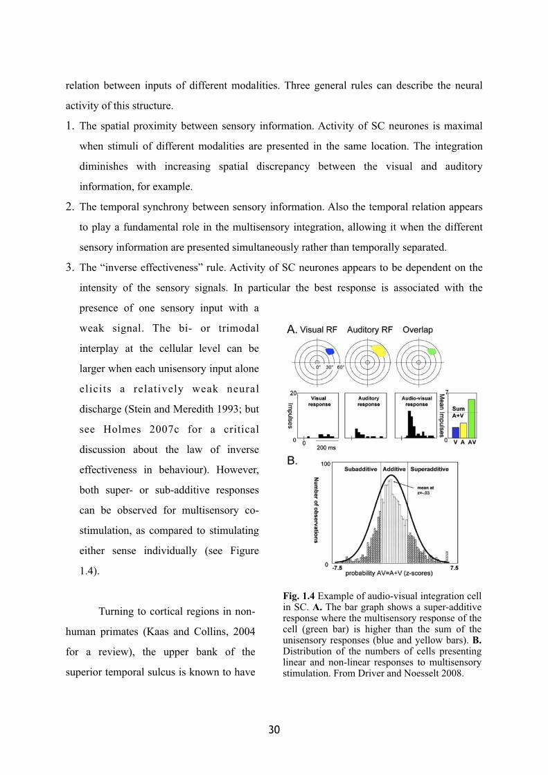
3. The “inverse effectiveness” rule. Activity of SC neurones appears to be dependent on the
intensity of the sensory signals. In particular the best response is associated with the
presence of one sensory input with a
weak signal. The bi- or trimodal
interplay at the cellular level can be
larger when each unisensory input alone
elicits a relatively weak neural
discharge (Stein and Meredith 1993; but
see Holmes 2007c for a critical
discussion about the law of inverse
effectiveness in behaviour). However,
both super- or sub-additive responses
can be observed for multisensory co-
stimulation, as compared to stimulating
either sense individually (see Figure
1.4).
Turning to cortical regions in non-
human primates (Kaas and Collins, 2004
for a review), the upper bank of the
superior temporal sulcus is known to have
30
Fig. 1.4 Example of audio-visual integration cell in SC. A. The bar graph shows a super-additive response where the multisensory response of the cell (green bar) is higher than the sum of the unisensory responses (blue and yellow bars). B.Distribution of the numbers of cells presenting linear and non-linear responses to multisensory stimulation. From Driver and Noesselt 2008.
bidirectional connections with unisensory auditory, visual and somatosensory cortices
(Padberg et al. 2003; Schmahmann and Pandya 1991) and to contain multisensory neurons
(Bruce et al. 1981) that, similarly to the SC, receive inputs from different sensory modalities
converging on the same single cell. Several regions within parietal and frontal areas also
present a multisensory convergence of information (see the second chapter of this
dissertation).
One of the most elegant research in humans focussing on cross-modal integration is
represented by a series of studies conducted in fMRI (functional Magnetic Resonance
Imaging, Macaluso and Driver 2001; Macaluso et al. 2000; Macaluso and Driver 2005 for a
review). This constitutes one of the first research investigating which areas in the human brain
present an activity correlated with a detection task performed on stimulation coming from
different modalities. A region corresponding to IPS (Intraparietal sulcus) appeared to be
activated both for a visual and for a tactile detection task (Macaluso and Driver 2001). These
results are in agreement with the results coming from an independent research by another
group showing an activation of parietal regions in response to visual or tactile stimulations
(Bremmer et al. 2001).
4. Multisensory interactions through feedback on unisensory areas and inter-connections among unisensory areas
As noted in the introduction, the revolutionary advance in multisensory research is the
discovery that the multisensoriality exists “beyond modularity and convergence” (Driver and
Spence 2000). The interplay between different modalities can indeed arise not only through a
convergence mechanism which integrates the information coming from different senses in the
same neuron. Other mechanisms have been recently proposed, following some studies whose
results showed the existence of modulations of unisensory low-level areas from the activity of
multisensory high-level areas, or as a result of direct early modulations from different low-
level unisensory areas (Figure 1.5).
� 31� �

4.1.Back-projections from multisensory higher-level to unisensory lower-level areas
The “new look” in this field is based on studies showing that once believed sensory-specific
areas can be influenced by multisensory interactions. Some evidence was present in the early
70’s (Fishman and Micheal 1973; Morrell 1972; Spinelli et al. 1968) showing a modulation of
the visual regions response as a consequence of the presence of auditory information.
However due to the technological limits of these early studies one cannot exclude non-
specific confounding factors. More recent researches have clearly showed that traditionally
considered unisensory regions can be influenced by multisensory interactions (Macaluso et al.
2000; Macaluso et al. 2002). Several fMRI studies have now reported modulation of
traditional unisensory areas (usually defined as occipital for the visual system, post-central for
tactile sensation and temporal for the auditory information) due to multisensory co-
stimulation (Amedi et al. 2002; Buchel et al. 1998; Calvert et al. 1999; 2001; Macaluso et al.
2000; Martuzzi et al. 2007; Miller and D'Esposito 2005). Using high-resolution fMRI in
monkeys, together with separate mapping of specific auditory-cortex regions, Kayser and
colleagues (2005) observed increased BOLD signal in secondary auditory cortex due to the
presence of tactile stimulations. Even primary auditory areas were affected during the
presence of visual information (Kayser et al. 2007).
Also in human, evidence has been found in favour of a modulation of primary sensory
areas in case of multisensory stimulation. Auditory cortex, for instance, appears to be
differentially modulated when subjects perceived audiovisual speech stimuli as synchronous
32
Fig. 1.5 Schematic representation of a)multisensory integration and b) multisensory feedback interaction from integration areas over unisensory areas. F r o m D r i v e r a n d Spence 2000.
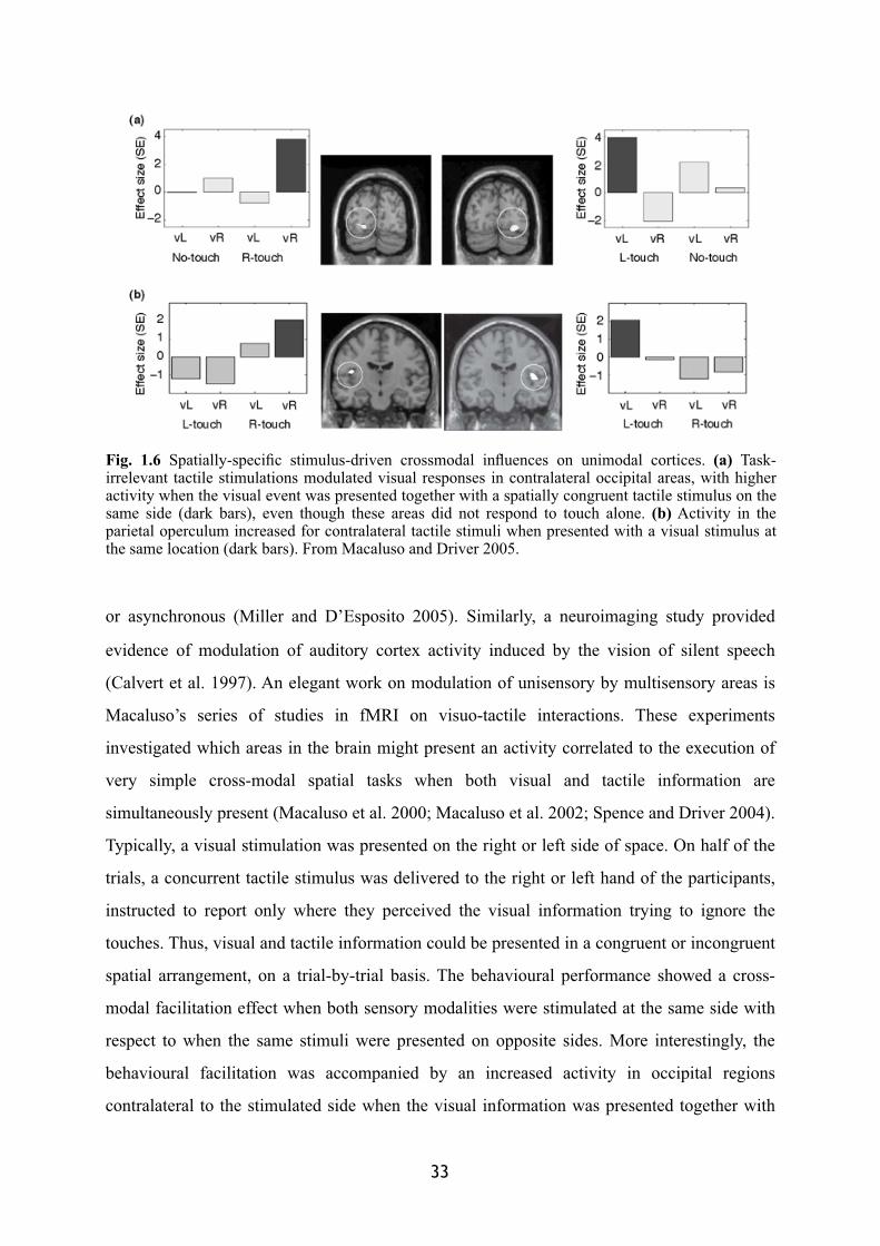
or asynchronous (Miller and D’Esposito 2005). Similarly, a neuroimaging study provided
evidence of modulation of auditory cortex activity induced by the vision of silent speech
(Calvert et al. 1997). An elegant work on modulation of unisensory by multisensory areas is
Macaluso’s series of studies in fMRI on visuo-tactile interactions. These experiments
investigated which areas in the brain might present an activity correlated to the execution of
very simple cross-modal spatial tasks when both visual and tactile information are
simultaneously present (Macaluso et al. 2000; Macaluso et al. 2002; Spence and Driver 2004).
Typically, a visual stimulation was presented on the right or left side of space. On half of the
trials, a concurrent tactile stimulus was delivered to the right or left hand of the participants,
instructed to report only where they perceived the visual information trying to ignore the
touches. Thus, visual and tactile information could be presented in a congruent or incongruent
spatial arrangement, on a trial-by-trial basis. The behavioural performance showed a cross-
modal facilitation effect when both sensory modalities were stimulated at the same side with
respect to when the same stimuli were presented on opposite sides. More interestingly, the
behavioural facilitation was accompanied by an increased activity in occipital regions
contralateral to the stimulated side when the visual information was presented together with
� 33� �
Fig. 1.6 Spatially-specific stimulus-driven crossmodal influences on unimodal cortices. (a) Task-irrelevant tactile stimulations modulated visual responses in contralateral occipital areas, with higher activity when the visual event was presented together with a spatially congruent tactile stimulus on the same side (dark bars), even though these areas did not respond to touch alone. (b) Activity in the parietal operculum increased for contralateral tactile stimuli when presented with a visual stimulus at the same location (dark bars). From Macaluso and Driver 2005.
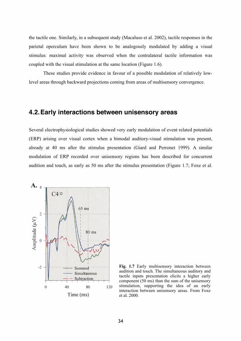
the tactile one. Similarly, in a subsequent study (Macaluso et al. 2002), tactile responses in the
parietal operculum have been shown to be analogously modulated by adding a visual
stimulus: maximal activity was observed when the contralateral tactile information was
coupled with the visual stimulation at the same location (Figure 1.6).
These studies provide evidence in favour of a possible modulation of relatively low-
level areas through backward projections coming from areas of multisensory convergence.
4.2.Early interactions between unisensory areas
Several electrophysiological studies showed very early modulation of event related potentials
(ERP) arising over visual cortex when a bimodal auditory-visual stimulation was present,
already at 40 ms after the stimulus presentation (Giard and Perronet 1999). A similar
modulation of ERP recorded over unisensory regions has been described for concurrent
audition and touch, as early as 50 ms after the stimulus presentation (Figure 1.7; Foxe et al.
34
Fig. 1.7 Early multisensory interaction between audition and touch. The simultaneous auditory and tactile inputs presentation elicits a higher early component (50 ms) than the sum of the unisensory stimulation, supporting the idea of an early interaction between unisensory areas. From Foxe et al. 2000.
2000; Molholm et al. 2002). Effects at such short latencies do not readily fit with backward
projections via longer pathways from multisensory areas. Thus they are presumably
suggesting some direct connections between sensory-specific areas. Evidence in favour of this
possibility comes from retrograde tracing studies in monkeys, showing for example direct
connections between primary auditory and visual cortex (Falchier et al. 2002). Moreover,
electrophysiology has also demonstrated some response to somatosensory stimuli in a region
closely adjacent to the primary auditory cortex (Fu et al. 2003; Lakatos et al. 2007).
5. Conclusion
The multisensory research presented in this chapter clearly shows how pervasive might be the
presence of multisensory mechanisms in the brain. It is obvious that the brain adapted such
mechanisms through evolution in a world that can essentially be experienced in a
multisensory way. Since also the low-level primary areas can be affected as well by the
presence of information coming from a different sensory channel, the question arises as to
whether it is still valuable to distinguish unisensory processing and unimodal areas from
multisensory areas (Ghazanfar and Schroeder 2006). However, despite this evidence,
unisensory areas show stronger preference for one particular modality with respect to others.
They thus are more “sensory-specific” rather than “unisensory” but they still retain their
functional specialisation in the elaboration of sensory information. On the other side, the
proper multisensory integration, arising at the single cell level should also present an adaptive
function with respect to other form of multisensory modulation. In the next chapter I will
present in detail the mechanisms underlying the multisensory convergence between visual and
tactile information and the characteristics of this multisensory link between information
external to the body and information present onto the body.
� 35� �
Chapter II
Multisensory and motor representations of peripersonal space
The binding of visual information available far outside the body with tactile information
arising, by definition, on the body, allows for the construction of a representation of the space
which lies in-between. This reachable part of the external space is often the theatre of our
interactions with objects. This representation of intermediate space has become known as
“peripersonal space” (Rizzolatti et al. 1981b, c). The definition of peripersonal space
originates from neurophysiological studies in monkeys where single cell investigations have
highlighted the existence of bimodal neurons responding to visual as well as tactile
stimulations. In this chapter I will review some basic features of visuo-tactile interactions, as
revealed by the single units recording studies in monkeys. A crucial point that will be stressed
is that these neuronal populations are not only responsive to sensory solicitation from the
environment, but they also present motor-related activity. The peculiar nature of their
activation qualifies them as multisensory-motor interfaces, a bridge between the
(multisensory) perception and the planning and execution of actions.
36
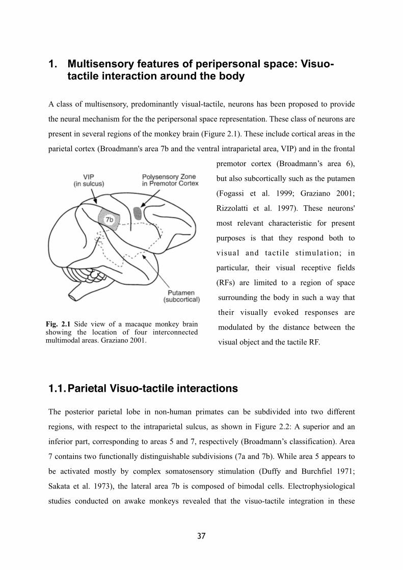
1. Multisensory features of peripersonal space: Visuo-tactile interaction around the body
A class of multisensory, predominantly visual-tactile, neurons has been proposed to provide
the neural mechanism for the the peripersonal space representation. These class of neurons are
present in several regions of the monkey brain (Figure 2.1). These include cortical areas in the
parietal cortex (Broadmann's area 7b and the ventral intraparietal area, VIP) and in the frontal
premotor cortex (Broadmann’s area 6),
but also subcortically such as the putamen
(Fogassi et al. 1999; Graziano 2001;
Rizzolatti et al. 1997). These neurons'
most relevant characteristic for present
purposes is that they respond both to
visual and tactile stimulation; in
particular, their visual receptive fields
(RFs) are limited to a region of space
surrounding the body in such a way that
their visually evoked responses are
modulated by the distance between the
visual object and the tactile RF.
1.1.Parietal Visuo-tactile interactions
The posterior parietal lobe in non-human primates can be subdivided into two different
regions, with respect to the intraparietal sulcus, as shown in Figure 2.2: A superior and an
inferior part, corresponding to areas 5 and 7, respectively (Broadmann’s classification). Area
7 contains two functionally distinguishable subdivisions (7a and 7b). While area 5 appears to
be activated mostly by complex somatosensory stimulation (Duffy and Burchfiel 1971;
Sakata et al. 1973), the lateral area 7b is composed of bimodal cells. Electrophysiological
studies conducted on awake monkeys revealed that the visuo-tactile integration in these
37
Fig. 2.1 Side view of a macaque monkey brain showing the location of four interconnected multimodal areas. Graziano 2001.
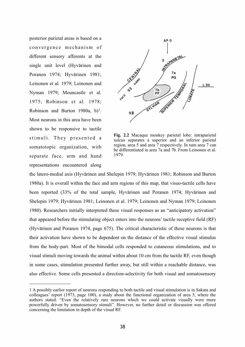
posterior parietal areas is based on a
c o n v e rg e n c e m e c h a n i s m o f
different sensory afferents at the
single unit level (Hyvärinen and
Poranen 1974; Hyvärinen 1981;
Leinonen et al. 1979; Leinonen and
Nyman 1979; Mouncastle et al.
1975; Robinson et al. 1978;
Robinson and Burton 1980a, b)1.
Most neurons in this area have been
shown to be responsive to tactile
s t i m u l i . T h e y p r e s e n t e d a
somatotopic organization, with
separate face, arm and hand
representations encountered along
the latero-medial axis (Hyvärinen and Shelepin 1979; Hyvärinen 1981; Robinson and Burton
1980a). It is overall within the face and arm regions of this map, that visuo-tactile cells have
been reported (33% of the total sample, Hyvärinen and Poranen 1974; Hyvärinen and
Shelepin 1979; Hyvärinen 1981; Leinonen et al. 1979; Leinonen and Nyman 1979; Leinonen
1980). Researchers initially interpreted these visual responses as an “anticipatory activation”
that appeared before the stimulating object enters into the neurons’ tactile receptive field (RF)
(Hyvärinen and Poranen 1974, page 675). The critical characteristic of these neurons is that
their activation have shown to be dependent on the distance of the effective visual stimulus
from the body-part. Most of the bimodal cells responded to cutaneous stimulations, and to
visual stimuli moving towards the animal within about 10 cm from the tactile RF, even though
in some cases, stimulation presented further away, but still within a reachable distance, was
also effective. Some cells presented a direction-selectivity for both visual and somatosensory
38
1 A possibly earlier report of neurons responding to both tactile and visual stimulation is in Sakata and colleagues’ report (1973, page 100), a study about the functional organization of area 5, where the authors stated: “Even the relatively rare neurons which we could activate visually were more powerfully driven by somatosensory stimuli”. However, no further detail or discussion was offered concerning the limitation in depth of the visual RF.
Fig. 2.2 Macaque monkey parietal lobe: intraparietal sulcus separates a superior and an inferior parietal region, area 5 and area 7 respectively. In turn area 7 can be differentiated in area 7a and 7b. From Leinonen et al. 1979.
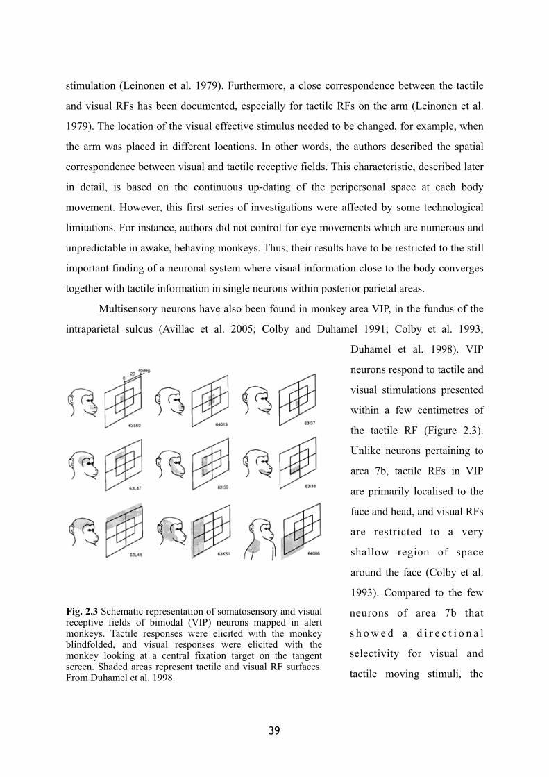
stimulation (Leinonen et al. 1979). Furthermore, a close correspondence between the tactile
and visual RFs has been documented, especially for tactile RFs on the arm (Leinonen et al.
1979). The location of the visual effective stimulus needed to be changed, for example, when
the arm was placed in different locations. In other words, the authors described the spatial
correspondence between visual and tactile receptive fields. This characteristic, described later
in detail, is based on the continuous up-dating of the peripersonal space at each body
movement. However, this first series of investigations were affected by some technological
limitations. For instance, authors did not control for eye movements which are numerous and
unpredictable in awake, behaving monkeys. Thus, their results have to be restricted to the still
important finding of a neuronal system where visual information close to the body converges
together with tactile information in single neurons within posterior parietal areas.
Multisensory neurons have also been found in monkey area VIP, in the fundus of the
intraparietal sulcus (Avillac et al. 2005; Colby and Duhamel 1991; Colby et al. 1993;
Duhamel et al. 1998). VIP
neurons respond to tactile and
visual stimulations presented
within a few centimetres of
the tactile RF (Figure 2.3).
Unlike neurons pertaining to
area 7b, tactile RFs in VIP
are primarily localised to the
face and head, and visual RFs
are restricted to a very
shallow region of space
around the face (Colby et al.
1993). Compared to the few
neurons of area 7b that
s h o w e d a d i r e c t i o n a l
selectivity for visual and
tactile moving stimuli, the
� 39
Fig. 2.3 Schematic representation of somatosensory and visual receptive fields of bimodal (VIP) neurons mapped in alert monkeys. Tactile responses were elicited with the monkey blindfolded, and visual responses were elicited with the monkey looking at a central fixation target on the tangent screen. Shaded areas represent tactile and visual RF surfaces. From Duhamel et al. 1998.
VIP cells are strongly characterised by a preferred direction that often matches between both
sensory modalities. Their visual RFs have been clearly shown to be anchored to the face
independently of the gaze direction (Colby et al. 1993). Noteworthy, VIP is a multisensory
region of the cortex in which the majority of VIP neurons perform multisensory integration
following the same general rules (e.g., spatial congruency and temporal synchrony) that have
been previously documented in other cortical and subcortical regions (Avillac et al. 2007).
An interesting recent finding showed that visuo-tactile neurons within area 7b and VIP
also respond when another individual’s body-part is approached by a visual stimulus (Ishida
et al. 2009). Similarly to the multisensory neurons described above, these “body-matching
neurons” respond to visual stimuli presented near the tactile RF. Moreover, these “body-
matching neurons” are responsive to a visual stimulus presented close to the corresponding
body-part of another individual (a human experimenter). For instance, a neuron displaying a
tactile RF on the arm responded to a visual stimulus presented close to the monkey’s own
arm, but also responded to visual stimuli presented close to another individual’s arm (Figure
2.4). For some of these neurons, this matching property seems to be independent of the
position of the observed individual with respect to the observing monkey (up to 35 degrees of
rotation).
The studies presented in this section indicate that the initial cortical convergence of
vision and somatosensation occurs in a pool of areas within the parietal lobe. These cortical
regions present also several direct and indirect connections. The intraparietal sulcus receives
projections from both somatosensory and visual areas respectively in its medial (Jones and
Powell 1970), and lateral bank (Seltzer and Pandya 1980; Maunsell and Van Essen 1983;
Ungerleider and Desimone 1986; Boussaoud et al. 1990; Baizer et al. 1991). Most
importantly, both projections overlap in the fundus, where VIP is located (Maunsell and Van
Essen 1983; Ungerleider and Desimone 1986; Colby and Duhamel 1991). These three regions
of the intraparietal sulcus send projections to area 7b (Jones and Powell 1970; Mesulam et al.
1977), which also receives more direct somatosensory input from SII (Stanton et al. 1977) and
from area 5 (Jones and Powell 1970), the superior part of the posterior parietal area.
In this way, multisensory perception arises as the result of a physiological convergence
of visual and tactile input on the same neurons (Jones and Powell 1970) from the respective
40
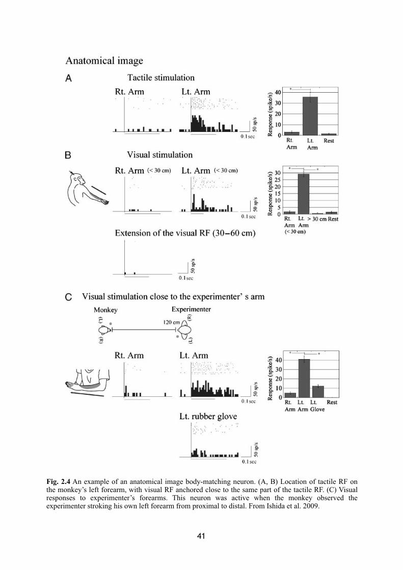
� 41
Fig. 2.4 An example of an anatomical image body-matching neuron. (A, B) Location of tactile RF on the monkey’s left forearm, with visual RF anchored close to the same part of the tactile RF. (C) Visual responses to experimenter’s forearms. This neuron was active when the monkey observed the experimenter stroking his own left forearm from proximal to distal. From Ishida et al. 2009.
areas of initial elaboration. Moreover, the multisensory parietal areas appear to be, to a certain
extent, differentially involved in the representation of the space around different sectors of the
body. Most part of the VIP neurons present RFs on the face and head while neurons within
parietal area 7b are more representative of the arm and hand regions.
1.2.Premotor visuo-tactile interactions
A detailed series of studies on the properties of visuo-tactile neurons has been also made
available by investigating monkeys’ bimodal neurons contained in the premotor cortex.
Numerous studies have focussed on the single cell recording on units within F4 and F5
regions of macaque monkey (Matelli et al. 1985), composing respectively the rostral and the
caudal inferior part of area 6, in the proximity of the arcuate sulcus as illustrated in Figure 2.5
(Fogassi et al. 1992; Fogassi et al. 1996; Gentilucci et al. 1983; Gentilucci et al. 1988;
Godschalk et al. 1985; Graziano et al. 1994; Graziano and Gandhi 2000; Rizzolatti et al.
1981a, b, c; Rizzolatti et al. 1987; Rizzolatti and Gentilucci 1988; Rizzolatti and Luppino
2001).
Neurons in the F4 sub-region are strongly responsive to tactile stimulation. They are
characterised by relatively large tactile RFs located primarily on the animal’s face or arm/
hand regions. Some of these neurons also present RFs in separated body parts. They are
responsive for tactile stimuli on the peribuccal region, for instance, as well as for touches
delivered on the hand (Gentilucci et al. 1988; Rizzolatti et al. 1981a). A large proportion
(85%) of the tactile cells in this area discharges also in response to visual stimuli. According
to the depth of the visual RFs extending out from the body, multisensory neurons have been
subdivided into pericutaneous (54%) and distant peripersonal neurons (46%) (Figure 2.6,
upper and lower panel). The former proved to be better responding to stimuli presented a few
centimetres from the skin (10 cm or less, Rizzolatti et al. 1981b); the latter are, instead,
activated by stimuli delivered a bit farther from the body, but always within the animal's
reaching distance. Therefore, an important property of these neurons, like the cells described
in multisensory parietal areas, is that their visual RFs are limited in depth to a few centimetres
(in most cases from ~5 to ~50 cm) from the tactile RFs. The visual RFs are generally
42
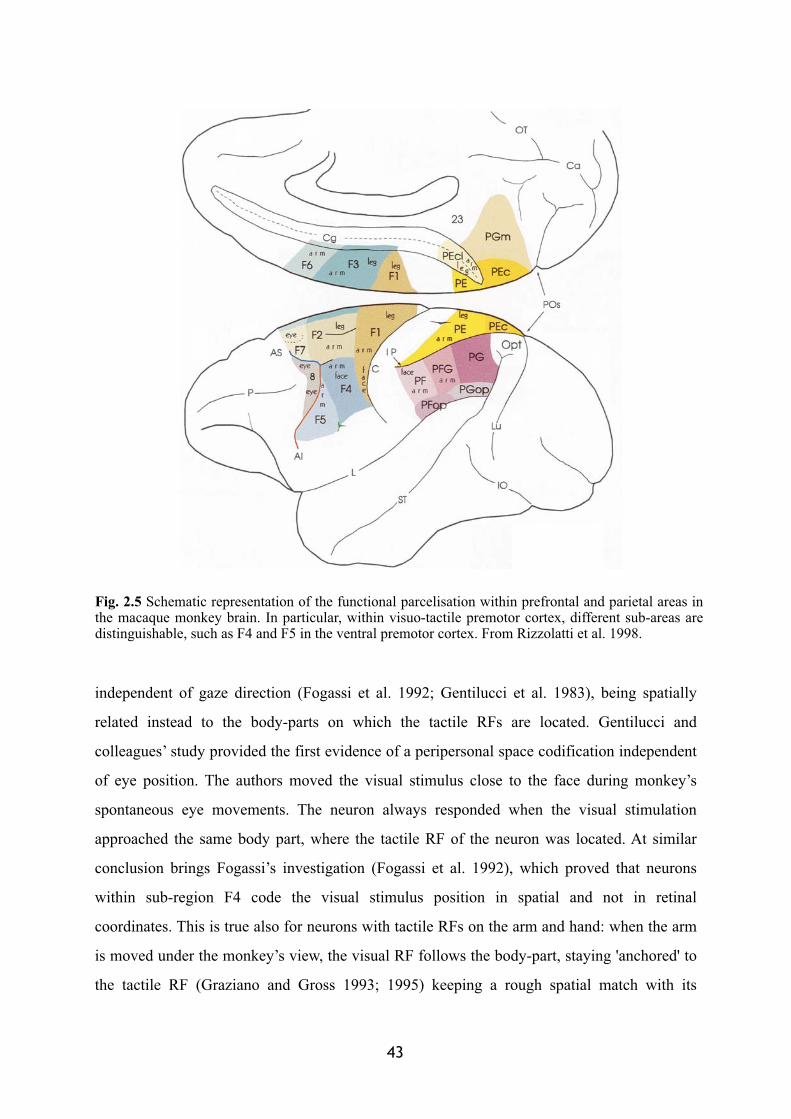
independent of gaze direction (Fogassi et al. 1992; Gentilucci et al. 1983), being spatially
related instead to the body-parts on which the tactile RFs are located. Gentilucci and
colleagues’ study provided the first evidence of a peripersonal space codification independent
of eye position. The authors moved the visual stimulus close to the face during monkey’s
spontaneous eye movements. The neuron always responded when the visual stimulation
approached the same body part, where the tactile RF of the neuron was located. At similar
conclusion brings Fogassi’s investigation (Fogassi et al. 1992), which proved that neurons
within sub-region F4 code the visual stimulus position in spatial and not in retinal
coordinates. This is true also for neurons with tactile RFs on the arm and hand: when the arm
is moved under the monkey’s view, the visual RF follows the body-part, staying 'anchored' to
the tactile RF (Graziano and Gross 1993; 1995) keeping a rough spatial match with its
� 43
Fig. 2.5 Schematic representation of the functional parcelisation within prefrontal and parietal areas in the macaque monkey brain. In particular, within visuo-tactile premotor cortex, different sub-areas are distinguishable, such as F4 and F5 in the ventral premotor cortex. From Rizzolatti et al. 1998.
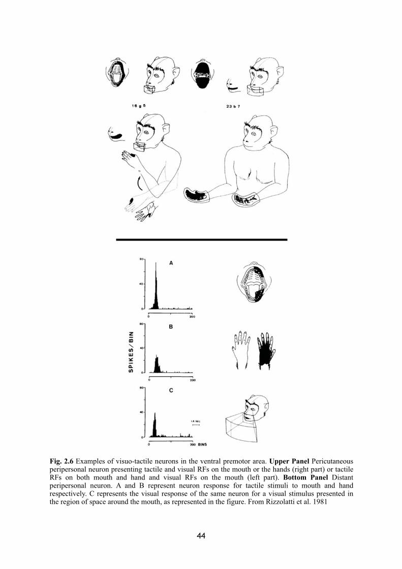
44
Fig. 2.6 Examples of visuo-tactile neurons in the ventral premotor area. Upper Panel Pericutaneousperipersonal neuron presenting tactile and visual RFs on the mouth or the hands (right part) or tactile RFs on both mouth and hand and visual RFs on the mouth (left part). Bottom Panel Distant peripersonal neuron. A and B represent neuron response for tactile stimuli to mouth and hand respectively. C represents the visual response of the same neuron for a visual stimulus presented in the region of space around the mouth, as represented in the figure. From Rizzolatti et al. 1981
location at every displacement (see Figure 2.7; Graziano et al. 1994; Graziano et al. 1997).
Although less numerous, visuo-tactile neurons are present also in the rostral sub-
region F5 of area 6, presenting smaller tactile RFs than F4 neurons, frequently located on the
hand, the face or both. However, the visual properties of these neurons were shown to be quite
different: While a stronger response was induced by stimuli presented within the reaching
distance, visual RFs were usually difficult to define (Rizzolatti et al. 1988; Rizzolatti and
Gentilucci 1988).
45
Fig. 2.7 Visual and tactile RFs of visuo-tactile neurons are in rough spatial register. When the arm is on the right side (A), for example, the visuo-tactile neuron presenting a tactile RF on the arm, also respond for a visual stimulus approaching the arm along the trajectory 3 (dark grey line in the graph C). When the arm moves on the left side (B), the same visuo-tactile neuron now responds better to a visual stimulus approaching the arm along the trajectory 2 (light grey line in the graph C). Modified from Graziano et al. 1997.
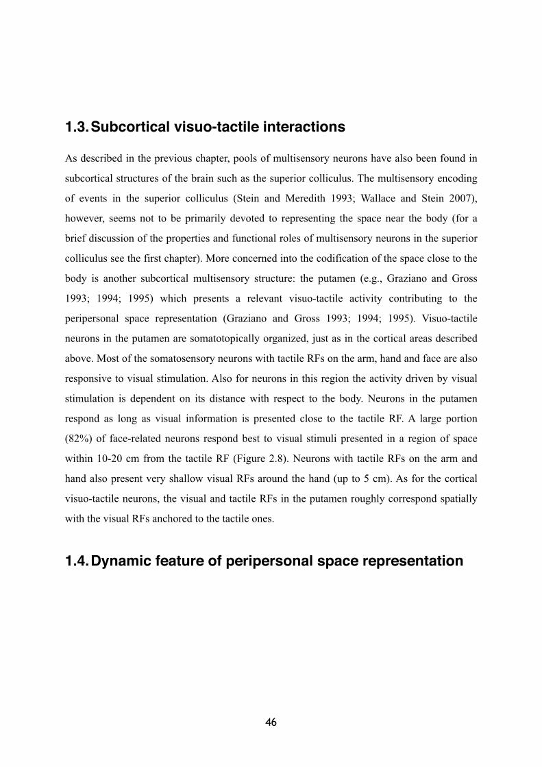
1.3.Subcortical visuo-tactile interactions
As described in the previous chapter, pools of multisensory neurons have also been found in
subcortical structures of the brain such as the superior colliculus. The multisensory encoding
of events in the superior colliculus (Stein and Meredith 1993; Wallace and Stein 2007),
however, seems not to be primarily devoted to representing the space near the body (for a
brief discussion of the properties and functional roles of multisensory neurons in the superior
colliculus see the first chapter). More concerned into the codification of the space close to the
body is another subcortical multisensory structure: the putamen (e.g., Graziano and Gross
1993; 1994; 1995) which presents a relevant visuo-tactile activity contributing to the
peripersonal space representation (Graziano and Gross 1993; 1994; 1995). Visuo-tactile
neurons in the putamen are somatotopically organized, just as in the cortical areas described
above. Most of the somatosensory neurons with tactile RFs on the arm, hand and face are also
responsive to visual stimulation. Also for neurons in this region the activity driven by visual
stimulation is dependent on its distance with respect to the body. Neurons in the putamen
respond as long as visual information is presented close to the tactile RF. A large portion
(82%) of face-related neurons respond best to visual stimuli presented in a region of space
within 10-20 cm from the tactile RF (Figure 2.8). Neurons with tactile RFs on the arm and
hand also present very shallow visual RFs around the hand (up to 5 cm). As for the cortical
visuo-tactile neurons, the visual and tactile RFs in the putamen roughly correspond spatially
with the visual RFs anchored to the tactile ones.
1.4.Dynamic feature of peripersonal space representation
46
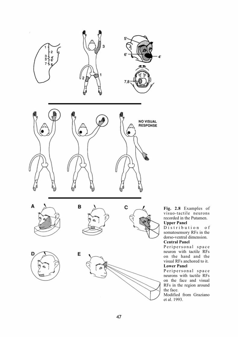
47
Fig. 2.8 Examples of visuo-tactile neurons recorded in the Putamen.Upper Panel D i s t r i b u t i o n o f somatosensory RFs in the dorso-ventral dimension.Central Panel P e r i p e r s o n a l s p a c e neuron with tactile RFs on the hand and the visual RFs anchored to it.Lower PanelP e r i p e r s o n a l s p a c e neurons with tactile RFs on the face and visual RFs in the region around the face.Modified from Graziano et al. 1993.
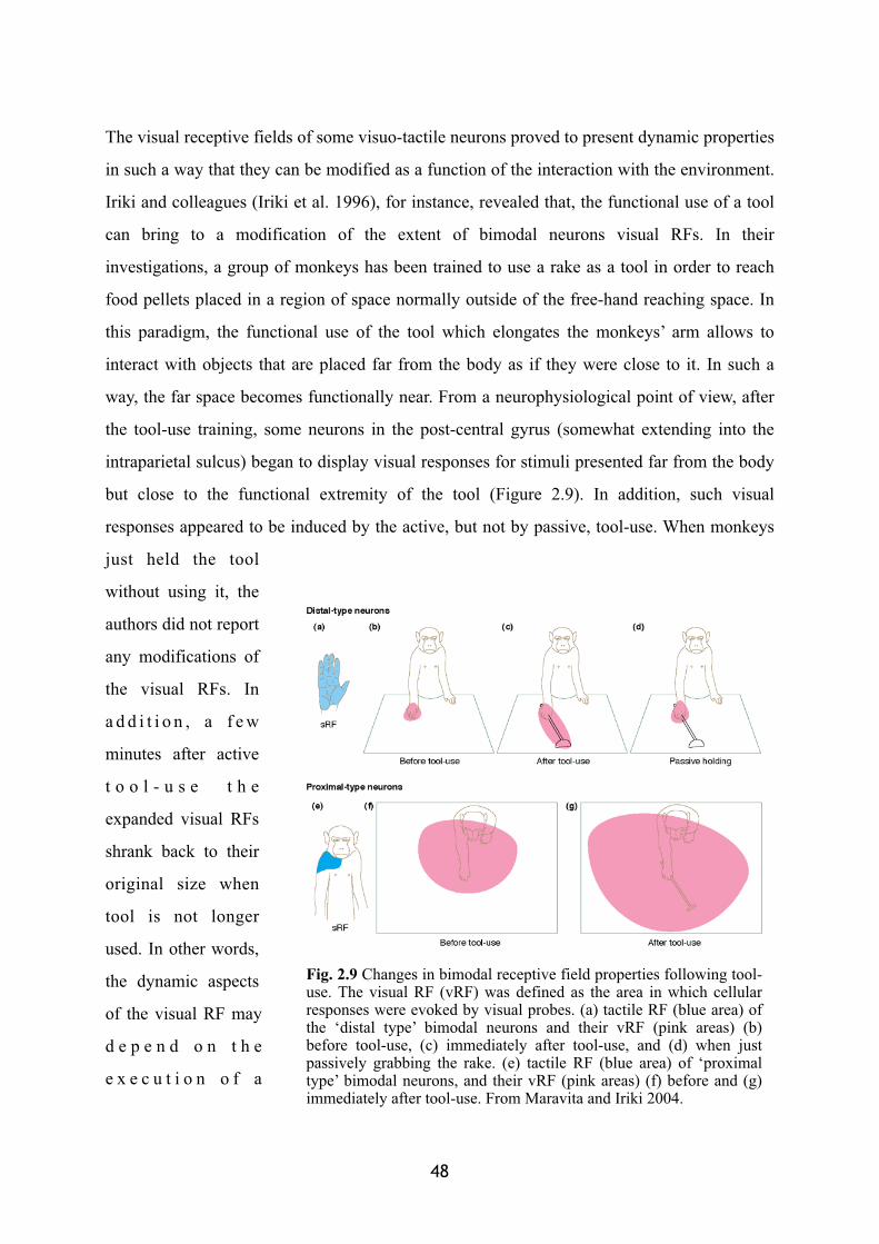
The visual receptive fields of some visuo-tactile neurons proved to present dynamic properties
in such a way that they can be modified as a function of the interaction with the environment.
Iriki and colleagues (Iriki et al. 1996), for instance, revealed that, the functional use of a tool
can bring to a modification of the extent of bimodal neurons visual RFs. In their
investigations, a group of monkeys has been trained to use a rake as a tool in order to reach
food pellets placed in a region of space normally outside of the free-hand reaching space. In
this paradigm, the functional use of the tool which elongates the monkeys’ arm allows to
interact with objects that are placed far from the body as if they were close to it. In such a
way, the far space becomes functionally near. From a neurophysiological point of view, after
the tool-use training, some neurons in the post-central gyrus (somewhat extending into the
intraparietal sulcus) began to display visual responses for stimuli presented far from the body
but close to the functional extremity of the tool (Figure 2.9). In addition, such visual
responses appeared to be induced by the active, but not by passive, tool-use. When monkeys
just held the tool
without using it, the
authors did not report
any modifications of
the visual RFs. In
a d d i t i o n , a f e w
minutes after active
t o o l - u s e t h e
expanded visual RFs
shrank back to their
original size when
tool is not longer
used. In other words,
the dynamic aspects
of the visual RF may
d e p e n d o n t h e
e x e c u t i o n o f a
48
Fig. 2.9 Changes in bimodal receptive field properties following tool-use. The visual RF (vRF) was defined as the area in which cellular responses were evoked by visual probes. (a) tactile RF (blue area) of the ‘distal type’ bimodal neurons and their vRF (pink areas) (b) before tool-use, (c) immediately after tool-use, and (d) when just passively grabbing the rake. (e) tactile RF (blue area) of ‘proximal type’ bimodal neurons, and their vRF (pink areas) (f) before and (g) immediately after tool-use. From Maravita and Iriki 2004.
specific motor action (Rizzolatti et al. 1998). However, as Holmes and colleagues noticed
(Holmes and Spence 2004), the intraparietal sulcus is a large and heterogeneous area in the
monkey, and at least five functionally- and neuroanatomically distinct sub-regions are found
there (Colby and Duhamel, 1991; Rizzolatti et al. 1998), with a variety of neuronal response
properties ranging from purely somatosensory, to purely visual. It is therefore important to
plot visual and somatosensory RFs while controlling for eye, head, and body movements. By
contrast, in Iriki’s study no control was exerted on eye and head movements. Even then, the
influence of attention and response preparation may be serious confounding factors. Neurons
in the area studied by Iriki and colleagues (1996) have been indeed studied elsewhere
(Mountcastle et al. 1975). Two thirds of the cells in this area have been reported to be purely
somatosensory, responding mainly to joint manipulation, and responding more vigorously for
active than for passive movements (Mountcastle et al. 1975).
In a similar vein, Fogassi and colleagues (Fogassi et al. 1996) found that the depth of
the visual RFs of F4 visuo-tactile neurons can increase with increases in the velocity (20-80
cm/s) of a visual stimulus approaching the cutaneous RF. The authors trained the monkey to
fixate a LED while moving three-dimensional moving stimuli were presented in the visual
scene through a robot arm. By changing the position of the visual fixation, it could be possible
to dissociate the retinocentred from the somatocentred codification of the stimulus. Moreover,
through the precise robotic presentation device, these authors could measure the extent in
depth of the visual RFs, as well as their rapid modification as a function of the stimulus’
approaching velocity. Results clearly showed that the extension in depth of F4 visual RFs
revealed to be not fixed. It dynamically changed as a function of the stimulus velocity, in
particular expanding in the direction of the approaching stimulus for higher velocities. This
neurophysiological result could be linked to the behaviour described in humans performing
reaching movements toward moving targets (Chieffi et al. 1992). Although the kinematic of
the arm movement was not affected by the velocity of the target, the movement onset was
modulated of target velocity. The dynamic property of visuo-tactile neurons could thus be
crucial for preparing and/or executing actions towards nearby objects.
� 49
1.5.A visuo-tactile network
The neurophysiological findings described in the previous sections define a set of at least four
distinctive areas with similar visuo-tactile responses: parietal area 7b and VIP, part of inferior
premotor cortex (mainly F4) and the putamen. These regions are heavily interconnected,
forming a tight network (Matelli and Luppino 2001; Rizzolatti et al. 1997; Rizzolatti et al.
1998). Posterior parietal areas send projections to the premotor cortex. In particular, superior
parietal area 5 and the inferior parietal area 7b project respectively to superior and inferior
part of premotor area 6 (Pandya and Vignolo 1971; Strick and Kim 1978; Godschalk et al.
1984; Matelli et al. 1984a and b; Luppino et al. 1999). Reciprocal connections are also sent
back from premotor area 6 to the respective parietal areas (Rizzolatti et al. 1997). This
reciprocal interconnected parieto-frontal network projects also to the putamen (Kunzle 1978;
Weber and Yin 1984; Cavada and Goldman-Rakic 1991; Parthasarathy et al. 1992).
Neurons in this network share several common features:
1) The visual responses lie primarily within a head-face or arm-hand centred somatosensory
representation of the body;
2) Visual stimuli near the monkey drive the cells better than farther stimuli;
3) The visual and tactile receptive fields are in spatial register, independent of gaze since they
relation is up-dated with the movement of the concerned body-part.
This suggests that these neurons allow for body-part centred coding of visual stimuli within
sectors of space adjacent to the tactile surface. This network possesses all of the necessary
properties to bind together external visual information around the body and tactile information
on a specific body part (Fogassi et al. 1992; Graziano and Gross 1993; Rizzolatti et al. 1997).
Indeed, in the somatotopically organised maps encountered in these areas, each
somatosensory body-part representation contains also the representation of the visual space
that closely surrounds this same body-part. For instance, the arm region contains a
representation of visual space around the arms as well as the face region contains a
representation of visual space around the head (particularly in area VIP). This modular
representation allows the brain to compute the spatial relation between an external object and
a specific body-part. Furthermore, the spatial correspondence between visual and tactile RFs
of these cells allows up-dating of the spatial relation between external objects and body-parts
50
whenever the body moves. In conclusion, this bimodal visuo-tactile network provides the
brain with the neurophysiological basis for the representation of the space between the body
and the external world, where each body-part becomes the reference system: the peripersonal
space.
2. Motor features of peripersonal space: Visuo-tactile interactions around the acting body
Why should the brain maintain a representation of the space around the body separate from a
representation of far extrapersonal space? One possibility is that it could serve purely
perceptual aims, giving a greater perceptual relevance to visual events occurring in the
vicinity of the body. Following this idea, the parieto-frontal network together with the
subcortical visuo-tactile maps would code for a visual space, with individual body-parts as the
reference. This is suggested by the most obvious sensory properties of this set of neurons.
However, this interpretation does not describe fully the potential functional aspects of this
system, since it does not correspond with some of the evidence described above. First, a
purely perceptual account does not fit with the presence of such multisensory neurons in a
predominantly 'motor' area such as the premotor cortex. Second, it may be difficult to
interpret the complex tactile RFs of some of these neurons (for instance, single cells
representing both the hand and peribuccal territories, as reported by Rizzolatti et al. 1981a, b).
Similarly, the dynamic changes of their visual RFs observed in case of objects approaching
the body (Fogassi et al. 1996) or after active tool-use (Iriki et al. 1996) point to a more
“active” role of this multisensory network. More critically, a purely perceptual hypothesis
does not take into account the intriguing common point of the visuo-tactile areas described so
far: their motor properties. Numerous visuo-tactile cells in the parietal areas 7b (Hyvärinen
1981; Hyvärinen and Poranen 1974; Hyvärinen and Shelepin 1979; Leinonen 1980; Leinonen
et al. 1979; Leinonen and Nyman 1979; Robinson et al. 1978), inferior area 6 (Gentilucci et
al. 1988; Rizzolatti et al. 1981c; Rizzolatti et al. 1987; Rizzolatti et al. 1988; Rizzolatti and
� 51
Gentilucci 1988; Rizzolatti et al. 1997) and putamen (Crutcher and DeLong 1984) respond
not only passively to visual and tactile stimulation, but also during motor activity.
These findings raise the more compelling possibility that the multisensory
representation of peripersonal space might serve motor functions. Objects in the vicinity of
the body are indeed more relevant by virtue of all the possible interactions the body can
establish with them (Graziano et al. 1993; Rizzolatti et al. 1997, 1998). The hand-centred
representation of peripersonal space provides us with extremely valuable information
regarding the spatial position of objects with respect to our hands.
2.1. Inferior premotor cortex
The motor properties of neurons in inferior premotor cortex appear to be very relevant
concerning the perception-action link characterising the peripersonal space. In particular, the
visual responses of such neurons may be enhanced when a reaching movement is performed
towards an object (Godschalk et al. 1985). Moreover, inferior area 6 neurons are active during
reaching and grasping movements of the arms (Godschalk et al. 1981; Godschalk et al. 1985;
Kurata et al. 1985; Kurata and Tanji 1986; Rizzolatti and Gentilucci 1988), as well as the
mouth (Rizzolatti et al. 1981c). They also show a rather precise degree of motor
representation (Gentilucci et al. 1988; Kurata and Tanji 1986; Murata et al. 1997; Raos et al.
2006; Rizzolatti et al. 1987; Rizzolatti et al. 1988; Rizzolatti and Gentilucci 1988). Proximal
and distal movements are represented separately, in area F4 (Figure 2.10) and medially in the
rostral part of area F1 for the former, and in the periarcuate region F5 for the latter. Distal
neurons discharge in relation to specific and complex motor acts such as “grasping with hand
and mouth” or “grasping with the hand” or simply “holding”. In the case of “grasping”
neurons, the activation occurred only if the approaching movement of the hand (or mouth) to
an object was aiming to grasp the target. Movements with other purposes, even activating
similar pattern of muscles, did not produce any response of the neuron. Even more
noteworthy is the fact that grasping neurons show selectivity for the type of prehension, so
that some neurons are selectively activated during a grasping action performed with a
precision grip (only the thumb and index finger) rather than during a whole hand prehension.
52
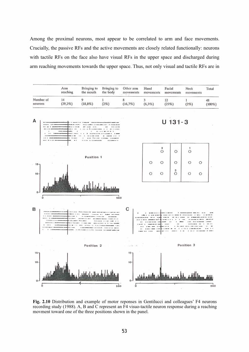
Among the proximal neurons, most appear to be correlated to arm and face movements.
Crucially, the passive RFs and the active movements are closely related functionally: neurons
with tactile RFs on the face also have visual RFs in the upper space and discharged during
arm reaching movements towards the upper space. Thus, not only visual and tactile RFs are in
� 53
Fig. 2.10 Distribution and example of motor reponses in Gentilucci and colleagues’ F4 neurons recording study (1988). A, B and C represent an F4 visuo-tactile neuron response during a reaching movment toward one of the three positions shown in the panel.
spatial register, but the motor activity of these neurons is maximal when the animal’s
movement is directed to reach into the region of space corresponding to its visual RF. This
suggests that the sensory and motor responses are expressed in a common coordinate system
for locating objects in the space close to the body and for guiding a movement toward them.
The premotor cortex, indeed, has both direct (Martino and Strick 1987) and indirect
(Godschalk et al. 1984; Matsumura and Kubota 1979; Muakkassa and Strick 1979; Pandya
and Vignolo 1971) access to the control of upper limbs movements, via projections to the
spinal cord and the primary motor cortex, respectively. That is, visuo-tactile premotor neurons
could potentially modulate motor activity and control down to the final stages of movement.
Indeed, coupled electrical stimulation experiments in the two corresponding hand fields of the
premotor (F5 region) and motor cortex, clearly showed the modulatory effects of premotor
over motor cortex during a grasping action (Cerri et al. 2003; Prabhu et al. 2009; Shimazu et
al. 2004). Depending on the temporal interval between the conditioning electrical stimulation
over the premotor cortex and the target stimulation over the correspondent hand field in the
motor cortex, premotor neurons can both excite or inhibit the primary motor area, producing
effects on distal muscle control.
This complex link between sensory and motor properties shown by the premotor
bimodal system of neurons already points out they can not subserve a mere perceptual
function. Rather, they appear more as a functional system to guide the acting body in the
sensory-motor transformation from perception to action.
2.1.1. Mirror neurons: a particular class of visuo-motor neuron
Inferior area 6, in the F5 sub-region is also characterized by the presence of 'mirror' neurons, a
special class of motor neurons with visual properties (di Pellegrino et al. 1992; Gallese et al.
1996; Rizzolatti et al. 1996)2. Activity of these neurons appears to be correlated to the
54
2 A first report of neurons responding while the monkey was watching an action performed by another individual is already present in an early electrophysiological study over the parietal area 7b (Leinonen 1980, page 305) : « […] two cells discharged when the monkey grasped an object […] or when the monkey saw an investigator grasp an object »
execution of a specific motor act, such as a grasping. Moreover, they discharge also when the
monkey observes someone else executing the same or a similar action. Relevant for this
dissertation is a recent finding which showed how a sub-group of mirror neurons can be
characterised by selectivity for actions performed within the observer’s peripersonal space
(Caggiano et al. 2009). Indeed, a sub-population of mirror neurons appeared to be activated
preferentially by the sight of an action performed in the observer monkey’s peripersonal space
rather than in its extrapersonal space (peripersonal mirror neurons). A different sub-population
of mirror neurons showed the opposite preference. Moreover, peripersonal and extrapersonal
space appeared to be defined according to a functional criterion: Reducing the extent of the
accessible peripersonal space by putting a screen in front of the monkey, reduced also the
responses of several peripersonal mirror neurons during the observation of actions performed
in the inaccessible portion of the peripersonal space. This suggests that the portion of no more
accessible peripersonal space had been re-coded as farther extrapersonal space. This was
supported by the finding that extrapersonal mirror neurons started to respond to the
observation of actions performed in the inaccessible peripersonal space.
2.2.Parietal areas
The more posterior parietal areas also have motor properties and, similarly to the premotor
cortex, parietal motor functions seem to be related to approaching movements of a body-part
toward an object (Debowy et al. 2001; Fogassi and Luppino 2005; Gardner et al. 2002;
Lacquaniti and Caminiti 1998; Rizzolatti et al. 1997; Stepniewska et al. 2005; Ferraina et al.
2009). The posterior parietal cortex, indeed, is part of the dorsal stream of action-oriented
visual processing (Milner and Goodale 1995). Here, as described in the previous sections, the
sensory information converge and start to be linked to the motor activity. Both inferior and
superior parietal lobules are interconnected with premotor cortex.
Gardner’s studies investigated the “anticipatory activation”, as it was defined in the
early studies in the 70‘s, during a monkey’s voluntary grasping movement. Through an
interesting approach, where the authors conjugated low-resolution kinematic and single cell
recordings, they revealed the pattern of activation in posterior parietal areas as a function of
� 55
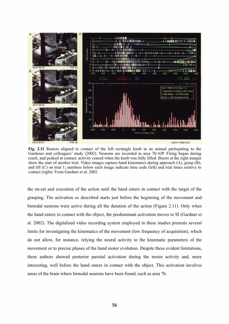
the on-set and execution of the action until the hand enters in contact with the target of the
grasping. The activation so described starts just before the beginning of the movement and
bimodal neurons were active during all the duration of the action (Figure 2.11). Only when
the hand enters in contact with the object, the predominant activation moves to SI (Gardner et
al. 2002). The digitalised video recording system employed in these studies presents several
limits for investigating the kinematics of the movement (low frequency of acquisition), which
do not allow, for instance, relying the neural activity to the kinematic parameters of the
movement or to precise phases of the hand motor evolution. Despite these evident limitations,
these authors showed posterior parietal activation during the motor activity and, more
interesting, well before the hand enters in contact with the object. This activation involves
areas of the brain where bimodal neurons have been found, such as area 7b.
56
Fig. 2.11 Rasters aligned to contact of the left rectangle knob in an animal particpating to the Gardener and colleagues’ study (2002). Neurons are recorded in area 7b/AIP. Firing began during reach, and peaked at contact; activity ceased when the knob was fully lifted. Bursts at the right margin show the start of another trial. Video images capture hand kinematics during approach (A), grasp (B), and lift (C) on trial 1; numbers below each image indicate time code (left) and trial times relative to contact (right). From Gardner et al. 2002.
Similar evidence of parietal bimodal neurons motor properties comes from electrical
micro-stimulation studies. Electrical micro-stimulation of portions of parietal cortex produces
motor activity of the arm/hand or the head/face. In particular, the parietal lobe appears
functionally fractionated in specialised sub-regions as a function of different complex
movements such as a reaching or a retrieving movement of the arm (Cooke et al. 2003;
Stepniewska et al. 2005).
3. Lesion studies
Another possible approach to identify the motor role of parietal and premotor bimodal areas is
to investigate the motor behaviour that might be affected by a dysfunction of one or both of
these areas. Permanent ablation and reversible inactivation studies in monkeys have tested for
the behavioral consequences of a lesion within premotor and posterior parietal areas, where
visuo-tactile neurons have been found.
Interestingly, lesions to the anterior or posterior part of this network seem to produce
very similar patterns of impairments, most of which affect in particular the execution of
visually guided reaching actions (Battaglini et al. 2002; Deuel and Regan 1985; Ettlinger and
Kalsbeck 1962; Faugier-Grimaud et al. 1978; Gallese et al. 1994; Halsban and Passingham
1982; Moll and Kuypers 1977; Rizzolatti et al. 1983). After premotor region ablation, for
instance, the animals were unable to reach around an obstacle with the contralesional arm.
Arm movements were executed without correctly taking into account visual information
within action space (Battaglini et al. 2002; Moll and Kuypers 1977). Similarly, removal of
postarcuate regions where the mouth is represented caused a severe impairment in grasping
with the mouth (Rizzolatti et al. 1983). Attentional deficits have also been reported after
selective damage of visuo-tactile parietal and premotor regions (Rizzolatti et al. 1983) in the
form of spatial hemineglect and extinction. The animals appeared to be unaware of visual (or
tactile) stimuli presented in the contralesional space. Crucially, the deficit was selective for
the space around the body.
� 57
4. Conclusion: A multisensory-motor network for peripersonal space
The above reviewed studies provide a large body of evidence in favour of the proposal that
this parieto-frontal network binds together visual and tactile information, in order to generate
an appropriate motor program, based on the available sensory information. When an object is
visually available in the space around the body, it is also potentially available to the tactile
system, by virtue of the possibility to physically interact with it. The brain can link the visual
information about the object to the tactile input which comes or could come from it and can
use the result of this sensory processing to establish the most correct motor plan as a function
of the desired interaction. According to this model, it is impossible to draw a strict separation
between perception and action, due to both multisensory and motor processing occurring
within these areas. The network should thus be considered of as providing an interface
between perception and action.
Here I would therefore suggest that the multisensory body-part centred representations
are coded to generate the appropriate motor responses towards objects in the world.
58

Chapter III
Multisensory based peripersonal space in humans
Several studies investigated the characteristics of the multisensory perception in humans, as
described in the first chapter. Here, in particular, examples of interactions between visual and
tactile information in humans will be presented. More relevant for this dissertation is the
strong dependence of the visuo-tactile interactions on the distance of visual information from
the body. This peculiarity has been taken as evidence in humans of the existence of a
representation of the space around the body similar to what I described in monkeys in the
previous chapter. In this respect, the study of a neuropsychological condition called
‘extinction’ (Bender 1952) has provided considerable insight into the behavioural
characteristics of multisensory spatial representation in the human brain (Làdavas 2002;
Làdavas and Farnè 2004; Legrand et al. 2007). Evidence for visuo-tactile interactions is also
available in healthy people, in the form of distance-modulated interference exerted by visual
inputs over touch (Spence et al. 2004c, 2008). The crucial point of these studies is the
presence, both in the healthy and brain-damaged populations, of stronger visual-tactile
interactions when visual stimuli are displayed in near, as compared to far space, providing
support for the idea that the human brain also represents peripersonal space through an
integrated multisensory visuo-tactile system. In this section the pathological phenomenon of
extinction will be introduced and more deeply discussed in condition of multisensory
stimulation. Finally, I will review part of the literature supporting the idea of a homologous
representation of peripersonal space in humans.
60
1. Modularity of space through cross-modal extinction
Neuropsychology allows having an insight over brain mechanisms through behavioural
deficits affecting brain damaged patients in their life. The fundamental assumption is that a
circumscribed lesion in the brain can selectively affect the behaviour the damaged area is
responsible for. The patients’ deficient behaviour can thus open the possibility of studying the
modularity of the brain functions.
The deficit of extinction, generally consecutive to a lesion in the right inferior parietal
areas is a window over the processing underlying the representation of the space around the
body through cross-modal perception. Extinction patients are usually defective in reporting an
event presented contralesionally, but only when it is accompanied by an event in the
ipsilesional hemispace. The patients most often present no deficits in reporting a single
stimulation delivered in the contralesional side. The deficit thus arises despite no gross
sensory loss, suggesting that the problem might be more on the construction of spatial
representations rather than on low-level perceptual processes. The deficit can be limited to a
single sensory modality or arise in more than one sensory modality at the same time. Cases
are known of motor extinction, where the concurrent motor activity of both side of the body
results in a disruption of the contralesional side movements.
Extinction has been commonly associated to the more severe deficit of hemineglect,
where after a parietal and/or frontal lesion, patients are no more aware of the contralesional
side. Even though the two syndromes present some common characteristics, their
neurophysiological bases have been shown to be different (Vallar et al. 1994). In particular the
extinction phenomenon appears more related to a competition between spatial representations
which are activated by the simultaneous presence of bilateral sensory inputs1. By virtue of its
spatial nature, cross-modal visuo-tactile extinction has become an interesting phenomenon
through which approaching the study of the modularity of the spatial representations.
� 61� �
1� Extinction could be considered of as the effect of a limit of the physiological perceptual system responding to the presence of several inputs in different sectors of space. Evidence in favour of this possibility is the recent finding of an extinction-like phenomenon in a normal population (Marcel et al. 2004; Farnè et al. 2007a). Following this model, patients would present a constant bias toward the ipsilesional side, mostly due to the unbalanced representation of the two hemispaces caused by the unilateral lesion.

EXTINCTION
Neglect and extinction: Within and between
sensory modalities
Restorative Neurology and Neuroscience(2004)

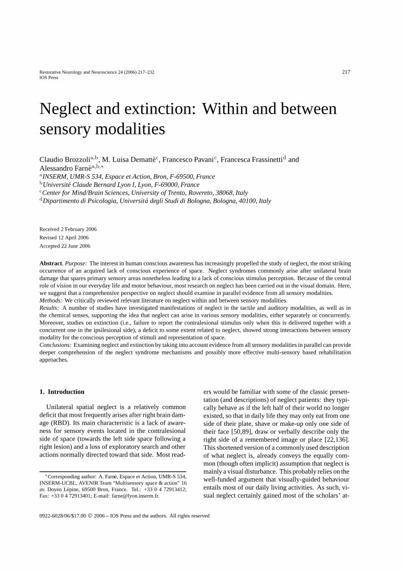
Restorative Neurology and Neuroscience 24 (2006) 217–232 217IOS Press
Neglect and extinction: Within and betweensensory modalities
Claudio Brozzolia,b, M. Luisa Demattec, Francesco Pavanic, Francesca Frassinettid andAlessandro Farnea,b,∗aINSERM, UMR-S 534, Espace et Action, Bron, F-69500, FrancebUniversite Claude Bernard Lyon I, Lyon, F-69000, FrancecCenter for Mind/Brain Sciences, University of Trento, Rovereto, 38068, ItalydDipartimento di Psicologia, Universita degli Studi di Bologna, Bologna, 40100, Italy
Received 2 February 2006
Revised 12 April 2006
Accepted 22 June 2006
Abstract. Purpose: The interest in human conscious awareness has increasingly propelled the study of neglect, the most strikingoccurrence of an acquired lack of conscious experience of space. Neglect syndromes commonly arise after unilateral braindamage that spares primary sensory areas nonetheless leading to a lack of conscious stimulus perception. Because of the centralrole of vision in our everyday life and motor behaviour, most research on neglect has been carried out in the visual domain. Here,we suggest that a comprehensive perspective on neglect should examine in parallel evidence from all sensory modalities.Methods: We critically reviewed relevant literature on neglect within and between sensory modalities.Results: A number of studies have investigated manifestations of neglect in the tactile and auditory modalities, as well as inthe chemical senses, supporting the idea that neglect can arise in various sensory modalities, either separately or concurrently.Moreover, studies on extinction (i.e., failure to report the contralesional stimulus only when this is delivered together with aconcurrent one in the ipsilesional side), a deficit to some extent related to neglect, showed strong interactions between sensorymodality for the conscious perception of stimuli and representation of space.Conclusions: Examining neglect and extinction by taking into account evidence from all sensory modalities in parallel can providedeeper comprehension of the neglect syndrome mechanisms and possibly more effective multi-sensory based rehabilitationapproaches.
1. Introduction
Unilateral spatial neglect is a relatively commondeficit that most frequently arises after right brain dam-age (RBD). Its main characteristic is a lack of aware-ness for sensory events located in the contralesionalside of space (towards the left side space following aright lesion) and a loss of exploratory search and otheractions normally directed toward that side. Most read-
∗Corresponding author: A. Farne, Espace et Action, UMR-S 534,INSERM-UCBL, AVENIR Team “Multisensory space & action” 16av. Doyen Lepine, 69500 Bron, France. Tel.: +33 0 4 72913412;Fax: +33 0 4 72913401; E-mail: [email protected].
ers would be familiar with some of the classic presen-tation (and descriptions) of neglect patients: they typi-cally behave as if the left half of their world no longerexisted, so that in daily life they may only eat from oneside of their plate, shave or make-up only one side oftheir face [50,89], draw or verbally describe only theright side of a remembered image or place [22,136].This shortened version of a commonly used descriptionof what neglect is, already conveys the equally com-mon (though often implicit) assumption that neglect ismainly a visual disturbance. This probably relies on thewell-funded argument that visually-guided behaviourentails most of our daily living activities. As such, vi-sual neglect certainly gained most of the scholars’ at-
0922-6028/06/$17.00 © 2006 – IOS Press and the authors. All rights reserved
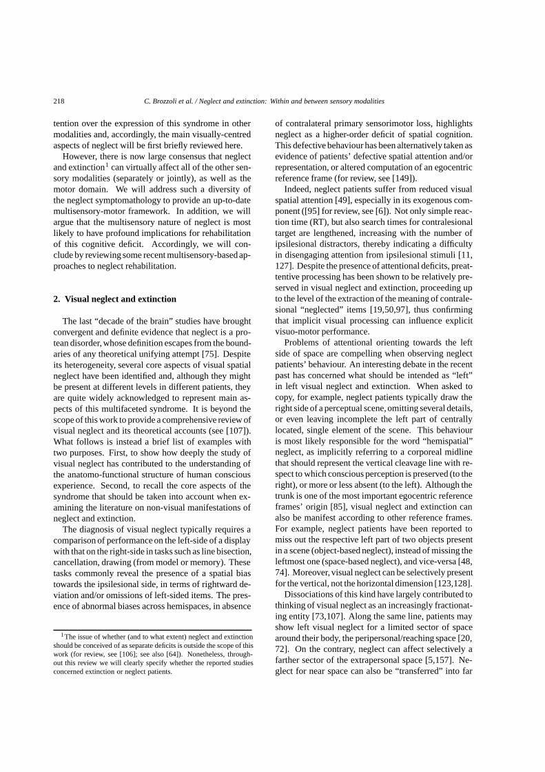
218 C. Brozzoli et al. / Neglect and extinction: Within and between sensory modalities
tention over the expression of this syndrome in othermodalities and, accordingly, the main visually-centredaspects of neglect will be first briefly reviewed here.
However, there is now large consensus that neglectand extinction1 can virtually affect all of the other sen-sory modalities (separately or jointly), as well as themotor domain. We will address such a diversity ofthe neglect symptomathology to provide an up-to-datemultisensory-motor framework. In addition, we willargue that the multisensory nature of neglect is mostlikely to have profound implications for rehabilitationof this cognitive deficit. Accordingly, we will con-clude by reviewing some recent multisensory-based ap-proaches to neglect rehabilitation.
2. Visual neglect and extinction
The last “decade of the brain” studies have broughtconvergent and definite evidence that neglect is a pro-tean disorder, whose definition escapes from the bound-aries of any theoretical unifying attempt [75]. Despiteits heterogeneity, several core aspects of visual spatialneglect have been identified and, although they mightbe present at different levels in different patients, theyare quite widely acknowledged to represent main as-pects of this multifaceted syndrome. It is beyond thescope of this work to provide a comprehensive review ofvisual neglect and its theoretical accounts (see [107]).What follows is instead a brief list of examples withtwo purposes. First, to show how deeply the study ofvisual neglect has contributed to the understanding ofthe anatomo-functional structure of human consciousexperience. Second, to recall the core aspects of thesyndrome that should be taken into account when ex-amining the literature on non-visual manifestations ofneglect and extinction.
The diagnosis of visual neglect typically requires acomparison of performance on the left-side of a displaywith that on the right-side in tasks such as line bisection,cancellation, drawing (from model or memory). Thesetasks commonly reveal the presence of a spatial biastowards the ipsilesional side, in terms of rightward de-viation and/or omissions of left-sided items. The pres-ence of abnormal biases across hemispaces, in absence
1The issue of whether (and to what extent) neglect and extinctionshould be conceived of as separate deficits is outside the scope of thiswork (for review, see [106]; see also [64]). Nonetheless, through-out this review we will clearly specify whether the reported studiesconcerned extinction or neglect patients.
of contralateral primary sensorimotor loss, highlightsneglect as a higher-order deficit of spatial cognition.This defective behaviour has been alternatively taken asevidence of patients’ defective spatial attention and/orrepresentation, or altered computation of an egocentricreference frame (for review, see [149]).
Indeed, neglect patients suffer from reduced visualspatial attention [49], especially in its exogenous com-ponent ([95] for review, see [6]). Not only simple reac-tion time (RT), but also search times for contralesionaltarget are lengthened, increasing with the number ofipsilesional distractors, thereby indicating a difficultyin disengaging attention from ipsilesional stimuli [11,127]. Despite the presence of attentional deficits, preat-tentive processing has been shown to be relatively pre-served in visual neglect and extinction, proceeding upto the level of the extraction of the meaning of contrale-sional “neglected” items [19,50,97], thus confirmingthat implicit visual processing can influence explicitvisuo-motor performance.
Problems of attentional orienting towards the leftside of space are compelling when observing neglectpatients’ behaviour. An interesting debate in the recentpast has concerned what should be intended as “left”in left visual neglect and extinction. When asked tocopy, for example, neglect patients typically draw theright side of a perceptual scene, omitting several details,or even leaving incomplete the left part of centrallylocated, single element of the scene. This behaviouris most likely responsible for the word “hemispatial”neglect, as implicitly referring to a corporeal midlinethat should represent the vertical cleavage line with re-spect to which conscious perception is preserved (to theright), or more or less absent (to the left). Although thetrunk is one of the most important egocentric referenceframes’ origin [85], visual neglect and extinction canalso be manifest according to other reference frames.For example, neglect patients have been reported tomiss out the respective left part of two objects presentin a scene (object-based neglect), instead of missing theleftmost one (space-based neglect), and vice-versa [48,74]. Moreover, visual neglect can be selectively presentfor the vertical, not the horizontal dimension [123,128].
Dissociations of this kind have largely contributed tothinking of visual neglect as an increasingly fractionat-ing entity [73,107]. Along the same line, patients mayshow left visual neglect for a limited sector of spacearound their body, the peripersonal/reaching space [20,72]. On the contrary, neglect can affect selectively afarther sector of the extrapersonal space [5,157]. Ne-glect for near space can also be “transferred” into far
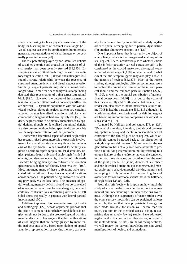
C. Brozzoli et al. / Neglect and extinction: Within and between sensory modalities 219
space when using tools as physical extensions of thebody for bisecting lines of constant visual angle [20].Visual neglect can even be confined to either internallygenerated representations of visual images or percep-tually presented scenes [70].
The role potentially played by non lateralised deficitsof sustained attention and arousal on the genesis of vi-sual neglect has been recently stressed [83,135]. Bystudying sustained attention through the use of an audi-tory target detection test, Hjaltason and colleagues [80]found a strong relationship between the presence ofsustained attention deficits and visual neglect severity.Similarly, neglect patients may show a significantlylonger “dwell time” for a secondary visual target beingdetected after presentation of a first target (attentionalblink [82]). However, the degree of impairment ontasks for sustained attention does not always differenti-ate between RBD patients populations with and withoutvisual neglect, although neglect patients are certainlyaffected by non lateralised attentional deficits whencompared with age-matched healthy subjects [55]. In-deed, neglect seems to be mainly characterised by spa-tial deficits, though non lateralised attentional deficitsare also present, without being specifically responsiblefor the major manifestations of the syndrome.
Another non-lateralised aspect of visual neglect thathas recently raised great interest is the possible involve-ment of a spatial working memory deficit in the gen-esis of the syndrome. When invited to ocularly ex-plore a scene to report targets amidst distractors, ne-glect patients do not only avoid exploring left-sided el-ements, but also produce a high number of rightwardssaccades bringing their eyes to re-fixate items on theiripsilesional side that had already been “visited” [100].Most important, many of these re-fixations were asso-ciated with a failure to keep track of spatial locationsacross saccades, the patients being unaware of revisit-ing previously visited locations. The presence of spa-tial working memory deficits should not be conceivedof as an alternative account for visual neglect, but couldcertainly contribute to exacerbating omission of leftsided items, especially in patients with lateral parietalinvolvement [100].
A different approach has been undertaken by Pisellaand Mattingley [122], whose arguments propose thatthe origin of some re-visiting behavioural deficits in ne-glect might not be due to the proposed spatial workingmemory disorder. They suggest that the manifestationsof visual neglect that are hardly grasped by more tra-ditional accounts solely based upon deficits of spatialattention, representation, or working memory can actu-
ally be accounted for by an additional underlying dis-order of spatial remapping due to parietal dysfunction(for another alternative account, see [139]).
One important issue that is currently the object ofa very lively debate is the fine-grained anatomy of vi-sual neglect. There is controversy as to whether lesionsof the inferior posterior parietal cortex are still to beconsidered as the crucial anatomo-pathological coun-terpart of visual neglect [150], or whether and to whatextent the mid-temporal gyrus may also play a role inthe genesis of neglect [86,137]. Most of the recentstudies, although employing different techniques, seemto confirm the crucial involvement of the inferior pari-etal lobule and the temporo-parietal junction [27,55,75,109], as well as the crucial contribution of parieto-frontal connections [44,46]. It is out of the scope ofthis review to fully address this topic, but the interestedreader can also refer to neurointerference studies us-ing TMS in healthy participants [58,111], although it isworth noting that the criteria used for defining neglectare becoming important for comparing anatomical le-sions studies [107].
As noted by Halligan and colleagues [75, p. 125],“Deficits of attention, intention, global-local process-ing, spatial memory and mental representation can allcontribute to the clinical picture of neglect, which ac-cordingly cannot be traced back to the disruption ofa single supramodal process.” More recently, the ne-glect literature has actually seen some attempts to pro-vide a re-unifying interpretation, not by referring to aunique feature of the syndrome, as was the tendencyin the past three decades, but by advocating the needof the joint presence of (some) deficits of lateralisedand non-lateralised attention, eye movement, and man-ual exploratory behaviour, spatial working memory andremapping to fully account for the puzzling lack ofawareness for contralesional events that is the hallmarkof neglect (see [35,83,122]).
From this brief review, it is apparent how much thestudy of visual neglect has contributed to the refine-ment of our understanding of human conscious aware-ness. Although this supremacy of vision studies overthe other sensory modalities can be explained, at leastin part, by the fact that the appropriate technology hasbeen made available for vision well before than fortouch, audition or the chemical senses, it is quite sur-prising that relatively few(er) studies have addressedneglect and extinction in the other senses, or even inthe motor domain [77,102]. In the following sections,we will review the current knowledge for non-visualmanifestations of neglect and extinctions.
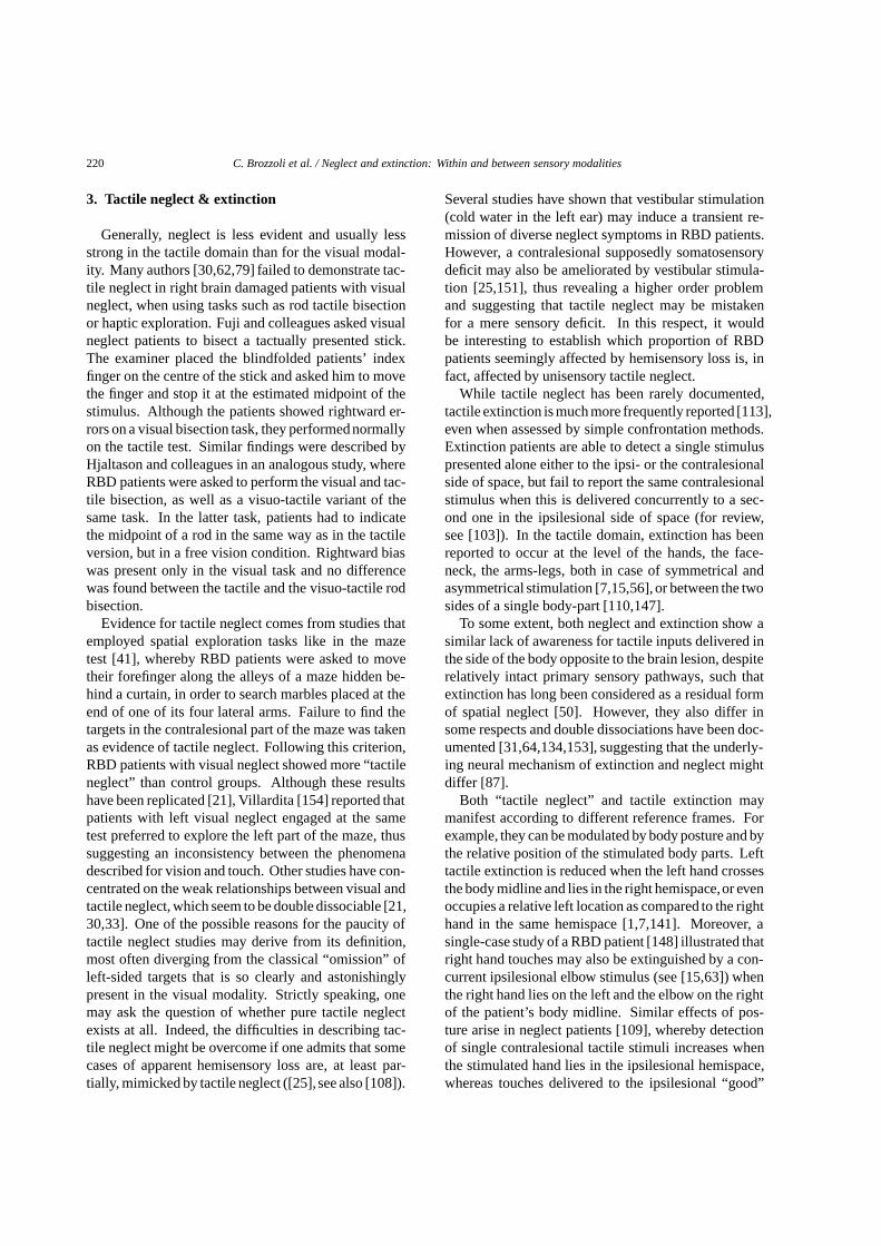
220 C. Brozzoli et al. / Neglect and extinction: Within and between sensory modalities
3. Tactile neglect & extinction
Generally, neglect is less evident and usually lessstrong in the tactile domain than for the visual modal-ity. Many authors [30,62,79] failed to demonstrate tac-tile neglect in right brain damaged patients with visualneglect, when using tasks such as rod tactile bisectionor haptic exploration. Fuji and colleagues asked visualneglect patients to bisect a tactually presented stick.The examiner placed the blindfolded patients’ indexfinger on the centre of the stick and asked him to movethe finger and stop it at the estimated midpoint of thestimulus. Although the patients showed rightward er-rors on a visual bisection task, they performed normallyon the tactile test. Similar findings were described byHjaltason and colleagues in an analogous study, whereRBD patients were asked to perform the visual and tac-tile bisection, as well as a visuo-tactile variant of thesame task. In the latter task, patients had to indicatethe midpoint of a rod in the same way as in the tactileversion, but in a free vision condition. Rightward biaswas present only in the visual task and no differencewas found between the tactile and the visuo-tactile rodbisection.
Evidence for tactile neglect comes from studies thatemployed spatial exploration tasks like in the mazetest [41], whereby RBD patients were asked to movetheir forefinger along the alleys of a maze hidden be-hind a curtain, in order to search marbles placed at theend of one of its four lateral arms. Failure to find thetargets in the contralesional part of the maze was takenas evidence of tactile neglect. Following this criterion,RBD patients with visual neglect showed more “tactileneglect” than control groups. Although these resultshave been replicated [21], Villardita [154] reported thatpatients with left visual neglect engaged at the sametest preferred to explore the left part of the maze, thussuggesting an inconsistency between the phenomenadescribed for vision and touch. Other studies have con-centrated on the weak relationships between visual andtactile neglect, which seem to be double dissociable [21,30,33]. One of the possible reasons for the paucity oftactile neglect studies may derive from its definition,most often diverging from the classical “omission” ofleft-sided targets that is so clearly and astonishinglypresent in the visual modality. Strictly speaking, onemay ask the question of whether pure tactile neglectexists at all. Indeed, the difficulties in describing tac-tile neglect might be overcome if one admits that somecases of apparent hemisensory loss are, at least par-tially, mimicked by tactile neglect ([25], see also [108]).
Several studies have shown that vestibular stimulation(cold water in the left ear) may induce a transient re-mission of diverse neglect symptoms in RBD patients.However, a contralesional supposedly somatosensorydeficit may also be ameliorated by vestibular stimula-tion [25,151], thus revealing a higher order problemand suggesting that tactile neglect may be mistakenfor a mere sensory deficit. In this respect, it wouldbe interesting to establish which proportion of RBDpatients seemingly affected by hemisensory loss is, infact, affected by unisensory tactile neglect.
While tactile neglect has been rarely documented,tactile extinction is much more frequently reported [113],even when assessed by simple confrontation methods.Extinction patients are able to detect a single stimuluspresented alone either to the ipsi- or the contralesionalside of space, but fail to report the same contralesionalstimulus when this is delivered concurrently to a sec-ond one in the ipsilesional side of space (for review,see [103]). In the tactile domain, extinction has beenreported to occur at the level of the hands, the face-neck, the arms-legs, both in case of symmetrical andasymmetrical stimulation [7,15,56], or between the twosides of a single body-part [110,147].
To some extent, both neglect and extinction show asimilar lack of awareness for tactile inputs delivered inthe side of the body opposite to the brain lesion, despiterelatively intact primary sensory pathways, such thatextinction has long been considered as a residual formof spatial neglect [50]. However, they also differ insome respects and double dissociations have been doc-umented [31,64,134,153], suggesting that the underly-ing neural mechanism of extinction and neglect mightdiffer [87].
Both “tactile neglect” and tactile extinction maymanifest according to different reference frames. Forexample, they can be modulated by body posture and bythe relative position of the stimulated body parts. Lefttactile extinction is reduced when the left hand crossesthe body midline and lies in the right hemispace,or evenoccupies a relative left location as compared to the righthand in the same hemispace [1,7,141]. Moreover, asingle-case study of a RBD patient [148] illustrated thatright hand touches may also be extinguished by a con-current ipsilesional elbow stimulus (see [15,63]) whenthe right hand lies on the left and the elbow on the rightof the patient’s body midline. Similar effects of pos-ture arise in neglect patients [109], whereby detectionof single contralesional tactile stimuli increases whenthe stimulated hand lies in the ipsilesional hemispace,whereas touches delivered to the ipsilesional “good”
C. Brozzoli et al. / Neglect and extinction: Within and between sensory modalities 221
hand are omitted to a variable degree when the righthand lies in the contralesional affected hemispace [1,7]. Similar to what has been reported for visual ne-glect [124], improvements in tactile neglect and extinc-tion have been observed following a reduction of grav-itational inputs, obtained by placing the subjects in asupine position [120].
Moreover, in the visual domain it is typically as-sumed that extinction is maximal under conditions inwhich competing stimuli occur simultaneously [95].One may ask whether, in addition to posture, time (e.g.,asynchronous stimulation) could also modulate tactileextinction in a similar fashion. The more a stimulus istemporally tied apart from another, the more reliablycontralesional events should be perceived [26]. In thisrespect, Mattingley et al. [103] examined tactile de-tection in a RBD patient with tactile extinction, intro-ducing a variable stimulus onset asynchrony (SOA) be-tween bilateral stimulations. The occurrence of a right-sided competitor interfered with detection of left targetsacross a range of asynchrony from −400 to 1200 ms(minus means that left leads), showing an asymmetry inthe effect. The point of subjective simultaneity appearsthus to be biased in favour of ipsilesional stimuli. Theseresults suggest that sensory timing problems might bepresent in both visual and somatosensory neglect [10].
In the case of tactile extinction considerable process-ing can still take place prior to the level at which lossof awareness arises. Although the extinguished tactilestimulus does not access consciousness, it may inter-fere with perception of the ipsilesional one [2]. Moredirect evidence comes from measures of patients’ neu-ral activity through functional imaging or event-relatedpotentials. Some studies in the visual domain observedthat the relatively early components of visual process-ing may be abnormal for contralesional stimuli in visualextinction [101]. Similarly, it is possible to examinethe fate of extinguished tactile stimuli in those bilat-eral conditions where extinction arises, by comparingcorrect unilateral ERPs with incorrect ones in case ofextinction [52]. In a single RBD patient study, bilateraltrials with extinction still revealed residual early com-ponents (P60 & N110) over the right hemisphere in re-sponse to the extinguished left touches. These compo-nents were completely absent in the right hemisphereafter a single right hand stimulation, although thesekind of stimuli have the same conscious report of theother ones. However, the somatosensory neural activityin the right hemisphere was reduced in amplitude whencompared to the one elicited by right hand stimulationon the left hemisphere. This suggests that, although
tactile extinction is not a pure sensory deficit and is de-fined in conditions of bilateral stimulation, there maybe an underlying pathology for the contralesional uni-lateral stimulation too, in agreement to what has beensuggested for visual extinction [101]. Finally, theseresults demonstrate that somatosensory cortex activityis not sufficient for tactile inputs to reach awareness.In the same vein, a PET study [129] revealed that tac-tile extinction is associated with reduced activity in thesecondary somatosensory cortex, but not in the primaryone, suggesting that processing of bilateral tactile stim-uli takes place at a “higher” stage and that extinctionarises at a high level of tactile input processing.
4. Chemical neglect & extinction
To date, only a limited number of investigationsconcerning the suppression of (or competition among)spatial information processed through the so-called‘chemical senses’ (i.e., olfaction and taste) have beenreported [12,13,16,18,105]. A number of variousdifferent reasons may account for this lack of re-search. First, the distinction between pure chemicalversus somatosensory information is often problematic(e.g. [18]). Second, it is widely assumed that olfactionand taste are senses that are not specialized for con-veying spatial information (e.g. [92]). In olfaction, inparticular, it is still unclear whether humans can local-ize at all the source of the olfactory stimulation by dis-tinguishing between odours that are processed throughthe right versus the left nostril. This is particularlytrue when the stimulus is a pure odorant rather thantrigeminal, that is when the odour does not cause anysomatosensory stimulation that is known to be encodedby the trigeminal system (see [47]).
4.1. Olfaction
With respect to olfaction, Mesulam [105] first de-scribed a case of left-sided olfactory extinction revealedunder double simultaneous stimulation of both nos-trils in a patient with a brain lesion localized in theright parietal cortex. A few years later, Bellas andcolleagues [12,13] assessed the ability of a group offifteen RBD patients who were affected by left tactileextinction on the hand to identify and localize a seriesof bilaterally presented olfactory stimuli. On each trial,patients were presented with two stimuli (one in eachnostril) using squeezing bottles and their task was toname each of the odours that were perceived. Partic-
222 C. Brozzoli et al. / Neglect and extinction: Within and between sensory modalities
ipants could receive either the same pure odorant inboth nostrils, or different odorants in each nostril, orelse an odorant in one nostril and a trigeminal odour(vinegar) in the other. As the authors could not find an-other appropriate trigeminal odour, the vinegar odourwas only presented singularly to one of the two nos-trils while the other nostril was stimulated with a pureodorant. Bellas and colleagues [12] reported the pres-ence of an extinction-like phenomenon in the patients’performance. Specifically, when two different stimuli(being either two pure odorants or an odorant and thetrigeminal odour) were delivered to each nostril, RBDpatients consistently failed to report the stimulus deliv-ered to the left nostril. As the olfactory system wouldpredominantly project its fibres ipsilaterally while thetrigeminal system would be a contralaterally innervatedsystem, the authors considered these results as evi-dence supporting the representational theory of neglect(see [22]). Indeed, if the sensory theory (see [37]) wasresponsible for the olfactory and trigeminal extinction,the pure odorants presented to the right nostril ratherthan those presented to the left nostril should have beenextinguished. Bellas et al. [13] also reported that thepatients affected by olfactory extinction showed a largenumber of displacements in that the correctly-identifiedstimuli presented to the left nostril were described asbeing in the right nostril.
The studies conducted by Bellas and colleagues rep-resent a first step in the investigation of phenomenasuch as extinction and neglect in the olfactory modality.Nevertheless, it is not completely possible to determinethe exact influence exerted by the nasal somatosensa-tion in the olfactory extinction reported, since one ofthe odours considered as being pure odorants was laterfound to be processed probably also by the trigeminalsystem (i.e., a soap odour; see [13]). Finally, the pos-sibility of highlighting deficits related to the localiza-tion of stimuli in the olfactory sensory modality shouldbe interpreted within a much wider debate regardingwhether the olfactory system could extract spatial in-formation from pure odorants (i.e., without any inter-ventions of the trigeminal system; e.g., see [47,92];though see [126,155]).
Kobal and colleagues [92], for instance, claimed thatthe human olfactory system appears to be able to local-ize the source of the olfactory stimulation only whenthe odour elicits also a trigeminal response. This wouldappear to be in contradiction with the pioneering workof von Bekesy [155] who showed that trained partici-pants localized both trigeminal stimuli and pure odor-ants between the two nostrils. Moreover, Porter et
al. [126] showed recently that naive participants wereable to reliably localize pure odorants between the twonostrils in a setting in which olfactory stimuli weredelivered by a computer-controlled air-dilution olfac-tometer that controlled for the exact timing in stimulipresentation. The stimuli were presented to the nosethrough a compartmentalized nasal mask that allowedfor mono-rhinal odour presentation and the sniff flow-rate was controlled in real-time. Clearly, if the abilityof the olfactory system to extract spatial informationfrom non-trigeminal stimuli turns out to be true, newlight could be shed on the extinction phenomena de-scribed for odours. In fact, the relative contributionof pure odorant and somatonsensory information to ol-factory localization could be disentangled using exper-imental methods similar to those described by Porterand colleagues.
4.2. Taste
The existence of neglect and/or extinction in taste hasbeen even less explored than in olfaction, even thoughin humans the ability to localize taste stimuli presentedon the tongue has been previously described (e.g. [140,156]). Bender and Feldman [17] first reported a sin-gle case of a patient with a wide parietal-occipital tu-mor and tactile extinction on the upper limbs who alsoshowed extinction of taste sensations on the left part ofthe tongue when two tastes were presented simultane-ously on each hemi-tongue. Taste stimuli were appliedon the tongue surface by means of cotton buds and theywere all accurately identified and localized by the pa-tient when presented singularly. The results of the as-sessment revealed that the patient was not only affectedby unimodal taste extinction, but that he also displacedtaste sensations under crossmodal taste-tactile stimula-tion. In particular, when a touch or a pinprick was de-livered to the right hemi-tongue and a taste was appliedon the left hemi-tongue, the patient repeatedly reportedbilateral taste stimulation, thus surprisingly extinguish-ing the right touch and partially misplacing the left tastestimulus. Unfortunately, Bender and Feldman did notdescribe in detail the method that was used to generatethe tactile sensations.
More recently, Berlucchi and colleagues [18] de-scribed a study carried on two groups of patients (i.e.,having a right or left brain lesion) and a control group.The RBD patients were affected by tactile (on thehands), visual, and/or auditory extinction with differentdegrees. By using a highly controlled stimulus presen-tation (e.g., use of micro-pipettes and controlled wa-
C. Brozzoli et al. / Neglect and extinction: Within and between sensory modalities 223
ter temperature), Berlucchi and colleagues could disen-tangle (contrary to [17]) between the presence of tasteand/or tactile extinction on the tongue. A sub-groupof the RBD patients showed tactile extinction under bi-lateral simultaneous stimulation of the tongue. How-ever, the authors failed to find any significant presenceof taste extinction even among these tactile extinguish-ing patients, thus highlighting for the first time the ex-istence of dissociations between extinction phenom-ena occurring in somatonsensory or in purely chemi-cal information processing. There also appeared not tobe any consistent correlations between the presence oftactile extinction on the tongue and that of tactile (onthe hands), visual, and auditory extinction. Berlucchiand colleagues suggested that a distributed taste repre-sentation could account for the existence of such dis-sociation. Namely, the processing of somatosensoryinformation coming from the tongue would be pre-dominantly contralateral, whereas the taste stimulationwould activate the brain areas ipsilateral to the hemi-tongue being stimulated (e.g., see [3,114]). Moreover,according to Berlucchi and colleagues, the fact that adissociation between gustatory and tactile extinctioncould be highlighted would suggest that gustatory ex-tinction occurs consequently to a severe tactile extinc-tion (see [17]). The patients involved in their studywould have been affected by a mild tactile extinction,thus allowing the gustatory information to be processedby the preserved left hemisphere.
To date, thus, there is still no clear evidence of the ex-istence of purely taste extinction and/or neglect, whilefew studies provided evidence about the presence oftactile extinction on the tongue or inside the mouthfollowing a right brain lesion [4,18]. In the study ofextinction and neglect, a wide number of questions re-lated to the chemical senses are still waiting for an-swers. Today, it would appear to be possible to devisestudies where information conveyed by the chemosen-sory modalities and by the collateral somatosensorymodality could finally be investigated separately [18,126,140]. Therefore, future research will be in chargeof furthering our understanding about odours and tastesand their links with spatial representations.
5. Auditory neglect & extinction
Patients with focal brain lesions can also suffer anumber of disturbances in the auditory modality thatcan be characterised as auditory manifestations of theneglect syndrome. Patients with right hemispheric le-
sions might either fail to respond when addressed ver-bally from the left, or more commonly behave as if theyheard the voice originating from their right (e.g. [16,38]). This suggested a deficit in detection and local-isation of auditory stimuli, especially when they orig-inate in contralesional space, which could emerge forhearing as well as for vision. Although this clinicalobservation has generally been confirmed, a number ofrecent evidence has now highlighted important differ-ences between the manifestations of neglect in hearingand vision.
5.1. Deficits of sound localisation
The disturbance for sound localisation, originally de-scribed as ‘alloacusis’ [16], has been the topic of severalexperimental works in the last two decades (see [118]for review). A first aspect that emerged from thesesystematic investigations is that auditory spatial distur-bances in neglect patients might reflect increased spa-tial uncertainty for sound position, especially for con-tralesional stimuli, instead of a strictly systematic shiftin heard azimuth towards the ipsilesional side. For in-stance, when asked to discriminate verbally the rela-tive position (same vs. different) of two sounds in closesuccession, neglect patients typically perform worsefor pairs of sounds originating from the contralesionalside (e.g. [117,146]; see also [40] for evidence of re-duced mismatch negativity response in scalp recordingsof event-related potentials for contralesional vs. ipsile-sional free-field sounds). In addition, it has recentlybeen shown that patients with neglect perform less ef-ficiently than control right-hemisphere patients with-out neglect in a discrimination task that concerns thevertical position free-field sounds [116,119]. Thus, adisturbance in auditory space perception emerges evenwhen localisation involved the vertical dimension, or-thogonal to any potential horizontal shift.
Horizontal bias in sound localisation have insteadbeen typically documented when neglect patients areasked to point to a sound presented in free-field (i.e.,from an external source [115,121]; but see [138]), orover headphones (pointing to a location on their head;e.g. [23]). In addition, deficits have been observedwhen using ‘auditory midline’ tasks, in which patientsadjust a continuous sound (or make judgments on adiscrete sound) to locate it relatively to the centre ofthe head or body midline (e.g. [23,90,146,152]; butsee [34]). For sounds presented over headphones (witheither varied intensity at the two ears, or varied interau-ral timing cues to sound localization), neglect patients
224 C. Brozzoli et al. / Neglect and extinction: Within and between sensory modalities
typically report a sound to be central when it is actuallylateralized towards the left (i.e. more intense or arrivingearlier at the left ear), as if there were a rightward shiftin perceived location (e.g. [23,146]). By contrast, forsounds presented free-field, neglect patients often re-ported that an external sound seemed aligned with theirhead/body midline when it was actually presented to theright (thus implying a leftward shift in sound localiza-tion if one assumes that perceived head/body midline isveridical, which it might not be in neglect patients [90,152]). As proposed recently [115], some of these dis-crepancies concerning the direction of lateral shifts insound localization for neglect patients might actuallyrelate to non-auditory aspects of the task. Specifically,motor or visuo-motor biases in pointing tasks [115], orpathological distortions of perceived head/body mid-line in auditory-midline tasks [57] could in principleaffect performance.
5.2. Detection and identification deficits
Although auditory spatial deficits have often beenreported in neglect patients for single auditory stimuli,especially when they originate in contralesional space,the patients usually detect these single sounds with ap-parent ease in most localization studies (e.g. [23,115–119,121,146]. This might appear to contrast with char-acteristic clinical deficits affecting the visual modalityin neglect patients, where complete failures to detect orrespond to contralesional visual events are commonlynoted, rather than merely failures in localization. Twocritical differences between hearing and vision may ac-count for this discrepancy. First, the anatomical orga-nization of the auditory system, which is less crossedthan for other senses, with some ipsilateral as wellas major contralateral cortical projections of the inputreaching each ear. Second, the typical reduced com-plexity of the auditory environment in experimental se-tups. Unlike experiments in vision, in which targetsare often embedded among many distracters, the typi-cal experiment in the auditory domain presents a singlestrong sound against silence [115]. Indeed, when evena minimal version of concurrent competing stimulationis produced, usually by presenting one sound on eachside of the head, a consistent failure to detect and/oridentify contralesional sounds emerged, for both free-field sounds [39,142] and headphone stimuli [14,42].Strictly speaking, however, such effects with two con-current competing sounds might be considered the au-ditory equivalent of visual or tactile extinction, ratherthan manifestations of neglect.
A long standing debate in relation to detection and/oridentification deficits under double simultaneous au-ditory stimulation has been whether poor detection ofsounds at the contralesional ear could be related to ne-glect of contralesional auditory space [81], or insteadshould be ascribed solely to poor processing (or sup-pression) of the auditory information entering the con-tralesional ear [8,9]. Indeed, free-field sounds pre-sented from a contralesional location will tend to bemore intense at the contralesional ear, and if presentedmonaurally over headphones, will only reach that ear.However, there is now mounting evidence suggestinga role for higher-level spatial factors (e.g., perceivedexternal position, spatial attention, relation to visualneglect) in the contralesional detection/identificationdeficits for auditory stimuli observed for neglect pa-tients (e.g. [14,29,142]). For instance, it has beenshown that identification of left free-field sounds cansometimes improve in the presence of a fictitious visi-ble sound source (a ‘dummy’ loudspeaker) on the right,which reportedly made it seem that the sounds orig-inated from the right side [142]. In addition, a di-rect investigation of the role of apparent sound locationwith respect to which ear the information enters wasrecently conducted by Bellman and colleagues [14],presenting each auditory stimulus (heard words) eitherto one ear only (‘dichotic’ stimulation), or binaurallybut with interaural time difference serving as the onlylateralization cue (‘diotic’ stimulation). Under doublesimultaneous presentation, two out of four neglect pa-tients tested in the study showed poorer performancefor left than right words only with dichotic presentation(consistent with a deficit for sounds entering the con-tralesional ear), whereas the other two patients wereimpaired in reporting left words for both methods oflateralized presentation (consistent with an identifica-tion deficit for sounds perceived as originating fromcontralesional space).
5.3. Non spatial auditory deficits
A final aspect that merits attention is the descriptionof non-spatially-lateralized auditory deficits in patientswith visual neglect [34,80,135]. Robertson and col-leagues, for instance, documented a non-spatial diffi-culty in sustaining attention and maintaining arousal inthe auditory modality, in a task where neglect patientswere required to count the number of occurrences ofa particular auditory target among a stream of sounds,of variable length, all presented centrally. Non-spatialauditory deficits have also emerged when patients with
C. Brozzoli et al. / Neglect and extinction: Within and between sensory modalities 225
visual neglect were asked to listen to a short rapid se-quence of auditory stimuli over headphones, to detectwhich of the stimuli had a higher pitch [34]. Despiteauditory stimuli were always presented centrally, andpatients were able to detect subtle pitch modulation forsingle auditory objects, they were severely impairedat any comparison between two sounds in a rapid se-quence, possibly as a result of pathologically limitedattentional capacity.
6. Multisensory neglect and extinction
The previous sections on non-visual manifestationsof neglect and extinction clearly showed that in manycircumstances neglect and extinction can emerge for asingle sensory modality, or for multiple sensory modal-ities in a given patient [43,153]. Note however that allthe works reviewed so far were concerned with stimu-lation delivered within a single sensory modality at atime. We now turn to examine how neglect and extinc-tion affecting a unimodal sensory system can be influ-enced (enhanced or degraded further) by the concurrentactivation of another modality.
A number of evidence has now systematically shownthat extinction in particular can emerge even whenconcurrent stimuli are presented in different sensorymodalities, i.e., different sensory inputs delivered to theipsi- and contra-lateral side of the patient’s body [17,45]. Tactile extinction, for example, can be modulatedby visual events simultaneously presented in the spaceregion near the tactile stimulation, increasing or reduc-ing tactile perception, depending upon the spatial ar-rangement of the stimuli. In particular, the visual stim-ulation in the ipsilesional side exacerbates contrale-sional tactile extinction, whereby the presentation ofvisual and tactile stimuli on the same contralesionalside can reduce the deficit [96]. Moreover, the mod-ulation described is most consistently manifest whenvisual-tactile interaction occurs in the space close to thebody than when the space far from the body is visuallystimulated.
In a similar way, visual and tactile information areintegrated in other peripersonal space regions, such asaround the face [56,99]. In this case, extinction patientswere presented with unilateral and bilateral tactile stim-ulation on both cheeks and, in addition, visual stimuliwere concurrently presented in the contralesional or ip-silesional side. As for the hand, exacerbation of thedeficit was found in the ipsilesional visual condition,whereby the visual stimulus enhanced tactile detection
when delivered in the contralesional side. The modula-tion, again, is more evident when the visual stimulus ispresented in a near-body region of space rather than ina farther region, thus implying that sensory integrationarising from the same near-the-body location allows forthe tactile input to reach awareness.
Similar modulations of tactile extinction have beenreported following another kind of multisensory inter-action, between audition and touch [98]. When soundsare concurrently presented with single touches deliv-ered at the level of the neck in tactile extinction patients,their contralesional tactile detection is most likely to behampered by proximal, as compared to far loudspeak-ers. Interestingly, such a multisensory effect observedin the front space with respect to the patients’ headwas even stronger when cross-modal auditory-tactileextinction was assessed in the patients’ back space,thus suggesting that different degrees of multisensoryintegration may occur depending upon the functionalrelevance of a given modality for that particular sectorof space [54].
These results support the existence of a peripersonalmultisensory space in humans, akin to that describedin animals studies [94]. Evidence from animal stud-ies [51,68,69,131,132] revealed a dissociation betweena space far from the body that can not be reached by asimple arm movement, and a near peripersonal space,a region of space extending only a few centimetres outfrom the body surface. Indeed, a strong multisensoryintegration takes place at single neuron level in this re-gion of space: the same neurons activated by tactilestimuli delivered on a given body-part are also activatedby visual or auditory stimuli delivered in the space nearthat body-part. In this respect, the selectivity of visual-tactile extinction for the proximal sector of space isreminding of the spatial bias observed in unimodal vi-sual neglect, which may selectively arise in the nearperipersonal space [32,72].
An interesting characteristic of the space region sur-rounding the body is its plasticity. Through tool-use,for example, it is possible to remap the space so that “farbecomes near” [20]. When asked to use a long stickto bisect distant horizontal lines the neglect patients’selective bias, formerly present only in the near space,was transferred to the far space. Similar results havebeen described in extinction patients who, after tool-use, showed increased contralesional tactile extinctionwhen a visual stimulus was presented far from the bodyat the extremity of a hand-held tool. Therefore, usinga tool to retrieve distant objects increases the strengthof visual-tactile integrative effects in a region of space
226 C. Brozzoli et al. / Neglect and extinction: Within and between sensory modalities
far from the patients’ body. Such a phenomenon hasbeen ascribed to a tool-use dependent size-change ofthe peri-hand multisensory space [53].
Altogether, these results show that the expression ofcross-modal interaction seems to be a rather frequentoccurrence, which can be selectively modulated by sev-eral parameters relative to the relationship between thestimulus and the body: like distance, spatial location,auditory complexity, spatial and temporal coincidence.Therefore, these findings are in good agreement witha modular organization of space in which several neu-ronal structures are devoted to the processing of dif-ferent space sectors, in different co-ordinates, acrossdifferent modalities, most probably for different behav-ioral purposes [143]. Among these structures, the rep-resentation of near and far peripersonal space in hu-mans parallels the functioning of the circuit of multi-sensory areas that has been well documented in mon-keys, which is similarly sensitive to the same parame-ters listed above.
7. Multisensory-based rehabilitation approaches
The reported frequency of hemispatial neglect varieswidely from 13% to 81% of patients who have had righthemisphere stroke [145]. The presence of neglect hasbeen associated with poor outcome measures on func-tional activities following a stroke [55,65,67]. Patientswith neglect2 have been found to have longer lengthsof stay in rehabilitation facilities and lower scores onthe Functional Independence Measure (FIM) [71] andthus require more assistance at discharge than patientswithout neglect [36,88]. Neglect severity also predictsthe degree of family burden more accurately than theextent of brain damage [27].
These are the main reasons why it is important toknow whether or not neglect spontaneous pattern ofevolution tends towards recovery, in which proportionof patients and to which degree. In this respect, arecent study [55] has shown that only 43% of neglectpatients improved spontaneously during a two-weeklong assessment in the acute phase (up to six weekspost-stroke) and only 9% of patients showed completerecovery. When a subset of this patient population was
2To date, rehabilitation studies have focussed on neglect ratherthan extinction, most likely because extinction is not known to havesuch a negative impact on patients’ everyday life as neglect, althoughsome approaches have nonetheless proved to ameliorate extinction,for example in the tactile modality [76,112].
re-assessed during the chronic phase, the proportion ofpatients who recovered increased up to 63%, althoughrecovery was complete only in 25% of them. Sincespontaneous recovery in the acute and chronic phaseof the disease is not axiomatic and, when present, doesnot allow for complete remission of neglect symptomsin most patients, it is very important to individuateefficient treatment strategies to improve recovery ofpatients with chronic and persistent unilateral neglect.
Neglect rehabilitation approaches have been classi-cally divided into two classes: rehabilitation proce-dures based on a voluntary reorientation of attentiontoward the contralesional space and rehabilitation pro-cedures based on the sensory stimulation of the af-fected (contralesional) field, or sensory deprivation ofthe good (ipsilesional) field. The second class of reha-bilitation procedures are based on an interpretation ofneglect as an attentional-representational deficit due tothe competition between left and right space represen-tations. After a right brain damage, the contralateralspace representation is weak and, as a consequence, thecompetition with intact ipsilesional space representa-tion induces neglect in that sector of space. The an-tagonism between left and right space representationmay be reduced by sensory stimulating the contrale-sional hemispace (i.e. vestibular, optokinetic, left-sidedtranscutaneous mechanical vibration, left-sided elec-trical nervous stimulation and left-limb proprioceptivestimulation), or by suppressing sensory inputs from theipsilesional hemispace (i.e., hemiblinding technique).Needless to say, most of the studies focussed on thevisual components of neglect, although several non-visual aspects of neglect and associated disorders mayalso benefit from some of these approaches [104,149].
More recently, many studies have outlined that spacerepresentation is based not only on input and outputresponses, that is on sensory and motor information,but on the integration of these information from multi-ple sensory modalities. As reviewed above, neuropsy-chological findings have shown the existence of mul-tisensory systems devoted to the integrated coding ofspatial information, e.g., a visuotactile system [96,99],an auditory – tactile system [54,98], and an auditory-visual system [59,61]. These integrated systems canoffer a unique opportunity to improve the performanceof patients with spatial representational deficit, such aspatients with visual neglect. As a consequence, poten-tial therapeutic implications could derive from the in-tegration of visual and proprioceptive information, andvisual and auditory information; for example, a multi-sensory based approach to neglect rehabilitation may
C. Brozzoli et al. / Neglect and extinction: Within and between sensory modalities 227
enable patients to detect “bimodal” stimuli for whichunimodal components are below behavioural threshold.Concerning the integration of proprioceptive and visualinformation it has been shown that passive movementsof the contralesional arm in the contralesional spacemay improve visual neglect. As far as the integrationof visual and auditory integration, bimodal audiovisualstimulation of the affected field can improve perceptionof the visual events in the neglected hemispace [59].This amelioration of visual detection was observed onlywhen the two simultaneous stimuli were spatially co-incident, or when they were located near one anotherin space (at a distance of 16◦). In contrast, when thespatial disparity between the two sensory stimuli waslarger than 16◦, patients’ visual performance remainedunvaried. Moreover, multisensory enhancement wasgreater when visual stimuli were presented in the mostperipheral positions of the affected visual field wherethe impairment was more severe. This is in keepingwith the functional properties of multisensory neuronsdescribed in animal studies [144]: a greater enhance-ment of bimodal neurons’ response is observed whenvisual and auditory stimuli originate at the same time(temporal rule) and from the same position and, as aconsequence, fall within the excitatory receptive fieldsof a visual-auditory multisensory neuron (spatial rule),and when two weaker, rather than two strong stim-uli are combined (inverse effectiveness rule). Thesefunctional integrative properties are well suited to ex-plain the amelioration of visual neglect patients follow-ing multisensory stimulation, thus providing a potentialneuronal substrate for a multisensory based treatmentof neglect.
Beyond the existence of beneficial effects of audio-visual stimulation, showing that a sound can amelioratevisual detection in neglect patients, the characteristicsof patients who can benefit from audio-visual integra-tion effects would be important to establish. It is wellknown that sensory deficits, such as visual field deficit(e.g. hemianopia), are frequently associated with ne-glect and may represent a negative predictive factorfor cross-modal audiovisual integration in neglect pa-tients [61]. Moreover, since it has been shown that,not only the superior colliculus [144], but also “het-eromodal” [66] and “sensory-specific” [28,66] corticesare involved in cross-modal integration, it is possiblethat the site of cerebral lesions may affect audio-visualintegration. The presence of cross-modal audio-visualintegration effects has been recently investigated in pa-tients with either neglect or hemianopia and in patientswith both hemianopia and neglect [61]. Patients were
asked to detect visual stimuli presented alone or in com-bination with auditory stimuli that could be spatiallyaligned or not with the visual ones. As in the previouslyreported study, an enhancement of visual detection wasfound when a sound was presented in the same positionof the visual one, but only in patients affected either byneglect or hemianopia; by contrast, enhancement de-pendent upon the multisensory integration did not occurwhen patients presented with both deficits. Moreover,a different influence of the site of the cortical lesionon multisensory integration has been found. When pa-tients’ lesion was mainly confined to fronto-temporo-parietal areas (neglect patients), or to the occipital ar-eas (hemianopic patients), the visual and auditory stim-uli were effectively integrated, whereas when the le-sion involved all the previous lobes, although to dif-ferent degrees in different patients, the effects of mul-tisensory integration were no longer present (neglectpatients with hemianopia).
The results of these studies underline the relevanceof cross-modal integration in enhancing visual process-ing in neglect patients and in patients with visual fielddeficits. The possibility of a sound improving the de-tection of visual stimuli is very promising with respectto the possibility to take advantage of the brain’s mul-tisensory capabilities for a rehabilitation approach ofvisual attention deficit and visual field defects [24,61].In this respect, one question which needs to be ad-dressed in the future is whether a systematic bimodalstimulation, by affecting orientation towards the ne-glected/blind hemifield and modulating the processingof visual events, can improve visual exploration, per-haps with long-lasting effects. A cross-modal trainingmight reinforce the innate ability of our brain to per-ceive multisensory events, hidden in the normal condi-tion in which unimodal processes are usually at workon unisensory events that are sufficiently salient to beperceived. This possibility is particularly relevant interms of rehabilitation perspectives because it is non-invasive, as compared with other rehabilitative proce-dures, and does not require the voluntary displacementof the patients’ attention to the affected side, which canbe particularly difficult for neglect patients.
8. Summary and conclusions
As it results from the experimental evidences re-viewed above, some core aspects of neglect and extinc-tion are observed across different modalities. What-ever explanation is proposed for the lack of perceptual
228 C. Brozzoli et al. / Neglect and extinction: Within and between sensory modalities
awareness, the deficits can not be solely attributed toearly sensory problems. For example, patients’ per-formance in visual and tactile detection may stronglyvary according to different reference frames and postu-ral changes. In addition, Pavani and colleagues [119]recently showed that discrimination performance forauditory stimuli presented in the contralesional audi-tory hemifield of visual neglect patients can actuallyimprove when patients gaze towards the left. A puresensory deficit would imply a complete loss of percep-tion irrespective of spatial relationship between body-parts or gaze direction. However, the presence of subtlesensory dysfunctions has been recently consistently re-ported in visual, tactile, and auditory studies of neglectand extinction. Although still unclear, the role possi-bly played by early sensory deficits can no longer beexcluded, as degradation or slowing of sensory inputsprocessing may concur to the difficulty in perceivingcontralesional events.
Other features of neglect and extinction studies re-cently gained considerable interest, such as the pres-ence of non lateralised deficits and their contributionto the syndrome. However, the latter have been mainlyreported in vision and audition, whereas their poten-tial role in the chemical and tactile modalities has notbeen systematically explored. In the same vein, clearevidence of processing without awareness is mainlyavailable for the visual and tactile modality.
Although we did not intend to provide an exhaustivecritical review of what the multisensory approach tellsus about the current neurocognitive models of neglectand extinction, we believe the study of unisensory andmultisensory neglect and extinction is both theoreti-cally and clinically relevant. The within- and between-modality approach would hopefully proceed in paral-lel, the other senses possibly filling the gap with vi-sion, which is still dominant. We undertook this direc-tion as it may provide a wider framework within whichmultisensory-based rehabilitation approaches may bedevised. An increasing attention devoted to non-visualmanifestations of neglect may be of great interest fordeepening our knowledge of human spatial awareness.
Acknowledgements
This work was supported by the European MobilityFellowship (CB), the Universita degli Studi di Trento(MLD), the MIUR and RFO (FF, AF, FP), the EC MarieCurie Fellowship #HPMF-CT-2000-00506 (FP), the In-tegrated Action Program “Galileo” (FP, AF) and theAVENIR project funding #R05265CS (AF, FP).
References
[1] S.M. Aglioti, N. Smania and A. Peru, Frames of reference formapping tactile stimuli in brain-damaged patients, Journalof Cognitive Neuroscience 11 (1999), 67–79.
[2] S.M. Aglioti, N. Smania, V. Moro and A. Peru, Tactilesalience influences extinction, Neurology 50 (1998), 1010–1014.
[3] S.M. Aglioti, G. Tassinari, M. Fabri, M. Del Pesce, A. Quat-trini, T. Manzoni et al., Taste laterality in the split brain,European Journal of Neuroscience 13 (2001), 195–200.
[4] J.-M. Andre, J.-M. Beis, N. Morin and J. Paysant, Buccalhemineglect, Archives of Neurology 57 (2000), 1734–1741.
[5] A.M. Barrett, R.L. Schwartz, G.P. Crucian, M. Kim and K.M.Heilman, Attentional grasp in far extrapersonal space afterthalamic infarction, Neuropsychologia 38 (2000), 778–784.
[6] P. Bartolomeo and S. Chokron, Orienting of attention in leftunilateral neglect, Neuroscience And Biobehavioral Reviews26 (2002), 217–234.
[7] P. Bartolomeo, R. Perri and G. Gainotti, The influence oflimb crossing on left tactile extinction, Journal of Neurology,Neurosurgery and Psychiatry 75 (2004), 49–55.
[8] A. Beaton and M. McCarthy, Auditory neglect after rightfrontal lobe and right pulvinar thalamic lesions: Commentson Hugdahl, Wester, and Asbjørnsen (1991) and some pre-liminary findings, Brain and Language 44 (1993), 121–126.
[9] A. Beaton and M. McCarthy, On the nature of auditory ne-glect: A reply to Hugdal and Wester, Brain and Language48 (1995), 351–358.
[10] C. Becchio and C. Bertone, The ontology of neglect, Con-sciousness And Cognition 14 (2005), 483–494.
[11] M. Behrmann, P. Ebert and S.E. Black, Hemispatial neglectand visual search: a large scale analysis, Cortex 40 (2004),247–263.
[12] D.N. Bellas, R.A. Novelly, B. Eskenazi and J. Wasserstein,The nature of unilateral neglect in the olfactory sensory sys-tem, Neuropsychologia 26 (1988), 45–52.
[13] D.N. Bellas, R.A. Novelly, B. Eskenazi and J. Wasserstein,Unilateral displacement in the olfactory sense: A manifesta-tion of the unilateral neglect syndrome, Cortex 24 (1988b),267–275.
[14] A. Bellmann, R. Meuli and S. Clarke, Two types of auditoryneglect, Brain 124 (2001), 676–687.
[15] M.B. Bender, Disorders of perception, C.C. Thomas, ed,Springfield, IL, 1952.
[16] M.B. Bender and S.P. Diamond, An analysis of auditoryperceptual defects with observations on the localization ofthe dysfunction, Brain 88 (1965), 675–686.
[17] M.B. Bender and D.S. Feldman, Extinction of taste sensationon double simultaneous stimulation, Neurology 2 (1952),195–202.
[18] G. Berlucchi, V. Moro, C. Guerrini and S.M. Aglioti, Disso-ciation between taste and tactile extinction on the tongue af-ter right brain damage, Neuropsychologia 42 (2004), 1007–1016.
[19] A. Berti, A. Allport, J. Driver, Z. Dienes, J. Oxbury and S.Oxbury, Levels of processing for visual stimuli in an extin-guished field, Neuropsychologia 5 (1992), 403–415.
[20] A. Berti and F. Frassinetti, When far becomes near: remap-ping of space by tool use, Journal of Cognitive Neuroscience12 (2000), 415–420.
[21] N. Beschin, M. Cazzani, R. Cubelli, S. Della Sala and L.Spinazzola, Ignoring left and far: an investigation of tactileneglect, Neuropsychologia 34 (1996), 41–49.
C. Brozzoli et al. / Neglect and extinction: Within and between sensory modalities 229
[22] E. Bisiach and C. Luzzatti, Unilateral neglect of representa-tional space, Cortex 14 (1978), 129–133.
[23] E. Bisiach, L. Cornacchia, R. Sterzi and G. Vallar, Disordersof perceived auditory lateralization after lesions of the righthemisphere, Brain 107 (1984), 37–52.
[24] N. Bolognini, F. Rasi, M. Coccia and E. Ladavas, Vi-sual search improvement in hemianopic patients after audio-visual stimulation, Brain 128 (2005), 2830–2842.
[25] G. Bottini, E. Paulesu, M. Gandola, S. Loffredo, P. Scarpa,R. Sterzi, I. Santilli, C.A. Defanti, G. Scialfa, F. Fazio and G.Vallar, Left caloric vestibular stimulation ameliorates righthemianesthesia, Neurology 65 (2005), 1278–1283.
[26] D. Bueti, M. Costantini, B. Forster and S.M. Aglioti, Uni-and cross-modal temporal modulation of tactile extinction inright brain damaged patients, Neuropsychologia 42 (2004),1689–1696.
[27] L.J. Buxbaum, M.K. Ferraro, T. Veramonti, A. Farne, J.Whyte, E. Ladavas, F. Frassinetti and H.B. Coslett, Hemis-patial neglect: Subtypes, neuroanatomy, and disability, Neu-rology 62 (2004), 749–756.
[28] G.A. Calvert, M.J. Brammer, E.T. Bullmore, R. Campbell,S.D. Iversen and A.S. David, Response amplification insensory-specific cortices during crossmodal binding, Neu-roreport 10 (1999), 2619–2623.
[29] R.P. Carlyon, R. Cusack, J.M. Foxton and I.H. Robertson,Effects of attention and unilateral neglect on auditory streamsegregation, Journal of Experimental Psychology: HumanPerception and Performance 27 (2001), 115–127.
[30] S. Chokron, P. Colliot, P. Bartolomeo, F. Rhein, E. Eusop,P. Vassel and T. Ohlmann, Visual, proprioceptive and tac-tile performance in left neglect patients, Neuropsychologia40(12) (2002), 1965–1976.
[31] G. Cocchini, R. Cubelli, S. Della Sala and N. Beschin, Ne-glect without extinction, Cortex 35 (1999), 285–313.
[32] A. Cowey, M. Small and S. Ellis, Left visuospatial neglectcan be worse in far than in near space, Neuropsychologia 32(1994), 1059–1066.
[33] R. Cubelli, P. Nichelli, V. Bonito, A. De Tanti and M.G.Inzaghi, Different patterns of dissociation in lateral spatialneglect, Brain Cognition 15 (1991), 139–159.
[34] R. Cusack, R.P. Carlyon and I.H. Robertson, Neglect be-tween but not within auditory objects, Journal of CognitiveNeuroscience 12 (2000), 1056–1065.
[35] J. Danckert and S. Ferber, Revisiting unilateral neglect, Neu-ropsychologia, in press.
[36] G. Denes, C. Semenza, E. Stoppa and A. Lis, Unilateralspatial neglect and recovery from hemiplegia: a follow-upstudy, Brain 105(pt 3) (1982), 543–552.
[37] D. Denny-Brown and B.Q. Bamker, Amorphosynthesis fromleft parietal lesions, Archives of Neurology and Psychiatry71 (1954), 302–313.
[38] D. Denny-Brown, J.S. Meyer and G. Horenstein, The sig-nificance of perceptual rivalry resulting from parietal lesion,Brain 75 (1952), 433–471.
[39] L.Y. Deouell and N. Soroker, What is extinguished in audi-tory extinction? NeuroReport 11 (2000), 3059–3062.
[40] L.Y. Deouell, S. Bentin and N. Soroker, Electrophysiologicalevidence for an early (pre-attentive) information processingdeficit in patients with right hemisphere damage and unilat-eral neglect, Brain 123 (2000), 353–365.
[41] E. de Renzi, P. Faglioni and G. Scotti, Hemispheric contri-bution to exploration of space through the visual and tactilemodality, Cortex 6 (1970), 191–203.
[42] E. de Renzi, M. Gentilini and C. Barcieri, Auditory ne-glect, Journal of Neurology, Neurosurgery, and Psychiatry52 (1989), 613–617.
[43] E. de Renzi, M. Gentilini and F. Pattacini, Auditory extinc-tion following hemisphere damage, Neuropsychologia 22(6)(1984), 733–744.
[44] M.T. de Schotten, M. Urbanski, H. Duffau, E. Volle, R. Levy,B. Dubois and P. Bartolomeo, Direct Evidence for a Parietal-Frontal Pathway Subserving Spatial Awareness in Humans,Science 309 (2005), 2226–2228.
[45] G. di Pellegrino, G. Basso and F. Frassinetti, Spatial extinc-tion on double asynchronous stimulation, Neuropsychologia35(9) (1997), 1215–1223.
[46] F. Doricchi and F. Tomaiuolo, The anatomy of neglect with-out hemianopia: a key role for parietal-frontal disconnec-tion? Neuroreport 14(17) (2003), 2239–2243.
[47] R.L. Doty and J.E. Cometto-Muniz, Trigeminal chemosen-sation, in: Handbook of olfaction and gestation, R.L. Doty,ed., Marcel Dekker, New York, 2003, pp. 981–999.
[48] J. Driver and P.W. Halligan, Can visual neglect operatein object-centred co-ordinates? An affirmative single-casestudy, Cogn Neuropsychol 8 (1991), 475–496.
[49] J. Driver and J.B. Mattingley, Parietal neglect and visualawareness, Nature Neuroscience 1 (1998), 17–22.
[50] J. Driver and P. Vuilleumier, Perceptual awareness and itsloss in unilateral neglect and extinction, Cognition 79 (2001),39–88.
[51] J.R. Duhamel, C.L. Colby and M.E. Goldberg, Ventral intra-parietal area of the macaque: congruent visual and somaticresponse properties, Journal of Neurophysiology 79 (1998),126–136.
[52] M. Eimer, A. Maravita, J. Van Velzen, M. Husain and J.Driver, The electrophysiology of tactile extinction: ERP cor-relates of unconscious somatosensory processing, Neuropsy-chologia 40 (2002), 2438–2447.
[53] A. Farne and E. Ladavas, Dynamic size-change of handperipersonal space following tool use, Neuroreport 11(8)(2000), 1645–1649.
[54] A. Farne and E. Ladavas, Auditory peripersonal space inhumans, Journal of Cognitive Neuroscience 14(7) (2002),1030–1043.
[55] A. Farne, L.J. Buxbaum, M. Ferraro, F. Frassinetti, J. Whyte,T. Veramonti, V. Angeli, H.B. Coslett and E. Ladavas, Pat-terns of spontaneous recovery of neglect and associated dis-orders in acute right brain-damaged patients, Journal of Neu-rology Neurosurgery And Psychiatry 75(10) (2004), 1401–1410.
[56] A. Farne, M.L. Dematte and E. Ladavas, Neuropsychologicalevidence of modular organization of the near peripersonalspace, Neurology 65(11) (2005), 1754–1758.
[57] S. Ferber and H.O. Karnath, Parietal and occipital lobe con-tributions to perception of straight ahead orientation, Jour-nal of Neurology Neurosurgery and Psychiatry 67 (1999),572–578.
[58] B. Fierro, F. Brighina, M. Oliveri, A. Piazza, V. La Bua, D.Buffa and E. Bisiach, Contralateral neglect induced by rightposterior parietal rTMS in healthy subjects, Neuroreport 11(2000), 1519–1521.
[59] F. Frassinetti, F. Pavani and E. Ladavas, Acoustical visionof neglected stimuli! Interaction among spatially convergingaudio-visual inputs in neglect patients, Journal of CognitiveNeuroscience 14 (2002), 62–69.
230 C. Brozzoli et al. / Neglect and extinction: Within and between sensory modalities
[60] F. Frassinetti, M. Rossi and E. Ladavas, Passive limbmovements improve visual neglect, Neuropsychologia 39(7)(2001), 725–733.
[61] F. Frassinetti, N. Bolognini, D. Bottari, A. Bonora and E. La-davas, Audiovisual integration in patients with visual deficit,Journal of Cognitive Neuroscience 17(9) (2005), 1442–1452.
[62] T. Fuji, R. Fukatsu, I. Kimura, S.-I. Saso and K. Kogure,Unilateral spatial neglect in visual and tactile modalitites,Cortex 27 (1991), 339–343.
[63] G. Gainotti, C. De Bonis, A. Daniele and C. Caltagirone,Contralateral and ipsilateral tactile extinction in patients withright and left focal brain damage, International Journal ofNeuroscience 45(1–2) (1989), 81–89.
[64] S. Geeraerts, K. Michiels, C. Lafosse, E. Vandenbusscheand K. Verfaillie, The relationship of visual extinction toluminance-contrast imbalances between left and right hemi-field stimuli, Neuropsychologia 43(4) (2005), 542–553.
[65] S. Giaquinto, S. Buzzelli, L. Di Francesco et al., On the prog-nosis of outcome after stroke, Acta Neurologica Scandinav-ica 100 (1999), 202–208.
[66] M.H. Giard and F. Peronnet, Auditory–visual integration dur-ing multimodal object recognition in humans: A behavioraland electrophysiological study, Journal of Cognitive Neuro-science 11 (1999), 473–490.
[67] E.E. Gordon, V. Drenth, L. Jarvis et al., Neurophysiologicsyndromes in stroke as predictors of outcome, Archives ofPhysical Medicine And Rehabilitation 59 (1978), 399–403.
[68] M.S.A. Graziano and C.G. Gross, The representation of ex-trapersonal space: a possible role for bimodal visual-tactileneurons, The Cognitive Neurosciences, M.S. Gazzaniga, ed.,MIT Press, Cambridge, MA, 1995.
[69] M.S.A. Graziano, L.A.J. Reiss and C.G. Gross, A neuronalrepresentation of the location of nearby sounds, Nature 397(1999), 428–430.
[70] C. Guariglia, A. Padovani, P. Pantano and L. Pizzamiglio,Unilateral neglect restricted to visual imagery, Nature 364(1993), 235–237.
[71] Guide for the Uniform Data Set for Medical Rehabilitation(Including the FIMSM Instrument). Buffalo, NY: State Uni-versity of New York at Buffalo, 1996.
[72] P.W. Halligan and J.C. Marshall, Left neglect for near but notfar space in man, Nature 350 (1991), 498–500.
[73] P.W. Halligan and J.C. Marshall, Left visuo-spatial neglect:a meaningless entity? Cortex 28 (1992), 525–535.
[74] P.W. Halligan and J.C. Marshall, When two is one: a casestudy of spatial parsing in visual neglect, Perception 22(1993), 309–312.
[75] P.W. Halligan, G.R. Fink, J.C. Marshall and G. Vallar, Spatialcognition: evidence from visual neglect, Trends In CognitiveScience 7 (2003), 125–133.
[76] B. Heldmann, G. Kerkhoff, A. Struppler, P. Havel and T.Jahn, Repetitive peripheral magnetic stimulation alleviatestactile extinction, Neuroreport 11 (2000), 3193–3198.
[77] K.M. Heilman, R.T. Watson and E. Valenstein, Neglect I:clinical and anatomic issues, in: Patient-based approachesto cognitive neuroscience, M.J. Farah and T.E. Feinberg, eds,MIT Press, Cambridge (MA), 2000.
[78] C.C. Hilgetag, H. Theoret and A. Pascual-Leone, Enhancedvisual spatial attention ipsilateral to rTMS-induced ‘virtuallesions’ of human parietal cortex, Nature Neuroscience 4(2001), 953–957.
[79] H. Hjaltason, G. Caneman and R. Tegner, Visual and tactilerod bisection in unilateral neglect, Cortex 29 (1993), 588–593.
[80] H. Hjaltason, R. Tegner, K. Tham, M. Levander and K.Ericson, Sustained attention and awareness of disability inchronic neglect, Neuropsychologia 34(12) (1996), 1229–1233.
[81] K. Hugdahl, K. Wester and A. Asbejornsen, Auditory neglectafter right frontal lobe and right pulvinar thalamic lesion,Brain and Language 41 (1991), 465–473.
[82] M. Husain and C. Kennard, Distractor-dependent frontal ne-glect, Neuropsychologia 35(6) (1997), 829–841.
[83] M. Husain and C. Rorden, Non-spatially lateralized mecha-nisms in hemispatial neglect, Nature Review of Neuroscience4(1) (2003), 26–36.
[84] A.W. Inhoff, R.D. Rafal and M.J. Posner, Bimodal extinc-tion without cross-modal extinction, Journal of NeurologyNeurosurgery And Psychiatry 55(1) (1992), 36–39.
[85] H.O. Karnath, Subjective body orientation in neglect and theinteractive contribution of neck muscle proprioception andvestibular stimulation, Brain 117 (1994), 1001–1012.
[86] H.O. Karnath, S. Ferber and M. Himmelbach, Spatial aware-ness is a function of the temporal not the posterior parietallobe, Nature 411 (2001), 950–953.
[87] H.O. Karnath, M. Himmelbach and W. Kuker, The corti-cal substrate of visual extinction, Neuroreport 14(3) (2003),437–442.
[88] N. Katz, A. Hartman-Maeir, H. Ring and N. Soroker, Func-tional disability and rehabilitation outcome in right hemi-sphere damage patients with and without unilateral spatialneglect, Archives of Physical Medicine And Rehabilitation80 (1999), 379–384.
[89] G. Kerkhoff, Spatial hemineglect in humans, Progress InNeurobiology 63 (2001), 1–27.
[90] G. Kerkhoff, F. Aartinger and W. Ziegler, Contrasting spatialhearing deficits in hemianopia and spatial neglect, NeuroRe-port 10 (1999), 3555–3560.
[91] A.J. King and A.R. Palmer, Cells responsive to free-field au-ditory stimuli in guinea-pig superior colliculus: Distributionand response properties, Journal of Physiology 342 (1983),361–381.
[92] G. Kobal, S. Van Toller and T. Hummel, Is there directionalsmelling? Experientia 45 (1989), 130–132.
[93] E.I. Knudsen, Auditory and visual maps of space in the optictectum of the owl, Journal of Neuroscience 2 (1983), 1177–1194.
[94] E. Ladavas, Functional and dynamic properties of visualperipersonal space in humans, Trends in Cognitive Science 6(2002), 17–22.
[95] E. Ladavas, M. Carletti and G. Gori, Automatic and voluntaryorienting of attention in patients with visual neglect: hori-zontal and vertical dimensions, Neuropsychologia 32 (1994),1195–1208.
[96] E. Ladavas, G. di Pellegrino, A. Farne and G. Zeloni, Neu-ropsychological evidence of an integrated visuo-tactile rep-resentation of peripersonal space in humans, Journal of Cog-nitive Neuroscience 10 (1998), 581–589.
[97] E. Ladavas, R. Paladini and R. Cubelli, Implicit associativepriming in a patient with left visual neglect, Neuropsycholo-gia 31 (1993), 1307–1320.
[98] E. Ladavas, F. Pavani and A. Farne, Auditory peripersonalspace in humans: A case of auditory-tactile extinction, Neu-rocase 7 (2001), 97–103.
[99] E. Ladavas, G. Zeloni and A. Farne, Visual peripersonalspace centred on the face in humans, Brain 121(Pt 12) (1998),2317–2326.
C. Brozzoli et al. / Neglect and extinction: Within and between sensory modalities 231
[100] S.K. Mannan, D.J. Mort, T.L. Hodgson, J. Driver, C. Kennardand M. Husain, Revisiting previously searched locations invisual neglect: role of right parietal and frontal lesions inmisjudging old locations as new, Journal of Cognitive Neu-roscience 17(2) (2005), 340–354.
[101] C.A. Marzi, M. Girelli, C. Miniussi, N. Smania and A. Mar-avita, Electrophysiological correlates of conscious vision:evidence from unilateral extinction, Journal of CognitiveNeuroscience 12(5) (2000), 869–877.
[102] J.B. Mattingley, J.G. Phillips and J.L. Bradshaw, Impairmentsof movement execution in unilateral neglect: a kinematicanalysis of directional bradykinesia, Neuropsychologia 32(9)(1994), 1111–1134.
[103] J.B. Mattingley, Spatial extinction and its relation to mech-anism of normal attention, H.O. Karnath, D. Milner and G.Vallar, eds., Oxford University Press, 2002.
[104] R.D. McIntosh, Y. Rossetti and A.D. Milner, Prism adapta-tion improves chronic visual and haptic neglect: a single casestudy, Cortex 38(3) (2002), 309–320.
[105] M.A. Mesulam, A cortical network for directed attention andunilateral neglect, Annals of Neurology 10 (1981), 309–325.
[106] A.D. Milner, Neglect, extinction, and the cortical streams ofvisual processing, in: parietal lobe contributions to orienta-tion in 3D space, P. Thier and H. O. Karnath, eds, Springer-Verlag, Berlin, 1997.
[107] A.D. Milner and R.D. McIntosh, The neurological basis ofvisual neglect, Current Opinion in Neurology 18(6) (2005),748–753.
[108] V. Moro, M. Zampini and S.M. Aglioti, Changes in spatialposition of hands modify tactile extinction but not disown-ership of controlesional hand in two right brain-damagedpatients, Neurocase 10(6) (2004), 437–443.
[109] D.J. Mort, P. Malhotra, S.K. Mannan, C. Rorden, A. Pam-bakian, C. Kennard and M. Husain, The anatomy of visualneglect, Brain 126(Pt 9) (2003), 1986–1997.
[110] M. Moscovitch and M. Behrmann, Coding of spatial infor-mation in the somatosensory system: evidence from patientswith neglect following parietal lobe damage, Journal of Cog-nitive Neuroscience 6 (1994), 151–155.
[111] M. Oliveri, E. Bisiach, F. Brighina, A. Piazza, V. La Bua,D. Buffa and B. Fierro, rTMS of the unaffected hemispheretransiently reduces contralesional visuospatial hemineglect,Neurology 57 (2001), 1338–1340.
[112] M. Oliveri, P.M. Rossini, R. Traversa, P. Cicinelli, M.M. Fil-ippi, P. Pasqualetti, F. Tomaiuolo and C. Caltagirone, Leftfrontal transcranial magnetic stimulation reduces contrale-sional extinction in patients with unilateral right brain dam-age, Brain 122 (1999), 1731–1739.
[113] E. Olson, M. Stark and A. Chatterjee, Evidence for a uni-modal somatosensory attention system, Experimental BrainResearch 151(1) (2003), 15–23.
[114] J.V. Pardo, T.D. Wood, P.A. Costello, P.J. Pardo and J.T.Lee, PET study of the localization and laterality of lingualsomatosensory processing in humans, Neuroscience Letters234 (1997), 23–26.
[115] F. Pavani, A. Farne and E. Ladavas, Poor hand-pointing tosounds in right brain-damaged patients: not just a problemof spatial-hearing, Brain Cognition 59 (2005), 215–224.
[116] F. Pavani, E. Ladavas and J. Driver, Selective deficit of au-ditory localisation in patients with visuospatial neglect, Neu-ropsychologia 40 (2002), 291–301.
[117] F. Pavani, F. Meneghello and E. Ladavas, Deficit of audi-tory space perception in patients with visuospatial neglect,Neuropsychologia 39 (2001), 1401–1409.
[118] F. Pavani, M. Husain, E. Ladavas and J. Driver, Auditorydeficits in visuospatial neglect patients, Cortex 40 (2004),347–365.
[119] F. Pavani, E. Ladavas and J. Driver, Gaze direction modulatesauditory spatial deficits in stroke patients with neglect, Cortex41 (2005), 181–188.
[120] A. Peru, V. Moro, L. Sattibaldi, J.S. Morgant and M.S. Agli-oti, Gravitational influences on reference frames for map-ping somatic stimuli in brain-damaged patients, Experimen-tal Brain Research 10 (2005).
[121] B. Pinek, J.-R. Duhamel, C. Cave and M. Brouchon, Audio-spatial deficit in humans: Differential effects associatedwith left versus right hemisphere parietal damage, Cortex 25(1989), 175–186.
[122] L. Pisella and J.B. Mattingley, The contribution of spatialremapping impairments to unilateral visual neglect, Neuro-science and Biobehavioral Reviews 28(2) (2004), 181–200.
[123] S. Pitzalis, F. Di Russo, F. Figliozzi and D. Spinelli, Under-estimation of contralateral space in neglect: a deficit in the“where” task, Exp Brain Res 159 (2004), 319–328.
[124] L. Pizzamiglio, G. Vallar and F. Doricchi, Gravitational in-puts modulate visuospatial neglect, Experimental Brain Re-search 117 (1997), 341–345.
[125] L. Pizzamiglio, G. Vallar and F. Doricchi, Gravity and hem-ineglect, Neuroreport 7 (1995), 370–371.
[126] J. Porter, T. Anand, B. Johnson, R.M. Khan and N. Sobel,Brain mechanisms for extracting spatial information fromsmell, Neuron 47 (2005), 581–592.
[127] M.I.Posner, J.A. Walker, F.J. Friedrich and R.D. Rafal, Ef-fects of parietal injury on covert orienting of attention, Jour-nal of Neuroscience 4(7) (1984), 1863–1874.
[128] S.Z. Rapcsak, C.R. Cimino and K.M. Heilman, Altitudinalneglect, Neurology 38(2) (1988), 277–281.
[129] P. Remy, M. Zilbovicius, J.D. Degos, A.C. Bachoud-Levy, G.Rancurel, P. Cesaro and Y. Samson, Somatosensory corticalactivations are suppressed in patients with tactile extinction:a PET study, Neurology 52 (1999), 571–577.
[130] R. Ricci and A. Chatterjee, Sensory and response contribu-tions to visual awareness in extinction, Experimental BrainResearch 157 (2004), 85–93.
[131] G. Rizzolatti, C. Scandolara, M. Matelli and M. Gentilucci,Afferent properties of periarcuate neurons in macaque mon-keys. II Visual responses, Behavioral Brain Research 2(1981), 147–163.
[132] G. Rizzolatti, M. Matelli and G. Pavesi, Deficits in attentionand movement following the removal of postarcuate (area 6)and prearcuate (area 8) cortex in macaque monkeys, Brain106(Pt 3) (1983), 655–673.
[133] G. Rizzolatti, G. Luppino and M. Matelli, The organiza-tion of the cortical motor system: new concepts, Electroen-cephalography and Clinical Neurophysiology 106 (1998),283–296.
[134] T. Ro and A. Farne, Within-modal anti-extinction in multi-modal “neglect”, Cognitive Neuroscience Society Abstracts103E (2001), 139.
[135] I.H. Robertson, T. Manly, N. Beschin, R. Daini, H. Haeske-Dewich, V. Homberg, M. Jehkonen, G. Pizzamiglio, A. Shieland E. Weber, Auditory sustained attention is a marker of uni-lateral spatial neglect, Neuropsychologia 35 (1997), 1527–1532.
[136] G. Rode, Y. Rossetti and D. Boisson, Prism adaptationimproves representational neglect, Neuropsychologia 39(2001), 1250–1254.
232 C. Brozzoli et al. / Neglect and extinction: Within and between sensory modalities
[137] C. Rorden, M. Fruhmann Berger and H.O. Karnath, Dis-turbed line bisection is associated with posterior brain le-sions, Brain Research Cognitive Brain Research 19 (2005).
[138] R.M. Ruff, N.A. Hersch and K.H. Pribram, Auditory spatialdeficit in the personal and extrapersonal frames of referencedue to cortical lesions, Neuropsychologia 19 (1981), 435–443.
[139] M.L. Rusconi, A. Maravita, G. Bottini and G. Vallar, Is theintact side really intact? Perseverative responses in patientswith unilateral neglect: a productive manifestation, Neu-ropsychologia 40(6) (2002), 594–604.
[140] H. Shikata, D.B. McMahon and P.A. Breslin, Psychophysicsof taste lateralization on arterior tongue, Perception and Psy-chophysics 62 (2000), 684–694.
[141] N. Smania and S. Aglioti, Sensory and spatial componentsof somaesthetic deficits following right brain damage, Neu-rology 45 (1995), 1725–1730.
[142] N. Soroker, N. Calamaro, J. Glicksohn and M.S. Myslobod-sky, Auditory inattention in right-hemisphere-damaged pa-tients with and without visual neglect, Neuropsychologia 35(1997), 249–256.
[143] B.E. Stein and M.O. Arigbede, Unimodal and multimodalresponse properties of neurons in the cat’s superior colliculus,Experimental of Neurology 36 (1972), 179–196.
[144] B.E. Stein and M.A. Meredith, The merging of the Senses,MIT Press, Cambridge, MA, 1993.
[145] S.P. Stone, P. Patel, R.J. Greenwood and P.W. Halligan, Mea-suring visual neglect in acute stroke and predicting its recov-ery: the visual neglect recovery index, Journal of NeurologyNeurosurgery and Psychiatry 55 (1992), 431–436.
[146] H. Tanaka, K. Hachisuka and H. Ogata, Sound lateralisationin patients with left or right cerebral hemispheric lesions:Relation with unilateral visuospatial neglect, Journal of Neu-rology, Neurosurgery and Psychiatry 67 (1999), 481–486.
[147] M. Tinazzi, G. Ferraria, M. Zampini and S.M. Aglioti, Neu-ropsychological evidence that somatic stimuli are spatiallycoded according to multiple frames of reference in a strokepatient with tactile extinction, Neuroscience Letters (2000),133–136.
[148] N. Valenza, M.L. Seghier, S. Schwartz, F. Lazeyras and P.Vuilleumier, Tactile awareness and limb position in neglect:Functional magnetic resonance imaging, Annals of Neurol-ogy 55 (2004), 139–143.
[149] G. Vallar, Spatial hemineglect in humans, Trends In Cogni-tive, Science 2 (1998), 87–97.
[150] G. Vallar and D. Perani, The anatomy of unilateral neglectafter right-hemisphere stroke lesions. A clinical/CT-scan cor-relation study in man, Neuropsychologia 24(5) (1986), 609–622.
[151] G. Vallar, G. Bottini, M.L. Rusconi and R. Sterzi, Exploringsomatosensory hemineglect by vestibular stimulation, Brain116(Pt 1) (1993), 71–86.
[152] G. Vallar, C. Guariglia, D. Nico and E. Bisiach, Spatial hem-ineglect in back space, Brain 118 (1995), 467–471.
[153] G. Vallar, M.L. Rusconi, L. Bignamini, G. Geminiani and D.Perani, Anatomical correlates of visual and tactile extinctionin humans: a clinical CT scan study, Journal of NeurologyNeurosurgery and Psychiatry 57 (1994), 464–470.
[154] C. Villardita, Tactile exploration of space and visual neglectin brain-damaged patients, Journal of Neurology 234 (1987),292–297.
[155] G. von Bekesy, Olfactory analogue to directional hearing,Journal of Applied Physiology 19 (1964), 369–373.
[156] G. von Bekesy, Rhythmical variations accompanying gusta-tory stimulation observed by means of localization phenom-ena, Journal of General Physiology 47 (1964), 809–825.
[157] P. Vuilleumier, N. Valenza, E. Mayer, A. Reverdin and T.Landis, Near and far visual space in unilateral neglect, Annalsof Neurology 43(3) (1998), 406–410.
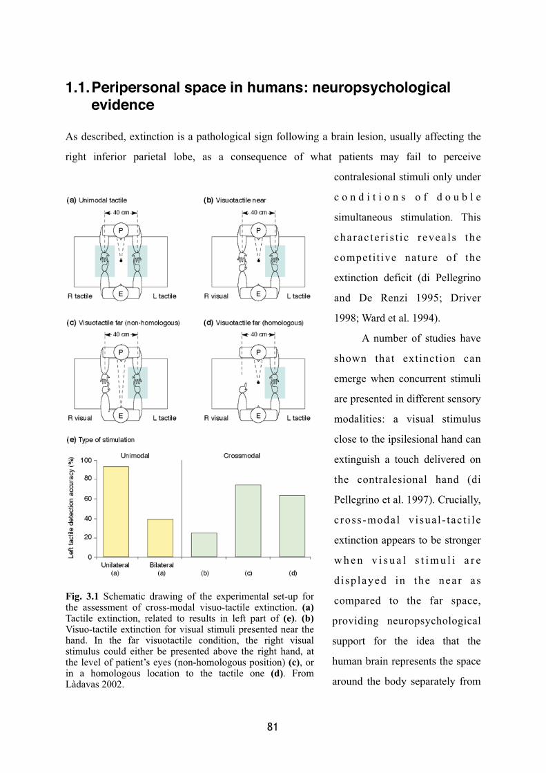
1.1.Peripersonal space in humans: neuropsychological evidence
As described, extinction is a pathological sign following a brain lesion, usually affecting the
right inferior parietal lobe, as a consequence of what patients may fail to perceive
contralesional stimuli only under
c o n d i t i o n s o f d o u b l e
simultaneous stimulation. This
charac te r i s t i c revea ls the
competi t ive nature of the
extinction deficit (di Pellegrino
and De Renzi 1995; Driver
1998; Ward et al. 1994).
A number of studies have
shown that extinction can
emerge when concurrent stimuli
are presented in different sensory
modalities: a visual stimulus
close to the ipsilesional hand can
extinguish a touch delivered on
the contralesional hand (di
Pellegrino et al. 1997). Crucially,
c ross-modal v isua l - tac t i le
extinction appears to be stronger
w h e n v i s u a l s t i m u l i a r e
d i sp layed in the near as
compared to the far space,
providing neuropsychological
support for the idea that the
human brain represents the space
around the body separately from
� 81� �
Fig. 3.1 Schematic drawing of the experimental set-up for the assessment of cross-modal visuo-tactile extinction. (a)Tactile extinction, related to results in left part of (e). (b)Visuo-tactile extinction for visual stimuli presented near the hand. In the far visuotactile condition, the right visual stimulus could either be presented above the right hand, at the level of patient’s eyes (non-homologous position) (c), or in a homologous location to the tactile one (d). From Làdavas 2002.
the space far from the body (Figure 3.1). In particular the spatial representation close to the
body is characterised by a stronger interaction between visual and tactile information. Thus,
the near space in humans presents similarities with the peripersonal space described before
and represented in monkeys through an integrated multisensory visuo-tactile system. Another
common point is the fact that, as described in monkey studies, also in humans the visual
representation of stimuli in near-hand space remains anchored to the hand when this is moved
in another hemi-space, suggesting that visual information in the space around the hand is
coded in a hand-centred coordinate system (di Pellegrino et al. 1997; Farnè et al. 2003b). As
for the hand, a multisensory mechanism is involved in representing the space close to the
human head. By showing stronger visual-tactile extinction for homologous (left and right
cheek) than non-homologous combinations of stimuli (e.g., left hand and right cheek), Farnè
and colleagues’ elegant study demonstrated the modular organisation of the representation of
space around the body. That is, authors found that different spatial regions, adjacent to
different body-parts, are represented separately (Farnè et al. 2005b).
In a series of further studies, Farnè and colleagues (unpublished data) formally tested
four cross-modal extinction patients by presenting visual stimuli close and far from the
patients’ as well as the experimenters’ hand. However, no effect was found in the direction of
a possible body-matching property of the human visuo-tactile system similar to that described
in monkeys (see chapter II, paragraph 1.1). One possibility is that in their research authors
employed a more radical change in orientation between the observer's own and the observed
hands (more than 35 degrees).
Finally, it has been shown that the representation of the region of space around the
human body also features plastic properties, akin to those shown in the monkey. Authors
employed a similar paradigm as that employed in monkeys. A group of cross-modal extinction
patients has been tested before and after a training period during which they became familiar
with a tool. A re-coding visual stimuli located in far space, as if they were closer to the
participants’ body, has been documented behaviourally in extinction patients following the
use of a tool to retrieve (or act upon) distant objects (Farnè and Làdavas 2000; see also Berti
and Frassinetti 2000; Bonifazi et al. 2007; Farnè et al. 2005a, 2007b; Maravita and Iriki
2004). In this interesting study, cross-modal visual-tactile extinction was assessed by
82
presenting visual stimuli far from the patients’ ipsilesional hand, at the distal edge of a 38 cm-
long rake, passively held in their hand. The patients’ performance was evaluated before tool-
use, immediately after a 5 minute period of tool-use, and after a further 5 to 10 minute resting
period. Far visual stimuli were found to induce more severe contralesional extinction
immediately after tool-use, than before tool-use (Figure 3.2). These results demonstrate that
while near and far spaces are separately represented, this spatial division is not defined a
priori. Rather the definition of near and far is derived functionally, depending upon
movements that allow the body to interact with objects in space. Other researches confirmed
this finding, also showing how much dependent might be the re-mapping on the functional
relationship between space and body. Berti and Frassinetti’s elegant study on a neglect patient
(Berti and Frassinetti 2000), for example, showed how the far space can be re-mapped as near
only when a physical continuity between the body and the far target of the action is present.
They asked a right brain damaged patient presenting neglect for left near space to perform a
visual line bisection task, classically employed to assess the presence of neglect symptoms.
Since this kind of patients “neglect” the contralesional side of space (generally the left side
following a right brain lesion), when requested to mark the middle point on a line they
manifest a rightward bias. In this study, the patient had to bisect lines placed far from the
body by using either a stick or a laser-pointer. While the patient transferred the rightward bias
when bisecting far lines with a stick, this re-mapping of far space as near did not arise when
the laser-pointer was used.
The research here presented on extinction patients’ pathological behaviour shows that
also in humans it is possible to define the peripersonal space with similar properties as the
monkeys’ peripersonal space:
• Peripersonal space in humans is a region of space limited in depth with respect to the
body, where visual and tactile information strongly interact.
• Human peripersonal space is a body-part centred representation of space, so that when
hands cross the mid-line, the respective peripersonal space is up-dated consequently.
• Human peripersonal space appears as dynamical as that described in monkeys, as tool-
use studies revealed.
� 83� �
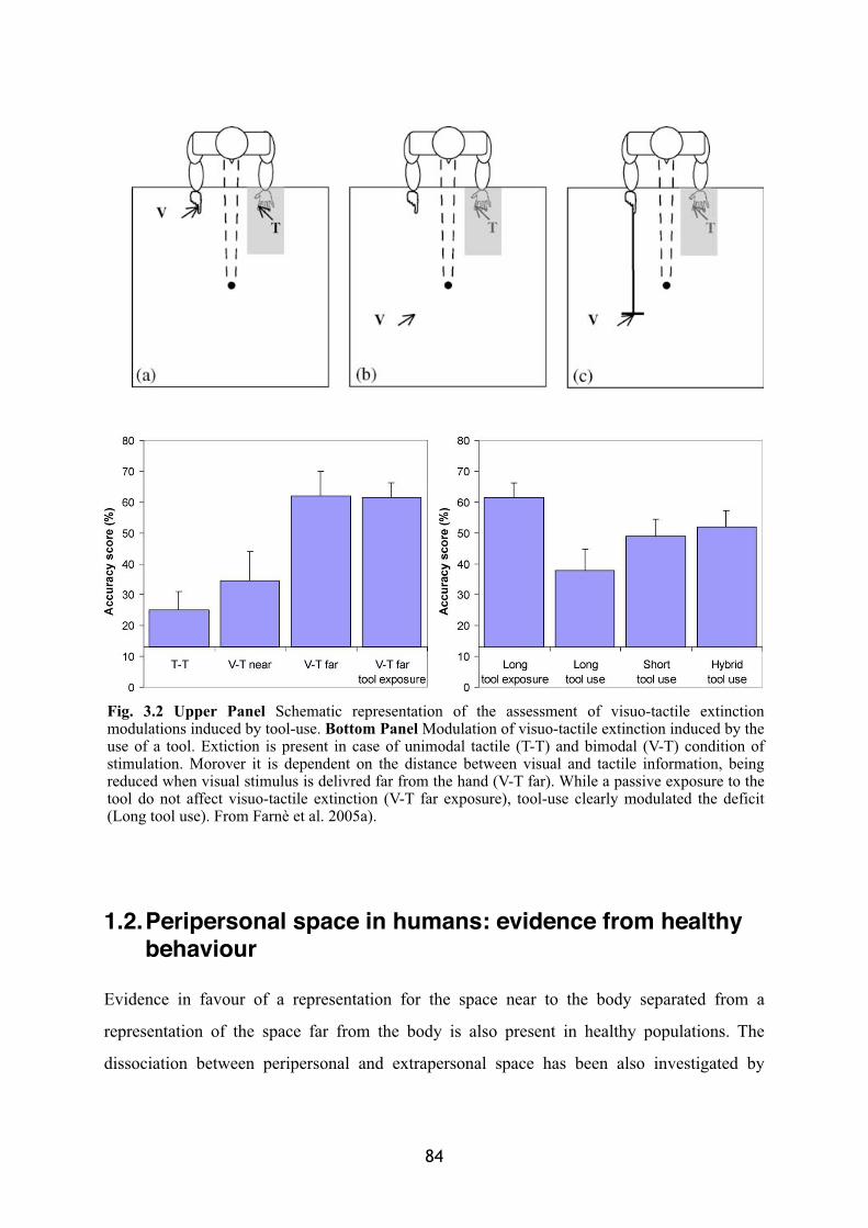
1.2.Peripersonal space in humans: evidence from healthy behaviour
Evidence in favour of a representation for the space near to the body separated from a
representation of the space far from the body is also present in healthy populations. The
dissociation between peripersonal and extrapersonal space has been also investigated by
84
Fig. 3.2 Upper Panel Schematic representation of the assessment of visuo-tactile extinction modulations induced by tool-use. Bottom Panel Modulation of visuo-tactile extinction induced by the use of a tool. Extiction is present in case of unimodal tactile (T-T) and bimodal (V-T) condition of stimulation. Morover it is dependent on the distance between visual and tactile information, being reduced when visual stimulus is delivred far from the hand (V-T far). While a passive exposure to the tool do not affect visuo-tactile extinction (V-T far exposure), tool-use clearly modulated the deficit (Long tool use). From Farnè et al. 2005a).
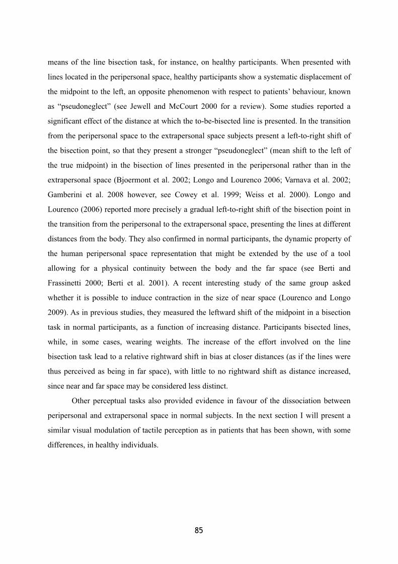
means of the line bisection task, for instance, on healthy participants. When presented with
lines located in the peripersonal space, healthy participants show a systematic displacement of
the midpoint to the left, an opposite phenomenon with respect to patients’ behaviour, known
as “pseudoneglect” (see Jewell and McCourt 2000 for a review). Some studies reported a
significant effect of the distance at which the to-be-bisected line is presented. In the transition
from the peripersonal space to the extrapersonal space subjects present a left-to-right shift of
the bisection point, so that they present a stronger “pseudoneglect” (mean shift to the left of
the true midpoint) in the bisection of lines presented in the peripersonal rather than in the
extrapersonal space (Bjoermont et al. 2002; Longo and Lourenco 2006; Varnava et al. 2002;
Gamberini et al. 2008 however, see Cowey et al. 1999; Weiss et al. 2000). Longo and
Lourenco (2006) reported more precisely a gradual left-to-right shift of the bisection point in
the transition from the peripersonal to the extrapersonal space, presenting the lines at different
distances from the body. They also confirmed in normal participants, the dynamic property of
the human peripersonal space representation that might be extended by the use of a tool
allowing for a physical continuity between the body and the far space (see Berti and
Frassinetti 2000; Berti et al. 2001). A recent interesting study of the same group asked
whether it is possible to induce contraction in the size of near space (Lourenco and Longo
2009). As in previous studies, they measured the leftward shift of the midpoint in a bisection
task in normal participants, as a function of increasing distance. Participants bisected lines,
while, in some cases, wearing weights. The increase of the effort involved on the line
bisection task lead to a relative rightward shift in bias at closer distances (as if the lines were
thus perceived as being in far space), with little to no rightward shift as distance increased,
since near and far space may be considered less distinct.
Other perceptual tasks also provided evidence in favour of the dissociation between
peripersonal and extrapersonal space in normal subjects. In the next section I will present a
similar visual modulation of tactile perception as in patients that has been shown, with some
differences, in healthy individuals.
� 85� �
1.2.1. Cross-modal congruency effect: a perceptual paradigm for the investigation of the peripersonal space representation in healthy humans
Highly converging evidence in favour of a distinct visuo-tactile representation of the
peripersonal space with respect to the extrapersonal space comes from studies about the
interference that visual information can exert over touch percepton in healthy humans. In a
series of studies, participants were asked to discriminate the location of a tactile stimulus
while a visual distractor could be delivered in a
congruent or incongruent spatial position. More in
detail, in a typical crossmodal congruency study,
participants are required to hold two foam cubes,
one in each hand (see Figure 3.3 for a schematic
illustration). A target tactile stimulation is
presented together with a visual distractor,
independently in an unpredictable trial-by-trial
basis from one of the four possible stimulus
locations. Participants are required to make a
series of speeded elevation discrimination
responses, reporting whether tactile target stimuli
are presented at the index finger or thumb of
either hand. Simultaneously, participants have to
try ignoring the visual distractors presented at
approximately the same time. The overall effect is
that participants are normally significantly worse
(both slower and less accurate) at discriminating
the elevation of the touches when the visual
distractors are presented at an incongruent rather
than at a congruent elevation. The difference in
performance between incongruent and congruent
trials is thus a measure of the amount of the
86
Fig. 3.3 Schematic view of the typical set-up for the study of visuo-tactile congruency effect. Participants held a foam cube in each of their left and right hands. Two vibrotactile stimulators (shaded rectangles) and two visual distractor lights (filled circles) were embedded in each cube, by the thumb and index finger. Participants made speeded elevation discrimination responses (by raising the toe or heel of their right foot), in response to vibrotactile targets presented either from the ‘top’ by the index finger of either hand, or from the ‘bottom’ by either t h u m b r e s p e c t i v e l y. M a x i m a l crossmodal congruency effects were always reported for visual distractors placed closest to the location of the vibrotactile target (i.e., on the same foam cube), no matter whether the hands were held in an uncrossed or crossed posture. From Spence et al. 2004c.
cross-modal visuo-tactile interaction. In particular, this value is known as Cross-modal
Congruency Effect (CCE), as it has been termed after the original work presented by Spence
and colleagues (Spence et al. 1998). The modulation exerted by visual distractors over
touches in this paradigm seems to be resistant to practice. People cannot ignore what they see,
even if they are instructed to respond only to what they feel.
Crossmodal congruency effects have also been observed when the role of the two
stimulus modalities is reversed (Walton and Spence 2004), with participants instructed to
respond to the elevation of the visual stimuli, while ignoring the elevation of the vibrotactile
stimuli. However, the crossmodal congruency effects elicited by vibrotactile distractors on
visual elevation discrimination responses tend to be somewhat smaller in magnitude.
There are at least two possible explanations for the crossmodal congruency effect. One
possibility is that this modulation reflects a response competition, between the response
tendencies elicited by the target and distractor in case of incongruent trials. However,
whenever a delay is inserted between the stimulation and the response (750 ms), a procedure
which is meant to reduce errors merely due to a response choice difficulty, the cross-modal
congruency effect is still significantly present. This finding suggests that this visuo-tactile
interaction should at least reflect another kind of mechanism.
The other possibility is indeed that the CCE is a phenomenon reflecting the integration
of visual and tactile information, possibly sensitive to the kind of visual-tactile interaction
occurring around the body and described in the previous sections. The presence of a visual
distractor integrated with the tactile information would bias the localisation of the second
input (tactile target) in a sort of visuo-tactile ventriloquism phenomenon (such as the audio-
visual example described in chapter I). The interesting point of this account is that, if so, the
cross-modal congruency effect could reveal itself as a perceptual measure of the extent of the
region where visual and tactile information are tightly interconnected as that investigated in
monkeys: the peripersonal space. I will discuss consequently whether the CCE reflects the
three main characteristics of the visuo-tactile integration arising in the peripersonal space
coding.
� 87� �
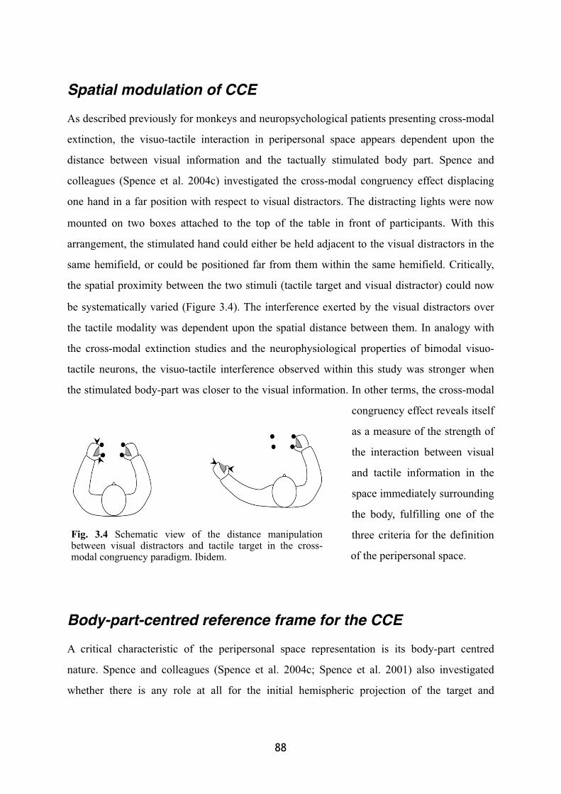
Spatial modulation of CCE
As described previously for monkeys and neuropsychological patients presenting cross-modal
extinction, the visuo-tactile interaction in peripersonal space appears dependent upon the
distance between visual information and the tactually stimulated body part. Spence and
colleagues (Spence et al. 2004c) investigated the cross-modal congruency effect displacing
one hand in a far position with respect to visual distractors. The distracting lights were now
mounted on two boxes attached to the top of the table in front of participants. With this
arrangement, the stimulated hand could either be held adjacent to the visual distractors in the
same hemifield, or could be positioned far from them within the same hemifield. Critically,
the spatial proximity between the two stimuli (tactile target and visual distractor) could now
be systematically varied (Figure 3.4). The interference exerted by the visual distractors over
the tactile modality was dependent upon the spatial distance between them. In analogy with
the cross-modal extinction studies and the neurophysiological properties of bimodal visuo-
tactile neurons, the visuo-tactile interference observed within this study was stronger when
the stimulated body-part was closer to the visual information. In other terms, the cross-modal
congruency effect reveals itself
as a measure of the strength of
the interaction between visual
and tactile information in the
space immediately surrounding
the body, fulfilling one of the
three criteria for the definition
of the peripersonal space.
Body-part-centred reference frame for the CCE
A critical characteristic of the peripersonal space representation is its body-part centred
nature. Spence and colleagues (Spence et al. 2004c; Spence et al. 2001) also investigated
whether there is any role at all for the initial hemispheric projection of the target and
88
Fig. 3.4 Schematic view of the distance manipulation between visual distractors and tactile target in the cross-modal congruency paradigm. Ibidem.
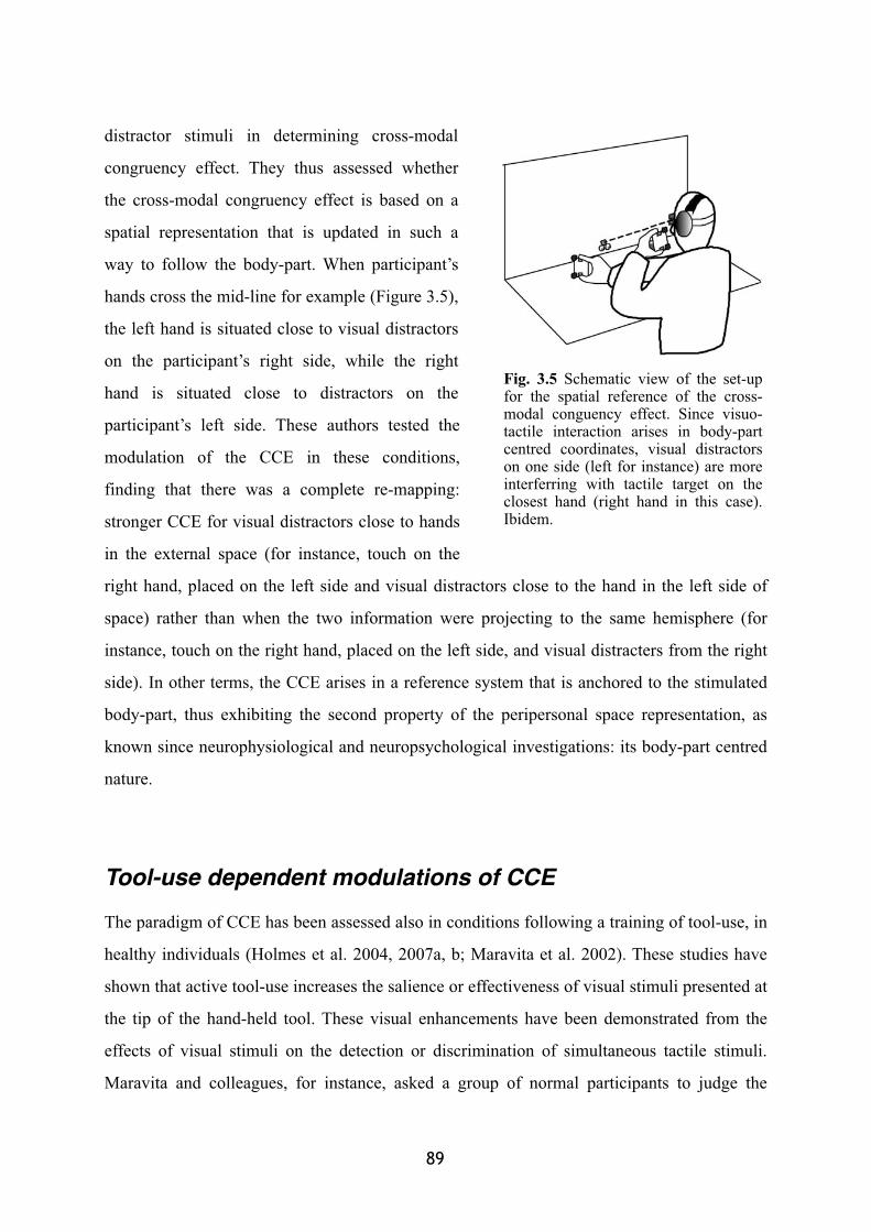
distractor stimuli in determining cross-modal
congruency effect. They thus assessed whether
the cross-modal congruency effect is based on a
spatial representation that is updated in such a
way to follow the body-part. When participant’s
hands cross the mid-line for example (Figure 3.5),
the left hand is situated close to visual distractors
on the participant’s right side, while the right
hand is situated close to distractors on the
participant’s left side. These authors tested the
modulation of the CCE in these conditions,
finding that there was a complete re-mapping:
stronger CCE for visual distractors close to hands
in the external space (for instance, touch on the
right hand, placed on the left side and visual distractors close to the hand in the left side of
space) rather than when the two information were projecting to the same hemisphere (for
instance, touch on the right hand, placed on the left side, and visual distracters from the right
side). In other terms, the CCE arises in a reference system that is anchored to the stimulated
body-part, thus exhibiting the second property of the peripersonal space representation, as
known since neurophysiological and neuropsychological investigations: its body-part centred
nature.
Tool-use dependent modulations of CCE
The paradigm of CCE has been assessed also in conditions following a training of tool-use, in
healthy individuals (Holmes et al. 2004, 2007a, b; Maravita et al. 2002). These studies have
shown that active tool-use increases the salience or effectiveness of visual stimuli presented at
the tip of the hand-held tool. These visual enhancements have been demonstrated from the
effects of visual stimuli on the detection or discrimination of simultaneous tactile stimuli.
Maravita and colleagues, for instance, asked a group of normal participants to judge the
� 89� �
Fig. 3.5 Schematic view of the set-up for the spatial reference of the cross-modal conguency effect. Since visuo-tactile interaction arises in body-part centred coordinates, visual distractors on one side (left for instance) are more interferring with tactile target on the closest hand (right hand in this case). Ibidem.
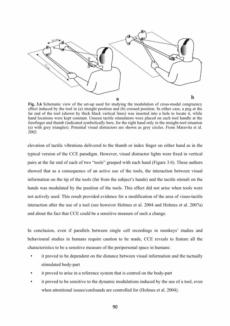
elevation of tactile vibrations delivered to the thumb or index finger on either hand as in the
typical version of the CCE paradigm. However, visual distractor lights were fixed in vertical
pairs at the far end of each of two “tools” grasped with each hand (Figure 3.6). These authors
showed that as a consequence of an active use of the tools, the interaction between visual
information on the tip of the tools (far from the subject’s hands) and the tactile stimuli on the
hands was modulated by the position of the tools. This effect did not arise when tools were
not actively used. This result provided evidence for a modification of the area of visuo-tactile
interaction after the use of a tool (see however Holmes et al. 2004 and Holmes et al. 2007a)
and about the fact that CCE could be a sensitive measure of such a change.
In conclusion, even if parallels between single cell recordings in monkeys’ studies and
behavioural studies in humans require caution to be made, CCE reveals to feature all the
characteristics to be a sensitive measure of the peripersonal space in humans:
• it proved to be dependent on the distance between visual information and the tactually
stimulated body-part
• it proved to arise in a reference system that is centred on the body-part
• it proved to be sensitive to the dynamic modulations induced by the use of a tool, even
when attentional issues/confounds are controlled for (Holmes et al. 2004).
90
Fig. 3.6 Schematic view of the set-up used for studying the modulation of cross-modal congruency effect induced by the tool in (a) straight position and (b) crossed position. In either case, a peg at the far end of the tool (shown by thick black vertical lines) was inserted into a hole to locate it, while hand locations were kept constant. Unseen tactile stimulators were placed on each tool handle at the forefinger and thumb (indicated symbolically here, for the right hand only in the straight-tool situation (a) with grey triangles). Potential visual distractors are shown as grey circles. From Maravita et al. 2002.
1.3.Peripersonal space in humans: neuroimaging studies
Evidence is now accumulating also from neuroimaging studies in healthy humans that can
provide further support for the homologies between some of the neurophysiological evidence
reviewed in the previous chapter and the neural bases of peripersonal space in the human
brain. Specifically, brain areas that represent near-face and near-hand space in body-centred
coordinates through visual and tactile maps have been reported in the anterior section of the
intraparietal sulcus and in the ventral premotor cortex (Bremmer et al. 2001; Makin et al.
2007; Sereno and Huang 2006). These findings correspond nicely to the anatomical locations
of the monkey visuo-tactile network. Moreover, recent studies have identified the superior
parietal occipital junction as a potential site for representing near-face and near-hand visual
space (Gallivan et al. 2009; Quinlan et al. 2007). This new evidence extends our current
knowledge of the peripersonal space neural network, and may guide further
electrophysiological studies to come.
2. Conclusions
The findings presented here for human beings, coming from neuropsychological, behavioural
and imaging studies, converge supporting the existence in the human brain of mechanisms
that are specialised for representing visual information selectively when it arises from the
space near the hand (and face). In other terms, the human brain, similarly to monkey’s, builds
a peripersonal space representation separated from a representation of the space that is farther
from the body. This distinction is based on a different multisensory integration arising in the
region close to the body, where visual information appears more tightly connected to tactile
information on the close body-part. As the previous section highlighted, indeed, a strong
binding mechanism of visual and tactile inputs has repeatedly been shown also in humans.
The homologous representation of the peripersonal space in humans is codified in a body-part
centred reference system and is up-dated with body posture changes. Finally, the human
homologous of peripersonal space also presents dynamic properties, induced for instance by
� 91� �
the active use of a tool that can change the functional relationship between the body and the
reachable space. Importantly, the CCE described in this section appears as a sensitive
paradigm to study the modulations of peripersonal space.
These converging results have refined and extended our understanding of the neural
processes underlying multisensory representation of peripersonal space in the human brain,
namely, by identifying various cortical areas that are involved in different sensory-motor
aspects of peripersonal space representation.
92
Chapter IV
Peripersonal space: a multisensory interface for body-object interactions?
The review in the previous sections showed that visual and somatosensory information are
integrated in the brain, both cortically in parietal and frontal areas and sub-cortically in the
putamen. The characteristics of the visuo-tactile integration through systems of bimodal
neurons differentiate the representation of the space around the body from a farther space. The
peripersonal space so defined, becomes the space between the body and the external space
where visual information are not mere events occurring outside the body, but they also
represent the possibility of an interaction between the body and the external world. When I
leave my cup of coffee close to me while working, for instance, not only I can interact with it
as it commonly -and preferably- happens when I grasp it in order to drink my coffee. In fact, I
have also to be careful when I’m moving because, for the simple reason that my cup is close
to my body, in the space where my movements take place, I could also accidentally -and
dangerously- collide with it. Also true is the fact that often, when an unknown insect is flying
around our arm, for instance, we could have the involuntary tendency to move the arm in
order to avoid the contact between the body and the unknown -and potentially threatening-
insect. It is therefore conceivable that an object in the peripersonal space is not visible in the
same way as are objects or events occurring in the far space: only the former might be
intrinsically available for active interactions with body-parts without needing the full body to
move. The peripersonal space could be thus considered of as an interface between the body
and the objects.
For these reasons, it appears striking that, until recently, all investigations of
peripersonal space representation in humans have assessed its characteristics in completely
static conditions. I will present a series of findings in monkeys that, together with the
electrophysiological findings on bimodal neurons, clearly indicate the strong link existing
� 93� �
between peripersonal space and action. As I will discuss in this chapter, also research about
human peripersonal space representation is ready to fill the gap between the multisensory
perceptual investigation of this representation and its involvement in the execution of actions.
This point represents the novel contribution of this dissertation to our knowledge about the
peripersonal space. In particular, two original methodological considerations will be
presented, through which I have tried to experimentally assess the link between multisensory
space representation and the execution of actions.
94
1. What kind of body-object interactions can the body-centered PpS representation subserve?
Since the multisensory space based on the visuo-tactile properties of bimodal neurons defines
a boundary zone limited in depth between the body and the environment, some researchers
have suggested that it might have evolved for the execution of defence or object-avoidance
movements (Cooke and Graziano 2004; Cooke et al. 2003; Graziano et al. 2002; Stepniewska
et al. 2005). Protect the body against physical threats is one of the vital function the system
should guarantee. By acting as an anticipatory multisensory-motor interface, the peripersonal
space representation may serve for the early detection of potential threats approaching the
body (Fogassi et al. 1996) in order to drive involuntary defensive movements (Cooke and
Graziano 2004; Graziano and Cooke 2006). As already described, the visual RFs of bimodal
neurons are sensitive to objects approaching the body and more specifically also to the
velocity of their approaching, as a function of which they can modify their area of response.
The most direct evidence in favour of the hypothesis that peripersonal space serves defensive
actions is the result of cortical stimulation studies, although some concerns have been raised
in this respect (Strick 2002; Graziano et al. 2002). The electrocortical microstimulation allows
to artificially induce electrical activity directly in a specific area in order to study, for
instance, the cartography of the motor control in the motor areas. Graziano and colleagues
applied this method for the study of the motor properties of visuo-tactile network (Graziano
and Cooke 2006). They thus described that the electrocortical stimulation of visuo-tactile
areas can elicit a pattern of complex movements that is compatible with defensive arm (and
face) movements and withdrawing of the arm or the head (Cooke and Graziano 2003;
Stepniewska et al. 2005). The most important concern other electrophysiologists underlined
was the intensity and the duration/length of train stimulation employed in this research (Strick
2002). By employing a similar paradigm, however, other researchers have found that the
stimulation of parietal visuo-tactile areas can induce not only movements that are compatible
with defensive behaviour, but also movements compatible with “appetitive” behaviours, such
as reaching and grasping (Stepniewska et al. 2005).
As a consequence, the same anticipatory feature might also have evolved to serve
voluntary object-oriented actions (Gardner et al. 2007; Kaas 2004; Rizzolatti et al. 1981a, b;
� 95� �
Rizzolatti et al. 1997; Stepniewska et al. 2005). In support of this view are the results of the
described single unit recording studies showing the motor properties of both parietal and
periarcuate visuo-tactile neurons, whose discharge is mostly correlated with reaching and
grasping movements (see Chapter II, section 2). Moreover, since peripersonal space
represents a boundary-zone between one’s own body and the external environment, it could
also indirectly have a role in the self-attribution of sensory signals (Makin et al. 2008). This
system may therefore be involved in the multisensory representation of limb position: the
visuo-tactile neurons, constantly updated with the varying positions of the body parts, achieve
a body-part-centered representation of visual space, even while the body moves.
The thesis of this dissertation, I would like to propose, is that the multisensory-motor
neural machinery represented by the bimodal visuo-tactile network has been selected
throughout evolution, not only to drive involuntary avoidance reactions, but also voluntary
approaching movements towards objects, via the continuous monitoring of action space, the
space within reach.
2. CCE in action
Until recently, however, the characteristics of visuo-tactile peripersonal space in humans had
been assessed exclusively while the relevant body parts were held statically. Even the most
‘dynamic’ properties of peripersonal space, the post tool-use modulation of the visuo-tactile
interaction, have been studied in the static phase preceding or following the active use of the
tool. Some researches have assessed tool-use induced modulations of peripersonal space after
very brief intervals (i.e., several seconds) following the use of the tool (Holmes et al. 2007b),
or after several minutes of tool use (Maravita et al. 2002), and have showed rapid changes in
visuo-tactile interactions on these time-scales. However, in these studies too, the perceptual
visuo-tactile task was performed in a “transient” but still static post tool-use session,
interleaved with the tool-use training sessions. An exception could be found in those studies
showing dynamic changes of peripersonal space during tasks such as line bisection, where
distant stimuli were acted on through the use of a long tool (e.g., Berti and Frassinetti 2000).
96
However, multisensory integration was not explicitly measured in these studies, meaning the
link with peripersonal space remains speculative.
If the peripersonal space representation is indeed directly involved in body-object
interactions, then modulations of visuo-tactile interaction should be found without the need of
any tools. On the contrary, the visuo-tactile interaction, or the dynamic 're-mapping' of near
space should be a basic, primary property that only secondarily can be generalised to tool-use.
In this respect, the execution of a voluntary free-hand action, for instance grasping an object,
should induce a rapid on-line re-mapping of visuo-tactile spatial interactions, as the action
unfolds.
2.1.The multisensori-motor task
Even if the CCE paradigm revealed to be a useful frame of investigation for the assessing of
the multisensory interaction in peripersonal space, an adaptation was needed in order to assess
the visuo-tactile interaction on-line during action.
First of all, an action is required and in particular, in a first step, we choose a grasping
action. Three main reasons supported this choice. First, the execution of grasping, as the
execution of reaching actions, has been deeply investigated in the neurophysiological studies
on visuo-tactile network, as described in other sections of this dissertation. These studies
revealed the high degree of functional specialisation of the bimodal network for different kind
of grasping (i.e., precision vs. whole-hand grip). Second, the kinematics of grasping in human
and non human primates has been revealed to be highly comparable (Castiello 2005; Roy et
al. 2002; 2006) so that the possible functional homologies, previously established on the basis
of bimodal neurons properties, will hold true across species when the same action of grasping
an object is performed. Finally, grasping objects is one of the most common actions we
execute in our everyday life. Thus, grasping appeared the right choice to link our research to
animal’s studies and at the same time to maintain an ecological approach to our investigation
in humans. Participants were thus requested to grasp a cylinder along its longitudinal axes,
with a precision grip (thumb and index fingers) and extract it from the horizontal support in
which it was inserted. The cylinder was placed far from the hand (47 cm), and presented in
� 97� �

different orientations. In this way, participants
had to perform a complex action composed by
a hand transport phase, a finger grip and a
wrist rotation component (Figure 4.1).
Secondly, the CCE paradigm has been
implemented in these active conditions. The
same perceptual discrimination task of the
elevation of tactile stimuli delivered on the
thumb (“down” position) or on the index
finger (“up” position) has been maintained.
However, instead of presenting the visual
distractors from a position close to the hand,
as in the typical CCE paradigm, they have
been embedded in the object, target of the
action. The cylinder presented two LEDs
above and below the fixation point marked at
its centre. The spatially congruent and
incongruent visuo-tactile stimuli could be thus
reproduced. At each trial, participants were
requested to respond where they perceived the
tactile stimulation trying to ignore the visual distractor on the object.
This arrangement allowed us to couple the perceptual and motor task at each trial and
at different timings of the unfolding action. The evolution of the visuo-tactile interaction
could thus be assessed along the execution of the action and in particular three epochs have
been chosen. The visuo-tactile interaction was assessed either before the on-set of the action,
on the action on-set itself, or in the early execution phase. The first constitutes a baseline,
where the hand does not move yet, to be compared with the two ‘dynamical’ phases. The
hypothesis supporting the involvement of peripersonal space in action execution would
predict a modulation of the visuo-tactile interaction in terms of CCE as a function of the
moment when it is assessed along the execution of the action.
98
Fig 4.1 View of the cross-modal congruency paradigm modified in order to assess the multisensory interaction during the execution of an action. (a) tactile target on the hand as in the typical CCE condition while visual distractors are embedded into the to-be-grasped object. (b) and (c) are example of grasping actions.
3. The kinematic recording of movements: a real-time method for linking multisensory perception and action
In order to study the relationship of the multisensory based peripersonal space and the
execution of an action, it is necessary to measure the updating of the visuo-tactile interplay in
real-time with the unfolding action. This aim revealed challenging since, as we described in
previous sections, the peripersonal space has been assessed only in static conditions. The
adaptation of the CCE paradigm allowed the real-time assessment of the multisensory
perception induced by the execution of an action. However, in order to investigate the link
possibly existing between the perceptual and the motor domain, also the latter needs to be
measured.
In the early 80’s, the development of kinematic methods allowed the on-line recording
and a quantitative description of movements executed during actions. For instance,
Jeannerod’s studies that were mainly focussed on grasping actions, provided a model of the
grasping as a two components movement: transport and grip components. The two
components could be thus defined by their respective kinematic parameters, such as velocity
and acceleration for the transport component and velocity of finger aperture and maximum
grip aperture for the grip component.
With the aim of investigating the link between peripersonal space and execution of
voluntary actions, we assessed the multisensory perception during action execution and
simultaneously recorded the kinematic of each movement. This novel approach conjugates a
multisensory perceptual task in real-time with the movement and provided the necessary
method to link the perception to the kinematic characteristics of the action.
In this section, the main characteristics of the kinematics will be briefly reviewed in
particular showing the normal structure of a grasping movement from a kinematic point of
view. The neural bases of grasping will also be briefly presented.
� 99� �
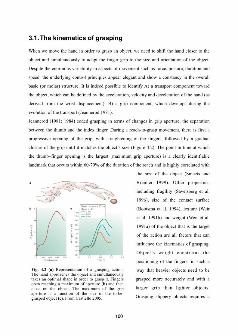
3.1.The kinematics of grasping
When we move the hand in order to grasp an object, we need to shift the hand closer to the
object and simultaneously to adapt the finger grip to the size and orientation of the object.
Despite the enormous variability in aspects of movement such as force, posture, duration and
speed, the underlying control principles appear elegant and show a constancy in the overall
basic (or molar) structure. It is indeed possible to identify A) a transport component toward
the object, which can be defined by the acceleration, velocity and deceleration of the hand (as
derived from the wrist displacement); B) a grip component, which develops during the
evolution of the transport (Jeannerod 1981).
Jeannerod (1981; 1984) coded grasping in terms of changes in grip aperture, the separation
between the thumb and the index finger. During a reach-to-grasp movement, there is first a
progressive opening of the grip, with straightening of the fingers, followed by a gradual
closure of the grip until it matches the object’s size (Figure 4.2). The point in time at which
the thumb–finger opening is the largest (maximum grip aperture) is a clearly identifiable
landmark that occurs within 60-70% of the duration of the reach and is highly correlated with
the size of the object (Smeets and
Brenner 1999). Other properties,
including fragility (Savelsberg et al.
1996), size of the contact surface
(Bootsma et al. 1994), texture (Weir
et al. 1991b) and weight (Weir et al.
1991a) of the object that is the target
of the action are all factors that can
influence the kinematics of grasping.
Object’s weight constrains the
positioning of the fingers, in such a
way that heavier objects need to be
grasped more accurately and with a
larger grip than lighter objects.
Grasping slippery objects requires a
100
Fig. 4.2 (a) Representation of a grasping action. The hand approaches the object and simultaneously takes an optimal shape in order to grasp it. Fingers open reaching a maximum of aperture (b) and then close on the object. The maximum of the grip aperture is a function of the size of the to-be-grasped object (c). From Castiello 2005.
larger approach parameter, leading to a larger grip earlier in the movement compared with
grasping rough-surfaced objects. On these bases Jeannerod proposed a model based on two
different visuo-motor channels (Jeannerod and Biguer, 1982). The transport component, that
is the spatial shift of the hand toward the object, requires the knowledge of the spatial
configuration of the to-be-grasped object with respect to the grasping body-part. To reach an
object in the external space with our hand, we need indeed to know its spatial location (and its
orientation) with respect to the hand. In other words, we need to know the extrinsic
characteristic of the object, i.e., those characteristics that not dependent upon the object itself,
but are determined by the spatial configuration and location of the object with respect to the
body. By contrast, intrinsic information is required for the development of the grip
component, such as the size, the texture and the shape of the object itself.
It is clear from these considerations that large part of information about how an object
has to be optimally grasped derives from the visual modality (orientation, shape, size of the
available landing surfaces) sometime associated with the experience of tactile information
(texture or weight). The tactile system, nonetheless, seems to be important for the optimal
evolution of the hand-shaping (Gentilucci et al. 1997; Witney et al. 2004): cutaneous
receptors in the hand provide information about the action of the hand, including its
kinematics and posture, as well as the grip forces and load forces that are used during
grasping and manipulation of objects. Finally, the visual information about the object can also
create expectancies about the tactile feedback the grasping hand will receive at the end of the
action, when it will enter in contact with the object itself (Gentilucci et al. 1997).
The kinematics recording represents a powerful technique which allows quantitatively
describing movement. More important for the experimental contribution of this thesis, the
real-time kinematics of the movement allows for establishing direct link between the
perceptual assessment of the visuo-tactile interaction and the evolution of the grasping action.
For this reason, each grasping movement performed by participants has been kinematically
recorded and analysed off-line. Three infrared emitters have been placed in the standard
locations on the grasping hand: one on the wrist, for recording the parameters associated to
the transport component; two on the last phalanx of the thumb and of the index finger for
recording the development of the grip component.
� 101� �
3.2.Neural network for grasping
Three specific areas involved in hand grasping execution have been identified in the monkey
cortex: the primary motor cortex (F1), part of ventral premotor cortex (F5) and the anterior
intraparietal sulcus (AIP). The integrity of the primary motor cortex (F1) is necessary for
performing a successful grasping action. A lesion of this area indeed produce a severe deficit
in the control of individual fingers and consequently disrupt normal grasping (Lawrence and
Kuypers 1968a). Lesioning the corticospinal tract, through which motor commands are
conveyed from the primary motor cortex to cells in the spinal cord, also impairs the control of
independent finger movements (Lawrence and Kuypers 1968b). However, after such kind of
lesion, synergistic finger flexion typical of a power grip prehension might be not affected.
More interesting for this dissertation, is another fundamental process needed for a
successful grasp to be executed. To grasp an object involves indeed a transformation of the
intrinsic properties of the object, which are visually available, into the correct motor shaping
of the hand. The visuo-motor transformations for grasping seem to involve two key cortical
areas: the AIP and area F5, already described in the second chapter of this dissertation. AIP
and F5 neurons activity shows striking similarities and important differences (Rizzolatti et al.
2002). For example, both AIP and F5 show a functional specialisation for type of grasping,
which in turn depends on the visual characteristic of the object to be grasped (Murata et al.
1997; 2000). Different populations of neurons are activated during different kinds of grasping
(i.e., precision grip or whole-hand prehension). However, AIP neurons seem to represent the
entire action, whereas F5 neurons seem to be concerned with a particular segment of the
action (Rizzolatti et al. 1998). Another important difference is that AIP, more than F5,
presents a visual response to the presentation of three-dimensional objects (Murata et al.
2000). Based on these findings, Fagg and Arbib (1998) have thus proposed a model in which
area AIP provides multiple descriptions of three-dimensional objects for the purpose of
manipulation, whereas area F5 might be mainly involved in selecting the most appropriate
motor prototype from a “motor vocabulary” (Rizzolatti et al. 1988a) for example, the type of
grip that is effective in interacting with a specific target object.
102
Similarly to monkeys, lesions of the human primary motor cortex or corticospinal
fibres dramatically disrupt the execution of a grasping movement. Such lesions typically lead
to grasping movements that are initially characterised by the loss of independent control of
the fingers, although synergistic movements of all fingers (a power grip) might remain intact
(Lang and Shieber 2004).
Due to the difficulty of establishing a correct homology between human and monkey
brains, it is difficult to integrate the known grasping deficits in humans after lesions of the
superior parietal lobe (SPL) with the results of neurophysiological work in monkeys. For
instance, in monkeys, the SPL (possibly area 5) seems to be mainly related to the
somatosensory rather than visuomotor elaboration (Rizzolatti et al. 2002). Secondly, although
in monkeys some parts of the SPL do receive visual information, activity in these areas seem
to be more correlated with reaching rather than grasping (Galletti et al. 1997). Finally, there is
reason to believe that, the relative expansion of the parietal lobes characterising humans’ brain
relative to monkeys’, the human SPL might be the homologue of the monkey IPL (Milner
1997). However, with all the possible cautions, there is evidence that a possible homologue of
AIP has a specific role in grasping in humans. Binkofski and colleagues (Binkofski et al.
1998) localised the area responsible for grasping in humans to the AIP, contralateral to the
impaired hand. In line with the results obtained from monkeys in which AIP has been
inactivated, human patients with AIP lesions present deficits in grasping, whereas reaching
remained relatively intact.
The most compelling evidence for a deficit in visually guided grasping in humans
comes from patients presenting a deficit known as optic ataxia (Perenin and Vighetto 1988;
Rossetti et al. 2003) classically considered to be a specific disorder of the visuomotor
transformations caused by posterior parietal lesions. Various optic ataxic patients have been
described showing specific deficits in the on-line control of reaching (Pisella et al.2009;
Blangero et al. 2008), but only a few cases have investigated the kinematics of grasping
actions. Patient V.K. (Jakobson et al. 1991), for example, showed an apparently normal early
phase of hand opening during attempts to grasp an object, but her on-line control of grip
aperture quickly degenerated, resulting in numerous secondary peaks in the grip aperture
profile, rather than a single peak, which is typical of a healthy subject. Patient I.G. (Milner et
� 103� �
al. 2003) also showed considerable deficits in the scaling of her maximum grip aperture to the
size of an object. Patient G.T.’s prehension movements were not normally tuned either by
intrinsic or extrinsic visual properties of objects (Roy et al. 2004), his grasping component not
being appropriately modulated by object’s size. Overall, these patients present a deficit in
translating the visually available information about an object into the correct motor
configuration to interact with it.
4. Conclusion: kinematic-perceptual co-recordingThe general hypothesis underlying the experimental contribution presented here is that
peripersonal space constitutes a multisensory interface serving for the interactions between
the body and the objects in the external space. This hypothesis is based on the neural
mechanism on which peripersonal space representation is built through. The kinematic
recording coupled with an on-line assessment of the multisensory interaction during action is
the method through which I tried to investigate the link between the multisensory perception
and the execution of action. This novelty meant to fill the gap till now existing between a
static and dynamic view of multisensory perception.
104

RESULTS

As described, electro-cortical microstimulation studies in monkeys provided evidence in
favour of an involvement of visuo-tactile parieto-frontal structures in the execution of
movements that are compatible with a defensive or avoidance reaction. This is compatible
with the hypothesis that the peripersonal space representation might be a multisensory
interface with a protective function in response to possible threatening objects approaching
the body. In the first study presented here, we tested this hypothesis in healthy humans,
investigating the reference frame the brain employs for coding the location of real three-
dimensional objects suddenly appearing near the body. We used a single pulse transcranial
magnetic stimulation (TMS) approach in order to measure the excitability of the motor cortex
in correspondence to the hand representation. TMS is a technique allowing to momentarily
interfere with the normal electrical activity of a relatively small area of the brain. When
applied to the motor cortex, TMS single pulse elicits a motor evoked potential (MEP) that can
be measure in the peripheral muscle, which reflects the level of cortical excitability in the
respective motor cortex representation. The cortical excitability reflects in turn, the readiness
for a motor response. We hypothesised that an object suddenly approaching the hand, for
instance, should modulate the cortical activity in correspondence of the motor cortex hand
representation. We also hypothesised that, if peripersonal space is involved in the detection
and localisation of stimuli approaching the body, the cortical activity modulation should arise
in a reference frame centred to the body-part (the hand in the example).
We asked subjects to make a simple response to a visual ‘Go’ signal while they were
simultaneously presented with a task-irrelevant distractor ball, which could rapidly approach
the peripersonal space in a location either near to or far from participants’ responding hand.
As expected, we found that motor excitability was modulated as a function of the
distance of approaching balls from the hand: MEPs amplitude was selectively reduced when
the ball approached near the hand, both when the hand was on the left and on the right of the
midline. Strikingly, this reduction occurred as early as 70 ms after ball appearance. The effect
arose in hand-centred coordinates since it was not modified by the location of visual fixation
relative to the hand. Furthermore, it was selective for approaching balls, since static visual
distractors did not modulate MEP amplitude.
� 107
Together with additional behavioral measurements, this series of experiments provides
direct and converging evidence for automatic hand-centered coding of visual space in the
human motor system during motor preparation. These results provide direct evidence in
favour of the interpretation of peripersonal space as a multisensory interface for translating
potentially relevant visual information into rapid actions.
108
STUDY 1
Coding of visual space during motor preparation:
approaching objects rapidly modulate corticospinal
excitability in hand-centred coordinates
Journal of Neuroscience (2009)

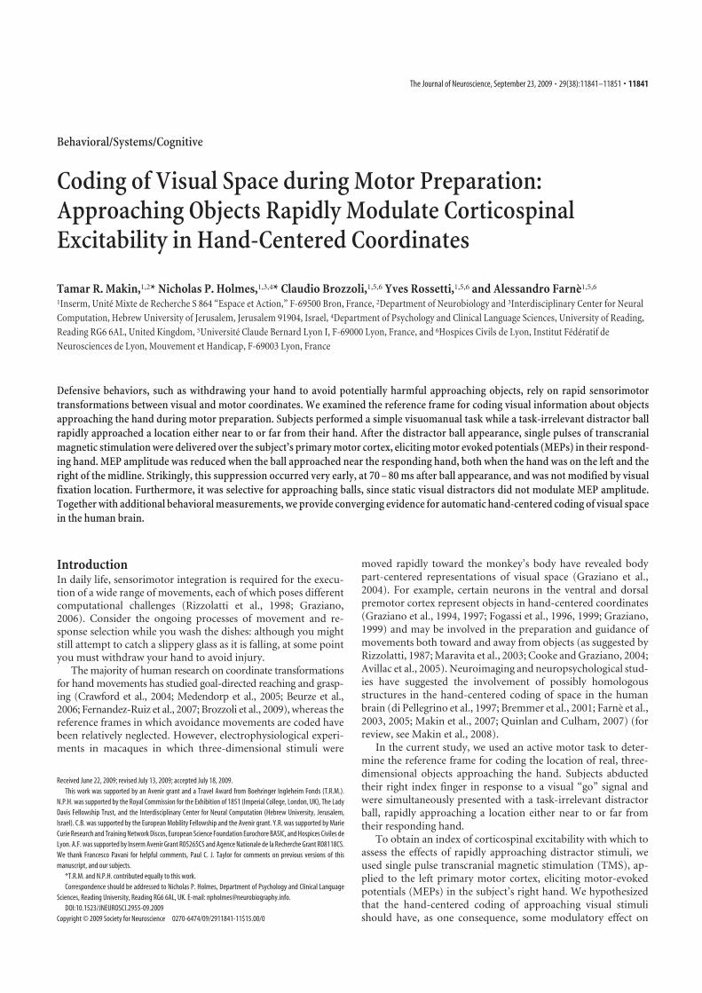
Behavioral/Systems/Cognitive
Coding of Visual Space during Motor Preparation:Approaching Objects Rapidly Modulate CorticospinalExcitability in Hand-Centered Coordinates
Tamar R. Makin,1,2* Nicholas P. Holmes,1,3,4* Claudio Brozzoli,1,5,6 Yves Rossetti,1,5,6 and Alessandro Farne1,5,6
1Inserm, Unite Mixte de Recherche S 864 “Espace et Action,” F-69500 Bron, France, 2Department of Neurobiology and 3Interdisciplinary Center for NeuralComputation, Hebrew University of Jerusalem, Jerusalem 91904, Israel, 4Department of Psychology and Clinical Language Sciences, University of Reading,Reading RG6 6AL, United Kingdom, 5Universite Claude Bernard Lyon I, F-69000 Lyon, France, and 6Hospices Civils de Lyon, Institut Federatif deNeurosciences de Lyon, Mouvement et Handicap, F-69003 Lyon, France
Defensive behaviors, such as withdrawing your hand to avoid potentially harmful approaching objects, rely on rapid sensorimotortransformations between visual and motor coordinates. We examined the reference frame for coding visual information about objectsapproaching the hand during motor preparation. Subjects performed a simple visuomanual task while a task-irrelevant distractor ballrapidly approached a location either near to or far from their hand. After the distractor ball appearance, single pulses of transcranialmagnetic stimulation were delivered over the subject’s primary motor cortex, eliciting motor evoked potentials (MEPs) in their respond-ing hand. MEP amplitude was reduced when the ball approached near the responding hand, both when the hand was on the left and theright of the midline. Strikingly, this suppression occurred very early, at 70 – 80 ms after ball appearance, and was not modified by visualfixation location. Furthermore, it was selective for approaching balls, since static visual distractors did not modulate MEP amplitude.Together with additional behavioral measurements, we provide converging evidence for automatic hand-centered coding of visual spacein the human brain.
IntroductionIn daily life, sensorimotor integration is required for the execu-tion of a wide range of movements, each of which poses differentcomputational challenges (Rizzolatti et al., 1998; Graziano,2006). Consider the ongoing processes of movement and re-sponse selection while you wash the dishes: although you mightstill attempt to catch a slippery glass as it is falling, at some pointyou must withdraw your hand to avoid injury.
The majority of human research on coordinate transformationsfor hand movements has studied goal-directed reaching and grasp-ing (Crawford et al., 2004; Medendorp et al., 2005; Beurze et al.,2006; Fernandez-Ruiz et al., 2007; Brozzoli et al., 2009), whereas thereference frames in which avoidance movements are coded havebeen relatively neglected. However, electrophysiological experi-ments in macaques in which three-dimensional stimuli were
moved rapidly toward the monkey’s body have revealed bodypart-centered representations of visual space (Graziano et al.,2004). For example, certain neurons in the ventral and dorsalpremotor cortex represent objects in hand-centered coordinates(Graziano et al., 1994, 1997; Fogassi et al., 1996, 1999; Graziano,1999) and may be involved in the preparation and guidance ofmovements both toward and away from objects (as suggested byRizzolatti, 1987; Maravita et al., 2003; Cooke and Graziano, 2004;Avillac et al., 2005). Neuroimaging and neuropsychological stud-ies have suggested the involvement of possibly homologousstructures in the hand-centered coding of space in the humanbrain (di Pellegrino et al., 1997; Bremmer et al., 2001; Farne et al.,2003, 2005; Makin et al., 2007; Quinlan and Culham, 2007) (forreview, see Makin et al., 2008).
In the current study, we used an active motor task to deter-mine the reference frame for coding the location of real, three-dimensional objects approaching the hand. Subjects abductedtheir right index finger in response to a visual “go” signal andwere simultaneously presented with a task-irrelevant distractorball, rapidly approaching a location either near to or far fromtheir responding hand.
To obtain an index of corticospinal excitability with which toassess the effects of rapidly approaching distractor stimuli, weused single pulse transcranial magnetic stimulation (TMS), ap-plied to the left primary motor cortex, eliciting motor-evokedpotentials (MEPs) in the subject’s right hand. We hypothesizedthat the hand-centered coding of approaching visual stimulishould have, as one consequence, some modulatory effect on
Received June 22, 2009; revised July 13, 2009; accepted July 18, 2009.
This work was supported by an Avenir grant and a Travel Award from Boehringer Ingleheim Fonds (T.R.M.).
N.P.H. was supported by the Royal Commission for the Exhibition of 1851 (Imperial College, London, UK), The Lady
Davis Fellowship Trust, and the Interdisciplinary Center for Neural Computation (Hebrew University, Jerusalem,
Israel). C.B. was supported by the European Mobility Fellowship and the Avenir grant. Y.R. was supported by Marie
Curie Research and Training Network Discos, European Science Foundation Eurochore BASIC, and Hospices Civiles de
Lyon. A.F. was supported by Inserm Avenir Grant R05265CS and Agence Nationale de la Recherche Grant R08118CS.
We thank Francesco Pavani for helpful comments, Paul C. J. Taylor for comments on previous versions of this
manuscript, and our subjects.
*T.R.M. and N.P.H. contributed equally to this work.
Correspondence should be addressed to Nicholas P. Holmes, Department of Psychology and Clinical Language
Sciences, Reading University, Reading RG6 6AL, UK. E-mail: [email protected].
DOI:10.1523/JNEUROSCI.2955-09.2009
Copyright © 2009 Society for Neuroscience 0270-6474/09/2911841-11$15.00/0
The Journal of Neuroscience, September 23, 2009 • 29(38):11841–11851 • 11841
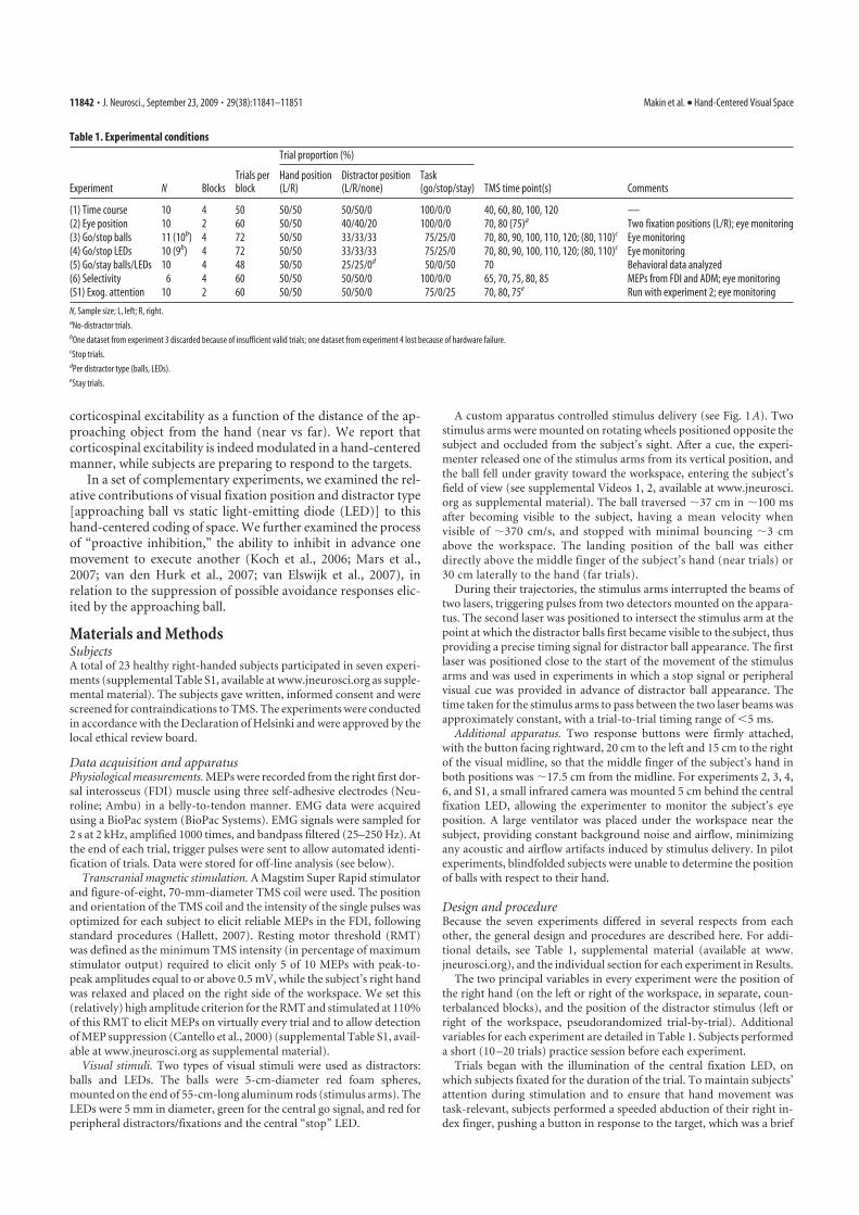
corticospinal excitability as a function of the distance of the ap-proaching object from the hand (near vs far). We report thatcorticospinal excitability is indeed modulated in a hand-centeredmanner, while subjects are preparing to respond to the targets.
In a set of complementary experiments, we examined the rel-ative contributions of visual fixation position and distractor type[approaching ball vs static light-emitting diode (LED)] to thishand-centered coding of space. We further examined the processof “proactive inhibition,” the ability to inhibit in advance onemovement to execute another (Koch et al., 2006; Mars et al.,2007; van den Hurk et al., 2007; van Elswijk et al., 2007), inrelation to the suppression of possible avoidance responses elic-ited by the approaching ball.
Materials and MethodsSubjectsA total of 23 healthy right-handed subjects participated in seven experi-ments (supplemental Table S1, available at www.jneurosci.org as supple-mental material). The subjects gave written, informed consent and werescreened for contraindications to TMS. The experiments were conductedin accordance with the Declaration of Helsinki and were approved by thelocal ethical review board.
Data acquisition and apparatusPhysiological measurements. MEPs were recorded from the right first dor-sal interosseus (FDI) muscle using three self-adhesive electrodes (Neu-roline; Ambu) in a belly-to-tendon manner. EMG data were acquiredusing a BioPac system (BioPac Systems). EMG signals were sampled for2 s at 2 kHz, amplified 1000 times, and bandpass filtered (25–250 Hz). Atthe end of each trial, trigger pulses were sent to allow automated identi-fication of trials. Data were stored for off-line analysis (see below).
Transcranial magnetic stimulation. A Magstim Super Rapid stimulatorand figure-of-eight, 70-mm-diameter TMS coil were used. The positionand orientation of the TMS coil and the intensity of the single pulses wasoptimized for each subject to elicit reliable MEPs in the FDI, followingstandard procedures (Hallett, 2007). Resting motor threshold (RMT)was defined as the minimum TMS intensity (in percentage of maximumstimulator output) required to elicit only 5 of 10 MEPs with peak-to-peak amplitudes equal to or above 0.5 mV, while the subject’s right handwas relaxed and placed on the right side of the workspace. We set this(relatively) high amplitude criterion for the RMT and stimulated at 110%of this RMT to elicit MEPs on virtually every trial and to allow detectionof MEP suppression (Cantello et al., 2000) (supplemental Table S1, avail-able at www.jneurosci.org as supplemental material).
Visual stimuli. Two types of visual stimuli were used as distractors:balls and LEDs. The balls were 5-cm-diameter red foam spheres,mounted on the end of 55-cm-long aluminum rods (stimulus arms). TheLEDs were 5 mm in diameter, green for the central go signal, and red forperipheral distractors/fixations and the central “stop” LED.
A custom apparatus controlled stimulus delivery (see Fig. 1 A). Twostimulus arms were mounted on rotating wheels positioned opposite thesubject and occluded from the subject’s sight. After a cue, the experi-menter released one of the stimulus arms from its vertical position, andthe ball fell under gravity toward the workspace, entering the subject’sfield of view (see supplemental Videos 1, 2, available at www.jneurosci.org as supplemental material). The ball traversed 37 cm in 100 msafter becoming visible to the subject, having a mean velocity whenvisible of 370 cm/s, and stopped with minimal bouncing 3 cmabove the workspace. The landing position of the ball was eitherdirectly above the middle finger of the subject’s hand (near trials) or30 cm laterally to the hand (far trials).
During their trajectories, the stimulus arms interrupted the beams oftwo lasers, triggering pulses from two detectors mounted on the appara-tus. The second laser was positioned to intersect the stimulus arm at thepoint at which the distractor balls first became visible to the subject, thusproviding a precise timing signal for distractor ball appearance. The firstlaser was positioned close to the start of the movement of the stimulusarms and was used in experiments in which a stop signal or peripheralvisual cue was provided in advance of distractor ball appearance. Thetime taken for the stimulus arms to pass between the two laser beams wasapproximately constant, with a trial-to-trial timing range of �5 ms.
Additional apparatus. Two response buttons were firmly attached,with the button facing rightward, 20 cm to the left and 15 cm to the rightof the visual midline, so that the middle finger of the subject’s hand inboth positions was 17.5 cm from the midline. For experiments 2, 3, 4,6, and S1, a small infrared camera was mounted 5 cm behind the centralfixation LED, allowing the experimenter to monitor the subject’s eyeposition. A large ventilator was placed under the workspace near thesubject, providing constant background noise and airflow, minimizingany acoustic and airflow artifacts induced by stimulus delivery. In pilotexperiments, blindfolded subjects were unable to determine the positionof balls with respect to their hand.
Design and procedureBecause the seven experiments differed in several respects from eachother, the general design and procedures are described here. For addi-tional details, see Table 1, supplemental material (available at www.jneurosci.org), and the individual section for each experiment in Results.
The two principal variables in every experiment were the position ofthe right hand (on the left or right of the workspace, in separate, coun-terbalanced blocks), and the position of the distractor stimulus (left orright of the workspace, pseudorandomized trial-by-trial). Additionalvariables for each experiment are detailed in Table 1. Subjects performeda short (10 –20 trials) practice session before each experiment.
Trials began with the illumination of the central fixation LED, onwhich subjects fixated for the duration of the trial. To maintain subjects’attention during stimulation and to ensure that hand movement wastask-relevant, subjects performed a speeded abduction of their right in-dex finger, pushing a button in response to the target, which was a brief
Table 1. Experimental conditions
Trial proportion (%)
Experiment N BlocksTrials perblock
Hand position(L/R)
Distractor position(L/R/none)
Task(go/stop/stay) TMS time point(s) Comments
(1) Time course 10 4 50 50/50 50/50/0 100/0/0 40, 60, 80, 100, 120 —
(2) Eye position 10 2 60 50/50 40/40/20 100/0/0 70, 80 (75)a Two fixation positions (L/R); eye monitoring
(3) Go/stop balls 11 (10b) 4 72 50/50 33/33/33 75/25/0 70, 80, 90, 100, 110, 120; (80, 110)c Eye monitoring
(4) Go/stop LEDs 10 (9b) 4 72 50/50 33/33/33 75/25/0 70, 80, 90, 100, 110, 120; (80, 110)c Eye monitoring
(5) Go/stay balls/LEDs 10 4 48 50/50 25/25/0d 50/0/50 70 Behavioral data analyzed
(6) Selectivity 6 4 60 50/50 50/50/0 100/0/0 65, 70, 75, 80, 85 MEPs from FDI and ADM; eye monitoring
(S1) Exog. attention 10 2 60 50/50 50/50/0 75/0/25 70, 80, 75e Run with experiment 2; eye monitoring
N, Sample size; L, left; R, right.aNo-distractor trials.bOne dataset from experiment 3 discarded because of insufficient valid trials; one dataset from experiment 4 lost because of hardware failure.cStop trials.dPer distractor type (balls, LEDs).eStay trials.
11842 • J. Neurosci., September 23, 2009 • 29(38):11841–11851 Makin et al. • Hand-Centered Visual Space
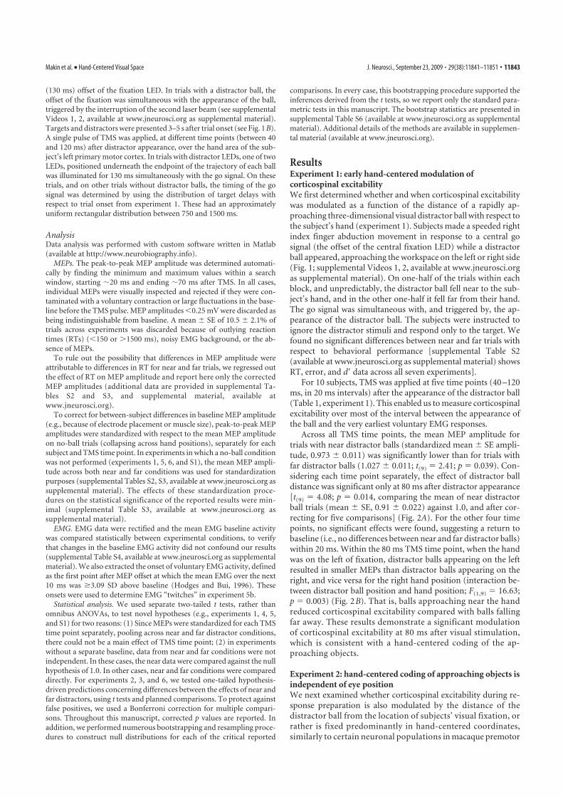
(130 ms) offset of the fixation LED. In trials with a distractor ball, theoffset of the fixation was simultaneous with the appearance of the ball,triggered by the interruption of the second laser beam (see supplementalVideos 1, 2, available at www.jneurosci.org as supplemental material).Targets and distractors were presented 3–5 s after trial onset (see Fig. 1 B).A single pulse of TMS was applied, at different time points (between 40and 120 ms) after distractor appearance, over the hand area of the sub-ject’s left primary motor cortex. In trials with distractor LEDs, one of twoLEDs, positioned underneath the endpoint of the trajectory of each ballwas illuminated for 130 ms simultaneously with the go signal. On thesetrials, and on other trials without distractor balls, the timing of the gosignal was determined by using the distribution of target delays withrespect to trial onset from experiment 1. These had an approximatelyuniform rectangular distribution between 750 and 1500 ms.
AnalysisData analysis was performed with custom software written in Matlab(available at http://www.neurobiography.info).
MEPs. The peak-to-peak MEP amplitude was determined automati-cally by finding the minimum and maximum values within a searchwindow, starting 20 ms and ending 70 ms after TMS. In all cases,individual MEPs were visually inspected and rejected if they were con-taminated with a voluntary contraction or large fluctuations in the base-line before the TMS pulse. MEP amplitudes �0.25 mV were discarded asbeing indistinguishable from baseline. A mean � SE of 10.5 � 2.1% oftrials across experiments was discarded because of outlying reactiontimes (RTs) (�150 or 1500 ms), noisy EMG background, or the ab-sence of MEPs.
To rule out the possibility that differences in MEP amplitude wereattributable to differences in RT for near and far trials, we regressed outthe effect of RT on MEP amplitude and report here only the correctedMEP amplitudes (additional data are provided in supplemental Ta-bles S2 and S3, and supplemental material, available atwww.jneurosci.org).
To correct for between-subject differences in baseline MEP amplitude(e.g., because of electrode placement or muscle size), peak-to-peak MEPamplitudes were standardized with respect to the mean MEP amplitudeon no-ball trials (collapsing across hand positions), separately for eachsubject and TMS time point. In experiments in which a no-ball conditionwas not performed (experiments 1, 5, 6, and S1), the mean MEP ampli-tude across both near and far conditions was used for standardizationpurposes (supplemental Tables S2, S3, available at www.jneurosci.org assupplemental material). The effects of these standardization proce-dures on the statistical significance of the reported results were min-imal (supplemental Table S3, available at www.jneurosci.org assupplemental material).
EMG. EMG data were rectified and the mean EMG baseline activitywas compared statistically between experimental conditions, to verifythat changes in the baseline EMG activity did not confound our results(supplemental Table S4, available at www.jneurosci.org as supplementalmaterial). We also extracted the onset of voluntary EMG activity, definedas the first point after MEP offset at which the mean EMG over the next10 ms was �3.09 SD above baseline (Hodges and Bui, 1996). Theseonsets were used to determine EMG “twitches” in experiment 5b.
Statistical analysis. We used separate two-tailed t tests, rather thanomnibus ANOVAs, to test novel hypotheses (e.g., experiments 1, 4, 5,and S1) for two reasons: (1) Since MEPs were standardized for each TMStime point separately, pooling across near and far distractor conditions,there could not be a main effect of TMS time point; (2) in experimentswithout a separate baseline, data from near and far conditions were notindependent. In these cases, the near data were compared against the nullhypothesis of 1.0. In other cases, near and far conditions were compareddirectly. For experiments 2, 3, and 6, we tested one-tailed hypothesis-driven predictions concerning differences between the effects of near andfar distractors, using t tests and planned comparisons. To protect againstfalse positives, we used a Bonferroni correction for multiple compari-sons. Throughout this manuscript, corrected p values are reported. Inaddition, we performed numerous bootstrapping and resampling proce-dures to construct null distributions for each of the critical reported
comparisons. In every case, this bootstrapping procedure supported theinferences derived from the t tests, so we report only the standard para-metric tests in this manuscript. The bootstrap statistics are presented insupplemental Table S6 (available at www.jneurosci.org as supplementalmaterial). Additional details of the methods are available in supplemen-tal material (available at www.jneurosci.org).
ResultsExperiment 1: early hand-centered modulation ofcorticospinal excitabilityWe first determined whether and when corticospinal excitabilitywas modulated as a function of the distance of a rapidly ap-proaching three-dimensional visual distractor ball with respect tothe subject’s hand (experiment 1). Subjects made a speeded rightindex finger abduction movement in response to a central gosignal (the offset of the central fixation LED) while a distractorball appeared, approaching the workspace on the left or right side(Fig. 1; supplemental Videos 1, 2, available at www.jneurosci.orgas supplemental material). On one-half of the trials within eachblock, and unpredictably, the distractor ball fell near to the sub-ject’s hand, and in the other one-half it fell far from their hand.The go signal was simultaneous with, and triggered by, the ap-pearance of the distractor ball. The subjects were instructed toignore the distractor stimuli and respond only to the target. Wefound no significant differences between near and far trials withrespect to behavioral performance [supplemental Table S2(available at www.jneurosci.org as supplemental material) showsRT, error, and d� data across all seven experiments].
For 10 subjects, TMS was applied at five time points (40 –120ms, in 20 ms intervals) after the appearance of the distractor ball(Table 1, experiment 1). This enabled us to measure corticospinalexcitability over most of the interval between the appearance ofthe ball and the very earliest voluntary EMG responses.
Across all TMS time points, the mean MEP amplitude fortrials with near distractor balls (standardized mean � SE ampli-tude, 0.973 � 0.011) was significantly lower than for trials withfar distractor balls (1.027 � 0.011; t(9) � 2.41; p � 0.039). Con-sidering each time point separately, the effect of distractor balldistance was significant only at 80 ms after distractor appearance[t(9) � 4.08; p � 0.014, comparing the mean of near distractorball trials (mean � SE, 0.91 � 0.022) against 1.0, and after cor-recting for five comparisons] (Fig. 2A). For the other four timepoints, no significant effects were found, suggesting a return tobaseline (i.e., no differences between near and far distractor balls)within 20 ms. Within the 80 ms TMS time point, when the handwas on the left of fixation, distractor balls appearing on the leftresulted in smaller MEPs than distractor balls appearing on theright, and vice versa for the right hand position (interaction be-tween distractor ball position and hand position; F(1,9) � 16.63;p � 0.003) (Fig. 2B). That is, balls approaching near the handreduced corticospinal excitability compared with balls fallingfar away. These results demonstrate a significant modulationof corticospinal excitability at 80 ms after visual stimulation,which is consistent with a hand-centered coding of the ap-proaching objects.
Experiment 2: hand-centered coding of approaching objects isindependent of eye positionWe next examined whether corticospinal excitability during re-sponse preparation is also modulated by the distance of thedistractor ball from the location of subjects’ visual fixation, orrather is fixed predominantly in hand-centered coordinates,similarly to certain neuronal populations in macaque premotor
Makin et al. • Hand-Centered Visual Space J. Neurosci., September 23, 2009 • 29(38):11841–11851 • 11843
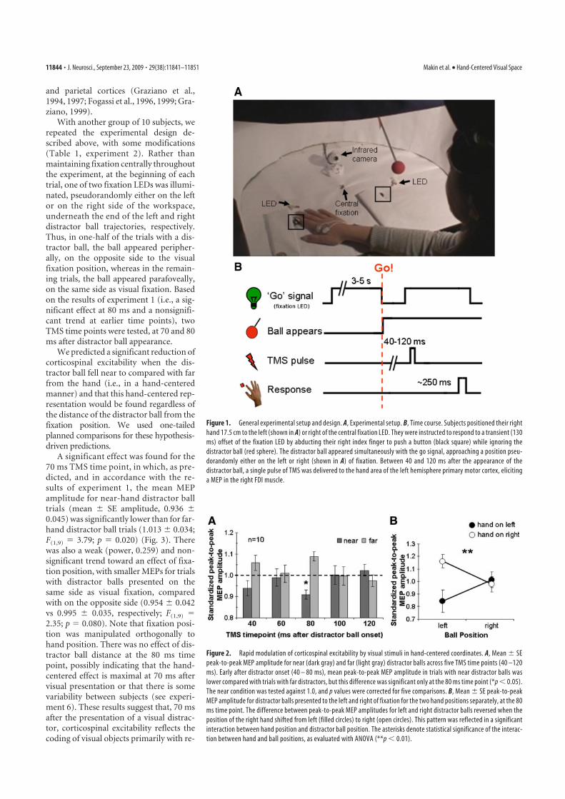
and parietal cortices (Graziano et al.,1994, 1997; Fogassi et al., 1996, 1999; Gra-ziano, 1999).
With another group of 10 subjects, werepeated the experimental design de-scribed above, with some modifications(Table 1, experiment 2). Rather thanmaintaining fixation centrally throughoutthe experiment, at the beginning of eachtrial, one of two fixation LEDs was illumi-nated, pseudorandomly either on the leftor on the right side of the workspace,underneath the end of the left and rightdistractor ball trajectories, respectively.Thus, in one-half of the trials with a dis-tractor ball, the ball appeared peripher-ally, on the opposite side to the visualfixation position, whereas in the remain-ing trials, the ball appeared parafoveally,on the same side as visual fixation. Basedon the results of experiment 1 (i.e., a sig-nificant effect at 80 ms and a nonsignifi-cant trend at earlier time points), twoTMS time points were tested, at 70 and 80ms after distractor ball appearance.
We predicted a significant reduction ofcorticospinal excitability when the dis-tractor ball fell near to compared with farfrom the hand (i.e., in a hand-centeredmanner) and that this hand-centered rep-resentation would be found regardless ofthe distance of the distractor ball from thefixation position. We used one-tailedplanned comparisons for these hypothesis-driven predictions.
A significant effect was found for the70 ms TMS time point, in which, as pre-dicted, and in accordance with the re-sults of experiment 1, the mean MEPamplitude for near-hand distractor balltrials (mean � SE amplitude, 0.936 �0.045) was significantly lower than for far-hand distractor ball trials (1.013 � 0.034;F(1,9) � 3.79; p � 0.020) (Fig. 3). Therewas also a weak (power, 0.259) and non-significant trend toward an effect of fixa-tion position, with smaller MEPs for trialswith distractor balls presented on thesame side as visual fixation, comparedwith on the opposite side (0.954 � 0.042vs 0.995 � 0.035, respectively; F(1,9) �2.35; p � 0.080). Note that fixation posi-tion was manipulated orthogonally tohand position. There was no effect of dis-tractor ball distance at the 80 ms timepoint, possibly indicating that the hand-centered effect is maximal at 70 ms aftervisual presentation or that there is somevariability between subjects (see experi-ment 6). These results suggest that, 70 msafter the presentation of a visual distrac-tor, corticospinal excitability reflects thecoding of visual objects primarily with re-
Figure 1. General experimental setup and design. A, Experimental setup. B, Time course. Subjects positioned their right
hand 17.5 cm to the left (shown in A) or right of the central fixation LED. They were instructed to respond to a transient (130
ms) offset of the fixation LED by abducting their right index finger to push a button (black square) while ignoring the
distractor ball (red sphere). The distractor ball appeared simultaneously with the go signal, approaching a position pseu-
dorandomly either on the left or right (shown in A) of fixation. Between 40 and 120 ms after the appearance of the
distractor ball, a single pulse of TMS was delivered to the hand area of the left hemisphere primary motor cortex, eliciting
a MEP in the right FDI muscle.
Figure 2. Rapid modulation of corticospinal excitability by visual stimuli in hand-centered coordinates. A, Mean � SE
peak-to-peak MEP amplitude for near (dark gray) and far (light gray) distractor balls across five TMS time points (40 –120
ms). Early after distractor onset (40 – 80 ms), mean peak-to-peak MEP amplitude in trials with near distractor balls was
lower compared with trials with far distractors, but this difference was significant only at the 80 ms time point (*p � 0.05).
The near condition was tested against 1.0, and p values were corrected for five comparisons. B, Mean � SE peak-to-peak
MEP amplitude for distractor balls presented to the left and right of fixation for the two hand positions separately, at the 80
ms time point. The difference between peak-to-peak MEP amplitudes for left and right distractor balls reversed when the
position of the right hand shifted from left (filled circles) to right (open circles). This pattern was reflected in a significant
interaction between hand position and distractor ball position. The asterisks denote statistical significance of the interac-
tion between hand and ball positions, as evaluated with ANOVA (**p � 0.01).
11844 • J. Neurosci., September 23, 2009 • 29(38):11841–11851 Makin et al. • Hand-Centered Visual Space
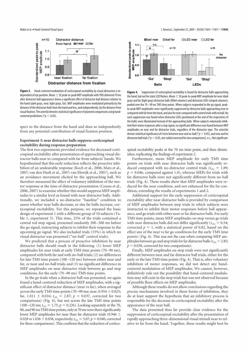
spect to the distance from the hand and does so independentlyfrom any potential contribution of visual fixation position.
Experiment 3: near distractor balls suppress corticospinalexcitability during response preparationThe first two experiments provided evidence for decreased corti-cospinal excitability after presentation of approaching visual dis-tractor balls near to compared with far from subjects’ hands. Wehypothesized that this early reduction reflects the proactive inhi-bition of an undesirable response (Koch et al., 2006; Mars et al.,2007; van den Hurk et al., 2007; van Elswijk et al., 2007), such asan avoidance movement elicited by the approaching ball. Wetherefore measured the effect of voluntary inhibition of the mo-tor response at the time of distractor presentation (Coxon et al.,2006, 2007) to examine whether this would suppress MEP ampli-tudes to a similar level as found with near distractor balls. Addi-tionally, we included a no-distractor “baseline” condition toassess whether near balls decrease, or else far balls increase, cor-ticospinal excitability. For this purpose, we repeated the basicdesign of experiment 1 with a different group of 10 subjects (Ta-ble 1, experiment 3). This time, 25% of the trials contained acentral red stop signal, presented randomly 200 –300 ms beforethe go signal, instructing subjects to inhibit their response to theupcoming go signal. We also included trials (33%) in which novisual distractor was presented (“no-ball” control trials).
We predicted that a process of proactive inhibition by neardistractor balls should result in the following: (1) lower MEPamplitudes for near trials at early TMS time points (70 –90 ms),compared with both far and with no-ball trials; (2) no differencesfor late TMS time points (100 –120 ms) between either near andfar, or near and no-ball trials; and (3) no significant difference inMEP amplitudes on near distractor trials between go and stopconditions, for the early (70 –90 ms) TMS time points.
In the go trials when a distractor ball was presented, we againfound a hand-centered reduction of MEP amplitudes, with a sig-nificant effect of distractor distance (near vs far), when averagedacross the early TMS time points (70 –90 ms; near, 0.954 � 0.025;far, 1.011 � 0.016; t(9) � 2.87; p � 0.037, corrected for twocomparisons) (Fig. 4), but not across the late TMS time points(100–120 ms; t(9) � 1.75; p � 0.226). Looking separately at the 70,80, and 90 ms TMS time points, only at 70 ms were there significantlylower MEP amplitudes for near than far distractor trials (0.946 �0.028 vs 1.036 � 0.038, respectively; t(9) � 3.07; p � 0.040, correctedfor three comparisons). This confirms that the reduction of cortico-
spinal excitability peaks at the 70 ms time point, and then dimin-ishes, replicating the findings of experiment 2.
Furthermore, mean MEP amplitude for early TMS timepoints on trials with near distractor balls was significantly re-duced compared with no-distractor control trials (t(9) � 1.89;p � 0.046, compared against 1.0), whereas MEPs for trials withfar distractor balls were not significantly different from no-balltrials (Fig. 4). These results show that MEP amplitudes were re-duced for the near condition, and not enhanced for the far con-dition, extending the results of experiments 1 and 2.
Additional support for the early suppression of corticospinalexcitability after near distractor balls is provided by comparisonof MEP amplitudes between stop trials in which subjects wereinstructed to inhibit their motor responses before ball appear-ance, and go trials with either near or far distractor balls. For earlyTMS time points, mean MEP amplitudes on stop versus go trialswith near distractor balls did not differ significantly (t(9) � 0.54;corrected p � 1, with a statistical power of 0.82, based on theeffect size of the near vs far go conditions for the early TMS timepoints) (Fig. 4). This was not the case when comparing MEP am-plitudes between go and stop trials for far distractor balls (t(9) � 2.85;p � 0.038, corrected for two comparisons).
Finally, MEP amplitudes on stop trials were not significantlydifferent between near and far distractor ball trials, either for theearly or the late TMS time points (Fig. 4). That is, after voluntaryinhibition of motor responses, we did not detect any hand-centered modulation of MEP amplitudes. We cannot, however,definitively rule out the possibility that hand-centered modula-tion may still exist in the stop trials but was not observed becauseof possible floor effects on MEP amplitudes.
Although these results do not allow conclusions regarding theprecise mechanisms involved in these forms of inhibition, theydo at least support the hypothesis that an inhibitory process isresponsible for the decrease in corticospinal excitability after theappearance of the near ball.
The data presented thus far provide clear evidence for thesuppression of corticospinal excitability after the presentation ofrapidly approaching three-dimensional visual stimuli near to rel-ative to far from the hand. Together, these results might best be
Figure 3. Hand-centered modulation of corticospinal excitability by visual distractors is in-
dependent of eye position. Mean � SE peak-to-peak MEP amplitude with TMS delivered 70 ms
after distractor ball appearance shows a significant effect of distractor ball distance relative to
the hand (dark grays, near; light grays, far). MEP amplitudes were modulated primarily by the
distance of the distractor balls from the hand and less, and independently, by the distance from
visual fixation. The asterisk denotes statistical significance of planned comparisons using hand-
centered predictions (*p � 0.05).
Figure 4. Suppression of corticospinal excitability is found for distractor balls approaching
the hand, but not for static LED flashes. Mean � SE peak-to-peak MEP amplitude for near (dark
gray) and far (light gray) distractor balls (filled columns) and distractor LEDs (striped columns),
pooled over the 70 –90 ms TMS time points. When subjects responded to the go signal, peak-
to-peak MEP amplitudes were significantly suppressed by distractor balls approaching near to
compared with far from the hand, and also for near compared with control trials with no ball. No
such suppression was found when distractor LEDs (positioned at the end of the trajectories of
the balls) were illuminated instead of the approaching balls. When subjects voluntarily inhib-
ited their motor responses after a stop signal, no significant difference was found between MEP
amplitudes on near and far distractor trials, regardless of the distractor type. The asterisks
denote statistical significance of t tests between near and no-ball (*p � 0.05), and near and far
distractor ball trials (*p � 0.05, one-tailed corrected for two comparisons). n.s., Not significant.
Makin et al. • Hand-Centered Visual Space J. Neurosci., September 23, 2009 • 29(38):11841–11851 • 11845
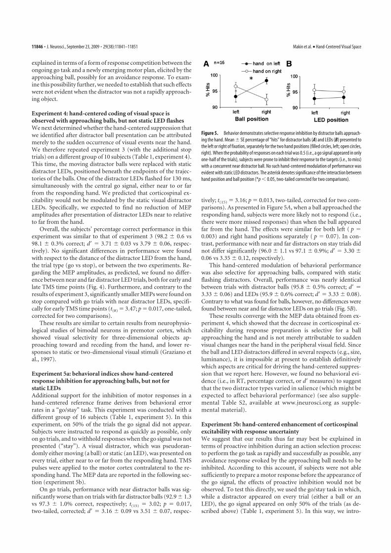
explained in terms of a form of response competition between theongoing go task and a newly emerging motor plan, elicited by theapproaching ball, possibly for an avoidance response. To exam-ine this possibility further, we needed to establish that such effectswere not evident when the distractor was not a rapidly approach-ing object.
Experiment 4: hand-centered coding of visual space isobserved with approaching balls, but not static LED flashesWe next determined whether the hand-centered suppression thatwe identified after distractor ball presentation can be attributedmerely to the sudden occurrence of visual events near the hand.We therefore repeated experiment 3 (with the additional stoptrials) on a different group of 10 subjects (Table 1, experiment 4).This time, the moving distractor balls were replaced with staticdistractor LEDs, positioned beneath the endpoints of the trajec-tories of the balls. One of the distractor LEDs flashed for 130 ms,simultaneously with the central go signal, either near to or farfrom the responding hand. We predicted that corticospinal ex-citability would not be modulated by the static visual distractorLEDs. Specifically, we expected to find no reduction of MEPamplitudes after presentation of distractor LEDs near to relativeto far from the hand.
Overall, the subjects’ percentage correct performance in thisexperiment was similar to that of experiment 3 (98.2 � 0.6 vs98.1 � 0.3% correct; d� � 3.71 � 0.03 vs 3.79 � 0.06, respec-tively). No significant differences in performance were foundwith respect to the distance of the distractor LED from the hand,the trial type (go vs stop), or between the two experiments. Re-garding the MEP amplitudes, as predicted, we found no differ-ence between near and far distractor LED trials, both for early andlate TMS time points (Fig. 4). Furthermore, and contrary to theresults of experiment 3, significantly smaller MEPs were found onstop compared with go trials with near distractor LEDs, specifi-cally for early TMS time points (t(8) � 3.47; p � 0.017, one-tailed,corrected for two comparisons).
These results are similar to certain results from neurophysio-logical studies of bimodal neurons in premotor cortex, whichshowed visual selectivity for three-dimensional objects ap-proaching toward and receding from the hand, and lower re-sponses to static or two-dimensional visual stimuli (Graziano etal., 1997).
Experiment 5a: behavioral indices show hand-centeredresponse inhibition for approaching balls, but not forstatic LEDsAdditional support for the inhibition of motor responses in ahand-centered reference frame derives from behavioral errorrates in a “go/stay” task. This experiment was conducted with adifferent group of 16 subjects (Table 1, experiment 5). In thisexperiment, on 50% of the trials the go signal did not appear.Subjects were instructed to respond as quickly as possible, onlyon go trials, and to withhold responses when the go signal was notpresented (“stay”). A visual distractor, which was pseudoran-domly either moving (a ball) or static (an LED), was presented onevery trial, either near to or far from the responding hand. TMSpulses were applied to the motor cortex contralateral to the re-sponding hand. The MEP data are reported in the following sec-tion (experiment 5b).
On go trials, performance with near distractor balls was sig-nificantly worse than on trials with far distractor balls (92.9 � 1.3vs 97.3 � 1.0% correct, respectively; t(15) � 3.02; p � 0.017,two-tailed, corrected; d� � 3.16 � 0.09 vs 3.51 � 0.07, respec-
tively; t(15) � 3.16; p � 0.013, two-tailed, corrected for two com-parisons). As presented in Figure 5A, when a ball approached theresponding hand, subjects were more likely not to respond (i.e.,there were more missed responses) than when the ball appearedfar from the hand. The effects were similar for both left ( p �0.003) and right hand positions separately ( p � 0.07). In con-trast, performance with near and far distractors on stay trials didnot differ significantly (96.0 � 1.1 vs 97.1 � 0.9%; d� � 3.30 �0.06 vs 3.35 � 0.12, respectively).
This hand-centered modulation of behavioral performancewas also selective for approaching balls, compared with staticflashing distractors. Overall, performance was nearly identicalbetween trials with distractor balls (95.8 � 0.5% correct; d� �3.33 � 0.06) and LEDs (95.9 � 0.6% correct; d� � 3.33 � 0.08).Contrary to what was found for balls, however, no differences werefound between near and far distractor LEDs on go trials (Fig. 5B).
These results converge with the MEP data obtained from ex-periment 4, which showed that the decrease in corticospinal ex-citability during response preparation is selective for a ballapproaching the hand and is not merely attributable to suddenvisual changes near the hand in the peripheral visual field. Sincethe ball and LED distractors differed in several respects (e.g., size,luminance), it is impossible at present to establish definitivelywhich aspects are critical for driving the hand-centered suppres-sion that we report here. However, we found no behavioral evi-dence (i.e., in RT, percentage correct, or d� measures) to suggestthat the two distractor types varied in salience (which might beexpected to affect behavioral performance) (see also supple-mental Table S2, available at www.jneurosci.org as supple-mental material).
Experiment 5b: hand-centered enhancement of corticospinalexcitability with response uncertaintyWe suggest that our results thus far may best be explained interms of proactive inhibition during an action selection process:to perform the go task as rapidly and successfully as possible, anyavoidance response evoked by the approaching ball needs to beinhibited. According to this account, if subjects were not ablesufficiently to prepare a motor response before the appearance ofthe go signal, the effects of proactive inhibition would not beobserved. To test this directly, we used the go/stay task in which,while a distractor appeared on every trial (either a ball or anLED), the go signal appeared on only 50% of the trials (as de-scribed above) (Table 1, experiment 5). In this way, we intro-
Figure 5. Behavior demonstrates selective response inhibition by distractor balls approach-
ing the hand. Mean � SE percentage of “hits” for distractor balls (A) and LEDs (B) presented to
the left or right of fixation, separately for the two hand positions (filled circles, left; open circles,
right). When the probability of responses on each trial was 0.5 (i.e., a go signal appeared in only
one-half of the trials), subjects were prone to inhibit their response to the targets (i.e., to miss)
with a concurrent near distractor ball. No such hand-centered modulation of performance was
evident with static LED distractors. The asterisk denotes significance of the interaction between
hand position and ball position (*p � 0.05, two-tailed corrected for two comparisons).
11846 • J. Neurosci., September 23, 2009 • 29(38):11841–11851 Makin et al. • Hand-Centered Visual Space
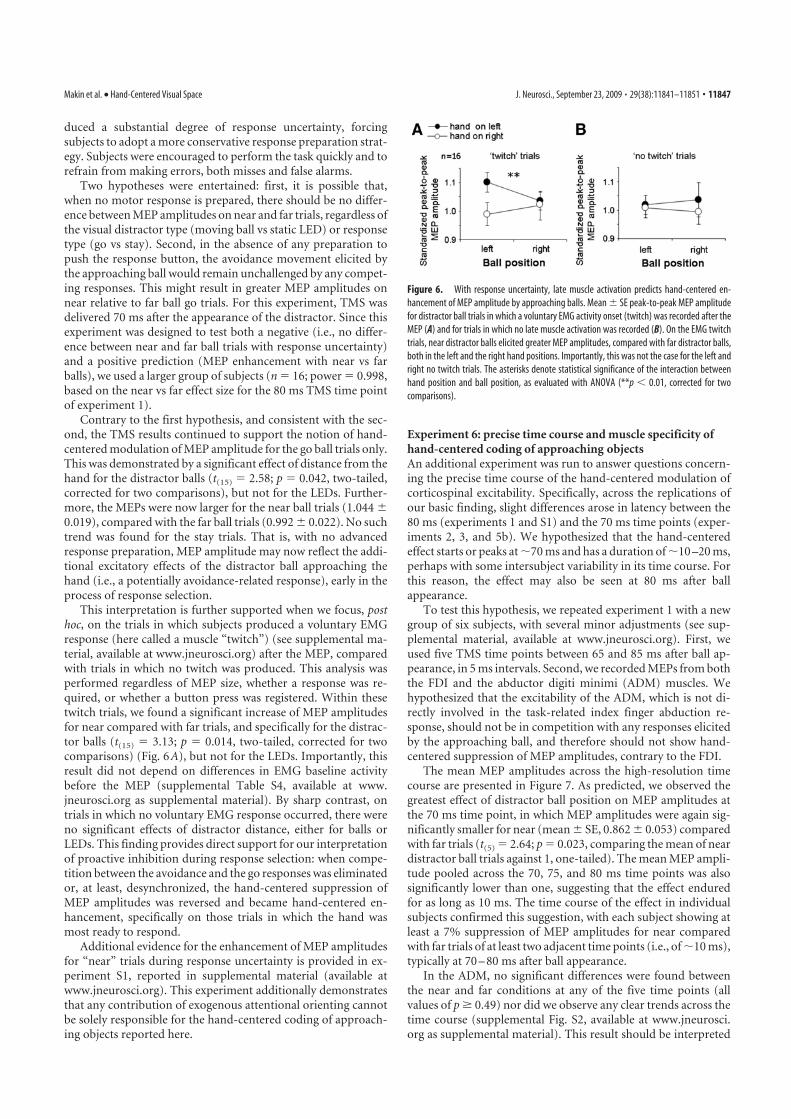
duced a substantial degree of response uncertainty, forcingsubjects to adopt a more conservative response preparation strat-egy. Subjects were encouraged to perform the task quickly and torefrain from making errors, both misses and false alarms.
Two hypotheses were entertained: first, it is possible that,when no motor response is prepared, there should be no differ-ence between MEP amplitudes on near and far trials, regardless ofthe visual distractor type (moving ball vs static LED) or responsetype (go vs stay). Second, in the absence of any preparation topush the response button, the avoidance movement elicited bythe approaching ball would remain unchallenged by any compet-ing responses. This might result in greater MEP amplitudes onnear relative to far ball go trials. For this experiment, TMS wasdelivered 70 ms after the appearance of the distractor. Since thisexperiment was designed to test both a negative (i.e., no differ-ence between near and far ball trials with response uncertainty)and a positive prediction (MEP enhancement with near vs farballs), we used a larger group of subjects (n � 16; power � 0.998,based on the near vs far effect size for the 80 ms TMS time pointof experiment 1).
Contrary to the first hypothesis, and consistent with the sec-ond, the TMS results continued to support the notion of hand-centered modulation of MEP amplitude for the go ball trials only.This was demonstrated by a significant effect of distance from thehand for the distractor balls (t(15) � 2.58; p � 0.042, two-tailed,corrected for two comparisons), but not for the LEDs. Further-more, the MEPs were now larger for the near ball trials (1.044 �0.019), compared with the far ball trials (0.992 � 0.022). No suchtrend was found for the stay trials. That is, with no advancedresponse preparation, MEP amplitude may now reflect the addi-tional excitatory effects of the distractor ball approaching thehand (i.e., a potentially avoidance-related response), early in theprocess of response selection.
This interpretation is further supported when we focus, posthoc, on the trials in which subjects produced a voluntary EMGresponse (here called a muscle “twitch”) (see supplemental ma-terial, available at www.jneurosci.org) after the MEP, comparedwith trials in which no twitch was produced. This analysis wasperformed regardless of MEP size, whether a response was re-quired, or whether a button press was registered. Within thesetwitch trials, we found a significant increase of MEP amplitudesfor near compared with far trials, and specifically for the distrac-tor balls (t(15) � 3.13; p � 0.014, two-tailed, corrected for twocomparisons) (Fig. 6A), but not for the LEDs. Importantly, thisresult did not depend on differences in EMG baseline activitybefore the MEP (supplemental Table S4, available at www.jneurosci.org as supplemental material). By sharp contrast, ontrials in which no voluntary EMG response occurred, there wereno significant effects of distractor distance, either for balls orLEDs. This finding provides direct support for our interpretationof proactive inhibition during response selection: when compe-tition between the avoidance and the go responses was eliminatedor, at least, desynchronized, the hand-centered suppression ofMEP amplitudes was reversed and became hand-centered en-hancement, specifically on those trials in which the hand wasmost ready to respond.
Additional evidence for the enhancement of MEP amplitudesfor “near” trials during response uncertainty is provided in ex-periment S1, reported in supplemental material (available atwww.jneurosci.org). This experiment additionally demonstratesthat any contribution of exogenous attentional orienting cannotbe solely responsible for the hand-centered coding of approach-ing objects reported here.
Experiment 6: precise time course and muscle specificity ofhand-centered coding of approaching objectsAn additional experiment was run to answer questions concern-ing the precise time course of the hand-centered modulation ofcorticospinal excitability. Specifically, across the replications ofour basic finding, slight differences arose in latency between the80 ms (experiments 1 and S1) and the 70 ms time points (exper-iments 2, 3, and 5b). We hypothesized that the hand-centeredeffect starts or peaks at 70 ms and has a duration of 10 –20 ms,perhaps with some intersubject variability in its time course. Forthis reason, the effect may also be seen at 80 ms after ballappearance.
To test this hypothesis, we repeated experiment 1 with a newgroup of six subjects, with several minor adjustments (see sup-plemental material, available at www.jneurosci.org). First, weused five TMS time points between 65 and 85 ms after ball ap-pearance, in 5 ms intervals. Second, we recorded MEPs from boththe FDI and the abductor digiti minimi (ADM) muscles. Wehypothesized that the excitability of the ADM, which is not di-rectly involved in the task-related index finger abduction re-sponse, should not be in competition with any responses elicitedby the approaching ball, and therefore should not show hand-centered suppression of MEP amplitudes, contrary to the FDI.
The mean MEP amplitudes across the high-resolution timecourse are presented in Figure 7. As predicted, we observed thegreatest effect of distractor ball position on MEP amplitudes atthe 70 ms time point, in which MEP amplitudes were again sig-nificantly smaller for near (mean � SE, 0.862 � 0.053) comparedwith far trials (t(5) � 2.64; p � 0.023, comparing the mean of neardistractor ball trials against 1, one-tailed). The mean MEP ampli-tude pooled across the 70, 75, and 80 ms time points was alsosignificantly lower than one, suggesting that the effect enduredfor as long as 10 ms. The time course of the effect in individualsubjects confirmed this suggestion, with each subject showing atleast a 7% suppression of MEP amplitudes for near comparedwith far trials of at least two adjacent time points (i.e., of 10 ms),typically at 70 – 80 ms after ball appearance.
In the ADM, no significant differences were found betweenthe near and far conditions at any of the five time points (allvalues of p � 0.49) nor did we observe any clear trends across thetime course (supplemental Fig. S2, available at www.jneurosci.org as supplemental material). This result should be interpreted
Figure 6. With response uncertainty, late muscle activation predicts hand-centered en-
hancement of MEP amplitude by approaching balls. Mean � SE peak-to-peak MEP amplitude
for distractor ball trials in which a voluntary EMG activity onset (twitch) was recorded after the
MEP (A) and for trials in which no late muscle activation was recorded (B). On the EMG twitch
trials, near distractor balls elicited greater MEP amplitudes, compared with far distractor balls,
both in the left and the right hand positions. Importantly, this was not the case for the left and
right no twitch trials. The asterisks denote statistical significance of the interaction between
hand position and ball position, as evaluated with ANOVA (**p � 0.01, corrected for two
comparisons).
Makin et al. • Hand-Centered Visual Space J. Neurosci., September 23, 2009 • 29(38):11841–11851 • 11847
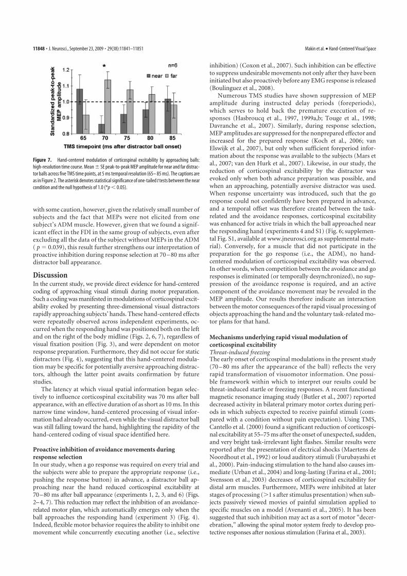
with some caution, however, given the relatively small number ofsubjects and the fact that MEPs were not elicited from onesubject’s ADM muscle. However, given that we found a signif-icant effect in the FDI in the same group of subjects, even afterexcluding all the data of the subject without MEPs in the ADM( p � 0.039), this result further strengthens our interpretation ofproactive inhibition during response selection at 70 – 80 ms afterdistractor ball appearance.
DiscussionIn the current study, we provide direct evidence for hand-centeredcoding of approaching visual stimuli during motor preparation.Such a coding was manifested in modulations of corticospinal excit-ability evoked by presenting three-dimensional visual distractorsrapidly approaching subjects’ hands. These hand-centered effectswere repeatedly observed across independent experiments, oc-curred when the responding hand was positioned both on the leftand on the right of the body midline (Figs. 2, 6, 7), regardless ofvisual fixation position (Fig. 3), and were dependent on motorresponse preparation. Furthermore, they did not occur for staticdistractors (Fig. 4), suggesting that this hand-centered modula-tion may be specific for potentially aversive approaching distrac-tors, although the latter point awaits confirmation by futurestudies.
The latency at which visual spatial information began selec-tively to influence corticospinal excitability was 70 ms after ballappearance, with an effective duration of as short as 10 ms. In thisnarrow time window, hand-centered processing of visual infor-mation had already occurred, even while the visual distractor ballwas still falling toward the hand, highlighting the rapidity of thehand-centered coding of visual space identified here.
Proactive inhibition of avoidance movements duringresponse selectionIn our study, when a go response was required on every trial andthe subjects were able to prepare the appropriate response (i.e.,pushing the response button) in advance, a distractor ball ap-proaching near the hand reduced corticospinal excitability at70–80 ms after ball appearance (experiments 1, 2, 3, and 6) (Figs.2–4, 7). This reduction may reflect the inhibition of an avoidance-related motor plan, which automatically emerges only when theball approaches the responding hand (experiment 3) (Fig. 4).Indeed, flexible motor behavior requires the ability to inhibit onemovement while concurrently executing another (i.e., selective
inhibition) (Coxon et al., 2007). Such inhibition can be effectiveto suppress undesirable movements not only after they have beeninitiated but also proactively before any EMG response is released(Boulinguez et al., 2008).
Numerous TMS studies have shown suppression of MEPamplitude during instructed delay periods (foreperiods),which serves to hold back the premature execution of re-sponses (Hasbroucq et al., 1997, 1999a,b; Touge et al., 1998;Davranche et al., 2007). Similarly, during response selection,MEP amplitudes are suppressed for the nonprepared effector andincreased for the prepared response (Koch et al., 2006; vanElswijk et al., 2007), but only when sufficient foreperiod infor-mation about the response was available to the subjects (Mars etal., 2007; van den Hurk et al., 2007). Likewise, in our study, thereduction of corticospinal excitability by the distractor wasevoked only when both advance preparation was possible, andwhen an approaching, potentially aversive distractor was used.When response uncertainty was introduced, such that the goresponse could not confidently have been prepared in advance,and a temporal offset was therefore created between the task-related and the avoidance responses, corticospinal excitabilitywas enhanced for active trials in which the ball approached nearthe responding hand (experiments 4 and S1) (Fig. 6; supplemen-tal Fig. S1, available at www.jneurosci.org as supplemental mate-rial). Conversely, for a muscle that did not participate in thepreparation for the go response (i.e., the ADM), no hand-centered modulation of corticospinal excitability was observed.In other words, when competition between the avoidance and goresponses is eliminated (or temporally desynchronized), no sup-pression of the avoidance response is required, and an activecomponent of the avoidance movement may be revealed in theMEP amplitude. Our results therefore indicate an interactionbetween the motor consequences of the rapid visual processing ofobjects approaching the hand and the voluntary task-related mo-tor plans for that hand.
Mechanisms underlying rapid visual modulation ofcorticospinal excitabilityThreat-induced freezingThe early onset of corticospinal modulations in the present study(70 – 80 ms after the appearance of the ball) reflects the veryrapid transformation of visuomotor information. One possi-ble framework within which to interpret our results could bethreat-induced startle or freezing responses. A recent functionalmagnetic resonance imaging study (Butler et al., 2007) reporteddecreased activity in bilateral primary motor cortex during peri-ods in which subjects expected to receive painful stimuli (com-pared with a condition without pain expectation). Using TMS,Cantello et al. (2000) found a significant reduction of corticospi-nal excitability at 55–75 ms after the onset of unexpected, sudden,and very bright task-irrelevant light flashes. Similar results werereported after the presentation of electrical shocks (Maertens deNoordhout et al., 1992) or loud auditory stimuli (Furubayashi etal., 2000). Pain-inducing stimulation to the hand also causes im-mediate (Urban et al., 2004) and long-lasting (Farina et al., 2001;Svensson et al., 2003) decreases of corticospinal excitability fordistal arm muscles. Furthermore, MEPs were inhibited at laterstages of processing (1 s after stimulus presentation) when sub-jects passively viewed movies of painful simulation applied tospecific muscles on a model (Avenanti et al., 2005). It has beensuggested that such inhibition may act as a sort of motor “decer-ebration,” allowing the spinal motor system freely to develop pro-tective responses after noxious stimulation (Farina et al., 2003).
Figure 7. Hand-centered modulation of corticospinal excitability by approaching balls:
high-resolution time course. Mean � SE peak-to-peak MEP amplitude for near and far distrac-
tor balls across five TMS time points, at 5 ms temporal resolution (65– 85 ms). The captions are
as in Figure 2. The asterisk denotes statistical significance of one-tailed t tests between the near
condition and the null hypothesis of 1.0 (*p � 0.05).
11848 • J. Neurosci., September 23, 2009 • 29(38):11841–11851 Makin et al. • Hand-Centered Visual Space
We believe that the results described in the studies mentionedabove do not directly correspond with our findings, and thatpain- or startle-related inhibition cannot explain the results wereport here, for two reasons. First, it cannot explain the reversal ofthe direction of hand-centered modulation when subjects couldnot confidently prepare a response in advance. Second, thetime course of startle-related inhibition based on TMS studies(Cantello et al., 2000) just precedes the corticospinal modula-tions reported here, which are restricted to 70 – 80 ms after dis-tractor ball appearance. Furthermore, Cantello et al. (2000) didnot provide (or search for) evidence for any hand-centeredmechanisms, so any direct links with our results remain specula-tive. We therefore propose that, although fear-induced startlemight play some role in the mechanism under investigation, ourhand-centered modulation represents a later stage of selectionbetween an appropriate avoidance/defensive response and thetask-related response.
Cortical mechanismsNumerous cortical areas have been shown to modulate cortico-spinal excitability and may therefore contribute to the resultsreported here. These include the primary somatosensory area(Avenanti et al., 2007), posterior parietal areas, and the premotorcortex (for review, see O’Shea et al., 2008).
Cooke and Graziano (2003) studied macaque muscle activityduring defensive movements evoked by aversive cutaneous stim-ulation. They distinguished between startle-related EMG activityoccurring as early as 20 –30 ms after stimulus onset and latermuscle activity starting 70 ms after stimulus onset [Cooke andGraziano (2003), their Fig. 2C]. Only the latter activity was spa-tially specific to the muscles involved in the defensive response.Very similar motor responses were evoked by electrical macro-stimulation of bimodal regions of the premotor cortex (Grazianoet al., 2002). Indeed, M1 and the premotor cortex are denselyinterconnected, both in humans and in monkeys (Shimazu et al.,2004; Dum and Strick, 2005; Koch et al., 2006; O’Shea et al., 2007;Davare et al., 2008). The modulation of MEP amplitude withrespect to approaching balls that we report here may thereforereflect ongoing activity in the premotor cortex.
Some premotor neurons are active after visual stimulation attime periods similar to the ones we have described and arethought to encode the significance of visual cues for responseselection (Cisek and Kalaska, 2005). In humans, the premotorcortex has been shown to participate in the visuomotor transfor-mations required to configure hand posture with respect to ob-jects (Davare et al., 2006). Furthermore, recent studies haveshown a direct influence of premotor over primary motor cortexas early as 75 ms after a go signal (Koch et al., 2006; O’Shea et al.,2007). Although any comparisons between data drawn frommonkeys and humans, and using such different methods, shouldbe made with caution, given the timing and the spatial specificityof the above responses with respect to visual events, these mech-anisms fit very well with our results and suggest the involvementof human premotor areas with the hand-centered coding of vi-sual space. Following Band and van Boxtel (1999), we tentativelysuggest that one source of the hand-centered modulation that wereport may be the premotor cortex, the site of modulation may bethe primary motor cortex, and the modulation is manifested bychanges in MEP amplitudes (Band and van Boxtel, 1999).
Pruszynski et al. (2008) demonstrated rapid arm muscle re-sponses that were sensitive to the direction and distance of visualtargets at 95 ms after a go signal. This timing fits very well withour findings, assuming a 20 –25 ms conduction time between the
primary motor cortex and the intrinsic hand muscles. Pruszynskiand colleagues suggested that such rapid motor responses arecapable of all the sophistication attributed to voluntary control,within the constraints of their limited processing time. Additionalresearch is required to determine the precise neurophysiologicalmechanisms that might enable such rapid and sophisticated visuo-motor spatial processing in the human brain.
Subcortical mechanismsIn monkeys, spinal interneurons have been shown to participatein the control of movement during preparation periods, display-ing both excitatory changes in the same direction as the subse-quent movement-related activity, and inhibitory changes thatmay reflect superimposed mechanisms suppressing muscularoutput (Prut and Fetz, 1999). It is possible that the hand-centeredmodulation that we observed reflects such subcortical movementpreparation, although it is important to note that, in our results,the critical movement selection processes occurred based on theposition of the visual distractor ball relative to the respondinghand, so any candidate subcortical source of the reported modu-lations of corticospinal excitability would have to have access tosuch information.
Hand-centered visual space and attentionIn our experiments, both near and far balls were distractors; how-ever, it is possible that near distractors were more salient than fardistractors and that this somehow resulted in reduced levels ofcorticospinal excitability. Such differences in salience could havebeen the result of subjects covertly attending toward their re-sponding hand, despite the instructions and the central visualtask. We addressed these concerns in several experiments.
First, when the balls were substituted with LEDs positionednear and far from the hand, we found no significant hand-centered modulations of corticospinal excitability (experiment4) (Fig. 3) or of percentage correct performance (experiment 5)(Fig. 5). Second, we manipulated subject’s overt attention, byshifting their fixation between positions on the same or oppositeside as the impending distractor ball (experiment 2). When shift-ing the fixation position with respect to the ball and hand posi-tions, we found a small, but nonsignificant difference betweenballs approaching the same side, and balls approaching the oppo-site side of fixation (Fig. 3). At the same time, however, the dis-tance of the ball from the hand (manipulated orthogonally andindependently from the distance from fixation) significantlymodulated corticospinal excitability. Finally, we used a cueingparadigm to attract subjects’ exogenous covert visuospatial atten-tion toward versus away from the distractor ball, just before itsappearance (experiment S1; supplemental Fig. S1A; supplemen-tal material, available at www.jneurosci.org). Again, this manip-ulation did not significantly modulate corticospinal excitability,while the distance of the distractor from the hand remained mosteffective (albeit marginally significantly), regardless of whethersubjects’ attention was cued toward or away from the hand (sup-plemental Fig. S1B, available at www.jneurosci.org as supple-mental material).
We therefore conclude that the role of overt and covert visuo-spatial attention in the results that we report here is marginal, or,at most, complementary, to the role of hand position. A rapid andautomatic hand-centered response to potentially aversive ap-proaching visual stimuli is a better description of our results as awhole.
Makin et al. • Hand-Centered Visual Space J. Neurosci., September 23, 2009 • 29(38):11841–11851 • 11849
ConclusionsWe demonstrate here that the human motor system is rapidlyinformed about potential collisions between nearby objects andspecific body parts that are about to move. This specialized sys-tem for transforming nearby sensory inputs into rapid and ap-propriate motor outputs is ideally suited to serve as asensorimotor interface for driving defensive movements awayfrom potentially harmful approaching objects.
ReferencesAvenanti A, Bueti D, Galati G, Aglioti SM (2005) Transcranial magnetic
stimulation highlights the sensorimotor side of empathy for pain. NatNeurosci 8:955–960.
Avenanti A, Bolognini N, Maravita A, Aglioti SM (2007) Somatic and motorcomponents of action simulation. Curr Biol 17:2129 –2135.
Avillac M, Deneve S, Olivier E, Pouget A, Duhamel JR (2005) Referenceframes for representing visual and tactile locations in parietal cortex. NatNeurosci 8:941–949.
Band GPH, van Boxtel GJM (1999) Inhibitory motor control in stop para-digms: review and reinterpretation of neural mechanisms. Acta Psychol101:179 –211.
Beurze SM, Van Pelt S, Medendorp WP (2006) Behavioral reference framesfor planning human reaching movements. J Neurophysiol 96:352–362.
Boulinguez P, Jaffard M, Granjon L, Benraiss A (2008) Warning signals in-duce automatic EMG activations and proactive volitional inhibition: ev-idence from analysis of error distribution in simple RT. J Neurophysiol99:1572–1578.
Bremmer F, Schlack A, Shah NJ, Zafiris O, Kubischik M, Hoffmann K, ZillesK, Fink GR (2001) Polymodal motion processing in posterior parietaland premotor cortex: a human fMRI study strongly implies equivalenciesbetween humans and monkeys. Neuron 29:287–296.
Brozzoli C, Pavani F, Urquizar C, Cardinali L, Farne A (2009) Graspingactions remap peripersonal space. Neuroreport 20:913–917.
Butler T, Pan H, Tuescher O, Engelien A, Goldstein M, Epstein J, Weisholtz D,Root JC, Protopopescu X, Cunningham-Bussel AC, Chang L, Xie XH,Chen Q, Phelps EA, Ledoux JE, Stern E, Silbersweig DA (2007) Humanfear-related motor neurocircuitry. Neuroscience 150:1–7.
Cantello R, Civardi C, Cavalli A, Varrasi C, Vicentini R (2000) Effects of a photicinput on the human cortico-motoneuron connection. Clin Neurophysiol111:1981–1989.
Cisek P, Kalaska JF (2005) Neural correlates of reaching decisions in dorsalpremotor cortex: specification of multiple direction choices and finalselection of action. Neuron 45:801– 814.
Cooke DF, Graziano MS (2003) Defensive movements evoked by air puff inmonkeys. J Neurophysiol 90:3317–3329.
Cooke DF, Graziano MS (2004) Sensorimotor integration in the precentralgyrus: polysensory neurons and defensive movements. J Neurophysiol91:1648 –1660.
Coxon JP, Stinear CM, Byblow WD (2006) Intracortical inhibition duringvolitional inhibition of prepared action. J Neurophysiol 95:3371–3383.
Coxon JP, Stinear CM, Byblow WD (2007) Selective inhibition of move-ment. J Neurophysiol 97:2480 –2489.
Crawford JD, Medendorp WP, Marotta JJ (2004) Spatial transformationsfor eye-hand coordination. J Neurophysiol 92:10 –19.
Davare M, Andres M, Cosnard G, Thonnard JL, Olivier E (2006) Dissociat-ing the role of ventral and dorsal premotor cortex in precision grasping.J Neurosci 26:2260 –2268.
Davare M, Lemon R, Olivier E (2008) Selective modulation of interactionsbetween ventral premotor cortex and primary motor cortex during pre-cision grasping in humans. J Physiol 586:2735–2742.
Davranche K, Tandonnet C, Burle B, Meynier C, Vidal F, Hasbroucq T(2007) The dual nature of time preparation: neural activation and sup-pression revealed by transcranial magnetic stimulation of the motor cor-tex. Eur J Neurosci 25:3766 –3774.
di Pellegrino G, Ladavas E, Farne A (1997) Seeing where your hands are.Nature 388:730.
Dum RP, Strick PL (2005) Frontal lobe inputs to the digit representations ofthe motor areas on the lateral surface of the hemisphere. J Neurosci25:1375–1386.
Farina S, Valeriani M, Rosso T, Aglioti S, Tamburin S, Fiaschi A, Tinazzi M(2001) Transient inhibition of the human motor cortex by capsaicin-
induced pain: a study with transcranial magnetic stimulation. NeurosciLett 314:97–101.
Farina S, Tinazzi M, Le Pera D, Valeriani M (2003) Pain-related modulationof the human motor cortex. Neurol Res 25:130 –142.
Farne A, Dematte ML, Ladavas E (2003) Beyond the window: multisensoryrepresentation of peripersonal space across a transparent barrier. IntJ Psychophysiol 50:51– 61.
Farne A, Dematte ML, Ladavas E (2005) Neuropsychological evidence ofmodular organization of the near peripersonal space. Neurology 65:1754 –1758.
Fernandez-Ruiz J, Goltz HC, DeSouza JF, Vilis T, Crawford JD (2007) Hu-man parietal “reach region” primarily encodes intrinsic visual direction,not extrinsic movement direction, in a visual motor dissociation task.Cereb Cortex 17:2283–2292.
Fogassi L, Gallese V, Fadiga L, Luppino G, Matelli M, Rizzolatti G (1996)Coding of peripersonal space in inferior premotor cortex (area F4). J Neu-rophysiol 76:141–157.
Fogassi L, Raos V, Franchi G, Gallese V, Luppino G, Matelli M (1999) Visualresponses in the dorsal premotor area F2 of the macaque monkey. ExpBrain Res 128:194 –199.
Furubayashi T, Ugawa Y, Terao Y, Hanajima R, Sakai K, Machii K, MochizukiH, Shiio Y, Uesugi H, Enomoto H, Kanazawa I (2000) The human handmotor area is transiently suppressed by an unexpected auditory stimulus.Clin Neurophysiol 111:178 –183.
Graziano MS (1999) Where is my arm? The relative role of vision and pro-prioception in the neuronal representation of limb position. Proc NatlAcad Sci U S A 96:10418 –10421.
Graziano MS (2006) Progress in understanding spatial coordinate systemsin the primate brain. Neuron 6:7–9.
Graziano MS, Yap GS, Gross CG (1994) Coding of visual space by premotorneurons. Science 266:1054 –1057.
Graziano MS, Hu XT, Gross CG (1997) Visuospatial properties of ventralpremotor cortex. J Neurophysiol 77:2268 –2292.
Graziano MS, Taylor CS, Moore T (2002) Complex movements evoked bymicrostimulation of precentral cortex. Neuron 34:841– 851.
Graziano MS, Gross CG, Taylor CSR, Moore T (2004) A system of multi-modal areas in the primate brain. In: Crossmodal space and crossmodalattention (Spence C, Driver J, eds), pp 51– 67. Oxford: Oxford UP.
Hallett M (2007) Transcranial magnetic stimulation: a primer. Neuron55:187–199.
Hasbroucq T, Kaneko H, Akamatsu M, Possamaï CA (1997) Preparatoryinhibition of cortico-spinal excitability: a transcranial magnetic stimula-tion study in man. Brain Res Cogn Brain Res 5:185–192.
Hasbroucq T, Kaneko H, Akamatsu M, Possamaï CA (1999a) The time-course of preparatory spinal and cortico-spinal inhibition: an H-reflexand transcranial magnetic stimulation study in man. Exp Brain Res124:33– 41.
Hasbroucq T, Osman A, Possamaï CA, Burle B, Carron S, Depy D, Latour S,Mouret I (1999b) Cortico-spinal inhibition reflects time but not eventpreparation: neural mechanisms of preparation dissociated by transcra-nial magnetic stimulation. Acta Psychol 101:243–266.
Hodges PW, Bui BH (1996) A comparison of computer-based methods forthe determination of onset of muscle contraction using electromyogra-phy. Electroencephalogr Clin Neurophysiol 101:511–519.
Koch G, Franca M, Del Olmo MF, Cheeran B, Milton R, Alvarez Sauco M,Rothwell JC (2006) Time course of functional connectivity betweendorsal premotor and contralateral motor cortex during movement selec-tion. J Neurosci 26:7452–7459.
Maertens de Noordhout A, Rothwell JC, Day BL, Dressler D, Nakashima K,Thompson PD, Marsden CD (1992) Effect of digital nerve stimuli onresponses to electrical or magnetic stimulation of the human brain.J Physiol 447:535–548.
Makin TR, Holmes NP, Zohary E (2007) Is that near my hand? Multisensoryrepresentation of peripersonal space in human intraparietal sulcus. J Neu-rosci 27:731–740.
Makin TR, Holmes NP, Ehrsson HH (2008) On the other hand: dummyhands and peripersonal space. Behav Brain Res 191:1–10.
Maravita A, Spence C, Driver J (2003) Multisensory integration and thebody schema: close to hand and within reach. Curr Biol 13:R531–R539.
Mars RB, Bestmann S, Rothwell JC, Haggard P (2007) Effects of motorpreparation and spatial attention on corticospinal excitability in adelayed-response paradigm. Exp Brain Res 182:125–129.
11850 • J. Neurosci., September 23, 2009 • 29(38):11841–11851 Makin et al. • Hand-Centered Visual Space
Medendorp WP, Goltz HC, Crawford JD, Vilis T (2005) Integration of tar-get and effector information in human posterior parietal cortex for theplanning of action. J Neurophysiol 93:954 –962.
O’Shea J, Sebastian C, Boorman ED, Johansen-Berg H, Rushworth MF(2007) Functional specificity of human premotor-motor cortical inter-actions during action selection. Eur J Neurosci 26:2085–2095.
O’Shea J, Taylor PC, Rushworth MF (2008) Imaging causal interactionsduring sensorimotor processing. Cortex 44:598 – 608.
Pruszynski JA, Kurtzer I, Scott SH (2008) Rapid motor responses are appro-priately tuned to the metrics of a visuo-spatial task. J Neurophysiol100:224 –238.
Prut Y, Fetz EE (1999) Primate spinal interneurons show pre-movementinstructed delay activity. Nature 401:590 –594.
Quinlan DJ, Culham JC (2007) fMRI reveals a preference for near viewing inthe human parieto-occipital cortex. Neuroimage 36:167–187.
Rizzolatti G (1987) Functional organization of inferior area 6. Ciba FoundSymp 132:171–186.
Rizzolatti G, Luppino G, Matelli M (1998) The organization of the corticalmotor system: new concepts. Electroencephalogr Clin Neurophysiol106:283–296.
Shimazu H, Maier MA, Cerri G, Kirkwood PA, Lemon RN (2004) Macaqueventral premotor cortex exerts powerful facilitation of motor cortex out-puts to upper limb motoneurons. J Neurosci 24:1200 –1211.
Svensson P, Miles TS, McKay D, Ridding MC (2003) Suppression of motorevoked potentials in a hand muscle following prolonged painful stimula-tion. Eur J Pain 7:55– 62.
Touge T, Taylor JL, Rothwell JC (1998) Reduced excitability of the cortico-spinal system during the warning period of a reaction time task. Electro-encephalogr Clin Neurophysiol 109:489 – 495.
Urban PP, Solinski M, Best C, Rolke R, Hopf HC, Dieterich M (2004) Dif-ferent short-term modulation of cortical motor output to distal and prox-imal upper-limb muscles during painful sensory nerve stimulation.Muscle Nerve 29:663– 669.
van den Hurk P, Mars RB, van Elswijk G, Hegeman J, Pasman JW, BloemBR, Toni I (2007) Online maintenance of sensory and motor repre-sentations: effects on corticospinal excitability. J Neurophysiol97:1642–1648.
van Elswijk G, Kleine BU, Overeem S, Stegeman DF (2007) Expectancy in-duces dynamic modulation of corticospinal excitability. J Cogn Neurosci19:121–131.
Makin et al. • Hand-Centered Visual Space J. Neurosci., September 23, 2009 • 29(38):11841–11851 • 11851
HAND-CENTERED VISUAL SPACE
S 1
SUPPLEMENTAL MATERIALS
General methods
Subjects
Healthy right-handed subjects (n = 23; 12 female; mean±s.e. age=29.9±1.3
years, range=22-47 years; mean±s.e. handedness laterality
quotient=88.1±1.7, range=38-100, Oldfield, 1971) were recruited from the
local population of research staff and students, and gave their written,
informed consent to participate. Over the course of twenty-five months,
individual subjects participated in a mean±s.e. of 3.0±1.3 experiments
(range=1-7), with a mean±s.e. duration between experimental sessions of
108±26 days (range=3-598) (Supplemental Table S1). There were seven
independent experiments, involving transcranial magnetic stimulation (TMS),
and electromyographic (EMG) and motor-evoked potential (MEP)
measurements. Behavioral data were extracted from one of the TMS
experiments (Experiment 5a) and are presented separately from the MEP
data (Experiment 5b) in the main text. Of the seven TMS experiments, five
longer experiments were conducted on different days, and two shorter
experiments were conducted in different blocks on the same day in the same
subjects.
Additional apparatus, materials, & data acquisition
PCs
All stimulus presentation and response collection was controlled via the
parallel port of one PC running in MS DOS and programmed using
TurboPascal. This PC sent trigger signals to a second PC running the EMG
HAND-CENTERED VISUAL SPACE
S 2
data acquisition software (‘EMG PC’). The EMG PC received 2 or 3 channels
of input from a BioPac (http://www.biopac.com) data acquisition unit via the
USB port.
Apparatus
A custom apparatus was built to control stimulus delivery. Two stimulus arms
were mounted in a wooden frame (50 x 76 x 80cm depth x height x width) on
a rotating wheel, attached to the far side of the frame from the subjects’
perspective. The side of the frame closest to the subjects was open, enabling
the subjects to position their hand in the workspace. The upper half of the
workspace was occluded from the subjects’ sight by a dark fabric curtain,
determining the point of initial visibility of the arm-mounted balls when falling
downwards (Figure 1, Supplemental Movie 1 and 2).
An LED panel, mounted on the rear of the frame and invisible to the
subject, was used to instruct the experimenter regarding the timing and
position of 3D stimulus delivery.
Procedures
After giving informed written consent, subjects completed a handedness
questionnaire (Oldfield, 1971), and the experimenter explained the task. The
skin over the belly of the subjects’ first dorsal interosseus (FDI) muscle, the
distal insertion of the muscle (the base of the forefinger), and at the styloid
process of the ulna (the lateral bony protrusion at the wrist) of the right wrist
was prepared, and an electrode was positioned at each site. A tight-fitting cap
was placed on subjects’ heads. The distances along the axes between the
inion and nasion, and between the tragi of the two ears were measured, and
HAND-CENTERED VISUAL SPACE
S 3
the intersection of the two axes was defined as the vertex. The approximate
expected location of the left primary motor cortex hand area was marked on
the cap. One experimenter then began searching for the hand area of the
motor cortex, using single pulses of TMS to elicit visible muscle contractions
selectively in the FDI of the right hand. The second experimenter inspected
the EMG trace and the MEPs. The position and orientation of the TMS coil
and the intensity of the single pulses was varied to elicit reliable MEPs in the
FDI (Supplemental Table S1).
Throughout each experiment, the TMS coil was held in place by a
mechanical arm and tripod, and one experimenter stood directly behind the
subject, continuously monitoring the coil position, and correcting the position
of the head and/or coil following any small (~2mm) head movements. The
second experimenter controlled the hardware and stimulus presentation. If
MEPs were not observed in two or three consecutive trials, the TMS coil was
repositioned. Trials with absent MEPs (defined as a peak-to-peak amplitude
of <0.25mV) were excluded during data analysis.
Analysis
MEP/EMG
Offline, the data were segmented into 2s epochs, linear trends were removed
with a high-pass filter, and MEP data were extracted. Following the end of the
MEP, EMG data were rectified, and a number of parameters describing the
EMG data were extracted: 1) The mean and standard deviation of rectified
EMG between 30-60ms after trial onset (in Experiment 1, between 1-30ms),
and; 2) The onset of voluntary EMG activity, defined as the first time-point
HAND-CENTERED VISUAL SPACE
S 4
after the offset of the MEP for which the following 10ms had a mean EMG
activity greater than 3.09 standard deviations above the baseline mean (i.e.,
where p<0.001) EMG activity (see Hodges and Bui, 1996). These parameters
were chosen primarily for their robustness in ignoring any small spikes of
EMG activity, which were not followed by full EMG responses. These same
onset criteria were used to determine EMG ‘twitches’ in Experiment 5b.
MEP pre-processing: Regressing-out the effect of RT on MEP amplitude
The measurement of MEP amplitude reflects excitability of the corticospinal
tract, and may therefore be affected by the motor responses that the subjects
were performing in the task. Indeed, throughout the experiments, shorter RTs
(faster responses) were associated with greater peak-to-peak MEP
amplitudes (Supplemental Table S2). Importantly, this negative correlation
between RT and MEP amplitude cannot explain our principal finding of
smaller MEP amplitude for near, as compared to far ball trials: Since RTs
were overall slightly shorter for near than far trials (Supplemental Table S2),
they should therefore elicit larger MEP amplitudes than far trials, if RT was the
only variable influencing MEP amplitude. Nevertheless, in order to reduce the
influence of RT on MEP amplitudes, we regressed out the effect of RT on
MEP amplitude, pooling the data across all conditions. This was done for
each subject in each experiment, by adjusting the MEP amplitudes based on
the slope of the regression of MEP amplitude on RT, as follows:
rMEPi = MEPi + m( ( RT/n) – RTi)
where rMEPi = regressed MEP amplitude for trial i; MEPi = original
MEP amplitude for trial i; m = slope of the regression of MEP on RT across all
trials for this subject; RT/n = mean RT of this subject across all trials; RTi =
HAND-CENTERED VISUAL SPACE
S 5
individual RT for trial i. As can be seen in Supplemental Table S3, this
procedure increased the t-statistics and decreased the p-values for several of
the near vs. far comparisons, but in no cases did this result in a ‘non-
significant’ effect becoming ‘significant’ or vice versa.
‘Bootstrap’ analysis of main reported findings
For each comparison reported in Supplemental Table S6, the data from the
significant or principle condition of interest (e.g., the near distractor ball trials
with TMS delivered at 70ms after distractor appearance) were compared
against a null distribution. The null distribution was constructed by randomly
sampling with replacement one data point per subject from all the data pooled
across several conditions (e.g., pooled across the near and far distractor ball
trials and across all TMS time-points). The mean across these (e.g., ten) data
points was recorded, and the resampling process was repeated 10,000 times
for each comparison of interest. The obtained means were given a 'p' value by
counting the number of resampled means that were lower (or higher,
depending on the prediction) than the actual mean from the null distribution,
and dividing by 10,000. p-values lower than 0.05 were considered significant.
All the raw data and the Octave/Matlab scripts used to run these resampling
procedures are available from http://www.neurobiography.info.
Experiment 6
The experimental design of Experiment 1 was repeated, with the following
changes: A MagStim (Magstim Company Ltd.) Rapid2 stimulator was used
and two pairs of Delsys bar electrodes, one attached to the FDI and another
attached to the ADM, both on the right (responding) hand. The reference
HAND-CENTERED VISUAL SPACE
S 6
electrode was placed over the bone near the elbow (the olecranon process).
The position and orientation of the TMS coil, and the intensity of the single
pulses, was optimised for each subject in order to elicit reliable MEPs both in
the FDI and the abductor digiti minimi (ADM). The MEP peak-to-peak
amplitude criterion for establishing the resting motor threshold (RMT) was
reduced to 0.05mV, and during the experiment we stimulated at 117% of this
RMT. Since the signal to noise ratio was higher, and the bar electrodes closer
together using the new apparatus, the exclusion criterion for excluding MEPs
as being indiscernible from the baseline was decreased to a peak-to-peak
amplitude of 0.05mV for the FDI and 0.025mV for the ADM. One subject’s
ADM data were excluded from the analysis, after discarding, or else failing to
observe, MEPs from 67% of the trials.
HAND-CENTERED VISUAL SPACE
S 7
Supplemental Results
Experiment S1: Hand-centred coding of visual space is independent of
visuospatial attention shifts
It is possible that distractor balls approaching near the subjects’ hand were
more salient visual stimuli, as compared with balls approaching the far
position, because subjects may have been selectively attending towards their
responding hand or to the space nearby. One way to test this hypothesis
would be to manipulate the location of subjects’ exogenous attention (e.g.,
Klein & Shore, 2000; van der Lubbe and Postma, 2005), either towards or
away from the upcoming distractor ball, with the prediction that directing
attention towards the distractor position should increase its salience, and
therefore modify the hand-centred effect. For this final experiment, we used
the same group of subjects as in Experiment 2, with a slightly modified
experimental design (see Experiment S1, Table 1, and Supplemental Figure
S1A): A peripheral cue (red LED) was illuminated 75ms before distractor ball
appearance, either to the left or to the right of central fixation, with equal
probability for each position. The ‘Go’ signal appeared in only 75% of trials,
thus creating response uncertainty, as in Experiment 5.
At the 70ms TMS time-point, we found no significant effect of the
distance of the distractor ball from either the hand or from the peripheral cue.
However, at the 80ms time-point we found a marginally significant effect of
distractor ball distance from the hand, regardless of its position relative to the
peripheral cue (t(9)=1.74, p=0.058, 1-tailed, compared against 1.0): As in the
‘Go/Stay’ task (Experiment 5), mean MEP amplitude was greater when
distractor balls were presented near to (1.050±0.029), as compared to far
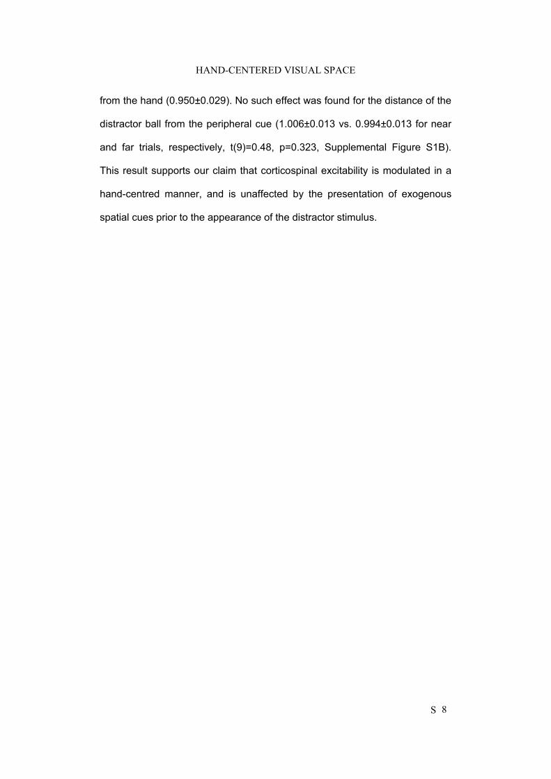
HAND-CENTERED VISUAL SPACE
S 8
from the hand (0.950±0.029). No such effect was found for the distance of the
distractor ball from the peripheral cue (1.006±0.013 vs. 0.994±0.013 for near
and far trials, respectively, t(9)=0.48, p=0.323, Supplemental Figure S1B).
This result supports our claim that corticospinal excitability is modulated in a
hand-centred manner, and is unaffected by the presentation of exogenous
spatial cues prior to the appearance of the distractor stimulus.
HAND-CENTERED VISUAL SPACE
S 9
Supplemental References
Cantello R, Civardi C, Cavalli A, Varrasi C, Vicentini R (2000) Effects of a
photic input on the human cortico-motoneuron connection. Clin
Neurophysiol 111:1981-1989.
Hodges PW, Bui BH (1996) A comparison of computer-based methods for the
determination of onset of muscle contraction using electromyography.
Electroencephalogr Clin Neurophysiol 101:511-519.
Klein RM, Shore D (2000) Relations among modes of visual orientation. In
Attention and performance XVIII: Control of cognitive processes, Monsell
S, Driver J, Eds (Cambridge, MA: MIT Press), pp. 195-208.
Oldfield RC (1971) The assessment and analysis of handedness: The
Edinburgh inventory. Neuropsychologia 9:97-113.
van der Lubbe RH, Postma A (2005) Interruption from irrelevant auditory and
visual onsets even when attention is in a focused state. Exp Brain Res
164:464-471.
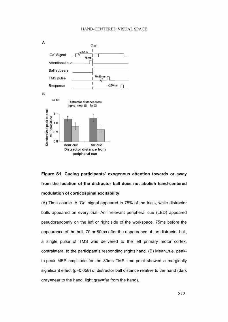
HAND-CENTERED VISUAL SPACE
S10
Figure S1. Cueing participants’ exogenous attention towards or away
from the location of the distractor ball does not abolish hand-centered
modulation of corticospinal excitability
(A) Time course. A ‘Go’ signal appeared in 75% of the trials, while distractor
balls appeared on every trial. An irrelevant peripheral cue (LED) appeared
pseudorandomly on the left or right side of the workspace, 75ms before the
appearance of the ball. 70 or 80ms after the appearance of the distractor ball,
a single pulse of TMS was delivered to the left primary motor cortex,
contralateral to the participant’s responding (right) hand. (B) Mean±s.e. peak-
to-peak MEP amplitude for the 80ms TMS time-point showed a marginally
significant effect (p=0.058) of distractor ball distance relative to the hand (dark
gray=near to the hand, light gray=far from the hand).
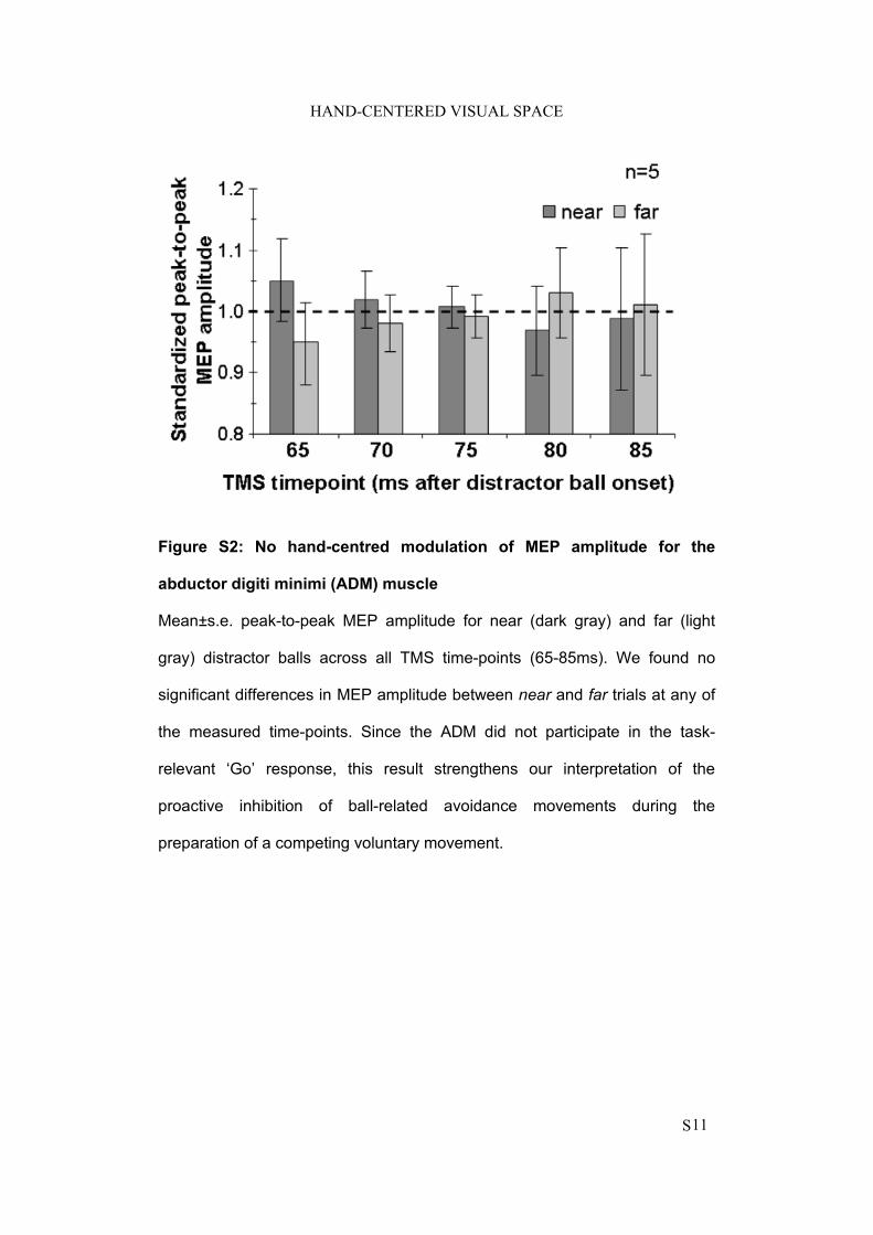
HAND-CENTERED VISUAL SPACE
S11
Figure S2: No hand-centred modulation of MEP amplitude for the
abductor digiti minimi (ADM) muscle
Mean±s.e. peak-to-peak MEP amplitude for near (dark gray) and far (light
gray) distractor balls across all TMS time-points (65-85ms). We found no
significant differences in MEP amplitude between near and far trials at any of
the measured time-points. Since the ADM did not participate in the task-
relevant ‘Go’ response, this result strengthens our interpretation of the
proactive inhibition of ball-related avoidance movements during the
preparation of a competing voluntary movement.
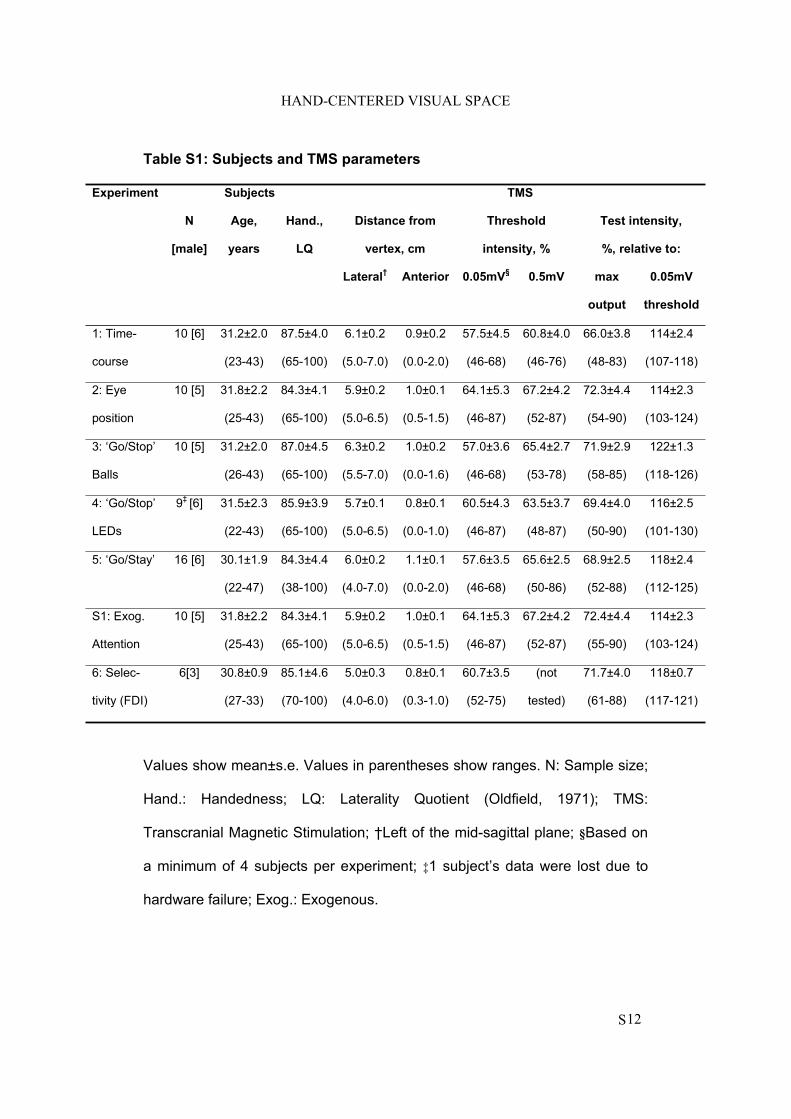
HAND-CENTERED VISUAL SPACE
S12
Table S1: Subjects and TMS parameters
Experiment Subjects TMS
N
[male]
Age,
years
Hand.,
LQ
Distance from
vertex, cm
Threshold
intensity, %
Test intensity,
%, relative to:
Lateral†
Anterior 0.05mV§
0.5mV max
output
0.05mV
threshold
1: Time-
course
10 [6] 31.2±2.0
(23-43)
87.5±4.0
(65-100)
6.1±0.2
(5.0-7.0)
0.9±0.2
(0.0-2.0)
57.5±4.5
(46-68)
60.8±4.0
(46-76)
66.0±3.8
(48-83)
114±2.4
(107-118)
2: Eye
position
10 [5] 31.8±2.2
(25-43)
84.3±4.1
(65-100)
5.9±0.2
(5.0-6.5)
1.0±0.1
(0.5-1.5)
64.1±5.3
(46-87)
67.2±4.2
(52-87)
72.3±4.4
(54-90)
114±2.3
(103-124)
3: ‘Go/Stop’
Balls
10 [5] 31.2±2.0
(26-43)
87.0±4.5
(65-100)
6.3±0.2
(5.5-7.0)
1.0±0.2
(0.0-1.6)
57.0±3.6
(46-68)
65.4±2.7
(53-78)
71.9±2.9
(58-85)
122±1.3
(118-126)
4: ‘Go/Stop’
LEDs
9‡ [6] 31.5±2.3
(22-43)
85.9±3.9
(65-100)
5.7±0.1
(5.0-6.5)
0.8±0.1
(0.0-1.0)
60.5±4.3
(46-87)
63.5±3.7
(48-87)
69.4±4.0
(50-90)
116±2.5
(101-130)
5: ‘Go/Stay’ 16 [6] 30.1±1.9
(22-47)
84.3±4.4
(38-100)
6.0±0.2
(4.0-7.0)
1.1±0.1
(0.0-2.0)
57.6±3.5
(46-68)
65.6±2.5
(50-86)
68.9±2.5
(52-88)
118±2.4
(112-125)
S1: Exog.
Attention
10 [5] 31.8±2.2
(25-43)
84.3±4.1
(65-100)
5.9±0.2
(5.0-6.5)
1.0±0.1
(0.5-1.5)
64.1±5.3
(46-87)
67.2±4.2
(52-87)
72.4±4.4
(55-90)
114±2.3
(103-124)
6: Selec-
tivity (FDI)
6[3] 30.8±0.9
(27-33)
85.1±4.6
(70-100)
5.0±0.3
(4.0-6.0)
0.8±0.1
(0.3-1.0)
60.7±3.5
(52-75)
(not
tested)
71.7±4.0
(61-88)
118±0.7
(117-121)
Values show mean±s.e. Values in parentheses show ranges. N: Sample size;
Hand.: Handedness; LQ: Laterality Quotient (Oldfield, 1971); TMS:
Transcranial Magnetic Stimulation; †Left of the mid-sagittal plane; §Based on
a minimum of 4 subjects per experiment; ‡1 subject’s data were lost due to
hardware failure; Exog.: Exogenous.
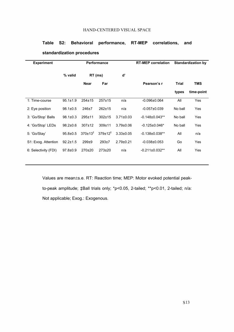
HAND-CENTERED VISUAL SPACE
S13
Table S2: Behavioral performance, RT-MEP correlations, and
standardization procedures
Experiment Performance RT-MEP correlation Standardization by
% valid RT (ms) d’
Near Far Pearson’s r Trial
types
TMS
time-point
1: Time-course 95.1±1.9 254±15 257±15 n/a -0.096±0.064 All Yes
2: Eye position 98.1±0.5 246±7 262±15 n/a -0.057±0.039 No ball Yes
3: ‘Go/Stop’ Balls 98.1±0.3 295±11 302±15 3.71±0.03 -0.148±0.043** No ball Yes
4: ‘Go/Stop’ LEDs 98.2±0.6 307±12 309±11 3.79±0.06 -0.125±0.046* No ball Yes
5: ‘Go/Stay’ 95.8±0.5 370±13‡ 379±12‡ 3.33±0.05 -0.138±0.038** All n/a
S1: Exog. Attention 92.2±1.5 299±9 293±7 2.79±0.21 -0.038±0.053 Go Yes
6: Selectivity (FDI) 97.8±0.9 270±20 273±20 n/a -0.211±0.032** All Yes
Values are mean±s.e. RT: Reaction time; MEP: Motor evoked potential peak-
to-peak amplitude; ‡Ball trials only; *p<0.05, 2-tailed; **p<0.01, 2-tailed; n/a:
Not applicable; Exog.: Exogenous.
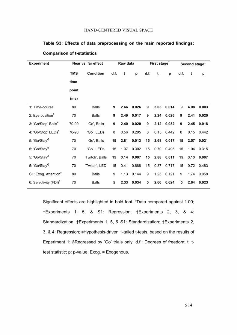
HAND-CENTERED VISUAL SPACE
S14
Table S3: Effects of data preprocessing on the main reported findings:
Comparison of t-statistics
Experiment Near vs. far effect Raw data First stage†
Second stage‡
TMS
time-
point
(ms)
Condition d.f. t p d.f. t p d.f. t p
1: Time-course 80 Balls 9 2.66 0.026 9 3.05 0.014 *9 4.08 0.003
2: Eye position# 70 Balls 9 2.49 0.017 9 2.24 0.026 9 2.41 0.020
3: ‘Go/Stop’ Balls# 70-90 ‘Go’, Balls 9 2.40 0.020 9 2.12 0.032 9 2.45 0.018
4: ‘Go/Stop’ LEDs# 70-90 ‘Go’, LEDs 8 0.56 0.295 8 0.15 0.442 8 0.15 0.442
5: ‘Go/Stay’§ 70 ‘Go’, Balls 15 2.81 0.013 15 2.68 0.017 15 2.57 0.021
5: ‘Go/Stay’§ 70 ‘Go’, LEDs 15 1.07 0.302 15 0.70 0.495 15 1.04 0.315
5: ‘Go/Stay’§ 70 ‘Twitch’, Balls 15 3.14 0.007 15 2.88 0.011 15 3.13 0.007
5: ‘Go/Stay’§ 70 ‘Twitch’, LED 15 0.41 0.688 15 0.37 0.717 15 0.72 0.483
S1: Exog. Attention# 80 Balls 9 1.13 0.144 9 1.25 0.121 9 1.74 0.058
6: Selectivity (FDI)# 70 Balls 5 2.33 0.034 5 2.60 0.024 *5 2.64 0.023
Significant effects are highlighted in bold font. *Data compared against 1.00;
†Experiments 1, 5, & S1: Regression; †Experiments 2, 3, & 4:
Standardization; ‡Experiments 1, 5, & S1: Standardization; ‡Experiments 2,
3, & 4: Regression; #Hypothesis-driven 1-tailed t-tests, based on the results of
Experiment 1; §Regressed by ‘Go’ trials only; d.f.: Degrees of freedom; t: t-
test statistic; p: p-value; Exog. = Exogenous.
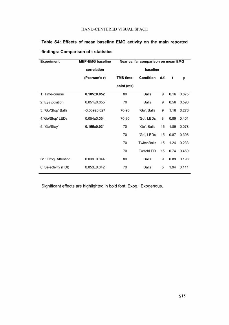
HAND-CENTERED VISUAL SPACE
S15
Table S4: Effects of mean baseline EMG activity on the main reported
findings: Comparison of t-statistics
Experiment MEP-EMG baseline
correlation
Near vs. far comparison on mean EMG
baseline
(Pearson’s r) TMS time-
point (ms)
Condition d.f. t p
1: Time-course 0.185±0.052 80 Balls 9 0.16 0.875
2: Eye position 0.051±0.055 70 Balls 9 0.56 0.590
3: ‘Go/Stop’ Balls -0.039±0.027 70-90 ‘Go’, Balls 9 1.16 0.276
4:’Go/Stop’ LEDs 0.054±0.054 70-90 ‘Go’, LEDs 8 0.89 0.401
5: ‘Go/Stay’ 0.155±0.031 70 ‘Go’, Balls 15 1.89 0.078
70 ‘Go’, LEDs 15 0.87 0.398
70 TwitchBalls 15 1.24 0.233
70 TwitchLED 15 0.74 0.469
S1: Exog. Attention 0.039±0.044 80 Balls 9 0.89 0.198
6: Selectivity (FDI) 0.053±0.042 70 Balls 5 1.94 0.111
Significant effects are highlighted in bold font; Exog.: Exogenous.
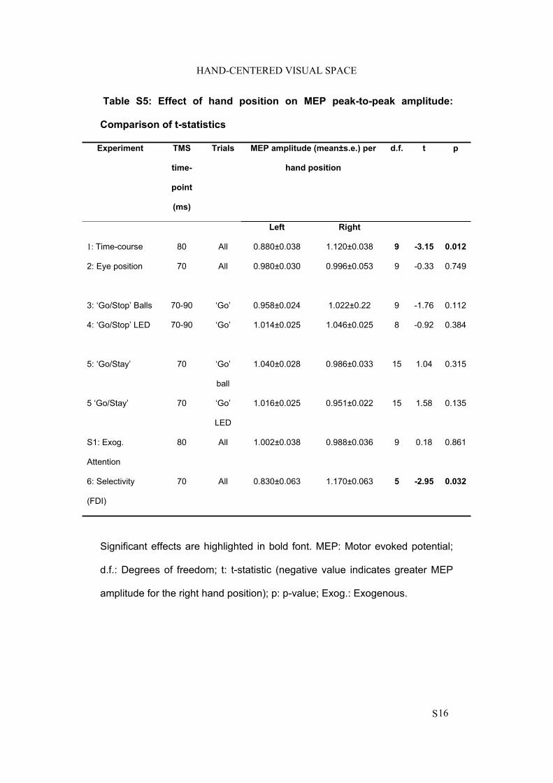
HAND-CENTERED VISUAL SPACE
S16
Table S5: Effect of hand position on MEP peak-to-peak amplitude:
Comparison of t-statistics
Experiment TMS
time-
point
(ms)
Trials MEP amplitude (mean±s.e.) per
hand position
d.f. t p
Left Right
1: Time-course 80 All 0.880±0.038 1.120±0.038 9 -3.15 0.012
2: Eye position 70 All 0.980±0.030 0.996±0.053 9 -0.33 0.749
3: ‘Go/Stop’ Balls 70-90 ‘Go’ 0.958±0.024 1.022±0.22 9 -1.76 0.112
4: ‘Go/Stop’ LED 70-90 ‘Go’ 1.014±0.025 1.046±0.025 8 -0.92 0.384
5: ‘Go/Stay’ 70 ‘Go’
ball
1.040±0.028 0.986±0.033 15 1.04 0.315
5 ‘Go/Stay’ 70 ‘Go’
LED
1.016±0.025 0.951±0.022 15 1.58 0.135
S1: Exog.
Attention
80 All 1.002±0.038 0.988±0.036 9 0.18 0.861
6: Selectivity
(FDI)
70 All 0.830±0.063 1.170±0.063 5 -2.95 0.032
Significant effects are highlighted in bold font. MEP: Motor evoked potential;
d.f.: Degrees of freedom; t: t-statistic (negative value indicates greater MEP
amplitude for the right hand position); p: p-value; Exog.: Exogenous.
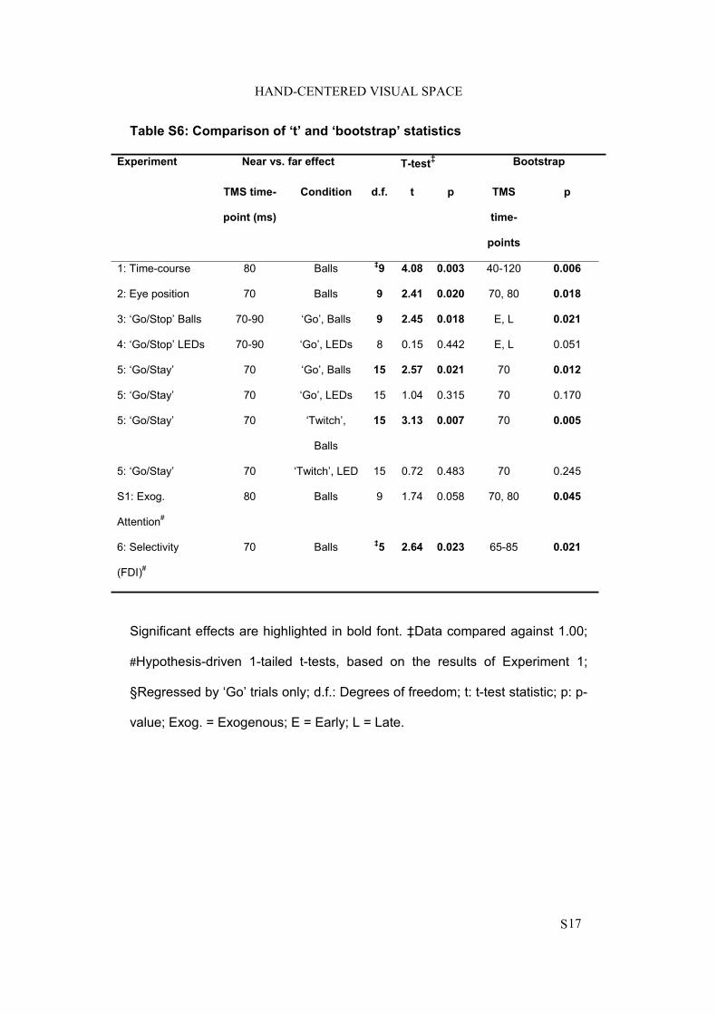
HAND-CENTERED VISUAL SPACE
S17
Table S6: Comparison of ‘t’ and ‘bootstrap’ statistics
Experiment Near vs. far effect T-test‡ Bootstrap
TMS time-
point (ms)
Condition d.f. t p TMS
time-
points
p
1: Time-course 80 Balls ‡9 4.08 0.003 40-120 0.006
2: Eye position 70 Balls 9 2.41 0.020 70, 80 0.018
3: ‘Go/Stop’ Balls 70-90 ‘Go’, Balls 9 2.45 0.018 E, L 0.021
4: ‘Go/Stop’ LEDs 70-90 ‘Go’, LEDs 8 0.15 0.442 E, L 0.051
5: ‘Go/Stay’ 70 ‘Go’, Balls 15 2.57 0.021 70 0.012
5: ‘Go/Stay’ 70 ‘Go’, LEDs 15 1.04 0.315 70 0.170
5: ‘Go/Stay’ 70 ‘Twitch’,
Balls
15 3.13 0.007 70 0.005
5: ‘Go/Stay’ 70 ‘Twitch’, LED 15 0.72 0.483 70 0.245
S1: Exog.
Attention#
80 Balls 9 1.74 0.058 70, 80 0.045
6: Selectivity
(FDI)#
70 Balls ‡5 2.64 0.023 65-85 0.021
Significant effects are highlighted in bold font. ‡Data compared against 1.00;
#Hypothesis-driven 1-tailed t-tests, based on the results of Experiment 1;
§Regressed by ‘Go’ trials only; d.f.: Degrees of freedom; t: t-test statistic; p: p-
value; Exog. = Exogenous; E = Early; L = Late.
The results of study 1 show that the location of a visually available three-dimensional object
near to the body is coded in a reference frame that is centred on the body-part (the hand in the
experiment present here) and that this information is rapidly available to the motor system.
The results of the study 1 in this respect, support the idea that peripersonal space can play a
role not only in the mere detection of visual information close to the body but also in
subserving the possible consequent motor reaction. In particular, an effect of inhibition of
irrelevant motor plans seems to be elicited by the suddenly appearance of an object near the
hand. Possibly, this inhibition of irrelevant plans would be meant to readily prepare to an
avoidance movement.
However, as previously pointed out, the same multisensory interface might be
involved in other kinds of body-object interactions, where the body-part is voluntary
projected toward the object location in order to manipulate it, such as in the everyday life
when we grasp an object. The next study will thus investigate this possibility, employing the
paradigm of the cross-modal congruency effect, adaptively modified for assessing the
multisensory interaction during the execution of action. Participants were requested at each
trial to grasp the object with the right stimulated hand index finger and thumb in a precision
grip. At the same time, participants were requested to respond to the tactile target
discriminating their elevation (“up” or “down”) on the grasping hand, trying to ignore the
visual distractors embedded into the to-be-grasped object. The visuo-tactile stimulation was
presented at one of three different timings with respect to the execution of the action: Either in
a static phase, when the grasping hand had not yet moved; At the onset of the movement (0
ms); Or, in the early execution phase (200 ms after movement onset).
When participants performed the action with the tactually stimulated hand, the
multisensory interaction was enhanced (i.e., there was more interference from the visual
distractor on the tactile task) as compared to the static phase. This effect was even more
pronounced when the multisensory interaction was assessed during the early execution phase
of the grasping.
The experiment was also repeated asking participants to grasp the object with the non-
stimulated hand. Crucially, when the same action was performed with the non-stimulated
hand, no multisensory modulation was observed.
141
The co-recording of the kinematics of the movement for each grasping action
performed by participants, provided a quantitative description of the motor performance. On
one hand this could rule out any possible confounds of differences in the left/right hand
movements; on the other hand, it provided the necessary information in order to investigate
the links possibly existing between the multisensory peripersonal space modulation during
action and the action itself.
142
142
STUDY 2
Grasping actions remap peripersonal space
NeuroReport (2009)

Cognitive neuroscience and neuropsychology 1
Grasping actions remap peripersonal spaceClaudio Brozzolia,b, Francesco Pavanic,d, Christian Urquizara,Lucilla Cardinalia,b and Alessandro Farnea,b
The portion of space that closely surrounds our body parts
is termed peripersonal space, and it has been shown to be
represented in the brain through multisensory processing
systems. Here, we tested whether voluntary actions, such
as grasping an object, may remap such multisensory
spatial representation. Participants discriminated touches
on the hand they used to grasp an object containing
task-irrelevant visual distractors. Compared with a static
condition, reach-to-grasp movements increased the
interference exerted by visual distractors over tactile
targets. This remapping of multisensory space was
triggered by action onset and further enhanced in real
time during the early action execution phase.
Additional experiments showed that this phenomenon is
hand-centred. These results provide the first evidence of a
functional link between voluntary object-oriented actions
and multisensory coding of the space around us.
NeuroReport 00:000–000 �c 2009 Wolters Kluwer Health |
Lippincott Williams & Wilkins.
NeuroReport 2009, 00:000–000
Keywords: bimodal neurons, cross-modal congruency effect, grasping,kinematics, motor control, multisensory, peripersonal space
aINSERM UMR-S 864 ‘Espace et Action’, Bron, bUniversite Claude Bernard LyonI, Lyon, France, cDepartment of Cognitive Sciences and Education and dCenterfor Mind/Brain Sciences, University of Trento, Rovereto, Italy
Correspondence to Claudio Brozzoli, INSERM, 16, avenue du Doyen Lepine,Lyon 69002, FranceTel: + 33 472 913420; fax: + 33 472 913401;e-mail: [email protected]; [email protected]
Received 13 March 2009 accepted 24 March 2009
IntroductionThe representation of the space near the body, termed
‘peripersonal’ space (PpS) [1,2], relies on multisensory
processing, both in human and non-human primates. In
monkeys, bimodal neurons in parieto-frontal and sub-
cortical structures code for tactile events on a body part
(e.g. the hand) and visual events near that body part,
thus giving rise to body-centred representations of PpS
[3–6]. In humans, a functionally homologous coding of PpS
is largely supported by behavioural studies showing
stronger visuotactile interaction in near rather than far
space in brain-damaged [7–9] and healthy individuals
[10–13]. For example, visual events occurring in the
immediate proximity to the body induce more severe
tactile extinction than farther ones [7–9]. Recent func-
tional neuroimaging studies further support the exis-
tence of similar multisensory integrative structures in
the human brain [11–13].
Despite the large body of knowledge accumulated across
species on the multisensory properties of PpS, little is
known about its function, and this issue has never been
directly assessed in humans. By acting as an anticipatory
sensorimotor interface, PpS may serve early detection
of potential threats approaching the body to drive in-
voluntary defensive movements [3]. The same antici-
patory feature, however, may also have evolved to serve
voluntary object-oriented actions [1,2,14]. Here, we
tested the latter hypothesis by assessing the effects
of grasping objects on the multisensory coding of PpS.
In Experiment 1, we modified a cross-modal paradigm
[10], whereby participants indicate the elevation (up or
down) of a tactile target delivered to a finger (index or
thumb), while a visual distractor is presented at either
congruent or incongruent elevation (Fig. 1). We then con-
ducted three experiments in which participants were
additionally required to grasp the object in which the
visual distractors were embedded. Although the percep-
tual task was always performed on the right hand, the
motor task was performed by either the right (Experi-
ments 2 and 4) or left (Experiment 3) hand. This simple
manipulation is crucial in two respects: it equalizes
attentional demands for the target object in the sti-
mulated and the nonstimulated hand actions, and it
allows assessing whether any modulation of multisensory
processing is hand-centred.
Experimental proceduresParticipants
Fifteen healthy participants (nine men, mean age 27±5
years) took part in Experiments 1, 19 (10 men, mean age
26±6 years) in Experiments 2 and 3, and 16 (8 male,
mean age 25±3) in Experiment 4. All gave their verbal
informed consent to take part in this study, approved by
the local INSERM Ethics Board.
Supplementary data are available at The NeuroReport Online (http://links.lww.com/A1251; http://links.lww.com/A1250; http://links.lww.com/A1249; http://links.lww.com/A1248; http://links.lww.com/A1247; http://links.lww.com/A1246)
0959-4965 �c 2009 Wolters Kluwer Health | Lippincott Williams & Wilkins DOI: 10.1097/WNR.0b013e32832c0b9b
Copyright © Lippincott Williams & Wilkins. Unauthorized reproduction of this article is prohibited.
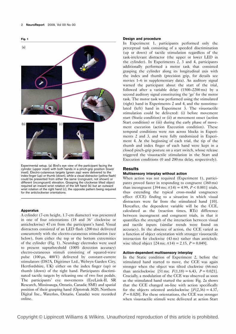
Apparatus
A cylinder (7-cm height, 1.7-cm diameter) was presented
in one of four orientations (18 and 361 clockwise or
anticlockwise) 47 cm from the participant’s hand. Visual
distractors consisted of an LED flash (200ms) delivered
concurrently with the electro-cutaneous stimulation (see
below), from either the top or the bottom extremities
of the cylinder (Fig. 1). Neurology electrodes were used
to present suprathreshold (100% detection accuracy)
electro-cutaneous stimuli consisting of squared-wave
pulse (100 ms, 400 V) delivered by constant-current
stimulators (DS7A, Digitimer Ltd., Welwyn Garden City,
Hertfordshire, UK) either on the index finger (up) or
thumb (down) of the right hand. Participants discrimi-
nated tactile targets by releasing one of two foot pedals.
The participants’ eye movements (EyeLink-II, SR
Research, Mississauga, Ontario, Canada; SMI) and spatial
position of their grasping hand (Optotrak 3020, Northern
Digital Inc., Waterloo, Ontario, Canada) were recorded
online.
Design and procedure
In Experiment 1, participants performed only the
perceptual task consisting of a speeded discrimination
(up or down) of tactile stimulation regardless of the
task-irrelevant distractor (the upper or lower LED in
the cylinder). In Experiments 2, 3 and 4, participants
additionally performed a motor task that consisted
grasping the cylinder along its longitudinal axis with
the index and thumb (precision grip, for details see
movies 1–6 in supplementary data). An auditory signal
warned the participant about the start of the trial,
followed after a variable delay (1500–2200ms) by a
second auditory signal constituting the ‘go’ for the motor
task. The motor task was performed using the stimulated
(right) hand in Experiments 2 and 4, and the nonstimu-
lated (left) hand in Experiment 3. The visuotactile
stimulation could be delivered: (i) before movement
start (Static condition) or (ii) at movement onset (action
Start condition) or (iii) during the early phase of move-
ment execution (action Execution condition). These
temporal conditions were run across blocks in Experi-
ments 2 and 3, and were fully randomized in Experi-
ment 4. At the beginning of each trial, the tip of the
thumb and index finger of each hand were kept in a
closed pinch-grip posture on a start switch, whose release
triggered the visuotactile stimulation in the Start and
Execution conditions (0 and 200ms delay, respectively).
ResultsMultisensory interplay without action
When action was not required (Experiment 1), partici-
pants proved faster in responding to congruent (360ms)
than incongruent [394ms; t(14)=4.99, P<0.001] trials,
thus extending the typical cross-modal congruency
effect (CCE) finding to a situation in which visual
distractors were far from the stimulated hand [10].
Hereafter, the dependent variable will be the CCE,
calculated as the (reaction times, RTs) difference
between incongruent and congruent trials, in that it
quantifies the strength of the interaction between visual
and tactile inputs (similar trends were found on
accuracy). In the absence of action, the CCE varied as
a function of object orientation with stronger visuotactile
interaction for clockwise (43ms) rather than anticlock-
wise tilted object [24ms; t(14)= 2.15, P=0.049].
Action-dependent multisensory interplay
In the Static condition of Experiment 2, before the
stimulated hand started to move, the CCE was again
stronger when the object was tilted clockwise (66ms)
than anticlockwise [51ms; F(1,18)=6.43, P=0.021].
Crucially, a modulation of the CCE was observed as soon
as the stimulated hand started the action: Fig. 2a shows
that the CCE changed on-line with action specifically
for the objects oriented anticlockwise [F(2,36)=4.37,
P=0.020]. For these orientations, the CCE was stronger
when visuotactile stimuli were delivered at action Start
Fig. 1
Experimental setup. (a) Bird’s eye view of the participant facing thecylinder (upper inset) with both hands in a pinch-grip position (lowerinset). Electro-cutaneous targets (green zap) were delivered to theindex finger (up) or thumb (down), while a visual distractor (yellow flash)could be presented from either the same (congruent, not shown) ordifferent (incongruent) elevation. Grasping the clockwise tilted objectrequired an inward wrist rotation of the left hand (b) but an outwardwrist rotation of the right hand (c), the opposite pattern being requiredfor the anticlockwise orientations.
2 NeuroReport 2009, Vol 00 No 00
Copyright © Lippincott Williams & Wilkins. Unauthorized reproduction of this article is prohibited.
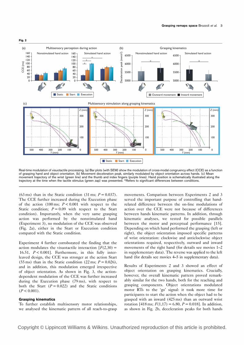
(63ms) than in the Static condition (31ms; P=0.037).
The CCE further increased during the Execution phase
of the action (100ms; P<0.001 with respect to the
Static condition; P=0.09 with respect to the Start
condition). Importantly, when the very same grasping
action was performed by the nonstimulated hand
(Experiment 3), no modulation of the CCE was observed
(Fig. 2a), either in the Start or Execution condition
compared with the Static condition.
Experiment 4 further corroborated the finding that the
action modulates the visuotactile interaction [F(2,30)=16.51, P<0.001]. Furthermore, in this fully inter-
leaved design, the CCE was stronger at the action Start
(55ms) than in the Static condition (22ms; P=0.026),
and in addition, this modulation emerged irrespective
of object orientation. As shown in Fig. 3, the action-
dependent modulation of the CCE was further increased
during the Execution phase (79ms), with respect to
both the Start (P=0.022) and the Static conditions
(P<0.001).
Grasping kinematics
To further establish multisensory motor relationships,
we analysed the kinematic pattern of all reach-to-grasp
movements. Comparison between Experiments 2 and 3
served the important purpose of controlling that hand-
related difference between the on-line modulations of
action over the CCE were not because of differences
between hands kinematic patterns. In addition, through
kinematic analyses, we tested for possible parallels
between the motor and perceptual performance [15].
Depending on which hand performed the grasping (left or
right), the object orientation imposed specific patterns
of wrist orientation: clockwise and anticlockwise object
orientations required, respectively, outward and inward
movements of the right hand (for details see movies 1–2
in supplementary data). The reverse was applied to the left
hand (for details see movies 4–5 in supplementary data).
Results of Experiments 2 and 3 showed an effect of
object orientation on grasping kinematics. Crucially,
however, the overall kinematic pattern proved remark-
ably similar for the two hands, both for the reaching and
grasping components. Object orientations modulated
motor RTs to the ‘go’ signal: it took more time for
participants to start the action when the object had to be
grasped with an inward (425ms) than an outward wrist
rotation [418ms; F(1,17)=6.80, P=0.018]. In addition,
as shown in Fig. 2b, deceleration peaks for both hands
Fig. 2
Multisensory perception during action(a) (b)
(c) Multisensory stimulation along grasping kinematics
Grasping kinematics
Nonstimulated hand action Nonstimulated hand action
∗ ∗
160140120100806040200
1606500
6000
5500
5000Dec
eler
atio
n pe
ak (
mm
/s2 )
6500
6000
5500
Outward movement Inward movement
5000
140120100806040200
CC
E (m
s)
Stimulated hand action Stimulated hand action
500 400 300 200 100 0
100
200
300
Y (mm)
Z (mm)
0
100
200
300
Z (mm)
100
200
300
Z (mm)
500 400 300 200 100Y (mm)
500 400 300 200 100 0Y (mm)
∗∗
∗
Static Start Execution
Static Start Execution
Real-time modulation of visuotactile processing. (a) Bar plots (with SEM) show the modulation of cross-modal congruency effect (CCE) as a functionof grasping hand and object orientation. (b) Movement deceleration peak, similarly modulated by object orientation across hands. (c) Meanmovement trajectory of the wrist (green line) and the thumb and index fingers (purple lines). Hand position is schematically illustrated along thetrajectory at the time when the tactile stimulus (green zap) was presented. *Refers to significant differences between conditions.
Grasping remaps space Brozzoli et al. 3
Copyright © Lippincott Williams & Wilkins. Unauthorized reproduction of this article is prohibited.
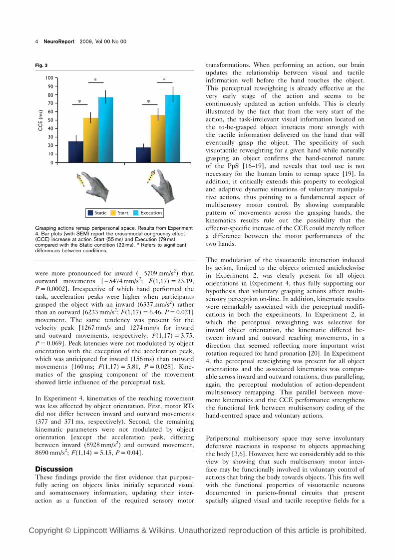
were more pronounced for inward ( – 5709mm/s2) than
outward movements [ – 5474mm/s2; F(1,17)=23.19,
P=0.0002]. Irrespective of which hand performed the
task, acceleration peaks were higher when participants
grasped the object with an inward (6337mm/s2) rather
than an outward [6233mm/s2; F(1,17)=6.46, P=0.021]
movement. The same tendency was present for the
velocity peak [1267mm/s and 1274mm/s for inward
and outward movements, respectively; F(1,17)=3.75,
P=0.069]. Peak latencies were not modulated by object
orientation with the exception of the acceleration peak,
which was anticipated for inward (156ms) than outward
movements [160ms; F(1,17)= 5.81, P=0.028]. Kine-
matics of the grasping component of the movement
showed little influence of the perceptual task.
In Experiment 4, kinematics of the reaching movement
was less affected by object orientation. First, motor RTs
did not differ between inward and outward movements
(377 and 371ms, respectively). Second, the remaining
kinematic parameters were not modulated by object
orientation [except the acceleration peak, differing
between inward (8928mm/s2) and outward movement,
8690mm/s2; F(1,14)=5.15, P=0.04].
DiscussionThese findings provide the first evidence that purpose-
fully acting on objects links initially separated visual
and somatosensory information, updating their inter-
action as a function of the required sensory motor
transformations. When performing an action, our brain
updates the relationship between visual and tactile
information well before the hand touches the object.
This perceptual reweighting is already effective at the
very early stage of the action and seems to be
continuously updated as action unfolds. This is clearly
illustrated by the fact that from the very start of the
action, the task-irrelevant visual information located on
the to-be-grasped object interacts more strongly with
the tactile information delivered on the hand that will
eventually grasp the object. The specificity of such
visuotactile reweighting for a given hand while naturally
grasping an object confirms the hand-centred nature
of the PpS [16–19], and reveals that tool use is not
necessary for the human brain to remap space [19]. In
addition, it critically extends this property to ecological
and adaptive dynamic situations of voluntary manipula-
tive actions, thus pointing to a fundamental aspect of
multisensory motor control. By showing comparable
pattern of movements across the grasping hands, the
kinematics results rule out the possibility that the
effector-specific increase of the CCE could merely reflect
a difference between the motor performances of the
two hands.
The modulation of the visuotactile interaction induced
by action, limited to the objects oriented anticlockwise
in Experiment 2, was clearly present for all object
orientations in Experiment 4, thus fully supporting our
hypothesis that voluntary grasping actions affect multi-
sensory perception on-line. In addition, kinematic results
were remarkably associated with the perceptual modifi-
cations in both the experiments. In Experiment 2, in
which the perceptual reweighting was selective for
inward object orientation, the kinematic differed be-
tween inward and outward reaching movements, in a
direction that seemed reflecting more important wrist
rotation required for hand pronation [20]. In Experiment
4, the perceptual reweighting was present for all object
orientations and the associated kinematics was compar-
able across inward and outward rotations, thus paralleling,
again, the perceptual modulation of action-dependent
multisensory remapping. This parallel between move-
ment kinematics and the CCE performance strengthens
the functional link between multisensory coding of the
hand-centred space and voluntary actions.
Peripersonal multisensory space may serve involuntary
defensive reactions in response to objects approaching
the body [3,6]. However, here we considerably add to this
view by showing that such multisensory motor inter-
face may be functionally involved in voluntary control of
actions that bring the body towards objects. This fits well
with the functional properties of visuotactile neurons
documented in parieto-frontal circuits that present
spatially aligned visual and tactile receptive fields for a
Fig. 3
100
90
80
70
*
* *
*
60
50
CC
E (m
s)
40
30
20
10
0
Static Start Execution
Grasping actions remap peripersonal space. Results from Experiment4. Bar plots (with SEM) report the cross-modal congruency effect(CCE) increase at action Start (55ms) and Execution (79ms)compared with the Static condition (22ms). * Refers to significantdifferences between conditions.
4 NeuroReport 2009, Vol 00 No 00
Copyright © Lippincott Williams & Wilkins. Unauthorized reproduction of this article is prohibited.
given body part [1–6,21]. This feature allows bimodal
neural systems to represent an object in a body-centred
reference frame and to be continuously updated during
bodily movements. This multisensory spatial representa-
tion has been suggested to serve involuntary defensive
movements, because electrical microstimulation of
some bimodal areas in the monkey brain [3] elicits stereo-
typed arm or face movements that are compatible
with defensive behaviour. Remarkably, however, some
bimodal neurons also respond when the arm is voluntarily
moved within reaching space [14,15,22,23], and have
been previously proposed to code goal-directed actions
[1,2,22]. Neurophysiological studies on monkeys have
shown activation in the posterior parietal cortex during
grasping, in the early phase of the action when the hand
has not yet reached the object. The activation gradually
shifts towards the somatosensory cortex when the hand
enters in contact with the object [14]. Finally, the on-line
enlargement of the visual receptive fields of bimodal
neurons in response to approaching objects [6] or tool use
[17–19,24,25] also emphasizes the dynamic nature of
their multisensory space coding, providing converging
evidence for the involvement of the bimodal system in
dynamic updating of the PpS. The multisensory motor
neural machinery acting as an anticipatory interface
between the body and nearby events may thus have
been selected throughout evolution to drive both in-
voluntary avoidance reactions and voluntary approaching
movements, with common adaptive advantages for
defensive and manipulative actions.
ConclusionVoluntarily acting on objects triggers a hand-centred
remapping of multisensory spatial processing that paral-
lels action requirements and is regulated in real time as
action unfolds.
AcknowledgementsThe authors thank A.C. Roy and N. Holmes for
thoughtful discussions and comments on an earlier
version of the manuscript. This study was supported
by the European Mobility Fellowship, the INSERM
AVENIR Grant No. R05265CS and the ANR Grants No.
JCJC06133960 and RPV08085CSA, the Galileo PHC
Grant and the PRIN Grant from the MIUR.
References1 Rizzolatti G, Scandolara C, Matelli M, Gentilucci M. Afferent properties of
periarcuate neurons in macque monkeys. II. Visual responses. Behav BrainRes 1981; 2:147–163.
2 Rizzolatti G, Fadiga L, Fogassi L, Gallese V. The space around us. Science1997; 11:190–191.
3 Graziano MS, Cooke DF. Parieto-frontal interactions, personal space, anddefensive behavior. Neuropsychologia 2006; 44:2621–2635.
4 Fogassi L, Gallese V, Fadiga L, Luppino G, Matelli M, Rizzolatti G. Codingof peripersonal space in inferior premotor cortex (area F4). J Neurophysiol1996; 76:141–157.
5 Duhamel JR, Colby CL, Goldberg ME. Ventral Intraparietal areaof the macaque: congruent visual and somatic response properties.J Neurophysiol 1998; 79:126–136.
6 Graziano MSA, Gross CG. The representation of extrapersonal space:a possible role for bimodal, visuo-tactile neurons. In: Gazzaniga M, editor.The Cognitive Neurosciences. Cambridge, Massachusetts: MIT Press;1995. pp. 1021–1034.
7 di Pellegrino G, Ladavas E, Farne A. Seeing where your hands are. Nature1997; 21:730.
8 Farne A, Ladavas E. Dynamic size-change of hand peripersonal spacefollowing tool use. Neuroreport 2000; 11:1645–1649.
9 Brozzoli C, Dematte ML, Pavani F, Frassinetti F, Farne A. Neglect andextinction: within and between sensory modalities. Restor Neurol Neurosci2006; 24:217–232.
10 Spence C, Pavani F, Maravita A, Holmes N. Multisensory contributionsto the 3-D representation of visuotactile peripersonal space in humans:evidence from the crossmodal congruency task. J Physiol Paris 2004;98:171–189.
11 Bremmer F, Schlack A, Shah NJ, Zafiris O, Kubischik M, Hoffmann K, et al.Polymodal motion processing in posterior parietal and premotor cortex:a human fMRI study strongly implies equivalencies between humans andmonkeys. Neuron 2001; 29:287–296.
12 Sereno MI, Huang RS. A human parietal face area contains alignedhead-centered visual and tactile maps. Nat Neurosci 2006;9:1337–1343.
13 Makin TR, Holmes NP, Zohary E. Is that near my hand? Multisensoryrepresentation of peripersonal space in human intraparietal sulcus.J Neurosci 2007; 27:731–740.
14 Gardner EP, Babu KS, Reitzen SD, Ghosh S, Brown AS, Chen J, et al.Neurophysiology of prehension. I. Posterior parietal cortex andobject-oriented hand behaviors. J Neurophysiol 2007; 97:387–406.
15 Jeannerod M. Mechanisms of visuomotor coordination: a study in normaland brain-damaged subjects. Neuropsychologia 1986; 24:41–78.
16 Farne A, Dematte M, Ladavas E. Beyond the window: multisensoryrepresentation of peripersonal space across a transparent barrier. J PhysiolParis 2003; 50:51–61.
17 Maravita A, Spence C, Driver J. Multisensory integration and the bodyschema: Close to hand and within reach. Curr Biol 2003; 13:531–539.
18 Farne A, Iriki A, Ladavas E. Shaping multisensory action-space with tools:evidence from patients with cross-modal extinction. Neuropsychologia2005; 43:238–248.
19 Holmes NP, Spence C, Hansen PC, Mackay CE, Calvert GA. Themultisensory attentional consequences of tool-use: an fMRI study. PLoSONE 2008; 3:e3502.
20 Mamassian P. Prehension of objects oriented in three-dimensional space.Exp Brain Res 1997; 114:223–245.
21 Avillac M, Deneve S, Olivier E, Pouget A, Duhamel JR. Reference frames forrepresenting visual and tactile locations in parietal cortex. Nat Neurosci2005; 8:941–949.
22 Gentilucci M, Fogassi L, Luppino G, Matelli M, Camarda R, Rizzolatti G.Somatotopic representation in inferior area 6 of the macaque monkey.Exp Brain Res 1988; 71:475–490.
23 Fattori P, Kutz DF, Breveglieri R, Marzocchi N, Galletti C. Spatial tuning ofreaching activity in the medial parieto-occipital cortex (area V6A) ofmacaque monkey. Eur J Neurosci 2005; 22:956–972.
24 Iriki A, Tanaka M, Iwamura Y. Coding of modified body schema during tooluse by macaque postcentral neurones. Neuroreport 1996; 7:2325–2330.
25 Berti A, Frassinetti F. When far becomes near: remapping of space bytool use. J Cogn Neurosci 2000; 12:415–420.
Grasping remaps space Brozzoli et al. 5
Copyright © Lippincott Williams & Wilkins. Unauthorized reproduction of this article is prohibited.

Results of the second study here presented clearly show that when grasping an object, a
remapping of spatial representation arises in such a way that visual information on the to-be-
grasped object, far from the hand, become more interacting with tactile information onto the
grasping hand, with respect to a static condition. This remapping arises already at the on-set
of the action and continues along the execution of the grasping. Furthermore, the remapping
of the peripersonal space representation induced by action, arises in a coordinates system that
is centred on the body-part (the hand in this experiment), since when participants employ the
unstimulated left hand, no modulation is present. The kinematics of the movement rule out
any possible confound due to a difference in the execution of the action between right and left
hand. Moreover, it allowed investigating the link between the remapping of peripersonal
space and the kinematic difficulty of the movements. These results constitute the first
evidence in humans of a role played by the multisensory representation of peripersonal space
in the execution of voluntary actions.
The intriguing relationship between kinematics of the movement and perceptual
remapping of the peripersonal space is the further aspect investigated in the third study
presented here. If, as the previous experiment results suggest, the remapping of peripersonal
space is linked to the kinematic difficulty of the actions to be executed, then a difference
should be found in the modulation of peripersonal space induced by actions that differ with
respect to their kinematic complexity. In order to investigate more deeply the relationship
between peripersonal remapping and the motor characteristics of the action, we tested
whether different multisensory interactions may arise as a function of the required sensory-
motor transformations. For example, we would expect that action-dependent multisensory
remapping should be more important whenever performing actions requires relatively more
complex sensory-motor transformations. Pointing towards an object without touching it, for
instance, is a kind of action that only requires the computation of extrinsic properties of the
object, such as the position of the target relative to the pointing hand. In contrast, in order to
grasp the same object, the brain also needs to take into account the intrinsic properties of the
object, such as its shape, size, and spatial orientation (Jeannerod 1988). Moreover, grasping,
by definition, requires hand-object contact, and the intrinsic properties of the object will
determine the available landing surface for the fingers and the wrist orientation that is needed
151
for adopting a functional grip on the object. The pre-shaping of the hand (grip component) is
smoothly implemented in the reaching phase (transport component), when the hand
approaches the object (see Castiello 2005 for a review). Therefore, while both pointing and
grasping have a reaching phase during which the hand is brought into the object’s vicinity,
grasping differs by requiring additional sensory-motor transformations mainly concerning the
final part of the movement.
Participants were thus submitted to participate to the same motor-perceptual task as
for the previous experiment. However, they had to perform either a grasping or a pointing
toward the object.
For both actions, the interaction between task-irrelevant visual information on the
object and the tactile information delivered on the acting hand increased already in the early
component of the action, thus replicating our previous findings. However, a differential
updating of the CCE took place during the execution phase of the two action types. While in
the grasping condition the magnitude of the multisensory interaction was further increased
during the execution phase, with respect to movement onset, in the pointing condition it did
not present any further increase. In other words, during the phase in which the hand
approached the object, the grasping movement triggered stronger CCE than pointing. Thus,
not only a continuous updating of peripersonal space occurs during action execution, but this
remapping varies with the characteristics of the required motor act.
� 152
STUDY 3
Action dependent modulations of peripersonal space
Neuropsychologia (In Press)

Accepted Manuscript
Title: Action-Specific Remapping Of Peripersonal Space
Authors: C. Brozzoli, L. Cardinali, F. Pavani, A. Farne
PII: S0028-3932(09)00412-6DOI: doi:10.1016/j.neuropsychologia.2009.10.009Reference: NSY 3450
To appear in: Neuropsychologia
Received date: 21-4-2009Revised date: 23-9-2009Accepted date: 8-10-2009
Please cite this article as: Brozzoli, C., Cardinali, L., Pavani, F., & Farne,A., Action-Specific Remapping Of Peripersonal Space, Neuropsychologia (2008),doi:10.1016/j.neuropsychologia.2009.10.009
This is a PDF file of an unedited manuscript that has been accepted for publication.As a service to our customers we are providing this early version of the manuscript.The manuscript will undergo copyediting, typesetting, and review of the resulting proofbefore it is published in its final form. Please note that during the production processerrors may be discovered which could affect the content, and all legal disclaimers thatapply to the journal pertain.
Page 1 of 30
Accep
ted
Man
uscr
ipt
1 2 3 4 5 6 7 8 9 1011121314151617181920212223242526272829303132333435363738394041424344454647484950515253545556575859606162636465
ACTION-SPECIFIC REMAPPING OF PERIPERSONAL SPACE
Brozzoli C1, 2, 3, Cardinali L1, 2, 3, Pavani F4, 5, Farnè A1, 2, 3
1. INSERM, U864, Espace et Action, Bron, France.
2. Université Claude Bernard Lyon 1, Lyon, France.
3. Hospices Civils de Lyon, Hôpital Neurologique, Mouvement et Handicap, Lyon, France.
4. Dep. Cognitive Science and Education, University of Trento, Italy
5. Center for Mind/Brain Sciences, University of Trento, Italy
RUNNING HEAD: Action-specific modulations of peripersonal space
*Manuscript
Page 2 of 30
Accep
ted
Man
uscr
ipt
1 2 3 4 5 6 7 8 9 1011121314151617181920212223242526272829303132333435363738394041424344454647484950515253545556575859606162636465
1
Abstract
-tactile neurons activated by
now well documented also in humans, in brain damaged patients presenting cross-modal
extinction as well as in healthy subjects and typically takes the form of stronger visuo-tactile
interactions in peripersonal than far space. We recently showed in healthy humans the
existence of a functional link between voluntary object-oriented actions (grasping) and the
multisensory coding of the space around us (as indexed by visual-tactile interaction). Here,
we investigated whether performing different actions towards the same object implies
differential modulations of peripersonal space. Healthy subjects were asked to either grasp or
point towards a target object. In addition, they discriminated whether tactile stimuli were
delivered on their right index finger (up), or thumb (down), while ignoring visual distractors.
Visuo-tactile interaction was probed in baseline static conditions (before the movement) and
in dynamic conditions (action onset and execution). Results showed that, compared to the
static baseline both actions similarly strengthened visuo-tactile interaction at the action onset,
when grasping and pointing were kinematically indistinguishable. Crucially, grasping induced
further enhancement than pointing in the execution phase, i.e. when the two actions
kinematically diverged. These findings reveal that performing actions induce a continuous re-
mapping of the multisensory peripersonal space as a function of on-line sensory-motor
requirements, thus supporting the hypothesis of a role for peripersonal space in the motor
control of voluntary actions.
Page 3 of 30
Accep
ted
Man
uscr
ipt
1 2 3 4 5 6 7 8 9 1011121314151617181920212223242526272829303132333435363738394041424344454647484950515253545556575859606162636465
2
Introduction
The
Scandolara, Matelli & Gentilucci 1981, Rizzolatti, Fadiga, Fogassi & Gallese 1997), relies on
multisensory processing both in human and non-human primates. In monkeys, bimodal
neurons have been described in inferior parietal areas (Hyvärinen & Poranen 1974) and
premotor cortex (Duhamel, Colby & Goldberg 1998, Fogassi, Gallese, Fadiga, Luppino,
Matelli & Rizzolatti 1996, Graziano & Cooke 2006, Graziano & Gross 1995). These neurons
have the characteristics to be activated by visual as well as somatosensory stimulations with a
higher activity for closer than farther visual stimuli. The activity of these cell assemblies
across parieto-frontal and subcortical structures codes for tactile events on a body-part (e.g.,
the hand) and visual events near that body-part, thus giving rise to body-centred
representations of peripersonal space (Rizzolatti et al 1981, Rizzolatti et al 1997, Gentilucci,
Scandolara, Pigarev & Rizzolatti 1983, see for review Rizzolatti, Fogassi & Gallese 2002). In
humans, a functionally homologous coding of peripersonal space is largely supported by
behavioural studies, showing stronger visual-tactile interaction in near than far space in brain-
damaged (di Pellegrino, Làdavas & Farné 1997, Farnè, Demattè & Làdavas 2005, Farnè,
Pavani, Meneghello & Làdavas 2000, Brozzoli, Demattè, Pavani, Frassinetti & Farnè 2006)
and healthy individuals (Spence, Pavani & Driver 2004, Spence, Pavani, Maravita & Holmes
2004, Pavani & Castiello 2004, Bremmer, Schlack, Shah, Zafiris, Kubishik et al 2001). The
investigation in neurological patients, for example, revealed that visual events occurring in the
immediate proximity to the body induce more severe visual-tactile extinction than farther
events (Farnè et al 2000; Brozzoli et al 2006). Similarly, in healthy individuals, visual-tactile
interaction has been shown to be stronger when visual information is presented close to the
body than far from it (Spence et al 2004a; Spence et al 2004b). In addition to behavioural
Page 4 of 30
Accep
ted
Man
uscr
ipt
1 2 3 4 5 6 7 8 9 1011121314151617181920212223242526272829303132333435363738394041424344454647484950515253545556575859606162636465
3
evidence, recent functional neuroimaging and electrophysiological studies support the
existence of similar multisensory integrative structures in the human brain (Bremmer et al
2001a, 2001b; Sereno & Huang 2006; Makin, Holmes & Zohary 2007; Sarri, Blankenburg &
Driver 2006).
Despite the large body of knowledge accumulated across species on the multisensory
properties of peripersonal space, little is known about its function as this issue has been
assessed in humans only recently (Brozzoli, Pavani, Urquizar, Cardinali & Farnè, 2009;
Cardinali, Brozzoli & Farnè, 2009). Two not mutually exclusive hypotheses have been
proposed on the basis of the neurophysiological findings in the monkey. By acting as an
anticipatory sensorimotor interface, peripersonal space may serve early detection of potential
threats approaching the body to drive involuntary defensive movements (Graziano & Cooke
2006). The most direct evidence in favour of this hypothesis is the result of cortical
stimulation studies. When multisensory areas where visuo-tactile neurons have been found
(Graziano & Cooke 2006) are electrically stimulated, a pattern of movements is elicited that is
compatible with defensive arm movements and withdrawing of the arm or the head.
Analogously, in humans, corticospinal excitability during motor preparation has been shown
to be modulated by visually approaching objects in a hand-centred fashion (Makin, Holmes,
Brozzoli, Rossetti & Farnè, in press).
The same anticipatory feature, however, may have also evolved to serve voluntary
object-oriented actions (Rizzolatti et al 1981, Rizzolatti et al 1997; Gardner, Babu, Reitzen,
Ghosh, Brown et al. 2007; Galletti, Kutz, Gamberini, Breveglieri & Fattori 2003). In support
to this view are the results of single units recording studies showing the motor properties of
both parietal (Hyvärinen & Poranen 1974; Mountcastle, Lynch, Georgopoulos, Sakata, &
Acuna 1975; Leinonen 1980; Gardener et al 2007) and periarcuate (Rizzolatti et al 1981a, b,
Gentilucci et al 1983; Rizzolatti, Gentilucci, Fogassi, Luppino, Matelli et al 1987, Rizzolatti

Page 5 of 30
Accep
ted
Man
uscr
ipt
1 2 3 4 5 6 7 8 9 1011121314151617181920212223242526272829303132333435363738394041424344454647484950515253545556575859606162636465
4
& Gentilucci 1988) visuo-tactile neurons. In particular, the visual sensory activation of
bimodal neurons seems to be enhanced whenever a reaching movement is performed towards
an object (Godschalk, Lemon, Kuypers & van der Steen 1985). Such a parieto-frontal network
would thus compute the sensory-motor transformations that allow a body-centred coding of
space. This, in turn, could be useful for the execution of voluntary actions toward objects
(e.g., Fogassi, Gallese, di Pellegrino, Fadiga, Gentilucci, et al 1992). Recently, we provided
evidence in humans of an involvement of the peripersonal space representation in the
execution of a grasping (Brozzoli et al 2009). When compared to a static (no action)
condition, the initiation of a grasping movement increased the interaction between visual
inputs originating from the to-be-grasped object and tactile inputs delivered to the grasping
hand. This action-dependent multisensory remapping was further enhanced during movement
execution when the hand was brought towards the object, but still well before any hand-object
contact. Notably, when the stimulated right hand remained still while the left (unstimulated)
hand performed the same action these modulations of the multisensory interactions no longer
emerged, suggesting that the observed effect was strictly hand-centred. These hand-centred
effects of action on multisensory perception support the role of peripersonal space in
voluntary object-oriented actions in humans.
In the present study, we took a step forward in the understanding of the functional link
between peripersonal space coding and voluntary control of action by testing whether
different object-oriented actions would induce different on-line modulations of multisensory
coding of peripersonal space. We hypothesized that, if peripersonal space serves the control
of voluntary object-oriented actions, action-dependent multisensory remapping should be
more important whenever the action to be performed requires relatively more complex
sensory-motor transformations. Two hand actions that differ clearly in this respect are reach-
to-point and reach-to-grasp. When pointing the hand (closed in a fist configuration) towards
Page 6 of 30
Accep
ted
Man
uscr
ipt
1 2 3 4 5 6 7 8 9 1011121314151617181920212223242526272829303132333435363738394041424344454647484950515253545556575859606162636465
5
an
position relative to the acting body-part (a so-called extrinsic object property; Jeannerod
1988). In contrast, when grasping the same object with the hand, the brain needs to take into
account the spatial position of the target object with respect to the grasping hand (extrinsic
object properties), as well as the shape, size of the target object (intrinsic object properties;
Jeannerod 1988). In addition, because grasping requires hand-object contact by definition, the
intrinsic object properties determine the available landing surface for the fingers and the
appropriate wrist orientation for achieving an optimal functional grip on the object. The pre-
shaping of the hand (grip component) is smoothly implemented in the reaching phase
(transport component), while the hand approaches the object (see Castiello 2005 for a
review). Therefore, while both pointing and grasping are composed by a reaching phase
during which the hand
different in the final part of the movement, with additional sensorimotor transformations
implemented selectively for grasping movements.
To test our prediction that action-dependent multisensory remapping is modulated as a
function of the sensorimotor transformations complexity, we contrasted the visual-tactile
effects produced by performing a relatively simple action (pointing) with those produced by
performing a more complex action (grasping). In the former condition, participants were
asked to reach a target object with their right hand without touching it, keeping a fist hand-
configuration (with a closed index-thumb pinch grip) along the entire movement. In the latter
condition, participants reached and grasped the object using a precision grip with the same
right hand. Kinematic recording was used to compare the reaching phase of both actions.
Visual-tactile interaction was measured online in both conditions, by asking participants to
discriminate the elevation of tactile stimuli delivered on either the index finger (up) or thumb
(down) of the acting hand while ignoring visual distractors embedded into the target object.
Page 7 of 30
Accep
ted
Man
uscr
ipt
1 2 3 4 5 6 7 8 9 1011121314151617181920212223242526272829303132333435363738394041424344454647484950515253545556575859606162636465
6
< Insert Figure 1 about here >
Materials and methods
Participants
Sixteen neurologically healthy participants (8 male, mean age 25 year old, ± 4) took part in
the experiment. Participants had normal or corrected-to-normal visual acuity and reported
normal tactile sensitivity. All participants gave their verbal informed consent to take part in
this study, which was approved by the local INSERM U864 ethics board.
Apparatus
The target object was a wooden cylinder (7 cm height, 1.7 cm diameter) located at a distance
of 47 cm from the starting position of the particip -level (Figure 1a). Two
red LEDs were used to present visual distractor stimuli. These were embedded into the
flash (200 ms) from either the top or bottom LED embedded into the cylinder, delivered
concurrently with the electro-cutaneous stimulation (see below). A dot (1 cm diameter) was
marked in the center of the cylinder (between the two LEDs) to serve as visual fixation
(Figure 1a, upper circle). To ensure that subjects planned a new action on each trial, the
cylinder was unpredictably rotated manually from behind the wooden panel into one of four
different orientations: 18° or 36° from the vertical position, in clockwise or anti-clockwise
directions, around the virtual axis perpendicular to the longitudinal axis of the cylinder and
passing through the fixation point. Disposable neurology electrodes (700 15-K, Neuroline,
Ambu) were used to present supra-threshold electro-cutaneous stimuli. Tactile target
stimulation consisted of squared-wave single pulse (100 μs, 400 V) delivered through
Page 8 of 30
Accep
ted
Man
uscr
ipt
1 2 3 4 5 6 7 8 9 1011121314151617181920212223242526272829303132333435363738394041424344454647484950515253545556575859606162636465
7
constant-current electrical stimulators (DS7A Digitimer Ltd., UK) either on the index finger
(upper stimulation) or on the thumb (lower stimulation) of the right hand (Figure 1a, lower
circle). With respect to the classical static studies adopting the cross-modal congruency task,
here electrods were fixated on the acting hand and stayed attached to it during the duration of
the action movement. Electro-cutaneous target intensities were set out individually for each
subject and stimulated location, so that participants could detect 100% of the stimuli in a serie
of ten trials for index finger and thumb [Thumb mean current (s.e.m.) = 11,0 mA (0.6); Index
finger mean current 11,0 mA (0.5)]. Participants were instructed to respond to the tactile
target as fast as possible by releasing one of two foot-pedals (Herga electric Ltd, England).
The real- y means of an
Optotrak 3020 system (Northern Digital Inc., sampling at 100 Hz, 0.01 mm 3D resolution at
2.25 m distance). The infra-red emissions were sampled at 100 Hz. Two infra-red emitting
diodes (IRED) were stuck on the lateral part of the nail of the thumb and index fingers and
one was placed on the interior part of the wrist at the styloid process level. These markers
were used to reconstruct the transport component of both actions (the change over time of the
wrist marker position) and the grip component of the grasping action (the change over time of
the distance between the index and the thumb).
Design and Procedure
Participants sat at a table in a quiet and dimly illuminated room, in front of the apparatus, with
two response pedals under their right foot. They were instructed to maintain fixation on the
the black dot painted in the middle of the target cylinder. Participants performed two
concurrent tasks during each trial: The perceptual task (speeded tactile discrimination) and the
motor task. Across blocks, the motor task consisted in grasping the cylinder along its
longitudinal axis with the index and thumb (precision grip, Fig 1b, upper circle) or pointing
Page 9 of 30
Accep
ted
Man
uscr
ipt
1 2 3 4 5 6 7 8 9 1011121314151617181920212223242526272829303132333435363738394041424344454647484950515253545556575859606162636465
8
the closed hand towards the centre of the cylinder longitudinal axis without opening the
fingers (Fig 1b, lower circle). In this latter condition, participants were instructed to stop the
movement in close vicinity of the object but avoid contact with it. Each trial started with an
auditory warning signal. After a variable delay (1500 2200 ms) a second auditory signal
constituted the GO for the motor task. The motor task was performed using the stimulated
right hand in both the pointing and grasping blocks. Regardless of which action had to be
performed, visuo-tactile stimulation was unpredictably delivered between trials: 1) before
movement start (Static condition) or, 2) at the beginning of the movement (action Start
condition) or, 3) during the execution of the movement (action Execution condition). At the
beginning of each trial the tip of the thumb and the index finger of each hand were kept in a
closed pinch-grip posture on the start-switches. Start-switch release was used to trigger the
visuo-tactile stimulation in the Start (0 ms) and Execution condition (with a 200 ms delay).
Each trial was started manually by the experimenter concealed behind the apparatus, after the
dowel had been rotated into one of the four possible orientations.
Results
Multisensory remapping of space
In a first ANOVA analysis, we contrasted all four factors: Action (Grasping vs. Pointing),
Phase (Static vs. Start vs. Execution), Object Orientation (Anti-Clockwise vs. Clockwise) and
Stimulation (Congruent vs. Incongruent). Briefly, we found a highly significant main effect of
Stimulation [F(1,15)=15.57, p<.001], confirming the presence of the typical Cross-modal
Congruency Effect (CCE, see Spence et al 2004a; Spence et al 2004b; Brozzoli et al, 2009):
participants proved faster in responding to congruent (420 ms) than incongruent (469 ms)
trials, with a similar pattern of results also for the accuracy score [95% for congruent vs. 85%
for incongruent trials, F(1,15)=17.61, p<.001]. Hereafter, the dependent variable will thus be
Page 10 of 30
Accep
ted
Man
uscr
ipt
1 2 3 4 5 6 7 8 9 1011121314151617181920212223242526272829303132333435363738394041424344454647484950515253545556575859606162636465
9
the CCE, calculated as the performance difference between incongruent and congruent trials,
in that it quantifies the strength of the interaction between visual and tactile inputs. Unless
otherwise specified, CCE will refer to the performance difference in terms of response times,
which typically proved more sensitive in this type of experimental paradigms. However, we
also examined the accuracy CCE (i.e., accuracy difference between congruent and
incongruent trials) to exclude any speed-accuracy trade-offs. As a consequence, the following
analyses only included Action (Grasping vs. Pointing), Phase (Static vs. Start vs. Execution)
and Object Orientation (Anti-Clockwise vs. Clockwise) as variables.
A significant main effect of Phase [F(2,30)=16.18, p<.0001] showed the dependence
of CCE on the phase of the action in which subjects received the visuo-tactile stimulation. As
Figure 2 (upper panel) shows, an increase of the CCE was observed as soon as the stimulated
hand started the action: the CCE was stronger when visuo-tactile stimuli were delivered at
action Start (55 ms) than in the Static condition (26 ms; p<.001, Newman-Keuls post-hoc
test). The increase of CCE was also observed during the Execution phase of the action (66
ms; p<0.001 with respect to the Static condition). The same effect was present in the accuracy
CCE [F(2,30)=4.43, p<0.05); 6% for the Static condition, 7% for the Start (p=.05 with respect
to Static) and 9% for the Execution (p<.05 with respect to Static)]. Critically, the effect of the
Phase was differently modulated as a function of the action that participants were performing,
as witnessed by the significant Phase X Action interaction [F(2,30)=6.19, p<.01] in the CCE
(Figure 2, upper panel). In the Grasping condition, the CCE increased significantly in the
Start (55 ms) with respect to the Static condition (22 ms, p<.001); and increased even further
in the Execution phase (79 ms, p<.001 with respect to Static and p<.01 with respect to Start
condition). In the Pointing condition, the CCE was similarly increased in the Start (56 ms)
with respect to the Static condition (29 ms, p<.01), but no further increase was observed
during the Execution phase (53 ms, p<.01 only with respect to Static condition, see Figure 2,
Page 11 of 30
Accep
ted
Man
uscr
ipt
1 2 3 4 5 6 7 8 9 1011121314151617181920212223242526272829303132333435363738394041424344454647484950515253545556575859606162636465
10
upper panel). Finally, a direct comparison between the two types of action showed a
significant difference in the level of CCE in the Execution phase, with stronger visuo-tactile
interactions for the Grasping (79 ms) than the Pointing action (53 ms, p<.01). The post-hoc
comparisons did not present any other significant difference between Grasping and Pointing,
in the amount of the CCE in Static (22 and 29 ms respectively for Grasping and Pointing,
p=.32) and Start (55 and 56 ms respectively, p=.89). A separate ANOVA performed only on
Static and Start conditions confirmed this finding, revealing a significant effect of the Phase
[F(1,15)=15.65, p<.01] but no interaction with the type of action [F(1,15)=0.56, p=.46].
When analysed in terms of accuracy CCE the interaction between Phase X Action was not
significant, however a similar numerical trend was nonetheless evident excluding any speed-
accuracy trade-off in the interpretation of the RT CCE results.
< Insert Figure 2 about here >
Differences in kinematics between Grasping and Pointing
Separate analyses were conducted on the kinematic parameters of reach-to-grasp and pointing
movements recorded in 3D space throughout the study. This served the important purpose of
assessing any possible parallel between the motor and the perceptual performance (see
Figure 3 for a representative example from the Execution condition of one participant), in
addition to documenting the difference between the two types of action. The following
kinematic parameters of the movement were analysed: Acceleration, Velocity, Deceleration
Peaks and relative Latencies since movement onset, Movement reaction time and Duration
(up to the closing of the fingers on the object to be then extracted from its support to complete
the required action). Each parameter was entered in an ANOVA with the following
independent variables: Action (Grasping vs. Pointing), Phase (Static vs. Start vs. Execution),
Page 12 of 30
Accep
ted
Man
uscr
ipt
1 2 3 4 5 6 7 8 9 1011121314151617181920212223242526272829303132333435363738394041424344454647484950515253545556575859606162636465
11
Object Orientation (Anti-Clockwise vs. Clockwise) and Stimulation (Congruent vs.
Incongruent).
<Insert Figure 3 about here>
As expected, the two actions differed in the ending part of the reaching phase. As
shown in Figure 2 (lower panel), the deceleration peak was influenced by the kind of action as
a main effect [F(1,15)=14.72, p<.01]: when participants pointed to the object, their
deceleration was more important (9668 mm/s2) than when they had to grasp it (8104 mm/s2).
No modulation was observed for the Velocity and Acceleration Peaks of both Grasping and
Pointing movements. The latencies of these kinematic peaks were only partially modulated by
the different kinds of actions to be performed. Indeed, the kind of action impacted only on the
Latency of Deceleration Peak, which occurred later in case of Pointing (434 ms) than in case
of Grasping (424 ms) in all experimental conditions (p<.05 in all comparisons) with only two
exceptions, where the same non-significant trend was present (movements towards anti-
clockwise tilted object in the Start condition and movements toward clockwise tilted object in
the Execution condition).
<Insert Table 1 about here>
Influence of the perceptual task on kinematics
Movement kinematics was partially affected by the different temporal coupling between the
motor task and visuo-tactile stimulation. The incongruent trials presented delayed latencies of
acceleration, velocity and deceleration peaks when stimulation occurred in Static (145 ms,
310 ms and 434 ms, respectively for the three parameters) rather than Start (136 ms, 305 ms
and 423 ms, p<.05) or Execution (136 ms, 304 ms and 428 ms, p<.05) conditions. This is
Page 13 of 30
Accep
ted
Man
uscr
ipt
1 2 3 4 5 6 7 8 9 1011121314151617181920212223242526272829303132333435363738394041424344454647484950515253545556575859606162636465
12
witnessed by the significant Phase X Stimulation interactions for Acceleration [F(2,30)=4.04,
p<.05], Velocity [F(2,30)=7.00, p<.01] and Deceleration [F(2,30)=7.14, p<.01]. These
modulations were present regardless of which action was performed, grasping or pointing.
Among the peaks, only the Acceleration Peak showed to be marginally modulated by the
stimulation [Phase X Stimulation interaction, F(2,30)=4.45, p<.05]: in case of congruent
trials, Acceleration Peak resulted more important when stimulation occurred before (Static
condition, 9411 mm/s2) than on (Start condition, 9059 mm/s2, p<.05) or after (Execution
condition, 9101 mm/s2, p=.055) the movement onset. Again, this effect was present both in
case of Grasping and Pointing movements. No modulation was present for Velocity and
Deceleration Peak as a function of the Stimulation. Finally, neither movement time nor motor
reaction times resulted affected by the perceptual task (Table 1).
Discussion
In a recent study we showed that, when performing an action, our brain updates the
relationship between distal visual input and tactile information at the acting hand well before
the hand contacts with the object (Brozzoli et al. 2009). This perceptual re-weighting occurs
already at the very early stages of the action, when the hand has barely moved from its
starting position. Furthermore, it is updated continuously as the action unfolds. This finding
showed for the first time that the brain can update the interaction between initially separated
visual and somatosensory inputs, as a function of the sensori-motor transformations required
by the action itself and suggested a functional link between visuo-tactile peripersonal space
and voluntary actions.
Action-specific remapping of peripersonal space
Page 14 of 30
Accep
ted
Man
uscr
ipt
1 2 3 4 5 6 7 8 9 1011121314151617181920212223242526272829303132333435363738394041424344454647484950515253545556575859606162636465
13
In the present study, we took one step forward and contrasted the effects of two different
actions, grasping and pointing, on visual-tactile interaction to assess any specificity in the
modulation of peripersonal space as a function of the required action. For both actions,
the interaction between task-irrelevant visual information on the target object and the tactile
information delivered on the acting hand increased already at action start, thus providing
further support to our original observation (Brozzoli et al., 2009). However, a different
updating of the visuo-tactile interplay took place during the execution phase of the two
different actions. While in the grasping action the magnitude of the CCE was further
increased during the execution phase, in the pointing action the visuo-tactile interaction did
not present any further increase during execution with respect to movement onset. In other
words, during the actual approaching phase of the movement, grasping triggered stronger
visual-tactile interaction than pointing.
This finding adds to our previous results by revealing not only that a continuous up-
dating of peripersonal space occurs during action execution, but also that this remapping
varies with the characteristics of a given motor act. If (part of) the remapping of peripersonal
space is already effective at the onset of the motor program, the perceptual modulation can be
either further enhanced in case of a relatively complex object-oriented actions like grasping,
or kept unchanged in case of simpler pointing actions. This on-line, motor-evoked
pace opens the possibility of very fast modulations of the
peripersonal space representation as a function of more ecological needs during actions
execution. One could speculate that such rapid on-line updating, for instance, could parallel
the fast, on-line motor corrections documented in motor control studies (Paulignan,
MacKenzie, Marteniuk & Jeannerod 1991; Desmurget, Epstein, Turner, Prablanc, Alexander
et al 1999; Pisella, Gréa, Tilikete, Vighetto, Desmurget 2000; Farnè, Roy, Paulignan, Rode,
Rossetti, Boisson & Jeannerod 2003). Since deficits of the so-
Page 15 of 30
Accep
ted
Man
uscr
ipt
1 2 3 4 5 6 7 8 9 1011121314151617181920212223242526272829303132333435363738394041424344454647484950515253545556575859606162636465
14
et al. 2000) have been documented after lesions o the posterior perietal cortex in humans, as
well as in healthy subjects after parietal TMS (Desmurget et al 1999), one could suggest that
the mechanism underlying the rapid remapping of peripersonal space could be damaged in
research in our laboratory will help clarifying this hypothesis.
Peripersonal space remapping is selectively modified by specific kinematic patterns
The second main finding of the present study is that the kinematic results appeared
remarkably associated with the perceptual modifications. We directly compared the transport
component of the pointing and grasping actions. The overall kinematic structure of either
pointing or grasping was not disrupted by the concurrent perceptual task. Whenever the tactile
task affected some movement parameters both types of movement were affected similarly,
thus showing that the visuo-tactile task did not differentially affected the kinematic pattern of
pointing and grasping actions. Crucially, however, the opposite was not true, in that different
kinematic patterns between pointing and grasping had a clear impact on the visuo-tactile task,
which we used as a proxy of peripersonal space remapping. The only substantial difference
appeared towards the end of the reaching component of the movement, during the
deceleration phase. Namely, when subjects pointed towards the object, they needed to
decelerate more than when they grasped it. This difference reflects the need for the subject to
stop the movement before the collision with the object, with respect to the grasping, where
deceleration is weaker due to the need of achieving a stable grip when the fingers contact the
target. It is to worth noting the parallel between the kinematic evolution of the two actions
and the perceptual modulation of the visuo-tactile interaction. When the visuo-tactile interplay
was assessed in the phase of the movement that did not present kinematic differences between
pointing and grasping (i.e., start phase), a similar remapping of peripersonal space was found
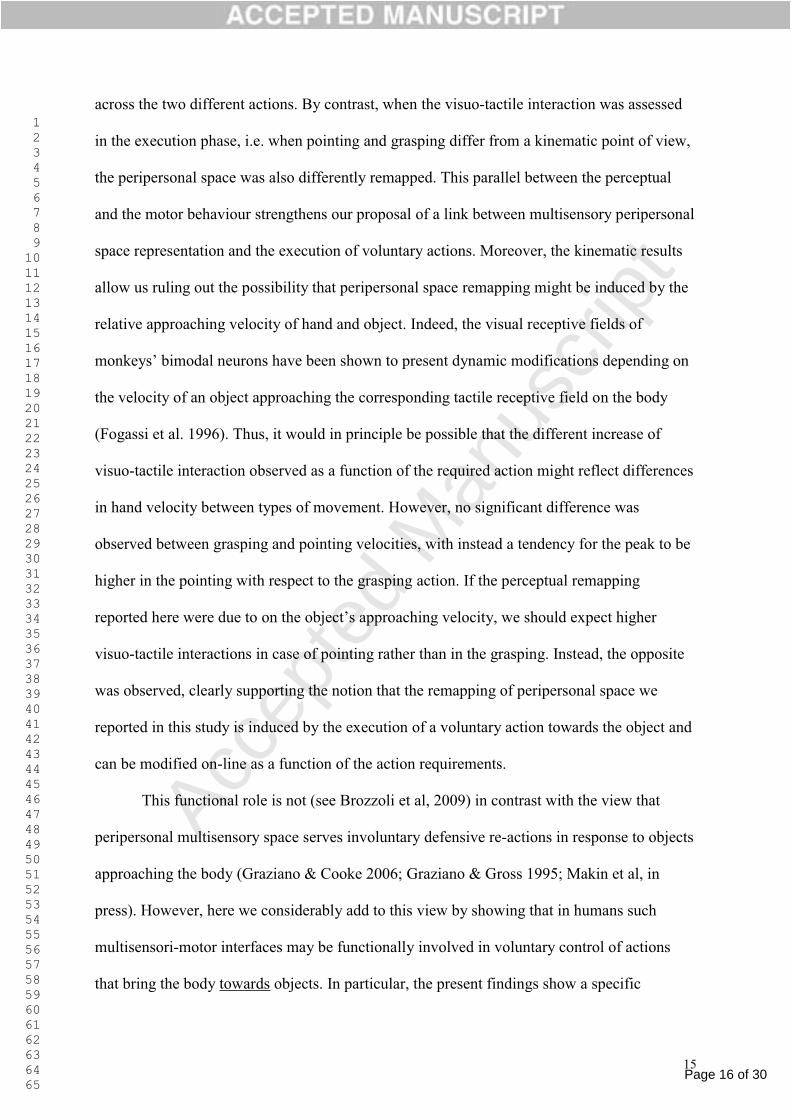
Page 16 of 30
Accep
ted
Man
uscr
ipt
1 2 3 4 5 6 7 8 9 1011121314151617181920212223242526272829303132333435363738394041424344454647484950515253545556575859606162636465
15
across the two different actions. By contrast, when the visuo-tactile interaction was assessed
in the execution phase, i.e. when pointing and grasping differ from a kinematic point of view,
the peripersonal space was also differently remapped. This parallel between the perceptual
and the motor behaviour strengthens our proposal of a link between multisensory peripersonal
space representation and the execution of voluntary actions. Moreover, the kinematic results
allow us ruling out the possibility that peripersonal space remapping might be induced by the
relative approaching velocity of hand and object. Indeed, the visual receptive fields of
the velocity of an object approaching the corresponding tactile receptive field on the body
(Fogassi et al. 1996). Thus, it would in principle be possible that the different increase of
visuo-tactile interaction observed as a function of the required action might reflect differences
in hand velocity between types of movement. However, no significant difference was
observed between grasping and pointing velocities, with instead a tendency for the peak to be
higher in the pointing with respect to the grasping action. If the perceptual remapping
proaching velocity, we should expect higher
visuo-tactile interactions in case of pointing rather than in the grasping. Instead, the opposite
was observed, clearly supporting the notion that the remapping of peripersonal space we
reported in this study is induced by the execution of a voluntary action towards the object and
can be modified on-line as a function of the action requirements.
This functional role is not (see Brozzoli et al, 2009) in contrast with the view that
peripersonal multisensory space serves involuntary defensive re-actions in response to objects
approaching the body (Graziano & Cooke 2006; Graziano & Gross 1995; Makin et al, in
press). However, here we considerably add to this view by showing that in humans such
multisensori-motor interfaces may be functionally involved in voluntary control of actions
that bring the body towards objects. In particular, the present findings show a specific
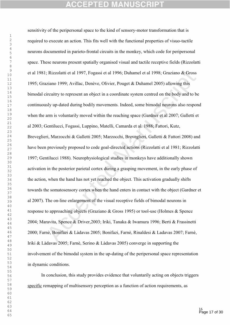
Page 17 of 30
Accep
ted
Man
uscr
ipt
1 2 3 4 5 6 7 8 9 1011121314151617181920212223242526272829303132333435363738394041424344454647484950515253545556575859606162636465
16
sensitivity of the peripersonal space to the kind of sensory-motor transformation that is
required to execute an action. This fits well with the functional properties of visuo-tactile
neurons documented in parieto-frontal circuits in the monkey, which code for peripersonal
space. These neurons present spatially organised visual and tactile receptive fields (Rizzolatti
et al 1981; Rizzolatti et al 1997, Fogassi et al 1996; Duhamel et al 1998; Graziano & Gross
1995; Graziano 1999; Avillac, Denève, Olivier, Pouget & Duhamel 2005) allowing this
bimodal circuitry to represent an object in a coordinate system centred on the body and to be
continuously up-dated during bodily movements. Indeed, some bimodal neurons also respond
when the arm is voluntarily moved within the reaching space (Gardner et al 2007; Galletti et
al 2003; Gentilucci, Fogassi, Luppino, Matelli, Camarda et al. 1988; Fattori, Kutz,
Breveglieri, Marzocchi & Galletti 2005; Marzocchi, Breveglieri, Galletti & Fattori 2008) and
have been previously proposed to code goal-directed actions (Rizzolatti et al 1981; Rizzolatti
1997; Gentilucci 1988). Neurophysiological studies in monkeys have additionally shown
activation in the posterior parietal cortex during a grasping movement, in the early phase of
the action, when the hand has not yet reached the object. This activation gradually shifts
towards the somatosensory cortex when the hand enters in contact with the object (Gardner et
al 2007). The on-line enlargement of the visual receptive fields of bimodal neurons in
response to approaching objects (Graziano & Gross 1995) or tool-use (Holmes & Spence
2004; Maravita, Spence & Driver,2003; Iriki, Tanaka & Iwamura 1996; Berti & Frassinetti
2000; Farnè, Bonifazi & Làdavas 2005; Bonifazi, Farnè, Rinaldesi & Ladavas 2007; Farnè,
Iriki & Làdavas 2005; Farnè, Serino & Làdavas 2005) converge in supporting the
involvement of the bimodal system in the up-dating of the peripersonal space representation
in dynamic conditions.
In conclusion, this study provides evidence that voluntarily acting on objects triggers
specific remapping of multisensory perception as a function of action requirements, as
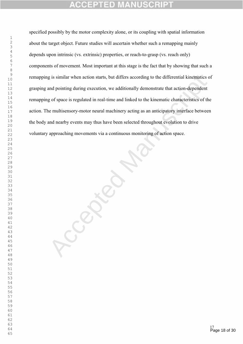
Page 18 of 30
Accep
ted
Man
uscr
ipt
1 2 3 4 5 6 7 8 9 1011121314151617181920212223242526272829303132333435363738394041424344454647484950515253545556575859606162636465
17
specified possibly by the motor complexity alone, or its coupling with spatial information
about the target object. Future studies will ascertain whether such a remapping mainly
depends upon intrinsic (vs. extrinsic) properties, or reach-to-grasp (vs. reach only)
components of movement. Most important at this stage is the fact that by showing that such a
remapping is similar when action starts, but differs according to the differential kinematics of
grasping and pointing during execution, we additionally demonstrate that action-dependent
remapping of space is regulated in real-time and linked to the kinematic characteristics of the
action. The multisensory-motor neural machinery acting as an anticipatory interface between
the body and nearby events may thus have been selected throughout evolution to drive
voluntary approaching movements via a continuous monitoring of action space.
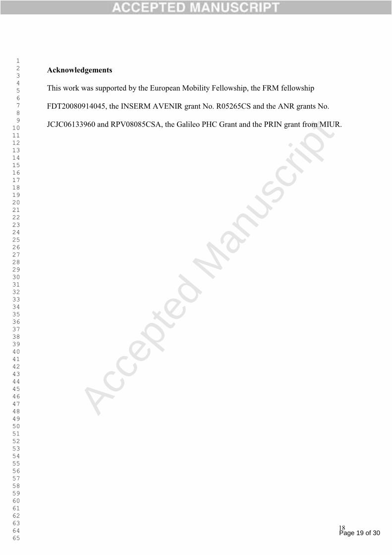
Page 19 of 30
Accep
ted
Man
uscr
ipt
1 2 3 4 5 6 7 8 9 1011121314151617181920212223242526272829303132333435363738394041424344454647484950515253545556575859606162636465
18
Acknowledgements
This work was supported by the European Mobility Fellowship, the FRM fellowship
FDT20080914045, the INSERM AVENIR grant No. R05265CS and the ANR grants No.
JCJC06133960 and RPV08085CSA, the Galileo PHC Grant and the PRIN grant from MIUR.

Page 20 of 30
Accep
ted
Man
uscr
ipt
1 2 3 4 5 6 7 8 9 1011121314151617181920212223242526272829303132333435363738394041424344454647484950515253545556575859606162636465
19
References
Avillac, M., Denève, S., Olivier, E., Pouget, A., & Duhamel, J.R. (2005). Reference
frames for representing visual and tactile locations in parietal cortex. Nature
Neuroscience, 8, 941-949.
Berti, A., & Frassinetti, F. (2000). When far becomes near: remapping of space by tool
use. Journal of Cognitive Neuroscience, 12, 415-420.
Bonifazi, S., Farnè, A., Rinaldesi, L., & Làdavas E. (2007). Dynamic size-change of peri-
hand space through tool-use: Spatial extension or shift of the multi-sensory area. Journal
of Neuropsychology, 1, 101-114.
Bremmer, F., Schlack, A., Duhamel, J.R., Graf, W., & Fink, G.R. (2001). Space coding in
primate posterior parietal cortex. Neuroimage, 14, S46-51.
Bremmer, F., Schlack, A., Shah, N.J., Zafiris, O., Kubischik, M., et al. (2001). Polymodal
motion processing in posterior parietal and premotor cortex: a human fMRI study strongly
implies equivalencies between humans and monkeys. Neuron, 29, 287-296.
Brozzoli, C., Demattè, M.L., Pavani, F., Frassinetti, F., & Farnè A. (2006). Neglect and
extinction: within and between sensory modalities. Restorative Neurology &
Neuroscience, 24, 217-232.
Brozzoli, C., Pavani, F., Urquizar, C., Cardinali, L., & Farnè A. (2009). Grasping actions
remap peripersonal space. NeuroReport, 20, 913-917.
Cardinali, L., Brozzoli, C., & Farnè A. (2009). Peripersonal space and body schema: two
labels for the same concept? Brain Topography. 21, 252-260.
Castiello, U. (2005). The neuroscience of grasping. Nature Review Neuroscience, 6, 726-
736.
Desmurget, M., Epstein, C.M., Turner, R.S., Prablanc, C., Alexander, G.E., & Grafton
S.T. (1999). Role of the posterior parietal cortex in updating reaching movements to a
visual target. Nature Neuroscience, 2, 563-567.
di Pellegrino, G., Làdavas, E., & Farné A. (1997). Seeing where your hands are. Nature,
21, 730.
Duhamel, J.R., Colby, C.L., & Goldberg, M.E. (1998). Ventral Intraparietal area of the
macaque: congruent visual and somatic response properties. Journal of Neurophysiology,
79, 126-136.
Page 21 of 30
Accep
ted
Man
uscr
ipt
1 2 3 4 5 6 7 8 9 1011121314151617181920212223242526272829303132333435363738394041424344454647484950515253545556575859606162636465
20
Farnè, A., Bonifazi, S., & Ladavas, E. (2005). The role played by tool-use and tool-length
on the plastic elongation of peri-hand space: A single case study. Cognitive
Neuropsychology, 22, 408-418.
Farnè, A., Demattè, M.L. & Làdavas, E. (2005). Neuropsychological evidence of modular
organization of the near peripersonal space. Neurology, 13, 1754-1758.
Farnè, A., Iriki, A., & Ladavas, E. (2005). Shaping multisensory action-space with tools:
evidence from patients with cross-modal extinction. Neuropsychologia, 43, 238-248.
Farnè, A., Pavani, F., Meneghello, F., & Làdavas, E. (2000) Left tactile extinction
following visual stimulation of a rubber hand. Brain, 123, 2350-2360.
Farnè, A., Roy, A.C., Paulignan, Y., Rode, G., Rossetti, Y., Boisson, D., & Jeannerod, M.
(2003). Visuo-motor control of the ipsilateral hand: evidence from right brain-damaged
patients. Neuropsychologia, 41, 739-757.
Farnè, A., Serino, A., & Ladavas, E. (2005). Dynamic size-change of peri-hand space
following tool-use: determinants and spatial characteristics revealed through cross-modal
extinction. Cortex, 43, 436-443.
Fattori, P., Kutz, D.F., Breveglieri, R., Marzocchi, N., & Galletti, C. (2005). Spatial
tuning of reaching activity in the medial parieto-occipital cortex (area V6A) of macaque
monkey. European Journal of Neuroscience, 22, 956-972.
Fogassi, L., Gallese, V., di Pellegrino, G., Fadiga, L., Gentilucci, M., Luppino, G.,
Matelli, M., Pedotti, A., & Rizzolatti, G. (1992). Space coding by premotor cortex.
Experimental Brain Research, 89, 686-690.
Fogassi, L., Gallese, V., Fadiga, L., Luppino, G., Matelli, M., & Rizzolatti G. (1996).
Coding of peripersonal space in inferior premotor cortex (area F4). Journal of
Neurophysiology, 76, 141-157.
Galletti, C., Kutz, D.F., Gamberini, M., Breveglieri, R., & Fattori, P. (2003). Role of the
medial parieto-occipital cortex in the control of reaching and grasping movements.
Experimental Brain Research, 153, 158-170.
Gardner, E.P., Babu, K.S., Reitzen, S.D., Ghosh, S., Brown, A.S. et al. (2007).
Neurophysiology of prehension. I. Posterior parietal cortex and object-oriented hand
behaviors. Journal of Neurophysiology, 97, 387-406.
Gentilucci, M., Fogassi, L., Luppino, G., Matelli, M., Camarda, R. et al (1988).
Somatotopic representation in inferior area 6 of the macaque monkey. Experimental Brain
Research, 71, 475-490.
Page 22 of 30
Accep
ted
Man
uscr
ipt
1 2 3 4 5 6 7 8 9 1011121314151617181920212223242526272829303132333435363738394041424344454647484950515253545556575859606162636465
21
Gentilucci, M., Scandolara, C., Pigarev, I.N., & Rizzolatti, G. (1983). Visual responses in
the postarcuate cortex (area 6) of the monkey that are independent of eye position.
Experimental Brain Research, 50, 464-468.
Godschalk, M., Lemon, R.N., Kuypers, H.G., & van der Steen J. (1985). The involvement
of monkey premotor cortex neurones in preparation of visually cued arm movements.
Behavioral Brain Research, 18, 143-157.
Graziano, M.S.A. (1999). Where is my arm? The relative role of vision and
proprioception in the neuronal representation of limb position. Proceedings of the
National Academy of Science, 97, 10418-10421.
Graziano, M.S.A., & Cooke, D.F. (2006). Parieto-frontal interactions, personal space, and
defensive behavior. Neuropsychologia, 44, 2621-2635.
Graziano, M.S.A., & Gross, C.G. (1995). The representation of extrapersonal space: a
possible role for bimodal, visuo-tactile neurons. in The Cognitive Neurosciences, Ed. M.
Gazzaniga, MIT Press: 1021-1034.
Holmes, N.P., & Spence, C. (2004). The body schema and multisensory representations of
peripersonale space. Cognitive Processing, 5, 94-105.
Hyvärinen, J., & Poranen, A. (1974). Function of the parietal associative area 7 as
revealed from cellular discharges in alert monkeys. Brain, 97, 673-692.
Iriki, A., Tanaka, M., & Iwamura, Y. (1996). Coding of modified body schema during
tool use by macaque postcentral neurons. Neuroreport, 7, 2325-2330.
Jeannerod, M. (1986). Mechanisms of visuomotor coordination: a study in normal and
brain-damaged subjects. Neurpsychologia, 24, 41-78.
Jeannerod, M. (1988). Motor Control: concepts and issues New York: Wiley J.
Làdavas, E., & Farnè, A. (2004). Visuo-tactile representation of near-the-body space.
Journal Physiology Paris, 98, 161-170.
Leinonen, L. (1980). Functional properties of neurones in the posterior part of area 7 in
awake monkey. Acta Physiologica Scandinavica, 108, 301-308.
Makin, T.R., Holmes, N.P., & Zohary, E. (2007). Is that near my hand? Multisensory
representation of peripersonal space in human intraparietal sulcus. Journal of
Neuroscience, 27, 731-740.
Makin, T.R., Holmes, N.P., Brozzoli, C., Rossetti, Y., & Farnè A. (in press). Coding of
visual space during motor preparation: Approaching objects rapidly modulate
corticospinal excitability in hand-centred coordinates. Journal of Neuroscience.
Page 23 of 30
Accep
ted
Man
uscr
ipt
1 2 3 4 5 6 7 8 9 1011121314151617181920212223242526272829303132333435363738394041424344454647484950515253545556575859606162636465
22
Maravita, A., Spence, C., & Driver, J. (2003). Multisensory integration and the body
schema: Close to hand and within reach. Current Biology, 13, 531-539.
Marzocchi, N., Breveglieri, R., Galletti, C., & Fattori, P. (2008). Reaching activity in
parietal area V6A of macaque: eye influence on arm activity or retinocentric coding of
reaching movements? European Journal of Neuroscience, 27, 775-789.
Mountcastle, V.B., Lynch, J.C., Georgopoulos, A., Sakata, H., & Acuna, C. (1975).
Posterior parietal association cortex of the monkey: command functions for operations
within extrapersonal space. Journal of Neurophysiology, 38, 871-908.
Paulignan, Y., MacKenzie, C., Marteniuk, R., & Jeannerod, M. (1991). Selective
perturbation of visual input during prehension movements. 1. The effects of changing
object position. Experimental Brain Research, 83, 502-512.
Pavani, F., & Castiello, U. (2004). Binding personal and extrapersonal space through body
shadows. Nature Neuroscience, 7, 14-16.
Pisella, L., Gréa, H., Tilikete, C., Vighetto, A., Desmurget, M., Rode, G., Boisson, D., &
Rossetti, Y. (2000). An 'automatic pilot' for the hand in human posterior parietal cortex:
toward reinterpreting optic ataxia. Nature Neuroscience, 3, 729-736.
Rizzolatti, G., Fadiga, L., Fogassi, L., & Gallese, V. (1997). The space around us. Science,
11, 190-191.
Rizzolatti, G., Fogassi, L., Gallese, V. (2002). Motor and cognitive functions of the
ventral premotor cortex. Current Opinion Neurobiology, 12, 149-154.
Rizzolatti, G., & Gentilucci, M. (1988). Motor and visual-motor functions of the premotor
cortex. In Rakic P & Singer W (eds.) Neurobiology of Neocortex. John Wiley & sons Ltd,
pp. 269-284.
Rizzolatti, G., Gentilucci, M., Fogassi, L., Luppino, G., Matelli, M., & Ponzoni-Maggi, S.
(1987). Neurons related to goal-directed motor acts in inferior area 6 of the macaque
monkey. Experimental Brain Research, 67, 220-224.
Rizzolatti, G., Scandolara, C., Matelli, M., & Gentilucci, M. (1981). Afferent properties of
periarcuate neurons in macque monkeys. II. Visual responses. Behavioural Brain
Research, 2, 147-163.
Sarri, M., Blankenburg, F., & Driver, J. (2006). Neural correlates of crossmodal visual-
tactile extinction and of tactile awareness revealed by fMRI in a right-hemisphere stroke
patient. Neuropsychologia, 44, 2398-2410.
Sereno, M.I., & Huang, R.S. (2006). A human parietal face area contains aligned head-
centered visual and tactile maps. Nature Neuroscience, 9, 1337-1343.
Page 24 of 30
Accep
ted
Man
uscr
ipt
1 2 3 4 5 6 7 8 9 1011121314151617181920212223242526272829303132333435363738394041424344454647484950515253545556575859606162636465
23
Spence, C., Pavani, F., & Driver, J. (2004a). Spatial constraints on visual-tactile cross-
modal distractor congruency effects. Cognitve Affective and Behavioral Neuroscience, 4,
148-169.
Spence, C., Pavani, F., Maravita, A., & Holmes, N. (2004b). Multisensory contributions to
the 3-D representation of visuotactile peripersonal space in humans: evidence from the
crossmodal congruency task. Journal Physiology Paris, 98, 171-189.
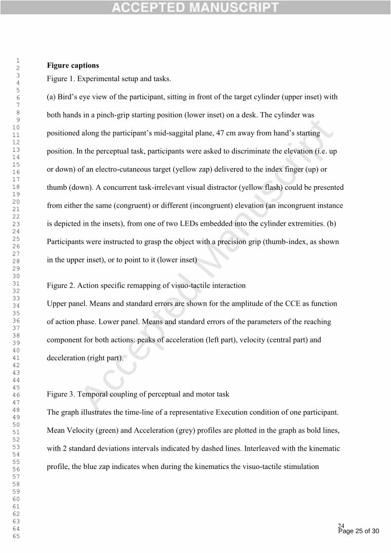
Page 25 of 30
Accep
ted
Man
uscr
ipt
1 2 3 4 5 6 7 8 9 1011121314151617181920212223242526272829303132333435363738394041424344454647484950515253545556575859606162636465
24
Figure captions
Figure 1. Experimental setup and tasks.
both hands in a pinch-grip starting position (lower inset) on a desk. The cylinder was
-
position. In the perceptual task, participants were asked to discriminate the elevation (i.e. up
or down) of an electro-cutaneous target (yellow zap) delivered to the index finger (up) or
thumb (down). A concurrent task-irrelevant visual distractor (yellow flash) could be presented
from either the same (congruent) or different (incongruent) elevation (an incongruent instance
is depicted in the insets), from one of two LEDs embedded into the cylinder extremities. (b)
Participants were instructed to grasp the object with a precision grip (thumb-index, as shown
in the upper inset), or to point to it (lower inset)
Figure 2. Action specific remapping of visuo-tactile interaction
Upper panel. Means and standard errors are shown for the amplitude of the CCE as function
of action phase. Lower panel. Means and standard errors of the parameters of the reaching
component for both actions: peaks of acceleration (left part), velocity (central part) and
deceleration (right part).
Figure 3. Temporal coupling of perceptual and motor task
The graph illustrates the time-line of a representative Execution condition of one participant.
Mean Velocity (green) and Acceleration (grey) profiles are plotted in the graph as bold lines,
with 2 standard deviations intervals indicated by dashed lines. Interleaved with the kinematic
profile, the blue zap indicates when during the kinematics the visuo-tactile stimulation
Page 26 of 30
Accep
ted
Man
uscr
ipt
1 2 3 4 5 6 7 8 9 1011121314151617181920212223242526272829303132333435363738394041424344454647484950515253545556575859606162636465
25
occurred. Means and standard errors of Reaction Times for Congruent (full blue vertical line)
and Incongruent (dashed blue vertical line) stimulations are also plotted.
Table 1. Means and standard errors for motor reaction time (ms) from the go signal, duration
(ms) of movements and absolute reaction time (ms) to the perceptual visuo-tactile task in all
experimental conditions.
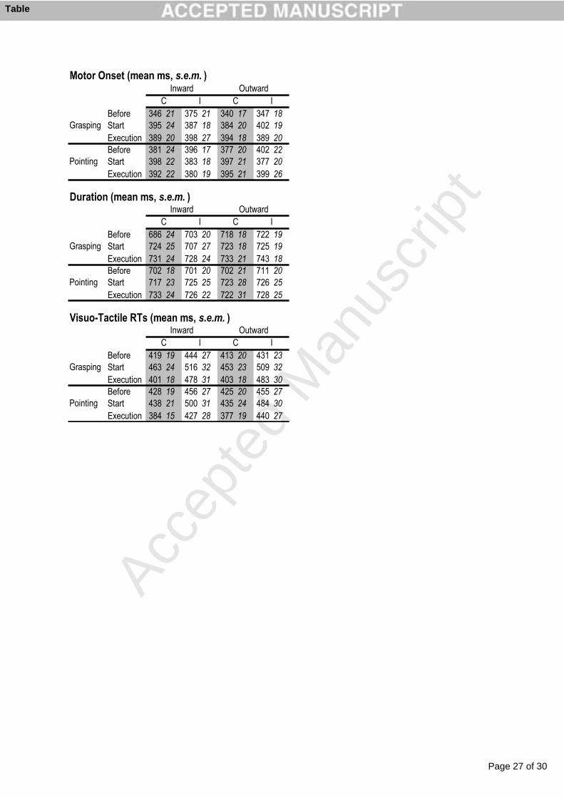
Page 27 of 30
Accep
ted
Man
uscr
ipt
Before 346 21 375 21 340 17 347 18Start 395 24 387 18 384 20 402 19Execution 389 20 398 27 394 18 389 20Before 381 24 396 17 377 20 402 22Start 398 22 383 18 397 21 377 20Execution 392 22 380 19 395 21 399 26
Before 686 24 703 20 718 18 722 19Start 724 25 707 27 723 18 725 19Execution 731 24 728 24 733 21 743 18Before 702 18 701 20 702 21 711 20Start 717 23 725 25 723 28 726 25Execution 733 24 726 22 722 31 728 25
Before 419 19 444 27 413 20 431 23Start 463 24 516 32 453 23 509 32Execution 401 18 478 31 403 18 483 30Before 428 19 456 27 425 20 455 27Start 438 21 500 31 435 24 484 30Execution 384 15 427 28 377 19 440 27
Motor Onset (mean ms, s.e.m. )
C I C I
Grasping
Pointing
Inward OutwardDuration (mean ms, s.e.m. )
C I C I
C I
Grasping
Pointing
Inward Outward
Grasping
Pointing
Inward Outward
Visuo-Tactile RTs (mean ms, s.e.m. )
C I
Table
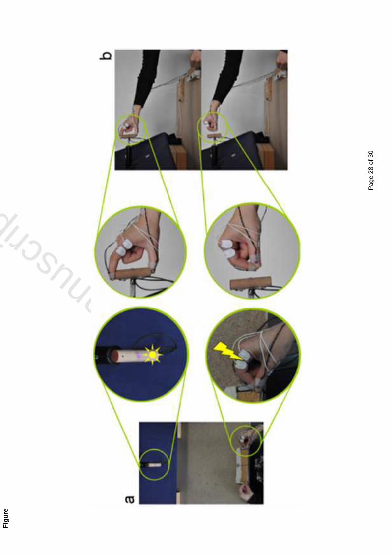
Pag
e 28
of 3
0
Accepted M
anuscrip
t
Fig
ure
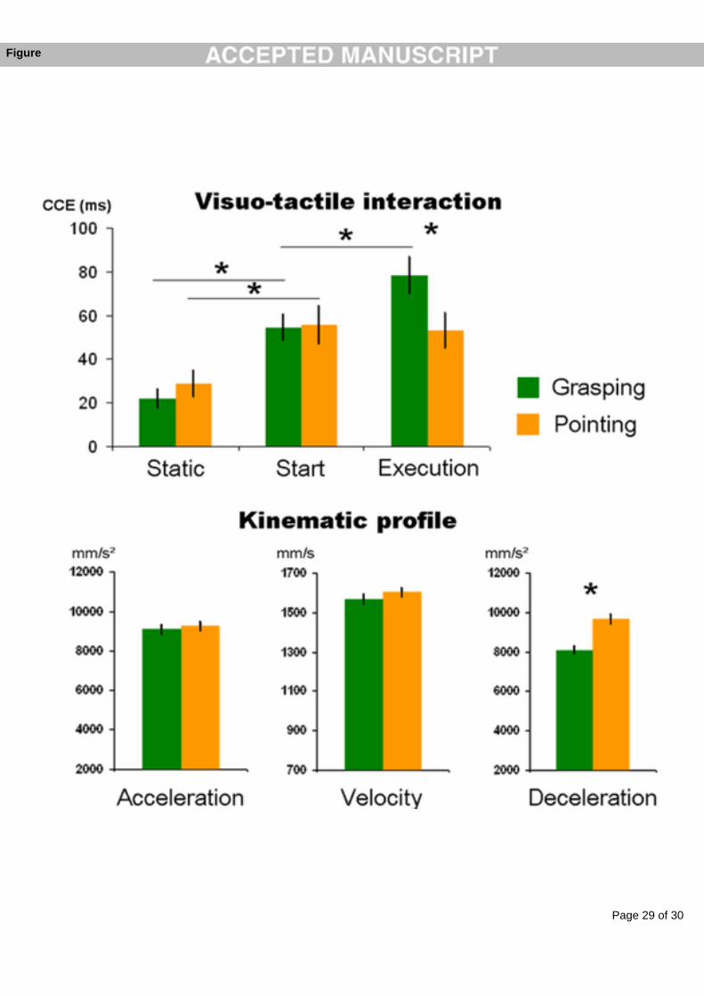
Page 29 of 30
Accep
ted
Man
uscr
ipt
Figure
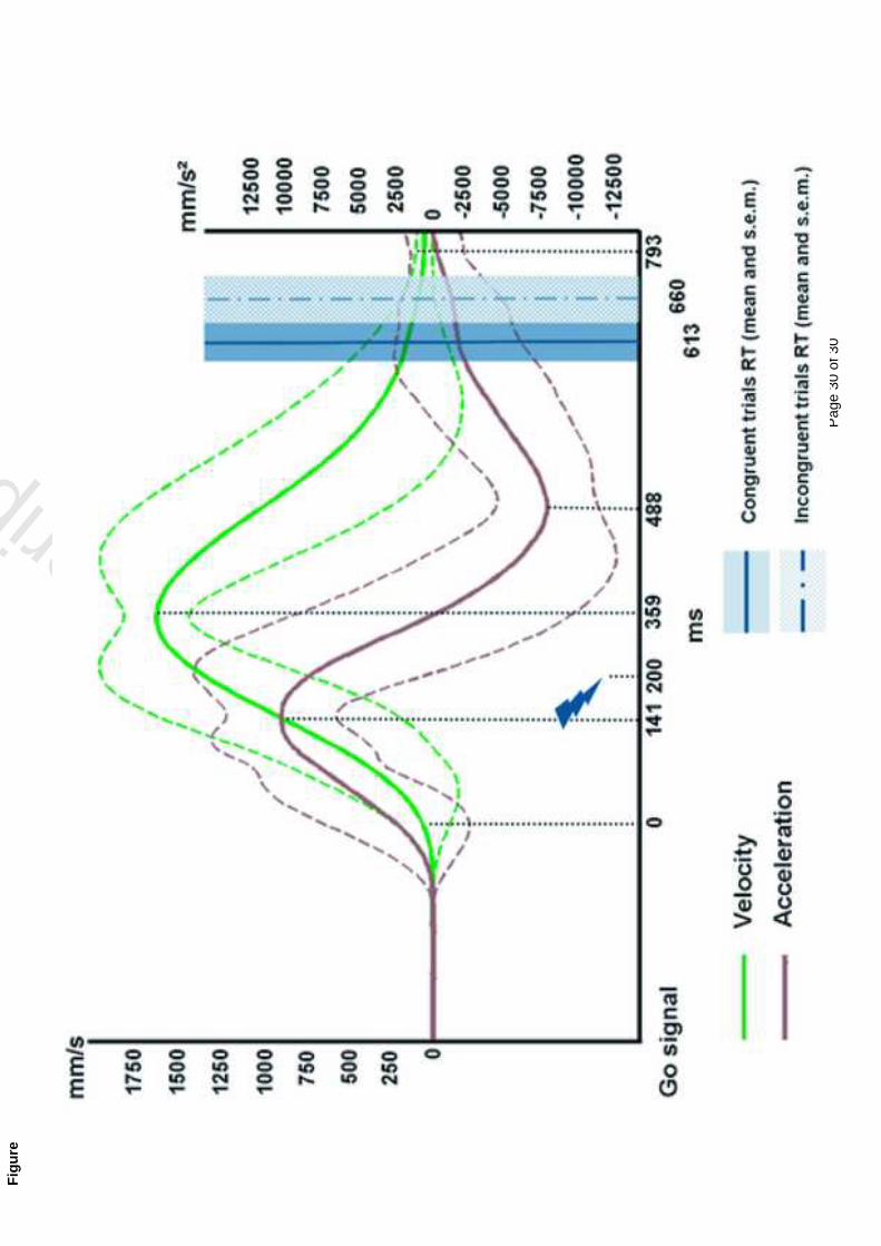
Pag
e 30
of 3
0
Accepted M
anuscrip
t
Fig
ure

DISCUSSION

Main results
Aim of the first study was to test the hypothesis of an involvement of the peripersonal space
representation in the rapid detection of objects approaching the body for the preparation of
involuntary defensive movements. In particular we measured how the excitability of the
motor cortex in correspondence of the hand representation varied when an object become
visible in the space around the hand. The series of experiments presented in this study showed
that the motor cortex is very rapidly provided with information concerning the representation
of the visual space around a particular body-part. When an object is approaching the hand, the
visual information is thus available to the motor cortex in order to react in the most adaptive
way to the environment. This spatial representation is centred on the body, following more the
hand rather than the eye position. The inhibition of cortical excitability induced by an object
approaching the hand is interpreted as a proactive inhibition in case of voluntary motor plans
that are contradictory with the avoidance involuntary reaction. We have also suggested that
the source of such information is most likely to be individuated in the premotor cortex,
although other multisensory regions may also contribute.
The second study tested, the involvement of the peripersonal space representation in the
execution of voluntary actions toward an object, such as an everyday grasping action. The
well-known phenomenon of visuo-tactile interference has been used as a measure of the
integration of visual and somatosensory information and so as a measure of peripersonal
space. The novel approach of co-registration of perceptual multisensory effect and kinematics
of the movement allowed to establish possible parallels between the perceptual and the motor
domain. Results of this study showed that the multisensory link between visual information
on a target object and tactile information on the acting body-part is modulated by the action.
A remapping of peripersonal space is indeed induced since the onset of the movement.
Moreover, the effect appeared to be present for those conditions which revealed to be the most
demanding from a kinematic point of view.
187
Aim of the third study was to more deeply investigate the link between motor demands and
perceptual effects. Employing the same paradigm, we contrasted the peripersonal space
remapping induced by a grasping an object with that induced by a less complex action such as
a pointing towards (without contacting) the same object. The two kinds of action are known to
share a transport component of the hand toward the target; they diverge, by contrast, for the
additional presence in the grasping of a grip component where the hand needs to be adjusted
to fit the intrinsic characteristics of the target and its spatial orientation. Results of this study
showed that the remapping of peripersonal space induced by a voluntary action is continuous
and dependent on the sensori-motor transformations required by the particular action in
progress. Indeed, the two actions present a similar remapping at the on-set of the movement;
however during the execution phase, when the actions kinematically differ, they induce a
different remapping of peripersonal space.
The following discussion will try to integrate the results of these studies in a general
model of peripersonal space as an interface between the body and the external world.
� 188� �
Peripersonal space: an interface for avoidance reactions
The neurophysiological studies on monkeys have shown the body-centred nature of the
peripersonal space representation (Graziano and Gross 1995); the visual receptive fields are in
spatial register with the tactile ones; the visuo-tactile neurons areas in the brain receive
proprioceptive input in order to update the peripersonal space representation at each body
movement. Indeed, as Graziano’s studies clearly showed, the visual receptive fields of
bimodal visuo-tactile neurones follow the tactile receptive field, for example when the hand
changes its position. Importantly, the visual receptive fields are also dynamic, as Fogassi and
colleagues showed: an object approaching a body-part induces a modification of the visual
receptive fields connected to the tactile ones on the same body-part, as a function of the
approaching velocity.
On the basis of these characteristics, Graziano proposed the peripersonal space
representation as a “defensive space”, a sort of radar that, by anticipatorily detecting
approaching objects can prevent the body be hurt by the possible dangerous collisions by
rapidly feeding this information to the motor system. Following this hypothesis, Graziano’s
group collected data about defensive reactions in animals. They thus provided the only
available link between peripersonal space representation and involuntary defensive reactions
in animals, through the direct electrical microstimulation of bimodal visuo-tactile areas.
However, they only showed that such a stimulation induces patterns of movements
compatible with defensive reaction.
Our results of the first study reported here are the first direct demonstration in humans
that the peripersonal space coding may indeed make available such information to the motor
system very rapidly, providing compelling evidence in favour of the role that peripersonal
space can play in detecting approaching objects and thus in defensive reactions.
Motor evoked potentials are a measure of the excitability of the motor cortex and thus
considered a measure of the “readiness” of the motor cortex to send the motor command for
the required movement to be executed. Our results show that motor cortex excitability of the
hand representation is reduced when a three-dimensional object is approaching the hand
rather than falling far from it. This means that motor cortex receives the information about the
189
presence of a visible object near the body. Furthermore, it is worth noting that the location of
the object is coded in a reference frame centred on the body-part. Indeed, our results clearly
showed that the effect is more dependent on the position of the hand in the action space than
on the direction of visual fixation, or the orienting of attention.
We hypothesized that this early reduction of motor cortex excitability might reflect the
proactive inhibition of an undesirable response (Koch et al. 2006; Mars et al. 2007; van den
Hurk et al. 2007; van Elswijk et al. 2007), consequence of the conflict between a voluntary
motor response (to press the button) and an avoidance movement elicited by the approaching
ball. Two arguments are directly available from our results to support this view. The first is
the fact that after a voluntary inhibition of motor responses, we did not detect any hand-
centered modulation of MEP amplitudes with a general suppression of motor excitability. In
other words this result is compatible with the interpretation of the reduction in motor cortex
excitability as an inhibition. Second, and more important, MEPs recorded from another
muscle that is not involved in the task-related motor response, did not show any modulation
depending on the distance of the object with respect to the hand. Therefore, when competition
between the avoidance and go responses is eliminated (or temporally desynchronised), no
suppression of the avoidance response is required anymore. Our results therefore indicate an
interaction between the motor consequences of the rapid visual processing of objects
approaching the hand and the voluntary task-related motor plans for that hand. These results
represent the first evidence in humans in favour of the involvement of peripersonal space in
serving defensive reactions.
A comparison with non-human primates studies
The results of this first study are compatible with an involvement of premotor cortex and
posterior parietal areas in this phenomenon. M1 and premotor cortex are indeed densely
interconnected, as described in the introduction, both in humans and in monkeys (Davare et
al. 2008; Dum and Strick, 2005; Koch et al. 2006; O’Shea et al. 2007; Shimazu et al. 2004).
A point worth of consideration is the on-set of corticospinal modulations at 70-80 ms
following the appearance of the object, a very rapid transformation of visual information into
� 190� �
motor consequences. The modulation of motor cortex excitability with respect to approaching
object may therefore reflect ongoing activity in the premotor cortex. In monkeys, neurons of
premotor cortex have been found to be driven by visual stimulation at time periods similar to
the ones we have described, and are thought to encode the significance of visual cues for
response selection (Cisek and Kalaska, 2005). In humans, the premotor cortex has been
shown to participate in visuomotor transformations required to configure hand posture with
respect to objects (Davare et al. 2006). Furthermore, recent studies have shown a direct
influence of premotor over primary motor cortex as early as 75 ms following a ‘Go’ signal
(Koch et al. 2006; O’Shea et al. 2007).
The other point in favour of an involvement of posterior parietal areas is the selectivity
of the motor cortex excitability modulation with respect to the three-dimensional
characteristics of the object. Indeed, when the real three-dimensional balls were replaced by
virtually bi-dimensional static LEDs, no effect was found of the body-centred modulation of
the cortical excitability. As reviewed in the introduction, area AIP in the intraparietal sulcus of
macaque monkeys shows a strong sensibility to three-dimensional objects with respect to bi-
dimensional ones. Since AIP and premotor cortex are densely interconnected, it is plausible to
think of an involvement of posterior parietal areas in modulating motor cortex excitability
when a three-dimensional object becomes suddenly visible near the hand.
While any comparisons between data drawn from monkeys and humans using such
different methods should be made with caution, given the timing and the spatial specificity of
the above responses with respect to visual events, these mechanisms fit very well with our
results, and suggest the involvement of human posterior parietal and premotor areas with the
hand-centred coding of visual space.
These data and considerations support the idea of a parieto-frontal network as the
neurophysiological basis of the peripersonal space representations. This specialised system
for transforming nearby sensory inputs into rapid and appropriate motor outputs is ideally
suited to serve as a sensori-motor interface for driving defensive movements away from
potentially harmful approaching objects.
191
Peripersonal space: an interface for voluntary actions
When grasping an object, the brain updates the link between the visual information available
on the target object and the tactile information on the grasping body-part. The results we
provided in the second and third studies clearly show that this up-dating arises and that
peripersonal space is re-mapped since the on-set of the execution of a grasping. When the
hand starts to move - but well before it comes into contact with the object - visual inputs
coming from the object are more strongly interacting with tactile information on the body,
with respect to a static situation. Furthermore, the finding that such visuo-tactile re-weighting
was observed selectively when both perceptual and grasping tasks concerned the same hand
not only confirms the hand-centred nature of the peripersonal space representation, but
critically extends this property to ecological and adaptive dynamic situations of voluntary
manipulative actions. Any interpretation solely based on an attentional account can be ruled
out by our results. Indeed, performing the grasping with the left hand as in the second study
(experiment 3), required the participants to allocate the same amount of attention on the
object as in the right hand grasping condition. Another point worth to be underline is the fact
that visual and tactile information in our paradigm were delivered simultaneously. While in
most part of the CCE studies, the visual distractor preceded the tactile target, working more as
an attentional cue, in our paradigm we tried to avoid it in order to reduce the attentional
effects on the visuo-tactile interaction. These results thus constitute the first evidence in
humans of an involvement of peripersonal space in the execution of voluntary actions.
A crucial insight in these studies was gleaned from the kinematic analysis of the
grasping movements. During the visuo-tactile and object-grasping task, movements of both
hands were recorded kinematically. These findings ruled out the possibility that the effector-
specific increase in CCE reflected a difference between motor performances for the two
hands, since they displayed comparable kinematic profiles. Furthermore, the kinematics
analysis revealed possible parallels between the motor and perceptual performances, showing
that a difference in the kinematic pattern was reflected by a difference in the perceptual
domain. In particular, orientations of the target object that elicited kinematically more
demanding reaching movements (i.e., in terms of inward wrist rotation) also elicited stronger
CCE. In a different experiment, when the kinematic difficulty of the movement was equalised
� 192� �
across different object orientations, the action also triggered comparable perceptual
remapping across object orientations, thus showing a comparable amount of CCE when
grasping actions were comparably difficult in kinematic terms. This parallel between the
motor and perceptual performance strengthens the functional link between multisensory
coding of hand-centred space and voluntary action.
The increase in CCE triggered by the action, even if already present at the very on-set
of the movement, kept increasing during the early execution phase. That is, an even stronger
interference of visual on tactile information was revealed, as the action unfolded in time and
space. This suggests that performing a voluntary action triggers a continuous monitoring of
action space, which keeps ‘assisting’ the motor execution of the action during its whole
dynamic phase. This consideration is indeed supported by the results of the third experiment
were two kinematically different actions were proved to elicit different remapping of
peripersonal space. If part of the remapping of peripersonal space is already effective at the
onset of the motor program, the perceptual modulation can be either further enhanced in the
case of relatively complex object-oriented actions like grasping, or kept unchanged in the case
of simpler pointing actions. This on-line, motor-evoked “monitoring” of action space opens
the possibility of very fast modulations of peripersonal space representation as a function of
more ecological needs during action execution. One could speculate that such rapid on-line
updating, for instance, could parallel the fast, on-line motor corrections documented in motor
control studies (Desmurget et al. 1999; Farnè et al. 2003a; Paulignan et al. 1991; Pisella et al.
2000). Since deficits of the so-called “automatic pilot” (Pisella et al. 2001) have been
documented after lesions of the posterior parietal cortex in humans, as well as in healthy
subjects after parietal TMS (Desmurget et al. 1999), we suggest that the mechanism
underlying the rapid remapping of peripersonal space could be damaged in these patients, thus
depriving them from the “monitoring” of action space.
A comparison with non-human primates studies
What kind of mechanism could be responsible for the rapid on-line re-mapping of
peripersonal space induced by the execution of a voluntary action?
193
It is plausible to suggest that a parieto-frontal network would be implied in the visuo-
tactile re-weighting described in these experiments. From neuroimaging investigations in
humans, as reviewed earlier in this dissertation, both parietal and frontal areas are involved in
the execution of reaching and grasping movements. Also investigations on multisensory
visuo-tactile perception pointed out the role of parietal and premotor areas. This series of
neuroimaging data has provided some evidence about the network of neural structures which
appears to be involved in the multisensory representation of near space in humans, namely the
IPS and ventral premotor cortex. Activation in the posterior aspect of the medial IPS in
humans has been shown to play a role in tasks requiring visuomotor coordination of hand
movements with respect to targets (Chaminade and Decety, 2002; Simon et al. 2002; Grefkes
et al. 2004). The same area has been reported to show a topographic mapping of space both
for saccades and for pointing to targets, which is updated with eye movements (Medendorp et
al. 2003, 2005). According to Ehrsson et al. (2004), activity in the medial wall of the IPS
reflects the seen position of the hand. Futhermore, IPS has been shown to be activated for
objects near the hand rather than far from it (Makin et al. 2006). These findings support the
potential role of posterior IPS as an area that integrates visual and spatial information in hand-
centered coordinates. The most anterior part of the IPS showed, instead, a significant
preference for objects near to the hand also when the hand is not visible. Consistent with
properties featured by the macaque anterior intraparietal (AIP) area, the human possible
analogue hAIP (Culham et al. 2006) is highly activated by visuomotor tasks such as visually
guided grasping (Binkofski et al. 1998; Shikata et al. 2003) and also responds to hand
manipulation without visual feedback (Stoeckel et al. 2003). Furthermore, Macaluso and
colleagues (2003) found visuo–tactile integration in this area, in addition to activations
correlated with motor responses. Also the ventral premotor area in humans appears to be
related to visuo-tactile peri-hand space representation. It is worth noting that it could represent
the homologue of the premotor cortical area recognised as a region containing perihand
neurons in monkeys.
Even if all monkey-human parallels require caution it is plausible to hypothesise a
parieto-frontal circuit in humans corresponding to the posterior parietal-premotor cortex
� 194� �
circuit in monkeys (Rizzolatti et al. 1997; 2002) that could provide the representation of
peripersonal space and a rapid remapping of it during action execution.
195

CONCLUSIONSAND
PERSPECTIVES

The spatial region where we can directly interact with objects has a limited extension. This
simple consideration highlights the relevance of the region of space around the body. Indeed,
a visually available object close to the body not only represents visual information per se, but
from this information the brain has to select among all the possible motor interactions that can
occur between the object and the body. When we see our mug of coffee next to us, for
instance, not only can we appreciate its color and shape (which may be useful to recognise it
as our own mug), but our brain is also translating the object into the movements required to
grasp it and bring it to the mouth. Up to date, research partly revealed the multisensory
mechanisms that allow this translation process, in monkeys as well as in humans. In
particular, the studies of this dissertation revealed that the brain directly links the visual
information available outside of the body with the tactile information on the body. Through a
system of visuo-tactile neurons, the brain can relate visual information in the proximity of a
body-part to the body-part itself, building a peripersonal space representation. Proprioceptive
information also reaches this system, updating the link between visual and tactile information
as each body-part moves. The brain has thus a multisensory interface between the body and
the objects in the external world available for possible body-object interactions. In particular,
we showed in humans that the action related properties of the peripersonal space
representation feature the basic aspect necessary for allowing rapid avoidance reactions, i.e. a
hand centred processing of proximal visual objects. We additionally provide converging
evidence that peripersonal space representations are intimately tighted, and possibly deserve,
voluntary action execution on nearby objects. Remapping of the peripersonal space
representation is time-locked with the onset of an action and continuously operates during its
execution, as a function of the sensory-motor transformations required by the action
kinematics.
The two hypotheses (involuntary and voluntary object-oriented actions) are not
mutually exclusive and one could speculate that, from a more primordial defensive function
of this machinery, a more fine-grained and sophisticated function could have developed using
the same, relatively basic visuo-tactile spatial computational capabilities. This development
could lead to its involvement in the control of the execution of voluntary actions towards
objects. Some comparative data showed, for instance, that prosimian sensory areas
199
corresponding to the monkeys' parietal areas already present some approximate motor
activity; however, the most represented movements are just very stereotyped limb retractions
and defensive movements (Fogassi et al. 1994; see also Kaas 2004 for an interesting
comparative approach about parietal functions).
The novel findings presented in this dissertation raise several important issues. First of
all, a better knowledge about the homology of the involved areas in the human brain with
respect to non-humans primates is necessary. Some interesting clues may come from parietal
optic ataxic patients, who show a deficit in manual reaching of visual, but also somatosensory
targets (Blangero et al. 2007). In particular, these patients are impaired in reaching with the
contralesional hand toward both hemispaces (hand effect) and in reaching with both hands
toward the contralesional hemispace (field effect). One possibility could be that the parietal
damage impairs the multisensory interface normally available in healthy people to help
guiding voluntary actions toward objects. The hypothesis could be made that the motor
difficulties presented by optic ataxic patients parallel a lack of the action-triggered remapping
of peripersonal space. In this respect, the novel perceptual and kinematic co-recording
approach introduced in the experiments 2 and 3 presented here, appears as the best way to
investigate the link between an impaired motor performance and a possible deficit in the
remapping of peripersonal space. Preliminary results recently collected show indeed a
different pattern of visuo-tactile interaction during the execution of an action with respect to
healthy participants.
Other point worth discussing is how parietal and premotor areas interact in the
modulation of peripersonal space. This is true for the functional involvement of peripersonal
space both in defensive and voluntary actions, discussed in this thesis. A double pulse
paradigm, with a first conditioning TMS pulse on parietal and/or premotor areas preceding the
conditioned TMS pulse on the motor cortex, might be an interesting approach to study the
timing of parietal and frontal areas interaction in the modulation of the MEPs.
Another interesting point still un-investigated is how the different sectors of the
peripersonal space may interact. Everyday motor acts are rarely limited to a grasping without
any other consequences; more often, in fact, we are confronted with complex series of motor
acts which involve different parts of the body each with its respective peripersonal space
� 200� �
representation. When we answer the phone or when we grasp a pistachio, for instance, we
nest bring the object to the ear or into the mouth (respectively!). These simple actions put in a
dynamic relation the hand-centred and the head-centred peripersonal space representations.
Among the possibilities that could be investigated, the brain may use each representation in a
serial way, as a function of the sequential mounting of the two motor components of the entire
action: the hand-centred for the grasping and head-centred for the bringing-to-the-head
movement. Alternatively, the brain might anticipate the consequences of the entire action,
triggering a remapping of the head-centred representation already at the start of the hand
movement toward the object.
Nonetheless, when an object is grasped it can also follow a different destiny. I can
grasp a telephone or a pistachio, following the previous example, in order to offer it to
someone else. In this case, another order of interaction is required, that is a “social”
interaction with someone else’s acting body. Recent research on monkeys showed that parietal
visuo-tactile neurons can also have a representation of another individual’s body, matching
the observer’s own body representation. This correspondence, first of all opens the possibility
of a remapping of the peripersonal space of an observer during the simple observation of an
action performed by someone else. Preliminary results from our laboratory support this view.
A fourth studyexperiment has been indeed performed, where an observer was present beside
the active participants. The observer was requested to perform the same perceptual task as in
studies 2 and 3, however without executing any actions. At the same time, the observer could
see the active participant to reach and grasp the object. Results show that the simple
observation of an action can elicit a remapping of peripersonal space also in the observer of
the action. Secondly, this matching representation of peripersonal and other individuals’
peripersonal space could be useful both in case of competition in interacting with external
object and in case of co-execution of actions. The peripersonal space appears thus to be the
multisensory interface the brain developed to follow the dynamic relationship between the
body and the world.
201


REFERENCES

-A-
Amedi, A., Jacobson, G., Hendler, T., Malach, R. and Zohary, E. 2002. Convergence of visual and
tactile shape processing in the human lateral occipital complex. Cerebral Cortex, 12: 1202–12.
Avillac M., Ben H.S. and Duhamel, J.R. 2007. Multisensory integration in the ventral intraparietal
area of the macaque monkey. J Neurosci, 27: 1922-32.
Avillac, M., Denève, S., Olivier, E., Pouget, A. and Duhamel, J.R. 2005. Reference frames for
representing visual and tactile locations in parietal cortex. Nature Neuroscience, 8: 941-49.
Azañón, E. and Soto-Faraco, S. 2008. Changing reference frames during the encoding of tactile
events. Curr Biol. 18: 1044-9. Erratum in: Curr Biol. 2008, 18: 1267.
-B-
Baizer, J.S., Ungerleider, L.G. and Desimone, R. 1991. Organization of visual inputs to the inferior
temporal and posterior parietal cortex in macaques. Journal of Neuroscience, 11: 168-90.
Battaglini, P.P., Muzur, A., Galletti, C., Skrap, M., Brovelli, A. and Fattori, P. 2002. Effects of lesions
to area V6A in monkeys. Experimental Brain Research, 144: 419-22.
Bender, M. 1952. Disorders in perception. Springfield: Thomas.
Berti, A. and Frassinetti, F. 2000. When far becomes near: remapping of space by tool use. Journal
of Cognitive Neuroscience, 12: 415-20.
Berti, A., Smania, N. and Allport, A. 2001. Coding of far and near space in neglect patients.
Neuroimage, 14: S98-102.
Binkofski, F. et al. 1998. Human anterior intraparietal area subserves prehension. Neurology, 50:
1253–59.
Bjoertomt, O., Cowey, A. and Walsh, V. 2002. Spatial neglect in near and far space investigated by
repetitive transcranial magnetic stimulation. Brain, 125: 2012-22.
Blangero, A. et al. 2007. Optic ataxia is not only 'optic': impaired spatial integration of
proprioceptive information. Neuroimage, 36: T61-8.
Blangero, A., Gaveau, V., Luauté, J., Rode, G., Salemme, R., Guinard, M., Boisson, D., Rossetti, Y.
and Pisella, L. 2008. A hand and a field effect in on-line motor control in unilateral optic ataxia.
Cortex. 44: 560-8.
Bootsma, R. J., Marteniuk, R. G., MacKenzie, C. L. and Zaal, F. T. J. M. 1994. The speed-accuracy
trade off in manual prehension: effects of movement amplitude, object size and object width on
kinematic characteristics. Experimental Brain Research, 98: 535–541.
Bremmer, F. et al. 2001. Polymodal motion processing in posterior parietal and premotor cortex: a
human fMRI study strongly implies equivalencies between humans and monkeys. Neuron, 29:
287-96.
205
Bonifazi, S, Farnè, A., Rinaldesi, L. and Ladavas, E. 2007. Dynamic size-change of peri-hand space
through tool-use: spatial extension or shift of the multi-sensory area. Journal of
Neuropsychology, 1: 101-14.
Boussaoud, D., Ungerleider, L.G. and Desimone, R. 1990. Pathways for motion analysis: cortical
connections of the medial superior temporal and fundus of the superior temporal visual areas in
the macaque. Journal of Comparative Neurology, 296: 462-95.
Bruce, C., Desimone, R. and Gross, C.G. 1981. Visual properties of neurons in a polysensory area in
superior temporal sulcus of the macaque. J. Neurophysiol. 46: 369–84.
Buchel, C., Price, C. and Friston, K. 1998. A multimodal language region in the ventral visual
pathway. Nature, 394: 274–77.
-C-
Caggiano, V., Fogassi, L., Rizzolatti, G., Thier, P. and Casile, A. 2009. Mirror neurons differentially
encode the peripersonal and extrapersonal space of monkeys. Science, 324: 403-6.
Caclin, A., Soto-Faraco, S., Kingstone, A. and Spence, C. 2002. Tactile "capture" of audition. Percept
Psychophys, 64: 616-30.
Calvert, G.A., Brammer, M.J., Bullmore, E.T., Campbell, R., Iversen, S.D. and David A.S. 1999.
Response amplification in sensory-specific cortices during crossmodal binding. Neuroreport 10:
2619–23.
Calvert, G.A., Bullmore, E.T., Brammer, M.J., Campbell, R., Williams, S.C., McGuire, P.K., Woodruff,
P.W., Iversen, S.D. and David, A.S. 1997. Activation of auditory cortex during silent lipreading.
Science, 276: 593–96.
Calvert, G.A., Hansen, P.C., Iversen, S.D. and Brammer, M.J. 2001. Detection of audio-visual
integration sites in humans by application of electrophysiological criteria to the BOLD effect.
Neuroimage, 14: 427–38.
Calvert, G.A., Stein, B.E. and Spence, C. MIT Press; Cambridge, MA: 2004. The Handbook of
Multisensory Processing.
Castiello, U. 2005. The neuroscience of grasping. Nature Review Neuroscience, 6: 726-36.
Cerri, G., Shimazu, H., Maier, M.A. and Lemon, R.N. 2003. Facilitation from ventral premotor cortex
of primary motor cortex outputs to macaque hand muscles. Journal of Neurophysiology, 90:
832-42.
Chaminade, T. and Decety, J. 2002. Leader or follower? Involvement of the inferior parietal lobule in
agency. NeuroReport 13:1975–1978.
Chieffi, S., Fogassi, L., Gallese, V. and Gentilucci, M. 1992. Prehension movements directed to
approaching objects: influence of stimulus velocity on the transport and the grasp components.
Neuropsychologia, 30: 877-97.
Cisek, P. and Kalaska, J.F. 2005. Neural correlates of reaching decisions in dorsal premotor cortex:
Specification of multiple direction choices and final selection of action. Neuron 45:801-814.
� 206� �
Colby, C.L. and Duhamel, J.R. 1991. Heterogeneity of extrastriate visual areas and multiple parietal
areas in the macaque monkey. Neuropsychologia, 29: 517-37.
Colby, C.L., Duhamel, J.R. and Goldberg, M.E. 1993. Ventral intraparietal area of the macaque:
anatomic location and visual response properties. Journal of Neurophysiology, 69: 902-14.
Cooke, D.F. and Graziano, M.S. 2003. Defensive movements evoked by air puff in monkeys. Journal
of Neurophysiology, 90: 3317-29.
Cooke, D.F. and Graziano, M.S. 2004. Sensorimotor integration in the precentral gyrus: polysensory
neurons and defensive movements. Journal of Neurophysiology, 91: 1648-60.
Cooke, D.F., Taylor, C.S., Moore, T. and Graziano, M.S. 2003. Complex movements evoked by
microstimulation of the ventral intraparietal area. Proceedings of the National academy of
Science, USA,100: 6163-8.
Cowey, A., Small, M. and Ellis, S. 1999. No abrupt change in visual hemineglect from near to far
space. Neuropsychologia, 37, 1–6.
Crutcher, M.D. and DeLong, M.R. 1984. Single cell studies of the primate putamen. II.Relations to
direction of movement and pattern of muscular activity. Experimental Brain Research, 53:
244-58.
Culham, J.C., Cavina-Pratesi, C.C. and Singhal, A. 2006. The role of parietal cortex in visuomotor
control: what have we learned from neuroimaging? Neuropsychologia 44:2668 –2684.
-D-
Davare, M., Lemon, R. and Olivier, E. 2008. Selective modulation of interactions between ventral
premotor cortex and primary motor cortex during precision grasping in humans. J Physiol
586:2735-2742.
Dehaene, S., Bossini, S. and Giraux, P. 1993. The mental representation of parity and number
magnitude. Journal of experimental psychology. General, 122: 71-396.
Deuel, R.K. and Regan, D.J. 1985. Parietal hemineglect and motor deficits in the monkey.
Neuropsychologia, 23: 305-14.
Desmurget, M., Epstein, C.M., Turner, R.S., Prablanc, C., Alexander, G.E., and Grafton S.T. 1999.
Role of the posterior parietal cortex in updating reaching movements to a visual target. Nature
Neuroscience, 2: 563-67.
di Pellegrino, G. and De Renzi, E. 1995. An experimental investigation on the nature of extinction.
Neuropsychologia, 33: 153-70.
di Pellegrino, G., Fadiga, L., Fogassi, L., Gallese, V. and Rizzolatti, G. 1992. Understanding motor
events: a neurophysiological study. Experimental Brain Research, 91: 176-80.
di Pellegrino, G., Ladavas, E., and Farné, A. 1997. Seeing where your hands are. Nature, 21: 730.
Driver, J. 1996. Enhancement of selective listening by illusory mislocation of speech sounds due to
lip-reading. Nature, 381: 66-8.
207
Driver, J. 1998. The neuropsychology of spatial attention. In Attention, ed. H. Pashler, 297-340.
Hove: Psychology Press.
Driver, J. and Spence, C. 1998. Cross-modal links in spatial attention. Philos Trans R Soc Lond B Biol
Sci, 353: 1319-31.
Driver, J. and Spence, C. 2000. Multisensory perception: beyond modularity and convergence. Curr.
Biol, 10: 731–35.
Driver, J. and Noesselt, T. 2008. Multisensory interplay reveals crossmodal influences on 'sensory-
specific' brain regions, neural responses, and judgments. Neuron, 57: 11-23.
Duhamel, J.R., Colby, C.L., and Goldberg, M.E. 1998. Ventral Intraparietal area of the macaque:
congruent visual and somatic response properties. Journal of Neurophysiology, 79: 126-36.
Duffy, F.H. and Burchfiel, J.L. 1971. Somatosensory system: organizational hierarchy from single
units in monkey area 5. Science, 172: 273-5.
Dum, R.P. and Strick, P.L. 2005. Frontal lobe inputs to the digit representations of the motor areas
on the lateral surface of the hemisphere. J Neurosci 25:1375-1386.
-E-
Ehrsson, H.H., Spence, C. and Passingham, R.E. 2004. That’s my hand! Activity in premotor cortex
reflects feeling of ownership of a limb. Science 305:875– 877.
Ettlinger, G. and Kalsbeck, J.E. 1962. Changes in tactile discrimination and in visual reaching after
successive and simultaneous bilateral posterior parietal ablations in the monkey. Journal of
Neurology, Neurosurgery and Psychiatry, 25: 256-68.
-F-
Falchier, A., Clavagnier, S., Barone, P. and Kennedy, H. 2002. Anatomical evidence of multimodal
integration in primate striate cortex. Journal of Neuroscience, 22: 5749-59.
Fagg, A.H. and Arbib, M. A. 1998. Modeling parietal-premotor interactions in primate control of
grasping. Neural Networks, 11: 1277–1303.
Farnè, A. et al. 2003a. Visuo-motor control of the ipsilateral hand: evidence from right brain-
damaged patients. Neuropsychologia, 41: 739-57.
Farnè, A., Bonifazi, S. and Ladavas, E. 2005a. The role played by tool-use and tool-length on the
plastic elongation of peri-hand space: A single case study. Cognitive Neuropsychology, 22: 408-18.
Farnè, A., Brozzoli, C., Làdavas, E. and Ro, T. 2007a. Investigating multisensory spatial cognition
through the phenomenon of extinction. In P. Haggard, Y. Rossetti and M. Kawato (Eds.), Attention
& Performance XXII, New York, NY: Oxford University Press.
� 208� �
Farnè, A., Demattè, M., and Ladavas, E. 2003b. Beyond the window: multisensory representation of
peripersonal space across a transparent barrier. Journal of Physiology Paris, 50: 51-61.
Farnè, A., Demattè, M.L. and Ladavas, E. 2005b. Neuropsychological evidence of modular
organization of the near peripersonal space. Neurology, 13: 1754-58.
Farnè, A., Iriki, A. and Ladavas, E. 2005b. Shaping multisensory action-space with tools: evidence
from patients with cross-modal extinction. Neuropsychologia, 43: 238-48.
Farnè, A. and Ladavas, E. 2000. Dynamic size-change of hand peripersonal space following tool use.
NeuroReport, 11: 1645-9.
Farnè, A., Serino, A. and Ladavas, E. 2007b. Dynamic size-change of peri-hand space following tool-
use: determinants and spatial characteristics revealed through cross-modal extinction. Cortex,
43: 436-43.
Faugier-Grimaud, S., Frenois, C. and Stein, D.G. 1978. Effects of posterior parietal lesions on
visually guided behavior in monkeys. Neuropsychologia,16: 151-68.
Ferraina, S., Brunamonti, E., Giusti, M.A., Costa, S., Genovesio, A. and Caminiti, R. 2009. Reaching
in Depth: Hand Position Dominates over Binocular Eye Position in the Rostral Superior Parietal
Lobule. Journal of Neuroscience, 29: 11461-470.
Fischer, M.H., Castel, A.D., Dodd, M.D. and Pratt, J. 2003. Perceiving numbers causes spatial shifts
of attention. Nat Neurosci, 6: 555-6.
Fishman, M.C. and Michael, P. 1973. Integration of auditory information in the cat's visual cortex.
Vision Res, 13: 1415–19.
Fogassi, L. et al. 1992. Space coding by premotor cortex. Experimental Brain Research, 89: 686-90.
Fogassi, L. et al. 1996. Coding of peripersonal space in inferior premotor cortex (area F4). Journal
of Neurophysiology, 76: 141-57.
Fogassi, L., Gallese, V., Gentilucci, M., Luppino, G., Matelli, M. and Rizzolatti, G. 1994. The fronto-
parietal cortex of the prosimian Galago: patterns of cytochrome oxidase activity and motor
maps. Behavioral Brain Research, 60: 91-113.
Fogassi. L. and Luppino, G. 2005. Motor functions of the parietal lobe. Current Opinion in
Neurobiology, 15: 626-31.
Fogassi, L., Raos, V., Franchi, G., Gallese, V., Luppino, G. and Matelli, M. 1999. Visual responses in
the dorsal premotor area F2 of the macaque monkey. Experimental Brain Research, 128: 194-9.
Foxe, J.J., Morocz, I.A., Murray, M.M., Higgins, B.A., Javitt, D.C. and Schroeder, C.E. 2000.
Multisensory auditory-somatosensory interactions in early cortical processing revealed by high-
density electrical mapping. Brain Res Cogn Brain Res. 10: 77-83.
Fu, K.M., Shah, A.S., O'Connell, M.N., McGinnis, T., Eckholdt, H., Lakatos, P., Smiley, J. and
Schroeder, C.E. 2004. Timing and laminar profile of eye-position effects on auditory responses in
primate auditory cortex. J. Neurophysiol. 92: 3522–31.
209
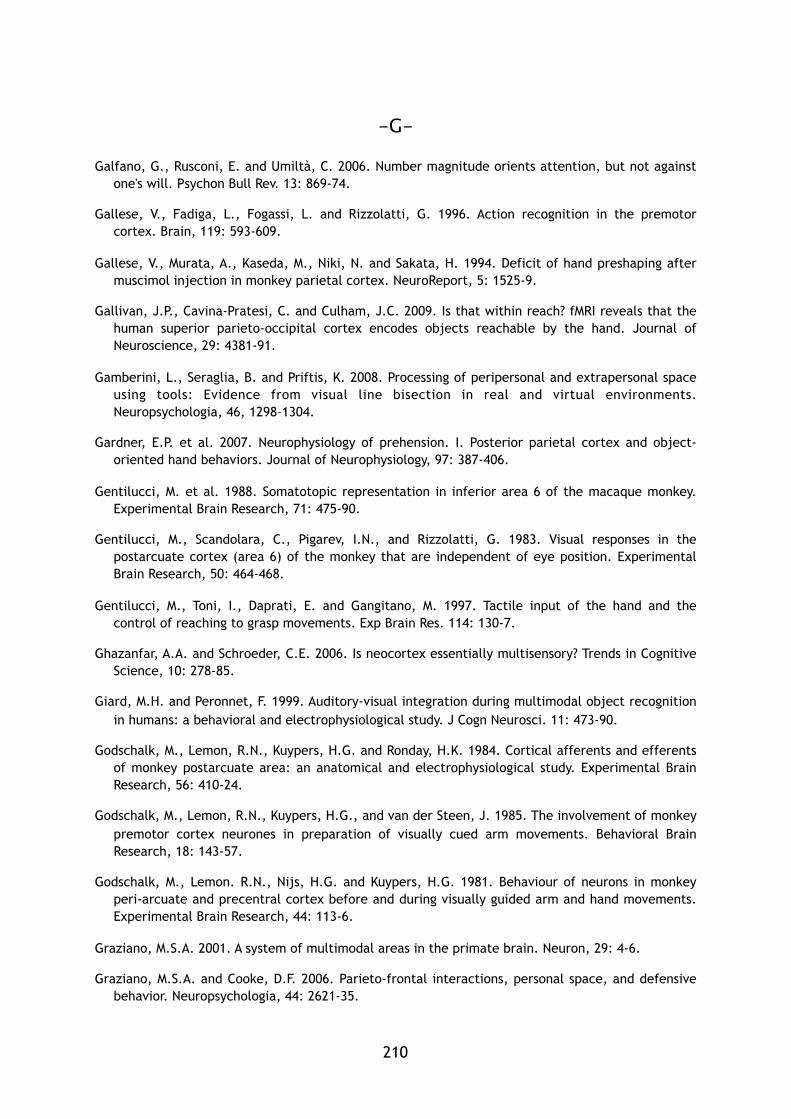
-G-
Galfano, G., Rusconi, E. and Umiltà, C. 2006. Number magnitude orients attention, but not against
one's will. Psychon Bull Rev. 13: 869-74.
Gallese, V., Fadiga, L., Fogassi, L. and Rizzolatti, G. 1996. Action recognition in the premotor
cortex. Brain, 119: 593-609.
Gallese, V., Murata, A., Kaseda, M., Niki, N. and Sakata, H. 1994. Deficit of hand preshaping after
muscimol injection in monkey parietal cortex. NeuroReport, 5: 1525-9.
Gallivan, J.P., Cavina-Pratesi, C. and Culham, J.C. 2009. Is that within reach? fMRI reveals that the
human superior parieto-occipital cortex encodes objects reachable by the hand. Journal of
Neuroscience, 29: 4381-91.
Gamberini, L., Seraglia, B. and Priftis, K. 2008. Processing of peripersonal and extrapersonal space
using tools: Evidence from visual line bisection in real and virtual environments.
Neuropsychologia, 46, 1298–1304.
Gardner, E.P. et al. 2007. Neurophysiology of prehension. I. Posterior parietal cortex and object-
oriented hand behaviors. Journal of Neurophysiology, 97: 387-406.
Gentilucci, M. et al. 1988. Somatotopic representation in inferior area 6 of the macaque monkey.
Experimental Brain Research, 71: 475-90.
Gentilucci, M., Scandolara, C., Pigarev, I.N., and Rizzolatti, G. 1983. Visual responses in the
postarcuate cortex (area 6) of the monkey that are independent of eye position. Experimental
Brain Research, 50: 464-468.
Gentilucci, M., Toni, I., Daprati, E. and Gangitano, M. 1997. Tactile input of the hand and the
control of reaching to grasp movements. Exp Brain Res. 114: 130-7.
Ghazanfar, A.A. and Schroeder, C.E. 2006. Is neocortex essentially multisensory? Trends in Cognitive
Science, 10: 278-85.
Giard, M.H. and Peronnet, F. 1999. Auditory-visual integration during multimodal object recognition
in humans: a behavioral and electrophysiological study. J Cogn Neurosci. 11: 473-90.
Godschalk, M., Lemon, R.N., Kuypers, H.G. and Ronday, H.K. 1984. Cortical afferents and efferents
of monkey postarcuate area: an anatomical and electrophysiological study. Experimental Brain
Research, 56: 410-24.
Godschalk, M., Lemon, R.N., Kuypers, H.G., and van der Steen, J. 1985. The involvement of monkey
premotor cortex neurones in preparation of visually cued arm movements. Behavioral Brain
Research, 18: 143-57.
Godschalk, M., Lemon. R.N., Nijs, H.G. and Kuypers, H.G. 1981. Behaviour of neurons in monkey
peri-arcuate and precentral cortex before and during visually guided arm and hand movements.
Experimental Brain Research, 44: 113-6.
Graziano, M.S.A. 2001. A system of multimodal areas in the primate brain. Neuron, 29: 4-6.
Graziano, M.S.A. and Cooke, D.F. 2006. Parieto-frontal interactions, personal space, and defensive
behavior. Neuropsychologia, 44: 2621-35.
� 210� �
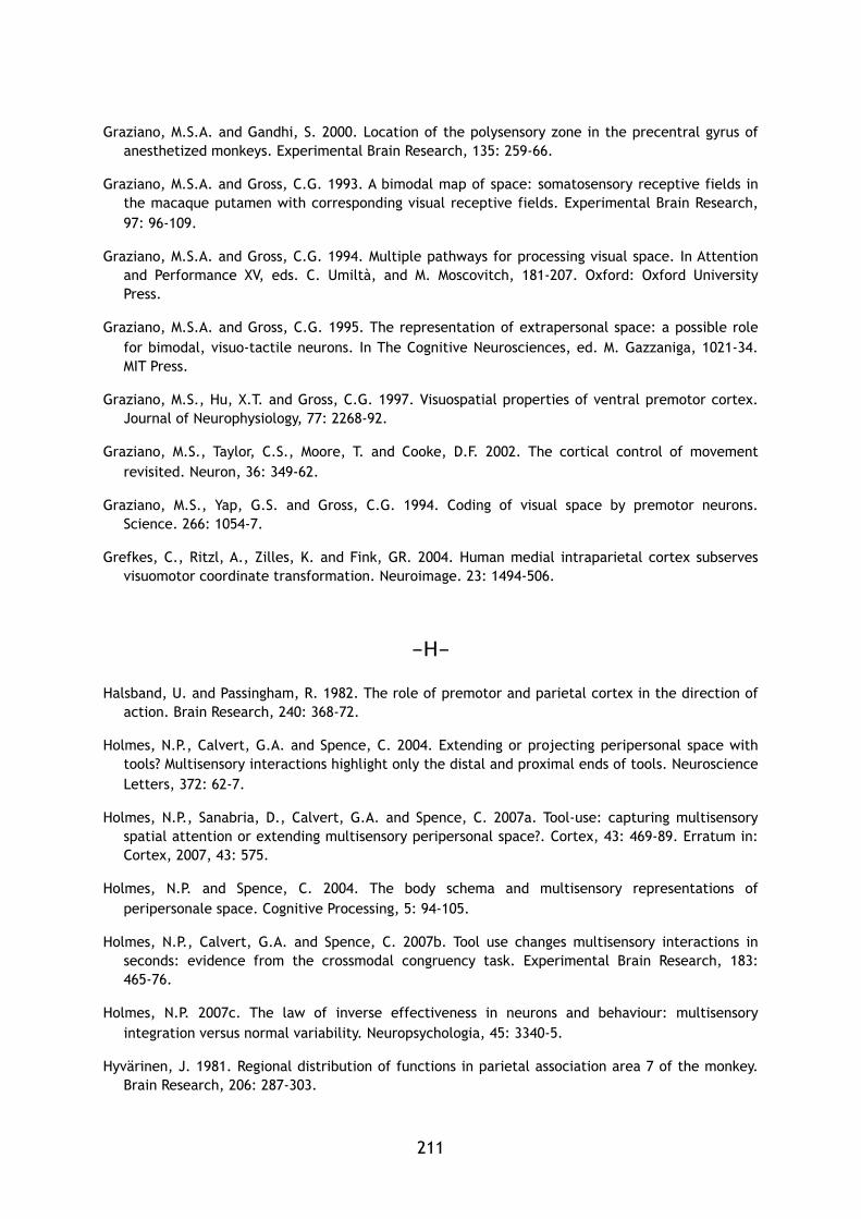
Graziano, M.S.A. and Gandhi, S. 2000. Location of the polysensory zone in the precentral gyrus of
anesthetized monkeys. Experimental Brain Research, 135: 259-66.
Graziano, M.S.A. and Gross, C.G. 1993. A bimodal map of space: somatosensory receptive fields in
the macaque putamen with corresponding visual receptive fields. Experimental Brain Research,
97: 96-109.
Graziano, M.S.A. and Gross, C.G. 1994. Multiple pathways for processing visual space. In Attention
and Performance XV, eds. C. Umiltà, and M. Moscovitch, 181-207. Oxford: Oxford University
Press.
Graziano, M.S.A. and Gross, C.G. 1995. The representation of extrapersonal space: a possible role
for bimodal, visuo-tactile neurons. In The Cognitive Neurosciences, ed. M. Gazzaniga, 1021-34.
MIT Press.
Graziano, M.S., Hu, X.T. and Gross, C.G. 1997. Visuospatial properties of ventral premotor cortex.
Journal of Neurophysiology, 77: 2268-92.
Graziano, M.S., Taylor, C.S., Moore, T. and Cooke, D.F. 2002. The cortical control of movement
revisited. Neuron, 36: 349-62.
Graziano, M.S., Yap, G.S. and Gross, C.G. 1994. Coding of visual space by premotor neurons.
Science. 266: 1054-7.
Grefkes, C., Ritzl, A., Zilles, K. and Fink, GR. 2004. Human medial intraparietal cortex subserves
visuomotor coordinate transformation. Neuroimage. 23: 1494-506.
-H-
Halsband, U. and Passingham, R. 1982. The role of premotor and parietal cortex in the direction of
action. Brain Research, 240: 368-72.
Holmes, N.P., Calvert, G.A. and Spence, C. 2004. Extending or projecting peripersonal space with
tools? Multisensory interactions highlight only the distal and proximal ends of tools. Neuroscience
Letters, 372: 62-7.
Holmes, N.P., Sanabria, D., Calvert, G.A. and Spence, C. 2007a. Tool-use: capturing multisensory
spatial attention or extending multisensory peripersonal space?. Cortex, 43: 469-89. Erratum in:
Cortex, 2007, 43: 575.
Holmes, N.P. and Spence, C. 2004. The body schema and multisensory representations of
peripersonale space. Cognitive Processing, 5: 94-105.
Holmes, N.P., Calvert, G.A. and Spence, C. 2007b. Tool use changes multisensory interactions in
seconds: evidence from the crossmodal congruency task. Experimental Brain Research, 183:
465-76.
Holmes, N.P. 2007c. The law of inverse effectiveness in neurons and behaviour: multisensory
integration versus normal variability. Neuropsychologia, 45: 3340-5.
Hyvärinen, J. 1981. Regional distribution of functions in parietal association area 7 of the monkey.
Brain Research, 206: 287-303.
211
Hyvärinen, J. and Poranen, A. 1974. Function of the parietal associative area 7 as revealed from
cellular discharges in alert monkeys. Brain, 97: 673-92.
Hyvärinen, J. and Shelepin, Y. 1979. Distribution of visual and somatic functions in the parietal
associative area 7 of the monkey. Brain Research, 169: 561-4.
-I-
Iriki, A., Tanaka, M. and Iwamura, Y. 1996. Coding of modified body schema during tool use by
macaque postcentral neurons. NeuroReport, 7: 2325-30.
Ishida H., Nakajima, K., Inase, M. and Murata, A. 2009. Shared mapping of own and others’bodies in
visuo-tactile bimodal area of monkey parietal cortex. Journal of Cognitive Neuroscience, : 1-14.
-J-
Jakobson, L. S., Archibald, Y., Carey, D. and Goodale, M. A. 1991. A kinematic analysis of reaching
and grasping movements in a patient recovering from optic ataxia. Neuropsychologia, 29: 803–
09.
Jeannerod, M. 1981. In Attention and Performance IX (eds Long, J. and Baddeley, A.) 153–168
(Erlbaum, Hillsdale).
Jeannerod, M. 1984. The timing of natural prehension movements. J. Mot. Behav. 16: 235–54.
Jeannerod, M. 1988. Motor Control: concepts and issues. New York: Wiley J.
Jewell, G. and McCourt, M. E. 2000. Pseudoneglect: A review and metaanalysis of performance
factors in line bisection tasks. Neuropsychologia, 38: 93–110.
Jones, E.G. and Powell, T.P. 1970. An anatomical study of converging sensory pathways within the
cerebral cortex of the monkey. Brain. 93: 793-820.
Jousmäki, V. and Hari, R. 1998. Parchment-skin illusion: sound-biased touch. Curr Biol. 8: R190.
-K-
Kaas, J.H. 2004. Evolution of somatosensory and motor cortex in primates. The Anatomical Record.
Part A, Discoveries in Molecular, Cellular, and Evolutionary Biology, 281: 1148-56.
Kaas, J.H. and Collins, C.E. The Resurrection of Multisensory Cortex in Primates. In: Calvert, G.A.,
Spence, C. and Stein, B.E. eds. The Handbook of Multisensory Processes. Bradford; Cambridge,
MA: 2004. p. 285-94.
Kayser, C., Petkov, C.I., Augath, M. and Logothetis, N.K. 2005. Integration of touch and sound in
auditory cortex. Neuron, 48: 373–84.
� 212� �
Kayser, C., Petkov, C.I., Augath, M. and Logothetis, N.K. 2007. Functional imaging reveals visual
modulation of specific fields in auditory cortex. J. Neurosci. 27: 1824–35.
Klein, RM. 2000. Inhibition of return. Trends Cogn Sci. 4: 138-47.
Koch, G., Franca, M., Del Olmo, M.F., Cheeran, B., Milton, R., Alvarez Sauco, M. and Rothwell, J.C.
2006. Time course of functional connectivity between dorsal premotor and contralateral motor
cortex during movement selection. J Neurosci 26:7452-7459.
Kurata, K. and Tanji, J. 1986. Premotor cortex neurons in macaques: activity before distal and
proximal forelimb movements. Journal of Neuroscience, 6: 403-11.
Kurata, K., Okano, K. and Tanji, J. 1985. Distribution of neurons related to a hindlimb as opposed to
forelimb movement in the monkey premotor cortex. Experimental Brain Research, 60: 188-91.
-L-
Lacquaniti, F. and Caminiti, R. 1998. Visuo-motor transformations for arm reaching. European
Journal of Neuroscience, 10: 195-203. Review. Erratum in: European Journal of Neuroscience,
1998, 10: 810.
Lakatos, P., Chen, C.M., O'Connell, M.N., Mills, A. and Schroeder, C.E. 2007. Neuronal oscillations
and multisensory interaction in primary auditory cortex. Neuron, 53: 279–92.
Làdavas, E. 2002. Functional and dynamic properties of visual peripersonal space. Trends in
Cognitive Sciences, 6: 17–22.
Làdavas, E. and Farnè, A. 2004. Visuo-tactile representation of near-the-body space. Journal of
Physiology Paris, 98: 161-70.
Lang, C.E. and Schieber, M.H. 2004. Reduced muscle selectivity during individuated finger
movements in humans after damage to the motor cortex or corticospinal tract. J. Neurophysiol.
91: 1722–33.
Lawrence, D.G. and Kuypers, H.G. 1968a. The functional organization of the motor system in the
monkey. I. The effects of bilateral pyramidal lesions. Brain, 91: 1–14.
Lawrence, D.G. and Kuypers, H.G. 1968b. The functional organization of the motor system in the
monkey. II. The effects of lesions of the descending brain-stem pathways. Brain, 91: 15–36.
Legrand, D., Brozzoli, C., Rossetti, Y. and Farnè, A. 2007. Close to me: multisensory space
representations for action and pre-reflexive consciousness of oneself-in-the-world. Consciousness
and Cognition, 16: 687-99.
Leinonen, L. 1980. Functional properties of neurones in the posterior part of area 7 in awake
monkey. Acta Physiologica Scandinava, 108: 301-8.
Leinonen, L., Hyvärinen, J., Nyman, G. and Linnankoski, I. 1979. I. Functional properties of neurons
in lateral part of associative area 7 in awake monkeys. Experimental Brain Research, 34:
299-320.
213
Leinonen, L. and Nyman, G. 1979. II. Functional properties of cells in anterolateral part of area 7
associative face area of awake monkeys. Experimental Brain Research, 34: 321-33.
Lewald, J. and Guski, R. 2003. Cross-modal perceptual integration of spatially and temporally
disparate auditory and visual stimuli. Brain Res Cogn Brain Res. 16: 468-78.
Longo, M.R. and Lourenco, S F. 2006. On the nature of near space. Effects of tool use and the
transition to far space. Neuropsychologia, 44, 977–81.
Lourenco, S.F. and Longo, M.R. 2009. The plasticity of near space: evidence for contraction.
Cognition, 112: 451-6.
Luppino, G., Murata, A., Govoni, P. and Matelli, M. 1999. Largely segregated parietofrontal
connections linking rostral intraparietal cortex (areas AIP and VIP) and the ventral premotor
cortex (areas F5 and F4). Experimental Brain Research, 128: 181-7.
-M-
Macaluso, E. and Driver, J. 2001. Spatial attention and crossmodal interactions between vision and
touch. Neuropsychologia, 39: 1304-16.
Macaluso, E. and Driver, J. 2005. Multisensory spatial interactions: a window onto functional
integration in the human brain. Trends Neuroscience 28: 264-71.
Macaluso, E., Eimer, M., Frith, C.D. and Driver, J. 2003. Preparatory states in crossmodal spatial
attention: spatial specificity and possible control mechanisms. Exp Brain Res 149:62–74.
Macaluso, E., Frith, C.D. and Driver, J. 2000. Modulation of human visual cortex by crossmodal
spatial attention. Science, 289: 1206–08.
Macaluso, E., Frith, C.D. and Driver, J. 2002. Supramodal effects of covert spatial orienting
triggered by visual or tactile events. J Cogn Neurosci., 14: 389-401.
Makin, T.R., Holmes, N.P, and Ehrsson, H.H. 2008. On the other hand: dummy hands and
peripersonal space. Behavioral Brain Research, 191: 1-10.
Makin, T.R., Holmes, N.P. and Zohary, E. 2007. Is that near my hand? Multisensory representation of
peripersonal space in human intraparietal sulcus. Journal of Neuroscience, 27: 731-40.
Maravita, A. and Iriki, A. 2004. Tools for the body (schema). Trends In Cognitive Science, 8: 79-86.
Maravita, A., Spence, C., Kennett, S. and Driver, J. 2002. Tool-use changes multimodal spatial
interactions between vision and touch in normal humans. Cognition, 83: B25-34.
Marcel, A., Postma, P., Gillmeister, H., Cox, S., Rorden, C., Nimmo-Smith, I. and Mackintosh, B.
2004. Migration and fusion of tactile sensation—premorbid susceptibility to allochiria, neglect
and extinction? Neuropsychologia, 42: 1749-67.
Mars, R.B., Bestmann, S., Rothwell, J.C. and Haggard, P. 2007. Effects of motor preparation and
spatial attention on corticospinal excitability in a delayed-response paradigm. Exp Brain Res 182:
125-129.
� 214� �
Martino, A.M. and Strick, P.L. 1987. Corticospinal projections originate from the arcuate premotor
area. Brain Research, 404: 307-12.
Martuzzi, R., Murray, M.M., Michel, C.M., Thiran, J.P., Maeder, P.P., Clarke, S. and Meuli, R.A. 2007.
Multisensory interactions within human primary cortices revealed by BOLD dynamics. Cereb.
Cortex, 17: 1672–9.
Matelli, M., Camarda, R., Glickstein, M. and Rizzolatti, G. 1984a. Interconnections within the
postarcuate cortex (area 6) of the macaque monkey. Brain Research, 310: 388-92.
Matelli, M., Camarda, R., Glickstein, M. and Rizzolatti, G. 1984b. Afferent and efferent projections
of the inferior area 6 in the macaque monkey. Journal of Comparative Neurology 251: 281-98.
Matelli, M. and Luppino, G. 2001. Parietofrontal circuits for action and space perception in the
macaque monkey. Neuroimage, 14: S27-32.
Matelli, M., Luppino, G. and Rizzolatti, G. 1985. Patterns of cytochrome oxidase activity in the
frontal agranular cortex of the macaque monkey. Behavioral Brain Research, 18: 125-36.
Matsumura, M. and Kubota, K. 1979. Cortical projection to hand-arm motor area from post-arcuate
area in macaque monkeys: a histological study of retrograde transport of horseradish peroxidase.
Neuroscience Letters, 11: 241-6.
Maunsell, J.H. and van Essen D.C. 1983. The connections of the middle temporal visual area (MT)
and their relationship to a cortical hierarchy in the macaque monkey. Journal of Neuroscience,
3: 2563-86.
McDonald, J.J., Teder-Salejarvi, W.A. and Hillyard, S.A. 2000. Involuntary orienting to sound
improves visual perception. Nature, 407: 906–8.
McGurk, H. and MacDonald, J. 1976. Hearing lips and seeing voices. Nature, 264: 746-8.
Medendorp, W.P., Goltz, H.C., Vilis, T. and Crawford, J.D. 2003. Gaze-centered updating of visual
space in human parietal cortex. J Neurosci 23:6209–6214.
Medendorp, W.P., Goltz, H.C., Crawford, J.D. and Vilis, T. 2005. Integration of target and effector information in human posterior parietal cortex for the planning of action. J Neurophysiol 93:954-962.
Meredith, M.A. and Stein, B.E. 1983. Interactions among converging sensory inputs in the superior
colliculus. Science, 221: 389–91.
Meredith, M.A. and Stein, B.E. 1986. Visual, auditory, and somatosensory convergence on cells in
superior colliculus results in multisensory integration. Journal of Neurophysiology, 56: 640-62.
Mesulam, M.M., Van Hoesen, G.W., Pandya, D.N. and Geschwind, N. 1977. Limbic and sensory
connections of the inferior parietal lobule (area PG) in the rhesus monkey: a study with a new
method for horseradish peroxidase histochemistry. Brain Research, 136: 393-414.
Miller, L.M. and D'Esposito, M. 2005. Perceptual fusion and stimulus coincidence in the cross-modal
integration of speech. J. Neurosci. 25: 5884–93.
Milner, A. D. and Goodale, M.A. 1995. The visual brain in action. Oxford: Oxford University Press.
215
Milner, A. D. in Parietal Lobe Contributions to Orientation in 3D Space (eds Thiers, P. and Karnath, H.
O.) 3–22 (Springer, Berlin, 1997).
Milner, A. D., Dijkerman, H. C., McIntosh, R. D., Rossetti, Y. and Pisella, L. 2003. Delayed reaching
and grasping in patients with optic ataxia. Progr. Brain Res. 142: 225–242.
Molholm, S., Ritter, W., Murray, M.M., Javitt, D.C., Schroeder, C.E. and Foxe, J.J. 2002. Multisensory
auditory-visual interactions during early sensory processing in humans: a high-density electrical
mapping study. Brain Res Cogn Brain Res. 14: 115-28.
Moll, L. and Kuypers, H.G. 1977. Premotor cortical ablations in monkeys: contralateral changes in
visually guided reaching behavior. Science. 198: 317-9.
Morrell, F. 1972. Visual system's view of acoustic space. Nature, 238: 44-6.
Mountcastle, V.B., Lynch, J.C., Georgopoulos, A., Sakata, H. and Acuna, C. 1975. Posterior parietal
association cortex of the monkey: command functions for operations within extrapersonal space.
Journal of Neurophysiology, 38: 871-908.
Muakkassa, K.F. and Strick, P.L. 1979. Frontal lobe inputs to primate motor cortex: evidence for four
somatotopically organized 'premotor' areas. Brain Research, 177: 176-82.
Murata, A., Gallese, V., Luppino, G., Kaseda, M. and Sakata, H. 2000. Selectivity for the shape, size,
and orientation of objects for grasping in neurons of monkey parietal area AIP. J. Neurophysiol,
83: 2580–601.
Murata, A., Fadiga, L., Fogassi, L., Gallese, V., Raos, V. and Rizzolatti, G. 1997. Object
representation in the ventral premotor cortex (area F5) of the monkey. Journal of
Neurophysiology, 78: 2226-30.
Murray, M.M. et al. 2005. Grabbing your ear: rapid auditory-somatosensory multisensory interactions
in low-level sensory cortices are not constrained by stimulus alignment. Cerebral Cortex, 15:
963-74.
-N-
Nagy, A., Eordegh, G., Paroczy, Z., Markus, Z. and Benedek G. 2006. Multisensory integration in the
basal ganglia. Eur. J. Neurosci. 24: 917–24.
-O-
O'Shea, J., Sebastian, C., Boorman, E.D., Johansen-Berg, H. and Rushworth, M.F.S. 2007. Functional
specificity of human premotor-motor cortical interactions during action selection. Eur J Neurosci
26:2085-2095.
-P-
� 216� �
Padberg, J., Seltzer, B. and Cusick, C.G. 2003. Architectonics and cortical connections of the upper
bank of the superior temporal sulcus in the rhesus monkey: an analysis in the tangential plane.
Comp. Neurol. 467: 418–34.
Pandya, D.N. and Vignolo, L.A. 1971. Intra- and interhemispheric projections of the precentral,
premotor and arcuate areas in the rhesus monkey. Brain Research, 26: 217-33.
Paulignan, Y., MacKenzie, C., Marteniuk, R. and Jeannerod, M. 1991. Selective perturbation of visual
input during prehension movements. 1. The effects of changing object position. Experimental
Brain Research, 83: 502-12.
Pavani, F., Spence, C. and Driver, J. 2000. Visual capture of touch: out-of-the-body experiences with
rubber gloves. Psychol Sci. 11: 353-9.
Pavani, F. and Castiello, U. 2004. Binding personal and extrapersonal space through body shadows.
Nature Neuroscience, 7: 14-16.
Perenin, M.T. and Vighetto, A. 1988. Optic ataxia: a specific disruption in visuomotor mechanisms. I.
Different aspects of the deficit in reaching for objects. Brain, 111: 643-74.
Pisella, L. et al. 2000. An 'automatic pilot' for the hand in human posterior parietal cortex: toward
reinterpreting optic ataxia. Nature Neuroscience, 3: 729-36.
Pisella, L., Sergio, L., Blangero, A., Torchin, H., Vighetto, A. and Rossetti, Y. 2009 Optic ataxia and
the function of the dorsal stream: Contributions to perception and action. Neuropsychologia, In
Press.
Posner, M.I. 1980. Orienting of attention. Q J Exp Psychol. 32: 3-25.
Posner, M.I. and Cohen, Y. 1984. Components of visual orienting. In: Attention and Performance
(Bouma X.H. and Bowhuis D. eds.), pp 531-556. Hillsdale, NJ: Lawrence Erlbaum Associates.
Prabhu, G. et al. 2009. Modulation of primary motor cortex outputs from ventral premotor cortex
during visually guided grasp in the macaque monkey. Journal of Physiology, 587: 1057-69.
-Q-
Quinlan, D.J. and Culham, J.C. 2007. fMRI reveals a preference for near viewing in the human
parieto-occipital cortex. Neuroimage, 36: 167-87.
-R-
Raos, V., Umiltá, M.A., Murata, A., Fogassi, L. and Gallese, V. 2006. Functional properties of
grasping-related neurons in the ventral premotor area F5 of the macaque monkey. Journal of
Neurophysiology, 95: 709-29.
Rizzolatti, G., Camarda, R., Fogassi, L., Gentilucci, M., Luppino, G. and Matelli, M. 1988a.
Functional organization of inferior area 6 in the macaque monkey. II. Area F5 and the control of
distal movements. Experimental Brain Research, 71: 491-507.
217
Rizzolatti, G., Fadiga, L., Fogassi, L. and Gallese, V. 1997. The space around us. Science, 11:
190-91.
Rizzolatti, G., Fadiga, L., Gallese, V. and Fogassi, L. 1996. Premotor cortex and the recognition of
motor actions. Brain Res Cogn Brain Research, 3: 131-41.
Rizzolatti, G., and Gentilucci, M. 1988b. Motor and visual-motor functions of the premotor cortex.
In Neurobiology of Neocortex, eds. P. Rakic, and W. Singer, 269-84. John Wiley and sons Ltd.
Rizzolatti, G., Gentilucci, M., Fogassi, L., Luppino, G., Matelli, M. and Ponzoni-Maggi, S. 1987.
Neurons related to goal-directed motor acts in inferior area 6 of the macaque monkey.
Experimental Brain Research, 67: 220-4.
Rizzolatti, G. and Luppino, G. 2001. The cortical motor system. Neuron, 31: 889-901.
Rizzolatti, G., Luppino, G. and Matelli, M. 1998. The organization of the cortical motor system: new
concepts. Electroencephalography and Clinical Neurophysiology, 106: 283-96.
Rizzolatti, G. and Matelli, M. 2003. Two different streams form the dorsal visual system: anatomy
and functions. Experimental Brain Research, 153: 146-57.
Rizzolatti, G., Scandolara, C., Gentilucci, M. and Camarda, R. 1981a. Response properties and
behavioral modulation of "mouth" neurons of the postarcuate cortex (area 6) in macaque
monkeys. Brain Research, 225: 421-4.
Rizzolatti, G., Scandolara, C., Matelli, M. and Gentilucci, M. 1981b. Afferent properties of
periarcuate neurons in macaque monkeys. I. Somatosensory responses. Behavioral Brain
Research, 2: 125-46.
Rizzolatti, G., Scandolara, C., Matelli, M. and Gentilucci, M. 1981c. Afferent properties of
periarcuate neurons in macque monkeys. II. Visual responses. Behavioral Brain Research, 2,
147-163.
Robinson, D.L., Goldberg, M.E. and Stanton, G.B. 1978. Parietal association cortex in the primate:
sensory mechanisms and behavioral modulations. Journal of Neurophysiology, 41: 910-32.
Robinson, C.J. and Burton, H. 1980a. Organization of somatosensory receptive fields in cortical
areas 7b, retroinsula, postauditory and granular insula of M. fascicularis. Journal of Comparative
Neurology, 192: 69-92.
Robinson, C.J. and Burton, H. 1980b. Somatic submodality distribution within the second
somatosensory (SII), 7b, retroinsular, postauditory, and granular insular cortical areas of M.
fascicularis. Journal of Comparative Neurology, 192: 93-108.
Rossetti, Y., Vighetto, A. and Pisella, L. 2003. Optic ataxia revisited: immediate motor control
versus visually guided action. Exp. Brain Res. 153: 171–9.
Roy, A.C., Paulignan, Y., Meunier, M. and Boussaoud, D. 2002 Prehension movements in the macaque
monkey: effects of object size and location. J Neurophysiol. 88: 1491-9.
Roy, A.C., Stefanini, S., Pavesi, G. and Gentilucci, M. 2004. Early movement impairments in a
patient recovering from optic ataxia. Neuropsychologia, 42: 847-54.
� 218� �
Roy, A.C., Paulignan, Y., Meunier, M. and Boussaoud, D. 2006. Prehension movements in the macaque
monkey: effects of perturbation of object size and location. Exp Brain Res. 169: 182-93.
-S-
Sakata, H., Takaoka, Y., Kawarasaki, A. and Shibutani, H. 1973. Somatosensory properties of neurons
in the superior parietal cortex (area 5) of the rhesus monkey. Brain Research, 64: 85-102.
Savelsbergh, G.J.P., Steenbergen, B. and van der Kamp, J. 1996. The role of fragility information in
the guidance of the precision grip. Hum. Mov. Sci. 15: 115–127.
Schmahmann, J.D. and Pandya, D.N. 1991. Projections to the basis pontis from the superior
temporal sulcus and superior temporal region in the rhesus monkey. J. Comp. Neurol. 308: 224–
48.
Seltzer, B. and Pandya, D.N. 1980. Converging visual and somatic sensory cortical input to the
intraparietal sulcus of the rhesus monkey. Brain Research, 192: 339-51.
Sereno, M.I. and Huang, R.S. 2006. A human parietal face area contains aligned head-centered
visual and tactile maps. Nature Neuroscience, 9: 1337-43.
Shams, L., Kamitani, Y. and Shimojo, S. 2000. Illusions. What you see is what you hear. Nature, 408:
788.
Shimazu, H., Maier, M.A., Cerri, G., Kirkwood, P.A. and Lemon, R.N. 2004. Macaque ventral
premotor cortex exerts powerful facilitation of motor cortex outputs to upper limb motoneurons.
Journal of Neuroscience, 24: 1200-11.
Shipley, T. 1964. Auditory flutter-driving of visual flicker. Science, 145: 1328–30.
Simon, O., Mangin, J.F., Cohen, L., Le Bihan, D. and Dehaene, S. 2002. Topographical layout of
hand, eye, calculation, and language-related areas in the human parietal lobe. Neuron 33:475–
487.
Smeets, J.B.J. and Brenner, E. 1999. A new view on grasping. Mot. Cont. 3: 237–71.
Soto-Faraco, S. and Alsius, A. 2009. Deconstructing the McGurk-MacDonald illusion. J Exp Psychol
Hum Percept Perform., 35: 580-7.
Soto-Faraco, S., Lyons, J., Gazzaniga, M., Spence, C. and Kingstone, A. 2002. The ventriloquist in
motion: illusory capture of dynamic information across sensory modalities. Brain Res Cogn Brain
Res, 14: 139-46.
Soto-Faraco, S., Navarra, J., & Alsius, A. 2004. Assessing automaticity in audiovisual speech
integration: Evidence from the speeded classification task. Cognition, 92: B13–B23.
Soto-Faraco, S., Spence, C. and Kingstone, A. 2004. Congruency effects between auditory and
tactile motion: extending the phenomenon of cross-modal dynamic capture. Cogn Affect Behav
Neurosci. 4: 208-17.
Spence, C. and Driver, J. 1996. Audiovisual links in endogenous covert spatial attention. J Exp
Psychol Hum Percept Perform. 22: 1005-30.
219
Spence, C. and Driver, J. 1997a. On measuring selective attention to an expected sensory modality.
Percept Psychophys. 59: 389-403.
Spence, C. and Driver, J. 1997b. Audiovisual links in exogenous covert spatial orienting. Percept
Psychophys. 59: 1-22.
Spence, C. and Driver, J. 1998a. Auditory and audiovisual inhibition of return. Percept Psychophys.
60: 125-39.
Spence, C. and Driver, J. 1998b. Inhibition of return following an auditory cue. The role of central
reorienting events. Exp Brain Res. 118: 352-60.
Spence, C. and Driver, J. 2004. Crossmodal Space and Crossmodal Attention. Oxford University
Press; Oxford.
Spence, C., Pavani, F. and Driver, J. 1998. What crossing the hands can reveal about crossmodal
links in spatial attention, Abstr. Psychon. Soc.
Spence, C., Nicholls, M.E., Gillespie, N. and Driver, J. 1998. Cross-modal links in exogenous covert
spatial orienting between touch, audition, and vision. Percept Psychophys. 60: 544-57.
Spence, C., Lloyd, D., McGlone, F., Nicholls, M.E. and Driver, J. 2000. Inhibition of return is
supramodal: a demonstration between all possible pairings of vision, touch, and audition. Exp
Brain Res. 134: 42-8.
Spence, C., McDonald, J. and Driver, J. 2004. Exogenous spatial-cuing studies of human crossmodal
attention and multisensory integration. In: Spence, C.; Driver, J., editors. Crossmodal Space and
Crossmodal Attention. Oxford University Press; Oxford: 277-319.
Spence, C., Pavani, F. and Driver, J. 2004. Spatial constraints on visual-tactile cross-modal distractor
congruency effects. Cognitve Affective and Behavioral Neuroscience, 4: 148-69.
Spence, C., Pavani, F., Maravita, A. and Holmes, N.P. 2004. Multisensory contributions to the 3-D
representation of visuotactile peripersonal space in humans: evidence from the crossmodal
congruency task. Journal of Physiology Paris, 98: 171-89.
Spence, C., Pavani, F., Maravita, A. and Holmes, N.P. 2008. Multisensory interactions. In Haptic
Rendering: Foundations, Algorithms, and Applications, eds M.C. Lin, and M.A. Otaduy, 21-52. A K
Peters Ltd., Wellesley, MA.
Spence, C., Shore, D.I., Gazzaniga, M.S., Soto-Faraco, S. and Kingstone A. 2001. Failure to remap
visuotactile space across the midline in the split-brain. Can J Exp Psychol., 55: 133-40.
Spinelli, D.N., Starr, A. and Barrett, T.W. 1968. Auditory specificity in unit recordings from cat's
visual cortex. Exp Neurol. 22: 75-84.
Stanton, G.B., Cruce, W.L., Goldberg, M.E. and Robinson, D.L. 1977. Some ipsilateral projections to
areas PF and PG of the inferior parietal lobule in monkeys. Neurosci Lett., 6: 243-50.
Stein, B.E. and Meredith, M.A. 1993. The merging of the Senses, Cambridge, MA: MIT Press.
Stepniewska, I., Fang, P.C. and Kaas, J.H. 2005. Microstimulation reveals specialized subregions for
different complex movements in posterior parietal cortex of prosimian galagos. Proceding of the
National Academy of Science U S A, 102: 4878-83.
� 220� �
Stoeckel, M.C., Weder, B., Binkofski, F., Buccino, G., Shah, N.J. and Seitz, R.J. 2003. A fronto-
parietal circuit for tactile object discrimination: an event-related fMRI study. NeuroImage
19:1103–1114.
Strick, P.L. and Kim, C.C. 1978. Input to primate motor cortex from posterior parietal cortex (area
5). I. Demonstration by retrograde transport. Brain Research, 157: 325-30.
Strick, P.L. 2002. Stimulating research on motor cortex. Nature Neuroscience, 5: 714-5.
-U-
Ungerleider, L.G. and Desimone, R. 1986. Cortical connections of visual area MT in the macaque.
Journal of Comparative Neurology, 248: 190-222.
-V-
Vallar, G., Rusconi, M.L., Bignamini, L., Geminiani, G. and Perani, D. 1994. Anatomical correlates of
visual and tactile extinction in humans: a clinical CT scan study. J Neurol Neurosurg Psychiatry.
57: 464-70.
van den Hurk, P., Mars, R.B., van Elswijk, G., Hegeman, J., Pasman, J.W., Bloem, B.R. and Toni, I.
2007. Online maintenance of sensory and motor representations: Effects on corticospinal
excitability. J Neurophysiol 97:1642-1648.
van Elswijk, G., Kleine, B.U., Overeem, S. and Stegeman, D.F. 2007. Expectancy induces dynamic
modulation of corticospinal excitability. J Cogn Neurosci 19:121-131.
Van Essen, D.C. 2005. Corticocortical and thalamocortical information flow in the primate visual
system. Prog Brain Res. 149: 173-85.
Varnava, A., McCarthy, M. and Beaumont, J.G. 2002. Line bisection in normal adults: Direction of
attentional bias for near and far space. Neuropsychologia, 40, 1372–8.
Violentyev, A., Shimojo, S. and Shams, L. 2005. Touch-induced visual illusion. Neuroreport, 16:
1107-10.
-W-
Wallace, M.T. and Stein, B.E. 2007. Early experience determines how the senses will interact.
Journal of Neurophysiology, 97: 921-6.
Walton M. and Spence C. 2004. Crossmodal congruency and visual capture in a visual elevation
discrimination task. Exp. Brain Res. 154: 113–20.
Wang, Y., Celebrini, S., Trotter, Y. and Barone, P. 2008. Visuo-auditory interactions in the primary
visual cortex of the behaving monkey: electrophysiological evidence. BMC Neuroscience, 9: 79.
221
Ward, R., Goodrich, S. and Driver, J. 1994. Grouping reduces visual extinction: neuropsychological
evidence for weight-linkage in visual selection. Visual Cognition, 1: 101-29.
Weir, P.L., MacKenzie, C.L., Marteniuk, R.G. and Cargoe, S.L. 1991b Is object texture a constraint on
human prehension?: kinematic evidence. J. Mot. Behav. 23, 205–210.
Weir, P.L., MacKenzie, C.L., Marteniuk, R.G., Cargoe, S.L. and Fraser, M.B. 1991a. The effects of
object weight on the kinematics of prehension. J. Mot. Behav. 23, 192–204.
Weiss, P.H., Marshall, J.C., Wunderlich, G., Tellmann, L., Halligan, P. W., Freund, H.-J., et al.
(2000). Neural consequences of acting in near versus far space: A physiological basis for clinical
dissociations. Brain, 123, 2531–41.
Witney, A.G., Wing, A., Thonnard, J.L. and Smith, A.M. 2004. The cutaneous contribution to
adaptive precision grip. Trends Neurosci. 27: 637–643.
� 222� �