Embed Size (px)
Citation preview
Perinatal Outcomes, Including Mother#to#Child Transmission of HIV, and Child
Mortality and Their Association withMaternal Vitamin D Status in Tanzania
The Harvard community has made thisarticle openly available. Please share howthis access benefits you. Your story matters
Citation Mehta, Saurabh, David J. Hunter, Ferdinand M. Mugusi, DonnaSpiegelman, Karim P. Manji, Edward L. Giovannucci, EllenHertzmark, Gernard I. Msamanga, and Wafaie W. Fawzi. 2009.“Perinatal Outcomes, Including Mother#to#Child Transmission ofHIV, and Child Mortality and Their Association with Maternal VitaminD Status in Tanzania.” J INFECT DIS 200 (7) (October): 1022–1030.doi:10.1086/605699.
Published Version doi:10.1086/605699
Citable link http://nrs.harvard.edu/urn-3:HUL.InstRepos:26951082
Terms of Use This article was downloaded from Harvard University’s DASHrepository, and is made available under the terms and conditionsapplicable to Other Posted Material, as set forth at http://nrs.harvard.edu/urn-3:HUL.InstRepos:dash.current.terms-of-use#LAA
Perinatal Outcomes, Including Mother-to-Child Transmission ofHIV, and Child Mortality and Their Association with MaternalVitamin D Status in Tanzania
Saurabh Mehta1,2, David J. Hunter1,2,5, Ferdinand M. Mugusi6, Donna Spiegelman1,3, KarimP. Manji7, Edward L. Giovannucci1,2,5, Ellen Hertzmark1, Gernard I. Msamanga8, and WafaieW. Fawzi1,2,41Department of Epidemiology, Harvard School of Public Health, Boston, Massachusetts2Department of Nutrition, Harvard School of Public Health, Boston, Massachusetts3Department of Biostatistics, Harvard School of Public Health, Boston, Massachusetts4Department of Global Health and Population, Harvard School of Public Health, Boston,Massachusetts5Channing Laboratory, Department of Medicine, Brigham and Women's Hospital and HarvardMedical School, Boston, Massachusetts6Department of Internal Medicine, Muhimbili University of Health and Allied Sciences, Dar esSalaam, Tanzania7Department of Pediatrics, Muhimbili University of Health and Allied Sciences, Dar es Salaam,Tanzania8Department of Community Health, Muhimbili University of Health and Allied Sciences, Dar esSalaam, Tanzania
AbstractBackground—Vitamin D is a strong immunomodulator and may protect against adverse pregnancyoutcomes, mother-to-child transmission (MTCT) of human immunodeficiency virus (HIV), and childmortality.
Methods—A total of 884 HIV-infected pregnant women who were participating in a vitaminsupplementation trial in Tanzania were monitored to assess pregnancy outcomes and child mortality.The association of these outcomes with maternal vitamin D status at enrollment was examined in anobservational analysis.
Results—No association was observed between maternal vitamin D status and adverse pregnancyoutcomes, including low birth weight and preterm birth. In multivariate models, a low maternalvitamin D level (<32 ng/mL) was associated with a 50% higher risk (95% confidence interval [CI],2%–120%) of MTCT of HIV at 6 weeks, a 2-fold higher risk of MTCT of HIV through breast-feedingamong children who were HIV uninfected at 6 weeks (95% CI, 1.08–3.82), and a 46% higher overallrisk of HIV infection (95% CI, 11%–91%). Children born to women with a low vitamin D level hada 61% higher risk of dying during follow-up (95% CI, 25%–107%).
2009 by the Infectious Diseases Society of America. All rights reserved.Reprints or correspondence: Saurabh Mehta, Dept. of Nutrition, Harvard School of Public Health, 655 Huntington Ave., Boston, MA02115 ([email protected])..Potential conflicts of interest: none reported.
NIH Public AccessAuthor ManuscriptJ Infect Dis. Author manuscript; available in PMC 2010 October 1.
Published in final edited form as:J Infect Dis. 2009 October 1; 200(7): 1022–1030. doi:10.1086/605699.
NIH
-PA Author Manuscript
NIH
-PA Author Manuscript
NIH
-PA Author Manuscript
Conclusions—If found to be efficacious in randomized trials, vitamin D supplementation couldprove to be an inexpensive method of reducing the burden of HIV infection and death among children,particularly in resource-limited settings.
Since the first demonstration of its antirachitic properties by Elmer McCollum in 1922 [1],vitamin D has been shown to have a potential role in several health outcomes, includingcolorectal cancer [2] and infectious diseases, such as tuberculosis [3,4]. Vitamin D is a knownimmunomodulator [5]; it can improve cell-mediated immunity [6] and the phagocytic capacityof macrophages [7], and it can also increase the number and the cytolytic activity of naturalkiller cells [8]. Recent research has also highlighted the importance of vitamin D in the innateimmune response, via the Toll-like receptor pathway [4]. Toll-like receptor stimulation ofhuman macrophages up-regulates expression of the vitamin D receptor and induces the enzymeCYP27b1, which catalyzes the conversion of 25-hydroxyvitamin D3 [25(OH)D] to 1,25-dihydroxyvitamin D3 [1,25(OH)D], the biologically active vitamin D metabolite. In thepresence of adequate 25(OH)D, activation of the up-regulated vitamin D receptors leads toinduction of cathelicidin, an antimicrobial peptide capable of killing such pathogens asMycobacterium tuberculosis intracellularly [4]. Cathelicidin also has several other importantbiological effects [9], such as neutralization of the effects of lipopolysaccharide [10],stimulation of angiogenesis [11], and chemotaxis of neutrophils, monocytes, and T cells [12].
Vitamin D may have a role in preventing adverse pregnancy outcomes, mother-to-childtransmission (MTCT) of human immunodeficiency virus (HIV), and child mortality. Theconventional role of vitamin D in calcium metabolism is important for fetal skeletaldevelopment [13]; approximately 25–30 g of calcium are transferred to the fetus, most of itduring the last trimester [13]. The importance of vitamin D in this process may be indicatedby the 50%–100% increase in 1,25(OH)D concentrations in the second and third trimesters[14]. In the last trimester, this increase is not accompanied by a similar increase in vitamin D–binding protein, leading to higher levels of free circulating 1,25(OH)D [15]. Recent researchhas suggested that vitamin D may also help regulate placental development and function[16], further contributing to fetal growth.
Vitamin D may also affect development of the fetal immune system [17], by virtue of itsimmunomodulatory properties. The potential ability to fight diseases such as tuberculosis, aleading cause of death in HIV-infected patients, could lead to decreased mortality [18].
A role for vitamin D in HIV infection has been postulated on the basis of a few laboratory andhuman studies [5]; however, there have been no published studies that have directly examinedthe association of vitamin D with pregnancy outcomes, MTCT of HIV, or death among childrenborn to HIV-infected women. In the Trial of Vitamins [19,20], which was conducted inTanzania, HIV-infected pregnant women received supplementation with a multivitaminregimen that did not include vitamin D and were monitored to observe pregnancy outcomesand disease progression, providing an opportunity to expand our knowledge about vitamin Dstatus and health. This knowledge is of particular relevance to HIV infection and developingcountries, because high rates of subclinical vitamin D deficiency have been identified in thesecountries [21].
METHODSStudy population
The study design has been described in detail elsewhere [22]. In brief, 1078 HIV-infectedpregnant women of 12–27 weeks’ gestation (hereafter referred to as “baseline”) wererandomized to receive daily doses of 1 of the following 4 regimens from enrollment until atleast 18 months postpartum: (1) vitamin A alone (30 mg of β-carotene and 5000 IU of
Mehta et al. Page 2
J Infect Dis. Author manuscript; available in PMC 2010 October 1.
NIH
-PA Author Manuscript
NIH
-PA Author Manuscript
NIH
-PA Author Manuscript
preformed vitamin A); (2) multivitamins, including vitamin A (30 mg of β-carotene and 5000IU of preformed vitamin A, 20 mg of vitamin B1, 20 mg of vitamin B2, 25 mg of vitamin B6,100 mg of niacin, 50 μg of vitamin B12, 500 mg of vitamin C, 30 mg of vitamin E, and 0.8 mgof folic acid); (3) multivitamins, excluding vitamin A; or (4) placebo. In addition, all womenreceived iron and folate tablets daily and chloroquine as malaria prophylaxis weekly, inaccordance with national guidelines for antenatal care in Tanzania. The primary objectives ofthis trial were to examine the effect of multivitamin supplementation on pregnancy outcomes,maternal and child mortality, and MTCT of HIV among HIV-infected pregnant women.
Informed consent was obtained from all the participants, and the institutional review boards atthe Muhimbili University College of Health Sciences, the National AIDS Control Program ofthe Tanzanian Ministry of Health, and The Harvard School of Public Health approved the studyprotocol.
Assessment of covariates at baselineStructured interviews were conducted during the baseline visit, to collect information ondemographic characteristics, including age and education and clinical and obstetric history.Gestational age was based on the women's recollection of the date of their last menstrual period.Study physicians performed a medical examination and collected blood, urine, and stoolsamples and vaginal swab specimens. The stage of HIV disease was classified in accordancewith World Health Organization (WHO) guidelines [23]. Trained research assistants obtainedanthropometric measurements, including weight and height, using standardized procedures.
Laboratory methodsBlood samples were obtained from participants at the enrollment visit, and plasma was storedat or below –70°C. Maternal vitamin D status was assessed using serum levels of 25(OH)D(hereafter referred to as “vitamin D levels”), which were measured by use of the fully automatedchemiluminescence Advantage 25(OH)D assay system obtained from Nicholas InstituteDiagnostics.
Total leukocyte counts were determined using a CBC5 Coulter Counter (Coulter), anddifferential white blood cell counts were determined manually. Absolute counts of CD4, CD8,and CD3 cells were measured using the FACSCount system (Becton Dickinson). HIV-1serostatus was determined using an enzyme-linked immunosorbent assay (ELISA)(Wellcozyme; Murex Biotech), and positive results were confirmed using Western blotanalysis (Bio-Rad).
Samples, such as serum and genital swab specimens, were used to diagnose candidiasis andsexually transmitted infections, including syphilis and gonorrhea. Malaria parasites wereidentified in Giemsa-stained thick-smear blood films. At the time of diagnosis, sexuallytransmitted infections, malaria, and intestinal parasitoses were treated according to standardsof prenatal care established by the Tanzanian Ministry of Health.
Assessment of outcome and outcome definitionsUsing a standard beam balance, a research midwife measured infant birth weight to the nearest10 g immediately after birth. Low birth weight was defined as a birth weight of <2500 g, pretermbirth was defined as delivery before 37 weeks’ gestation, severe preterm birth was defined asdelivery before 34 weeks’ gestation, and small-for-gestational-age status was defined as a birthweight below the 10th percentile for gestational age [24]. Fetal death was defined as eithermiscarriage (occurring at <28 weeks’ gestation) or stillbirth (occurring at ≥28 weeks’gestation). For live births, a composite adverse pregnancy end point was created that usedinformation on small-for-gestational-age status, birth weight <2500 g, and preterm birth; all 3
Mehta et al. Page 3
J Infect Dis. Author manuscript; available in PMC 2010 October 1.
NIH
-PA Author Manuscript
NIH
-PA Author Manuscript
NIH
-PA Author Manuscript
events were weighted equally. This outcome was defined a priori and is consistent withprevious publications from this trial [25,26].
Whole-blood samples were collected from the children at birth (range, 0–21 days), at 6 weeksafter birth (range, 21–49 days), and at 3-month intervals thereafter. The Amplicor 1.5 HIV-1detection kit (Roche Diagnostic System) was used to determine HIV status for infants <18months of age. Infants ≥18 months of age had HIV infection diagnosed by ELISA, and thediagnosis was confirmed by Western blot analysis. If a blood sample obtained at the last visittested negative for HIV DNA or a plasma specimen obtained from a child ≥18 months of ageat that same last visit tested negative for HIV by ELISA, then the child was considered to beHIV uninfected.
Infants who were HIV infected at birth were most likely infected in utero, whereas those whowere HIV uninfected at birth but were HIV infected at 6 weeks after birth probably acquiredinfection during the intrapartum period or as a result of breast-feeding in the first few weeksof life. Children who were first found to be HIV infected after 6 weeks of age were assumedto have acquired infection through breast-feeding. The vital status of the children was trackedby means of either regular clinic visits or home visits when a scheduled clinic visit was missed.The composite end point “HIV infected or dead at birth” was defined on the basis of whetheran infant survived and remained free of HIV infection during the first 21 days of life, whereasthe end point “HIV infected or dead by the end of follow-up” was used to denote whether achild survived follow-up and was free of HIV infection in the first 24 months of life. Both ofthese composite end points were defined a priori.
Statistical analysesThe cutoff value used to define vitamin D insufficiency was 32 ng/mL, which is considered tobe optimal for calcium homeostasis [27,28] and has been used in similar studies [29]. We alsodefined quintiles of vitamin D on the basis of its distribution in the population. Risk ratios and95% confidence intervals were estimated using binomial regression with the log-link functionfor all pregnancy outcomes and HIV transmission at birth and at 6 weeks [30]. When the log-binomial model failed to converge, log-Poisson models, which provide consistent but not fullyefficient estimates of the risk ratio and its confidence intervals, were used [31,32]. For deathand HIV transmission outcomes occurring after 6 weeks of age, Cox proportional hazardsmodels were fit [33]. In addition, we investigated evidence of any nonlinear associationbetween continuous vitamin D levels and the risk of major outcomes, including MTCT of HIVand child mortality, nonparametrically with stepwise-restricted cubic splines [34]. In theseanalyses, tests for nonlinearity used the likelihood ratio test, with the model using only thelinear term compared with the model using the linear and the cubic spline terms. We includedthe following maternal variables at baseline in all multivariate models: age at enrollment, WHOHIV disease stage, CD4 cell count, and multivitamin regimen. The approach proposed byRothman and Greenland [35] was used to control for confounding by additional variables inthis analysis. According to this approach, all known or suspected measured risk factors for theoutcome were included in the model, if they led to a >10% change in the estimate of the riskratio or hazard ratio for vitamin D status [36]. Observations for which data for covariates weremissing were retained in the analysis by use of the missing indicator method for variablesmissing >1% of the observations [37]. Statistical analyses were performed using SAS software(version 9.2; SAS Institute).
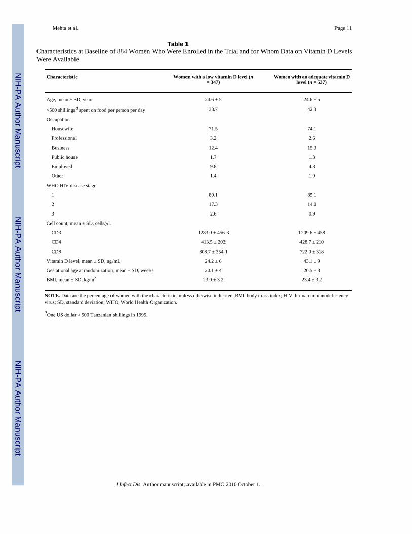
RESULTSVitamin D levels at baseline were known for 885 of the 1078 women enrolled in the trial. Onewoman was excluded because she had stage 4 HIV disease. The characteristics of these 884women at baseline are presented in table 1. The average maternal age (±SD) at enrollment was
Mehta et al. Page 4
J Infect Dis. Author manuscript; available in PMC 2010 October 1.
NIH
-PA Author Manuscript
NIH
-PA Author Manuscript
NIH
-PA Author Manuscript
24.6 ± 5 years, and >70% of the women described their occupation as housewife. More than80% of the women in this analysis had WHO stage 1 HIV disease, and >70% had a body massindex of 18.5–25 kg/m2 at enrollment. Twin births (n = 24) were excluded from the analysisof pregnancy outcomes but were retained in the analysis of HIV infection and death as endpoints. The results obtained in the analysis with low versus adequate vitamin D levels wereessentially similar to those obtained in the analysis with vitamin D quintiles; only the resultsfrom analyses of low versus adequate vitamin D levels are presented below (data not shown).
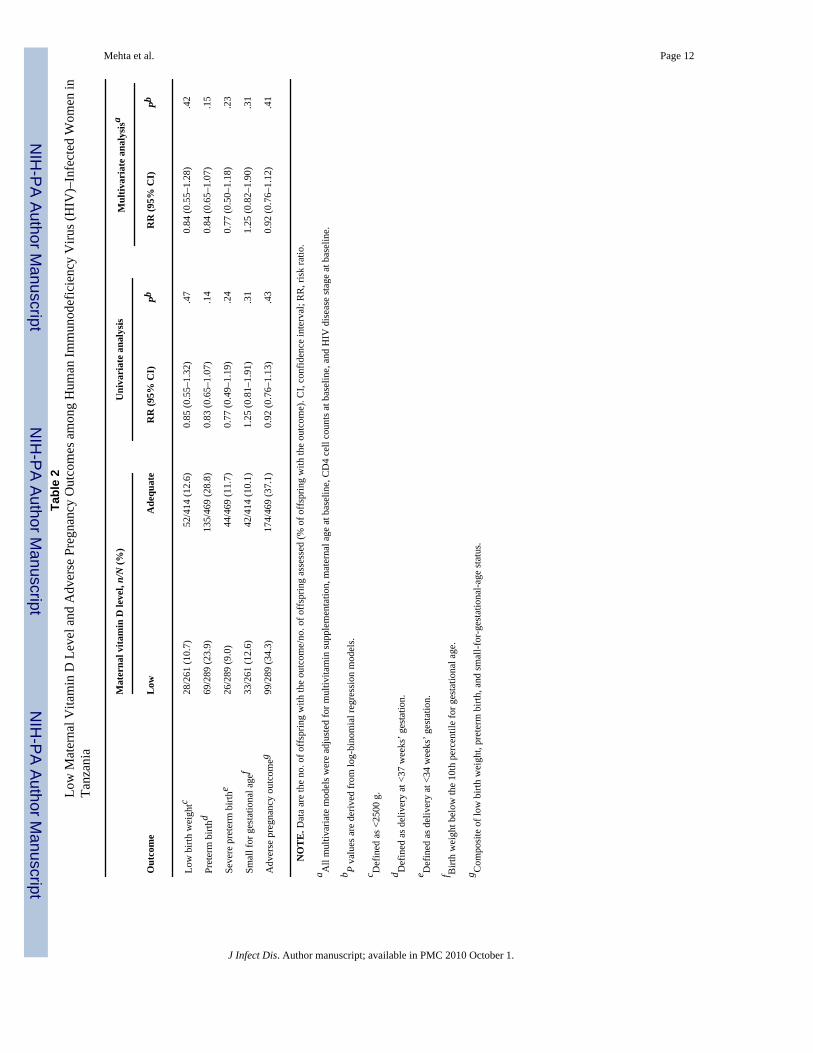
In this subset of the overall cohort, the risks of low birth weight, preterm birth, and small-for-gestational-age status were 12%, 27%, and 11%, respectively (table 2). For this subset ofwomen with vitamin D measurements, these incidences, along with those for other outcomespresented subsequently, are comparable to those obtained for the entire cohort [19,38,39]. Aftermultivariate adjustment for maternal age, HIV disease stage at baseline, CD4 cell counts, andmultivitamin supplementation, low levels of vitamin D were not associated with the risk oflow birth weight, preterm birth, small for gestational age, or the composite end point of anadverse pregnancy outcome (table 2).
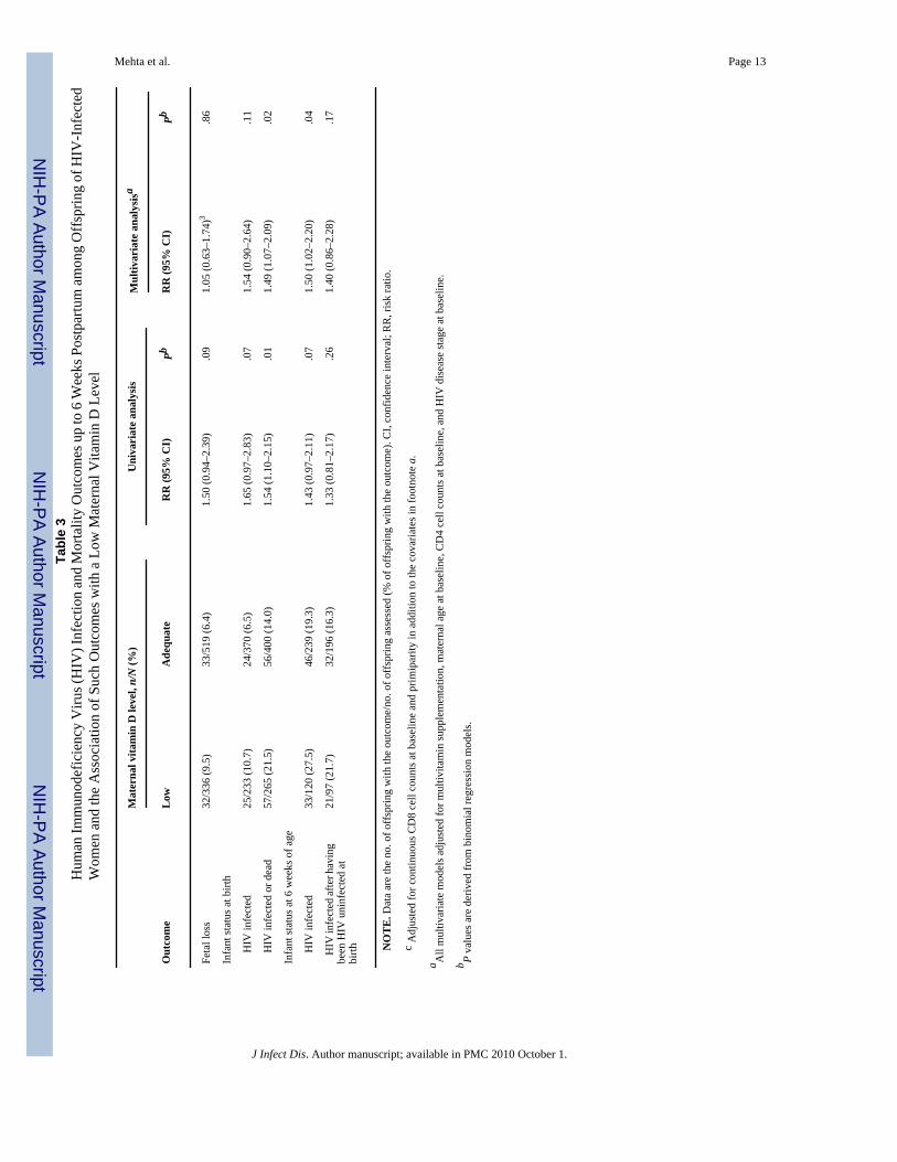
Fetal loss was the outcome of 7.6% of all pregnancies, and 8.1% of infants were HIV infectedat birth (table 3). Of the children who were known to be HIV uninfected at birth, 18.1% becameinfected by 6 weeks of age. There was no observed association between vitamin D status andfetal loss, HIV infection at birth, or HIV infection at 6 weeks of age, for those infants knownto be HIV uninfected at birth, after multivariate adjustment (table 3). However, a significantassociation was observed between vitamin D status and the composite end point of HIVinfection or death at delivery and HIV infection at 6 weeks. Children born to women with lowvitamin D levels had a 49% greater risk of dying or being HIV infected at birth than did childrenborn to women with sufficient vitamin D levels (95% CI, 7%–109%). A similar increase in therisk of HIV infection at 6 weeks of age was observed for children born to women with lowvitamin D levels at baseline (risk ratio [RR], 1.50 [95% CI, 1.02–2.20]).
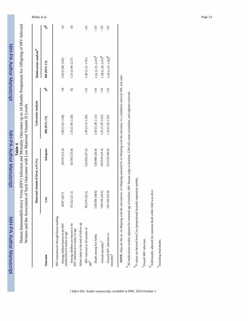
Over the first 24 months of follow-up, a total of 30.1% children were infected with HIV (table4). In multivariate Cox regression models that assessed time to HIV infection as the outcome(table 4), children who were born to women with low vitamin D levels at baseline and whowere known to be HIV uninfected at 6 weeks of age had a 2-fold higher incidence of acquiringHIV infection through breast-feeding than did children who were born to women with sufficientvitamin D levels (incidence rate ratio [IRR], 2.03 [95% CI, 1.08–3.82]). The overall rate ofincidence of HIV infection by 24 months of age was 46% higher in the group with low vitaminD levels (95% CI, 11%–91%) than in the group with sufficient vitamin D levels. A nonlinearassociation was observed between continuous vitamin D levels and the risk of HIVtransmission (P = .01) (figure 1); mothers with the lowest vitamin D levels had the highest riskof MTCT of HIV, and the risk decreased linearly as vitamin D levels increased. Although therisk of MTCT appeared to increase again at the highest levels of vitamin D, this risk was notsignificant.
During follow-up, 36.6% of the children died (fetal loss was included in this statistic); 46.0%of children died during follow-up or were HIV infected (table 4). A 61% increase in the rateof incidence of death during follow-up among live births was observed for infants born towomen with low vitamin D levels at baseline (95% CI, 25%–107%). We examined effectmodification of the association between a low vitamin D level and child mortality by HIVstatus, by introducing an interaction term in the model. This term was not statisticallysignificant (P = .15). For children who remained HIV uninfected during follow-up, a similarincrease in overall mortality was observed (IRR, 1.66 [95% CI, 1.23–2.23]); among childrenwho became HIV infected, the increase in mortality was lower and not statistically significant(IRR, 1.36 [95% CI, 0.95–1.94]), suggesting independent and separate effects of vitamin D on
Mehta et al. Page 5
J Infect Dis. Author manuscript; available in PMC 2010 October 1.
NIH
-PA Author Manuscript
NIH
-PA Author Manuscript
NIH
-PA Author Manuscript
HIV infection and death. Furthermore, children born to mothers with the lowest vitamin Dlevels had the highest risk of dying (P = .02) (figure 1), and the risk decreased linearly asvitamin D levels increased. The risk of child mortality appeared to increase again at the highestvitamin D levels; however, this finding was not significant. Finally, for the composite end pointof HIV infection or death by the end of follow-up, a 50% increase in risk (95% CI, 23%–63%)was observed for the group with low vitamin D levels.
DISCUSSIONIn the present study, we examined the association of maternal vitamin D status at baseline (12–27 weeks’ gestation) with adverse pregnancy outcomes, MTCT of HIV, and child mortality.No association was observed between a low maternal vitamin D level (<32 ng/mL) and suchadverse pregnancy outcomes as low birth weight, preterm birth, and small-for-gestational-agestatus. An increased risk of being HIV infected or of dying at birth was observed for childrenborn to women with a low vitamin D level at baseline; a low maternal vitamin D level was alsoassociated with HIV transmission via breast-feeding and with higher infant mortality duringfollow-up.
The role of maternal vitamin D status in pregnancy outcomes of HIV-infected women has notbeen previously studied; however, there is some literature suggesting a benefit of vitamin Din pregnant women who are not HIV infected. For example, Marya et al [40] conducted a studyof Asian-Indian women, randomizing them to receive either 600,000 IU of vitamin D twice(once each during the seventh and eighth months of pregnancy) or no vitamin D supplements.Infants of mothers who received vitamin D had greater intrauterine growth and a higher birthweight than did infants of women who did not receive vitamin D supplements. However, thebeneficial effects of vitamin D have been observed with relatively large doses of vitamin Dsupplements, which may lead to serum vitamin D levels that are much higher than thoseobserved in our cohort. This could be a potential explanation for the lack of any observedassociation with vitamin D levels and pregnancy outcomes in this cohort. Furthermore, therates of adverse pregnancy outcomes appear to be higher in HIV-infected women than in HIV-uninfected women. For example, in another trial involving 8468 HIV-uninfected pregnantwomen in Tanzania, our group determined that the incidence of preterm birth was only 17%,compared with the 27% incidence noted in the current analysis [41]. Thus, it is possible thatthe adverse effect of HIV infection on perinatal outcomes may mask the beneficial effect ofvitamin D on these outcomes, if any.
The association of maternal vitamin D status with HIV transmission and death among childrenhas not been previously studied. However, our results are in accordance with some small studiesof nonpregnant HIV-infected populations that have shown an association between low vitaminD levels and increased HIV disease progression and higher mortality [5]. For example, in onelongitudinal study in Norway, HIV-infected patients with 1,25(OH)D levels of <25 ng/L atbaseline (n = 9) had a significantly shorter survival time than did patients with levels in therange considered to be normal (n = 44), after adjustment for CD4 cell counts [42].
Recent research has highlighted the role of vitamin D in the regulation of the immune system,particularly innate immunity, which might explain this finding [43]. Vitamin D is also knownto have other immunomodulatory effects, such as improving the phagocytic capacity ofmacrophages, increasing the number of natural killer cells, and boosting cell-mediatedimmunity [6–8]. Vitamin D is known to contribute to the development of the fetal immunesystem; a stronger immune system may be more resistant to HIV infection and may explainthe decreased risk of MTCT observed in the present study. This finding would also likelycorrelate with fewer infections and opportunistic illnesses during follow-up and, consequently,with decreased mortality. In addition, there is increasing evidence supporting the role of
Mehta et al. Page 6
J Infect Dis. Author manuscript; available in PMC 2010 October 1.
NIH
-PA Author Manuscript
NIH
-PA Author Manuscript
NIH
-PA Author Manuscript
vitamin D in fighting tuberculosis; tuberculosis is one of the primary killers in HIV-infectedpopulations [18].
Vitamin D may also decrease MTCT of HIV or child mortality by reducing inflammation; inthis cohort, we observed an association between a low vitamin D level and a higher CD8 cellcount and erythrocyte sedimentation rate (S.M., D.S., S. Aboud, E.L.G., G.I.M., E.H., F.M.M.,D.J.H., and W.W.F., unpublished data). Although the conventional role of CD8 cells is tofunction as cytotoxic killer cells, they may also be the effector cells in inflammation [44]. Theinvolvement of vitamin D in modulating CD8 cells is also indicated by the fact that, of themajor immune cells (CD8, CD4, and B cells and macrophages), CD8 cells express the highestconcentration of vitamin D receptor [45].
The present study includes important and informative findings, considering the lack of similardata in other studies reported to date, as well as implications for potential interventions for theprevention of MTCT of HIV and child mortality. However, we had limited power to examinethe exact timing of MTCT of HIV. We also do not know whether the levels of vitamin D maybe depressed as a result of HIV disease and thus may be a consequence of an advanced stageof HIV disease and/or accelerated viral replication and not the cause thereof. However, mostwomen in our analysis did not have advanced HIV disease at baseline (>80% had stage 1disease). The association observed in the present study may be different in other populationswith a different underlying nutritional or immunologic status, as well as in populations withaccess to antiretroviral therapy, and therefore the findings may not be fully generalizable. Onelimitation of our analysis is that we had only one measurement of vitamin D levels at baseline;however, data from other studies suggest that there is a positive correlation between vitaminD levels in cord blood and vitamin D levels at baseline [29]. Therefore, it may be reasonableto assume that vitamin D levels at baseline are predictive of vitamin D levels in the postpartumperiod.
Another limitation of the present study is that the assay that we used to assess vitamin D statusdoes not measure vitamin D2 accurately; however, this form of vitamin D is obtained throughsupplements, the use of which was unlikely in this population. In addition, our choice of acutoff level for vitamin D is to make the results clinically more relevant and comparable tothose of other studies. However, estimates of a similar effect were obtained for such majoroutcomes as overall HIV transmission and overall child mortality, by use of quintiles of vitaminD, on the basis of its distribution in this population (data not shown).
Recent studies estimated that 1 billion people worldwide have vitamin D insufficiency [28].Daily intake of at least 800–1000 IU of vitamin D may be needed in the absence of adequatesun exposure, to maintain a circulating level of 25(OH)D >32 ng/mL [28,46]. If demonstratedto be effective in intervention studies, vitamin D supplementation could prove to be a relativelysimple and inexpensive method to lower mortality among children and to help prevent MTCTof HIV as an adjunct to antiretroviral therapy.
AcknowledgmentsWe thank the mothers and children, the field teams (including physicians, nurses, midwives, supervisors, andlaboratory staff), and administrative staff, all of whom made the study possible. We also thank Muhimbili MedicalCentre, Muhimbili University College of Health Sciences, and the National AIDS Control Program in Dar es Salaam,Tanzania, for their institutional support.
Financial support: National Institute of Child Health and Human Development (grant R01 32257), the FogartyInternational Center (National Institutes of Health grant D43 TW00004), and the Harvard School of Public Health.
Mehta et al. Page 7
J Infect Dis. Author manuscript; available in PMC 2010 October 1.
NIH
-PA Author Manuscript
NIH
-PA Author Manuscript
NIH
-PA Author Manuscript
References1. McCollum EV, Simmonds N, Becker JE, Shipley PG. Studies on experimental rickets. XXI. An
experimental demonstration of the existence of a vitamin which promotes calcium deposition. J BiolChem 1922;53:293–312.
2. Giovannucci E. The epidemiology of vitamin D and cancer incidence and mortality: a review (UnitedStates). Cancer Causes Control 2005;16:83–95. [PubMed: 15868450]
3. Nnoaham KE, Clarke A. Low serum vitamin D levels and tuberculosis: a systematic review and meta-analysis. Int J Epidemiol 2008;37:113–9. [PubMed: 18245055]
4. Liu PT, Stenger S, Li H, et al. Toll-like receptor triggering of a vitamin D-mediated humanantimicrobial response. Science 2006;311:1770–3. [PubMed: 16497887]
5. Villamor E. A potential role for vitamin D on HIV infection? Nutr Rev 2006;64:226–33. [PubMed:16770943]
6. Yang S, Smith C, Prahl JM, Luo X, DeLuca HF. Vitamin D deficiency suppresses cell-mediatedimmunity in vivo. Arch Biochem Biophys 1993;303:98–106. [PubMed: 8489269]
7. Bar-Shavit Z, Noff D, Edelstein S, Meyer M, Shibolet S, Goldman R. 1,25-dihydroxyvitamin D3 andthe regulation of macrophage function. Calcif Tissue Int 1981;33:673–6. [PubMed: 6275970]
8. Mariani E, Ravaglia G, Forti P, et al. Vitamin D, thyroid hormones and muscle mass influence naturalkiller (NK) innate immunity in healthy nonagenarians and centenarians. Clin Exp Immunol1999;116:19–27. [PubMed: 10209500]
9. Sorensen OE. The human cathelicidin hCAP-18. Dan Med Bull 2005;52:1–10. [PubMed: 16009068]10. Nagaoka I, Hirota S, Niyonsaba F, et al. Cathelicidin family of antibacterial peptides CAP18 and
CAP11 inhibit the expression of TNF-α by blocking the binding of LPS to CD14+ cells. J Immunol2001;167:3329–38. [PubMed: 11544322]
11. Koczulla R, von Degenfeld G, Kupatt C, et al. An angiogenic role for the human peptide antibioticLL-37/hCAP-18. J Clin Invest 2003;111:1665–72. [PubMed: 12782669]
12. De Yang, Chen Q, Schmidt AP, et al. LL-37, the neutrophil granule- and epithelial cell-derivedcathelicidin, utilizes formyl peptide receptor-like 1 (FPRL1) as a receptor to chemoattract humanperipheral blood neutrophils, monocytes, and T cells. J Exp Med 2000;192:1069–74. [PubMed:11015447]
13. Specker B. Vitamin D requirements during pregnancy. Am J Clin Nutr 2004;80:1740S–7S. [PubMed:15585798]
14. Cross NA, Hillman LS, Allen SH, Krause GF, Vieira NE. Calcium homeostasis and bone metabolismduring pregnancy, lactation, and postweaning: a longitudinal study. Am J Clin Nutr 1995;61:514–23. [PubMed: 7872214]
15. Bikle DD, Gee E, Halloran B, Haddad JG. Free 1,25-dihydroxyvitamin D levels in serum from normalsubjects, pregnant subjects, and subjects with liver disease. J Clin Invest 1984;74:1966–71. [PubMed:6549014]
16. Evans KN, Bulmer JN, Kilby MD, Hewison M. Vitamin D and placental-decidual function. J SocGynecol Investig 2004;11:263–71.
17. Reichrath J, Querings K. Vitamin D deficiency during pregnancy: a risk factor not only for fetalgrowth and bone metabolism but also for correct development of the fetal immune system? Am JClin Nutr 2005;81:1177. [PubMed: 15883446]author reply 1177–8
18. Chaisson RE, Martinson NA. Tuberculosis in Africa—combating an HIV-driven crisis. N Engl J Med2008;358:1089–92. [PubMed: 18337598]
19. Fawzi WW, Msamanga GI, Spiegelman D, et al. Randomised trial of effects of vitamin supplementson pregnancy outcomes and T cell counts in HIV-1-infected women in Tanzania. Lancet1998;351:1477–82. [PubMed: 9605804]
20. Fawzi WW, Msamanga GI, Spiegelman D, et al. A randomized trial of multivitamin supplements andHIV disease progression and mortality. N Engl J Med 2004;351:23–32. [PubMed: 15229304]
21. Prentice A. Vitamin D deficiency: a global perspective. Nutr Rev 2008;66:S153–64. [PubMed:18844843]
22. Fawzi WW, Msamanga GI, Spiegelman D, Urassa EJ, Hunter DJ. Rationale and design of the TanzaniaVitamin and HIV Infection Trial. Control Clin Trials 1999;20:75–90. [PubMed: 10027501]
Mehta et al. Page 8
J Infect Dis. Author manuscript; available in PMC 2010 October 1.
NIH
-PA Author Manuscript
NIH
-PA Author Manuscript
NIH
-PA Author Manuscript
23. The WHO International Collaborating Group for the Study of the WHO Staging System. Proposed‘World Health Organization staging system for HIV infection and disease’: preliminary testing byan international collaborative cross-sectional study. AIDS 1993;7:711–8. [PubMed: 8100422]
24. Brenner WE, Edelman DA, Hendricks CH. A standard of fetal growth for the United States ofAmerica. Am J Obstet Gynecol 1976;126:555–64. [PubMed: 984126]
25. Fawzi WW, Msamanga GI, Hunter D, et al. Randomized trial of vitamin supplements in relation totransmission of HIV-1 through breastfeeding and early child mortality. AIDS 2002;16:1935–44.[PubMed: 12351954]
26. Kupka R, Garland M, Msamanga G, Spiegelman D, Hunter D, Fawzi W. Selenium status, pregnancyoutcomes, and mother-to-child transmission of HIV-1. J Acquir Immune Defic Syndr 2005;39:203–10. [PubMed: 15905738]
27. Hollis BW. Circulating 25-hydroxyvitamin D levels indicative of vitamin D sufficiency: implicationsfor establishing a new effective dietary intake recommendation for vitamin D. J Nutr 2005;135:317–22. [PubMed: 15671234]
28. Holick MF. Vitamin D deficiency. N Engl J Med 2007;357:266–81. [PubMed: 17634462]29. Bodnar LM, Simhan HN, Powers RW, Frank MP, Cooperstein E, Roberts JM. High prevalence of
vitamin D insufficiency in black and white pregnant women residing in the northern United Statesand their neonates. J Nutr 2007;137:447–52. [PubMed: 17237325]
30. Wacholder S. Binomial regression in GLIM: estimating risk ratios and risk differences. Am JEpidemiol 1986;123:174–84. [PubMed: 3509965]
31. Zou G. A modified Poisson regression approach to prospective studies with binary data. Am JEpidemiol 2004;159:702–6. [PubMed: 15033648]
32. Spiegelman D, Hertzmark E. Easy SAS calculations for risk or prevalence ratios and differences. AmJ Epidemiol 2005;162:199–200. [PubMed: 15987728]
33. Cox D. Regression models and life tables. J Royal Stat Soc 1972;34:187–220.34. Durrleman S, Simon R. Flexible regression models with cubic splines. Stat Med 1989;8:551–61.
[PubMed: 2657958]35. Rothman, K.; Greenland, S. Modern epidemiology. Vol. 2nd ed.. Lippincott Williams & Wilkins;
Philadelphia: 1998.36. Greenland S. Modeling and variable selection in epidemiologic analysis. Am J Public Health
1989;79:340–9. [PubMed: 2916724]37. Miettinen, O. Theoretical epidemiology. Vol. 107. John Wiley & Sons; New York: 1985.38. Fawzi W, Msamanga G, Renjifo B, et al. Predictors of intrauterine and intrapartum transmission of
HIV-1 among Tanzanian women. AIDS 2001;15:1157–65. [PubMed: 11416718]39. Fawzi W, Msamanga G, Spiegelman D, et al. Transmission of HIV-1 through breastfeeding among
women in Dar es Salaam, Tanzania. J Acquir Immune Defic Syndr 2002;31:331–8. [PubMed:12439210]
40. Marya RK, Rathee S, Dua V, Sangwan K. Effect of vitamin D supplementation during pregnancy onfoetal growth. Indian J Med Res 1988;88:488–92. [PubMed: 3243609]
41. Fawzi WW, Msamanga GI, Urassa W, et al. Vitamins and perinatal outcomes among HIV-negativewomen in Tanzania. N Engl J Med 2007;356:1423–31. [PubMed: 17409323]
42. Haug C, Muller F, Aukrust P, Froland SS. Subnormal serum concentration of 1,25-vitamin D inhuman immunodeficiency virus infection: correlation with degree of immune deficiency and survival.J Infect Dis 1994;169:889–93. [PubMed: 7907645]
43. Adams JS, Liu PT, Chun R, Modlin RL, Hewison M. Vitamin D in defense of the human immuneresponse. Ann N Y Acad Sci 2007;1117:94–105. [PubMed: 17656563]
44. Meehan TF, DeLuca HF. CD8+ T cells are not necessary for 1 α,25-dihydroxyvitamin D3 to suppressexperimental autoimmune encephalomyelitis in mice. Proc Natl Acad Sci U S A 2002;99:5557–60.[PubMed: 11929984]
45. Veldman CM, Cantorna MT, DeLuca HF. Expression of 1,25-dihydroxyvitamin D3 receptor in theimmune system. Arch Biochem Biophys 2000;374:334–8. [PubMed: 10666315]
46. Holick MF, Chen TC. Vitamin D deficiency: a worldwide problem with health consequences. Am JClin Nutr 2008;87:1080S–6S. [PubMed: 18400738]
Mehta et al. Page 9
J Infect Dis. Author manuscript; available in PMC 2010 October 1.
NIH
-PA Author Manuscript
NIH
-PA Author Manuscript
NIH
-PA Author Manuscript
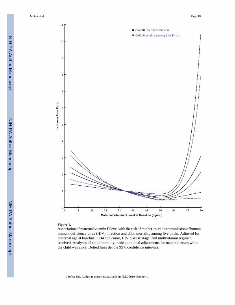
Figure 1.Association of maternal vitamin D level with the risk of mother-to-child transmission of humanimmunodeficiency virus (HIV) infection and child mortality among live births. Adjusted formaternal age at baseline, CD4 cell count, HIV disease stage, and multivitamin regimenreceived. Analyses of child mortality made additional adjustments for maternal death whilethe child was alive. Dotted lines denote 95% confidence intervals.
Mehta et al. Page 10
J Infect Dis. Author manuscript; available in PMC 2010 October 1.
NIH
-PA Author Manuscript
NIH
-PA Author Manuscript
NIH
-PA Author Manuscript
NIH
-PA Author Manuscript
NIH
-PA Author Manuscript
NIH
-PA Author Manuscript
Mehta et al. Page 11
Table 1Characteristics at Baseline of 884 Women Who Were Enrolled in the Trial and for Whom Data on Vitamin D LevelsWere Available
Characteristic Women with a low vitamin D level (n= 347)
Women with an adequate vitamin Dlevel (n = 537)
Age, mean ± SD, years 24.6 ± 5 24.6 ± 5
≤500 shillingsa spent on food per person per day 38.7 42.3
Occupation
Housewife 71.5 74.1
Professional 3.2 2.6
Business 12.4 15.3
Public house 1.7 1.3
Employed 9.8 4.8
Other 1.4 1.9
WHO HIV disease stage
1 80.1 85.1
2 17.3 14.0
3 2.6 0.9
Cell count, mean ± SD, cells/μL
CD3 1283.0 ± 456.3 1209.6 ± 458
CD4 413.5 ± 202 428.7 ± 210
CD8 808.7 ± 354.1 722.0 ± 318
Vitamin D level, mean ± SD, ng/mL 24.2 ± 6 43.1 ± 9
Gestational age at randomization, mean ± SD, weeks 20.1 ± 4 20.5 ± 3
BMI, mean ± SD, kg/m2 23.0 ± 3.2 23.4 ± 3.2
NOTE. Data are the percentage of women with the characteristic, unless otherwise indicated. BMI, body mass index; HIV, human immunodeficiencyvirus; SD, standard deviation; WHO, World Health Organization.
aOne US dollar ≈ 500 Tanzanian shillings in 1995.
J Infect Dis. Author manuscript; available in PMC 2010 October 1.
NIH
-PA Author Manuscript
NIH
-PA Author Manuscript
NIH
-PA Author Manuscript
Mehta et al. Page 12Ta
ble
2Lo
w M
ater
nal V
itam
in D
Lev
el a
nd A
dver
se P
regn
ancy
Out
com
es a
mon
g H
uman
Imm
unod
efic
ienc
y V
irus (
HIV
)–In
fect
ed W
omen
inTa
nzan
ia
Mat
erna
l vita
min
D le
vel,
n/N
(%)
Uni
vari
ate
anal
ysis
Mul
tivar
iate
ana
lysi
sa
Out
com
eL
owA
dequ
ate
RR
(95%
CI)
PbR
R (9
5% C
I)Pb
Low
birt
h w
eigh
tc28
/261
(10.
7)52
/414
(12.
6)0.
85 (0
.55–
1.32
).4
70.
84 (0
.55–
1.28
).4
2
Pret
erm
birt
hd69
/289
(23.
9)13
5/46
9 (2
8.8)
0.83
(0.6
5–1.
07)
.14
0.84
(0.6
5–1.
07)
.15
Seve
re p
rete
rm b
irthe
26/2
89 (9
.0)
44/4
69 (1
1.7)
0.77
(0.4
9–1.
19)
.24
0.77
(0.5
0–1.
18)
.23
Smal
l for
ges
tatio
nal a
gef
33/2
61 (1
2.6)
42/4
14 (1
0.1)
1.25
(0.8
1–1.
91)
.31
1.25
(0.8
2–1.
90)
.31
Adv
erse
pre
gnan
cy o
utco
meg
99/2
89 (3
4.3)
174/
469
(37.
1)0.
92 (0
.76–
1.13
).4
30.
92 (0
.76–
1.12
).4
1
NO
TE
. Dat
a ar
e th
e no
. of o
ffsp
ring
with
the
outc
ome/
no. o
f off
sprin
g as
sess
ed (%
of o
ffsp
ring
with
the
outc
ome)
. CI,
conf
iden
ce in
terv
al; R
R, r
isk
ratio
.
a All
mul
tivar
iate
mod
els w
ere
adju
sted
for m
ultiv
itam
in su
pple
men
tatio
n, m
ater
nal a
ge a
t bas
elin
e, C
D4
cell
coun
ts a
t bas
elin
e, a
nd H
IV d
isea
se st
age
at b
asel
ine.
b P va
lues
are
der
ived
from
log-
bino
mia
l reg
ress
ion
mod
els.
c Def
ined
as <
2500
g.
d Def
ined
as d
eliv
ery
at <
37 w
eeks
’ ges
tatio
n.
e Def
ined
as d
eliv
ery
at <
34 w
eeks
’ ges
tatio
n.
f Birt
h w
eigh
t bel
ow th
e 10
th p
erce
ntile
for g
esta
tiona
l age
.
g Com
posi
te o
f low
birt
h w
eigh
t, pr
eter
m b
irth,
and
smal
l-for
-ges
tatio
nal-a
ge st
atus
.
J Infect Dis. Author manuscript; available in PMC 2010 October 1.
NIH
-PA Author Manuscript
NIH
-PA Author Manuscript
NIH
-PA Author Manuscript
Mehta et al. Page 13Ta
ble
3H
uman
Imm
unod
efic
ienc
y V
irus (
HIV
) Inf
ectio
n an
d M
orta
lity
Out
com
es u
p to
6 W
eeks
Pos
tpar
tum
am
ong
Off
sprin
g of
HIV
-Inf
ecte
dW
omen
and
the
Ass
ocia
tion
of S
uch
Out
com
es w
ith a
Low
Mat
erna
l Vita
min
D L
evel
Mat
erna
l vita
min
D le
vel,
n/N
(%)
Uni
vari
ate
anal
ysis
Mul
tivar
iate
ana
lysi
sa
Out
com
eL
owA
dequ
ate
RR
(95%
CI)
PbR
R (9
5% C
I)Pb
Feta
l los
s32
/336
(9.5
)33
/519
(6.4
)1.
50 (0
.94–
2.39
).0
91.
05 (0
.63–
1.74
)3.8
6
Infa
nt st
atus
at b
irth
H
IV in
fect
ed25
/233
(10.
7)24
/370
(6.5
)1.
65 (0
.97–
2.83
).0
71.
54 (0
.90–
2.64
).1
1
H
IV in
fect
ed o
r dea
d57
/265
(21.
5)56
/400
(14.
0)1.
54 (1
.10–
2.15
).0
11.
49 (1
.07–
2.09
).0
2
Infa
nt st
atus
at 6
wee
ks o
f age
H
IV in
fect
ed33
/120
(27.
5)46
/239
(19.
3)1.
43 (0
.97–
2.11
).0
71.
50 (1
.02–
2.20
).0
4
H
IV in
fect
ed af
ter h
avin
gbe
en H
IV u
ninf
ecte
d at
birth
21/9
7 (2
1.7)
32/1
96 (1
6.3)
1.33
(0.8
1–2.
17)
.26
1.40
(0.8
6–2.
28)
.17
NO
TE
. Dat
a ar
e th
e no
. of o
ffsp
ring
with
the
outc
ome/
no. o
f off
sprin
g as
sess
ed (%
of o
ffsp
ring
with
the
outc
ome)
. CI,
conf
iden
ce in
terv
al; R
R, r
isk
ratio
.
c A
djus
ted
for c
ontin
uous
CD
8 ce
ll co
unts
at b
asel
ine
and
prim
ipar
ity in
add
ition
to th
e co
varia
tes i
n fo
otno
te a
.
a All
mul
tivar
iate
mod
els a
djus
ted
for m
ultiv
itam
in su
pple
men
tatio
n, m
ater
nal a
ge a
t bas
elin
e, C
D4
cell
coun
ts a
t bas
elin
e, a
nd H
IV d
isea
se st
age
at b
asel
ine.
b P va
lues
are
der
ived
from
bin
omia
l reg
ress
ion
mod
els.
J Infect Dis. Author manuscript; available in PMC 2010 October 1.
NIH
-PA Author Manuscript
NIH
-PA Author Manuscript
NIH
-PA Author Manuscript
Mehta et al. Page 14Ta
ble
4H
uman
Imm
unod
efic
ienc
y V
irus
(HIV
) Inf
ectio
n an
d M
orta
lity
Out
com
es u
p to
24
Mon
ths
Post
partu
m fo
r Off
sprin
g of
HIV
-Inf
ecte
dW
omen
and
the
Ass
ocia
tion
of S
uch
Out
com
es w
ith L
ow M
ater
nal V
itam
in D
Lev
els
Mat
erna
l vita
min
D le
vel,
n/N
(%)
Uni
vari
ate
anal
ysis
Mul
tivar
iate
ana
lysi
sa
Out
com
eL
owA
dequ
ate
RR
(95%
CI)
PbR
R (9
5% C
I)Pb
HIV
tran
smis
sion
thro
ugh
brea
st-f
eedi
ng
A
mon
g ch
ildre
n kn
own
to b
e HIV
unin
fect
ed a
t 6 w
eeks
of a
ge18
/87
(20.
7)24
/193
(12.
4)1.
89 (1
.03–
3.49
).0
42.
03 (1
.08–
3.82
).0
3
A
mon
g ch
ildre
n no
t kno
wn
to b
eH
IV in
fect
ed a
t 6 w
eeks
of a
ge47
/222
(21.
2)62
/392
(15.
8)1.
55 (1
.06–
2.26
).0
21.
55 (1
.06–
2.27
).0
2
Infa
nt st
atus
at t
he e
nd o
f fol
low
-up
H
IV in
fect
ed a
t 24
mon
ths o
fag
ec95
/270
(35.
2)12
2/45
2 (2
7.0)
1.49
(1.1
4–1.
95)
<.01
1.46
(1.1
1–1.
91)
<.01
D
eath
am
ong
live
birth
s11
8/30
4 (3
8.8)
130/
486
(26.
8)1.
64 (1
.28–
2.11
)<.
011.
61 (1
.25–
2.07
)d<.
01
O
vera
ll m
orta
litye
150/
336
(44.
6)16
3/51
9 (3
1.4)
1.61
(1.2
9–2.
01)
<.01
1.58
(1.2
6–1.
97)d
<.01
O
vera
ll H
IV in
fect
ion
orm
orta
litye
181/
336
(53.
9)21
2/51
9 (4
0.9)
1.54
(1.2
6–1.
87)
<.01
1.50
(1.2
3–1.
83)d
<.01
NO
TE
. Dat
a ar
e th
e no
. of o
ffsp
ring
with
the
outc
ome/
no. o
f off
sprin
g as
sess
ed (%
of o
ffsp
ring
with
the
outc
ome)
. CI,
conf
iden
ce in
terv
al; R
R, r
isk
ratio
.
a All
mul
tivar
iate
mod
els a
djus
ted
for m
ater
nal a
ge a
t bas
elin
e, H
IV d
isea
se st
age
at b
asel
ine,
CD
4 ce
ll co
unts
at b
asel
ine,
and
regi
men
rece
ived
.
b P va
lues
are
der
ived
from
Cox
(pro
porti
onal
haz
ards
) reg
ress
ion
mod
els.
c Ove
rall
HIV
infe
ctio
n.
d Add
ition
ally
adj
uste
d fo
r mat
erna
l dea
th w
hile
chi
ld w
as a
live.
e Incl
udin
g fe
tal d
eath
s.
J Infect Dis. Author manuscript; available in PMC 2010 October 1.