Embed Size (px)
Citation preview

Perinatal and neurodevelopmental outcome of late-onset growth restricted fetuses
Daniel Orós López
ADVERTIMENT. La consulta d’aquesta tesi queda condicionada a l’acceptació de les següents condicions d'ús: La difusió d’aquesta tesi per mitjà del servei TDX (www.tesisenxarxa.net) ha estat autoritzada pels titulars dels drets de propietat intel·lectual únicament per a usos privats emmarcats en activitats d’investigació i docència. No s’autoritza la seva reproducció amb finalitats de lucre ni la seva difusió i posada a disposició des d’un lloc aliè al servei TDX. No s’autoritza lapresentació del seu contingut en una finestra o marc aliè a TDX (framing). Aquesta reserva de drets afecta tant al resum de presentació de la tesi com als seus continguts. En la utilització o cita de parts de la tesi és obligat indicar el nom de lapersona autora.
ADVERTENCIA. La consulta de esta tesis queda condicionada a la aceptación de las siguientes condiciones de uso: La difusión de esta tesis por medio del servicio TDR (www.tesisenred.net) ha sido autorizada por los titulares de los derechos de propiedad intelectual únicamente para usos privados enmarcados en actividades de investigación y docencia. No se autoriza su reproducción con finalidades de lucro ni su difusión y puesta a disposición desde un sitio ajeno al servicio TDR. No se autoriza la presentación de su contenido en una ventana o marco ajeno a TDR (framing). Esta reserva de derechos afecta tanto al resumen de presentación de la tesis como a sus contenidos. En la utilización o cita de partes de la tesis es obligado indicar el nombre de la persona autora.
WARNING. On having consulted this thesis you’re accepting the following use conditions: Spreading this thesis by the TDX (www.tesisenxarxa.net) service has been authorized by the titular of the intellectual property rights only for private uses placed in investigation and teaching activities. Reproduction with lucrative aims is not authorized neither its spreading and availability from a site foreign to the TDX service. Introducing its content in a window or frame foreign to the TDX service is not authorized (framing). This rights affect to the presentation summary of the thesis as well as to its contents. Inthe using or citation of parts of the thesis it’s obliged to indicate the name of the author.


PhD Thesis Daniel Orós López
� �
PhD THESIS
Programa de Doctorat en Medicina
Universitat de Barcelona
Perinatal and neurodevelopmental outcome
of
late-onset growth restricted fetuses
AUTHOR: DANIEL ORÓS LÓPEZ
DIRECTORS: FRANCESC FIGUERAS RETUERTA
EDUARD GRATACÓS SOLSONA

PhD Thesis Daniel Orós López �
� �

PhD Thesis Daniel Orós López �
� �
Universitat de Barcelona
Divisió de Ciències de la Salud
Facultat de Medicina
Departament d’Obstetrícia i Ginecologia, Pediatria, Radiologia i Medicina
Física.
Programa de Doctorat de Medicina RD 1393/2007
A thesis submitted by Daniel Orós López for the degree of Doctor in Medicine
(Faculty of Medicine, University of Barcelona) under the direction of Francesc
Figueras Retuerta, Asociated Professor of Obstetrics and Gynecology at
Barcelona University, and Eduard Gratacós Solsona, Professor of Obstetrics
and Gynecology at Barcelona University.
Signed: Daniel Orós López
Barcelona, 22nd February 2010.

PhD Thesis Daniel Orós López �
� �
Francesc Figueras Retuerta, Asociated Professor of Obstetrics and Gynecology
at Barcelona University, and Eduard Gratacós Solsona, Professor of Obstetrics
and Gynecology in the Barcelona University.
DECLARE:
That Daniel Orós López has carried out the study entitled “Perinatal and
neurodevelopmental outcome of late-onset growth restricted fetuses” under our
direction for the degree of Doctor in Medicine, and that the mentioned study is
henceforth ready to be presented.
Signed,
Francesc Figueras Retuerta
Eduard Gratacós Solsona
Barcelona, 22nd February 2010.

PhD Thesis Daniel Orós López
� �
This thesis project has been structured following the normative for PhD thesis
as a compendium of publications. The studies included in the thesis belong to
the same research line leading to three papers already published or submitted
for publication in international journals:
1. Oros D, Francesc Figueras, Rogelio Cruz-Martinez, Eva Meler, Meritxell
Munmany, Eduard Gratacos. “Longitudinal changes in uterine, umbilical
and cerebral Doppler in late-onset small-for-gestational age fetuses.”
Ultrasound Obstet Gynecol. (submitted)
State: submitted (under review)
Impact factor: 2.69
Quartile: 1st
2. Francesc Figueras, Daniel Oros, Rogelio Cruz-Martinez, Nelly Padilla, Edgar
Hernandez-Andrade, Francesc Botet, Carmen Costas-Moragas, Eduard
Gratacos. “Neurobehavior in term, small-for-gestational age infants with
normal placental function.” Pediatrics. 2009 Nov;124(5):e934-41. Epub 2009
Oct 26.
State: published
Impact factor: 4.789
Quartile: 1st
3. Daniel Oros; Francesc Figueras; Rogelio Cruz-Martinez; Nelly Padilla; Eva
Meler; Edgar Hernandez-Andrade; Eduard Gratacos. “Middle versus anterior cerebral artery Doppler for the prediction of perinatal outcome and
neonatal neurobehavior in term small-for-gestational-age fetuses with normal umbilical artery Doppler.” Ultrasound Obstet Gynecol (in press)
State: accepted for publication
Impact factor: 2.69
Quartile: 1st


PhD Thesis Daniel Orós López
� �
TABLE OF CONTENTS


PhD Thesis Daniel Orós López
�
�
1. Introduction
15
16
18
21
23
25
26
1.1 Definition
1.2 Clinical impact of SGA
1.3. SGA versus IUGR: the value of umbilical artery Doppler
1.4. Uterine artery Doppler
1.5. Middle cerebral artery Doppler
1.6. Anterior cerebral artery Doppler
1.7. Cerebro-placental ratio
1.8. Relevance and justification of the research project 26
2. General hypotesis
2.1. Conceptual hypothesis 31
2.2. Secondary hypothesis 31
3. General objectives
35 3.1. Main objective
3.2. Specific objectives 35
4. Methods
39
39
40
40
41
42
42
4.1. Study design
4.2. Study population
4.3. Sample size
4.4. Predictive variables
4.5. Result variables
4.6. Ethical approvement
4.7. Study protocol
4.8. Specific projects 46

�
� �
5. Results
53 5.1. Project 1: Longitudinal evaluation of fetal hemodynamic status in late-onset IUGR fetuses.
5.2. Project 2: Evaluation of perinatal outcome and neonatal neurobehaviour of late-onset IUGR fetuses with normal UA Doppler versus controls.
58
5.3. Project 3: Evaluation of the anterior and middle cerebral arteries for the prediction of perinatal outcome and neonatal neurobehavior in late-onset IUGR fetuses with normal UA Doppler.
65
6. Discussion
6.1. General overview 75
6.2. Project 1: Longitudinal evaluation of fetal hemodynamic status in late-onset IUGR fetuses. 77
6.3. Project 2: Evaluation of perinatal outcome and neonatal neurobehaviour of late-onset IUGR fetuses with normal UA Doppler versus controls.
80
6.4. Project 3: Evaluation of the anterior and middle cerebral arteries for the prediction of perinatal outcome and neonatal neurobehavior in late-onset IUGR fetuses with normal UA Doppler.
83
7. Conclusions 89
8. Aknowlegments 93
9. Bibliography 97
10. Annexes
107
109
8.1. Ethic Committe approval
8.2. Inform consent – Patient information
8.3. Data form 111

PhD Thesis Daniel Orós López �
� ��
113 8.4. Aceptance letter proyect 3
8.5. Submission letter proyect 1 115
11. Papers
11.1. Longitudinal changes in uterine, umbilical and cerebral Doppler in late-onset small-for-gestational age fetuses.
119
11.2. Neurobehavior in term, small-for-gestational age infants with normal placental function.
147
11.3. Middle versus anterior cerebral artery Doppler for the prediction of perinatal outcome and neonatal neurobehavior in term small-for-gestational-age fetuses with normal umbilical artery Doppler.
159

�
� ��

Introduction
� ��
1) INTRODUCTION


Introduction
� ��
1.1. Definition
Conceptually, intrauterine growth restriction (IUGR) is failure of a fetus to
achieve its genetically endorsed growth potential. However, the difficulty in
accurately determining the growth potential for a given fetus limits the
applicability of this definition, although birthweight centiles (1) and estimated
fetal weight charts, customized for individual maternal and fetal characteristics
(2) represent attempts to use this concept in clinical practice.
Hence, in clinical pratice birthweight is classified using population-based sex-
adjusted centiles, with a newborn being small for gestational age (SGA) if the
birthweight is <10th centile. However, this definition includes a large proportion
of cases that are constitutionally small and otherwise healthy newborns.
Currently, the most accepted clinical classification of SGA, to further tailor their
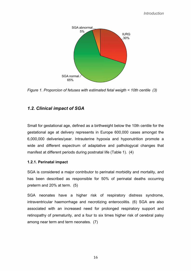
monitoring and management, is (Figure 1):
• IUGR: this category represents those fetuses that fail to reach their growth
potential, mostly because of chronic placental insufficiency.
• Abnormal SGA fetuses: this category comprises cases with congenital
anomalies, including chromosomopaties, genetic syndromes and fetal
infections.
• Normal SGA fetuses: this category includes constitutionally small fetuses
with a genetically determined lower growth potential. This group is
defined after negative screening for congenital anomalies, fetal infeccion
and signs of placental insufficiency.
Introduction �
� ��
Figure 1. Proporcion of fetuses with estimated fetal weigth < 10th centile (3)
1.2. Clinical impact of SGA
Small for gestational age, defined as a birthweight below the 10th centile for the
gestational age at delivery represents in Europe 600,000 cases amongst the
6,000,000 deliveries/year. Intrauterine hypoxia and hyponutrition promote a
wide and different espectrum of adaptative and pathologycal changes that
manifest at different periods during postnatal life (Table 1).� (4)
1.2.1. Perinatal impact
SGA is considered a major contributor to perinatal morbidity and mortality, and
has been described as responsible for 50% of perinatal deaths occurring
preterm and 20% at term. (5)
SGA neonates have a higher risk of respiratory distress syndrome,
intraventricular haemorrhage and necrotizing enterocolitis. (6) SGA are also
associated with an increased need for prolonged respiratory support and
retinopathy of prematurity, and a four to six times higher risk of cerebral palsy
among near term and term neonates. (7)

Introduction �
� ��
Table 1. Systemic effects of intrauterine growth restriction. (4)
Cardiovascular
• Increased afterload • Cardiac hypertrophy • Initial elevation of left ventricular output • Elevated brain natriuretic peptide (BNP) • Higher shunting through Ductus venosus • Decreases cardiac compliance
Lungs • Accelerated lung maturity • Decreased lung compliance
Kidneys • Reduced number of nephrons • Alterations in rennin–angiotensin–aldosterone
axis, hypertension • Accelerated nephrosclerosis • Shortened life span.
Skeletal muscle • Increase in thyroxine secretion • Altered mitochondrial function • Reduction and then cessation of foetal motor
activity
Brain • Increase in cerebral blood flow • Reduction in metabolic rate • Dishminuish oxigenation of temporal and occipital
cortex, hippocampus and cerebellum • Altering nitric oxide synthases (NOSs) genes and
protein expression. • Affect myelination and growth • White-matter injury • Increase in the dopaminergic system
Gastrointestinal tract
• Alter blood flow in the mesenteric artery • Postnatal intestinal motility syndrome and poor
nutrient absorption
1.2.2. Impact on childhood
In childhood, SGA is linked with an increased mortality due to infectious
diseases, congenital anomalies, central nervous system anomalies and
cardiovascular diseases. (8)
Introduction �
� ��
Furthermore, SGA neonates are at higher risk of developing cognitive deficits at
school age with impaired learning capacities. Even in the absence of overt
perinatal brain lesions, minor neurologic deficits have been reported. (7) The
most frequent cognitive difficulties are in memory performance, learning
abilities, visuomotor functions and attention span. (9) Both in the animal model
and in human pregnancies complicated by IUGR, autopsy of the brain showed
a significant reduction in myelination, whereas visual and frontal cortical
synaptogenesis seemed to be decreased. (10) In addition, magnetic resonance
imaging (MRI) studies have demonstrated specific differences in the brain
composition of neonates with IUGR in comparison with those without this
condition. Thus, significant volume reductions in cortical grey matter (11) and
both hippocampi (12) have been demonstrated. Moreover an important delay in
cortical development, a discordant pattern of gyrification and a pronounced
reduced cortical expansion has been observed. (13) In contrast, white matter
(WM) seems to be affected only in cases of evident ischemic injury. (11)
1.2.3. Impact on adulthood
Epidemiological evidence has long suggested a link between low birth weight
and increased cardiovascular mortality in adulthood. (14) This association is
essentially mediated through fetal growth restriction. (15) The mechanistic
pathways underlying the relationship between IUGR and cardiovascular risk are
poorly understood. A number of studies support that it might be partially
explained by fetal metabolic programming leading to conditions associated with
cardiovascular disease, such as obesity, diabetes and hypertension. (16-17)
1.3. SGA versus IUGR: the value of umbilical artery Doppler
Clinical management and prognosis differ in abnormal SGA fetuses depending
of their specific aetiology. SGA and IUGR have been used interchangeably, but
the two terms are not synonymous; not all small babies are growth-restricted
and not all growth-restricted infants are small. (18) Intrauterine placental
Introduction �
� ��
function evaluation by umbilical artery Doppler (UA) is currently the clinical
standard to distinguish between SGA and IUGR. (19)
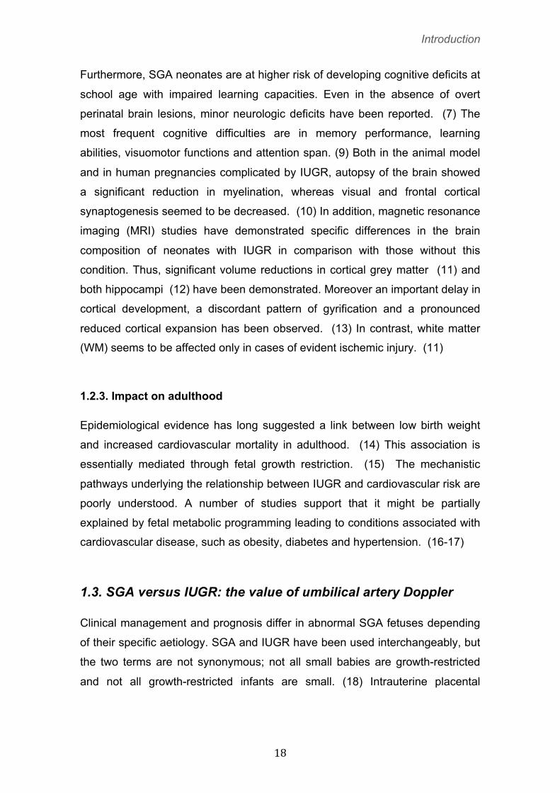
1.3.1. Physiopatology of UA Doppler abnormalities
The vasoconstriction phenomena of the tertiary stem villi are considered
responsible for the up river modifications in the normal wave flow velocity of the
UA, with a decrease in the diastolic velocities and an increase in the resistance
and impedance indices. (20) Umbilical artery Doppler resistance index only
increase when approximately 30% foetal villous vasculature is abnormal. At the
end of the spectrum, a pattern of absent or reversed end-diastolic flow
velocities (EDFV) appears when 60–70% of the villous vasculature is damaged.
(21) Accordingly, clinical studies on IUGR have established that UA impedance
progressively increases and, in advanced stages of placental histological and
functional damage, diastolic velocities become absent or even reversed (Figure
2).
Figure 2. Site of insonation of the umbilical artery (a). Progressive waveform patterns with advancing severity: normal UA waveform(b), increased impedance to flow(c), absent end-diastolic flow (d) and reversed end-diastolic flow (e). (20)
UA Doppler index correlate with foetal levels of glucose, aminoacids and blood
gases and, therefore, it could be considered a surrogate measurement of
placental functionality. (22).
1.3.2. Outcome of SGA with abnormal umbilical artery Doppler
There is an extensive body of evidence that those SGA fetuses with abnormal
UA flow are at higher risk of adverse perinatal outcome than those with normal
Introduction �
� �
flow. (23-25) This association is independent of gestational age at delivery.
(23,26) At the end of the spectrum, absent and reversed end-diastolic velocities
are independently correlated with perinatal morbidity and mortality, with a
relative risk of 4.0 and 10.6, respectively. (27) In addition, this pattern is also
associated with increased risk of long-term abnormal neurodevelopment. (28)
A meta-analysis, including nearly 7000 high-risk pregnancies monitored with UA
Doppler, has demonstrated a significant improvement of a number of perinatal
outcomes, with an overall reduction of perinatal mortality of 30%. (29) Thus,
there is a sound basis for the use of umbilical artery Doppler as a risk-
discriminator tool in the management of SGA fetuses.
1.3.3. Outcome of SGA with normal umbilical artery Doppler
While abnormal umbilical Doppler is associated with adverse perinatal and
neurodevelopmental outcome, (29) small fetuses with normal umbilical artery
Doppler have been traditionally considered to represent one end of the normal
size spectrum and the importance of managing them completely differently from
true IUGR babies has been stressed. (30)
However, recent evidence suggests that a substantial proportion of these
fetuses have true growth restriction.� (6) Studies over the last decade have
provided evidence that perinatal outcome may be significantly poorer in SGA
fetuses. (25,31-32) Trudinguer et al (23) showed that SGA fetuses with normal
UA Doppler born beyond 34 weeks still had a mean neonatal intensive care unit
stay of 5 days. It has also been reported a 3% mortality rate and a 6% rate of
intraventricular hemorrhage in a series of SGA fetuses with normal Doppler.
(24) In accordance with these results, other studies found that one-quarter of
small-for- gestational-age babies with normal umbilical Doppler were
hypoglycemic, and even after adjusting for gestational age the odds of SGA
with normal Doppler requiring prolonged newborn care was two-fold greater
than the general population. (25) Therefore, morbidity and mortality among
these fetuses could be increased in relation with normally grown fetuses. (6)
Introduction �
� ��
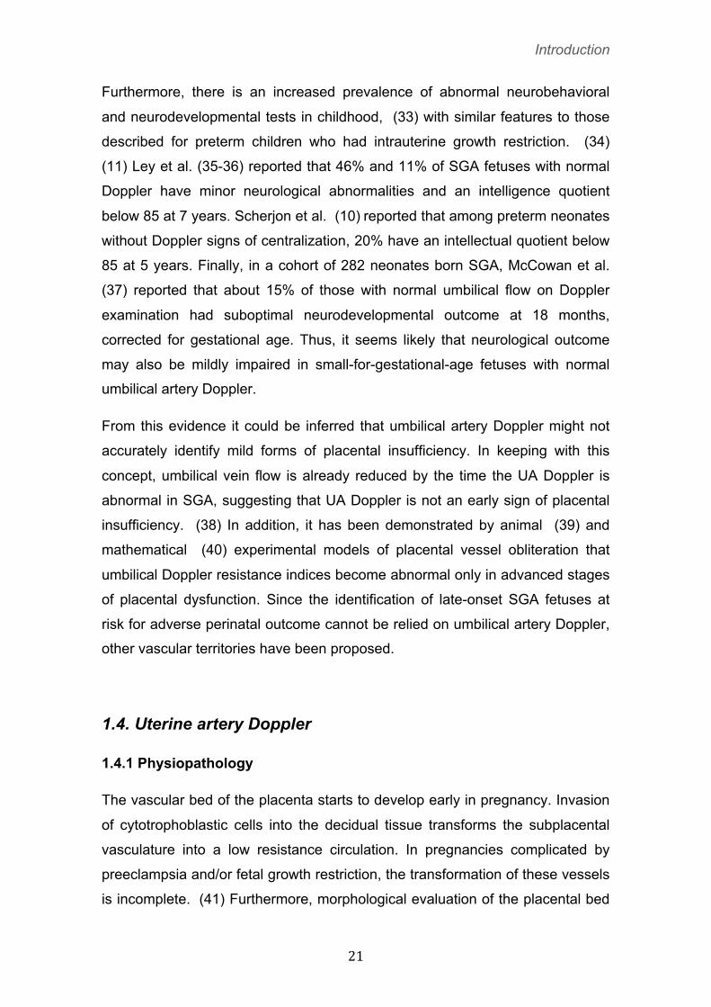
Furthermore, there is an increased prevalence of abnormal neurobehavioral
and neurodevelopmental tests in childhood, (33) with similar features to those
described for preterm children who had intrauterine growth restriction. (34)
(11) Ley et al. (35-36) reported that 46% and 11% of SGA fetuses with normal
Doppler have minor neurological abnormalities and an intelligence quotient
below 85 at 7 years. Scherjon et al. (10)�reported that among preterm neonates
without Doppler signs of centralization, 20% have an intellectual quotient below
85 at 5 years. Finally, in a cohort of 282 neonates born SGA, McCowan et al.
(37) reported that about 15% of those with normal umbilical flow on Doppler
examination had suboptimal neurodevelopmental outcome at 18 months,
corrected for gestational age. Thus, it seems likely that neurological outcome
may also be mildly impaired in small-for-gestational-age fetuses with normal
umbilical artery Doppler.
From this evidence it could be inferred that umbilical artery Doppler might not
accurately identify mild forms of placental insufficiency. In keeping with this
concept, umbilical vein flow is already reduced by the time the UA Doppler is
abnormal in SGA, suggesting that UA Doppler is not an early sign of placental
insufficiency. (38) In addition, it has been demonstrated by animal (39) and
mathematical (40) experimental models of placental vessel obliteration that
umbilical Doppler resistance indices become abnormal only in advanced stages
of placental dysfunction. Since the identification of late-onset SGA fetuses at
risk for adverse perinatal outcome cannot be relied on umbilical artery Doppler,
other vascular territories have been proposed.
1.4. Uterine artery Doppler
1.4.1 Physiopathology
The vascular bed of the placenta starts to develop early in pregnancy. Invasion
of cytotrophoblastic cells into the decidual tissue transforms the subplacental
vasculature into a low resistance circulation. In pregnancies complicated by
preeclampsia and/or fetal growth restriction, the transformation of these vessels
is incomplete.� (41) Furthermore, morphological evaluation of the placental bed

Introduction �
� ��
has shown that this lack of vessel transformation is associated with increased
vascular impedance of the uterine artery.� (42)
Doppler velocimetry of the uterine arteries reflects vascular impedance on the
maternal side of the placental circulation. Increasing impedance, due to
anomalous invasion of cytotrophoblastic cells into the decidual tissue of the
placental bed and defective remodelling of the spiral arteries, is reflected in
decreasing diastolic blood flow velocities and/or persistent early diastolic notch
in the uterine artery blood flow waveform. (43) (Figure 3).
Figure 3. Site of insonation of the uterine artery with color Dopper at the crossover of the iliac artery (a). Normal (b) and abnormal (increased impedance flow with early diastolic notching) (c) waveforms. (20)
1.4.2. Uterine Doppler as a screening tool for placental-related diseases
Increased pulsatility index (PI) and/or persistent notches in the blood flow
waveform can be detected early in the second trimester. Uterine artery (UtA)
Doppler evaluation has been proposed as a screening tool for early-onset IUGR
and has been associated with detection rates of approximately 25% and 75%,
for a false-positive rate of 10%.� (44-45) These sensitivities are higher for the
prediction of early-IUGR associated with preeclampsia. Different strategies
combining maternal risk factors, blood pressure and biochemical markers have
been published, with detection rates greater than 90% for early-onset
preeclampsia-associated IUGR. (46-47)
1.4.3. Uterine artery in the management of SGA
In pregnancies complicated by pre-eclampsia, increased uterine artery vascular
impedance in the third trimester has been correlated with small-for-gestational-
Introduction �
� ��
age (SGA) newborns, delivery by caesarean section, premature delivery,
admission to a neonatal intensive care unit (NICU) and maternal complications.
(48-49) In late-onset IUGR, abnormal uterine artery Doppler has shown to be
comparable to umbilical artery Doppler as a predictor of adverse outcome in
late-onset IUGR. (32,43,50) Futhermore, it has been suggested that UtA
Doppler could provide additional value to the umbilical and cerebral arteries in
predicting the occurrence of adverse perinatal outcome (32,51)
Despite these encouraging reports, uterine artery Doppler is not routinely used
in the evaluation of pregnancies suspected for fetal growth restriction.
Futhermore, a meta-analysis (52) showed a limited capacity of this parameter
to predict perinatal mortality.
1.5. Middle cerebral artery Doppler
1.5.1 Pathophysiology
Several studies in IUGR fetuses show redistribution of fetal blood flow in
response to hypoxia. Selective vasoconstriction results in a primary reduced
perfusion of the gastrointestinal organs, kidneys and skin. On the other hand,
blood supply to the brain, spleen and heart is increased to ensure adequate
oxygenation. (53) This vasodilatation leads to an increase in the diastolic
velocities and a decrease in the resistance and impedance indices in the
vessels supplying these organs. This centralisation of the fetal circulation with
the described brain sparing effect represents an essential development in the
hemodynamic adaptation of the fetus to an increasingly hypoxic environment.
(28,54) Accordingly, longitudinal monitoring of IUGR demonstrated a
progressive decrease in the impedance indices of the brain arteries. (55) In
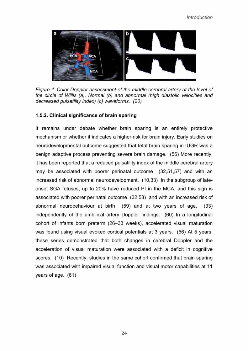
clinical practice the key sign to identify brain vasodilation is the demonstration
of a reduction in the pulsatility index of the middle cerebral artery (MCA).
Doppler evaluation of this artery has become a standard parameter for fetal
assessment and management. (Figure 4)
Introduction �
� ��
Figure 4. Color Doppler assessment of the middle cerebral artery at the level of the circle of Willis (a). Normal (b) and abnormal (high diastolic velocities and decreased pulsatility index) (c) waveforms. (20)
1.5.2. Clinical significance of brain sparing
It remains under debate whether brain sparing is an entirely protective
mechanism or whether it indicates a higher risk for brain injury. Early studies on
neurodevelopmental outcome suggested that fetal brain sparing in IUGR was a
benign adaptive process preventing severe brain damage. (56) More recently,
it has been reported that a reduced pulsatility index of the middle cerebral artery
may be associated with poorer perinatal outcome (32,51,57) and with an
increased risk of abnormal neurodevelopment. (10,33) In the subgroup of late-
onset SGA fetuses, up to 20% have reduced PI in the MCA, and this sign is
associated with poorer perinatal outcome (32,58) and with an increased risk of
abnormal neurobehaviour at birth (59) and at two years of age, (33)
independently of the umbilical artery Doppler findings. (60) In a longitudinal
cohort of infants born preterm (26–33 weeks), accelerated visual maturation
was found using visual evoked cortical potentials at 3 years. (56) At 5 years,
these series demonstrated that both changes in cerebral Doppler and the
acceleration of visual maturation were associated with a deficit in cognitive
scores. (10) Recently, studies in the same cohort confirmed that brain sparing
was associated with impaired visual function and visual motor capabilities at 11
years of age. (61)
Introduction �
� ��
1.6. Anterior cerebral artery Doppler
1.6.1. Pathophysiology
In animal models under hypoxia, blood supply differs substantially between
different brain areas depending on the gestational age and the type and
severity of the insult. (62) Intra-brain regional hemodynamic redistribution could
be one of the mechanisms behind the existence of a regional hierarchy in brain
deterioration, whereby certain areas are more susceptible than others to
hypoxic damage. (63) Frontal areas are philogenetically recently acquired and,
therefore, maturation and myelinization processes of these areas occur late in
the fetal development, making these structures vulnerable during a long period.
(64) Long-term outcome of growth-restricted fetuses revealed a specific profile
of neurocognitive difficulties with poor executive functioning, inflexibility-
creativity and language problems, (9) as signs of frontal lobe dysfunction. (65)
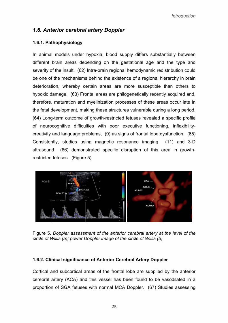
Consistently, studies using magnetic resonance imaging (11) and 3-D
ultrasound (66) demonstrated specific disruption of this area in growth-
restricted fetuses. (Figure 5)
� Figure 5. Doppler assessment of the anterior cerebral artery at the level of the circle of Willis (a); power Doppler image of the circle of Willis (b)
1.6.2. Clinical significance of Anterior Cerebral Artery Doppler
Cortical and subcortical areas of the frontal lobe are supplied by the anterior
cerebral artery (ACA) and this vessel has been found to be vasodilated in a
proportion of SGA fetuses with normal MCA Doppler. (67) Studies assessing
Introduction �
� ��
the temporal evolution of brain arteries in IUGR fetuses suggest that the ACA
shows vasodilatory changes earlier than the MCA (67) (62) Established
vasodilation of the MCA seemed to coincide with a decline in the relative
perfusion to the frontal lobe in relation with other regions such as the basal
ganglia. Furthermore, tissue perfusion studies in fetuses with IUGR suggest that
increased frontal perfusion occurs weeks before the MCA PI both in early and
late-onset IUGR. (59,68)
1.7. Cerebro-placental ratio
1.7.1 Pathophysiology
The unique arrangement of the fetal circulation allows afterload to affect
ventricles individually. Accordingly, relative contributions of individual ventricles
to the common cardiac output may change with individual alterations in
afterload. (69) In this setting, improved oxygen delivery to the brain is thought
to result from decreased left ventricular afterload. The cerebroplacental ratio
(CPR) quantifies redistribution of cardiac output by dividing Doppler indices
from representative cerebral and fetoplacental vessels: the MCA and the UA.
(70)
1.7.2. Clinical significance
Studies on animal models suggest that the CPR most closely reflects acute
changes in pO2 (71) than its individual components, that is the UA and MCA.
Finally, as supported by an increasing number of studies, cerebroplacental ratio
is an earlier and more sensitive predictor for adverse outcome than either the
middle cerebral artery or umbilical artery alone, both in severe (70,72-74) as in
mild forms of IUGR, (75) and it correlates better with adverse outcomes. (76)
1.8. Relevance and justification of the research project
The studies included in this project are part of a research line on fetal brain
circulation in growth-restricted fetuses and its prediction capacity for
neurological damage.
Introduction �
� ��
In early-onset IUGR, the sequence of changes in Doppler indexes has been
described. (27,55,75,77) However, in late-onset cases, these longitudinal trends
have not been investigated before. This information is essential to ascertain
whether there is a sound basis to use these parameters in the monitoring of
late-onset SGA. The first project was aimed at determining the longitudinal
trends and rate of conversion of normal to abnormal Doppler pulsatility indexes
of the uterine, umbilical and middle cerebral artery in late-onset SGA fetuses
from diagnosis to delivery.
Many studies have found associations between premature growth restricted
infants and later behavioral, (11,34,78) sensorial (10,79) and cognitive
dysfunctions. (9,65,80,81) Long-term outcome of preterm growth-restricted
infants has revealed a specific profile of neurocognitive difficulties with poor
executive functioning, inflexibility-creativity and language problem. (9,80) Some
studies have correlated these difficulties during childhood with behavioral
disruptions already present in the neonatal period (11,12,34) a time when
environmental influences are still minimal. Neurobehavior is a neurological
function that in the neonate is mainly related to neurological maturation. (82) In
low-risk preterm infants, individualized developmental interventions reportedly
prevent short-term neurobehavioral dysfunction. (83) Some studies have also
reported long-term cognitive disadvantages for full-term SGA, (84-85) but there
is no information on neurobehavioral performance of full-term SGA babies with
normal placental function. The purpose of the second project was to evaluate
the neonatal neurobehavior performance of full-term SGA fetuses with normal
placental function.
Some studies on IUGR fetuses have demonstrated a regional redistribution of
blood supply within the brain, (62) which contributes to the regional hierarchy in
brain deterioration, making certain areas more susceptible than others to
hypoxic damage. The frontal brain lobe, mainly supplied by the ACA, is one of
these highly susceptible structures in chronically hypoxic infants. The study of
this vessel may be superior to the standard parameter used to detect brain
redistribution, the MCA, detecting those fetuses at an early stage of brain
hypoxia. This may allow earlier interventions such as closer monitoring and
timely delivery. The third project was aimed to evaluate whether ACA Doppler

Introduction �
� ��
investigation is superior to middle cerebral artery Doppler investigation in the
prediction of adverse perinatal outcome in term SGA fetuses with normal
umbilical Doppler.

General hypothesis�
� ��
�
�
�
�
�
�
�
�
�
2) GENERAL HYPOTHESIS �

General hypothesis�
� ��
2.1. Conceptual hypothesis
• A proportion of late-onset growth restricted fetuses with normal placental
function have been exposed to mild in utero hypoxia.
2.2. Secondary hypothesis
• Longitudinal monitoring of late-onset growth restricted fetuses show that
Doppler pulsatility indices of the anterior cerebral artery (ACA), middle
cerebral artery (MCA) and cerebro-placental ratio (CPR) present earlier
and more frequent modifications than umbilical artery (UA) and maternal
uterine arteries (AUT).
• Late-onset growth restricted fetuses with normal umbilical artery Doppler,
have worse perinatal outcomes as well as a suboptimal neonatal
neurobehavior.
• Late-onset growth restricted fetuses with signs of cerebral hemodynamic
redistribution present neurological disrruptions affecting neonatal
neurobehavior.

�
� ��

General objectives�
� ��
3) GENERAL OBJECTIVES


General objectives�
� ��
3.1. Main objective
• To explore the temporal evolution of Doppler parameters in late-onset
growth restricted fetuses and to assess its association with adverse
perinatal and neurobehavioral outcome.
3.2. Specific objectives
• To describe during late pregnancy the trend of the Doppler longitudinal
pulsatility indices of the middle cerebral artery, umbilical and maternal
uterine arteries in late-onset growth restricted fetuses.
• To assess the neurobehavior and perinatal outcome of fetuses with an
estimated fetal weight less than p10 and normal umbilical artery Doppler.
• To assess the neurobehavior and perinatal outcome of late-onset growth
restricted fetuses with signs of intrauterine brain Doppler redistribution
defined by the anterior and middle cerebral arteries.

� ��

Methods
� ��
4) METHODS

Methods
� ��
4.1. Study design
Between November 2007 and August 2009, a prospective longitudinal cohort
study was performed in the Fetal Growth Unit of the Maternal-Fetal Medicine
Department of Hospital Clinic of Barcelona.
4.2. Study population
Study population was divided in two cohorts.
a) Case cohort
a.1) Inclusion criteria:
• Consecutive suspected SGA singleton fetuses at routine third trimester
ultrasound (30-36 weeks), with an estimated fetal weight <10 percentile
according to local standards (1).
• Normal admission Doppler examination, with mean uterine artery
pulsatility index (PI) < 95th percentile (86), umbilical artery pulsatility
index < 95th percentile (87), middle cerebral artery pulsatility index >5th
percentile and cerebroplacental ratio >5th percentile (76).
a.2) Exclusion criteria:
• Congenital defects, chromosomal abnormalities and infections,
birthweight > 10th percentile according to local standards (1).
b) Control cohort
b.1) Inclusion criteria:
• Adequate for gestational age controls were defined as singleton
neonates with birthweight between the 10th and 90th percentile
according to local standards. (1)
Controls were selected from our general population, with previous adequate
ultrasound estimated fetal weigth, individually matched with cases for

Methods �
� �
gestational age at inclusion (± 1 week), corrected by first trimester ultrasound.
(88)
b.2) Exclusion criteria:
• Congenital defects, chromosomal abnormalities and infections.
4.3. Sample size
Based on results of previous studies, the number of subjects that should be
included in the study to detect differences equal to or greater than 0.4 points in
the result of the Neonatal Behaviour Assesment Scale (NBAS) test and
differences equal to or greater than 15% for the onset of intrauterine cerebral
redistribution, assuming an alpha error of 5% and a beta error of 20%, was 102
children per branch.
Assuming an acceptance rate of 90% we aimed to include in the initial sample
116 patients in each branch.
4.4. Predictive variables
1. Maternal age at delivery; Continuous (years)
2. Smoking consumption during pregnancy; Continuous (cigarettes / day)
3. Maternal weight at the beginning of pregnancy; Continuous (kg)
4. Maternal height; Continuous (cm)
5. Maternal ethnicity; Categorical (Europe, Africa, South America, North
Africa, Asia, Others)
6. Parity (number of births> 22 weeks); Discrete
7. History of preeclampsia (89) Binary (Yes / No)
8. History of gestational hypertension (89)(Binary (Yes / No)
9. Previous history of intrauterine growth restriction in previous pregnancies
(birth weight below the 10th percentile (1); Binary (Yes / No)

Methods �
� ��
10. Gestational age at inclusion; Continuous (weeks)
11. Diastolic blood pressure at inclusion; Continuous (mmHg)
12. Sistolic blood pressure at inclusion; Continuous (mmHg)
13. Pulsatility index of uterine arteries at each examination (86); Continuous
(standardized for gestational age)
14. Pulsatility index of umbilical artery at each examination (87); Continuous
(standardized for gestational age)
15. Pulsatility index of middle cerebral artery at each examination (76);
Continuous (standardized for gestational age)
16. Pulsatility index of anterior cerebral artery at each examination (66);
Continuous (standardized for gestational age)
17. Cerebro-placental ratio at each examination (76); Continuous
(standardized for gestational age )
4.5. Result variables
a) Main result variable:
Neonatal Behavior Assessment Scale (90); Continuous (standardized)
b) Secondary result variables
1. Preeclampsia: Diastolic blood pressure (DBP) ≥ 90mmHg and/or systolic
(SBP) ≥ 140 in 2 separate determinations (≥ 4h), and proteinuria> 300
mg/24 h; Binary (Yes / No)
2. Severe preeclampsia: preeclampsia criteria DBP ≥110 mmHg,
proteinuria> 5 g./24 h, oliguria (<400 ml/24h), neurological symptoms
(cerebral or visual), acute pulmonary edema (radiological and blood gas
test) persistent epigastric pain, abnormal liver function (AST or ALT> 70
IU) Laboratory evidence of hemolysis (LDH> 700 U / L) and / or
thrombocytopenia (<100,000 / mL); Binary (Yes / No)
3. Gestational age at delivery; Continuous (weeks)
4. Intervention for fetal distress; Binary (Yes / No).

Methods �
� ��
5. Cesarean for fetal distress; Binary (Yes / No)
6. Neonatal weight; Continuous (g)
7. Neonatal acidosis (arterial pH <7.10 EB> 12mEq / L), Binary (Yes / No)
8. Perinatal mortality (> 22 weeks gestation - <28 days postpartum), Binary
(Yes / No)
9. Admission length in Neonatal Intensive Care Unit, Continuous (days)
10. Significant neonatal morbidity (seizures, intraventricular hemorrhage>
grade III, periventricular leukomalacia, hypoxic ischemic encephalopathy,
abnormal electroencephalogram, necrotizing enterocolitis, acute renal
failure (serum creatinine> 1.5 mg / dL) or heart failure (requiring inotropic
agents), Binary (Yes / No)
4.6. Ethical approvement
The research protocol was approved by the Local Ethics Committee. Informed
consent complies with the provisions of the RD 561/1993. A copy of the
approval of the CEIC, and patient or controls informed consent is given in the
annexes. (Annex 1)
4.7. Study protocol
1. Patients were recruited at routine third trimester ultrasound (30-36 weeks).
2. In those patients meeting the inclusion criteria, a study information sheet was
given to those agreeing to participate (Annex 2).
3. At a first visit within 7 days of inclusion, variables previously defined were
collected. (Annex 3)
4. From diagnosis to delivery, a biweekly ultrasound examination was
scheduled. Prenatal Doppler was performed using an Acuson Antares Premium
Edition (Siemens, Mountain View, CA) ultrasound machine equipped with a
2.3–4 MHz transabdominal transducer. A high pass wall filter of 70 Hz was used
to avoid artefacts.
Methods �
� ��
Uterine arteries were examined transabdominally. The probe was placed on the
lower quadrant of the abdomen, angled medially, using color Doppler to identify
the uterine artery at the apparent crossover with the external iliac artery.
Measurements were taken approximately at 1 cm distal to the crossover point.
The pulsatility index of the left and right arteries were measured, and the mean
index was calculated.
The umbilical artery Doppler flow spectrum was recorded from a free-floating
portion of the umbilical cord.
The middle cerebral artery Doppler was recorded in a transverse view of the
fetal brain, with the Doppler gate placed on the vessel 1 cm distal from the circle
of Willis.
For the first segment of ACA, the Doppler gate was placed immediately after the
origin of the ACA from internal carotid artery.
The cerebroplacental ratio was calculated as MCA PI / UA PI.
In these vessels, once it had been ensured that the angle was less than 30º, the
pulsed Doppler gate was placed over the whole width of the vessel. Angle
correction was then applied and the signal updated until three similar
consecutive waveforms were obtained in the absence of fetal movements and
voluntary maternal suspended breathing. All cases had a Doppler examination
within 7 days of delivery.
Either the MCA PI, the ACA PI and CPR values below the 5th centile were
considered indicative of cerebral blood flow redistribution and were reported as
abnormal. Values over the 95th centile were considered reported as abnormal
for both UA PI and mean uterine artery PI.
5. Labour induction was performed by cervical ripening with prostaglandins for
cases when (i) at 37 weeks of gestation there was an estimated fetal weight
below the 3rd centile; or (ii) at 40 weeks of gestation when estimated fetal weight
was below the 10th centile; (iii) at 37 weeks with abnormal umbilical artery
Doppler. Cases were managed at the discretion of an attending senior
obstetrician, following standard management guidelines, who was blinded to the
cerebral Doppler. A staff obstetrician assisted all the deliveries.
Methods �
� ��
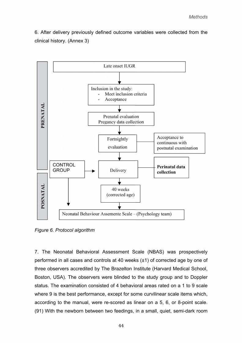
6. After delivery previously defined outcome variables were collected from the
clinical history. (Annex 3)
Figure 6. Protocol algorithm
7. The Neonatal Behavioral Assessment Scale (NBAS) was prospectively
performed in all cases and controls at 40 weeks (±1) of corrected age by one of
three observers accredited by The Brazelton Institute (Harvard Medical School,
Boston, USA). The observers were blinded to the study group and to Doppler
status. The examination consisted of 4 behavioral areas rated on a 1 to 9 scale
where 9 is the best performance, except for some curvilinear scale items which,
according to the manual, were re-scored as linear on a 5, 6, or 8-point scale.
(91) With the newborn between two feedings, in a small, quiet, semi-dark room

Methods �
� ��
at a temperature of between 22 to 27°C and in the presence of at least one
parent, the following areas were analyzed: habituation (habituation to light,
rattle, bell and tactile stimulation of the foot items), motor (which includes
general tone, motor maturity, pull-to-sit, defensive movements and level of
activity) social-interactive (which includes response to visual and acoustic
stimuli), state organization (which includes peak of excitement, rapidity of build-
up, irritability and lability of states). The behavioral items were converted into
percentiles according to normal curve references for our population, (90) and
each area was considered abnormal at a score below the 5th percentile.
8. Controls were recruited, matched for gestational age at delivery and gender,
of babies born at the hospital during the study period. After acceptance of
informed consent, Neonatal Behavior Assesment Scale was performed. The
psychology team was blinded to the study group.
Methods �
� ��
4.8. Specific projects
4.8.1. Project 1: Longitudinal evaluation of fetal hemodynamic
status in late-onset IUGR fetuses.
a) Hypothesis: In late-onset growth restricted fetuses cerebral haemodinamic
redistribution appears earlier and more frequently than umbilical artery (UA) and
maternal uterine arteries (AUT) haemodynamic changes.
b) Objective: To describe during late pregnancy the trend of the Doppler
longitudinal pulsatility indices of the middle cerebral artery, umbilical and
maternal uterine arteries in late-onset growth restricted fetuses without
hemodynamic changes at the time of diagnosis.
c) Study design: propective longitudinal cohort study.
d) Study population: a prospective cohort was created of consecutive
suspected late-onset IUGR singleton babies at routine third trimester ultrasound
(30-36 weeks) with confirmed birth weight < 10 percentile according to local
standards. (1) Only cases presented upon admission with mean uterine artery
pulsatility index (PI) < 95th percentile (86), umbilical artery pulsatility index <
95th percentile (87), middle cerebral artery pulsatility index >5th percentile and
cerebroplacental ratio >5th percentile (76) were included. Exclusion criteria
were congenital malformations (including chromosomopathies and infections)
and the diagnosis of preeclampsia.
e) Statistical analysis: All Doppler parameters were transformed into z-values
according to normative references. The longitudinal changes were analyzed by
Kaplan-Meier survival analysis, in which the endpoint was defined as an
abnormal Doppler value (middle cerebral artery pulsatility index and
cerebroplacental ratio < 5th percentile; umbilical and uterine arteries pulsatility
index > 95th percentile). The McNemar test was used to compare paired group
proportions. Statistical and survival analyses were performed using the
Statistical Package for Social Sciences (SPSS 15.0, SPSS Inc., Chicago, IL)
statistical software.
Longitudinal changes in z-values during the last 10 weeks before delivery were
modeled by means of multilevel analysis, fitting to second-degree polynomials:
Methods �
� ��
α + βt + γt2 , where α, β and γ were the parameters characterizing the
individual fetus and t the days before delivery. These parameters were
calculated assuming its randomly normal distribution in the population (random
effect model), which allows us to assume that both the individuals and the days
to delivery at which the scan was performed are random samples of their
respective populations. Standard errors of these parameters were used to
construct confidence intervals (CI). The software MLwiN 2.1 (Centre for
Multilevel Modelling, University of Bristol, UK) was used for the parameters´
estimation. Repeated measurements at different time points (gestational age) in
the same fetus comprised Level 1 and those in different fetuses comprised
Level 2. Individual regression lines for each variable were calculated for each
fetus and from these the regression lines for the whole group were derived.
4.8.2 Project 2: Evaluation of perinatal outcome and neonatal
neurobehaviour of late-onset IUGR fetuses with normal UA
Doppler versus controls.
a) Hypothesis: Late-onset growth restricted fetuses with normal umbilical
artery Doppler, have worse perinatal outcomes as well as a suboptimal
neonatal neurobehavior.
b) Objective: To assess the neurobehavioral and perinatal outcome of fetuses
with an estimated fetal weight less than p10 and normal umbilical artery
Doppler.
c) Study design: propective cohort study.
d) Study population: patients were divided in two cohorts:
A cohort was created of consecutive, suspected SGA; singleton infants
delivered at term with confirmed birth weights of <10th percentile according to
local standards. (1) Exclusion criteria included congenital malformations
(including chromosomopathies and infections) and umbilical artery pulsatility
index values of >95th percentile. (87)
Methods �
� ��
Control subjects were defined as singleton term infants with size appropriate for
gestational age (AGA) (>10th percentile, according to local standards (1)) and
were sampled from our general neonatal population during the same period;
they were matched with case subjects according to the date of delivery (±7
days).
e) Statistical analysis: Student’s t test for independent samples and Pearson’s
�χ2 test were used to compare quantitative and qualitative data, respectively.
Multivariate analyses were conducted through multivariate analysis of
covariance in which a model was run for each different set of skills (attention,
habituation, motor, state organization, state regulation, and autonomic nervous
system), with the study group included as a factor and the following variables as
covariates: (1) smoking during pregnancy (no smoking, 1–9 cigarettes per day,
or �10 cigarettes per day); (2) maternal BMI at booking; (3) low socioeconomic
level (routine occupations, long-term unemployment, or never worked; United
Kingdom National Statistics Socio-economic Classification); (4) onset of labor
(spontaneous versus induction); (5) mode of delivery (vaginal delivery versus
cesarean section); (6) number of doses of epidural anesthetic medication
(bupivacaine, 1.2–1.8 mg) during labor (none, 1–3 doses, or �4 doses); (7)
gestational age at delivery; (8) postnatal age (in days) at evaluation; and (9)
gender.
For each model, assumptions for the multivariate analysis of covariance were
checked and the multivariate significance of the F value was assessed with
Wilks’ P value. Also, the �Χ2 value was provided, which could be interpreted as
the proportion of the total variance of the dependent variables explained by
each factor and covariate. To rule out an expectation bias, the association
between birth weight and NBAS scores was evaluated through Pearson
correlation within each study group.
The software package SPSS 15.0 (SPSS, Chicago, IL) was used for the
statistical analyses.
Methods �
� ��
4.8.3) Project 3: Evaluation of the anterior and middle cerebral
arteries for the prediction of perinatal outcome and neonatal
neurobehavior in late-onset IUGR fetuses with normal UA
Doppler.
a) Hypothesis: Late-onset growth restricted fetuses with signs of cerebral
hemodynamic redistribution present neurological alterations affecting neonatal
neurobehavior as a result of secondary damage due to chronic hypoxia during
fetal development.
b) Objective: To assess the neurobehavioral and perinatal outcome of late-
onset growth restricted fetuses with signs of intrauterine brain Doppler
redistribution defined by the anterior and middle cerebral arteries.
c) Study design: prospective cohort study.
d) Study population: A prospective cohort was created of all suspected SGA
fetuses (estimated fetal weight below the 10th centile (1) at a routine third
trimester ultrasound) meeting inclusion and exclusion criteria previously
defined. Cases were classified according to their brain arteries vasodilation at
last examination before delivery into:
Group 1. SGA with MCA vasodilation: defined as SGA fetuses presented MCA
PI values below the 5th centile. (87)
Group 2. SGA with ACA vasodilation: defined as SGA fetuses presented ACA
PI values below the 5th centile. (66)
Group 3. Adequate for gestational age controls: defined as singleton neonates
with birthweight between the 10th and 90th percentile according to local
standards. (1) Controls were selected from our general population, with
previous adequate ultrasound estimated fetal weigth, individually matched with
cases for gestational age at inclusion (± 1 week), corrected by first trimester
ultrasound.
e) Statistical analysis: Student’s t-test and Pearson Chi-squared test or exact
Fisher test were used to compare quantitative and qualitative data, respectively.
Receiver operating characteristic curves evaluated diagnostic performance for

Methods �
� �
adverse perinatal outcome of both arteries. Following standard methodology,
neurobehavioral outcome was adjusted for smoking during pregnancy (no
smoking; 1-9 cigarettes/day; 10+ cigarettes/day), labor induction, mode of
delivery (cesarean section vs. vaginal delivery), gestational age at birth, gender
and postnatal days at evaluation by multiple linear or logistic regression.
Statistical analysis was performed using the SPSS 15.0 (Chicago, IL, USA) and
MedCalc 8.0 (Broekstraat, Belgium) statistical software.

Results
� ��
5) RESULTS

Results
� ��
5.1. Project 1: Longitudinal evaluation of fetal hemodynamic
status in late-onset IUGR fetuses.
The results of this project have been submitted for publication in an
international Journal:
Daniel Oros, Francesc Figueras, Rogelio Cruz-Martinez, Eva Meler, Meritxell
Munmany, Eduard Gratacos. “Longitudinal changes in uterine, umbilical and
cerebral Doppler in late-onset small-for-gestational age fetuses.” Ultrasound in
Obstetrics & Gynaecoly. (submitted)
These results have also been presented at the 19th World Congress of
Ultrasound in Obstetrics and Gynecology (October 2009, Hamburg, Germany):
M. Munmany, F. Figueras, E. Meler, R. Cruz-Martinez, D. Oros, E. Gratacos
“Rate of conversion of normal to abnormal umbilical, middle cerebral and
uterine arteries in late-onset small-for-gestational-age fetuses”. Ultrasound in
Obstetrics & Gynecology 2009; 34 (Suppl. 1): 62 – 176.
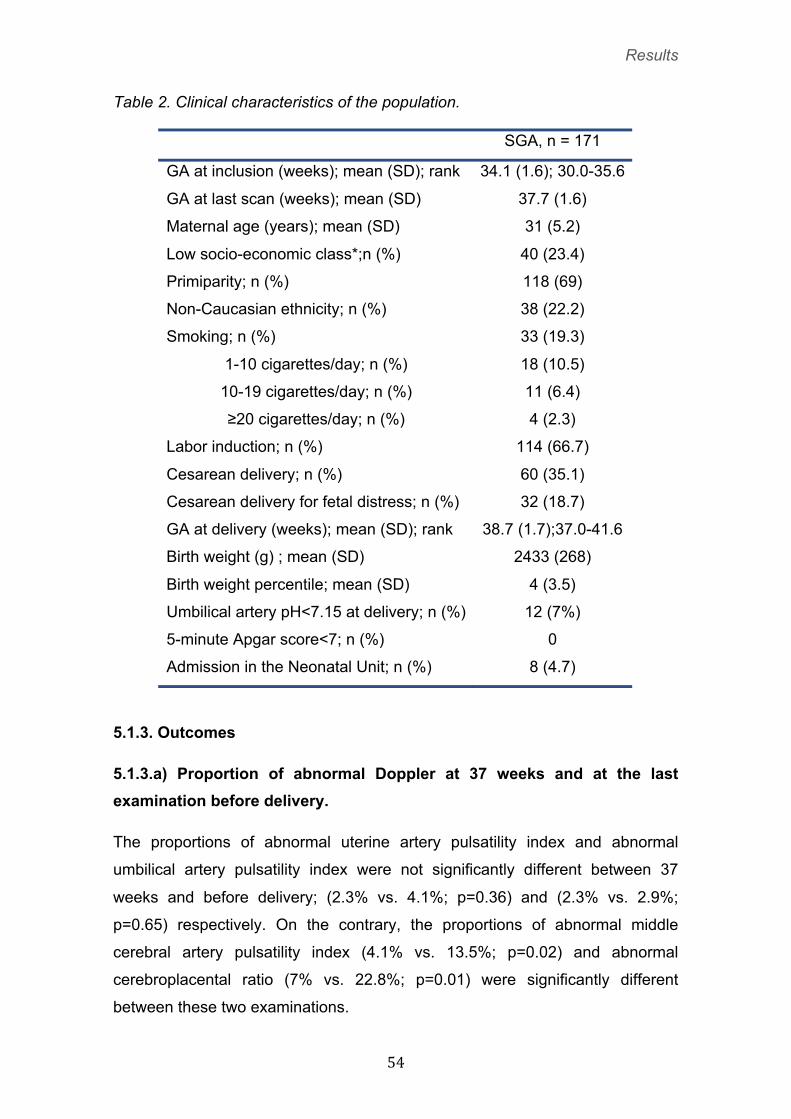
5.1.1. Study population
During the study period a total of 616 scans were performed on 171 SGA
fetuses. The median number of Doppler examinations was 3 (range 2-9). In 124
(62.5%) women more than two examinations were performed.
5.1.2. Clinical characteristics of the population
The mean gestational ages at inclusion and delivery were 34.1 (SD 1.6; range
30.0-35.6) and 38.7 (SD 1.7; range 37-41.6) weeks, respectively. The median
interval between the last examination and delivery was 3 (range 0-6) days.
Table 2 shows the maternal and neonatal clinical characteristics of the
population.
Results
� ��
Table 2. Clinical characteristics of the population.
5.1.3. Outcomes
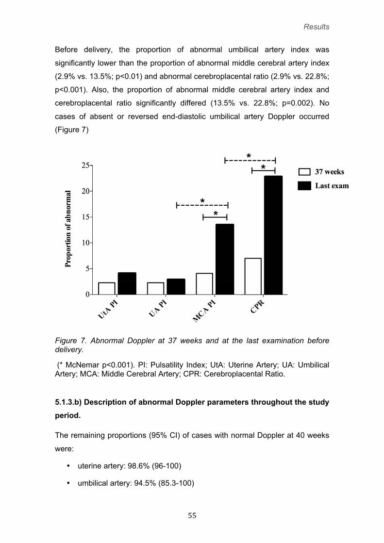
5.1.3.a) Proportion of abnormal Doppler at 37 weeks and at the last examination before delivery.
The proportions of abnormal uterine artery pulsatility index and abnormal
umbilical artery pulsatility index were not significantly different between 37
weeks and before delivery; (2.3% vs. 4.1%; p=0.36) and (2.3% vs. 2.9%;
p=0.65) respectively. On the contrary, the proportions of abnormal middle
cerebral artery pulsatility index (4.1% vs. 13.5%; p=0.02) and abnormal
cerebroplacental ratio (7% vs. 22.8%; p=0.01) were significantly different
between these two examinations.
SGA, n = 171
GA at inclusion (weeks); mean (SD); rank 34.1 (1.6); 30.0-35.6
GA at last scan (weeks); mean (SD) 37.7 (1.6)
Maternal age (years); mean (SD) 31 (5.2)
Low socio-economic class*;n (%) 40 (23.4)
Primiparity; n (%) 118 (69)
Non-Caucasian ethnicity; n (%) 38 (22.2)
Smoking; n (%) 33 (19.3)
1-10 cigarettes/day; n (%) 18 (10.5)
10-19 cigarettes/day; n (%) 11 (6.4)
≥20 cigarettes/day; n (%) 4 (2.3)
Labor induction; n (%) 114 (66.7)
Cesarean delivery; n (%) 60 (35.1)
Cesarean delivery for fetal distress; n (%) 32 (18.7)
GA at delivery (weeks); mean (SD); rank 38.7 (1.7);37.0-41.6
Birth weight (g) ; mean (SD) 2433 (268)
Birth weight percentile; mean (SD) 4 (3.5)
Umbilical artery pH<7.15 at delivery; n (%) 12 (7%)
5-minute Apgar score<7; n (%) 0
Admission in the Neonatal Unit; n (%) 8 (4.7)
Results
� ��
Before delivery, the proportion of abnormal umbilical artery index was
significantly lower than the proportion of abnormal middle cerebral artery index
(2.9% vs. 13.5%; p<0.01) and abnormal cerebroplacental ratio (2.9% vs. 22.8%;
p<0.001). Also, the proportion of abnormal middle cerebral artery index and
cerebroplacental ratio significantly differed (13.5% vs. 22.8%; p=0.002). No
cases of absent or reversed end-diastolic umbilical artery Doppler occurred
(Figure 7)
Figure 7. Abnormal Doppler at 37 weeks and at the last examination before delivery.
(* McNemar p<0.001). PI: Pulsatility Index; UtA: Uterine Artery; UA: Umbilical Artery; MCA: Middle Cerebral Artery; CPR: Cerebroplacental Ratio. �
5.1.3.b) Description of abnormal Doppler parameters throughout the study period.
The remaining proportions (95% CI) of cases with normal Doppler at 40 weeks
were:
• uterine artery: 98.6% (96-100)
• umbilical artery: 94.5% (85.3-100)
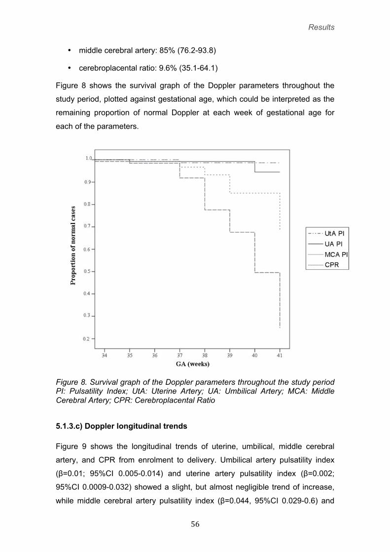
Results
� ��
• middle cerebral artery: 85% (76.2-93.8)
• cerebroplacental ratio: 9.6% (35.1-64.1)
Figure 8 shows the survival graph of the Doppler parameters throughout the
study period, plotted against gestational age, which could be interpreted as the
remaining proportion of normal Doppler at each week of gestational age for
each of the parameters.
Figure 8. Survival graph of the Doppler parameters throughout the study period PI: Pulsatility Index; UtA: Uterine Artery; UA: Umbilical Artery; MCA: Middle Cerebral Artery; CPR: Cerebroplacental Ratio 5.1.3.c) Doppler longitudinal trends
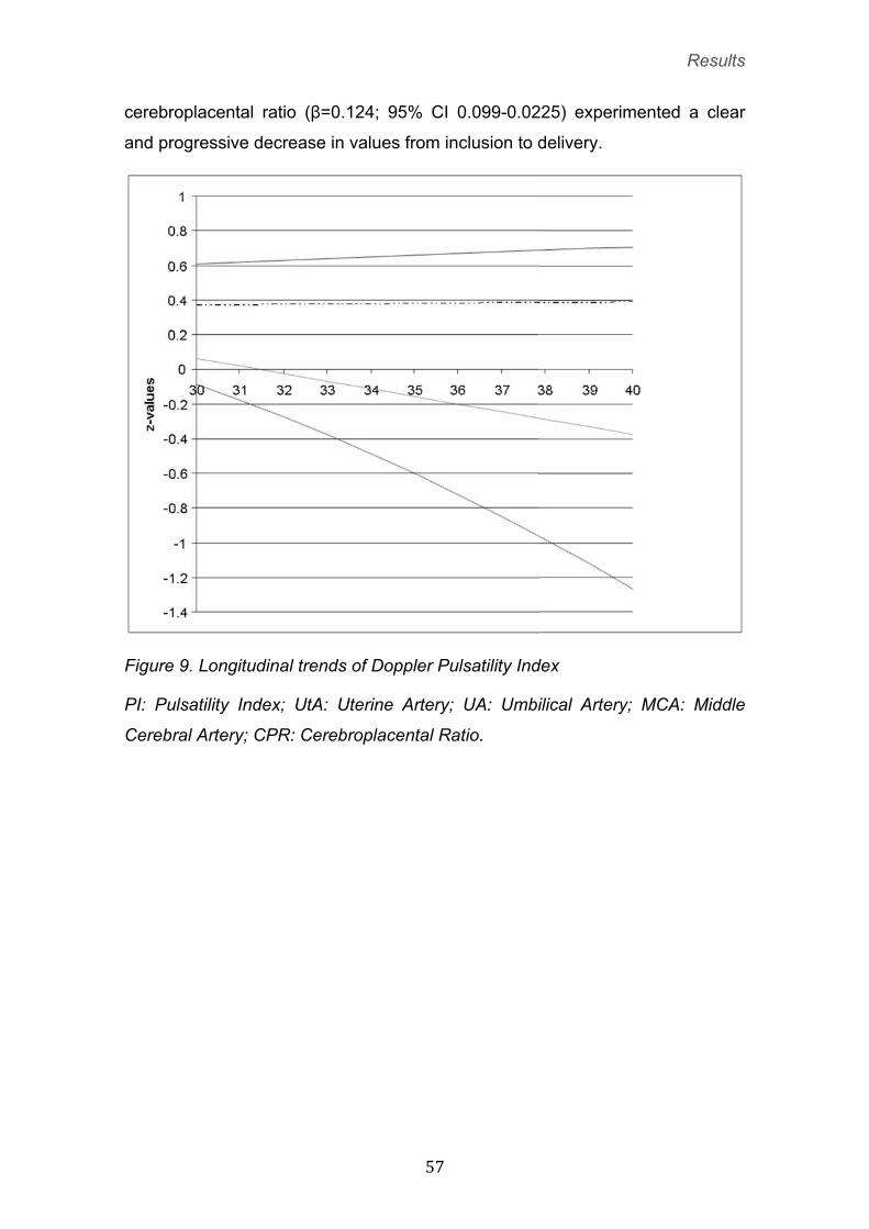
Figure 9 shows the longitudinal trends of uterine, umbilical, middle cerebral
artery, and CPR from enrolment to delivery. Umbilical artery pulsatility index
(β=0.01; 95%CI 0.005-0.014) and uterine artery pulsatility index (β=0.002;
95%CI 0.0009-0.032) showed a slight, but almost negligible trend of increase,
while middle cerebral artery pulsatility index (β=0.044, 95%CI 0.029-0.6) and
Results
� ��
cerebroplacental ratio (β=0.124; 95% CI 0.099-0.0225) experimented a clear
and progressive decrease in values from inclusion to delivery.
Figure 9. Longitudinal trends of Doppler Pulsatility Index
PI: Pulsatility Index; UtA: Uterine Artery; UA: Umbilical Artery; MCA: Middle
Cerebral Artery; CPR: Cerebroplacental Ratio.
Results
� ��
5.2. Project 2: Evaluation of perinatal outcome and neonatal
neurobehaviour of late-onset IUGR fetuses with normal UA
Doppler versus controls.
The results of this project have been published in an international Journal:
Francesc Figueras, Daniel Oros, Rogelio Cruz-Martinez, Nelly Padilla, Edgar
Hernandez-Andrade, Francesc Botet, Carmen Costas, Eduard Gratacos.
“Neurobehavioral performance of full-term small-for-gestational age infants with
normal placental function”. Pediatrics. 2009 Nov;124(5):e934-41. Epub 2009
Oct 26)
These results also have been presented at the 19th World Congress on
Ultrasound on Obstetrics and Gynecology (October 2009, Hamburg, Germany)
F. Figueras, R. Martinez-Cruz, D. Oros, N. Padilla, F. Botet, E. Gratacos.
“Neurobehavioral performance of term small-for-gestational age newborns with
normal umbilical artery Doppler”. Ultrasound in Obstetrics & Gynecology 2009;
34 (Suppl. 1): 1 – 61.).
This communication was selected as a top-10 for oral presentation in plenary
session.
5.2.1 Study population
A final population of 202 infants (102 SGA and 100 AGA) was studied. A total of
were of 216 infants were initialy included. Neurobehavioral assessment visit
was scheduled at corrected age of 40 ±1 weeks. Parents of 6 case subjects and
8 control subjects later declined to participate. The habituation area could not
be assessed for 50 newborns (21 SGA and 29 AGA) because of the absence of
a sleeping period during the evaluation.
5.2.2. Clinical characteristics of the population
Mothers in the SGA group had a lower body weight and more frequently were
from a low socioeconomic level. There were no cases of drug consumption
other than tobacco or alcohol. Table 3 depicts the clinical characteristics of the
Results
� ��
population.
Table 3. Clinical characteristics of the population
AGA SGA p+
(n=100) (n=102)
Primiparity (%) 61 67.6 0.32
Non-caucasian ethnicity (%) 21.2 16 0.34
Maternal age (years) 31.8(4.9) 33.6(4.9) 0.79
Body Mass Index (kg/m2) at booking 23.4 21.9 0.003
Low socioeconomic level* (%) 31 52 0.003
Smoking (%)
Non-Smoking 85 83.3 0.75
1-10 cigarettes/day 12 5.9
11-20 cigarettes/day 2 7.8
> 20 cigarettes/day 1 2.9
SD: Standard deviation; + Student’s t-test for independent samples or Pearson-χ2 test; Routine occupations, long-term unemployment or never worked (UK National Statistics Socio-Economic Classification��
5.2.3. Outcomes
5.2.3.a) Perinatal outcome
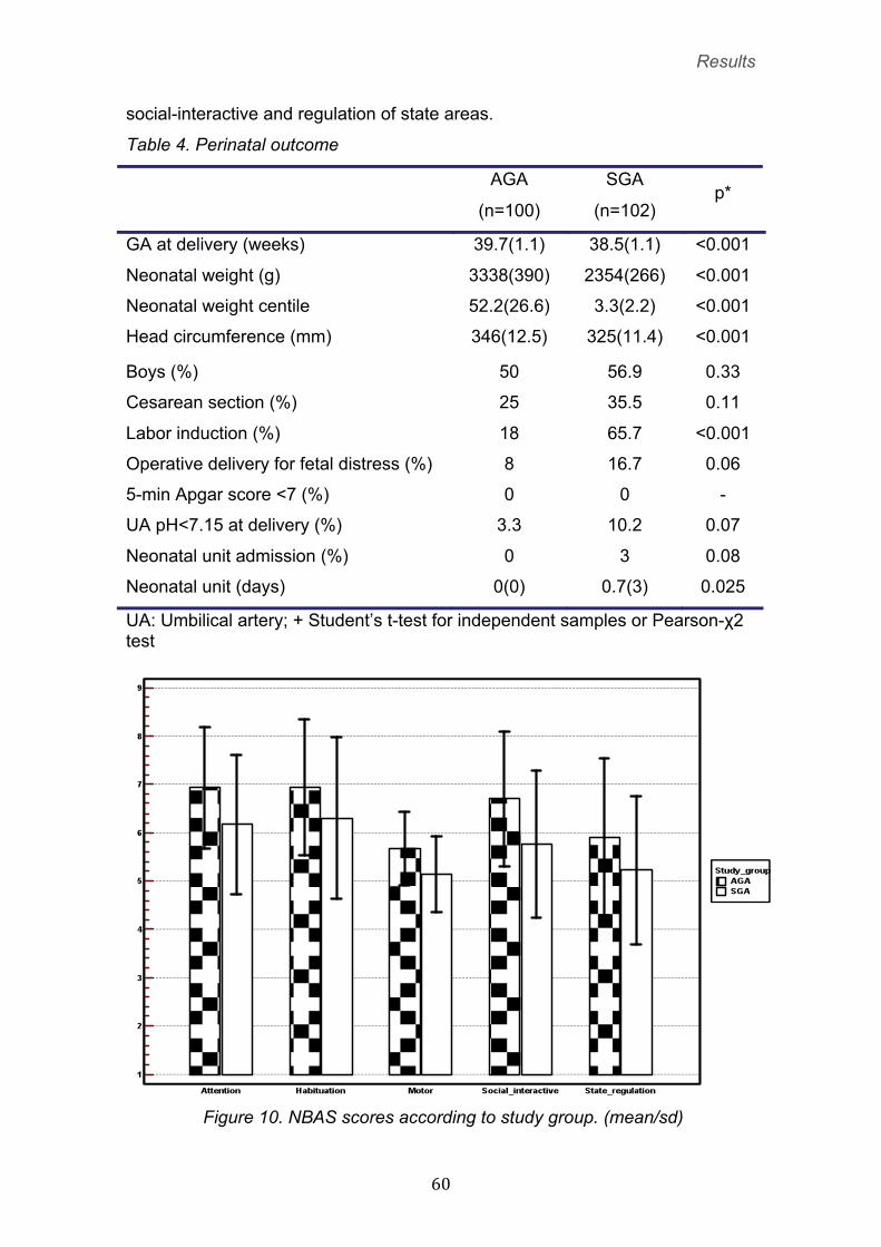
SGA newborns had a lower birth weight and a smaller head circumference.
Delivery in the SGA group was at an earlier gestational age (SGA: 38.5 weeks;
AGA: 39.7 weeks). SGA fetuses were more frequently induced. Operative
delivery because of fetal distress was twice as frequent in the SGA group.
Although no infants in the AGA group were admitted to the neonatal unit 3% in
the SGA group were. (Table 4)
5.2.3.b) Neonatal neurobehavior
Neonatal neurobehavior was assessed at 7.8 (±7.2) in the AGA group and at
10.5 (±9.6) days of life in the SGA groups.
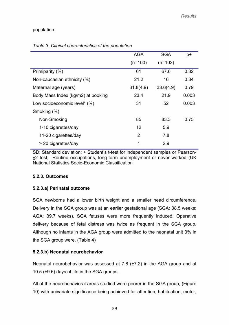
All of the neurobehavioral areas studied were poorer in the SGA group, (Figure
10) with univariate significance being achieved for attention, habituation, motor,
Results
� �
social-interactive and regulation of state areas.
Table 4. Perinatal outcome
AGA SGA
(n=100) (n=102) p*
GA at delivery (weeks) 39.7(1.1) 38.5(1.1) <0.001
Neonatal weight (g) 3338(390) 2354(266) <0.001
Neonatal weight centile 52.2(26.6) 3.3(2.2) <0.001
Head circumference (mm) 346(12.5) 325(11.4) <0.001
Boys (%) 50 56.9 0.33
Cesarean section (%) 25 35.5 0.11
Labor induction (%) 18 65.7 <0.001
Operative delivery for fetal distress (%) 8 16.7 0.06
5-min Apgar score <7 (%) 0 0 -
UA pH<7.15 at delivery (%) 3.3 10.2 0.07
Neonatal unit admission (%) 0 3 0.08
Neonatal unit (days) 0(0) 0.7(3) 0.025
UA: Umbilical artery; + Student’s t-test for independent samples or Pearson-χ2 test
Figure 10. NBAS scores according to study group. (mean/sd)
Results
� ��
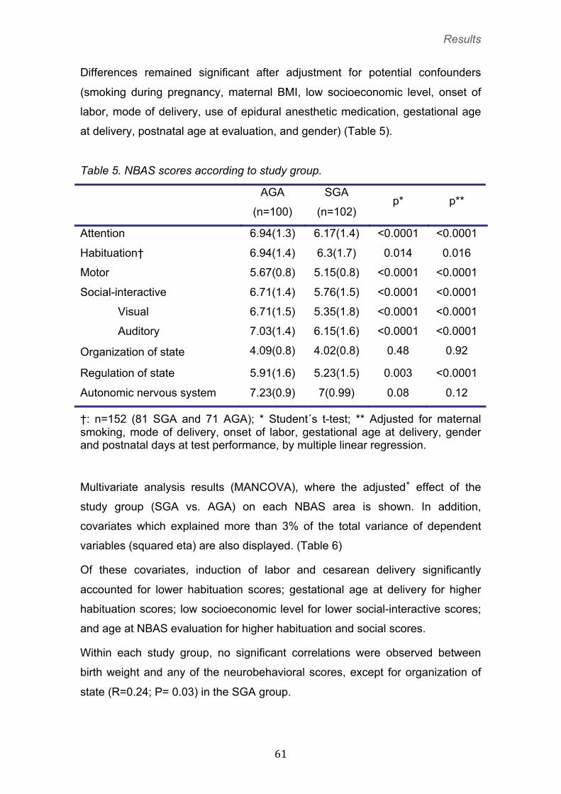
Differences remained significant after adjustment for potential confounders
(smoking during pregnancy, maternal BMI, low socioeconomic level, onset of
labor, mode of delivery, use of epidural anesthetic medication, gestational age
at delivery, postnatal age at evaluation, and gender) (Table 5). �
�
Table 5. NBAS scores according to study group.
AGA SGA
(n=100) (n=102) p* p**
Attention 6.94(1.3) 6.17(1.4) <0.0001 <0.0001
Habituation† 6.94(1.4) 6.3(1.7) 0.014 0.016
Motor 5.67(0.8) 5.15(0.8) <0.0001 <0.0001
Social-interactive 6.71(1.4) 5.76(1.5) <0.0001 <0.0001
Visual 6.71(1.5) 5.35(1.8) <0.0001 <0.0001
Auditory 7.03(1.4) 6.15(1.6) <0.0001 <0.0001
Organization of state 4.09(0.8) 4.02(0.8) 0.48 0.92
Regulation of state 5.91(1.6) 5.23(1.5) 0.003 <0.0001
Autonomic nervous system 7.23(0.9) 7(0.99) 0.08 0.12
†: n=152 (81 SGA and 71 AGA); * Student´s t-test; ** Adjusted for maternal smoking, mode of delivery, onset of labor, gestational age at delivery, gender and postnatal days at test performance, by multiple linear regression.
Multivariate analysis results (MANCOVA), where the adjusted+ effect of the
study group (SGA vs. AGA) on each NBAS area is shown. In addition,
covariates which explained more than 3% of the total variance of dependent
variables (squared eta) are also displayed. (Table 6)
Of these covariates, induction of labor and cesarean delivery significantly
accounted for lower habituation scores; gestational age at delivery for higher
habituation scores; low socioeconomic level for lower social-interactive scores;
and age at NBAS evaluation for higher habituation and social scores.
Within each study group, no significant correlations were observed between
birth weight and any of the neurobehavioral scores, except for organization of
state (R=0.24; P= 0.03) in the SGA group.
Results
� ��
Table 6. Multivariate analysis of covariance results (MANCOVA)
Attention
F Wilk´s lambda
p-value
Squared
Eta (%)
SGA 5.96 0.001 9.2
Smoking 0.65 0.59 1.1
BMI 0.35 0.79 0.6
Low socioeconomic level 0.67 0.57 1.1
Induction 2.14 0.1 3.5
Cesarean section 0.24 0.87 0.4
Epidural anesthetic medication 0.46 0.71 0.8
Gestational age at delivery 0.25 0.86 0.4
Days at NBAS evaluation 1.9 0.12 3.2
Male gender 0.04 0.99 0.1
Habituation†
SGA 4.59 0.002 17
Smoking 1.03 0.39 4.5
BMI 0.73 0.57 3.3
Low socioeconomic level 0.17 0.95 0.8
Induction 2.54 0.046 10.4
Cesarean section 2.44 0.05 10.1
Epidural anesthetic medication 0.32 0.86 1.5
Gestational age at delivery 2.59 0.04 10.6
Days at NBAS evaluation 3.57 0.01 14.1
Male gender 1.26 0.29 5.5
Motor
SGA 5.18 <0.001 12.8
Smoking 1.16 0.33 3.2
BMI 1.13 0.34 3.1
Low socioeconomic level 0.85 0.52 2.4
Induction 1.1 0.36 3
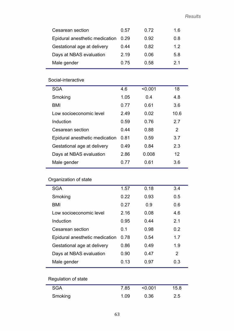
Results
� ��
Cesarean section 0.57 0.72 1.6
Epidural anesthetic medication 0.29 0.92 0.8
Gestational age at delivery 0.44 0.82 1.2
Days at NBAS evaluation 2.19 0.06 5.8
Male gender 0.75 0.58 2.1
Social-interactive
SGA 4.6 <0.001 18
Smoking 1.05 0.4 4.8
BMI 0.77 0.61 3.6
Low socioeconomic level 2.49 0.02 10.6
Induction 0.59 0.76 2.7
Cesarean section 0.44 0.88 2
Epidural anesthetic medication 0.81 0.59 3.7
Gestational age at delivery 0.49 0.84 2.3
Days at NBAS evaluation 2.86 0.008 12
Male gender 0.77 0.61 3.6
Organization of state
SGA 1.57 0.18 3.4
Smoking 0.22 0.93 0.5
BMI 0.27 0.9 0.6
Low socioeconomic level 2.16 0.08 4.6
Induction 0.95 0.44 2.1
Cesarean section 0.1 0.98 0.2
Epidural anesthetic medication 0.78 0.54 1.7
Gestational age at delivery 0.86 0.49 1.9
Days at NBAS evaluation 0.90 0.47 2
Male gender 0.13 0.97 0.3
Regulation of state
SGA 7.85 <0.001 15.8
Smoking 1.09 0.36 2.5
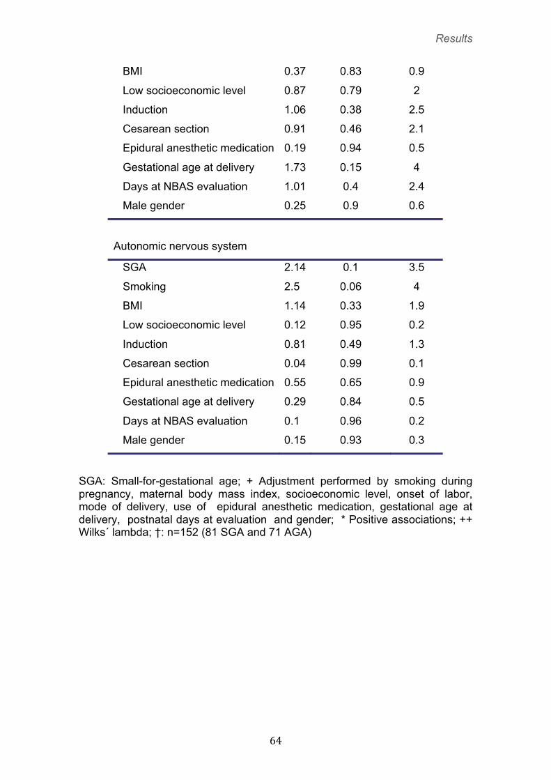
Results
� ��
BMI 0.37 0.83 0.9
Low socioeconomic level 0.87 0.79 2
Induction 1.06 0.38 2.5
Cesarean section 0.91 0.46 2.1
Epidural anesthetic medication 0.19 0.94 0.5
Gestational age at delivery 1.73 0.15 4
Days at NBAS evaluation 1.01 0.4 2.4
Male gender 0.25 0.9 0.6
Autonomic nervous system
SGA 2.14 0.1 3.5
Smoking 2.5 0.06 4
BMI 1.14 0.33 1.9
Low socioeconomic level 0.12 0.95 0.2
Induction 0.81 0.49 1.3
Cesarean section 0.04 0.99 0.1
Epidural anesthetic medication 0.55 0.65 0.9
Gestational age at delivery 0.29 0.84 0.5
Days at NBAS evaluation 0.1 0.96 0.2
Male gender 0.15 0.93 0.3
SGA: Small-for-gestational age; + Adjustment performed by smoking during pregnancy, maternal body mass index, socioeconomic level, onset of labor, mode of delivery, use of epidural anesthetic medication, gestational age at delivery, postnatal days at evaluation and gender; * Positive associations; ++ Wilks´ lambda; †: n=152 (81 SGA and 71 AGA)
Results
� ��
5.3. Project 3: Evaluation of the anterior and middle cerebral
arteries for the prediction of perinatal outcome and neonatal
neurobehavior in late-onset IUGR fetuses with normal UA
Doppler.
The results of this project have been published in two international journals:
Daniel Oros, Francesc Figueras, Rogelio Cruz-Martinez, Nelly Padilla, Eva
Meler, Edgar Hernandez-Andrade, Eduard Gratacos. “Middle versus anterior
cerebral artery Doppler for the prediction of perinatal outcome and neonatal
neurobehavior in term small-for-gestational-age fetuses with normal umbilical
artery Doppler.” Ultrasound Obstet Gynecol (in press)
Benavides-Serralde J.A, Hernández-Andrade E, Figueroa-Diesel H, Oros D,
Feria L.A, Scheier M, Figueras F, Gratacos E. Reference Values for Doppler
Parameters of the Fetal Anterior Cerebral Artery throughout Gestation. Gynecol
Obstet Invest 2010;69:33-39.
These results also have been presented at the 17th World Congress on
Ultrasound on Obstetrics and Gynecology (October 2009, Florence, Italy):
Oros D, Figueras F, Padilla N, Hernandez-Andrade E, Gratacos E. Anterior
cerebral artery improves the prediction of adverse perinatal outcome in small-
for-gestational age fetuses with normal umbilical artery. Ultrasound Obstet
Gynecol. 2007;30: 456-546.)
These results also have been presented at the 30th Congress of the Spanish
Society of Obstetrics and Gynaecology. (Barcelona, June 2009):
Oros D; Figueras F, Cruz-Martínez R, Meler E, Padilla N, Gratacós E. “Utilidad
de las arterias cerebral media y anterior para la predicción del resultado
perinatal y neurocomportamiento neonatal en fetos pequeños para edad
gestacional a término”.
Results
� ��
5.3.1. Study population
A final population of 199 babies (98 SGA and 101 AGA) was analyzed.
Inclusion criteria were fulfilled for 118 SGA fetuses. Seven were excluded
because of birthweight above the 10th centile, none of them had adverse
perinatal outcome. The remaining 111 cases were matched at delivery with
111 adequate-for-gestational-age (AGA) babies. Of these, the parents of 6
cases and 7 controls later declined to participate in the neurobehavioral
evaluation. Finally, in 7 cases and 3 controls the evaluation was not considered
satisfactory by the examiner due to the absence of a sleeping state during the
test.
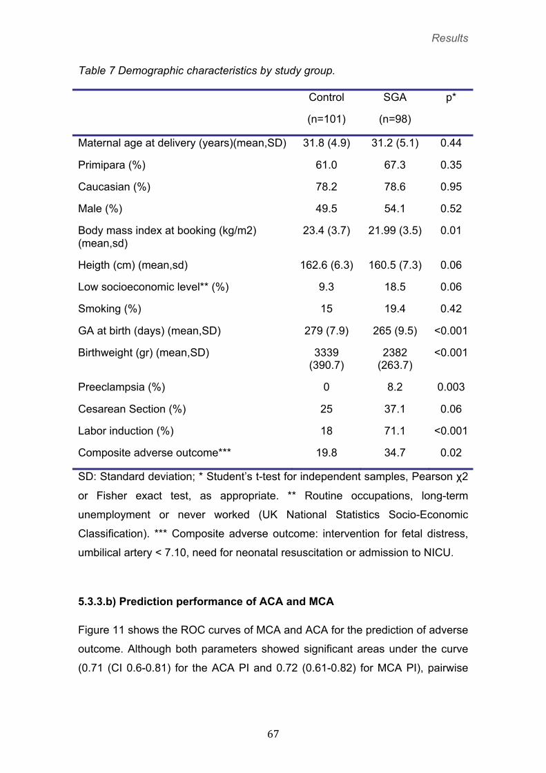
5.3.2. Clinical characteristics of the population
Women in the SGA group had a lower body mass index and showed a non-
significant trend corresponding to a lower socioeconomic level. (Table 7)
5.3.3. Outcomes
5.3.3.a) Perinatal outcome according to the MCA or ACA redistribution.
A total of 28 (29%) and 17 (17%) SGA fetuses had middle and anterior artery
Doppler redistribution, respectively. It is of note that in 14 cases MCA and ACA
simultaneously showed redistribution. Whereas ACA-redistributed fetuses had a
lower birthweight than non ACA-redistributed, no differences were observed
between MCA-redistributed and non ACA-redistributed fetuses. Both MCA and
ACA redistribution accounted for significant differences in cesarean section,
and only the MCA significantly differentiated cases at-risk for fetal distress.
(Table 8)
Results
� ��
Table 7 Demographic characteristics by study group.
Control SGA p*
(n=101) (n=98)
Maternal age at delivery (years)(mean,SD) 31.8 (4.9) 31.2 (5.1) 0.44
Primipara (%) 61.0 67.3 0.35
Caucasian (%) 78.2 78.6 0.95
Male (%) 49.5 54.1 0.52
Body mass index at booking (kg/m2) (mean,sd)
23.4 (3.7) 21.99 (3.5) 0.01
Heigth (cm) (mean,sd) 162.6 (6.3) 160.5 (7.3) 0.06
Low socioeconomic level** (%) 9.3 18.5 0.06
Smoking (%) 15 19.4 0.42
GA at birth (days) (mean,SD) 279 (7.9) 265 (9.5) <0.001
Birthweight (gr) (mean,SD) 3339 (390.7)
2382 (263.7)
<0.001
Preeclampsia (%) 0 8.2 0.003
Cesarean Section (%) 25 37.1 0.06
Labor induction (%) 18 71.1 <0.001
Composite adverse outcome*** 19.8 34.7 0.02
SD: Standard deviation; * Student’s t-test for independent samples, Pearson χ2
or Fisher exact test, as appropriate. ** Routine occupations, long-term
unemployment or never worked (UK National Statistics Socio-Economic
Classification). *** Composite adverse outcome: intervention for fetal distress,
umbilical artery < 7.10, need for neonatal resuscitation or admission to NICU.
5.3.3.b) Prediction performance of ACA and MCA
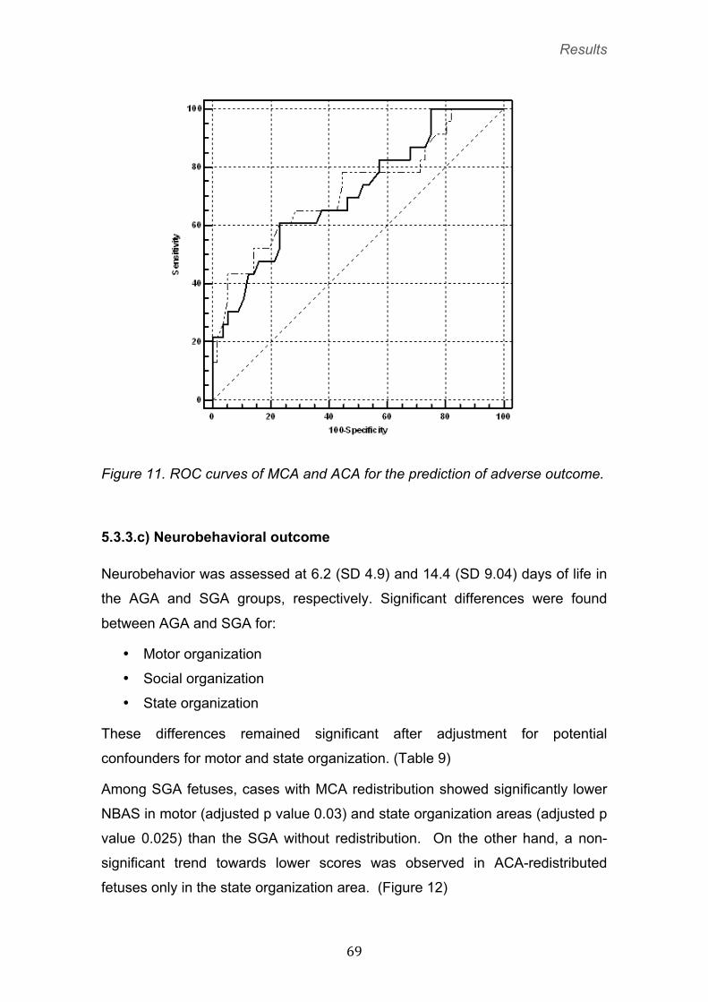
Figure 11 shows the ROC curves of MCA and ACA for the prediction of adverse
outcome. Although both parameters showed significant areas under the curve
(0.71 (CI 0.6-0.81) for the ACA PI and 0.72 (0.61-0.82) for MCA PI), pairwise
Results
� ��
comparison of both areas showed no significant differences between the two
parameters (p=0.82).
Table 8. Perinatal outcome according to the MCA or ACA redistribution.
MCA ACA
SGA not redistributed
SGA redistributed
p+ SGA not redistributed
SGA redistributed
p++
(n=70) (n=28) (n=81) (n=17)
GA at birth (days) 266 (9.9) 265 (8.7) 1 266 (9.5) 262 (8.7) 0.1
Birthweight (gr) 2411 (246.6) 2310 (294.4) 0.54 2426 (235.2) 2173 (296.5) 0.01
Birthweight <3rd centile (%)
31 (44.1) 18 (64.3) 0.07 39 (48.1) 10 (58.8) 0.42
Head circumference (cm)
32.7 (1) 32.3 (1) 0.41 32.6 (1) 32.3 (1) 0.99
Preeclampsia (%) 5 (7.1) 3 (10.7) 0.68 5 (6.2) 3 (17.6) 0.14
Labor induction (%) 52 (75.2) 17 (60.5) 0.14 58 (73.4) 10 (58.8) 0.25
Cesarean Section (%) 18 (26.1) 18 (64.3) 0.001 24 (30) 12 (70.6) 0.002
Intervention for Fetal Distress (%)
10 (14.9) 12 (42.9) 0.006 15 (19.5) 7 (41.2) 0.06
5´Apgar <7 (%) 0 0 1 0 0 1
Umbilical artery pH <7.10 (%)
4 (6.1) 2 (8.3) 0.65 4 (5.4) 2 (13.3) 0.26
Neonatal resuscitation (%)
2 (3.7) 4 (14.8) 0.09 3 (4.5) 3 (20) 0.07
NICU admission (%) 1 (1.5) 2 (7.1) 0.2 1 (1.3) 2 (11.8) 0.08
Composite adverse outcome* (%)
14 (20) 13 (48.1) 0.01 20 (25) 8 (50) 0.05
GA: Gestational Age; NICU: Neonatal Intensive Care Unit; * Composite adverse outcome: intervention for fetal distress, umbilical artery < 7.10, need for neonatal resuscitation or admission to NICU. Student’s t-test for independent samples, Pearson χ2 or Fisher exact test, as appropriate: +between the MCA groups; ++between the ACA groups;
Results
� ��
Figure 11. ROC curves of MCA and ACA for the prediction of adverse outcome.
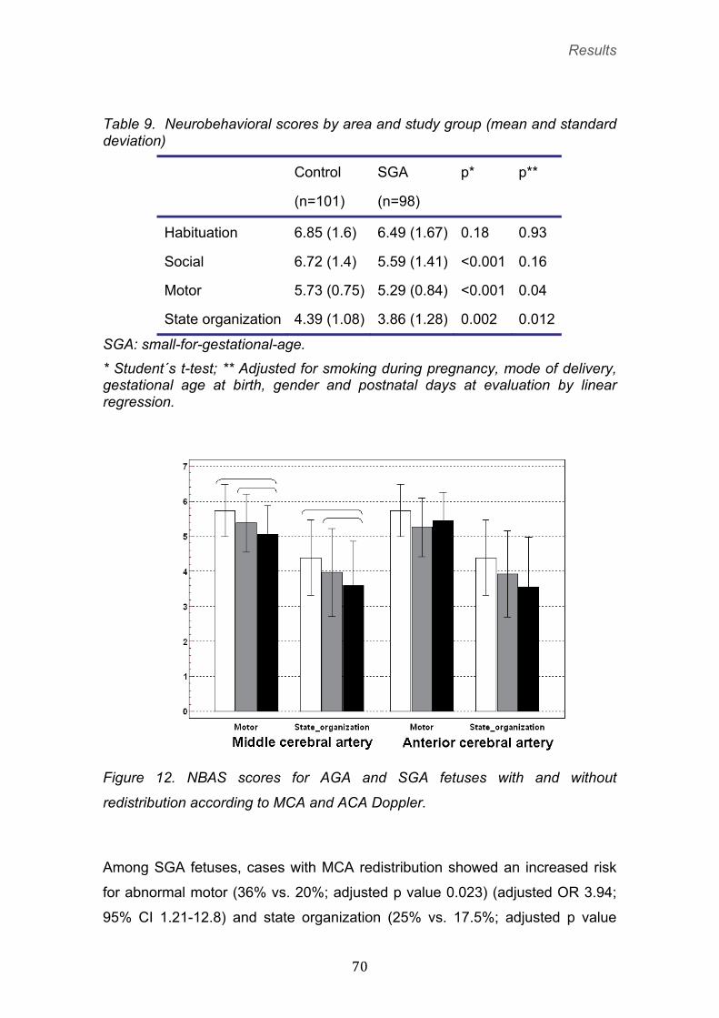
5.3.3.c) Neurobehavioral outcome
Neurobehavior was assessed at 6.2 (SD 4.9) and 14.4 (SD 9.04) days of life in
the AGA and SGA groups, respectively. Significant differences were found
between AGA and SGA for:
• Motor organization
• Social organization
• State organization
These differences remained significant after adjustment for potential
confounders for motor and state organization. (Table 9)
Among SGA fetuses, cases with MCA redistribution showed significantly lower
NBAS in motor (adjusted p value 0.03) and state organization areas (adjusted p
value 0.025) than the SGA without redistribution. On the other hand, a non-
significant trend towards lower scores was observed in ACA-redistributed
fetuses only in the state organization area. (Figure 12)
Results
� �
Table 9. Neurobehavioral scores by area and study group (mean and standard deviation)
Control SGA p* p**
(n=101) (n=98)
Habituation 6.85 (1.6) 6.49 (1.67) 0.18 0.93
Social 6.72 (1.4) 5.59 (1.41) <0.001 0.16
Motor 5.73 (0.75) 5.29 (0.84) <0.001 0.04
State organization 4.39 (1.08) 3.86 (1.28) 0.002 0.012
SGA: small-for-gestational-age.
* Student´s t-test; ** Adjusted for smoking during pregnancy, mode of delivery, gestational age at birth, gender and postnatal days at evaluation by linear regression.
Figure 12. NBAS scores for AGA and SGA fetuses with and without
redistribution according to MCA and ACA Doppler.
Among SGA fetuses, cases with MCA redistribution showed an increased risk
for abnormal motor (36% vs. 20%; adjusted p value 0.023) (adjusted OR 3.94;
95% CI 1.21-12.8) and state organization (25% vs. 17.5%; adjusted p value
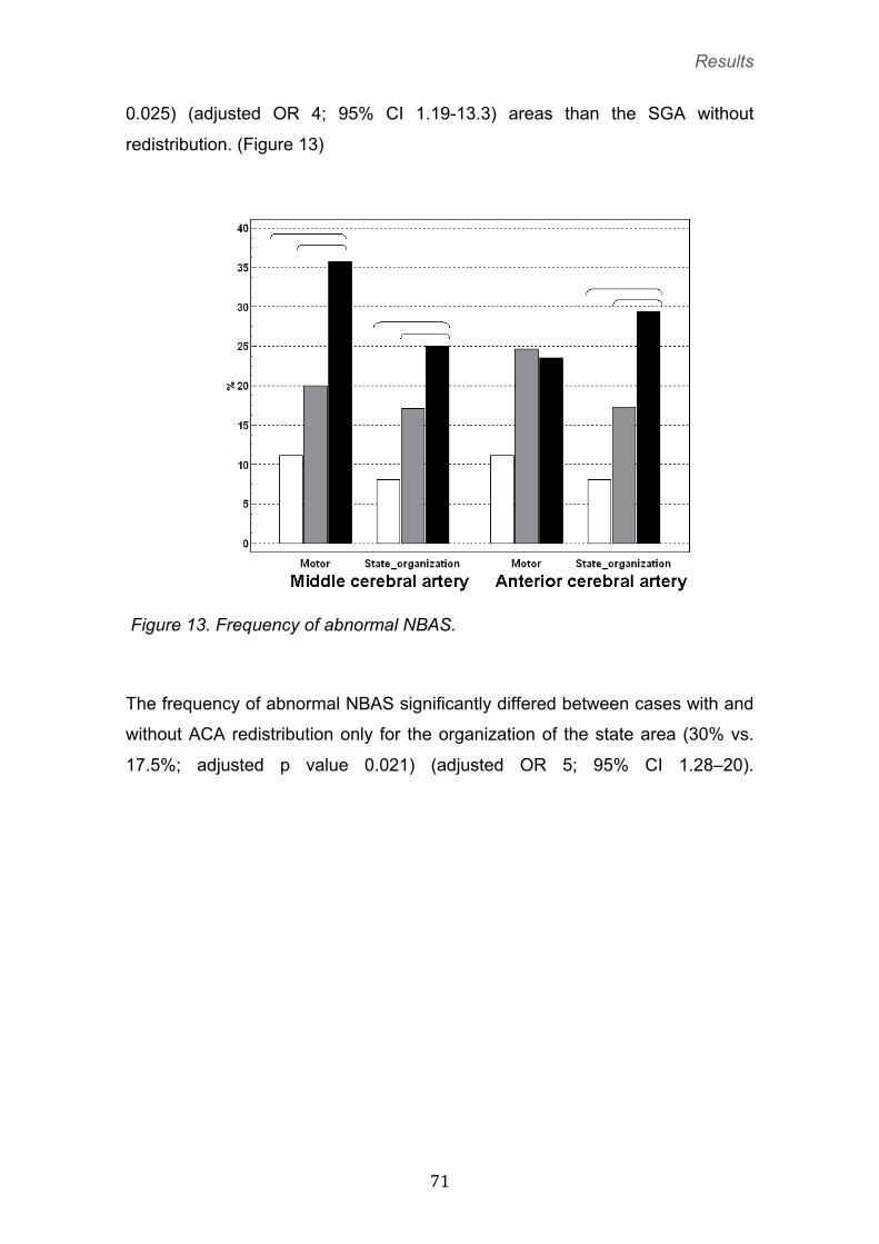
Results
� ��
0.025) (adjusted OR 4; 95% CI 1.19-13.3) areas than the SGA without
redistribution. (Figure 13)
Figure 13. Frequency of abnormal NBAS.
The frequency of abnormal NBAS significantly differed between cases with and
without ACA redistribution only for the organization of the state area (30% vs.
17.5%; adjusted p value 0.021) (adjusted OR 5; 95% CI 1.28–20).

Results
� ��

Discussion
� ��
�
�
�
�
�
�
�
�
�
�
6) DISCUSSION

Discussion
� ��
6.1. General overview
This research project provides evidence to the concept that a proportion of late-
onset small-for-gestational-age fetuses with no signs of placental insufficency
are true forms of growth restriction. This is supported by our findings that brain
redistribution occurs in a substantial proportion of late-onset SGA with normal
umbilical artery Doppler and by the fact that those fetuses have delayed brain
maturation.
Intrauterine growth restriction (IUGR) is predominantly a vascular disorder
secondary to placental insufficency. The effects can be documented with
Doppler ultrasound examination of a number of vessels: maternal uterine
arteries and the fetal umbilical arteries for the placenta; middle cerebral artery
(MCA) for preferential brain perfusion. As early-onset IUGR worsens, Doppler
abnormalities in these vascular territories also deteriorate, (92) suggesting a
sequential pattern of disease progression. This presumed sequence and the
anticipation of fetal deterioration form the basis for Doppler surveillance in
IUGR. Contrary to early-onset growth restriction, in late-onset cases, which are
far more prevalent, most events of adverse outcome attributable to IUGR occur
in fetuses with normal umbilical artery. (93) Thus, a substantial proportion of
late-onset small-for-gestational age (SGA) fetuses with normal umbilical artery
Doppler, considered to represent one end of the normal size spectrum, (30)
may have true growth restriction and are at-risk of adverse perinatal outcome.
(31,32,37) It seems that early and late-onset growth restricted fetuses followed
different pathophysiologycal pathways. (58)
The importance of managing small fetuses with normal umbilical artery Doppler
completely differently from true IUGR babies has been stressed. (30) The
identification of fetal growth restriction depends on adequate risk assessment
and surveillance. Failure to identify small-for-gestational age (SGA) fetuses has
been described as a major cause of perinatal morbidity with a 4-fold risk for
adverse perinatal outcome. (94) However, recent evidence suggests that
umbilical artery Doppler might not accurately identify mild forms of placental
insufficiency. (6) A substantial proportion of these so-called “normal” small

Discussion
� ��
fetuses are actually growing under a pathological environment, but the
consequences of this on fetal brain maturation had not been investigated
before. Our results suggest the existence of subtle degrees of neurological
injury from earlier stages of fetal hemodynamic adaptation to hypoxia.
Discussion
� ��
6.2. Project 1: Longitudinal evaluation of fetal hemodynamic
status in late-onset IUGR fetuses.
This is the first study describing the longitudinal changes of utero-placental and
fetal brain hemodynamics in late-onset SGA fetuses. Our study shows that on
average feto-placental Doppler indexes remain virtually unchanged from
diagnosis to delivery in these fetuses, and consequently the proportion of cases
progressing to abnormal Doppler in the umbilical and uterine arteries is
negligible. On the contrary, average middle cerebral artery and, more markedly,
cerebroplacental ratio values worsen significantly from 37 weeks until delivery,
with 14% and 23% of abnormal values on the last scan before delivery.
The study provides evidence to support the notion that uteroplacental Doppler
is of little clinical value in SGA fetuses and that cerebral Doppler may constitute
the most sensitive exam to identify cases at high risk of adverse perinatal
outcome.
6.2.1. Differencies between early-onset IUGR and late-onset IUGR fetal hemodinamics.
The findings of this study are in line with previous Doppler studies in IUGR and
SGA fetuses.
Concerning umbilical artery Doppler, comparison of our findings in SGA fetuses
with published longitudinal series on early-onset IUGR demonstrates that both
conditions have a different pattern of Doppler deterioration. In early-onset
IUGR, abnormal umbilical artery constitutes the mainstay for diagnosis and
deteriorates progressively, followed by the development of brain sparing. (60)
(75) On the contrary, as illustrated in this study, in most SGA fetuses the
umbilical atery remains normal despite the development of brain sparing in up
to 20% of cases. In a recent study on late-onset IUGR (beyond 30 weeks) with
abnormal umbilical artery, Turan et al (75) reported that fetal rapid
hemodynamic deterioration as observed in early-onset forms was very unlikely,
and the median interval between admission and the appearance of any other
Doppler abnormality was 33 days. The findings of Turan et al. together with
Discussion
� ��
those reported in this study support that as gestational age at onset increases,
abnormalities in the umbilical artery Doppler in small fetuses are more unlikely.
Regarding uterine artery Doppler, we and others (95-97) have demonstrated
that progression of uterine Doppler during the second trimester correlates with
perinatal outcome, but similarly to umbilical artery Doppler, this parameter
remained almost constant throughout the third trimester in SGA fetuses.
Finally, concerning cerebral Doppler measurements our findings are in line with
previous studies in early-onset IUGR reporting that cerebroplacental ratio
becomes abnormal earlier than the middle cerebral artery, (60,75) and with
animal models demonstrating that cerebroplacental ratio turns out to be the
hemodynamic parameter that reflects most closely PO2 changes. (71)
6.2.2. Clinical impact
Our study has several clinical implications.
First, since uterine and umbilical Doppler does not deteriorate from diagnosis to
delivery, little benefit can be expected by serial monitoring. McCowan et al (98)
demonstrated that fortnightly fetal surveillance by umbilical artery Doppler
compared to twice-weekly surveillance resulted in fewer inductions without any
impact on the perinatal outcome. They concluded that more studies are needed
to determine the safety of less frequent surveillance of SGA with normal
umbilical artery. Our findings suggest that in this population, isolated serial
umbilical or uterine artery Doppler would be unnecessary. On the other hand,
restricting the evaluation of near-term SGA to the umbilical artery Doppler could
be potentially harmful since a normal result could give false reassurance of fetal
wellbeing.
Secondly, cerebral Doppler seems to constitute the most sensitive parameter to
detect late-onset SGA fetuses at risk for adverse perinatal outcome.
Hershkovitz et al (58) first provided evidence that near-term SGA with isolated
middle cerebral artery vasodilatation are at-risk of adverse outcome. Others
have confirmed their findings. (32) In addition, cerebral Doppler studies seem
to detect the group of SGA fetuses with the highest risk of suboptimal
Discussion
� ��
neurodevelopmental outcome in these fetuses. (33) Notwithstanding its better
predictiveness as compared with umbilical artery Doppler, the middle cerebral
artery still fails to detect a substantial number of fetuses with abnormal neonatal
outcome. (59) As supported by an increasing number of studies,
cerebroplacental ratio is an earlier and more sensitive predictor for adverse
outcome than either the middle cerebral artery or umbilical artery alone, both in
severe (70-74) as in mild forms of IUGR. (75) The findings of this study are in
line with these publications and further support the use of cerebroplacental ratio
as a primary Doppler monitoring parameter in late-onset SGA.
6.2.3. Study limitations
This study has some limitations. Since abnormal umbilical artery Doppler was
an indication for delivery for ethical reasons, the temporal relationship between
the parameters studied may be biased. However, this occurred in a small
proportion of cases and, therefore, the effect of this bias should be negligible.
Any clinical study on the surveillance of fetal growth needs to be interpreted
with caution because it can never be entirely blinded and the clinical
management is influenced by the antenatal findings. However, clinicians
attending the deliveries were unaware of the middle cerebral artery, uterine and
cerebroplacental ratio Doppler status.
6.2.4. Conclusion
In conclusion, late-onset SGA with normal Doppler upon diagnosis show no
changes in umbilical and uterine artery Doppler. In contrast, a progression from
37 weeks onward with worsening cerebroplacental ratio in up to 24 % and of
middle cerebral artery vasodilation in 14% of cases is observed. These findings
discourage the use of umbilical artery as a single parameter for surveillance in
late-onset SGA and support the use of the cerebroplacental ratio as the most
sensitive Doppler parameter for the clinical monitoring of these fetuses.
Discussion
� �
6.3. Project 2: Evaluation of perinatal outcome and neonatal
neurobehaviour of late-onset IUGR fetuses with normal UA
Doppler versus controls.
It is accepted that SGA with abnormal umbilical artery Doppler, as a surrogate
sign of placental insufficiency, are at high risk of neurobehavioral and
neurocognitive outcome. (87,99,100) Our study extends the consequences of
being SGA on neurobehavior to full-term neonates with no signs of placental
insufficiency, a subgroup of neonates traditionally considered to be one end of
the size spectrum of normal babies. Contrary to the prevailing opinion, our
findings challenge the concept that umbilical artery Doppler is a reliable tool to
prenatally identify those constitutional term SGA babies at low risk. Subtle
degrees of neurological injury seem to have already occurred before umbilical
Doppler waveform becomes abnormal. In fact, animal (101) and mathematical
(102) experimental models of placental vessel obliteration have suggested that
umbilical artery Doppler become abnormal only in advanced stages of placental
dysfunction.
6.3.1. Late-onset growth restricted fetuses neurobehavior
We found a poorer neonatal response to both visual and auditory stimuli in SGA
babies. Long-term follow-up studies of SGA babies have found impaired visual-
spatial stimuli processing. (80) Chronic fetal growth restriction animal models
have found optic nerve reduced myelogenesis (103) along with reduced
synaptogenesis in the visual cortex. (39) These factors could restrict the
integration of cerebral cortex inputs. Additionally, mistimed cell migration could
also have an effect on synaptic plasticity with functional and behavioral
consequences. (40) Visual-spatial abilities also play a role in the neonatal
capacity to select environmental stimuli to process and act on, which provide
important scaffolding on which attention skills are constructed through early
childhood. (104) Indeed, fetal growth restriction is also associated with the
development of attention deficit symptoms in childhood and early adolescence.
Discussion
� ��
(37) Our finding of lower attention capacities in SGA babies is in line with this
hypothesis. Two previous studies (11,12) have found impaired attention skills
among preterm SGA babies. Structural correlations have been made between
attention scores and cortical grey matter (11) and hippocampal (12) volumes.
Whether NBAS predicts attention later in childhood is uncertain. However, some
studies have found that infant attention is predictive of intelligence in childhood
and adolescence. (105-106) Another study of temperament in early infancy (5-
6 months) reported a significant prediction of attention deficits among 8-year-old
children. (83) There is a substantial overlap between childhood attention
disorders and the diagnosis of motor impairment, with poor movement abilities.
(107) Our finding of lower motor performance among SGA babies is consistent
with this association. In a series of 40 SGA preterm neonates Feldman et al
(34) studied the correlation of neurobehavioral and cognitive function with the
Bayley Scale of Infant Development. They found that neonatal motor maturity
correlated with the Psychomotor Development Outcome at 24-months. Another
interesting finding of our study is the lower level of self-regulation among SGA
babies, as reflected by the regulation of state NBAS cluster. In a series of 38
full-term healthy babies, Lundqvist (108) reported that levels of self-regulation
were also correlated with the infants’ levels of cognitive development (personal-
social development, speech development and eye and hand coordination,
subvariables in Griffiths’ Mental Development Test) and with sleeping disorders
at 2 years of age.
6.3.2. Clinical implications
One strength of our study is that only included full-term SGA fetuses without
signs of placental dysfunction, as are low-risk SGA defined in everyday clinical
practice. No previous studies aimed to analyze the effect of growth restriction
on neurobehavior have reported which proportion of cases had abnormal
Doppler. This is a potential source of misinterpretation of the results, since an
unknown part of the babies could have been exposed to a very prolonged
period of intrauterine hypoxia. Another strong point of our study is that
Discussion
� ��
confounding was accounted by adjustment by potential factors independently
associated with SGA and the neonatal neurobehavioral performance.
In low-risk preterm infants, it has been reported that individualized
developmental interventions prevent short-term neurobehavioral dysfunction.
(83) It is unknown whether interventions could also be effective in term infants
and whether they would influence long-term outcome. Nevertheless,
identifying the at-risk infants is essential to understand the association between
fetal wellbeing and later neurodevelopmental problems and lays the basis for
possible preventive interventions.
6.3.3. Study limitations
This study also has some limitations. Although NBAS is a gold standard to
evaluate the neonate’s capacity to respond to the environment, reflecting brain
maturation, it only assesses neurobehavioral and not cognitive function.
However, several studies have demonstrated the correlation between neonatal
neurobehavioral performance and later neurocognitive development.
(11,12,34,108,109) It could also be argued that the study is prone to an
expectation bias, since the examiners could have been influenced by baby size
when assessing the neurobehavioral performance. However, within each study
group, no significant correlations were found between birthweight and the
NBAS scores, except for organization of state in the SGA group. This makes it
unlikely that this potential bias could explain the differences observed between
SGA and AGA babies in the other areas.
6.3.4. Conclusion
In summary, we found in a well-defined cohort of term SGA neonates with no
signs of placental insufficiency poorer neurobehavioral performance, suggesting
delayed neurological maturation. Whether earlier markers of placental
insufficiency or hypoxia could predict this outcome requires further
investigation.
Discussion
� ��
6.4. Project 3: Evaluation of the anterior and middle cerebral
arteries for the prediction of perinatal outcome and neonatal
neurobehavior in late-onset IUGR fetuses with normal UA
Doppler.
This is the first study to explore the ability of the ACA to predict perinatal and
neurobehavioural outcome in term SGA fetuses. Contrary to the hypothesis of
the study, we found no differences between MCA and ACA redistribution in
terms of association with perinatal outcome or neonatal neurobehavioral
performance. Therefore, MCA seems to remain unchallenged as a primary
clinical tool to evaluate term SGA fetuses without signs of placental
insufficiency.
6.4.1. Role of anterior cerebral artery Doppler for the prediction of neurological damage as a consecuence of intrauterine growth restriction
In preterm IUGR fetuses we have previously demonstrated that ACA
vasodilation takes place earlier than MCA vasodilation. (62) Dubiel et al.
reported similar results in pregnancies with pregnancy-induced hypertension.
(67) These findings are not in line with the results of the present study.
However, different population characteristics are likely to explain this
inconsistency. The above studies included a substantial proportion of early
onset growth restricted fetuses with abnormal umbilical artery Doppler. Since
gestational age was considerably different we cannot exclude differences in the
temporal patterns in the adaptation to chronic hypoxia due to maturity changes
in brain hemodynamic regulation. Indeed, Dubiel et al (67) reported a better
correlation of ACA PI with adverse perinatal outcome only for cases before 32
weeks’ gestation. For those delivering beyond that gestational age MCA and
ACA PI showed a similar association with perinatal outcome. Also in line with
this reasoning, other studies (110) have found no differences between ACA
and MCA in response to acute hypoxia in term SGA fetuses.
As a secondary explanation, the considerable degree of systemic fetal
hemodynamic adaptation to hypoxia in early onset IUGR, which is not present
Discussion
� ��
in term SGA, could also have influences in brain hemodynamics that we cannot
interpret.
The findings of this study may appear inconsistent with evidences pointing to a
higher vulnerability of frontal areas in fetuses with IUGR. (9,11,66,80,111)
However, the evaluated arteries provide blood supply to ill-defined anatomical
areas with a marked component of vascular shunting. Tissue perfusion
depends on local arteriolar phenomena and therefore the data cannot be used
to infer which territories are specifically involved by vascular changes. Using the
fractional moving blood volume estimate to assess tissue perfusion we have
previously suggested that increased perfusion in the frontal lobe seems to be
the earliest response to hypoxia, rather than changes in basal ganglia
perfusion. (68) It is unknown whether evaluation of tissue perfusion can detect
subtle differences in blood perfusion between cerebral areas in this population
of late-onset SGA. Studies evaluating the potential role of direct measurements
of perfusion towards different brain regions in this population are under way.
6.4.2. Clinical implications
This study confirms previous reports suggesting that late-onset SGA is
associated with an increased risk of abnormal neonatal neurobehavior (112-
113) and neurodevelopment in childhood. (33,108,114) Furthermore, we have
previously described that in late-onset SGA fetuses, MCA redistribution
differentiates those at risk of long-term suboptimal neurodevelopment. (33) In a
recent study, Roza et al. (114) showed that fetal ACA redistribution could be
superior to MCA in the prediction of neurobehavioral problems in childhood. In
the present study MCA and ACA Doppler were similarly associated with poorer
neonatal neurobehavior. These findings are difficult to compare with those of
Roza et al for two reasons. Our results refer exclusively to neonatal
neurobehavior and therefore they do not exclude that long term assessment
could demonstrate a higher sensitivity of ACA to predict poor
neurodevelopment. A second important difference is that the study of Roza et
al. (114) was based on a large cohort, which included all pregnancies, and not
Discussion
� ��
only SGA fetuses. Therefore, the value of ACA to predict long-term outcome in
small fetuses remains to be evaluated.
6.4.3 Study limitations
This study has some limitations. First, since no Doppler was performed in the
group of AGA babies, we cannot rule out that some fetuses in this group might
have shown brain redistribution. However, this potential bias would be
conservative, attenuating the differences between AGA and SGA fetuses.
Secondly, although NBAS is a gold standard to evaluate the neonate’s capacity
to respond to the environment, reflecting brain maturation, it only assesses
neurobehavioral and not cognitive function. However, several studies have
demonstrated the correlation between neonatal neurobehavioral performance
and later neurocognitive development. (11,12,34,108,109) We admit that
socioeconomic status may have confounded the association between SGA and
abnormal neurobehaviour. However, although socioeconomic status is a major
determinant of postnatal neurobehavior during childhood, its influence in the
neonatal period is still minimal if smoking is accounted for in our population.
Thirdly, any clinical study on the surveillance of fetal growth needs to be
interpreted with caution because it can never be entirely blinded and the clinical
management is influenced by the antenatal findings. This work-up bias could
account for differences between AGA and SGA. However, the clinicians
attending the deliveries were unaware of the MCA or ACA Doppler status. Thus
this potential bias could not explain differences between the two groups of SGA
fetuses. Besides, the neurobehavioral assessment could also have been biased
by the examiners´ knowledge of perinatal factors. However, since these
examiners were also blinded to the antenatal Doppler and due to the fact that
birthweight was similar between the two groups, this expectation bias is unlikely
to explain differences between Doppler groups.
6.4.4. Conclusion
In conclusion, the findings of this study do not support the inclusion of ACA
Doppler investigation in the management of term SGA fetuses.

Discussion
� ��

Conclusions �
� ��
7) CONCLUSIONS


Conclusions �
� ��
1. Umbilical artery Doppler failed as a single parameter for surveillance in
late-onset growth restricted fetuses.
2. Term late-onset growth restricted fetuses neonates with no signs of
placental insufficiency present poorer neurobehavioral performance,
suggesting delayed neurological maturation.
3. Term late-onset growth restricted fetuses neonates with no signs of
placental insufficiency present poorer perinatal outcome.
4. Cerebroplacental ratio is the most sensitive Doppler parameter for the
clinical monitoring of late-onset growth restricted fetuses
5. The inclusion of ACA Doppler investigation in the management of term
late-onset growth restricted fetuses does not improve neither perinatal
nor neurobehavioral outcomes.

� �

Acknowledgements �
� ��
8) ACKNOWLEDGEMENTS


Acknowledgements �
� ��
The Fetal and Perinatal Medicine Research Group (IDIBAPS) is founded by the
Centro de Investigación Biomédica en Red de Enfermedades Raras (CIBERER,
ISCIII, Spain), and supported by grants from the Fondo the Investigación
Sanitaria (PI/060347) (Spain), Cerebra Foundation for the Brain Injured Child
(Carmarthen, Wales, UK), Thrasher Research Fund (Salt Lake City, USA) and
the European Commission.
�
�
�
�
�
�
�
�
�
�
�
�

� ��

Bibliography
� ��
9) BIBLIOGRAPHY

Bibliography
� ��

Bibliography
� ��
1. Figueras F, Meler E, Iraola A, Eixarch E, Coll O, Figueras J, et al. Customized birthweight standards for a spanish population. Eur J Obstet Gynecol Reprod Biol 2008, Jan;136(1):20-4.
2. Gardosi J, Chang A, Kalyan B, Sahota D, Symonds EM. Customised antenatal growth charts. Lancet 1992, Feb 1;339(8788):283-7.
3. Wilcox AJ. Intrauterine growth retardation: Beyond birthweight criteria. Early Hum Dev 1983, Oct;8(3-4):189-93.
4. Sankaran S, Kyle PM. Aetiology and pathogenesis of IUGR. Best Pract Res Clin Obstet Gynaecol 2009, Dec;23(6):765-77.
5. M Kady S, Gardosi J. Perinatal mortality and fetal growth restriction. Best Pract Res Clin Obstet Gynaecol 2004, Jun;18(3):397-410.
6. Figueras F, Eixarch E, Meler E, Iraola A, Figueras J, Puerto B, Gratacos E. Small-For-Gestational-Age fetuses with normal umbilical artery doppler have suboptimal perinatal and neurodevelopmental outcome. Eur J Obstet Gynecol Reprod Biol 2008, Jan;136(1):34-8.
7. Tan TY, Yeo GS. Intrauterine growth restriction. Curr Opin Obstet Gynecol 2005, Apr;17(2):135-42.
8. Li CI, Daling JR, Emanuel I. Birthweight and risk of overall and cause-specific childhood mortality. Paediatr Perinat Epidemiol 2003, Apr;17(2):164-70.
9. Leitner Y, Fattal-Valevski A, Geva R, Eshel R, Toledano-Alhadef H, Rotstein M, et al. Neurodevelopmental outcome of children with intrauterine growth retardation: A longitudinal, 10-year prospective study. J Child Neurol 2007, May;22(5):580-7.
10. Scherjon S, Briët J, Oosting H, Kok J. The discrepancy between maturation of visual-evoked potentials and cognitive outcome at five years in very preterm infants with and without hemodynamic signs of fetal brain-sparing. Pediatrics 2000, Feb;105(2):385-91.
11. Tolsa CB, Zimine S, Warfield SK, Freschi M, Sancho Rossignol A, Lazeyras F, et al. Early alteration of structural and functional brain development in premature infants born with intrauterine growth restriction. Pediatr Res 2004, Jul;56(1):132-8.
12. Lodygensky GA, Seghier ML, Warfield SK, Tolsa CB, Sizonenko S, Lazeyras F, Hüppi PS. Intrauterine growth restriction affects the preterm infant's hippocampus. Pediatr Res 2008, Apr;63(4):438-43.
13. Dubois J, Benders M, Borradori-Tolsa C, Cachia A, Lazeyras F, Ha-Vinh Leuchter R, et al. Primary cortical folding in the human newborn: An early marker of later functional development. Brain 2008, Aug;131(Pt 8):2028-41.
14. Baker JL, Olsen LW, Sørensen TI. Weight at birth and all-cause mortality in adulthood. Epidemiology 2008, Mar;19(2):197-203.
15. Kaijser M, Bonamy AK, Akre O, Cnattingius S, Granath F, Norman M, Ekbom A. Perinatal risk factors for ischemic heart disease: Disentangling the roles of birth weight and preterm birth. Circulation 2008, Jan 22;117(3):405-10.
16. Palinski W, Napoli C. Impaired fetal growth, cardiovascular disease, and the need to move on. Circulation 2008, Jan 22;117(3):341-3.
17. Bukowski R, Smith GC, Malone FD, Ball RH, Nyberg DA, Comstock CH, et al. Fetal growth in early pregnancy and risk of delivering low birth weight infant: Prospective cohort study. BMJ 2007, Apr 21;334(7598):836.

Bibliography
� ��
18. Figueras F, Oros D, Cruz-Martinez R, Padilla N, Hernandez-Andrade E, Botet F, et al. Neurobehavior in term, small-for-gestational age infants with normal placental function. Pediatrics 2009, Nov;124(5):e934-41.
19. ACOG committee opinion. Utility of antepartum umbilical artery doppler velocimetry in intrauterine growth restriction. Number 188, october 1997 (replaces no. 116, november 1992). Committee on obstetric practice. American college of obstetricians and gynecologists. Int J Gynaecol Obstet 1997, Dec;59(3):269-70.
20. Cruz-Martinez R, Figueras F. The role of doppler and placental screening. Best Pract Res Clin Obstet Gynaecol 2009, Dec;23(6):845-55.
21. Baschat AA, Weiner CP. Umbilical artery doppler screening for detection of the small fetus in need of antepartum surveillance. Am J Obstet Gynecol 2000, Jan;182(1 Pt 1):154-8.
22. Nicolaides KH, Bilardo CM, Soothill PW, Campbell S. Absence of end diastolic frequencies in umbilical artery: A sign of fetal hypoxia and acidosis. BMJ 1988, Oct 22;297(6655):1026-7.
23. Trudinger BJ, Cook CM, Giles WB, Ng S, Fong E, Connelly A, Wilcox W. Fetal umbilical artery velocity waveforms and subsequent neonatal outcome. Br J Obstet Gynaecol 1991, Apr;98(4):378-84.
24. Gaziano EP, Knox H, Ferrera B, Brandt DG, Calvin SE, Knox GE. Is it time to reassess the risk for the growth-retarded fetus with normal doppler velocimetry of the umbilical artery? Am J Obstet Gynecol 1994, Jun;170(6):1734-41; discussion 1741-3.
25. McCowan LM, Harding JE, Stewart AW. Umbilical artery doppler studies in small for gestational age babies reflect disease severity. BJOG 2000, Jul;107(7):916-25.
26. Yoon BH, Lee CM, Kim SW. An abnormal umbilical artery waveform: A strong and independent predictor of adverse perinatal outcome in patients with preeclampsia. Am J Obstet Gynecol 1994, Sep;171(3):713-21.
27. Cosmi E, Ambrosini G, D'Antona D, Saccardi C, Mari G. Doppler, cardiotocography, and biophysical profile changes in growth-restricted fetuses. Obstet Gynecol 2005, Dec;106(6):1240-5.
28. Valcamonico A, Danti L, Frusca T, Soregaroli M, Zucca S, Abrami F, Tiberti A. Absent end-diastolic velocity in umbilical artery: Risk of neonatal morbidity and brain damage. Am J Obstet Gynecol 1994, Mar;170(3):796-801.
29. Neilson JP, Alfirevic Z. Doppler ultrasound for fetal assessment in high risk pregnancies. Cochrane Database Syst Rev 2000(2):CD000073.
30. Soothill PW, Bobrow CS, Holmes R. Small for gestational age is not a diagnosis. Ultrasound Obstet Gynecol 1999, Apr;13(4):225-8.
31. Doctor BA, O'Riordan MA, Kirchner HL, Shah D, Hack M. Perinatal correlates and neonatal outcomes of small for gestational age infants born at term gestation. Am J Obstet Gynecol 2001, Sep;185(3):652-9.
32. Severi FM, Bocchi C, Visentin A, Falco P, Cobellis L, Florio P, et al. Uterine and fetal cerebral doppler predict the outcome of third-trimester small-for-gestational age fetuses with normal umbilical artery doppler. Ultrasound Obstet Gynecol 2002, Mar;19(3):225-8.
33. Eixarch E, Meler E, Iraola A, Illa M, Crispi F, Hernandez-Andrade E, et al. Neurodevelopmental outcome in 2-year-old infants who were small-for-gestational age term fetuses with cerebral blood flow redistribution. Ultrasound Obstet Gynecol 2008, Dec;32(7):894-9.

Bibliography
� ��
34. Feldman R, Eidelman AI. Neonatal state organization, neuromaturation, mother-infant interaction, and cognitive development in small-for-gestational-age premature infants. Pediatrics 2006, Sep;118(3):e869-78.
35. Ley D, Laurin J, Bjerre I, Marsal K. Abnormal fetal aortic velocity waveform and minor neurological dysfunction at 7 years of age. Ultrasound Obstet Gynecol 1996, Sep;8(3):152-9.
36. Ley D, Tideman E, Laurin J, Bjerre I, Marsal K. Abnormal fetal aortic velocity waveform and intellectual function at 7 years of age. Ultrasound Obstet Gynecol 1996, Sep;8(3):160-5.
37. McCowan LM, Pryor J, Harding JE. Perinatal predictors of neurodevelopmental outcome in small-for-gestational-age children at 18 months of age. Am J Obstet Gynecol 2002, May;186(5):1069-75.
38. Rigano S, Bozzo M, Ferrazzi E, Bellotti M, Battaglia FC, Galan HL. Early and persistent reduction in umbilical vein blood flow in the growth-restricted fetus: A longitudinal study. Am J Obstet Gynecol 2001, Oct;185(4):834-8.
39. Morrow RJ, Adamson SL, Bull SB, Ritchie JW. Effect of placental embolization on the umbilical arterial velocity waveform in fetal sheep. Am J Obstet Gynecol 1989, Oct;161(4):1055-60.
40. Thompson RS, Stevens RJ. Mathematical model for interpretation of doppler velocity waveform indices. Med Biol Eng Comput 1989, May;27(3):269-76.
41. Matthiesen L, Berg G, Ernerudh J, Ekerfelt C, Jonsson Y, Sharma S. Immunology of preeclampsia. Chem Immunol Allergy 2005;89:49-61.
42. Olofsson P, Laurini RN, Marsál K. A high uterine artery pulsatility index reflects a defective development of placental bed spiral arteries in pregnancies complicated by hypertension and fetal growth retardation. Eur J Obstet Gynecol Reprod Biol 1993, May;49(3):161-8.
43. Ghosh GS, Gudmundsson S. Uterine and umbilical artery doppler are comparable in predicting perinatal outcome of growth-restricted fetuses. BJOG 2009, Feb;116(3):424-30.
44. Papageorghiou AT, Yu CK, Bindra R, Pandis G, Nicolaides KH, Fetal Medicine Foundation Second Trimester Screening Group. Multicenter screening for pre-eclampsia and fetal growth restriction by transvaginal uterine artery doppler at 23 weeks of gestation. Ultrasound Obstet Gynecol 2001, Nov;18(5):441-9.
45. Martin AM, Bindra R, Curcio P, Cicero S, Nicolaides KH. Screening for pre-eclampsia and fetal growth restriction by uterine artery doppler at 11-14 weeks of gestation. Ultrasound Obstet Gynecol 2001, Dec;18(6):583-6.
46. Spencer K, Cowans NJ, Avgidou K, Molina F, Nicolaides KH. First-Trimester biochemical markers of aneuploidy and the prediction of small-for-gestational age fetuses. Ultrasound Obstet Gynecol 2008, Jan;31(1):15-9.
47. Crispi F, Llurba E, Domínguez C, Martín-Gallán P, Cabero L, Gratacós E. Predictive value of angiogenic factors and uterine artery doppler for early- versus late-onset pre-eclampsia and intrauterine growth restriction. Ultrasound Obstet Gynecol 2008, Mar;31(3):303-9.
48. Li H, Gudnason H, Olofsson P, Dubiel M, Gudmundsson S. Increased uterine artery vascular impedance is related to adverse outcome of pregnancy but is present in only one-third of late third-trimester pre-eclamptic women. Ultrasound Obstet Gynecol 2005, May;25(5):459-63.

Bibliography
� �
49. Meler E, Figueras F, Gratacos E. Uterine artery for the mangement of SGA. Am J Obstet Gynecol (In press).
50. Vergani P, Roncaglia N, Andreotti C, Arreghini A, Teruzzi M, Pezzullo JC, Ghidini A. Prognostic value of uterine artery doppler velocimetry in growth-restricted fetuses delivered near term. Am J Obstet Gynecol 2002, Oct;187(4):932-6.
51. Vergani P, Roncaglia N, Locatelli A, Andreotti C, Crippa I, Pezzullo JC, Ghidini A. Antenatal predictors of neonatal outcome in fetal growth restriction with absent end-diastolic flow in the umbilical artery. Am J Obstet Gynecol 2005, Sep;193(3 Pt 2):1213-8.
52. Chien PF, Arnott N, Gordon A, Owen P, Khan KS. How useful is uterine artery doppler flow velocimetry in the prediction of pre-eclampsia, intrauterine growth retardation and perinatal death? An overview. BJOG 2000, Feb;107(2):196-208.
53. Robel-Tillig E, Knüpfer M, Vogtmann C. Cardiac adaptation in small for gestational age neonates after prenatal hemodynamic disturbances. Early Hum Dev 2003, Jun;72(2):123-9.
54. Battaglia C, Artini PG, Galli PA, D'Ambrogio G, Droghini F, Genazzani AR. Absent or reversed end-diastolic flow in umbilical artery and severe intrauterine growth retardation. An ominous association. Acta Obstet Gynecol Scand 1993, Apr;72(3):167-71.
55. Hecher K, Bilardo CM, Stigter RH, Ville Y, Hackelöer BJ, Kok HJ, et al. Monitoring of fetuses with intrauterine growth restriction: A longitudinal study. Ultrasound Obstet Gynecol 2001, Dec;18(6):564-70.
56. Scherjon SA, Oosting H, Smolders-DeHaas H, Zondervan HA, Kok JH. Neurodevelopmental outcome at three years of age after fetal 'brain-sparing'. Early Hum Dev 1998, Aug 28;52(1):67-79.
57. Strigini FA, De Luca G, Lencioni G, Scida P, Giusti G, Genazzani AR. Middle cerebral artery velocimetry: Different clinical relevance depending on umbilical velocimetry. Obstet Gynecol 1997, Dec;90(6):953-7.
58. Hershkovitz R, Kingdom JC, Geary M, Rodeck CH. Fetal cerebral blood flow redistribution in late gestation: Identification of compromise in small fetuses with normal umbilical artery doppler. Ultrasound Obstet Gynecol 2000, Mar;15(3):209-12.
59. Cruz-Martinez R, Figueras F, Oros D, Padilla N, Meler E, Hernandez-Andrade E, Gratacos E. Cerebral blood perfusion and neurobehavioral performance in full-term small-for-gestational-age fetuses. Am J Obstet Gynecol 2009, Nov;201(5):474.e1-7.
60. Harrington K, Thompson MO, Carpenter RG, Nguyen M, Campbell S. Doppler fetal circulation in pregnancies complicated by pre-eclampsia or delivery of a small for gestational age baby: 2. Longitudinal analysis. Br J Obstet Gynaecol 1999, May;106(5):453-66.
61. Kok JH, Prick L, Merckel E, Everhard Y, Verkerk GJ, Scherjon SA. Visual function at 11 years of age in preterm-born children with and without fetal brain sparing. Pediatrics 2007, Jun;119(6):e1342-50.
62. Figueroa-Diesel H, Hernandez-Andrade E, Acosta-Rojas R, Cabero L, Gratacos E. Doppler changes in the main fetal brain arteries at different stages of hemodynamic adaptation in severe intrauterine growth restriction. Ultrasound Obstet Gynecol 2007, Sep;30(3):297-302.
63. Jensen A, Roman C, Rudolph AM. Effects of reducing uterine blood flow on fetal blood flow distribution and oxygen delivery. J Dev Physiol 1991, Jun;15(6):309-23.

Bibliography
� ��
64. Capilla-González A, Fernández-González S, Campo P, Maestú F, Fernández-Lucas A, Mulas F, Ortiz T. [Magnetoencephalography in cognitive disorders involving frontal lobes]. Rev Neurol 2004;39(2):183-8.
65. Geva R, Eshel R, Leitner Y, Valevski AF, Harel S. Neuropsychological outcome of children with intrauterine growth restriction: A 9-year prospective study. Pediatrics 2006, Jul;118(1):91-100.
66. Benavides-Serralde A, Hernández-Andrade E, Fernández-Delgado J, Plasencia W, Scheier M, Crispi F, et al. Three-Dimensional sonographic calculation of the volume of intracranial structures in growth-restricted and appropriate-for-gestational age fetuses. Ultrasound Obstet Gynecol 2009, May;33(5):530-7.
67. Dubiel M, Gunnarsson GO, Gudmundsson S. Blood redistribution in the fetal brain during chronic hypoxia. Ultrasound Obstet Gynecol 2002, Aug;20(2):117-21.
68. Hernandez-Andrade E, Jansson T, Figueroa-Diesel H, Rangel-Nava H, Acosta-Rojas R, Gratacós E. Evaluation of fetal regional cerebral blood perfusion using power doppler ultrasound and the estimation of fractional moving blood volume. Ultrasound Obstet Gynecol 2007, May;29(5):556-61.
69. Al-Ghazali W, Chita SK, Chapman MG, Allan LD. Evidence of redistribution of cardiac output in asymmetrical growth retardation. Br J Obstet Gynaecol 1989, Jun;96(6):697-704.
70. Gramellini D, Folli MC, Raboni S, Vadora E, Merialdi A. Cerebral-Umbilical doppler ratio as a predictor of adverse perinatal outcome. Obstet Gynecol 1992, Mar;79(3):416-20.
71. Arbeille P, Maulik D, Fignon A, Stale H, Berson M, Bodard S, Locatelli A. Assessment of the fetal PO2 changes by cerebral and umbilical doppler on lamb fetuses during acute hypoxia. Ultrasound Med Biol 1995;21(7):861-70.
72. Bahado-Singh RO, Kovanci E, Jeffres A, Oz U, Deren O, Copel J, Mari G. The doppler cerebroplacental ratio and perinatal outcome in intrauterine growth restriction. Am J Obstet Gynecol 1999, Mar;180(3 Pt 1):750-6.
73. Arias F. Accuracy of the middle-cerebral-to-umbilical-artery resistance index ratio in the prediction of neonatal outcome in patients at high risk for fetal and neonatal complications. Am J Obstet Gynecol 1994, Dec;171(6):1541-5.
74. Murata S, Nakata M, Sumie M, Sugino N. The doppler cerebroplacental ratio predicts risk of non-reassuring fetal status for fetal growth restriction in term pregnancy. Ultrasound Obstet Gynecol 2009;34((S1)):56.
75. Turan OM, Turan S, Gungor S, Berg C, Moyano D, Gembruch U, et al. Progression of doppler abnormalities in intrauterine growth restriction. Ultrasound Obstet Gynecol 2008, Aug;32(2):160-7.
76. Baschat AA, Gembruch U. The cerebroplacental doppler ratio revisited. Ultrasound Obstet Gynecol 2003, Feb;21(2):124-7.
77. Baschat AA, Gembruch U, Harman CR. The sequence of changes in doppler and biophysical parameters as severe fetal growth restriction worsens. Ultrasound Obstet Gynecol 2001, Dec;18(6):571-7.
78. Schothorst PF, van Engeland H. Long-Term behavioral sequelae of prematurity. J Am Acad Child Adolesc Psychiatry 1996, Feb;35(2):175-83.
79. Scherjon SA, Smolders-DeHaas H, Kok JH, Zondervan HA. The "brain-sparing" effect: Antenatal cerebral doppler findings in relation to neurologic outcome in very preterm infants. Am J Obstet Gynecol 1993, Jul;169(1):169-75.

Bibliography
� ��
80. Geva R, Eshel R, Leitner Y, Fattal-Valevski A, Harel S. Memory functions of children born with asymmetric intrauterine growth restriction. Brain Res 2006, Oct 30;1117(1):186-94.
81. McCarton CM, Wallace IF, Divon M, Vaughan HG. Cognitive and neurologic development of the premature, small for gestational age infant through age 6: Comparison by birth weight and gestational age. Pediatrics 1996, Dec;98(6 Pt 1):1167-78.
82. Herschkowitz N, Kagan J, Zilles K. Neurobiological bases of behavioral development in the first year. Neuropediatrics 1997, Dec;28(6):296-306.
83. Buehler DM, Als H, Duffy FH, McAnulty GB, Liederman J. Effectiveness of individualized developmental care for low-risk preterm infants: Behavioral and electrophysiologic evidence. Pediatrics 1995, Nov;96(5 Pt 1):923-32.
84. Larroque B, Bertrais S, Czernichow P, Léger J. School difficulties in 20-year-olds who were born small for gestational age at term in a regional cohort study. Pediatrics 2001, Jul;108(1):111-5.
85. O'Keeffe MJ, O'Callaghan M, Williams GM, Najman JM, Bor W. Learning, cognitive, and attentional problems in adolescents born small for gestational age. Pediatrics 2003, Aug;112(2):301-7.
86. Gómez O, Figueras F, Fernández S, Bennasar M, Martínez JM, Puerto B, Gratacós E. Reference ranges for uterine artery mean pulsatility index at 11-41 weeks of gestation. Ultrasound Obstet Gynecol 2008, Aug;32(2):128-32.
87. Arduini D, Rizzo G. Normal values of pulsatility index from fetal vessels: A cross-sectional study on 1556 healthy fetuses. J Perinat Med 1990;18(3):165-72.
88. Robinson HP, Fleming JE. A critical evaluation of sonar "crown-rump length" measurements. Br J Obstet Gynaecol 1975, Sep;82(9):702-10.
89. Report of the national high blood pressure education program working group on high blood pressure in pregnancy. High Blood Pressure in Pregnancy 2000(1):S1-S22.
90. Costas Moragas C, Fornicles Deu A, Botet Mussons F, Boatella Costa E, Cáceres Zurita ML. Evaluación psicométrica de la escala de brazelton en una muestra de recién nacidos españoles. Psycothema ;19(1):140-9.
91. Brazelton TB, Nugent JK. Brazelton. 3 edition ed. .
92. Hecher K, Campbell S, Doyle P, Harrington K, Nicolaides K. Assessment of fetal compromise by doppler ultrasound investigation of the fetal circulation. Arterial, intracardiac, and venous blood flow velocity studies. Circulation 1995, Jan 1;91(1):129-38.
93. Chang TC, Robson SC, Spencer JA, Gallivan S. Prediction of perinatal morbidity at term in small fetuses: Comparison of fetal growth and doppler ultrasound. Br J Obstet Gynaecol 1994, May;101(5):422-7.
94. Lindqvist PG, Molin J. Does antenatal identification of small-for-gestational age fetuses significantly improve their outcome? Ultrasound Obstet Gynecol 2005, Mar;25(3):258-64.
95. Prefumo F, Güven M, Ganapathy R, Thilaganathan B. The longitudinal variation in uterine artery blood flow pattern in relation to birth weight. Obstet Gynecol 2004, Apr;103(4):764-8.
96. Gómez O, Figueras F, Martínez JM, del Río M, Palacio M, Eixarch E, et al. Sequential changes in uterine artery blood flow pattern between the first and second

Bibliography
� ��
trimesters of gestation in relation to pregnancy outcome. Ultrasound Obstet Gynecol 2006, Nov;28(6):802-8.
97. Groom KM, North RA, Stone PR, Chan EH, Taylor RS, Dekker GA, et al. Patterns of change in uterine artery doppler studies between 20 and 24 weeks of gestation and pregnancy outcomes. Obstet Gynecol 2009, Feb;113(2 Pt 1):332-8.
98. McCowan LM, Harding JE, Roberts AB, Barker SE, Ford C, Stewart AW. A pilot randomized controlled trial of two regimens of fetal surveillance for small-for-gestational-age fetuses with normal results of umbilical artery doppler velocimetry. Am J Obstet Gynecol 2000, Jan;182(1 Pt 1):81-6.
99. Nugent JK, Brazelton TB. Handbook of infant metal health. Vol 2: Early intervention, evaluation and assesment. Osofsky JD, Fitzgerald HE, ed. New York,NY:Wiley; 2000.
100. Sagiv SK, Nugent JK, Brazelton TB, Choi AL, Tolbert PE, Altshul LM, Korrick SA. Prenatal organochlorine exposure and measures of behavior in infancy using the neonatal behavioral assessment scale (NBAS). Environ Health Perspect 2008, May;116(5):666-73.
101. Vossbeck S, de Camargo OK, Grab D, Bode H, Pohlandt F. Neonatal and neurodevelopmental outcome in infants born before 30 weeks of gestation with absent or reversed end-diastolic flow velocities in the umbilical artery. Eur J Pediatr 2001, Feb;160(2):128-34.
102. Montenegro N, Santos F, Tavares E, Matias A, Barros H, Leite LP. Outcome of 88 pregnancies with absent or reversed end-diastolic blood flow (ARED flow) in the umbilical arteries. Eur J Obstet Gynecol Reprod Biol 1998, Jul;79(1):43-6.
103. Schreuder AM, McDonnell M, Gaffney G, Johnson A, Hope PL. Outcome at school age following antenatal detection of absent or reversed end diastolic flow velocity in the umbilical artery. Arch Dis Child Fetal Neonatal Ed 2002, Mar;86(2):F108-14.
104. Niegel S, Ystrom E, Hagtvet KA, Vollrath ME. Difficult temperament, breastfeeding, and their mutual prospective effects: The norwegian mother and child cohort study. J Dev Behav Pediatr 2008, Dec;29(6):458-62.
105. Barnett WS, Belfield CR. Early childhood development and social mobility. Future Child 2006;16(2):73-98.
106. Yoshikawa H. Long-Term effects of early childhood programs on social outcomes and delinquency. Future Child 1995;5(3):51-75.
107. Rees S, Bainbridge A. The structural and neurochemical development of the fetal guinea pig retina and optic nerve in experimental growth retardation. Int J Dev Neurosci 1992;10(1):93-108.
108. Lundqvist-Persson C. Correlation between level of self-regulation in the newborn infant and developmental status at two years of age. Acta Paediatr 2001, Mar;90(3):345-50.
109. Olson SL, Bates JE, Sandy JM, Schilling EM. Early developmental precursors of impulsive and inattentive behavior: From infancy to middle childhood. J Child Psychol Psychiatry 2002, May;43(4):435-47.
110. Fu J, Olofsson P. Intracerebral regional distribution of blood flow in response to uterine contractions in growth-restricted human fetuses. Early Hum Dev 2007, Sep;83(9):607-12.

Bibliography
� ��
111. Makhoul IR, Soudack M, Goldstein I, Smolkin T, Tamir A, Sujov P. Sonographic biometry of the frontal lobe in normal and growth-restricted neonates. Pediatr Res 2004, May;55(5):877-83.
112. Abrol P, Kapoor R, Gathwala G, Tiwari S, Tiwari AD. Neonatal behavior in full-term small for date. Indian Pediatr 1994, Jul;31(7):785-9.
113. Iyer RS, Chetan R, Venkatesh A. Neonatal behavior of small for gestational age infants. Indian Pediatr 1989, Oct;26(10):987-91.
114. Roza SJ, Steegers EA, Verburg BO, Jaddoe VW, Moll HA, Hofman A, et al. What is spared by fetal brain-sparing? Fetal circulatory redistribution and behavioral problems in the general population. Am J Epidemiol 2008, Nov 15;168(10):1145-52.

Annexes
� ��
�
�
10) ANNEXES


Annexes
� ��
ANNEX 1. ETHIC COMMITTEE APPROVAL

Annexes
� �
ANNEX 2. INFORM CONSENT- PATIENT INFORMATION
1
EVALUACION FETAL DE LA CIRCULACION CEREBRAL Y FUNCION CARDIACA Y SU ASOCIACION CON EVOLUCION NEUROLÓGICA POSNATAL
Como usted sabe, en la mayoría de hospitales y en particular en el Hospital Clínic, además del trabajo asistencial se lleva a cabo investigación biomédica. Esto requiere, entre otras cosas, recoger datos de pacientes para analizarlos y
obtener conclusiones que puedan ser útiles para futuros pacientes. Le invitamos a participar en un estudio cuyo objetivo es estudiar diversas complicaciones de la gestación, como la restricción del crecimiento fetal en diferentes momentos
del embarazo y la hipertensión o preeclampsia.
En la actualidad se acepta que la mayoría de estas complicaciones se deben a alteraciones en el funcionamiento de la
placenta. Por el momento no existe un tratamiento eficaz para estas patologías. El manejo de estas gestaciones se basa en un control prenatal estricto y en la finalización del embarazo una vez asumida una madurez fetal aceptable y
siempre antes de que se produzcan complicaciones fetales. Dentro del control fetal prenatal es especialmente importante el estudio de la circulación cerebral y del funcionamiento del corazón, ya que son los órganos que permiten
la adaptación fetal a estas patologías y que advierten de la posible aparición de complicaciones futuras. Actualmente
estamos intentando evaluar diferentes técnicas para estudiar el funcionamiento de estos órganos fetales y para mejorar el manejo del embarazo e incluso desarrollar posibles tratamientos futuros.
De acuerdo con las normas bioéticas y la legislación vigente, necesitamos su autorización para utilizar la información clínica (datos, imágenes, otros) y el material biológico que haya sobrado de las pruebas que formando parte del proceso
asistencial normal, le hayamos hecho o le hagamos a partir de ahora. En ningún caso se le harán pruebas de carácter experimental.
¿En qué consiste el estudio? Se realizará un estudio ecográfico completo del bebé, que incluirá el flujo sanguíneo en el cerebro y el funcionamiento del corazón. El estudio se realiza utilizando un aparato de ecografía convencional, es
totalmente inofensivo para el bebé y suele durar alrededor de 20-30 minutos de tiempo (dependiendo de la posición del bebé y de los movimientos que realice). En función de cómo se encuentre el bebé realizaremos un seguimiento
ecográfico, que puede ser semanal o en algunos casos incluso diario. También le propondremos obtener muestras de sangre, orina y/o líquido amniótico, intentando aprovechar analíticas y/o exploraciones (amniocentesis) que usted se
realice de forma programada durante el embarazo. En el momento del parto y de manera opcional se recogerán
muestras de sangre del cordón umbilical (no directamente del bebé) y muestras de placenta. Inicialmente todas estas muestras se congelarán y serán estudiadas en un futuro, siempre en relación a la patología que usted ha presentado
durante la gestación. Existen datos preliminares sobre la utilidad que podrían tener diferentes sustancias a la hora de identificar a aquellos fetos con mayor riesgo de presentar de problemas neurológicos tras el nacimiento.
Para conocer como acaba su embarazo, revisaremos su historia clínica y/o nos pondremos en contacto con usted telefónicamente. A su vez, nos interesa conocer como es el desarrollo neurológico de su bebé, para lo que le
realizaremos una detallada evaluación en momentos claves del crecimiento (al nacimiento y durante el primer y segundo año de vida). Para ello utilizaremos diversas pruebas o tests de neuropsicología y técnicas de imagen
cerebral como la ecografía o la resonancia magnética.
¿Cuales son los beneficios de participar en este estudio? Si identificamos una alteración de crecimiento del bebé
y/o la aparición de hipertensión inducida por la gestación le realizaremos un mayor número de ecografías para estudiar el funcionamiento de la placenta, el crecimiento del bebé y la circulación en sus diferentes órganos. Esto será
beneficioso para usted ya que podremos mejorar el control de su embarazo. Por otro lado, su bebé podrá optar a un
seguimiento tras el nacimiento para identificar muy precozmente posibles alteraciones en su crecimiento y desarrollo e iniciar posibles tratamientos. Además, creemos que este estudio puede ayudarnos a comprender mejor porque se
producen estas complicaciones para en un futuro ofrecer un mejor control del embarazo y posibles tratamientos.
¿Existe algún riesgo por participar en este estudio? No existen desventajas ni riesgos añadidos para usted ni para
su bebé. Tanto la ecografía como la resonancia magnética son técnicas totalmente inofensivas para el bebé y no
HOJA DE CONSENTIMIENTO INFORMADO

Annexes �
� ��
2
implican ningún riesgo para la salud del niño. Intentaremos aprovechar analíticas o pruebas ya programadas para
guardar muestras de suero, orina y/o o líquido amniótico. En cualquier momento puede usted cambiar de opinión y salir
del estudio.
¿Qué ocurre si se niega a participar en el estudio? Nada. El seguimiento de su embarazo se realizará igualmente.
Le agradecemos su colaboración y estamos a su disposición para contestar cualquier pregunta que quiera realizar.
Sus datos serán utilizados siempre de forma anónima y absolutamente confidencial, de forma que únicamente miembros autorizados dispondrán de acceso a la información obtenida (Ley de protección de datos). Usted dispondrá
en todo momento de toda la información obtenida y ésta se limitará a la mencionada en este texto. Por otro lado, ha de saber que cualquier estudio de investigación que se realiza en el Hospital Clínic dispone de la aprobación del Comité
Etico de Investigación Clínica del hospital.
Yo,
He leído la hoja de información que se me ha entregado. He podido hacer preguntas sobre los posibles beneficios e
inconvenientes de participar en el estudio, y he recibido suficiente información sobre el mismo.
He hablado con:
comprendo que mi participación es voluntaria y que puedo retirarme del estudio: En cualquier momento, sin tener que dar explicaciones, y sin que ello repercuta en mis cuidados médicos.
Fecha: / /
Firma: Paciente
Firma: Médico

Annexes
� ���
ANNEX 3. DATA FORM
PLACENTAL MEDIATED DISEASE: CLINICAL RECORD FORM
IDENTIFICATION MATERNAL DATA
Name & Surname:
Hospital’s ID |__|__|__|__|__|__|__|__|__|__|
Date of birth (dd/mm/yy) |__|__| |__|__| |__|__|
Phone numbers |__|__|__|__|__|__|__|__|__|__|
|__|__|__|__|__|__|__|__|__|__|
Address ______________________________________
______________________________________________
Newborn´s name:
Age (y) |__|__| Smoking (# cig./day) |__|__| (0 for non-smoking)
Height (cm) |__|__||__| Pregestational Weight (kg) |__|__|__|
TPAL : |__|__|__|__|
Ethnicity. |__| 1.Caucasian 2.Black 3.Latin-American 4.Asian 5.Others
Maternal History (0-No, 1-Yes):
|__| Chronic hypertension |__| Previous PE / IUGR
|__| Diabetes mellitus |__| Coagulation disorder
|__| Autoimmune disease |__| Others
Specify ___________________________________________
CURRENT PREGNANCY
LMP (dd/mm/yy) |__|__| |__|__| |__|__| (US corrected)
Pregnancy:|__| 1.Singleton 2.Dichorionic twins
Diagnosis (0-No, 1-Yes): GA ONSET |__|__I . |__| |__| IUGR-I |__| IUGR-II |__| IUGR-III |__| IUGR-IV |__| IUGR-V |__| PE – MILD |__| PE – SEVERE |__| HBP |__| SGA
PERINATAL OUTCOME
Death: |__| 0.NO 1.Intrauterine 2.Intrapartum 3.Neonatal Date of death (dd/mm/yy) |__|__| |__|__| |__|__|
Date (dd/mm/yy) |__|__| |__|__| |__|__| Mode:|__| 1.Spontaneous vaginal 2.Operative vaginal 3.Cesarean section
Indication for delivery : MATERNAL |__| FETAL |__|
Newborn gender: |__| 1.Male 2.Female Birth weight (g) |__|__|__|__| 1-min/5-min Apgar score |__|__|/ |__|__|
Cord arterial pH |__|.|__|__| venous pH |__|.|__|__| Base Excess (mmol/l) |__|__|__| pO2((mmol/l))|__|__|__|
Neonatal brain scan 3d: |__| 0.Not done, 1.Normal 2.IVH-I 3.IVH-II 4.IVH-III 5.IVH-IV 6.PV echodensity 7. Other
40w: |__| 0.Not done, 1.Normal 2.IVH-I 3.IVH-II 4.IVH-III 5.IVH-IV 6.PV echodensity 7. Other
Brazelton |__| 0.Not done, 1.Done Specify if needed ______________________________________________________________________________________________________________

Annexes �
� ���
PLACENTAL MEDIATED DISEASE: FOLLOW UP
IDENTIFICATION
Hospital’s ID |__|__|__|__|__|__|__|__|__|__| Name & Surname:
PRENATAL EVALUATION 1 Date (dd/mm/yy) |__|__| |__|__| |__|__| Gestational age (weeks.days) |__|__|. |__| Proteinuria |__|__|__| BP |__|__|__| / |__|__|__| Drugs Yes |__| / No |__| Specify: Date(dd/mm/yy) |__|__| |__|__| |__|__| Brain structure evaluation (3D volume): |__| 0.No 1.Yes Fractional Blood Moving Volume evaluation: |__| 0.No 1.Yes Brain Doppler evaluation |__| 0.No 1.Yes Heart evaluation: |__| 0.No 1.Yes Blood samples |__| 0.No 1.Yes
PRENATAL EVALUATION 2 Date (dd/mm/yy) |__|__| |__|__| |__|__| Gestational age (weeks.days) |__|__|. |__| Proteinuria |__|__|__| BP |__|__|__| / |__|__|__| Drugs Yes |__| / No |__| Specify: Date(dd/mm/yy) |__|__| |__|__| |__|__| Brain structure evaluation (3D volume): |__| 0.No 1.Yes Fractional Blood Moving Volume evaluation: |__| 0.No 1.Yes Brain Doppler evaluation |__| 0.No 1.Yes Heart evaluation: |__| 0.No 1.Yes Blood samples |__| 0.No 1.Yes
PRENATAL EVALUATION 3 Date (dd/mm/yy) |__|__| |__|__| |__|__| Gestational age (weeks.days) |__|__|. |__| Proteinuria |__|__|__| BP |__|__|__| / |__|__|__| Drugs Yes |__| / No |__| Specify: Date(dd/mm/yy) |__|__| |__|__| |__|__| Brain structure evaluation (3D volume): |__| 0.No 1.Yes Fractional Blood Moving Volume evaluation: |__| 0.No 1.Yes Brain Doppler evaluation |__| 0.No 1.Yes Heart evaluation: |__| 0.No 1.Yes Blood samples |__| 0.No 1.Yes
PRENATAL EVALUATION 4 Date (dd/mm/yy) |__|__| |__|__| |__|__| Gestational age (weeks.days) |__|__|. |__| Proteinuria |__|__|__| BP |__|__|__| / |__|__|__| Drugs Yes |__| / No |__| Specify: Date(dd/mm/yy) |__|__| |__|__| |__|__| Brain structure evaluation (3D volume): |__| 0.No 1.Yes Fractional Blood Moving Volume evaluation: |__| 0.No 1.Yes Brain Doppler evaluation |__| 0.No 1.Yes Heart evaluation: |__| 0.No 1.Yes Blood samples |__| 0.No 1.Yes
PRENATAL EVALUATION 5 Date (dd/mm/yy) |__|__| |__|__| |__|__| Gestational age (weeks.days) |__|__|. |__| Proteinuria |__|__|__| BP |__|__|__| / |__|__|__| Drugs Yes |__| / No |__| Specify: Date(dd/mm/yy) |__|__| |__|__| |__|__| Brain structure evaluation (3D volume): |__| 0.No 1.Yes Fractional Blood Moving Volume evaluation: |__| 0.No 1.Yes Brain Doppler evaluation |__| 0.No 1.Yes Heart evaluation: |__| 0.No 1.Yes Blood samples |__| 0.No 1.Yes

Annexes
� ���
ANNEX 4. ADMISSION LETTER PROYECT 3
Decision Letter (UOG-2009-0035.R2)
From: [email protected]
Subject: Accept - Manuscript UOG-2009-0035.R2
Body: Date:03-Jul-2009 Ref.: UOG-2009-0035.R2
Dear Dr Figueras
Thank you for submitting a further amended version of your manuscript, "Middle versus anteriorcerebral artery Doppler for the prediction of perinatal outcome and neonatal neurobehavior interm small-for-gestational-age fetuses with normal umbilical artery Doppler". I am pleased toinform you that it has now been accepted for publication in Ultrasound in Obstetrics andGynecology.
Please mail or fax a completed copyright transfer agreement (which can be downloaded from thejournal website http://www3.interscience.wiley.com/cgi-bin/jabout/99020267/ForAuthors.html) tothe address below.
Ultrasound in Obstetrics and Gynecology 4, Blythe Mews Blythe Road London W14 0HW United Kingdom Fax: 44 (0) 20 7471 9956
When the manuscript has been assigned an issue, it will be forwarded for typesetting and you willreceive the proof via e-mail containing a link to a pdf file. Further instructions will be sent withthe proof.
Thank you for choosing Ultrasound in Obstetrics and Gynecology for publication of your work.
Yours sincerely
Kurt Hecher Editor, Ultrasound in Obstetrics and Gynecology
Date Sent: 03-Jul-2009


Annexes
� ���
ANNEX 5. SUBMISSION LETTER PROYECT 1
Preview
From: [email protected]
CC:
Subject: Manuscript submitted to Ultrasound in Obstetrics and Gynecology - UOG-2010-0046, Authors Copy
Body: 29-Jan-2010
Manuscript number: UOG-2010-0046
Dear Dr Figueras
We are pleased to receive your manuscript entitled Longitudinal changes in uterine, umbilical andcerebral Doppler in late-onset small-for-gestational age fetuses by Oros, Daniel; Figueras,Francesc; Cruz-Martinez, Rogelio; Meler, Eva; Munmany, Meritxell; Gratacos, Eduard. We willshortly be assigning it to one of the Journal's Editors who will handle the peer review of thepaper.
To track the progress of your manuscript through the editorial process using our web-basedsystem, simply point your browser to:
http://mc.manuscriptcentral.com/uog
and log in using the following user ID and password:
(User ID): frafig(Password): (Person not available)
Please remember in any future correspondence regarding this article to always include itsmanuscript ID number UOG-2010-0046.
If you experience problems associated with the submission web site, please contact the Wileysupport staff directly at: [email protected]
Many thanks for submitting your manuscript
Yours sincerely
Sarah HatcherManaging Editor
Date Sent: 29-Jan-2010

Annexes �
� ���

Papers
� �
�
�
�
11) PAPERS �

Papers
� �
�
For Peer Review
����������� ������������������������ ���� ����������������������������������������������������������
��������� ��������������� ���������� �������
���������!��� ������
#��������������������� �� ����$�����
�����%���������������$�������
&���������������$�������� �����������*�+�������&�����!�!8$<%�������������������������=�����������= �������=�����*�+�������&�����!�!8$<%�������������������������=�����������&��>�������>��?� ���*�+�������&�����!�!8$<%����������������
���������=������������������NQ�*�+�������&�����!�!8$<%�������������������������=������������������������V���*�+�������&�����!�!8$<%�������������������������=��������������������N�����*�+�������&�����!�!8$<%�������������������������=�����������
���������&��� ������ ���������
X��Z������%��������� ����������� �������������� ��Z���������������� �������������� �����������������������������������������������������������������������������������������������������
John Wiley & Sons, Ltd.
Ultrasound in Obstetrics and Gynecology
For Peer Review
1
Longitudinal changes in uterine, umbilical and cerebral Doppler in late-
onset small-for-gestational age fetuses
Daniel Oros, MD; Francesc Figueras, PhD; Rogelio Cruz-Martinez, MD; Eva Meler,
MD; Meritxell Munmany, MD; Eduard Gratacos, PhD.
Department of Maternal-Fetal Medicine, ICGON, Hospital Clinic-IDIBAPS, University
of Barcelona and Centre for Biomedical Research on Rare Diseases (CIBER-ER),
Barcelona, Spain.
Corresponding author:
Francesc Figueras
Maternal-Fetal Medicine Department.
Hospital Clinic Barcelona. Spain.
Telephone: +34 932275600 Mobile +34 (0) 649835994
Fax: +34 (0) 932275605
e-mail: [email protected]
Short title: Doppler trends in late SGA
Key words: Small-for-gestational-age, Doppler, longitudinal monitoring, uterine artery,
umbilical artery, middle cerebral artery, cerebroplacental ratio.
Page 1 of 25
John Wiley & Sons, Ltd.
Ultrasound in Obstetrics and Gynecology
123456789101112131415161718192021222324252627282930313233343536373839404142434445464748495051525354555657585960

For Peer Review
2
ABSTRACT
Objective: To determine the longitudinal trends and rate of conversion of normal to
abnormal uterine, umbilical and cerebral Doppler throughout third trimester in late-
onset small-for-gestational age (SGA) fetuses.
Methods: Uterine, umbilical and cerebral Doppler was serially performed in a cohort of
singleton consecutive late-onset small-for gestational age fetuses with normal Doppler
values at diagnosis. The rate of conversion of normal to abnormal Doppler values was
evaluated by survival analysis. Longitudinal trends were modeled by means of
multilevel analysis.
Results: A total of 616 scans were performed on 171 SGA fetuses. Mean gestational
ages at inclusion and at delivery were 34.1 (standard deviation 1.6) and 38.7 (standard
deviation 1.7) weeks. The proportions of abnormal uterine (2.3% vs. 4.1%) and
umbilical (2.3% vs. 2.9%) artery indexes were not significantly different between 37
weeks and before delivery. On the contrary, the proportions of abnormal middle
cerebral artery index (4.1% vs. 13.5%) and cerebroplacental ratio (7% vs. 22.8%) were
significantly different between these two examinations. The remaining proportion of
cases with normal uterine, umbilical and middle cerebral artery indexes, and
cerebroplacental ratio at 40 weeks were 98.6%, 94.5%, 85% and 49.6%, respectively.
Whereas a slight increasing trend was observed for the uterine (�=0.002) and umbilical
(�=0.01) artery pulsatility indexes, middle cerebral artery pulsatility index (�=0.044)
and cerebroplacental ratio (�=0.124) showed a progressive decrease until delivery.
Page 2 of 25
John Wiley & Sons, Ltd.
Ultrasound in Obstetrics and Gynecology
123456789101112131415161718192021222324252627282930313233343536373839404142434445464748495051525354555657585960

For Peer Review
3
Conclusion: Late-onset SGA with normal Doppler upon diagnosis show progression
from 37 weeks with worsening cerebroplacental ratio followed by middle cerebral
artery vasodilation.
Page 3 of 25
John Wiley & Sons, Ltd.
Ultrasound in Obstetrics and Gynecology
123456789101112131415161718192021222324252627282930313233343536373839404142434445464748495051525354555657585960
For Peer Review
4
INTRODUCTION
Placental function evaluation by umbilical artery Doppler is the clinical standard to
identify early-onset intrauterine growth restriction (IUGR) 1-3 and there is evidence that
its use in these pregnancies improves a number of obstetric care outcomes and reduces
perinatal deaths 4. On the contrary, in late-onset cases, which are far more prevalent,
most events of adverse outcome attributable to IUGR occur in fetuses with normal
umbilical artery 5. Thus, a substantial proportion of late-onset small-for-gestational age
(SGA) fetuses with normal umbilical artery Doppler may have true growth restriction
and are at-risk of adverse perinatal outcome 6-8. In line with this notion, we have
reported an increased prevalence of suboptimal neurobehavioral 9,10 and
neurodevelopmental outcome in children born SGA 10-11, with similar features to those
described for children who had intrauterine growth restriction 11-12.
Since the identification of late-onset SGA fetuses at risk for adverse perinatal outcome
cannot be relied on umbilical artery Doppler (UA), other vascular territories have been
proposed, including the uterine and middle cerebral arteries. Abnormal uterine artery
Doppler (UtA) has shown to be comparable with umbilical artery Doppler as a predictor
of adverse outcome in late-onset IUGR 8,13,14. On the other hand, a reduced pulsatility in
the middle cerebral artery (MCA) is also associated with poorer perinatal outcome 8,15,16
and with an increased risk of abnormal neurodevelopment 17,18. Finally, the
cerebroplacental ratio CPR), (which combines the pulsatility index of the middle
cerebral and umbilical arteries, has demonstrated to be more sensitive to hypoxia than
its individual components in animal 19 and clinical 20 models, and it correlates better
with adverse outcome 21.
Page 4 of 25
John Wiley & Sons, Ltd.
Ultrasound in Obstetrics and Gynecology
123456789101112131415161718192021222324252627282930313233343536373839404142434445464748495051525354555657585960
For Peer Review
5
In early-onset IUGR, the sequence of changes in Doppler indexes has been described 22-
24. However, in late-onset cases, these longitudinal trends have not been investigated.
This information is essential to ascertain whether there is a sound basis to use these
parameters in the monitoring of late-onset SGA. This study was aimed at determining
the longitudinal trends and rate of conversion of normal to abnormal Doppler pulsatility
indexes of the uterine, umbilical and middle cerebral artery in late-onset SGA fetuses
from diagnosis to delivery.
Page 5 of 25
John Wiley & Sons, Ltd.
Ultrasound in Obstetrics and Gynecology
123456789101112131415161718192021222324252627282930313233343536373839404142434445464748495051525354555657585960
For Peer Review
6
METHODS
Between November 2007 and August 2009, a prospective cohort was created of
consecutive suspected SGA singleton babies at routine third trimester ultrasound (30-36
weeks) with confirmed birth weight <10 percentile according to local standards 25. Only
cases presented upon admission with mean uterine artery pulsatility index (PI) <95th
percentile 26, umbilical artery pulsatility index <95th percentile, middle cerebral artery
pulsatility index >5th percentile and cerebroplacental ratio >5th percentile 21 were
included. Exclusion criteria were congenital malformations (including
chromosomopathies and infections) and the diagnosis of preeclampsia. The study
protocol was approved by the Clinic Hospital Institutional Review Board and parents
provided their written informed consent.
Prenatal Doppler was performed using an Acuson Antares Premium Edition (Siemens,
Mountain View, CA) ultrasound machine equipped with a 2.3–4 MHz transabdominal
transducer. Every Doppler measurement was obtained in all cases by one of three
experienced observers (F.F, D.O and R.C.M). Uterine arteries were examined
transabdominally. The probe was placed on the lower quadrant of the abdomen, angled
medially, using color Doppler to identify the uterine artery at the apparent crossover
with the external iliac artery. Measurements were taken approximately at 1 cm distal to
the crossover point. The pulsatility index of the left and right arteries were measured,
and the mean index was calculated. The umbilical artery Doppler flow spectrum was
recorded from a free-floating portion of the umbilical cord. The middle cerebral artery
Doppler was recorded in a transverse view of the fetal brain, with the Doppler gate
placed on the vessel 1 cm distal from the circle of Willis. The cerebroplacental ratio was
Page 6 of 25
John Wiley & Sons, Ltd.
Ultrasound in Obstetrics and Gynecology
123456789101112131415161718192021222324252627282930313233343536373839404142434445464748495051525354555657585960

For Peer Review
7
calculated as MCA PI / UA PI. In these vessels, once it had been ensured that the angle
was less than 30º, the pulsed Doppler gate was placed over the whole width of the
vessel. Angle correction was then applied and the signal updated until three similar
consecutive waveforms were obtained in the absence of fetal movements and voluntary
maternal suspended breathing.
Pregnancies were dated by first-trimester crown-rump length measurement 27. Doppler
examination was performed fortnightly and biophysical profile was carried out weekly.
All cases had a Doppler examination within 7 days of delivery. Indications for delivery
included a persistent (12-hour apart) biophysical profile <6 and umbilical artery
pulsatility index >95th percentile beyond 37 weeks. Otherwise, labor was induced at
40+1 weeks. A staff obstetrician assisted all the deliveries.
All Doppler parameters were transformed into z-values according to normative
references 21-26.
The longitudinal changes were analyzed by Kaplan-Meier survival analysis, in which
the endpoint was defined as an abnormal Doppler value (middle cerebral artery
pulsatility index and cerebroplacental ratio <5th percentile; umbilical and uterine
arteries pulsatility index >95th percentile). The McNemar test was used to compare
paired group proportions. Statistical and survival analyses were performed using the
Statistical Package for Social Sciences (SPSS 15.0, SPSS Inc., Chicago, IL) statistical
software.
Page 7 of 25
John Wiley & Sons, Ltd.
Ultrasound in Obstetrics and Gynecology
123456789101112131415161718192021222324252627282930313233343536373839404142434445464748495051525354555657585960

For Peer Review
8
Longitudinal changes in z-values during the last 10 weeks before delivery were modeled
by means of multilevel analysis, fitting to second-degree polynomials: � + �t + �t2 ,
where �, � and � were the parameters characterizing the individual fetus and t the days
before delivery. These parameters were calculated assuming its randomly normal
distribution in the population (random effect model), which allows us to assume that
both the individuals and the days to delivery at which the scan was performed are
random samples of their respective populations. Standard errors of these parameters
were used to construct confidence intervals (CI). The software MLwiN 2.1 (Centre for
Multilevel Modelling, University of Bristol, UK) was used for the parameters´
estimation. Repeated measurements at different time points (gestational age) in the same
fetus comprised Level 1 and those in different fetuses comprised Level 2. Individual
regression lines for each variable were calculated for each fetus and from these the
regression lines for the whole group were derived 28.
Page 8 of 25
John Wiley & Sons, Ltd.
Ultrasound in Obstetrics and Gynecology
123456789101112131415161718192021222324252627282930313233343536373839404142434445464748495051525354555657585960

For Peer Review
9
RESULTS
During the study period a total of 616 scans were performed on 171 SGA fetuses. The
median number of Doppler examinations was 3 (range 2-9). In 124 (62.5%) women
more than two examinations were performed.
The mean gestational ages at inclusion and delivery were 34.1 (SD 1.6; range 30.0-35.6)
and 38.7 (SD 1.7; range 37-41.6) weeks, respectively. The median interval between the
last examination and delivery was 3 (range 0-6) days. Table 1 shows the maternal and
neonatal clinical characteristics of the population.
Figure 1 shows the proportion of abnormal Doppler at 37 weeks and at the last
examination before delivery. Remarkably, the proportions of abnormal uterine artery
pulsatility index (2.3% vs. 4.1%; p=0.36) and abnormal umbilical artery pulsatility
index (2.3% vs. 2.9%; p=0.65) were not significantly different between 37 weeks and
before delivery. On the contrary, the proportions of abnormal middle cerebral artery
pulsatility index (4.1% vs. 13.5%; p=0.02) and abnormal cerebroplacental ratio (7% vs.
22.8%; p=0.01) were significantly different between these two examinations. Before
delivery, the proportion of abnormal umbilical artery index was significantly lower than
the proportion of abnormal middle cerebral artery index (2.9% vs. 13.5%; p<0.01) and
abnormal cerebroplacental ratio (2.9% vs. 22.8%; p<0.001). Also, the proportion of
abnormal middle cerebral artery index and cerebroplacental ratio significantly differed
(13.5% vs. 22.8%; p=0.002). No cases of biophysical profile <6 or absent or reversed
end-diastolic umbilical artery Doppler occurred.
Page 9 of 25
John Wiley & Sons, Ltd.
Ultrasound in Obstetrics and Gynecology
123456789101112131415161718192021222324252627282930313233343536373839404142434445464748495051525354555657585960
For Peer Review
10
Figure 2 shows the survival graph of the Doppler parameters throughout the study
period, plotted against gestational age, which could be interpreted as the remaining
proportion of normal Doppler at each week of gestational age for each of the
parameters. The remaining proportions (95% CI) of cases with normal uterine,
umbilical, middle cerebral artery and cerebroplacental ratio at 40 weeks were 98.6%
(96-100), 94.5% (85.3-100), 85% (76.2-93.8) and 49.6% (35.1-64.1).
Figure 3 shows the longitudinal trends of uterine, umbilical, middle cerebral artery, and
cerebroplancental ratio from enrolment to delivery. Umbilical artery pulsatility index
(�=0.01; 95%CI 0.005-0.014) and uterine artery pulsatility index (�=0.002; 95%CI
0.0009-0.032) showed a slight, but almost negligible trend of increase, while middle
cerebral artery pulsatility index (�=0.044, 95%CI 0.029-0.6) and cerebroplacental ratio
(�=0.124; 95% CI 0.099-0.0225) demonstrated a clear and progressive decrease in
values from inclusion to delivery.
Page 10 of 25
John Wiley & Sons, Ltd.
Ultrasound in Obstetrics and Gynecology
123456789101112131415161718192021222324252627282930313233343536373839404142434445464748495051525354555657585960
For Peer Review
11
DISCUSSION
This is the first study describing the longitudinal changes of utero-placental and fetal
brain hemodynamics in late-onset SGA fetuses. Our study shows that on average feto-
placental Doppler indexes remain virtually unchanged from diagnosis to delivery in
these fetuses, and consequently the proportion of cases progressing to abnormal
Doppler in the umbilical and uterine arteries is negligible. On the contrary, average
middle cerebral artery and, more markedly, cerebroplacental ratio values worsen
significantly from 37 weeks until delivery, with 14% and 23% of abnormal values on
the last scan before delivery, respectively. The study provides evidence to support the
notion that uteroplacental Doppler is of little clinical value in SGA fetuses and that
cerebral Doppler may constitute the most sensitive exam to identify cases at high risk of
adverse perinatal outcome.
The findings of this study are in line with previous Doppler studies in IUGR and SGA
fetuses. Concerning umbilical artery Doppler, comparison of our findings in SGA
fetuses with published longitudinal series on early-onset IUGR demonstrates that both
conditions have a different pattern of Doppler deterioration. In early-onset IUGR,
abnormal umbilical artery constitutes the mainstay for diagnosis and deteriorates
progressively, followed by the development of brain sparing 29-30. On the contrary, as
illustrated in this study, umbilical atery remains normal in most SGA fetuses despite the
development of brain sparing in up to 20% of cases. In a recent study on late-onset
IUGR (beyond 30 weeks) with abnormal umbilical artery, Turan et al 30 reported that
fetal rapid hemodynamic deterioration as observed in early-onset forms was very
unlikely, and the median interval between admission and the appearance of any other
Page 11 of 25
John Wiley & Sons, Ltd.
Ultrasound in Obstetrics and Gynecology
123456789101112131415161718192021222324252627282930313233343536373839404142434445464748495051525354555657585960

For Peer Review
12
Doppler abnormality was 33 days. The findings of Turan et al. together with those
reported in this study support that as gestational age at onset increases abnormalities in
the umbilical artery Doppler in small fetuses are more unlikely. Regarding uterine artery
Doppler, we and others 31-33 have demonstrated that progression of uterine Doppler
during the second trimester correlates with perinatal outcome, but similarly to umbilical
artery Doppler, this parameter remained almost constant throughout the third trimester
in SGA fetuses. Finally, concerning cerebral Doppler measurements our findings are in
line with previous studies in early-onset IUGR reporting that cerebroplacental ratio
becomes abnormal earlier than the middle cerebral artery 29-30, and with animal models
demonstrating that cerebroplacental ratio turns out to be the hemodynamic parameter
that reflects most closely PO2 changes 34.
Our study has several clinical implications. First, since uterine and umbilical Doppler
does not deteriorate from diagnosis to delivery, little benefit can be expected by serial
monitoring. McCowan et al 35 demonstrated that fortnightly fetal surveillance by
umbilical artery Doppler compared to twice-weekly surveillance resulted in fewer
inductions without any impact on the perinatal outcome. They concluded that more
studies are needed to determine the safety of less frequent surveillance of SGA with
normal umbilical artery. Our findings suggest that in this population, isolated serial
umbilical or uterine artery Doppler would be unnecessary. On the other hand, restricting
the evaluation of near-term SGA to the umbilical artery Doppler could be potentially
harmful since a normal result could give false reassurance of fetal wellbeing. Secondly,
cerebral Doppler seems to constitute the most sensitive parameter to detect late-onset
SGA fetuses at risk for adverse perinatal outcome. Hershkovitz et al 15 first provided
Page 12 of 25
John Wiley & Sons, Ltd.
Ultrasound in Obstetrics and Gynecology
123456789101112131415161718192021222324252627282930313233343536373839404142434445464748495051525354555657585960

For Peer Review
13
evidence that near-term SGA with isolated middle cerebral artery vasodilatation are at-
risk of adverse outcome. Their findings have been confirmed by others 8. In addition,
cerebral Doppler studies seem to detect the group of SGA fetuses with the highest risk
of suboptimal neurodevelopmental outcome in these fetuses 17. Notwithstanding its
better predictiveness as compared with umbilical artery Doppler, the middle cerebral
artery still fails to detect a substantial number of fetuses with abnormal neonatal
outcome 10. As supported by an increasing number of studies, cerebroplacental ratio is
an earlier and more sensitive predictor for adverse outcome than either the middle
cerebral artery or umbilical artery alone, both in severe 19,20,36,37 as in mild forms of
IUGR 30. The findings of this study are in line with these studies and further support the
use of cerebroplacental ratio as a primary Doppler monitoring parameter in late-onset
SGA.
This study has some limitations. Since abnormal umbilical artery Doppler was an
indication for delivery for ethical reasons, the temporal relationship between the
parameters studied may be biased. However, this occurred in a small proportion of
cases and, therefore, the effect of this bias should be negligible. Any clinical study on
the surveillance of fetal growth needs to be interpreted with caution because it can never
be entirely blinded and the clinical management is influenced by the antenatal findings.
However, clinicians attending the deliveries were unaware of the middle cerebral artery,
uterine and cerebrolacental ratio Doppler status.
In conclusion, late-onset SGA with normal Doppler upon diagnosis show no changes in
umbilical and uterine artery Doppler. In contrast, a progression from 37 weeks with
worsening cerebroplacental ratio in up to 24 % and of middle cerebral artery
Page 13 of 25
John Wiley & Sons, Ltd.
Ultrasound in Obstetrics and Gynecology
123456789101112131415161718192021222324252627282930313233343536373839404142434445464748495051525354555657585960

For Peer Review
14
vasodilation in 14% of cases is observed. These findings discourage the use of
umbilical artery as a single parameter for surveillance in late-onset SGA and support the
use of the cerebroplacental ratio as the most sensitive Doppler parameter for the clinical
monitoring of these fetuses.
Page 14 of 25
John Wiley & Sons, Ltd.
Ultrasound in Obstetrics and Gynecology
123456789101112131415161718192021222324252627282930313233343536373839404142434445464748495051525354555657585960

For Peer Review
15
ACKNOWLEDGMENTS
The study was supported by grants from the Fondo the Investigación Sanitaria
(PI/060347) (Spain), Cerebra Foundation for the Brain Injured Child (Carmarthen,
Wales, UK) and Thrasher Research Fund (Salt Lake City, USA).
R.C.M. was supported by Marie Curie Host Fellowships for Early Stage Researchers,
FETAL-MED-019707-2.
Page 15 of 25
John Wiley & Sons, Ltd.
Ultrasound in Obstetrics and Gynecology
123456789101112131415161718192021222324252627282930313233343536373839404142434445464748495051525354555657585960

For Peer Review
16
REFERENCES
1. ACOG committee opinion. Utility of antepartum umbilical artery Doppler
velocimetry in intrauterine growth restriction. Number 188, October 1997 (replaces no.
116, November 1992). Committee on Obstetric Practice. American College of
Obstetricians and Gynecologists. Int J Gynaecol Obstet 1997;59(3):269-70.
2. The Investigation and Management of the Small-for-Gestational-Age Fetus.
[Internet] Royal College of Obstetrics and Gynaecology Green-Top Guidelines (UK).
[updated 2002 Nov; cited 2009 12 Dec]. Available from: http//:rcog.org.uk/files/rcog-
corp/uploaded-files/GT31SmallGestationalAgeFetus.pdf
3. SOGC Clinical Practice Guidelines. The use of fetal Doppler in obstetrics. J
Obstet Gynecol Can 2003;25(7):601-7.
4. Neilson JP, Alfirevic Z. Doppler ultrasound for fetal assessment in high risk
pregnancies. Cochrane Database Syst Rev 2000(2):CD000073.
5. Chang TC, Robson SC, Spencer JA, Gallivan S. Prediction of perinatal
morbidity at term in small fetuses: comparison of fetal growth and Doppler ultrasound.
Br J Obstet Gynaecol 1994;101(5):422-7.
6. Doctor BA, O'Riordan MA, Kirchner HL, Shah D, Hack M. Perinatal correlates
and neonatal outcomes of small for gestational age infants born at term gestation. Am J
Obstet Gynecol 2001;185(3):652-9.
7. McCowan LM, Harding JE, Stewart AW. Umbilical artery Doppler studies in
small for gestational age babies reflect disease severity. Bjog 2000;107(7):916-25.
8. Severi FM, Bocchi C, Visentin A, Falco P, Cobellis L, Florio P, et al. Uterine
and fetal cerebral Doppler predict the outcome of third-trimester small-for-gestational
Page 16 of 25
John Wiley & Sons, Ltd.
Ultrasound in Obstetrics and Gynecology
123456789101112131415161718192021222324252627282930313233343536373839404142434445464748495051525354555657585960

For Peer Review
17
age fetuses with normal umbilical artery Doppler. Ultrasound Obstet Gynecol
2002;19(3):225-8.
9. Figueras F, Oros D, Cruz-Martinez R, Padilla N, Hernandez-Andrade E, Botet F,
et al. Neurobehavior in Term, Small-for-Gestational Age Infants With Normal Placental
Function. Pediatrics 2009;124(5):e934-41.
10. Cruz-Martinez R, Figueras F, Oros D, Padilla N, Meler E, Hernandez-Andrade
E, et al. Cerebral blood perfusion and neurobehavioral performance in full-term small-
for-gestational-age fetuses. Am J Obstet Gynecol 2009;201(5):474 e1-7.
11. Feldman R, Eidelman AI. Neonatal state organization, neuromaturation, mother-
infant interaction, and cognitive development in small-for-gestational-age premature
infants. Pediatrics 2006;118(3):e869-78.
12. Tolsa CB, Zimine S, Warfield SK, Freschi M, Sancho Rossignol A, Lazeyras F,
et al. Early alteration of structural and functional brain development in premature
infants born with intrauterine growth restriction. Pediatr Res 2004;56(1):132-8.
13. Ghosh GS, Gudmundsson S. Uterine and umbilical artery Doppler are
comparable in predicting perinatal outcome of growth-restricted fetuses. BJOG
2009;116(3):424-30.
14. Vergani P, Roncaglia N, Andreotti C, Arreghini A, Teruzzi M, Pezzullo JC, et
al. Prognostic value of uterine artery Doppler velocimetry in growth-restricted fetuses
delivered near term. Am J Obstet Gynecol 2002;187(4):932-6.
15. Hershkovitz R, Kingdom JC, Geary M, Rodeck CH. Fetal cerebral blood flow
redistribution in late gestation: identification of compromise in small fetuses with
normal umbilical artery Doppler. Ultrasound Obstet Gynecol 2000;15(3):209-12.
Page 17 of 25
John Wiley & Sons, Ltd.
Ultrasound in Obstetrics and Gynecology
123456789101112131415161718192021222324252627282930313233343536373839404142434445464748495051525354555657585960

For Peer Review
18
16. Strigini FA, De Luca G, Lencioni G, Scida P, Giusti G, Genazzani AR. Middle
cerebral artery velocimetry: different clinical relevance depending on umbilical
velocimetry. Obstet Gynecol 1997;90(6):953-7.
17. Eixarch E, Meler E, Iraola A, Illa M, Crispi F, Hernandez-Andrade E, et al.
Neurodevelopmental outcome in 2-year-old infants who were small-for-gestational age
term fetuses with cerebral blood flow redistribution. Ultrasound Obstet Gynecol
2008;32(7):894-9.
18. Scherjon S, Briet J, Oosting H, Kok J. The discrepancy between maturation of
visual-evoked potentials and cognitive outcome at five years in very preterm infants
with and without hemodynamic signs of fetal brain-sparing. Pediatrics
2000;105(2):385-91.
19. Bahado-Singh RO, Kovanci E, Jeffres A, Oz U, Deren O, Copel J, et al. The
Doppler cerebroplacental ratio and perinatal outcome in intrauterine growth restriction.
Am J Obstet Gynecol 1999;180(3 Pt 1):750-6.
20. Gramellini D, Folli MC, Raboni S, Vadora E, Merialdi A. Cerebral-umbilical
Doppler ratio as a predictor of adverse perinatal outcome. Obstet Gynecol
1992;79(3):416-20.
21. Baschat AA, Gembruch U. The cerebroplacental Doppler ratio revisited.
Ultrasound Obstet Gynecol 2003;21(2):124-7.
22. Baschat AA, Gembruch U, Harman CR. The sequence of changes in Doppler
and biophysical parameters as severe fetal growth restriction worsens. Ultrasound
Obstet Gynecol 2001;18(6):571-7.
Page 18 of 25
John Wiley & Sons, Ltd.
Ultrasound in Obstetrics and Gynecology
123456789101112131415161718192021222324252627282930313233343536373839404142434445464748495051525354555657585960

For Peer Review
19
23. Cosmi E, Ambrosini G, D'Antona D, Saccardi C, Mari G. Doppler,
cardiotocography, and biophysical profile changes in growth-restricted fetuses. Obstet
Gynecol 2005;106(6):1240-5.
24. Hecher K, Bilardo CM, Stigter RH, Ville Y, Hackeloer BJ, Kok HJ, et al.
Monitoring of fetuses with intrauterine growth restriction: a longitudinal study.
Ultrasound Obstet Gynecol 2001;18(6):564-70.
25. Figueras F, Meler E, Iraola A, Eixarch E, Coll O, Figueras J, et al. Customized
birthweight standards for a Spanish population. Eur J Obstet Gynecol Reprod Biol
2008;136(1):20-4.
26. Gomez O, Figueras F, Fernandez S, Bennasar M, Martinez JM, Puerto B, et al.
Reference ranges for uterine artery mean pulsatility index at 11-41 weeks of gestation.
Ultrasound Obstet Gynecol 2008;32(2):128-32.
27. Robinson HP, Fleming JE. A critical evaluation of sonar "crown-rump length"
measurements. Br J Obstet Gynaecol 1975;82(9):702-10.
28. Goldstein H. Multilevel Statistical Models. 2nd ed. London: University of
London; 1995.
29. Harrington K, Thompson MO, Carpenter RG, Nguyen M, Campbell S. Doppler
fetal circulation in pregnancies complicated by pre-eclampsia or delivery of a small for
gestational age baby: 2. Longitudinal analysis. Br J Obstet Gynaecol 1999;106(5):453-
66.
30. Turan OM, Turan S, Gungor S, Berg C, Moyano D, Gembruch U, et al.
Progression of Doppler abnormalities in intrauterine growth restriction. Ultrasound
Obstet Gynecol 2008;32(2):160-7.
Page 19 of 25
John Wiley & Sons, Ltd.
Ultrasound in Obstetrics and Gynecology
123456789101112131415161718192021222324252627282930313233343536373839404142434445464748495051525354555657585960

For Peer Review
20
31. Gomez O, Figueras F, Martinez JM, del Rio M, Palacio M, Eixarch E, et al.
Sequential changes in uterine artery blood flow pattern between the first and second
trimesters of gestation in relation to pregnancy outcome. Ultrasound Obstet Gynecol
2006;28(6):802-8.
32. Prefumo F, Guven M, Ganapathy R, Thilaganathan B. The longitudinal variation
in uterine artery blood flow pattern in relation to birth weight. Obstet Gynecol
2004;103(4):764-8.
33. Groom KM, North RA, Stone PR, Chan EH, Taylor RS, Dekker GA, et al.
Patterns of change in uterine artery Doppler studies between 20 and 24 weeks of
gestation and pregnancy outcomes. Obstet Gynecol 2009;113(2 Pt 1):332-8.
34. Arbeille P, Maulik D, Fignon A, Stale H, Berson M, Bodard S, et al. Assessment
of the fetal PO2 changes by cerebral and umbilical Doppler on lamb fetuses during
acute hypoxia. Ultrasound Med Biol 1995;21(7):861-70.
35. McCowan LM, Harding JE, Roberts AB, Barker SE, Ford C, Stewart AW. A
pilot randomized controlled trial of two regimens of fetal surveillance for small-for-
gestational-age fetuses with normal results of umbilical artery doppler velocimetry. Am
J Obstet Gynecol 2000;182(1 Pt 1):81-6.
36. Arias F. Accuracy of the middle-cerebral-to-umbilical-artery resistance index
ratio in the prediction of neonatal outcome in patients at high risk for fetal and neonatal
complications. Am J Obstet Gynecol 1994;171(6):1541-5.
37. Murata S, Nakata M, Sumie M, Sugino N. The Doppler cerebroplacental ratio
predicts risk of non-reassuring fetal status for fetal growth restriction in term pregnancy.
Ultrasound in Obstetrics and Gynecology 2009;34(S1):56.
Page 20 of 25
John Wiley & Sons, Ltd.
Ultrasound in Obstetrics and Gynecology
123456789101112131415161718192021222324252627282930313233343536373839404142434445464748495051525354555657585960
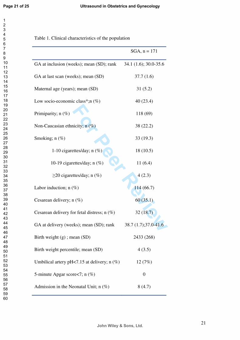
For Peer Review
21
Table 1. Clinical characteristics of the population
SGA, n = 171
GA at inclusion (weeks); mean (SD); rank 34.1 (1.6); 30.0-35.6
GA at last scan (weeks); mean (SD) 37.7 (1.6)
Maternal age (years); mean (SD) 31 (5.2)
Low socio-economic class*;n (%) 40 (23.4)
Primiparity; n (%) 118 (69)
Non-Caucasian ethnicity; n (%) 38 (22.2)
Smoking; n (%) 33 (19.3)
1-10 cigarettes/day; n (%) 18 (10.5)
10-19 cigarettes/day; n (%) 11 (6.4)
�20 cigarettes/day; n (%) 4 (2.3)
Labor induction; n (%) 114 (66.7)
Cesarean delivery; n (%) 60 (35.1)
Cesarean delivery for fetal distress; n (%) 32 (18.7)
GA at delivery (weeks); mean (SD); rank 38.7 (1.7);37.0-41.6
Birth weight (g) ; mean (SD) 2433 (268)
Birth weight percentile; mean (SD) 4 (3.5)
Umbilical artery pH<7.15 at delivery; n (%) 12 (7%)
5-minute Apgar score<7; n (%) 0
Admission in the Neonatal Unit; n (%) 8 (4.7)
Page 21 of 25
John Wiley & Sons, Ltd.
Ultrasound in Obstetrics and Gynecology
123456789101112131415161718192021222324252627282930313233343536373839404142434445464748495051525354555657585960

For Peer Review
22
SGA: small-for-gestational-age; GA: gestational age; *Routine occupations, long-term
unemployment or never worked (UK National Statistics Socio-Economic Classification)
Page 22 of 25
John Wiley & Sons, Ltd.
Ultrasound in Obstetrics and Gynecology
123456789101112131415161718192021222324252627282930313233343536373839404142434445464748495051525354555657585960
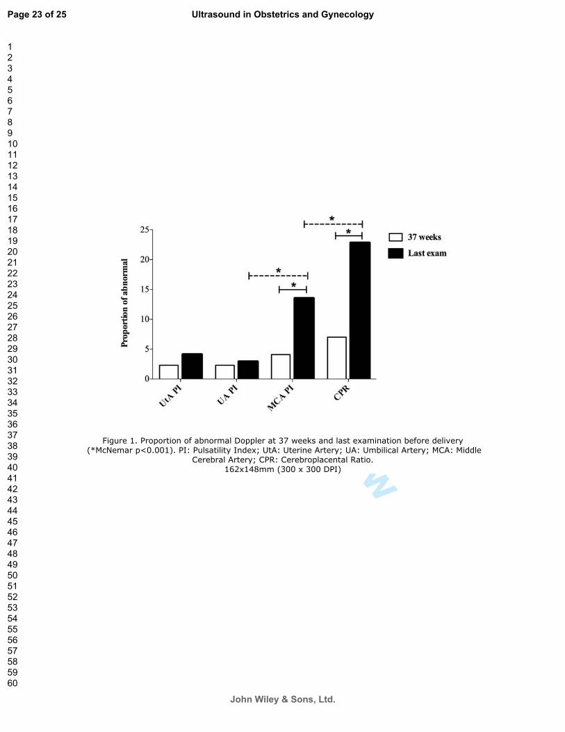
For Peer Review
= ����[\�<��������������������������������]^�Z��_������������V������������������Q�����`q�{������|}\}}[~\�<!��<���������!���V*���$���������$�����*��$���������$�����*��&$��������
&��������$�����*�&<?��&���������������?���\��[��V[�����`]}}�V�]}}��<!~��
Page 23 of 25
John Wiley & Sons, Ltd.
Ultrasound in Obstetrics and Gynecology
123456789101112131415161718192021222324252627282930313233343536373839404142434445464748495051525354555657585960
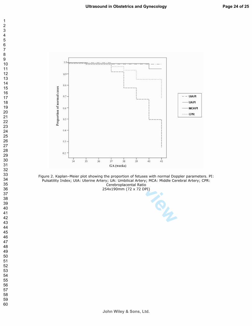
For Peer Review
= �����\�X�������������������Z� ��������������������������Z����������������������������\�<!��<���������!���V*���$���������$�����*��$���������$�����*��&$��������&��������$�����*�&<?��
&���������������?��������V[�}���`^��V�^���<!~��
Page 24 of 25
John Wiley & Sons, Ltd.
Ultrasound in Obstetrics and Gynecology
123456789101112131415161718192021222324252627282930313233343536373839404142434445464748495051525354555657585960
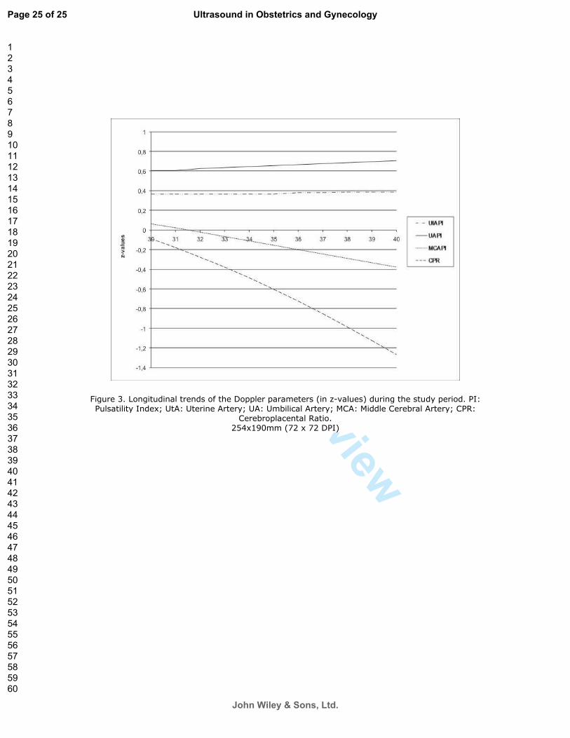
For Peer Review
= ����]\���� ����������������������������������������`��>�Q�����~����� ����������������\�<!��<���������!���V*���$���������$�����*��$���������$�����*��&$��������&��������$�����*�&<?��
&���������������?���\�����V[�}���`^��V�^���<!~��
Page 25 of 25
John Wiley & Sons, Ltd.
Ultrasound in Obstetrics and Gynecology
123456789101112131415161718192021222324252627282930313233343536373839404142434445464748495051525354555657585960
DOI: 10.1542/peds.2008-3346 2009;124;e934-e941; originally published online Oct 26, 2009; Pediatrics
Hernandez-Andrade, Francesc Botet, Carme Costas-Moragas and Eduard Gratacos Francesc Figueras, Daniel Oros, Rogelio Cruz-Martinez, Nelly Padilla, Edgar
Placental FunctionNeurobehavior in Term, Small-for-Gestational Age Infants With Normal
http://www.pediatrics.org/cgi/content/full/124/5/e934located on the World Wide Web at:
The online version of this article, along with updated information and services, is
rights reserved. Print ISSN: 0031-4005. Online ISSN: 1098-4275. Grove Village, Illinois, 60007. Copyright © 2009 by the American Academy of Pediatrics. All and trademarked by the American Academy of Pediatrics, 141 Northwest Point Boulevard, Elkpublication, it has been published continuously since 1948. PEDIATRICS is owned, published, PEDIATRICS is the official journal of the American Academy of Pediatrics. A monthly
. Provided by Health Internetwork on October 28, 2009 www.pediatrics.orgDownloaded from
Neurobehavior in Term, Small-for-Gestational Age
Infants With Normal Placental Function
WHAT’S KNOWN ON THIS SUBJECT: IUGR is associated with
behavioral, sensorial, and cognitive dysfunctions in childhood and
adolescence. Several studies on preterm infants correlated these
difficulties with behavioral disruptions already present in the neonatal
period.
WHAT THIS STUDY ADDS: This study extends the neurobehavioral
effects of being SGA to term neonates with no signs of placental
insufficiency, a subgroup of neonates traditionally considered to
be at one end of the size spectrum of normal infants.
abstractOBJECTIVE: The goal was to evaluate the neurobehavioral outcomes of
term, small-for-gestational age (SGA) newborns with normal placental
function.
METHODS: A cohort of consecutive term SGA newborns with normal
prenatal umbilical artery Doppler ultrasound findings was created and
compared with a group of term infants with size appropriate for ges-
tational age, who were sampled from our general neonatal population.
Neonatal behaviorwas evaluated at corrected age of 40� 1weekswith
the Neonatal Behavioral Assessment Scale. The effect of the study
group on each Neonatal Behavioral Assessment Scale area was ad-
justed, through multivariate analysis of covariance, for smoking dur-
ing pregnancy, maternal BMI, socioeconomic level, onset of labor,
mode of delivery, use of epidural anesthetic medication, gestational
age at delivery, postnatal age (in days) at evaluation, and gender.
RESULTS: A total of 202 newborns (102 SGA and 100 appropriate for
gestational age) were included. All of the neurobehavioral areas stud-
ied were poorer in the SGA group, with significance for attention, ha-
bituation, motor, social-interactive, and regulation of state. The aver-
age mean differences in scores between the study groups were 0.77
(95% confidence interval: 0.38–1. 14) for attention, 0.64 (95% confi-
dence interval: 0.13–1.14) for habituation, 0.52 (95% confidence inter-
val: 0.31–0.74) for motor, 0.95 (95% confidence interval: 0.54–1.37) for
social-interactive, and 0.68 (95% confidence interval: 0.23–1.13) for
regulation of state. These differences remained significant after ad-
justment for potential confounders.
CONCLUSION: Term SGA newborns with no signs of placental insuffi-
ciency had poorer neurobehavioral competencies, which suggests de-
layed neurologic maturation. Pediatrics 2009;124:e934–e941
AUTHORS: Francesc Figueras, MD,a,b,c Daniel Oros, MD,b
Rogelio Cruz-Martinez, MD,b Nelly Padilla, MD,b Edgar
Hernandez-Andrade, MD,b Francesc Botet, MD,b,c,d Carme
Costas-Moragas, PhD,e and Eduard Gratacos, PhDa,b,c
aMaternal-Fetal Medicine and dNeonatology Departments,
Hospital Clinic, and cCIBER-ER, University of Barcelona,
Barcelona, Spain; bFetal and Perinatal Research Group, August
Pi i Sunyer Institute for Biomedical Research, Barcelona, Spain;
and eDepartment of Clinical and Health Psychology, Autonomous
University of Barcelona, Barcelona, Spain
KEY WORDSsmall for gestational age, intrauterine growth restriction,
neurobehavioral outcome, Doppler ultrasonography
ABBREVIATIONSSGA—small for gestational age
AGA—appropriate for gestational age
IUGR—intrauterine growth restriction
NBAS—Neonatal Behavioral Assessment Scale
www.pediatrics.org/cgi/doi/10.1542/peds.2008-3346
doi:10.1542/peds.2008-3346
Accepted for publication Jun 4, 2009
Address correspondence to Francesc Figueras, MD, Maternal-
Fetal Medicine Department, Hospital Clinic, Barcelona, Spain.
E-mail: [email protected]
PEDIATRICS (ISSN Numbers: Print, 0031-4005; Online, 1098-4275).
Copyright © 2009 by the American Academy of Pediatrics
FINANCIAL DISCLOSURE: The authors have indicated they have
no financial relationships relevant to this article to disclose.
e934 FIGUERAS et al. Provided by Health Internetwork on October 28, 2009 www.pediatrics.orgDownloaded from
Small for gestational age (SGA) and in-
trauterine growth restriction (IUGR)
have been used interchangeably. The 2
terms are not synonymous, however;
not all small newborns have IUGR, and
not all infants with IUGR are small.1
Most instances of true IUGR corre-
spond to cases of placental insufficien-
cy.2 Intrauterine placental function
evaluation through umbilical artery
Doppler ultrasonography is the clini-
cal standard used to distinguish be-
tween SGA and IUGR.3–5 In addition,
there is evidence that umbilical artery
Doppler ultrasound use in these preg-
nancies improves a number of obstet-
ric care outcomes and reduces peri-
natal deaths.6 Although abnormal
umbilical artery Doppler ultrasound
findings are associated with adverse
perinatal and neurodevelopmental
outcomes,7–10 small fetuses with nor-
mal umbilical artery Doppler ultra-
sound findings are considered to rep-
resent one end of the normal size
spectrum, and the importance of treat-
ing them in a completely differentman-
ner, compared with infants with true
IUGR, has been stressed.11,12
Many studies have found associations
between prematurity with IUGR and
later behavioral,13–15 sensorial,16,17 and
cognitive18–21 dysfunctions. Long-term
outcomes for such infants reveal a
specific profile of neurocognitive diffi-
culties, with poor executive function-
ing, cognitive inflexibility, poor creativ-
ity, and language problems.18,19 Other
studies also reported long-term cogni-
tive disadvantages for term SGA in-
fants.22,23 Some studies on preterm in-
fants with IUGR correlated these
difficulties during childhood with be-
havioral disruptions already present
in the neonatal period,13,14,24 a time
when environmental influences are
still minimal. Behavioral competencies
in newborns are mainly related to neu-
rologic maturation, and increasing ev-
idence supports a neurobiological ba-
sis for infant behavior.25 Furthermore,
several studies demonstrated correla-
tions between neonatal neurobehavioral
skills and later neurocognitive develop-
ment for preterm13,14,24 and term26,27 in-
fants. There is no informationon theneo-
natal behavior of term SGA newborns
with normal placental function.
Preliminary evidence suggested indi-
rectly that a proportion of term SGA
infants without Doppler ultrasound
signs of placental insufficiency might
have been exposed to mild hypoxia in
utero,8,28,29 but the effects of this on
brain maturation have not been investi-
gated. Thehypothesis of this study is that
term SGA infants with normal placental
function might have neurobehavioral
disruptions attributable to abnormal
brainmaturation. Accordingly, this study
was aimed at evaluating the neurobe-
havior of term SGA newborns with nor-
mal placental function.
METHODS
Subjects
A cohort was created of consecutive,
suspected SGA, singleton infants deliv-
ered at gestational ages of �37 weeks
between November 2006 and August
2008, with confirmed birth weights of
�10th percentile according to local
standards.30 Pregnancies were dated
according to the first-trimester crown-
rump length measurement.31 Exclusion
criteria included congenital malforma-
tions (including chromosomopathies
and infections) and umbilical artery
pulsatility index values of �95th per-
centile.32 Control subjects were de-
fined as singleton term infants with
size appropriate for gestational age
(AGA) (�10th percentile, according to
local standards30) and were sampled
from our general neonatal population
during the same period; they were
matched with case subjects according
to the date of delivery (�7 days). A to-
tal of 108 SGA newborns fulfilled the
inclusion criteria and were matched
with 108 AGA infants. The study proto-
col was approved by the ethics com-
mittee, and parents provided written
informed consent.
Neurobehavioral Outcomes
Neurobehavioral performance was
evaluated at corrected age of 40 � 1
weekswith the Neonatal Behavioral As-
sessment Scale (NBAS),33 which as-
sesses both cortical and subcortical
functions by evaluating 35 items; the
items are rated on a scale of 1 to 9
(with 9 being the best performance)
except for 8 curvilinear scale items,
which, according to the manual, are
rescored as linear on a 5-, 6-, or 8-point
scale. Items are grouped into 6 clus-
ters, including habituation (habitua-
tion to light, rattle, bell, and tactile
stimulation of the foot), motor (gen-
eral tone, elicited activity, spontane-
ous activity, and motor maturity),
social-interactive (responses to visual,
animate, and inanimate auditory stim-
uli and alertness), organization of
state (irritability, state lability, maxi-
mal excitation, and reaction time), reg-
ulation of state (self-quieting and
hand-to-mouth responses), and auto-
nomic nervous system. The social-
interactive cluster was subscored for
visual and auditory stimuli. In addition,
as reported recently by the authors of
the NBAS,34 an aggregation of individ-
ual items (alertness, quality of the
alert responsiveness, and cost of at-
tention) was used to evaluate the ca-
pacity of infant attention.
All evaluations were performed by 1 of
3 trained examiners accredited by the
Brazelton Institute (Harvard Medical
School, Boston, MA); they had been
tested previously for reliability and
achieved an interrater reliability level
of �90%. The examiners were blinded
to the study group and perinatal out-
comes. Neonates were assessed in the
afternoon, between feedings, in a
small, semidark, quiet room with a
ARTICLES
PEDIATRICS Volume 124, Number 5, November 2009 e935. Provided by Health Internetwork on October 28, 2009 www.pediatrics.orgDownloaded from
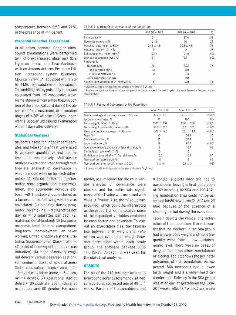
temperature between 22°C and 27°C,
in the presence of �1 parent.
Placental Function Assessment
In all cases, prenatal Doppler ultra-
sound examinations were performed
by 1 of 3 experienced observers (Drs
Figueras, Oros, and Cruz-Martinez),
with an Acuson Antares Premium Edi-
tion ultrasound system (Siemens,
Mountain View, CA) equipped with a 2.3-
to 4-MHz transabdominal transducer.
Theumbilical artery pulsatility indexwas
calculated from �3 consecutive wave-
forms obtained from a free-floating por-
tion of the umbilical cord during the ab-
sence of fetal movement, at insonation
angles of �30°. All case subjects under-
went a Doppler ultrasound examination
within 7 days after delivery.
Statistical Analyses
Student’s t test for independent sam-
ples and Pearson’s �2 test were used
to compare quantitative and qualita-
tive data, respectively. Multivariate
analyseswere conducted throughmul-
tivariate analysis of covariance in
which a model was run for each differ-
ent set of skills (attention, habituation,
motor, state organization, state regu-
lation, and autonomic nervous sys-
tem), with the study group included as
a factor and the following variables as
covariates: (1) smoking during preg-
nancy (no smoking, 1–9 cigarettes per
day, or �10 cigarettes per day); (2)
maternal BMI at booking; (3) low socio-
economic level (routine occupations,
long-term unemployment, or never
worked; United Kingdom National Sta-
tistics Socio-economic Classification);
(4) onset of labor (spontaneous versus
induction); (5) mode of delivery (vagi-
nal delivery versus cesarean section);
(6) number of doses of epidural anes-
thetic medication (bupivacaine, 1.2–
1.8 mg) during labor (none, 1–3 doses,
or �4 doses); (7) gestational age at
delivery; (8) postnatal age (in days) at
evaluation; and (9) gender. For each
model, assumptions for the multivari-
ate analysis of covariance were
checked and the multivariate signifi-
cance of the F value was assessed with
Wilks’ � P value. Also, the �2 value was
provided, which could be interpreted
as the proportion of the total variance
of the dependent variables explained
by each factor and covariate. To rule
out an expectation bias, the associa-
tion between birth weight and NBAS
scores was evaluated through Pear-
son correlation within each study
group. The software package SPSS
14.0 (SPSS, Chicago, IL) was used for
the statistical analyses.
RESULTS
For all of the 216 included infants, a
neurobehavioral assessment visit was
scheduled at corrected age of 40 � 1
weeks. Parents of 6 case subjects and
8 control subjects later declined to
participate, leaving a final population
of 202 infants (102 SGA and 100 AGA).
The habituation area could not be as-
sessed for 50 newborns (21 SGA and 29
AGA) because of the absence of a
sleeping period during the evaluation.
Table 1 depicts the clinical character-
istics of the population. It is notewor-
thy that the mothers in the SGA group
had a lower body weight and more fre-
quently were from a low socioeco-
nomic level. There were no cases of
drug consumption other than tobacco
or alcohol. Table 2 shows the perinatal
outcomes of the population. As ex-
pected, SGA newborns had a lower
birth weight and a smaller head cir-
cumference. Delivery in the SGA group
was at an earlier gestational age (SGA:
38.5 weeks; AGA: 39.7 weeks) and more
TABLE 1 Clinical Characteristics of the Population
AGA (N � 100) SGA (N � 102) Pa
Primiparity, % 61 67.6 .32
Nonwhite ethnicity, % 21.2 16 .34
Maternal age, mean � SD, y 31.8 � 4.9 33.6 � 4.9 .79
Maternal age of �21 y, % 5 2 .28
BMI at booking, mean, kg/m2 23.4 21.9 .003
Low socioeconomic level, %b 31 52 .003
Smoking, %
Nonsmoking 85 83.3 .75
1–9 cigarettes per d 12 5.9
11–19 cigarettes per d 2 7.8
�20 cigarettes per day 1 2.9
Alcohol consumption of �170 g/wk, % 5 3.9 .71
a Student’s t test for independent samples or Pearson’s �2 test.b Routine occupations, long-term unemployment, or never worked (United Kingdom National Statistics Socio-economic
Classification).
TABLE 2 Perinatal Outcomes for the Population
AGA (N � 100) SGA (N � 102) Pa
Gestational age at delivery, mean � SD, wk 39.7 � 1.1 38.5 � 1.1 �.001
Epidural anesthesia, % 92 100 .004
Birth weight, mean � SD, g 3338 � 390 2354 � 266 �.001
Birth weight percentile, mean � SD 52.2 � 26.6 3.3 � 2.2 �.001
Head circumference, mean � SD, mm 346 � 12.5 325 � 11.4 �.001
Male, % 50 56.9 .33
Cesarean section, % 25 35.5 .11
Labor induction, % 18 65.7 �.001
Operative delivery because of fetal distress, % 8 16.7 .06
5-min Apgar score of �7, % 0 0
Umbilical artery pH of �7.15 at delivery, % 3.3 10.2 .07
Neonatal unit admission, % 0 3 .08
Neonatal unit stay length, mean � SD, d 0 � 0 0.7 � 3 .025
a Student’s t test for independent samples or Pearson’s �2 test.
e936 FIGUERAS et al. Provided by Health Internetwork on October 28, 2009 www.pediatrics.orgDownloaded from
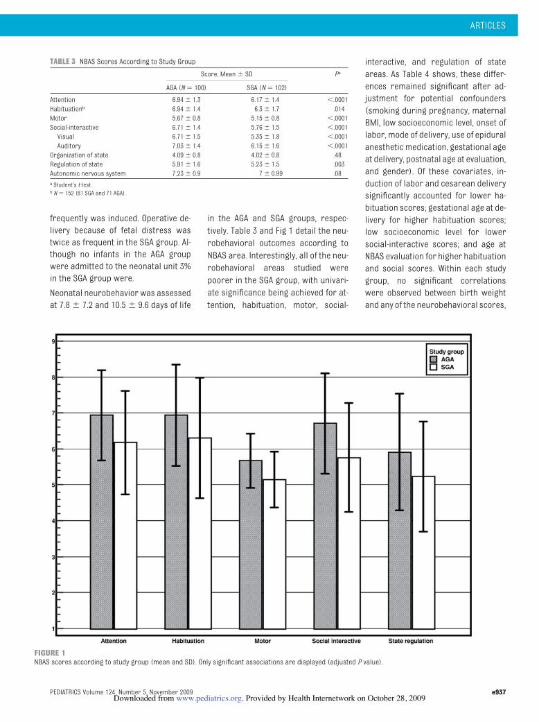
frequently was induced. Operative de-
livery because of fetal distress was
twice as frequent in the SGA group. Al-
though no infants in the AGA group
were admitted to the neonatal unit 3%
in the SGA group were.
Neonatal neurobehavior was assessed
at 7.8 � 7.2 and 10.5 � 9.6 days of life
in the AGA and SGA groups, respec-
tively. Table 3 and Fig 1 detail the neu-
robehavioral outcomes according to
NBAS area. Interestingly, all of the neu-
robehavioral areas studied were
poorer in the SGA group, with univari-
ate significance being achieved for at-
tention, habituation, motor, social-
interactive, and regulation of state
areas. As Table 4 shows, these differ-
ences remained significant after ad-
justment for potential confounders
(smoking during pregnancy, maternal
BMI, low socioeconomic level, onset of
labor, mode of delivery, use of epidural
anesthetic medication, gestational age
at delivery, postnatal age at evaluation,
and gender). Of these covariates, in-
duction of labor and cesarean delivery
significantly accounted for lower ha-
bituation scores; gestational age at de-
livery for higher habituation scores;
low socioeconomic level for lower
social-interactive scores; and age at
NBAS evaluation for higher habituation
and social scores. Within each study
group, no significant correlations
were observed between birth weight
and any of the neurobehavioral scores,
FIGURE 1NBAS scores according to study group (mean and SD). Only significant associations are displayed (adjusted P value).
TABLE 3 NBAS Scores According to Study Group
Score, Mean � SD Pa
AGA (N � 100) SGA (N � 102)
Attention 6.94 � 1.3 6.17 � 1.4 �.0001
Habituationb 6.94 � 1.4 6.3 � 1.7 .014
Motor 5.67 � 0.8 5.15 � 0.8 �.0001
Social-interactive 6.71 � 1.4 5.76 � 1.5 �.0001
Visual 6.71 � 1.5 5.35 � 1.8 �.0001
Auditory 7.03 � 1.4 6.15 � 1.6 �.0001
Organization of state 4.09 � 0.8 4.02 � 0.8 .48
Regulation of state 5.91 � 1.6 5.23 � 1.5 .003
Autonomic nervous system 7.23 � 0.9 7 � 0.99 .08
a Student’s t test.b N � 152 (81 SGA and 71 AGA).
ARTICLES
PEDIATRICS Volume 124, Number 5, November 2009 e937. Provided by Health Internetwork on October 28, 2009 www.pediatrics.orgDownloaded from
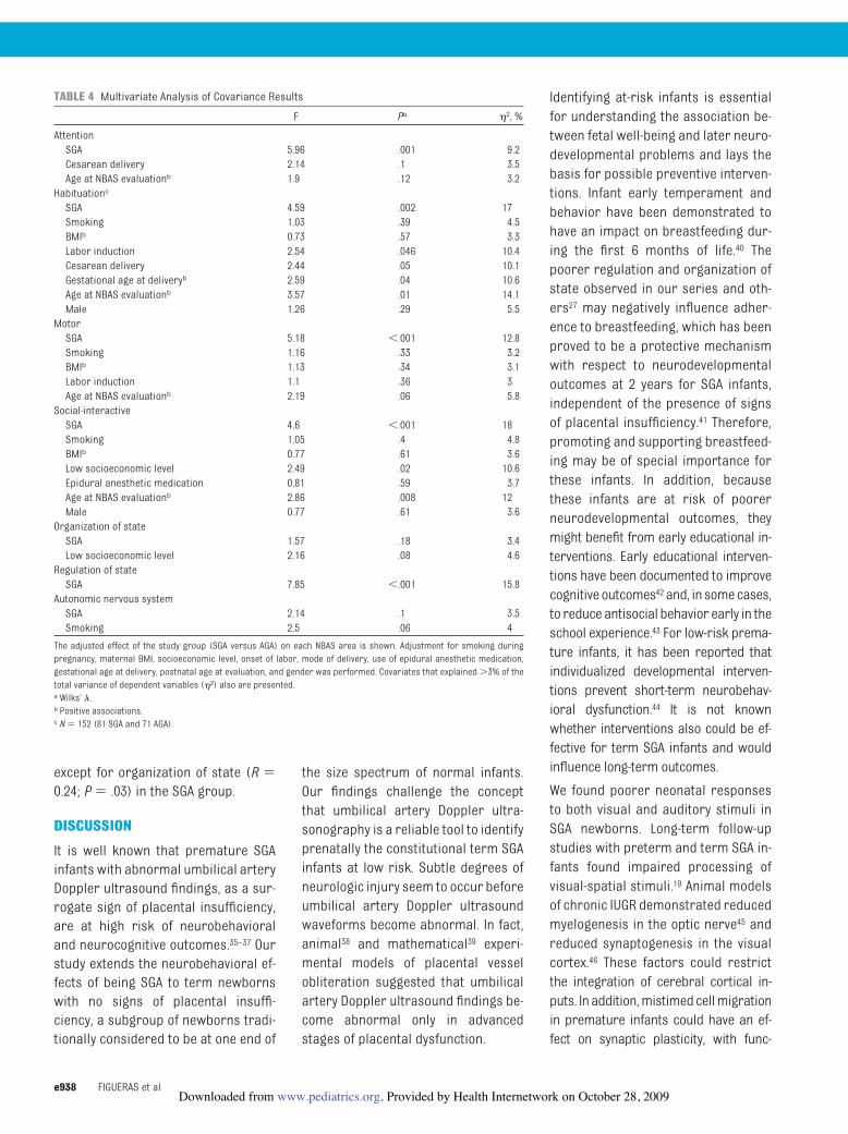
except for organization of state (R �0.24; P � .03) in the SGA group.
DISCUSSION
It is well known that premature SGA
infants with abnormal umbilical artery
Doppler ultrasound findings, as a sur-
rogate sign of placental insufficiency,
are at high risk of neurobehavioral
and neurocognitive outcomes.35–37 Our
study extends the neurobehavioral ef-
fects of being SGA to term newborns
with no signs of placental insuffi-
ciency, a subgroup of newborns tradi-
tionally considered to be at one end of
the size spectrum of normal infants.
Our findings challenge the concept
that umbilical artery Doppler ultra-
sonography is a reliable tool to identify
prenatally the constitutional term SGA
infants at low risk. Subtle degrees of
neurologic injury seem to occur before
umbilical artery Doppler ultrasound
waveforms become abnormal. In fact,
animal38 and mathematical39 experi-
mental models of placental vessel
obliteration suggested that umbilical
artery Doppler ultrasound findings be-
come abnormal only in advanced
stages of placental dysfunction.
Identifying at-risk infants is essential
for understanding the association be-
tween fetal well-being and later neuro-
developmental problems and lays the
basis for possible preventive interven-
tions. Infant early temperament and
behavior have been demonstrated to
have an impact on breastfeeding dur-
ing the first 6 months of life.40 The
poorer regulation and organization of
state observed in our series and oth-
ers27 may negatively influence adher-
ence to breastfeeding, which has been
proved to be a protective mechanism
with respect to neurodevelopmental
outcomes at 2 years for SGA infants,
independent of the presence of signs
of placental insufficiency.41 Therefore,
promoting and supporting breastfeed-
ing may be of special importance for
these infants. In addition, because
these infants are at risk of poorer
neurodevelopmental outcomes, they
might benefit from early educational in-
terventions. Early educational interven-
tions have been documented to improve
cognitiveoutcomes42 and, in somecases,
to reduce antisocial behavior early in the
school experience.43 For low-risk prema-
ture infants, it has been reported that
individualized developmental interven-
tions prevent short-term neurobehav-
ioral dysfunction.44 It is not known
whether interventions also could be ef-
fective for term SGA infants and would
influence long-term outcomes.
We found poorer neonatal responses
to both visual and auditory stimuli in
SGA newborns. Long-term follow-up
studies with preterm and term SGA in-
fants found impaired processing of
visual-spatial stimuli.19 Animal models
of chronic IUGR demonstrated reduced
myelogenesis in the optic nerve45 and
reduced synaptogenesis in the visual
cortex.46 These factors could restrict
the integration of cerebral cortical in-
puts. In addition,mistimedcellmigration
in premature infants could have an ef-
fect on synaptic plasticity, with func-
TABLE 4 Multivariate Analysis of Covariance Results
F Pa �2, %
Attention
SGA 5.96 .001 9.2
Cesarean delivery 2.14 .1 3.5
Age at NBAS evaluationb 1.9 .12 3.2
Habituationc
SGA 4.59 .002 17
Smoking 1.03 .39 4.5
BMIb 0.73 .57 3.3
Labor induction 2.54 .046 10.4
Cesarean delivery 2.44 .05 10.1
Gestational age at deliveryb 2.59 .04 10.6
Age at NBAS evaluationb 3.57 .01 14.1
Male 1.26 .29 5.5
Motor
SGA 5.18 �.001 12.8
Smoking 1.16 .33 3.2
BMIb 1.13 .34 3.1
Labor induction 1.1 .36 3
Age at NBAS evaluationb 2.19 .06 5.8
Social-interactive
SGA 4.6 �.001 18
Smoking 1.05 .4 4.8
BMIb 0.77 .61 3.6
Low socioeconomic level 2.49 .02 10.6
Epidural anesthetic medication 0.81 .59 3.7
Age at NBAS evaluationb 2.86 .008 12
Male 0.77 .61 3.6
Organization of state
SGA 1.57 .18 3.4
Low socioeconomic level 2.16 .08 4.6
Regulation of state
SGA 7.85 �.001 15.8
Autonomic nervous system
SGA 2.14 .1 3.5
Smoking 2.5 .06 4
The adjusted effect of the study group (SGA versus AGA) on each NBAS area is shown. Adjustment for smoking during
pregnancy, maternal BMI, socioeconomic level, onset of labor, mode of delivery, use of epidural anesthetic medication,
gestational age at delivery, postnatal age at evaluation, and gender was performed. Covariates that explained �3% of the
total variance of dependent variables (�2) also are presented.a Wilks’ �.b Positive associations.c N � 152 (81 SGA and 71 AGA).
e938 FIGUERAS et al. Provided by Health Internetwork on October 28, 2009 www.pediatrics.orgDownloaded from
tional and behavioral consequences.47
Visual-spatial competencies also play a
role in the neonatal capacity to select en-
vironmental stimuli toprocessand toact
on, which provides important scaffold-
ing on which attention skills are con-
structed through early childhood.48
IUGR also is associated with the develop-
ment of attention-deficit/hyperactivity
disorder symptoms in childhood and
early adolescence.49 Our finding of lower
attention capacities in SGA newborn is in
line with this finding. Two previous stud-
ies13,24 found impaired attention skills
among premature SGA infants. Struc-
tural correlations were made between
attention scores and cortical gray mat-
ter13 and hippocampal24 volumes in the
neonatal period. Whether NBAS findings
predict attention later in childhood is not
clear. However, somestudies have found
that, for term infants, attention is predic-
tive of intelligence in childhood and ado-
lescence.50,51 A study of temperament in
early infancy (5–6 months) performed
with infants born at term reported sig-
nificant prediction of attention deficits
among 8-year-old children.26
There is substantial overlap between
childhood attention disorders and the
diagnosis of motor impairment, with
poor movement skills.52 Our finding of
lower motor performance among SGA
newborns is consistent with this asso-
ciation. With a series of 40 SGA prema-
ture neonates, Feldman and Eidelman14
studied thecorrelationof neurobehavior
with later cognitive function, as mea-
suredwith the Bayley Scales of Infant De-
velopment. They found that neonatal
motor maturity was correlated with
Psychomotor Developmental Index val-
ues at 24 months.
Another interesting finding of our
study was the lower level of self-
regulation among SGA newborns, as
reflected by the regulation of state
NBAS cluster. For a series of 38 healthy
term infants, Lundqvist-Persson27 re-
ported that levels of self-regulation
were correlatedwith the infants’ levels
of cognitive development (personal-
social development, speech develop-
ment, and eye-hand coordination, sub-
variables in Griffiths’ Mental
Development Test) and with sleeping
disorders at 2 years of age. We found
no difference in autonomic nervous
system functioning in SGA infants. This
finding is consistent with previous stud-
ies that showed preserved sympathova-
gal balance in term SGA infants.53 Auto-
nomic nervous system problems are
more likely to occur in preterm infants
with acute and more-severe hypoxia.54
One strength of our study is that it in-
cluded only term SGA infants without
signs of placental dysfunction or con-
genital malformations, as low-risk SGA
infants are defined in everyday clinical
practice. No previous studies aimed at
analyzing the effect of IUGR on neu-
robehavior reported the proportion of
cases with placental insufficiency. This
is a potential source of misinterpreta-
tion of results, because an unknown
proportion of the newborns might
have been exposed to a very prolonged
period of intrauterine hypoxia. This
study also has some limitations.
Firstly, although NBAS assessment is a
standard method for evaluating new-
borns’ capacity to respond to the envi-
ronment, reflecting brain maturation,
it assesses only neurobehavior and
not cognitive function.55 However, sev-
eral studies demonstrated correla-
tions between neonatal neurobehavior
and later neurocognitive development
in preterm13,14,24 and term26,27 infants.
Secondly, it could be argued the differ-
ences between SGA and AGA infants
are likely to be clinically irrelevant, al-
beit statistically significant. However,
because behavioral clusters are rated
on a scale of 1 to 9, where 9 represents
the best performance in some areas
and 5 in others, a 1-point difference in
absolute values represents a relative
difference of 10% to 25%. Thirdly, it
could be argued that the study design
is prone to an expectation bias, be-
cause the examiners might have been
influenced by the size of the infants
during the neurobehavioral assess-
ments. Within each study group, how-
ever, no significant correlations were
found between birth weights and NBAS
scores, except for organization of state
in the SGA group. This makes it unlikely
that this potential bias could explain
the observed differences between SGA
and AGA newborns in the other areas.
In addition, our findings would be
more valid if we had repeated the NBAS
examinations to show that the behav-
ior was consistent over time. Finally,
mothers in the 2 study groups did not
match with respect to sociodemo-
graphic conditions. It is likely that other
factors associated with SGA also were
different for thesemothers. Although the
effect of the study group on neurobehav-
ior was adjusted with some of these po-
tential confounders, we cannot rule out
the possibility that some residual con-
founding is biasing our results.
CONCLUSIONS
We found poorer neurobehavioral com-
petencies, suggesting delayed neuro-
logic maturation, in a well-defined co-
hort of term SGA infants with no signs of
placental insufficiency. Whether earlier
markers of placental insufficiency or
hypoxia could be used to identify high-
risk infants andwhether timely interven-
tions for these infants could be of benefit
require further investigation.
ACKNOWLEDGMENTSFunding was provided by the Fund for
Health Research (Spain) (grant PI/
060347), the Cerebra Foundation for the
Brain-Injured Child (Carmarthen, Wales,
United Kingdom), and the Thrasher Re-
search Fund (Salt Lake City, UT). R.C. and
N.P. were supported by European Com-
missionMarieCurieHost Fellowships for
Early Stage Researchers, FETAL-MED-
019707-2.
ARTICLES
PEDIATRICS Volume 124, Number 5, November 2009 e939. Provided by Health Internetwork on October 28, 2009 www.pediatrics.orgDownloaded from
REFERENCES
1. Lee PA, Chernausek SD, Hokken-Koelega AC,
et al. International Small for Gestational
Age Advisory Board consensus develop-
ment conference statement: management
of short children born small for gestational
age, April 24–October 1, 2001. Pediatrics.
2003;111(6):1253–1261
2. Lackman F, Capewell V, Gagnon R, Richard-
son B. Fetal umbilical cord oxygen values
and birth to placental weight ratio in rela-
tion to size at birth. Am J Obstet Gynecol.
2001;185(3):674–682
3. Royal College of Obstetrics and Gynaecol-
ogy. The Investigation and Management of
the Small-for-Gestational-Age Fetus. Lon-
don, England: Royal College of Obstetrics
and Gynaecology; 2002. Green-top guideline
31
4. Gagnon R, Van den Hof M; Society of Obste-
tricians and Gynaecologists of Canada, Di-
agnostic Imaging Committee and Council.
The use of fetal Doppler in obstetrics. J Ob-
stet Gynaecol Can. 2003;25(7):601–614
5. American College of Obstetricians and Gy-
necologists, Committee on Obstetric Prac-
tice. Utility of antepartum umbilical artery
Doppler velocimetry in intrauterine growth
restriction: number 188, October 1997 (re-
places no. 116, November 1992). Int J Gynae-
col Obstet. 1997;59(3):269–270
6. Neilson JP, Alfirevic Z. Doppler ultrasound
for fetal assessment in high risk pregnan-
cies. Cochrane Database Syst Rev. 2000;(2):
CD000073
7. McCowan LM, Harding JE, Stewart AW. Um-
bilical artery Doppler studies in small for
gestational age babies reflect disease se-
verity. BJOG. 2000;107(7):916–925
8. Figueras F, Eixarch E, Gratacos E, Gardosi J.
Predictiveness of antenatal umbilical ar-
tery Doppler for adverse pregnancy out-
come in small-for-gestational-age babies
according tocustomisedbirthweight centiles:
population-based study. BJOG. 2008;115(5):
590–594
9. Valcamonico A, Danti L, Frusca T, et al. Ab-
sent end-diastolic velocity in umbilical
artery: risk of neonatal morbidity and brain
damage. Am J Obstet Gynecol. 1994;170(3):
796–801
10. Soothill PW, Ajayi RA, Campbell S, Nicolaides
KH. Prediction ofmorbidity in small and nor-
mally grown fetuses by fetal heart rate vari-
ability, biophysical profile score and umbil-
ical artery Doppler studies. Br J Obstet
Gynaecol. 1993;100(8):742–745
11. Soothill PW, Bobrow CS, Holmes R. Small for
gestational age is not a diagnosis. Ultra-
sound Obstet Gynecol. 1999;13(4):225–228
12. Bobrow CS, Soothill PW. Fetal growth
velocity: a cautionary tale. Lancet. 1999;
353(9163):1460
13. Tolsa CB, Zimine S, Warfield SK, et al. Early
alteration of structural and functional brain
development in premature infants born
with intrauterine growth restriction. Pedi-
atr Res. 2004;56(1):132–138
14. FeldmanR, Eidelman AI. Neonatal state orga-
nization, neuromaturation, mother-infant
interaction, and cognitive development in
small-for-gestational-age premature in-
fants. Pediatrics. 2006;118(3). Available at:
www.pediatrics.org/cgi/content/full/118/
3/e869
15. Schothorst PF, van Engeland H. Long-term
behavioral sequelae of prematurity. J Am
Acad Child Adolesc Psychiatry. 1996;35(2):
175–183
16. Scherjon S, Briet J, Oosting H, Kok J. The
discrepancy between maturation of visual-
evoked potentials and cognitive outcome at
five years in very preterm infants with and
without hemodynamic signs of fetal brain-
sparing. Pediatrics. 2000;105(2):385–391
17. Scherjon SA, Smolders-DeHaas H, Kok JH,
Zondervan HA. The “brain-sparing” effect:
antenatal cerebral Doppler findings in rela-
tion to neurologic outcome in very preterm
infants. Am J Obstet Gynecol. 1993;169(1):
169–175
18. Leitner Y, Fattal-Valevski A, Geva R, et al.
Neurodevelopmental outcome of children
with intrauterine growth retardation: a lon-
gitudinal, 10-year prospective study. J Child
Neurol. 2007;22(5):580–587
19. Geva R, Eshel R, Leitner Y, Fattal-Valevski A,
Harel S. Memory functions of children born
with asymmetric intrauterine growth re-
striction. Brain Res. 2006;1117(1):186–194
20. Geva R, Eshel R, Leitner Y, Valevski AF, Harel
S. Neuropsychological outcome of children
with intrauterine growth restriction: a
9-year prospective study. Pediatrics. 2006;
118(1):91–100
21. McCarton CM, Wallace IF, Divon M, Vaughan
HG Jr. Cognitive and neurologic develop-
ment of the premature, small for gesta-
tional age infant through age 6: comparison
by birth weight and gestational age. Pediat-
rics. 1996;98(6):1167–1178
22. Larroque B, Bertrais S, Czernichow P, Leger
J. School difficulties in 20-year-olds who
were born small for gestational age at term
in a regional cohort study. Pediatrics. 2001;
108(1):111–115
23. O’Keeffe MJ, O’Callaghan M, Williams GM,
Najman JM, Bor W. Learning, cognitive, and
attentional problems in adolescents born
small for gestational age. Pediatrics. 2003;
112(2):301–307
24. Lodygensky GA, Seghier ML, Warfield SK, et
al. Intrauterine growth restriction affects
the preterm infant’s hippocampus. Pediatr
Res. 2008;63(4):438–443
25. Herschkowitz N, Kagan J, Zilles K. Neurobio-
logical bases of behavioral development in
the first year. Neuropediatrics. 1997;28(6):
296–306
26. Olson SL, Bates JE, Sandy JM, Schilling EM.
Early developmental precursors of impul-
sive and inattentive behavior: from infancy
to middle childhood. J Child Psychol Psychi-
atry. 2002;43(4):435–447
27. Lundqvist-Persson C. Correlation between
level of self-regulation in the newborn in-
fant and developmental status at two years
of age. Acta Paediatr. 2001;90(3):345–350
28. Eixarch E, Meler E, Iraola A, et al. Neurode-
velopmental outcome in 2-year-old infants
whowere small-for-gestational age term fe-
tuses with cerebral blood flow redistribu-
tion. Ultrasound Obstet Gynecol. 2008;32(7):
894–899
29. Hershkovitz R, Kingdom JC, Geary M, Rodeck
CH. Fetal cerebral blood flow redistribution
in late gestation: identification of compro-
mise in small fetuses with normal umbilical
artery Doppler. Ultrasound Obstet Gynecol.
2000;15(3):209–212
30. Figueras F, Meler E, Iraola A, et al. Custom-
ized birthweight standards for a Spanish
population. Eur J Obstet Gynecol Reprod
Biol. 2008;136(1):20–24
31. Robinson HP, Fleming JE. A critical evalua-
tion of sonar “crown-rump length” mea-
surements. Br J Obstet Gynaecol. 1975;
82(9):702–710
32. Arduini D, Rizzo G. Normal values of pulsatility
index from fetal vessels: a cross-sectional
study on 1556 healthy fetuses. J Perinat Med.
1990;18(3):165–172
33. Nugent JK, Brazelton TB. Preventive infant
mental health: uses of the Brazelton scale.
In: Osofsky JD, Fitzgerald HE, eds. WAIMH
Handbook of Infant Mental Health, Vol 2:
Early Intervention, Evaluation and Assess-
ment. New York, NY: Wiley; 2000:157–202
34. Sagiv SK, Nugent JK, Brazelton TB, et al. Pre-
natal organochlorine exposure and mea-
sures of behavior in infancy using the Neo-
natal Behavioral Assessment Scale (NBAS).
Environ Health Perspect. 2008;116(5):
666–673
35. Vossbeck S, de Camargo OK, Grab D, Bode H,
Pohlandt F. Neonatal and neurodevelop-
mental outcome in infants born before 30
e940 FIGUERAS et al. Provided by Health Internetwork on October 28, 2009 www.pediatrics.orgDownloaded from

weeks of gestation with absent or reversed
end-diastolic flow velocities in the umbilical
artery. Eur J Pediatr. 2001;160(2):128–134
36. Montenegro N, Santos F, Tavares E, Matias
A, Barros H, Leite LP. Outcome of 88 preg-
nancies with absent or reversed end-
diastolic blood flow (ARED flow) in the um-
bilical arteries. Eur J Obstet Gynecol Reprod
Biol. 1998;79(1):43–46
37. Schreuder AM, McDonnell M, Gaffney G,
Johnson A, Hope PL. Outcome at school age
following antenatal detection of absent or
reversed end diastolic flow velocity in the
umbilical artery. Arch Dis Child Fetal Neona-
tal Ed. 2002;86(2):F108–F114
38. Morrow RJ, Adamson SL, Bull SB, Ritchie JW.
Effect of placental embolization on the um-
bilical arterial velocity waveform in fetal
sheep. Am J Obstet Gynecol. 1989;161(4):
1055–1060
39. Thompson RS, Stevens RJ. Mathematical
model for interpretation of Doppler velocity
waveform indices. Med Biol Eng Comput.
1989;27(3):269–276
40. Niegel S, Ystrom E, Hagtvet KA, Vollrath ME.
Difficult temperament, breastfeeding, and
their mutual prospective effects: the Nor-
wegian Mother and Child Cohort Study.
J Dev Behav Pediatr. 2008;29(6):458–462
41. McCowan LM, Pryor J, Harding JE. Perinatal
predictors of neurodevelopmental outcome
in small-for-gestational-age children at 18
months of age. Am J Obstet Gynecol. 2002;
186(5):1069–1075
42. Barnett WS. Long-term effects of early child-
hood programs on cognitive and school out-
comes. Future Child. 1995;5(3):25–50
43. Yoshikawa H. Long-term effects of early
childhood programs on social outcomes
and delinquency. Future Child. 1995;5(3):
51–75
44. Buehler DM, Als H, Duffy FH, McAnulty GB,
Liederman J. Effectiveness of individualized
developmental care for low-risk preterm
infants: behavioral and electrophysiologic
evidence. Pediatrics. 1995;96(5):923–932
45. Rees S, Bainbridge A. The structural and
neurochemical development of the fetal
guinea pig retina and optic nerve in experi-
mental growth retardation. Int J Dev Neuro-
sci. 1992;10(1):93–108
46. Bisignano M, Rees S. The effects of intra-
uterine growth retardation on synapto-
genesis and mitochondrial formation in
the cerebral and cerebellar cortices of
fetal sheep. Int J Dev Neurosci. 1988;6(5):
453–460
47. Hutton JL, Pharoah PO, Cooke RW, Steven-
son RC. Differential effects of preterm birth
and small gestational age on cognitive and
motor development. Arch Dis Child Fetal
Neonatal Ed. 1997;76(2):F75–F81
48. Smith SE, Chatterjee A. Visuospatial atten-
tion in children. Arch Neurol. 2008;65(10):
1284–1288
49. Hultman CM, Torrång A, Tuvblad C, Cnattin-
gius S, Larsson JO, Lichtenstein P. Birth
weight and attention-deficit/hyperactivity
symptoms in childhood and early ado-
lescence: a prospective Swedish twin study.
J Am Acad Child Adolesc Psychiatry. 2007;
46(3):370–377
50. Sigman M, Cohen S, Beckwith L. Why does
infant attention predict adolescent intelli-
gence? Infant Behav Dev. 1997;20(2):133–140
51. SigmanM, Cohen S, Beckwith L. Infant atten-
tion in relation to intellectual abilities in
childhood. Dev Psychol. 1986;22(6):788–792
52. Pitcher TM, Piek JP, Hay DA. Fine and gross
motor ability in males with ADHD. Dev Med
Child Neurol. 2003;45(8):525–535
53. Schaffer L, Burkhardt T, Muller-Vizentini D,
et al. Cardiac autonomic balance in small-
for-gestational-age neonates. Am J Physiol
Heart Circ Physiol. 2008;294(2):H884–H890
54. De Rogalski Landrot I, Roche F, Pichot V,
et al. Autonomic nervous system activity in
premature and full-term infants from theo-
retical term to 7 years. Auton Neurosci.
2007;136(1–2):105–109
55. Brazelton TB. Neonatal Intensive Care Unit
Network Neurobehavioral Scale: preface.
Pediatrics. 2004;113(3):632–633
ARTICLES
PEDIATRICS Volume 124, Number 5, November 2009 e941. Provided by Health Internetwork on October 28, 2009 www.pediatrics.orgDownloaded from

DOI: 10.1542/peds.2008-3346 2009;124;e934-e941; originally published online Oct 26, 2009; Pediatrics
Hernandez-Andrade, Francesc Botet, Carme Costas-Moragas and Eduard Gratacos Francesc Figueras, Daniel Oros, Rogelio Cruz-Martinez, Nelly Padilla, Edgar
Placental FunctionNeurobehavior in Term, Small-for-Gestational Age Infants With Normal
& ServicesUpdated Information
http://www.pediatrics.org/cgi/content/full/124/5/e934including high-resolution figures, can be found at:
References
http://www.pediatrics.org/cgi/content/full/124/5/e934#BIBLat: This article cites 50 articles, 13 of which you can access for free
Subspecialty Collections
nhttp://www.pediatrics.org/cgi/collection/premature_and_newbor
Premature & Newbornfollowing collection(s): This article, along with others on similar topics, appears in the
Permissions & Licensing
http://www.pediatrics.org/misc/Permissions.shtmltables) or in its entirety can be found online at: Information about reproducing this article in parts (figures,
Reprints http://www.pediatrics.org/misc/reprints.shtml
Information about ordering reprints can be found online:
. Provided by Health Internetwork on October 28, 2009 www.pediatrics.orgDownloaded from
Ultrasound Obstet Gynecol (2010)Published online in Wiley InterScience (www.interscience.wiley.com). DOI: 10.1002/uog.7588
Middle versus anterior cerebral artery Doppler for theprediction of perinatal outcome and neonatal neurobehaviorin term small-for-gestational-age fetuses with normalumbilical artery Doppler
D. OROS, F. FIGUERAS, R. CRUZ-MARTINEZ, N. PADILLA, E. MELER,E. HERNANDEZ-ANDRADE and E. GRATACOSDepartment of Maternal-Fetal Medicine, Institute Clinic for Gynecology, Obstetrics and Neonatology, Hospital Clinic-IDIBAPS, Universityof Barcelona and Centro de Investigacion Biomedica en Enfermedades Raras (CIBER-ER), Barcelona, Spain
KEYWORDS: anterior cerebral artery; Doppler; intrauterine growth restriction; middle cerebral artery; perinatal outcome
ABSTRACT
Objective To evaluate whether anterior cerebral artery(ACA) Doppler ultrasonography is superior to middlecerebral artery (MCA) Doppler in the prediction ofperinatal outcome and neonatal neurobehavior in termsmall-for-gestational age (SGA) fetuses with normalumbilical artery (UA) Doppler.
Methods MCA and ACA Doppler ultrasonography wasperformed in a cohort of SGA term fetuses withnormal UA Doppler. Perinatal outcome and neonatalneurobehavioral performance were compared with agroup of term appropriate-for-gestational age (AGA)infants. Neurobehavior was evaluated at 40 (± 1) weeksof corrected age with the Neonatal Behavioral AssessmentScale. Differences between the study groups were adjustedfor potential confounding variables by multiple linear orlogistic regression analyis.
Results A total of 199 newborns (98 SGA and 101 AGA)were included. Among the SGA fetuses, 28.6 and 17%had MCA and ACA redistribution, respectively. Caseswith either type of redistribution had an increased riskfor adverse outcome, with no differences in predictiveperformance between the two parameters. SGA fetuseswith MCA redistribution compared with controls had anincreased risk for abnormal neurobehavioral performancein motor (36 vs. 20%; adjusted P = 0.02) and stateorganization (25 vs. 17.5%; adjusted P = 0.03) areas.SGA fetuses with ACA redistribution had only anincreased risk for abnormal neurobehavioral performance
area in state organization compared with controls (30 vs.17.5%; adjusted P = 0.021).
Conclusion In term SGA newborns with no signs ofbrain-sparing, ACA Doppler investigation does notprovide any benefit over MCA in terms of the predictionof adverse perinatal outcome. Copyright © 2010 ISUOG.Published by John Wiley & Sons, Ltd.
INTRODUCTION
Small fetuses with normal umbilical artery (UA) Dopplerultrasound findings are currently defined as normalsmall-for-gestational-age (SGA) fetuses1,2. Earlier reportssuggested that this diagnostic category might essentiallycontain constitutionally small fetuses3, but recent evi-dence suggests that a substantial proportion of fetuseshave true growth restriction4. Studies over the last decadehave provided evidence that perinatal outcome may besignificantly poorer in SGA fetuses2–5,6. Furthermore,there is an increased prevalence of abnormal neurobe-havioral and neurodevelopmental tests in childhood, withsimilar features to those described for preterm childrenwho had intrauterine growth restriction4,7–9. Since theidentification of SGA fetuses with milder forms of growthrestriction cannot be based on UA Doppler, the use of mid-dle cerebral artery (MCA) ultrasonography might help toidentify these cases6,10,11. Up to 20% of SGA fetuses havea reduced pulsatility index (PI) in the MCA, a sign that isassociated with poorer perinatal outcome6,11 and with an
Correspondence to: Dr F. Figueras, Maternal-Fetal Medicine Department, ICGON Hospital Clinic, Barcelona, Spain(e-mail: [email protected])
Accepted: 3 July 2009
Copyright © 2010 ISUOG. Published by John Wiley & Sons, Ltd. ORIGINAL PAPER
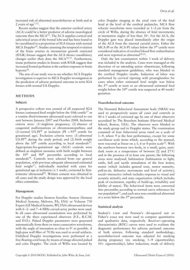
Oros et al.
increased risk of abnormal neurobehavior at birth and at2 years of age7,12.
Recent studies suggest that the anterior cerebral artery(ACA) could be a better predictor of adverse neurologicaloutcome than the MCA13. The ACA supplies cortical andsubcortical areas of the frontal lobe and has been found tobe vasodilated in a proportion of SGA fetuses with normalMCA Doppler14. Studies assessing the temporal evolutionof the brain arteries in intrauterine growth restricted(IUGR) fetuses suggest that the ACA shows vasodilatorychanges earlier than does the MCA14,15. Furthermore,tissue perfusion studies in fetuses with IUGR suggest thatincreased frontal perfusion is the earliest response to brainhypoxia16.
The aim of our study was to see whether ACA Dopplerinvestigation is superior to MCA Doppler investigation inthe prediction of adverse perinatal outcome in term SGAfetuses with normal UA Doppler.
METHODS
Subjects
A prospective cohort was created of all suspected SGAfetuses (estimated fetal weight below the 10th centile17 ata routine third-trimester ultrasound scan) referred to ourunit between January 2007 and October 2008. Inclusioncriteria were: (1) singleton pregnancy; (2) absence ofcongenital malformations or chromosomopathies; and(3) normal UA-PI18 at inclusion (PI < 95th centile forgestational age). Exclusion criteria were: (1) abnormalUA-PI18 during the study period; and (2) birth weightabove the 10th centile according to local standards19.Appropriate-for-gestational age (AGA) controls weredefined as singleton neonates with birth weight betweenthe 10th and 90th percentiles according to localstandards19. Controls were selected from our generalpopulation, with previous adequate ultrasound estimatedfetal weight17, individually matched with cases forgestational age at inclusion (± 1 week), corrected by first-trimester ultrasound20. Written consent was obtained inall cases and the study design was approved by the localethics committee.
Management
For Doppler studies Siemens Sonoline Antares (SiemensMedical Systems, Malvern, PA, USA) or Voluson 730Expert (GE Medical Systems, WI, USA) ultrasound deviceswith 6–2- and 7–4-MHz curved array probes were used.In all cases ultrasound examination was performed byone of the three experienced observers (F.F., R.C.M.and D.O.). Pulsed Doppler parameters were generatedautomatically from three or more consecutive waveforms,with the angle of insonation as close to 0◦ as possible. Ahigh-pass wall filter of 70 Hz was used to avoid artifacts.Umbilical Doppler investigations were performed at afree-floating cord loop, by means of image-directed pulsedand color Doppler. The circle of Willis was located by
color Doppler imaging in the axial view of the fetalhead at the level of the cerebral peduncles. MCA flowvelocity waveforms were recorded at 1–2 cm from thecircle of Willis, during the absence of fetal movements,at insonation angles of less than 30◦. For the ACA, theDoppler gate was placed immediately after the originof the ACA from the internal carotid artery. Either theMCA-PI or the ACA-PI values below the 5th centile wereconsidered indicative of cerebral blood flow redistributionand were reported as abnormal18,21.
Only the last examination within 1 week of deliverywas included in the analysis. Cases were managed at thediscretion of an attending senior obstetrician, followingstandard management guidelines, who was blinded tothe cerebral Doppler results. Induction of labor wasperformed by cervical ripening with prostaglandins forcases when either estimated fetal weight was belowthe 3rd centile at term or an ultrasound estimated fetalweight below the 10th centile was suspected at 40 weeks’gestation.
Neurobehavioral outcome
The Neonatal Behavioral Assessment Scale (NBAS) wasused to prospectively assess all cases and controls at40 ± 1 weeks of corrected age by one of three observersaccredited by The Brazelton Institute (Harvard MedicalSchool, Boston, USA). The observers were blinded tothe study group and to Doppler status. The examinationconsisted of four behavioral areas rated on a scale of1–9, where 9 is the best performance, except for somecurvilinear scale items which, according to the manual,were rescored as linear on a 5, 6 or 8-point scale22. Withthe newborn between two feeds, in a small, quiet, semi-dark room at a temperature of between 22 and 27◦Cand in the presence of at least one parent, the followingareas were analyzed: habituation (habituation to light,rattle, bell and tactile stimulation of the foot items);motor (which includes general tone, motor maturity,pull-to-sit, defensive movements and level of activity);social–interactive (which includes response to visual andacoustic stimuli); and state organization (which includespeak of excitement, rapidity of build-up, irritability andlability of states). The behavioral items were convertedinto percentiles according to normal curve references forour population23, and each area was considered abnormalat a score below the 5th percentile.
Statistical analysis
Student’s t-test and Pearson’s chi-squared test orFisher’s exact test were used to compare quantitativeand qualitative data, respectively. Receiver–operatingcharacteristics (ROC) curves were used to evaluate thediagnostic performance for adverse perinatal outcomeof both arteries. Following standard methodology,neurobehavioral outcome was adjusted for smokingduring pregnancy (no smoking; 1–9 cigarettes/day;10+ cigarettes/day); labor induction; mode of delivery
Copyright © 2010 ISUOG. Published by John Wiley & Sons, Ltd. Ultrasound Obstet Gynecol (2010).
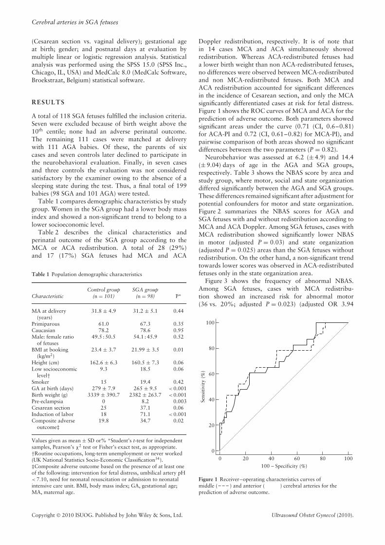
Cerebral arteries in SGA fetuses
(Cesarean section vs. vaginal delivery); gestational ageat birth; gender; and postnatal days at evaluation bymultiple linear or logistic regression analysis. Statisticalanalysis was performed using the SPSS 15.0 (SPSS Inc.,Chicago, IL, USA) and MedCalc 8.0 (MedCalc Software,Broekstraat, Belgium) statistical software.
RESULTS
A total of 118 SGA fetuses fulfilled the inclusion criteria.Seven were excluded because of birth weight above the10th centile; none had an adverse perinatal outcome.The remaining 111 cases were matched at deliverywith 111 AGA babies. Of these, the parents of sixcases and seven controls later declined to participate inthe neurobehavioral evaluation. Finally, in seven casesand three controls the evaluation was not consideredsatisfactory by the examiner owing to the absence of asleeping state during the test. Thus, a final total of 199babies (98 SGA and 101 AGA) were tested.
Table 1 compares demographic characteristics by studygroup. Women in the SGA group had a lower body massindex and showed a non-significant trend to belong to alower socioeconomic level.
Table 2 describes the clinical characteristics andperinatal outcome of the SGA group according to theMCA or ACA redistribution. A total of 28 (29%)and 17 (17%) SGA fetuses had MCA and ACA
Table 1 Population demographic characteristics
CharacteristicControl group
(n = 101)SGA group
(n = 98) P*
MA at delivery(years)
31.8 ± 4.9 31.2 ± 5.1 0.44
Primiparous 61.0 67.3 0.35Caucasian 78.2 78.6 0.95Male: female ratio
of fetuses49.5 : 50.5 54.1 : 45.9 0.52
BMI at booking(kg/m2)
23.4 ± 3.7 21.99 ± 3.5 0.01
Height (cm) 162.6 ± 6.3 160.5 ± 7.3 0.06Low socioeconomic
level†9.3 18.5 0.06
Smoker 15 19.4 0.42GA at birth (days) 279 ± 7.9 265 ± 9.5 < 0.001Birth weight (g) 3339 ± 390.7 2382 ± 263.7 < 0.001Pre-eclampsia 0 8.2 0.003Cesarean section 25 37.1 0.06Induction of labor 18 71.1 < 0.001Composite adverse
outcome‡19.8 34.7 0.02
Values given as mean ± SD or% *Student’s t-test for independentsamples, Pearson’s χ2 test or Fisher’s exact test, as appropriate.†Routine occupations, long-term unemployment or never worked(UK National Statistics Socio-Economic Classification34).‡Composite adverse outcome based on the presence of at least oneof the following: intervention for fetal distress, umbilical artery pH< 7.10, need for neonatal resuscitation or admission to neonatalintensive care unit. BMI, body mass index; GA, gestational age;MA, maternal age.
Doppler redistribution, respectively. It is of note thatin 14 cases MCA and ACA simultaneously showedredistribution. Whereas ACA-redistributed fetuses hada lower birth weight than non ACA-redistributed fetuses,no differences were observed between MCA-redistributedand non MCA-redistributed fetuses. Both MCA andACA redistribution accounted for significant differencesin the incidence of Cesarean section, and only the MCAsignificantly differentiated cases at risk for fetal distress.Figure 1 shows the ROC curves of MCA and ACA for theprediction of adverse outcome. Both parameters showedsignificant areas under the curve (0.71 (CI, 0.6–0.81)for ACA-PI and 0.72 (CI, 0.61–0.82) for MCA-PI), andpairwise comparison of both areas showed no significantdifferences between the two parameters (P = 0.82).
Neurobehavior was assessed at 6.2 (± 4.9) and 14.4(± 9.04) days of age in the AGA and SGA groups,respectively. Table 3 shows the NBAS score by area andstudy group, where motor, social and state organizationdiffered significantly between the AGA and SGA groups.These differences remained significant after adjustment forpotential confounders for motor and state organization.Figure 2 summarizes the NBAS scores for AGA andSGA fetuses with and without redistribution according toMCA and ACA Doppler. Among SGA fetuses, cases withMCA redistribution showed significantly lower NBASin motor (adjusted P = 0.03) and state organization(adjusted P = 0.025) areas than the SGA fetuses withoutredistribution. On the other hand, a non-significant trendtowards lower scores was observed in ACA-redistributedfetuses only in the state organization area.
Figure 3 shows the frequency of abnormal NBAS.Among SGA fetuses, cases with MCA redistribu-tion showed an increased risk for abnormal motor(36 vs. 20%; adjusted P = 0.023) (adjusted OR 3.94
100
80
60
Sens
itiv
ity
(%)
40
20
00 20 40
100 − Specificity (%)60 80 100
Figure 1 Receiver–operating characteristics curves ofmiddle ( ) and anterior ( ) cerebral arteries for theprediction of adverse outcome.
Copyright © 2010 ISUOG. Published by John Wiley & Sons, Ltd. Ultrasound Obstet Gynecol (2010).
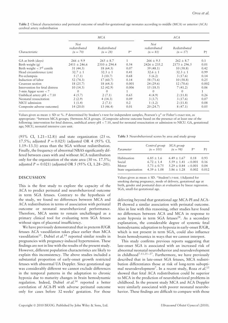
Oros et al.
Table 2 Clinical characteristics and perinatal outcome of small-for-gestational age neonates according to middle (MCA) or anterior (ACA)cerebral artery redistribution
MCA ACA
Characteristic
Notredistributed
(n = 70)Redistributed
(n = 28) P*
Notredistributed
(n = 81)Redistributed
(n = 17) P†
GA at birth (days) 266 ± 9.9 265 ± 8.7 1 266 ± 9.5 262 ± 8.7 0.1Birth weight (g) 2411 ± 246.6 2310 ± 294.4 0.54 2426 ± 235.2 2173 ± 296.5 0.01Birth weight < 3rd centile 31 (44.3) 18 (64.3) 0.07 39 (48.1) 10 (58.8) 0.42Head circumference (cm) 32.7 ± 1 32.3 ± 1 0.41 32.6 ± 1 32.3 ± 1 0.99Pre-eclampsia 5 (7.1) 3 (10.7) 0.68 5 (6.2) 3 (17.6) 0.14Induction of labor 52 (74.3) 17 (60.7) 0.14 58 (71.6) 10 (58.8) 0.25Cesarean section 18 (25.7) 18 (64.3) 0.001 24 (29.6) 12 (70.6) 0.002Intervention for fetal distress 10 (14.3) 12 (42.9) 0.006 15 (18.5) 7 (41.2) 0.065-min Apgar score < 7 0 0 1 0 0 1Umbilical artery pH < 7.10 4 (5.7) 2 (7.1) 0.65 4 (4.9) 2 (11.8) 0.26Neonatal resuscitation 2 (2.9) 4 (14.3) 0.09 3 (3.7) 3 (17.6) 0.07NICU admission 1 (1.4) 2 (7.1) 0.2 1 (1.2) 2 (11.8) 0.08Composite adverse outcome‡ 14 (20.0) 13 (46.4) 0.01 20 (24.7) 8 (47.1) 0.05
Values given as mean ± SD or %. P determined by Student’s t-test for independent samples, Pearson’s χ2 or Fisher’s exact test, asappropriate: *between MCA groups; †between ACA groups. ‡Composite adverse outcome based on the presence of at least one of thefollowing: intervention for fetal distress, umbilical artery pH < 7.10, need for neonatal resuscitation or admission to NICU. GA, gestationalage; NICU, neonatal intensive care unit.
(95% CI, 1.21–12.8)) and state organization (25 vs.17.5%; adjusted P = 0.025) (adjusted OR 4 (95% CI,1.19–13.3)) areas than the SGA without redistribution.Finally, the frequency of abnormal NBAS significantly dif-fered between cases with and without ACA redistributiononly for the organization of the state area (30 vs. 17.5%;adjusted P = 0.021) (adjusted OR 5 (95% CI, 1.28–20)).
DISCUSSION
This is the first study to explore the capacity of theACA to predict perinatal and neurobehavioral outcomein term SGA fetuses. Contrary to the hypothesis ofthe study, we found no differences between MCA andACA redistribution in terms of association with perinataloutcome or neonatal neurobehavioral performance.Therefore, MCA seems to remain unchallenged as aprimary clinical tool for evaluating term SGA fetuseswithout signs of placental insufficiency.
We have previously demonstrated that in preterm IUGRfetuses ACA vasodilation takes place earlier than MCAvasodilation15. Dubiel et al.14 reported similar results inpregnancies with pregnancy-induced hypertension. Thesefindings are not in line with the results of the present study.However, different population characteristics are likely toexplain this inconsistency. The above studies included asubstantial proportion of early-onset growth restrictedfetuses with abnormal UA Doppler. Since gestational agewas considerably different we cannot exclude differencesin the temporal patterns in the adaptation to chronichypoxia due to maturity changes in brain hemodynamicregulation. Indeed, Dubiel et al.14 reported a bettercorrelation of ACA-PI with adverse perinatal outcomeonly for cases before 32 weeks’ gestation. For those
Table 3 Neurobehavioral scores by area and study group
ParameterControl group
(n = 101)SGA group
(n = 98) P* P†
Habituation 6.85 ± 1.6 6.49 ± 1.67 0.18 0.93Social 6.72 ± 1.4 5.59 ± 1.41 < 0.001 0.16Motor 5.73 ± 0.75 5.29 ± 0.84 < 0.001 0.04State organization 4.39 ± 1.08 3.86 ± 1.28 0.002 0.012
Values given as mean ± SD. *Student’s t-test. †Adjusted forsmoking during pregnancy, mode of delivery, gestational age atbirth, gender and postnatal days at evaluation by linear regression.SGA, small-for-gestational age.
delivering beyond that gestational age MCA-PI and ACA-PI showed a similar association with perinatal outcome.Also in line with this reasoning, other studies have foundno differences between ACA and MCA in response toacute hypoxia in term SGA fetuses24. As a secondaryexplanation, the considerable degree of systemic fetalhemodynamic adaptation to hypoxia in early-onset IUGR,which is not present in term SGA, could also influencebrain hemodynamics in ways that we cannot interpret.
This study confirms previous reports suggesting thatlate-onset SGA is associated with an increased risk ofabnormal neonatal neurobehavior and neurodevelopmentin childhood7,13,25–27. Furthermore, we have previouslydescribed that in late-onset SGA fetuses, MCA redistri-bution differentiates those at risk of long-term subopti-mal neurodevelopment7. In a recent study, Roza et al.13
showed that fetal ACA redistribution could be superiorto MCA in the prediction of neurobehavioral problems inchildhood. In the present study MCA and ACA Dopplerwere similarly associated with poorer neonatal neurobe-havior. These findings are difficult to compare with those
Copyright © 2010 ISUOG. Published by John Wiley & Sons, Ltd. Ultrasound Obstet Gynecol (2010).
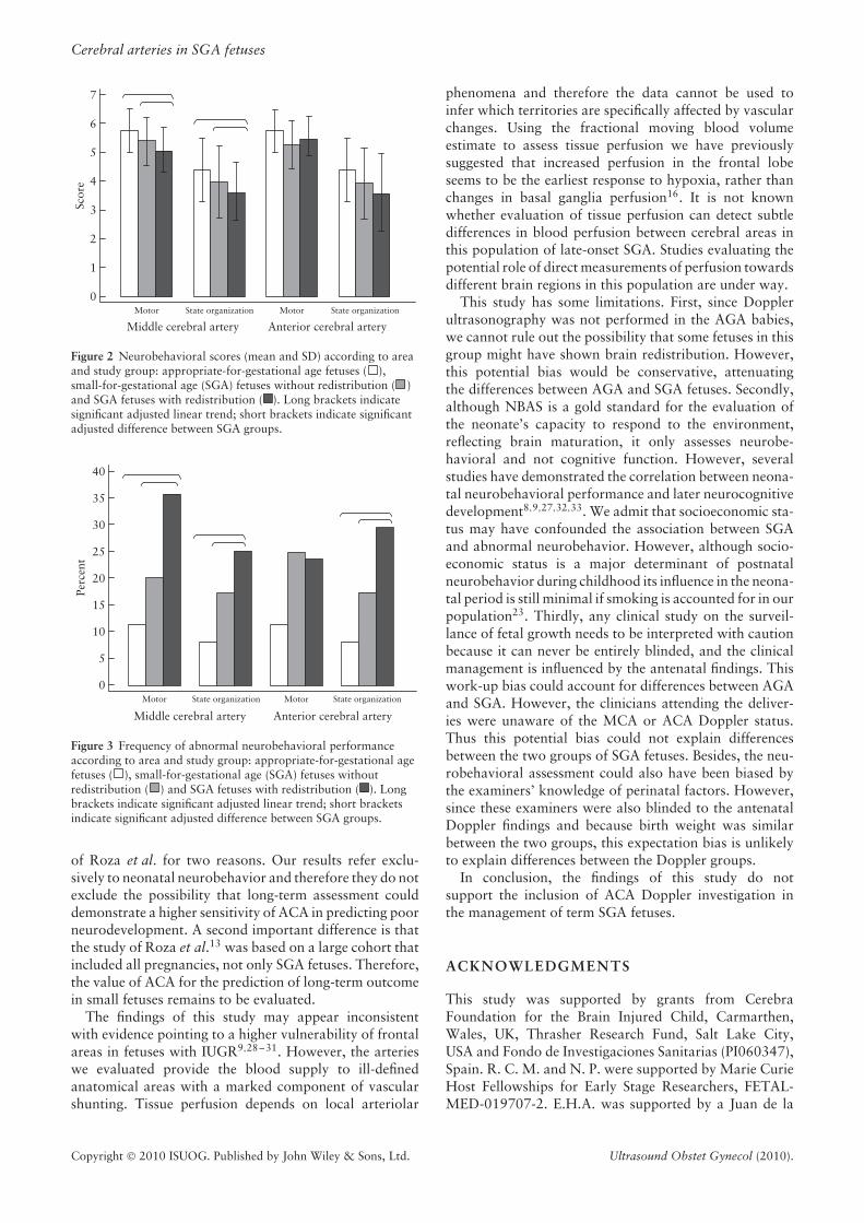
Cerebral arteries in SGA fetuses
7
6
5
4
3
2
1
0Motor State organization State organization
Anterior cerebral arteryMiddle cerebral artery
Motor
Scor
e
Figure 2 Neurobehavioral scores (mean and SD) according to areaand study group: appropriate-for-gestational age fetuses ( ),small-for-gestational age (SGA) fetuses without redistribution ( )and SGA fetuses with redistribution ( ). Long brackets indicatesignificant adjusted linear trend; short brackets indicate significantadjusted difference between SGA groups.
40
35
30
25
20
15
Perc
ent
10
5
0Motor State organization State organization
Anterior cerebral arteryMiddle cerebral artery
Motor
Figure 3 Frequency of abnormal neurobehavioral performanceaccording to area and study group: appropriate-for-gestational agefetuses ( ), small-for-gestational age (SGA) fetuses withoutredistribution ( ) and SGA fetuses with redistribution ( ). Longbrackets indicate significant adjusted linear trend; short bracketsindicate significant adjusted difference between SGA groups.
of Roza et al. for two reasons. Our results refer exclu-sively to neonatal neurobehavior and therefore they do notexclude the possibility that long-term assessment coulddemonstrate a higher sensitivity of ACA in predicting poorneurodevelopment. A second important difference is thatthe study of Roza et al.13 was based on a large cohort thatincluded all pregnancies, not only SGA fetuses. Therefore,the value of ACA for the prediction of long-term outcomein small fetuses remains to be evaluated.
The findings of this study may appear inconsistentwith evidence pointing to a higher vulnerability of frontalareas in fetuses with IUGR9,28–31. However, the arterieswe evaluated provide the blood supply to ill-definedanatomical areas with a marked component of vascularshunting. Tissue perfusion depends on local arteriolar
phenomena and therefore the data cannot be used toinfer which territories are specifically affected by vascularchanges. Using the fractional moving blood volumeestimate to assess tissue perfusion we have previouslysuggested that increased perfusion in the frontal lobeseems to be the earliest response to hypoxia, rather thanchanges in basal ganglia perfusion16. It is not knownwhether evaluation of tissue perfusion can detect subtledifferences in blood perfusion between cerebral areas inthis population of late-onset SGA. Studies evaluating thepotential role of direct measurements of perfusion towardsdifferent brain regions in this population are under way.
This study has some limitations. First, since Dopplerultrasonography was not performed in the AGA babies,we cannot rule out the possibility that some fetuses in thisgroup might have shown brain redistribution. However,this potential bias would be conservative, attenuatingthe differences between AGA and SGA fetuses. Secondly,although NBAS is a gold standard for the evaluation ofthe neonate’s capacity to respond to the environment,reflecting brain maturation, it only assesses neurobe-havioral and not cognitive function. However, severalstudies have demonstrated the correlation between neona-tal neurobehavioral performance and later neurocognitivedevelopment8,9,27,32,33. We admit that socioeconomic sta-tus may have confounded the association between SGAand abnormal neurobehavior. However, although socio-economic status is a major determinant of postnatalneurobehavior during childhood its influence in the neona-tal period is still minimal if smoking is accounted for in ourpopulation23. Thirdly, any clinical study on the surveil-lance of fetal growth needs to be interpreted with cautionbecause it can never be entirely blinded, and the clinicalmanagement is influenced by the antenatal findings. Thiswork-up bias could account for differences between AGAand SGA. However, the clinicians attending the deliver-ies were unaware of the MCA or ACA Doppler status.Thus this potential bias could not explain differencesbetween the two groups of SGA fetuses. Besides, the neu-robehavioral assessment could also have been biased bythe examiners’ knowledge of perinatal factors. However,since these examiners were also blinded to the antenatalDoppler findings and because birth weight was similarbetween the two groups, this expectation bias is unlikelyto explain differences between the Doppler groups.
In conclusion, the findings of this study do notsupport the inclusion of ACA Doppler investigation inthe management of term SGA fetuses.
ACKNOWLEDGMENTS
This study was supported by grants from CerebraFoundation for the Brain Injured Child, Carmarthen,Wales, UK, Thrasher Research Fund, Salt Lake City,USA and Fondo de Investigaciones Sanitarias (PI060347),Spain. R. C. M. and N. P. were supported by Marie CurieHost Fellowships for Early Stage Researchers, FETAL-MED-019707-2. E.H.A. was supported by a Juan de la
Copyright © 2010 ISUOG. Published by John Wiley & Sons, Ltd. Ultrasound Obstet Gynecol (2010).
Oros et al.
Cierva postdoctoral fellowship, Fondo de InvestigacionesSanitarias, Spain.
REFERENCES
1. Soothill PW, Ajayi RA, Campbell S, Nicolaides KH. Predictionof morbidity in small and normally grown fetuses by fetal heartrate variability, biophysical profile score and umbilical arteryDoppler studies. Br J Obstet Gynaecol 1993; 100: 742–745.
2. McCowan LM, Harding JE, Stewart AW. Umbilical arteryDoppler studies in small for gestational age babies reflect diseaseseverity. BJOG 2000; 107: 916–925.
3. Soothill PW, Bobrow CS, Holmes R. Small for gestational ageis not a diagnosis. Ultrasound Obstet Gynecol 1999; 13:225–228.
4. Figueras F, Eixarch E, Meler E, Iraola A, Figueras J, Puerto B,Gratacos E. Small-for-gestational-age fetuses with normalumbilical artery Doppler have suboptimal perinatal andneurodevelopmental outcome. Eur J Obstet Gynecol ReprodBiol 2008; 136: 34–38.
5. Doctor BA, O’Riordan MA, Kirchner HL, Shah D, Hack M.Perinatal correlates and neonatal outcomes of small forgestational age infants born at term gestation. Am J ObstetGynecol 2001; 185: 652–659.
6. Severi FM, Bocchi C, Visentin A, Falco P, Cobellis L, Florio P,Zagonari S, Pilu G. Uterine and fetal cerebral Doppler predictthe outcome of third-trimester small-for-gestational age fetuseswith normal umbilical artery Doppler. Ultrasound ObstetGynecol 2002; 19: 225–228.
7. Eixarch E. Neurodevelopmental outcome at two years of agein small-for-gestational age term babies with isolated middlecerebral artery vasodilation. Ultrasound Obstet Gynecol 2008;32: 894–899.
8. Feldman R, Eidelman AI. Neonatal state organization, neuro-maturation, mother–infant interaction, and cognitive develop-ment in small-for-gestational-age premature infants. Pediatrics2006; 118: e869–e878.
9. Tolsa CB, Zimine S, Warfield SK, Freschi M, Sancho Rossig-nol A, Lazeyras F, Hanquinet S, Pfizenmaier M, Huppi PS.Early alteration of structural and functional brain developmentin premature infants born with intrauterine growth restriction.Pediatr Res 2004; 56: 132–138.
10. Habek D, Salihagic A, Jugovic D, Herman R. Doppler cerebro-umbilical ratio and fetal biophysical profile in the assessmentof peripartal cardiotocography in growth-retarded fetuses. FetalDiagn Ther 2007; 22: 452–456.
11. Hershkovitz R, Kingdom JC, Geary M, Rodeck CH. Fetalcerebral blood flow redistribution in late gestation: identificationof compromise in small fetuses with normal umbilical arteryDoppler. Ultrasound Obstet Gynecol 2000; 15: 209–212.
12. Oros D, Figueras F, Padilla N, Hernandez-Andrade E, Grat-acos E. Anterior cerebral artery improves the prediction ofadverse perinatal outcome in small-for-gestational age fetuseswith normal umbilical artery. Ultrasound Obstet Gynecol 2007;30: 524.
13. Roza SJ, Steegers EA, Verburg BO, Jaddoe VW, Moll HA,Hofman A, Verhulst FC, Tiemeier H. What is spared by fetalbrain-sparing? Fetal circulatory redistribution and behavioralproblems in the general population. Am J Epidemiol 2008; 168:1145–1152.
14. Dubiel M, Gunnarsson GO, Gudmundsson S. Blood redistri-bution in the fetal brain during chronic hypoxia. UltrasoundObstet Gynecol 2002; 20: 117–121.
15. Figueroa-Diesel H, Hernandez-Andrade E, Acosta-Rojas R,Cabero L, Gratacos E. Doppler changes in the main fetal brainarteries at different stages of hemodynamic adaptation in severeintrauterine growth restriction. Ultrasound Obstet Gynecol2007; 30: 297–302.
16. Hernandez-Andrade E, Figueroa-Diesel H, Jansson T, Rangel-Nava H, Gratacos E. Changes in regional fetal cerebral bloodflow perfusion in relation to hemodynamic deterioration inseverely growth-restricted fetuses. Ultrasound Obstet Gynecol2008; 32: 71–76.
17. Hadlock FP, Harrist RB, Carpenter RJ, Deter RL, Park SK.Sonographic estimation of fetal weight. The value of femurlength in addition to head and abdomen measurements.Radiology 1984; 150: 535–540.
18. Arduini D, Rizzo G. Normal values of Pulsatility Index fromfetal vessels: a cross-sectional study on 1556 healthy fetuses.J Perinat Med 1990; 18: 165–172.
19. Figueras F, Meler E, Iraola A, Eixarch E, Coll O, Figueras J,Francis A, Gratacos E. Customized birthweight standards for aSpanish population. Eur J Obstet Gynecol Reprod Biol 2008;136: 20–24.
20. Robinson HP, Fleming JE. A critical evaluation of sonar‘‘crown–rump length’’ measurements. Br J Obstet Gynaecol1975; 82: 702–710.
21. Benavides-Serralde JA, Hernandez-Andrade E, Figueroa-Diesel H, Oros D, Feria LA, Scheier M, Figueras F, Gratacos E.Reference values for anterior cerebral artery throughoutDoppler parameters of the fetal gestation. Gynecol Obstet Invest2010; 69: 33–39.
22. Brazelton TB, Nugent JK. Neonatal Behavioral AssessmentScale (3rd edn). McKeith Press: London, 1995.
23. Costas Moragas C, Fornieles Deu A, Botet Mussons F, BoatellaCosta E, de Caceres Zurita ML. [Psychometric evaluation of theBrazelton Scale in a sample of Spanish newborns.] Psicothema2007; 19: 140–149.
24. Fu J, Olofsson P. Intracerebral regional distribution of bloodflow in response to uterine contractions in growth-restrictedhuman fetuses. Early Hum Dev 2007; 83: 607–612.
25. Abrol P, Kapoor R, Gathwala G, Tiwari S, Tiwari AD. Neona-tal behavior in full-term small for date. Indian Pediatr 1994;31: 785–789.
26. Iyer RS, Chetan R, Venkatesh A. Neonatal behavior of smallfor gestational age infants. Indian Pediatr 1989; 26: 987–991.
27. Lundqvist-Persson C. Correlation between level of self-regulation in the newborn infant and developmental statusat two years of age. Acta Paediatr 2001; 90: 345–350.
28. Geva R, Eshel R, Leitner Y, Fattal-Valevski AF, Harel S. Mem-ory functions of children born with asymmetric intrauterinegrowth restriction. Brain Res 2006; 1117: 186–194.
29. Leitner Y, Fattal-Valevski A, Geva R, Eshel R, Toledano-Alhadef H, Rotstein M, Bassan H, Radianu B, Bitchonsky O,Jaffa AJ, Harel S. Neurodevelopmental outcome of childrenwith intrauterine growth retardation: a longitudinal, 10-yearprospective study. J Child Neurol 2007; 22: 580–587.
30. Benavides-Serralde A, Hernandez-Andrade E, Fernandez-Delgado J, Plasencia W, Scheier M, Crispi F, Figueras F, Nico-laides KH, Gratacos E. Three-dimensional sonographic calcula-tion of the volume of intracranial structures in growth-restrictedand appropriate-for-gestational age fetuses. Ultrasound ObstetGynecol 2009; 33: 530–537.
31. Makhoul IR, Soudack M, Goldstein I, Smolkin T, Tamir A,Sujov P. Sonographic biometry of the frontal lobe in normal andgrowth-restricted neonates. Pediatr Res 2004; 55: 877–883.
32. Lodygensky GA, Seghier ML, Warfield SK, Tolsa CB, Sizo-nenko S, Lazeyras F, Huppi PS. Intrauterine growth restrictionaffects the preterm infant’s hippocampus. Pediatr Res 2008; 63:438–443.
33. Olson SL, Bates JE, Sandy JM, Schilling EM. Early develop-mental precursors of impulsive and inattentive behavior: frominfancy to middle childhood. J Child Psychol Psychiatry 2002;43: 435–447.
34. Office for National Statistics (UK). Standard OccupationalClassification. http://www.ons.gov.uk/about-statistics/classifications/current/SOC2000/about-soc2000/index.html. [Last up-dated 1 August 2008].
Copyright © 2010 ISUOG. Published by John Wiley & Sons, Ltd. Ultrasound Obstet Gynecol (2010).