Embed Size (px)
Citation preview

PERICARDITIS
Pericarditis is the inflammation of the membranous sac surrounding the heart. It may be manifested as an acute or chronic condition.

ACUTE PERICARDITIS
Infectious forms of acute pericarditis include….
-bacterial
-viral
-fungal
-tubercular
Noninfectious forms of acute pericarditis are….

-azotemia-presence or increase of nitrogenous waste products, especially urea, in the blood.
-acute MI
-lung cancer
-breast cancer
-leukemia
-Hodgkin’s disease
-lymphoma
-scleroderma
-trauma after thoracic surgery
-systemic lupus erythematosus
-radiation
-drug reactions
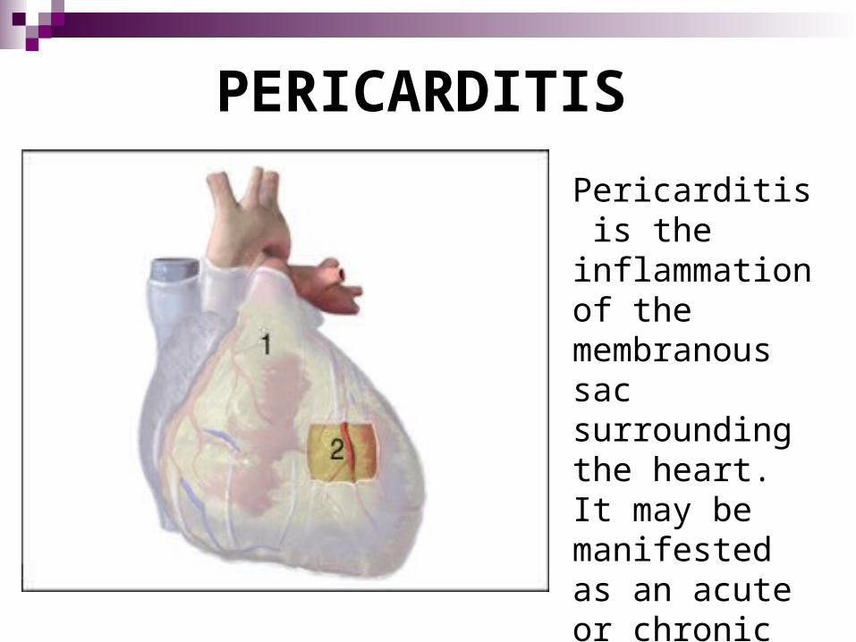
BACTERIAL PERICARDITIS
Pericardial sac
The pericardial sac has been opened to reveal an abundant amount of fibrin that is coating the heart’s surface.

VIRAL PERICARDITISThis is an example of severe viral pericarditis. Here, the pericardial sac surrounding the heart has been opened to show the amount of pus and fibrin that has accumulated around the heart.

FUNGAL PERICARDITIS
This is an example of purulent fungal pericarditis. Note the yellowish exudate that has pooled in the lower pericardial sac.
Fibrosis of the pericardial sac develops in the chronic form causing….
-fibrous constriction of the pericardium
-thickening of the pericardium
-severe compression of the pericardium due to the fibrous growth
-prevents normal filling during diastole-because of the fibrous constriction, the heart is not about to fill and pump effectively
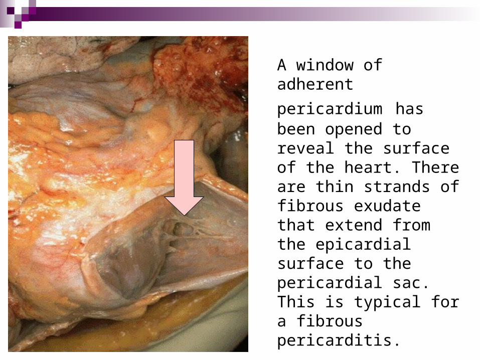
A window of adherent
pericardium has been opened to reveal the surface of the heart. There are thin strands of fibrous exudate that extend from the epicardial surface to the pericardial sac. This is typical for a fibrous pericarditis.

This is an example of a fibrous pericarditis. The surface appears roughened from the normal glistening appearance by the stands of pink and tan firbin.
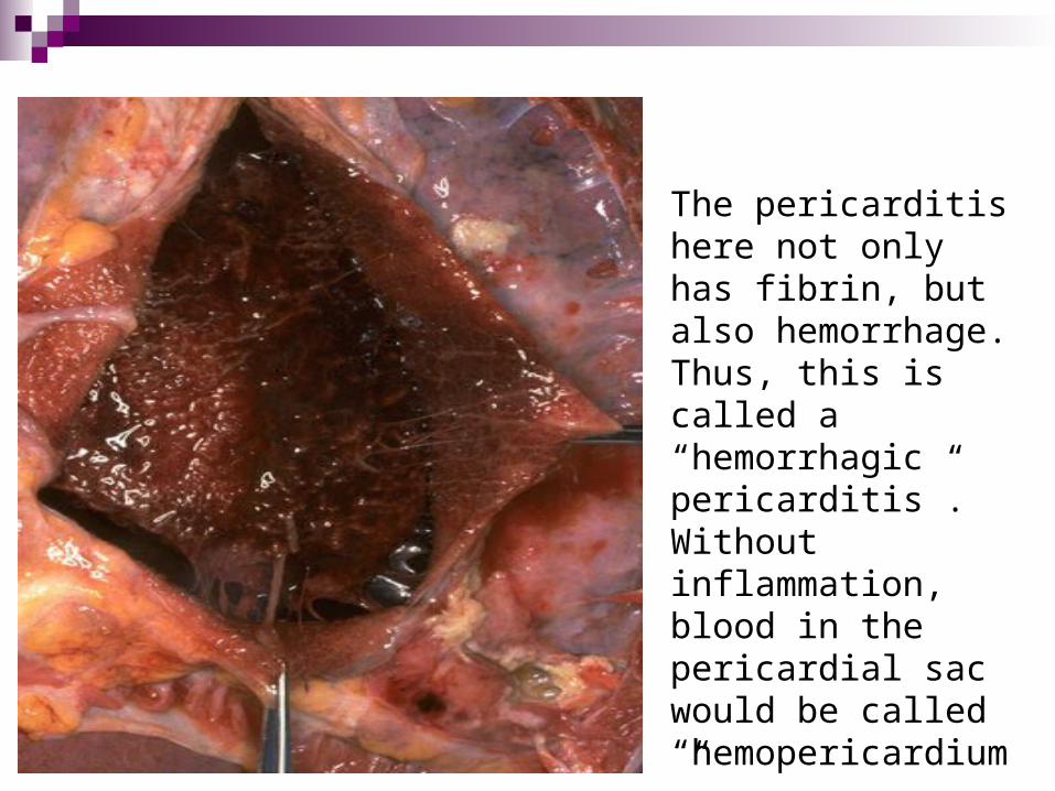
The pericarditis here not only has fibrin, but also hemorrhage. Thus, this is called a “hemorrhagic pericarditis”. Without inflammation, blood in the pericardial sac would be called “hemopericardium”.

Surgical removal of the pericardium may be necessary to restore normal cardiac output
The white stuff that you see on the pericardium is calcified, scar tissue that is filled with fibrous material. The cause of why it’s there is unknown. In this specific case they think that it was caused by a childhood virus….

Pericarditis differs clinically from other inflammatory conditions of the heart in that the presentation of debilitating pain-much like that of an MI-is common.
The pain is aggravated by…-lying supine-deep breathing-coughing-swallowing-moving the trunk of the body
The pain is alleviated by…-sitting up-leaning forward
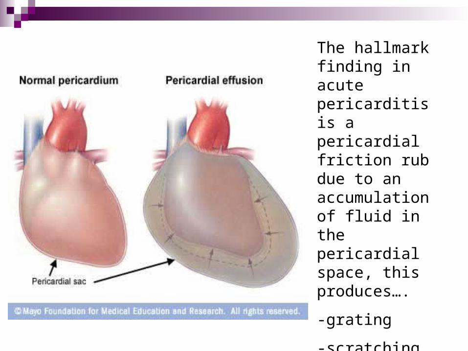
The hallmark finding in acute pericarditis is a pericardial friction rub due to an accumulation of fluid in the pericardial space, this produces….
-grating
-scratching
-leathery sounds on auscultation
-dysrhythmias
Other findings in pericarditis include….
-dypnea -nonproductive cough
-fever -anxiety
-chills -rapid pulse
-diaphoresis -shallow breaths
-leukocytosis
-muscle aches
-fatigue
-excruciating chest pain
radiating to the neck and
shoulders with severe and
sudden onset.

The most serious complication of pericarditis is a cardiac tamponade…
This is when a pericardial effusion is so large that it restricts heart movement and pumping

Electrocardiogram obtained in 17-year-old adolescent with chest pain caused by acute pericarditis. The ST segments are elevated in multiple leads. This represents the first stage of acute pericarditis.
An EKG will note changes or dysrhythmias

Visualization of the heart with an echocardiography device. The pericarditis is signaled by the arrow and corresponds to a removal of the two leaflets of the pericardium that is “the heart envelope”.
Echocardiography will show the presence of a pericardial effusion or a cardiac tamponade by ultrasound.
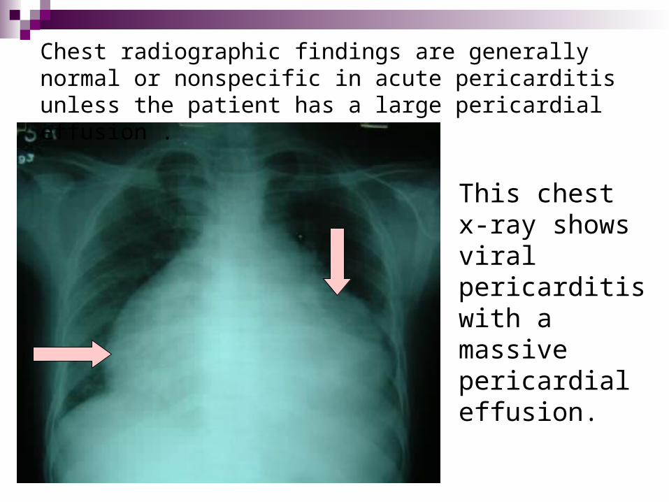
Chest radiographic findings are generally normal or nonspecific in acute pericarditis unless the patient has a large pericardial effusion .
This chest x-ray shows viral pericarditis with a massive pericardial effusion.

A CT scan gives you a picture of the body in a cross-section and it is as if you are looking down at the section. The red arrow in the picture is showing pericarditis.
Some medical management includes….
-analgesics for pain-oxygen -parenteral fluids-antibiotics if bacterial-salicylates for an elevated temperature-corticosteroids for the inflammatory process
Some surgical interventions are….
-pericardial fenestration (pericardial window)-a surgical drainage portal through the pericardium into the peritoneum to drain fluid from the pericardial space.
-pericardiocentesis
A pericardiocentesis is an invasive procedure in which the physician removes fluid from the pericardial sac. The procedure involves the insertion of a needle into the pericardium to withdraw fluid. A catheter may be inserted to allow further drainage. If necessary, the catheter will remain in place for several days for continuous drainage.
Complications of a pericardiocentesis include….
-atelectasis
-introduction to infectious agents

Pericardiocentesis

Nursing interventions….
-carefully evaluate vitals every two to four hours
-auscultate lungs and heart sounds
-administer meds as ordered
-provide physical and emotional support
-observe for further complications
-bed rest
-HOB elevated 45 degrees to decrease dyspnea
-hypothermia therapy may be necessary
-explain all procedures thoroughly
-monitor I&O and restrict sodium intake
Some nursing diagnoses….
-decreased cardiac output, related to inflammatory process
-pain, related to inflammatory process
-excess fluid volume, related to ineffective myocardial pumping action.
Prognosis….
The prognosis is fair in early stages but extremely poor if purulent and fibrous stages develop. Also, depending on the underlying cause, pericarditis usually subsides in one month or less. However, if pericarditis is caused by a disease like lupus or rheumatoid arthritis, it can persist for longer periods of time.
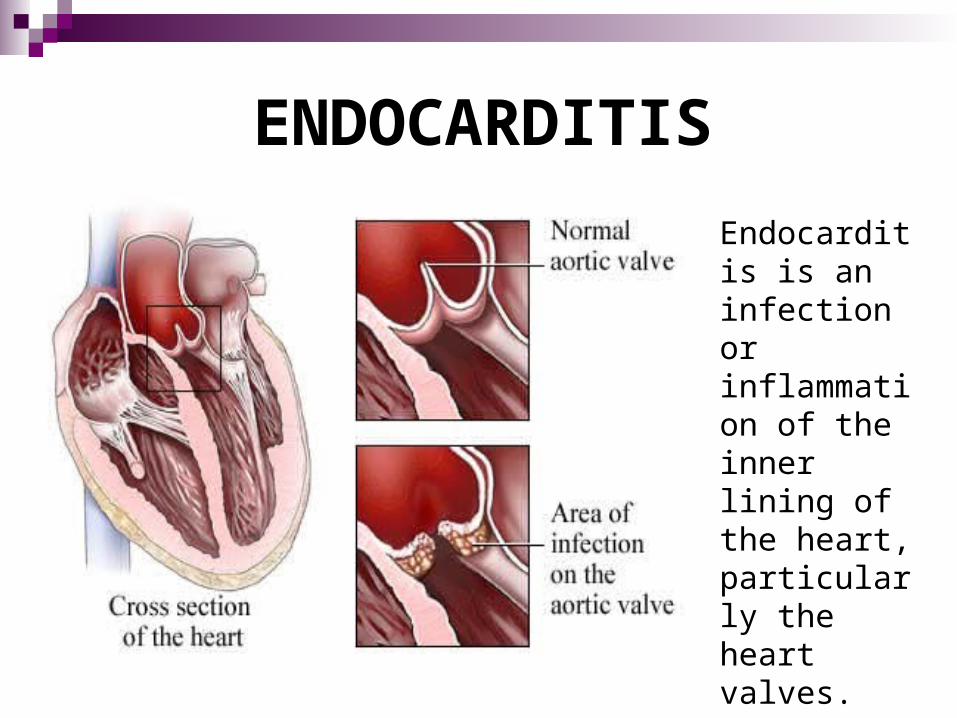
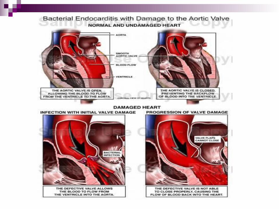
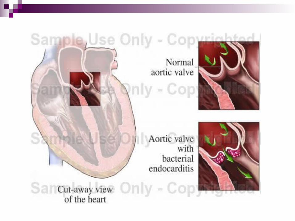
ENDOCARDITIS
Endocarditis is an infection or inflammation of the inner lining of the heart, particularly the heart valves.
It is termed infective endocarditis, causative organisms include….
-fungi -chlamydiae -rickettsiae -viruses -bacteria
Most common organisms…. -streptococcus viridans -streptococcus pyogenes -staphylococcus aureus -staphylococcus epidermidis
Endocarditis may result from….-invasion of an organism into the endocardium-injury to the lining of the endocardium-after cardiac surgery
Who’s at risk?-rheumatic heart disease-congestive heart disease-degenerative heart disease-street drugs-due to the possibility of bacterium from
contaminated needles-following intrusive procedures
-dental procedure-minor surgery-gynecological exams-insertion of indwelling urinary catheters

As organisms embed into the tissues, a vegetative (morbid outgrowth, wart-like projections made of fibrin, RBC’s, and WBC’s) growth perforates the chambers
or valve leaflets. Fibrin and calciferous growths may ulcerate and scar the valves.

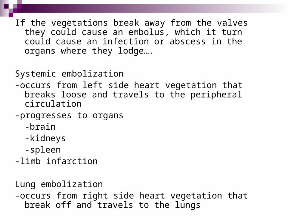
If the vegetations break away from the valves they could cause an embolus, which it turn could cause an infection or abscess in the organs where they lodge….
Systemic embolization-occurs from left side heart vegetation that breaks loose
and travels to the peripheral circulation-progresses to organs
-brain-kidneys-spleen
-limb infarction
Lung embolization-occurs from right side heart vegetation that break off and
travels to the lungs
Some signs and symptoms of endocarditis….
-flu like symptoms -splinter hemorrhages in nailbed
-fatigue -weight loss
-chest pain -rapid pulse
-headaches -onset of a murmur
-joint pain -aortic and mitral valves most
-chills commonly affected
-petechiae
-conjuntiva
-oral mucosa
-neck
-anterior chest
-abdomen
-legs

Roth spots-small white spots in the retina close to the optic disk, often surrounded by a systemic infection.
Osler nodes-small, tender cutaneous nodes, usually present in the fingers and toes. They are due to infected emboli from the heart.
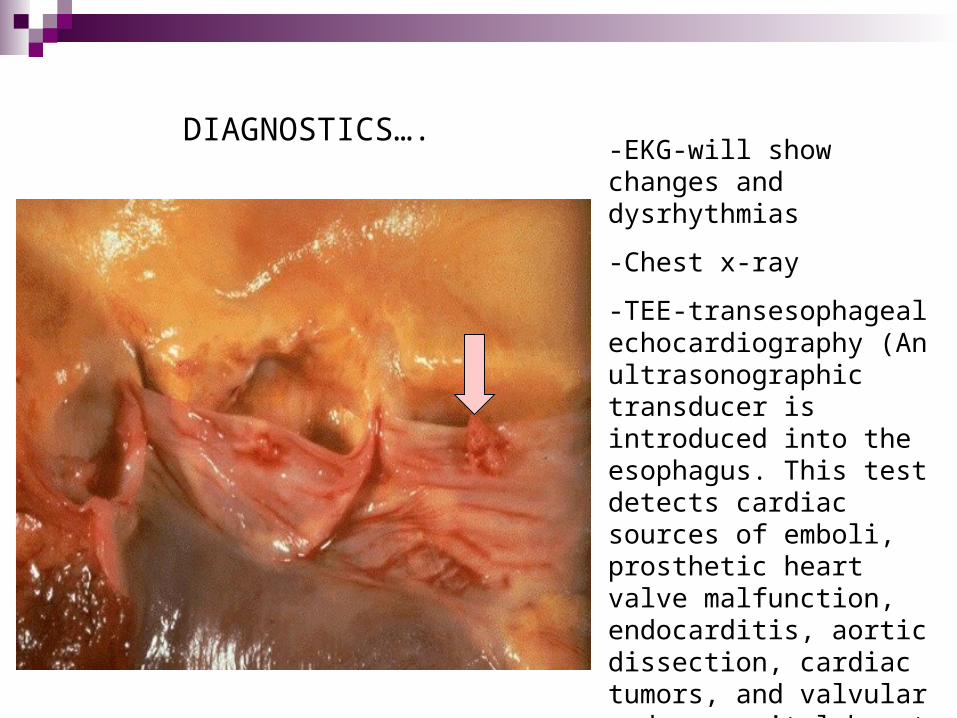
DIAGNOSTICS….-EKG-will show changes and dysrhythmias
-Chest x-ray
-TEE-transesophageal echocardiography (An ultrasonographic transducer is introduced into the esophagus. This test detects cardiac sources of emboli, prosthetic heart valve malfunction, endocarditis, aortic dissection, cardiac tumors, and valvular and congenital heart disease.

Labs would indicate….
-leukocytosis-elevated WBC count-increased erythrocyte sedimentation rate
(ESR)-test of the speed at which RBC’s settle out of un-clotted blood
-anemia-reduction in the amount of circulating red blood cells
-hyperglobulinemia-excess amount of globin (a plasma protein) in the blood.
-sensitivity tests for antibiotics
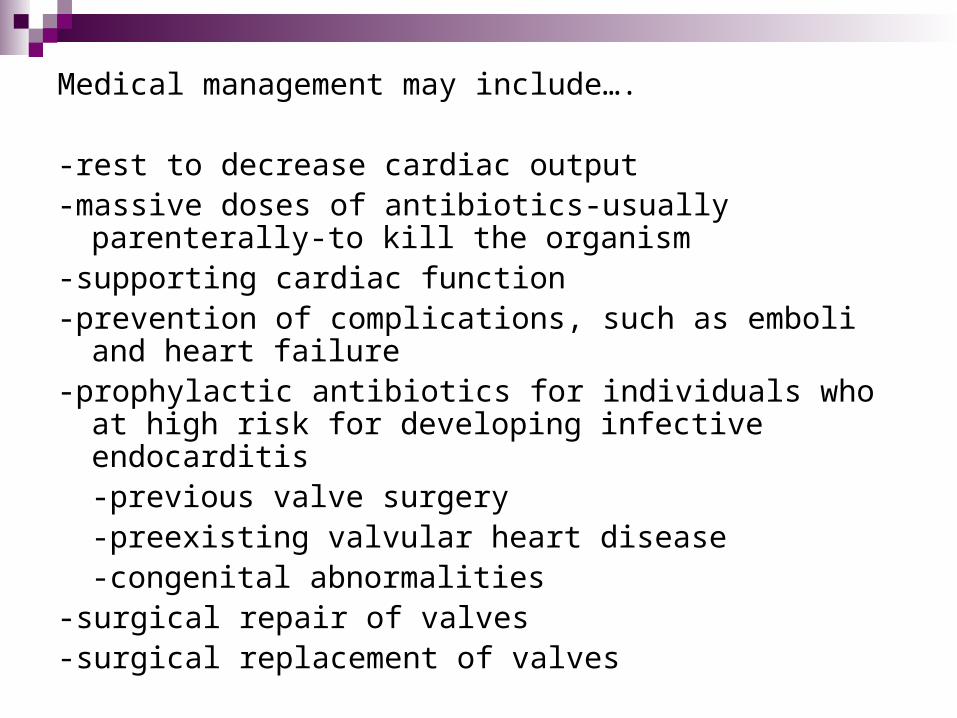
Medical management may include….
-rest to decrease cardiac output -massive doses of antibiotics-usually parenterally-to kill the
organism-supporting cardiac function-prevention of complications, such as emboli and heart
failure-prophylactic antibiotics for individuals who at high risk for
developing infective endocarditis-previous valve surgery-preexisting valvular heart disease-congenital abnormalities
-surgical repair of valves-surgical replacement of valves

Gross anatomy of aortic valve endocarditis. The left-sided image displays a quadricuspid aortic valve with prominent nodules of Arantii (arrow). The right-sided image displays a vegetation on the aortic valve (arrow). The pulmonary
valve is on the left side of this image and is bicuspid.

Nursing interventions….
-based on the signs and symptoms
-observe for petechiae
-location of pain
-vomiting
-fever
-decreased activity
-calm, quiet environment
-vitals every 4 hours, including apical pulse
-adequate nutrition
-give attractive meals to stimulate appetite and supplements in between meals
-promote rest and comfort
-preventing further inflammation and infection

Prognosis….
Before the advent of antibiotics, patients with infective endocarditis could be expected to live approximately one year; prompt treatment with intensive antibiotic therapy will now cure about 90% of patients with this condition.