Embed Size (px)
Citation preview
1
Perianal Crohn’s Disease in Canterbury,
New Zealand
Dr Tim W Eglinton
A thesis submitted for the degree of
Master of Medical Science
at the University of Otago, Dunedin,
New Zealand.
22nd December 2011
i
Acknowledgements
I would like to thank Associate Professor Richard Gearry and Professor Frank Frizelle
for their mentorship and supervision throughout the preparation of this thesis. I am
also very grateful to John Pearson of the Department of Public Health and General
Practice for his input into the statistical analysis in Chapter 3 and the resulting
publication in the American Journal of Gastroenterology. Similarly Rebecca Roberts
of the Department of Biochemistry provided genotyping and scientific advice for this
chapter. My gratitude also goes to Michaela Lion, a summer student in the
Department of Medicine, who collected the data used in Chapter 6.There are many
other members of the Departments of Surgery and Gastroenterology and their patients
whose support made this thesis possible.
Peer reviewed publications arising from the work described in this thesis
1. T Eglinton, R Roberts, J Pearson, M Barclay, T R Merriman, F A Frizelle, R B
Gearry. Clinical and Genetic Risk Factors for Perianal Crohn‟s Disease in a
Population Based Cohort. American Journal of Gastroenterology. Accepted 8th
November 2011.
2. M Lion, R B Gearry, A Day, T Eglinton. The cost of paediatric and perianal
Crohn‟s disease in Canterbury, New Zealand. NZMJ accepted 2nd
August 2011.
3. Eglinton T, Reilly M, Chang C, Barclay M, Frizelle F, Gearry R. Ileal Disease is
Associated with Surgery for Perianal Disease in a Population-Based Crohn‟s
Disease Cohort. British Journal of Surgery 2010; 97: 1103-1109.
4. Eglinton T, Gearry R. Clinical factors predicting disease course in Crohn‟s disease.
Expert Reviews in Clinical Immunology 2010; 6(1): 41-45.
ii
Peer reviewed conference proceedings arising from the work described in this
thesis
Oral Presentations
1. T. W. Eglinton, A. G. Balasingam, L. Dixon, R. B. Gearry, F. A. Frizelle.
Magnetic resonance imaging (MRI) in perianal Crohn‟s disease (CD): does it
change intra-operative decision making? New Zealand Society of
Gastroenterology, Annual Scientific Meeting, Dunedin, November 2011.
2. T. W. Eglinton, R. L. Roberts, J. F. Pearson, M. L. Barclay, L. R. Ferguson, D. Y.
Han, F. A. Frizelle, R. B. Gearry Clinical and genetic risk factors for perianal
Crohn‟s disease (CD) in a population based cohort. Tripartite Colorectal Meeting,
Cairns, July 2011.
Poster Presentations
1. T. W. Eglinton, R. L. Roberts, J. F. Pearson, T.R.Merriman, M. L. Barclay, F. A.
Frizelle, R. B. Gearry. Clinical and genetic risk factors for perianal Crohn‟s
disease (CD) in a population based cohort. New Zealand Society of
Gastroenterology, Annual Scientific Meeting, Dunedin, November 2011.
*Roche Best Poster Award.
2. T. W. Eglinton, M. L. Barclay, R.B Gearry, F.A. Frizelle. The spectrum of
perianal Crohn‟s disease in a population based cohort. New Zealand Society of
Gastroenterology, Annual Scientific Meeting, Dunedin, November 2011.
3. T. W. Eglinton, A. G. Balasingam, L. Dixon, R. B. Gearry, F. A. Frizelle.
Magnetic resonance imaging (MRI) in perianal Crohn‟s disease (CD): does it
change intra-operative decision making? Tripartite Colorectal Meeting, Cairns,
July 2011.
iii
Table of Contents
Chapter 1. Perianal CD Overview………….………………………………………….1
Chapter 2. The Spectrum of Perianal CD………………..…………………….……..36
Chapter 3. Risk Factors for Perianal CD………………………………………….….46
Chapter 4. The role of MRI in perianal CD……...…………………………………..61
Chapter 5. Perianal surgical intervention in CD……………...……………………....71
Chapter 6. Cost………...……………………………………………………………..84
Chapter 7. Discussion………………………………………………………………...95
Appendix A………………………………………………………………….……...101
Appendix B………………………………………………………………….….…..103
References……………………………………………………………….……..…...106
iv
List of Tables
1.1. The Cardiff Classification of perianal Crohn‟s Disease
1.2 The Perianal Disease Activity Index
1.3 Definition of the various types of perianal lesions in CD
1.4 The Vienna Classification of CD
1.5 The Montreal Classification of CD
1.6 Studies reporting the frequency of perianal involvement in CD
2.1 Classification of symptomatic perianal CD lesions in the Canterbury IBD cohort
2.2 Types of perianal lesions in 202 consecutive patients in a Crohn‟s disease follow-
up clinic
3.1 Clinical characteristics of CD patients with and without perianal disease
3.2 Allelic and genotypic associations between Crohn‟s disease and controls
3.3 Allelic and genotypic associations between perianal disease and controls
3.4 Allelic and genotypic associations for perianal disease within Crohn's disease
4.1 Montreal classification of included patients
4.2 Findings at EUA, MRI and final diagnosis using the outcome based gold standard
4.3 Discordant findings at EUA
5.1 Frequency of operation type for the group with a single perianal procedure
compared with those with multiple procedures
5.2 Perianal surgical interventions according to the Montreal Classification
5.3 Unadjusted and adjusted odds ratios for factors associated with PSI
v
List of Figures
1.1. Park‟s classification of cryptoglandular fistula-in-ano
1.2 MRI of perianal CD
1.3 Treatment algorithm for perianal CD fistulas
2.1 Temporal Relationship of perianal involvement to diagnosis of CD
2.2 Kaplan-Meier curve showing the proportion of patients developing perianal
Crohn‟s disease
2.3 Kaplan-Meier curve showing the proportion of patients developing perianal
fistulas
5.1 Study flow diagram
5.2 Frequency of PSI in 119 CD patients
5.3 Type of PSI in 119 patients with CD
5.4 The percentage of patients with PSI according to the Montreal Classification
5.5 Time to first PSI in 119 patients with CD
6.1 Overall cost of perianal CD divided into outpatient, hospital and indirect cost
6.2 Outpatient cost of perianal CD showing the components that made up the total
cost
6.3 Hospital cost of perianal CD showing the components that made up the total cost
6.4 Comparison of hospital and pharmaceutical cost for perianal CD
6.5 Indirect cost of perianal CD showing the components that made up the total cost
vi
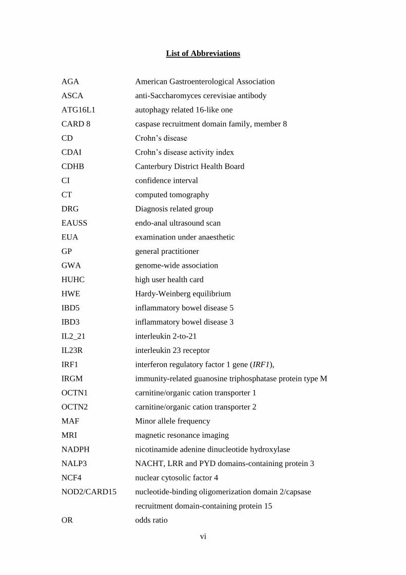
List of Abbreviations
AGA American Gastroenterological Association
ASCA anti-Saccharomyces cerevisiae antibody
ATG16L1 autophagy related 16-like one
CARD 8 caspase recruitment domain family, member 8
CD Crohn‟s disease
CDAI Crohn‟s disease activity index
CDHB Canterbury District Health Board
CI confidence interval
CT computed tomography
DRG Diagnosis related group
EAUSS endo-anal ultrasound scan
EUA examination under anaesthetic
GP general practitioner
GWA genome-wide association
HUHC high user health card
HWE Hardy-Weinberg equilibrium
IBD5 inflammatory bowel disease 5
IBD3 inflammatory bowel disease 3
IL2_21 interleukin 2-to-21
IL23R interleukin 23 receptor
IRF1 interferon regulatory factor 1 gene (IRF1),
IRGM immunity-related guanosine triphosphatase protein type M
OCTN1 carnitine/organic cation transporter 1
OCTN2 carnitine/organic cation transporter 2
MAF Minor allele frequency
MRI magnetic resonance imaging
NADPH nicotinamide adenine dinucleotide hydroxylase
NALP3 NACHT, LRR and PYD domains-containing protein 3
NCF4 nuclear cytosolic factor 4
NOD2/CARD15 nucleotide-binding oligomerization domain 2/capsase
recruitment domain-containing protein 15
OR odds ratio

vii
PCR polymerise chain reaction
PDAI perianal disease activity index
PHARMAC Pharmaceutical Management Agency in New Zealand
PHO primary health organisation
PSI perianal surgical intervention
SNP single nucleotide polymorphism
TNFα tumour necrosis factor-α
1
Chapter 1
Perianal Crohn’s Disease Overview.
1.1 Introduction
Crohn‟s disease (CD) is a chronic inflammatory bowel disease characterised by
transmural segmental inflammation that can occur in any portion of the
gastrointestinal tract from the oral cavity to the anus. It can be complicated by the
development of fibrotic strictures, perforation, abscess formation, and fistulisation [1].
The aetiology and pathogenesis remain undetermined although on-going research
suggests a multi-factorial aetiology combining genetic and environmental factors [2].
The fact that the incidence of CD is increasing worldwide [3] and that the peak age of
onset is in the productive years of life between ages 15 and 35 [4] mean it is an
increasingly significant health problem in terms of both morbidity and cost.
Crohn‟s disease has long been recognised as a protean disorder that incorporates
a range of disease phenotypes. A percentage of patients with CD will develop a
variety of perianal complications during the course of the disease which range from
mild problems requiring minimal intervention, such as anal skin tags, to severe
fistulising disease resulting in proctectomy. In addition to the morbidity associated
with the perianal lesions themselves, research suggests that patients with perianal CD
suffer from more severe intestinal disease [5, 6] requiring more intensive medical and
surgical management. Previously it was considered that perianal fistulas were closely
related to intestinal fistulas and were simply a manifestation of the same pathologic
process occurring higher in the gastrointestinal tract. However, recent research
suggests penetrating perianal disease often occurs independently of penetrating
intestinal disease [7-9] raising the possibility of separate underlying aetiological and
pathogenic factors for these disease phenotypes.
While much progress has been made in our understanding of inflammatory
bowel disease, perianal CD has attracted less attention and important gaps in our
knowledge exist. The present discussion reviews the current literature pertaining to
perianal CD incidence, classification, diagnosis, management and cost. In doing so, a
2
number of research questions are highlighted which are subsequently addressed in the
five separate but related studies presented in this thesis.
1.2 Classification and Incidence
Penner and Crohn first described the presence of a perianal fistula in a patient
with CD in 1938 [10]. This was six years after the original description of regional
ileitis [11] and a decade after Gabriel‟s finding of perianal granulomas in the absence
of tuberculosis [12]. Since then, perianal CD has become an increasingly recognised
entity with the documented perianal manifestations including; skin tags, haemorrhoids,
fissures, ulcers, perianal abscesses, fistulas, strictures and cancer.
1.2.1 Classification
1.2.1.1 Anal Skin Tags
Skin tags are common but often asymptomatic. While they may be associated
with fissures or fistulas, they can occur independently, possibly as a result of
lymphoedema from lymphatic obstruction. They have been reported to become larger
and firmer during an active CD flare.
1.2.1.2 Haemorrhoids
Haemorrhoids are a common problem in the general population but appear to
cause significant symptoms infrequently in CD patients. In a series of 50,000 patients
with haemorrhoids, 20 were noted to have CD (0.04%) [13].
1.2.1.3 Anal Fissures and Ulcers
Anal fissures in CD tend to be broad based and deep with undermining of the
edges. There may be associated skin tags and a bluish tinge to the surrounding skin.
Idiopathic anal fissures, not associated with CD, occur in the posterior midline, are
painful and are associated with high resting anal canal pressure. In contrast, fissures in
CD have classically been described as painless although reported series suggest pain
3
is a feature in a significant proportion of patients [14, 15]. CD fissures are more often
multiple and lateral than idiopathic fissures but around 40% still occur in the posterior
midline [14]. Early series of perianal CD patients also suggested CD fissures differed
from their idiopathic counterparts in that they were not associated with high resting
sphincter pressures [16]. However, more recent evidence suggests CD patients have
high resting anal canal pressures suggesting compromised anal circulation may also
play a role in the pathogenesis of CD fissures [17]. Fissures appear to be a relatively
common lesion in perianal CD occurring in 21-59% of patients in various series [18-
20]. Deep cavitating ulcers are less common than fissures and are usually associated
with rectal inflammation. They may erode into the sphincters and progress to
anorectal strictures or complex fistulas.
1.2.1.4 Perianal Abscess
Perianal abscesses are common and usually associated with a fistula-in-ano. Up
to 62% of perianal CD patients will develop an abscess at some stage [19]. Any of the
potential anorectal spaces may become infected with an abscess, however, ischiorectal
and transphincteric fistulas are particularly prone to abscess development meaning the
ischiorectal space is a common site with around 40% of abscesses occurring in this
location [21]. After surgical drainage, recurrence is common and is likely related to
the location of the associated fistula.
4
1.2.1.5 Fistula-in-ano
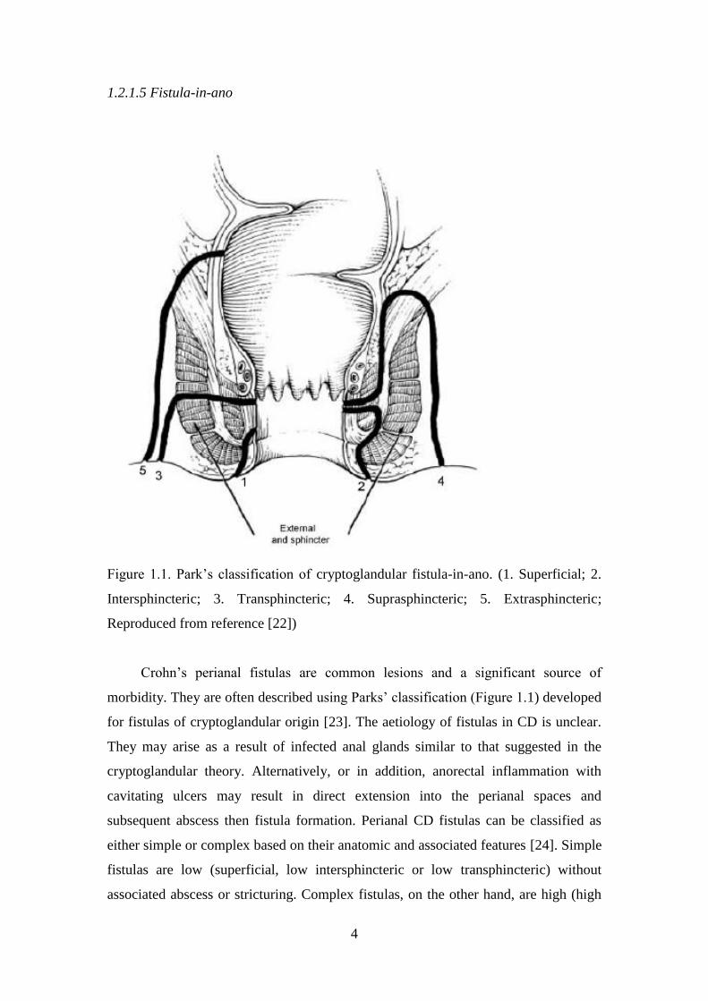
Figure 1.1. Park‟s classification of cryptoglandular fistula-in-ano. (1. Superficial; 2.
Intersphincteric; 3. Transphincteric; 4. Suprasphincteric; 5. Extrasphincteric;
Reproduced from reference [22])
Crohn‟s perianal fistulas are common lesions and a significant source of
morbidity. They are often described using Parks‟ classification (Figure 1.1) developed
for fistulas of cryptoglandular origin [23]. The aetiology of fistulas in CD is unclear.
They may arise as a result of infected anal glands similar to that suggested in the
cryptoglandular theory. Alternatively, or in addition, anorectal inflammation with
cavitating ulcers may result in direct extension into the perianal spaces and
subsequent abscess then fistula formation. Perianal CD fistulas can be classified as
either simple or complex based on their anatomic and associated features [24]. Simple
fistulas are low (superficial, low intersphincteric or low transphincteric) without
associated abscess or stricturing. Complex fistulas, on the other hand, are high (high
5
intersphincteric, transphincteric, extrasphincteric or suprasphincteric) and may be
associated with multiple external openings, an abscess, rectovaginal fistula or stricture.
This classification has significant clinical relevance as simple fistulas are more
straightforward to treat and have higher rates of healing [25].
Rectovaginal fistulas have been reported in up to 23% of women with perianal
CD [26]. As with other fistula-in-ano, they can be described using Park‟s
classification based on the relationship of the fistula tract to the sphincters;
intersphincteric, transphincteric, suprasphincteric and extrasphincteric. In the majority
(85%), the opening into the rectum or anal canal is anterior [27]. The aetiology is
likely from anorectal inflammation and ulceration penetrating into the vagina
although infected anal glands may contribute in some cases.
1.2.1.6 Anorectal strictures
These often occur in the low rectum and may be the result of chronic ulceration,
perianal sepsis or severe, active luminal inflammatory disease. Strictures appear less
common than other perianal manifestations but represent a severe form of the disease;
43% in one series requiring proctectomy [28].
1.2.1.7 Cancer
Both adenocarcinoma and squamous cell carcinoma (SCC) of the anus occur
with perianal CD. Patients with longstanding CD colitis are recognised to be at
increased risk for colorectal adenocarcinoma similar to that seen in ulcerative colitis
(UC) [29]. However this risk appears to predominantly involve colonic rather than
rectal cancer [30] when all CD patients are considered. Smaller studies looking at
perianal CD patients specifically suggest the incidence of adenocarcinoma is
increased and likely related to duration of disease and activity [31].
Anal SCC also appears to occur with greater frequency in perianal CD than the
general population. Slater et al found the relative incidence of anal cancer as a
proportion of all colorectal cancer was 14% in CD patients compared with 1.4% in
patients without CD [32]. Cancer risk is likely to be increased as a result of chronic,
6
long standing perineal inflammation and risk appears to be related to the duration of
disease. Delay in diagnosis may also occur as cancers have been demonstrated in
long standing fistulas and other perianal lesions.
1.2.1.8 Classification Systems for Perianal CD
Due to the variety of perianal CD lesions discussed above, systems have been
developed in an attempt to accurately classify the anatomic, pathologic and clinical
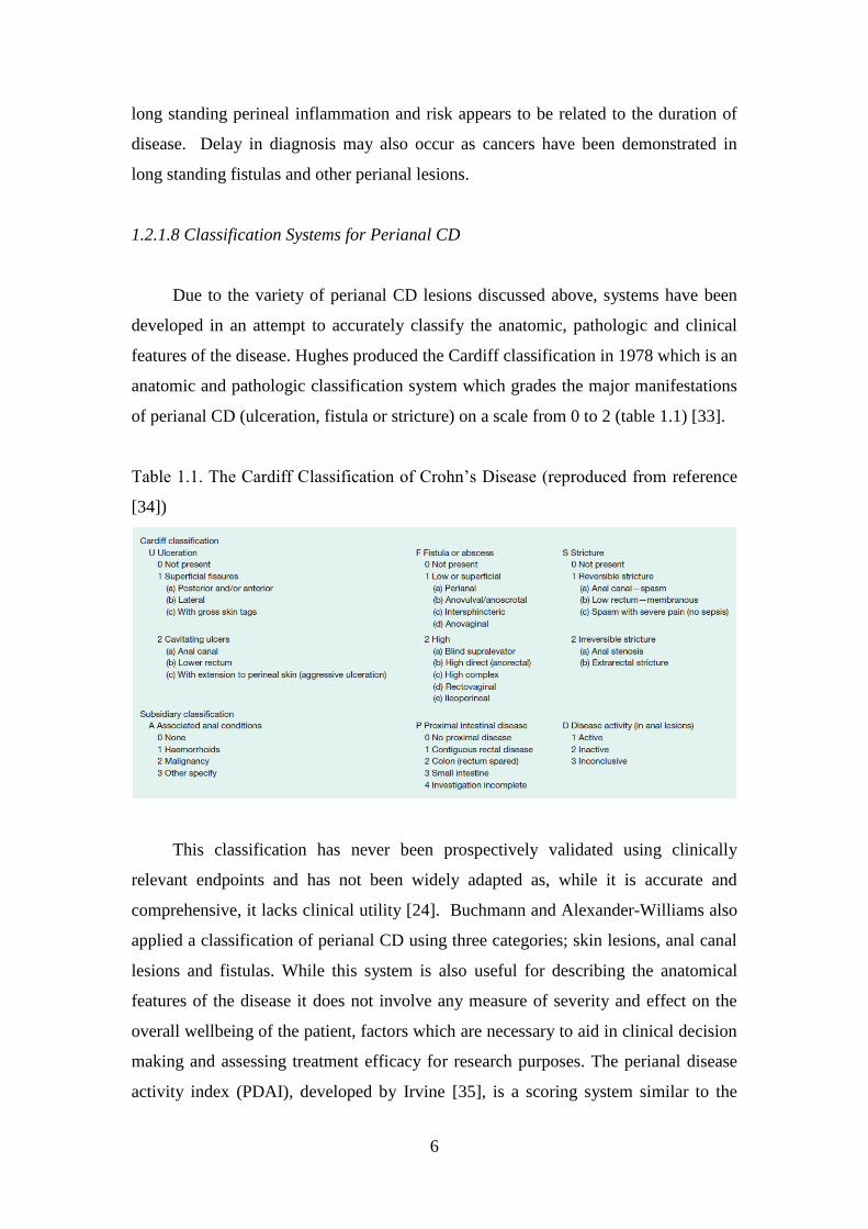
features of the disease. Hughes produced the Cardiff classification in 1978 which is an
anatomic and pathologic classification system which grades the major manifestations
of perianal CD (ulceration, fistula or stricture) on a scale from 0 to 2 (table 1.1) [33].
Table 1.1. The Cardiff Classification of Crohn‟s Disease (reproduced from reference
[34])
This classification has never been prospectively validated using clinically
relevant endpoints and has not been widely adapted as, while it is accurate and
comprehensive, it lacks clinical utility [24]. Buchmann and Alexander-Williams also
applied a classification of perianal CD using three categories; skin lesions, anal canal
lesions and fistulas. While this system is also useful for describing the anatomical
features of the disease it does not involve any measure of severity and effect on the
overall wellbeing of the patient, factors which are necessary to aid in clinical decision
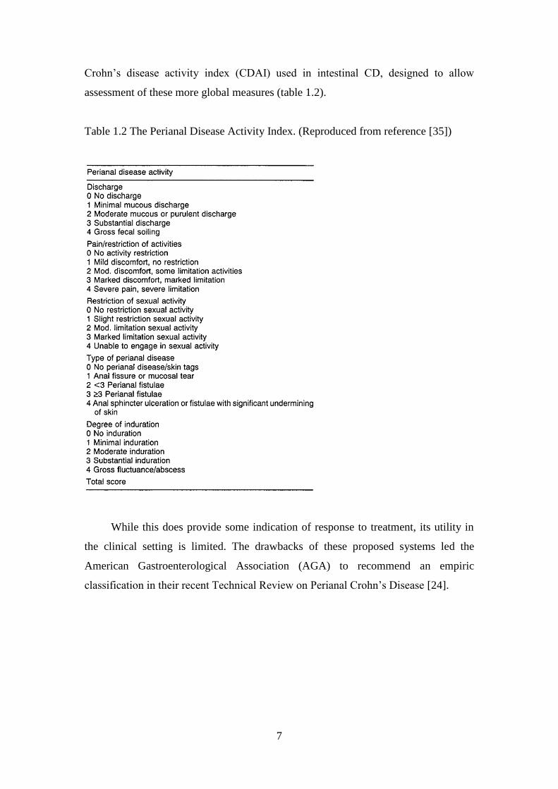
making and assessing treatment efficacy for research purposes. The perianal disease
activity index (PDAI), developed by Irvine [35], is a scoring system similar to the
7
Crohn‟s disease activity index (CDAI) used in intestinal CD, designed to allow
assessment of these more global measures (table 1.2).
Table 1.2 The Perianal Disease Activity Index. (Reproduced from reference [35])
While this does provide some indication of response to treatment, its utility in
the clinical setting is limited. The drawbacks of these proposed systems led the
American Gastroenterological Association (AGA) to recommend an empiric
classification in their recent Technical Review on Perianal Crohn‟s Disease [24].
8
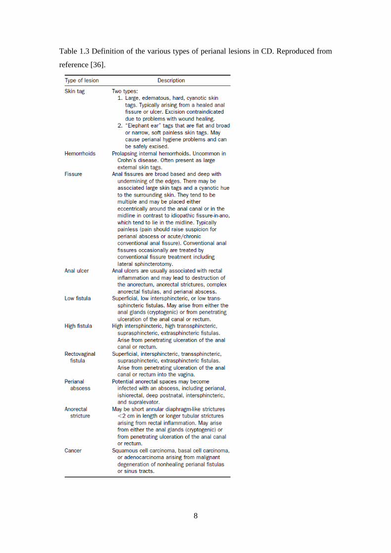
Table 1.3 Definition of the various types of perianal lesions in CD. Reproduced from
reference [36].
9
This paper recommends clinical examination to document the presence of any
of the perianal lesions discussed above. Fistulas are then classified either as simple or
complex based on the aforementioned criteria. This simple and clinically relevant
approach was adopted for use in this thesis. The definitions used for the various
perianal lesions are summarised in table 1.3.
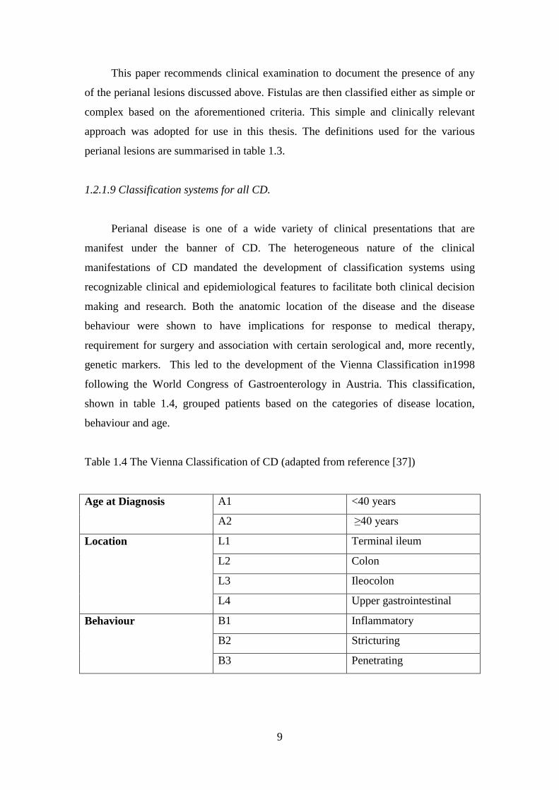
1.2.1.9 Classification systems for all CD.
Perianal disease is one of a wide variety of clinical presentations that are
manifest under the banner of CD. The heterogeneous nature of the clinical
manifestations of CD mandated the development of classification systems using
recognizable clinical and epidemiological features to facilitate both clinical decision
making and research. Both the anatomic location of the disease and the disease
behaviour were shown to have implications for response to medical therapy,
requirement for surgery and association with certain serological and, more recently,
genetic markers. This led to the development of the Vienna Classification in1998
following the World Congress of Gastroenterology in Austria. This classification,
shown in table 1.4, grouped patients based on the categories of disease location,
behaviour and age.
Table 1.4 The Vienna Classification of CD (adapted from reference [37])
Age at Diagnosis A1 <40 years
A2 ≥40 years
Location L1 Terminal ileum
L2 Colon
L3 Ileocolon
L4 Upper gastrointestinal
Behaviour B1 Inflammatory
B2 Stricturing
B3 Penetrating
10
The classification system was useful in predicting aspects of disease
presentation and natural history. In addition, using the Vienna Classification,
associations in disease location and behaviour with anti-Saccharomyces cerevisiae
antibody (ASCA) [38, 39] and the presence of the nucleotide-binding oligomerization
domain 2/capsase recruitment domain-containing protein 15 (NOD2/CARD15) gene
mutations [39-41] were demonstrated. However, significantly, the Vienna
classification did not separate luminal and perianal fistulising disease into different
categories. Subsequent evidence suggested that while perianal fistulas and luminal
fistulas are related, they tend to have distinct clinical associations and often occur
completely independently of each other [7]. This evidence was incorporated into the
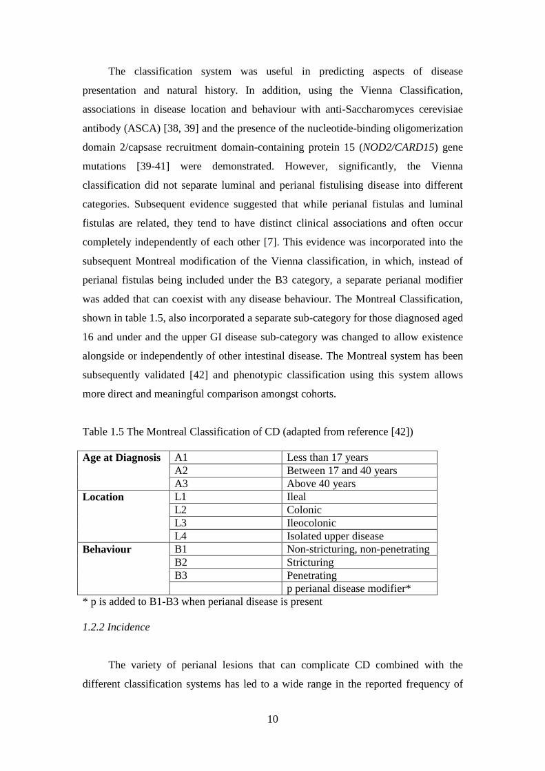
subsequent Montreal modification of the Vienna classification, in which, instead of
perianal fistulas being included under the B3 category, a separate perianal modifier
was added that can coexist with any disease behaviour. The Montreal Classification,
shown in table 1.5, also incorporated a separate sub-category for those diagnosed aged
16 and under and the upper GI disease sub-category was changed to allow existence
alongside or independently of other intestinal disease. The Montreal system has been
subsequently validated [42] and phenotypic classification using this system allows
more direct and meaningful comparison amongst cohorts.
Table 1.5 The Montreal Classification of CD (adapted from reference [42])
Age at Diagnosis A1 Less than 17 years
A2 Between 17 and 40 years
A3 Above 40 years
Location L1 Ileal
L2 Colonic
L3 Ileocolonic
L4 Isolated upper disease
Behaviour B1 Non-stricturing, non-penetrating
B2 Stricturing
B3 Penetrating
p perianal disease modifier*
* p is added to B1-B3 when perianal disease is present
1.2.2 Incidence
The variety of perianal lesions that can complicate CD combined with the
different classification systems has led to a wide range in the reported frequency of
11
perianal involvement in CD which varies from 10-80% [43]. In addition to differences
in the definition of perianal disease and the criteria for inclusion in different series
reporting on incidence, the wide range is also a result of whether the samples
originate from referral centres or population based cohorts. Given that perianal CD is
associated with a more complicated disease course, single centre, tertiary hospital
based studies can be expected to produce a different prevalence to that seen in the CD
population as a whole. Table 1.6 summarises the studies published to date which
document the prevalence of perianal CD. In general the lowest prevalence is reported
in population based cohorts including only fistulas and the highest prevalence in
referral centre cohorts that include all perianal lesions including less significant
lesions such as skin tags, haemorrhoids and fissures.
The duration of follow up will also impact on the reported frequency of perianal
involvement as the temporal relationship of the perianal to the intestinal
manifestations is variable. Previous research suggests that in up to one third of
patients the perianal disease precedes the intestinal CD [18, 44] and of those that
develop perianal CD, 75% do so within 10 years of the diagnosis of intestinal disease
[44]. In less than 5% of patients perianal disease is the sole manifestation of CD.
Hence, the longer the duration of follow up, the higher one would expect the
cumulative incidence of perianal CD to be.
12
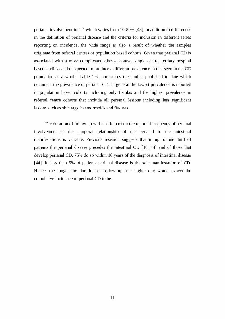
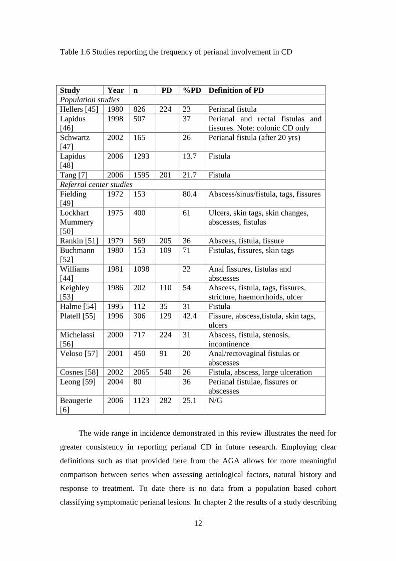
Table 1.6 Studies reporting the frequency of perianal involvement in CD
The wide range in incidence demonstrated in this review illustrates the need for
greater consistency in reporting perianal CD in future research. Employing clear
definitions such as that provided here from the AGA allows for more meaningful
comparison between series when assessing aetiological factors, natural history and
response to treatment. To date there is no data from a population based cohort
classifying symptomatic perianal lesions. In chapter 2 the results of a study describing
Study Year n PD %PD Definition of PD
Population studies
Hellers [45] 1980 826 224 23 Perianal fistula
Lapidus
[46]
1998 507 37 Perianal and rectal fistulas and
fissures. Note: colonic CD only
Schwartz
[47]
2002 165 26 Perianal fistula (after 20 yrs)
Lapidus
[48]
2006 1293 13.7 Fistula
Tang [7] 2006 1595 201 21.7 Fistula
Referral center studies
Fielding
[49]
1972 153 80.4 Abscess/sinus/fistula, tags, fissures
Lockhart
Mummery
[50]
1975 400 61 Ulcers, skin tags, skin changes,
abscesses, fistulas
Rankin [51] 1979 569 205 36 Abscess, fistula, fissure
Buchmann
[52]
1980 153 109 71 Fistulas, fissures, skin tags
Williams
[44]
1981 1098 22 Anal fissures, fistulas and
abscesses
Keighley
[53]
1986 202 110 54 Abscess, fistula, tags, fissures,
stricture, haemorrhoids, ulcer
Halme [54] 1995 112 35 31 Fistula
Platell [55] 1996 306 129 42.4 Fissure, abscess,fistula, skin tags,
ulcers
Michelassi
[56]
2000 717 224 31 Abscess, fistula, stenosis,
incontinence
Veloso [57] 2001 450 91 20 Anal/rectovaginal fistulas or
abscesses
Cosnes [58] 2002 2065 540 26 Fistula, abscess, large ulceration
Leong [59] 2004 80 36 Perianal fistulae, fissures or
abscesses
Beaugerie
[6]
2006 1123 282 25.1 N/G
13
the rate and classification of symptomatic perianal CD in a population based cohort
are presented.
1.3 Aetiologic factors in perianal CD
1.3.1 The aetiology of CD
The aetiology and pathogenesis of CD remain incompletely understood. Current
evidence, however, overwhelmingly favours a dysregulated immune response to
resident intestinal bacteria in genetically susceptible hosts [60, 61]. Familial
aggregation suggests that CD is heritable [62]. A significantly higher rate of disease in
monozygotic versus dizygotic twins [63], together with genome-wide association
(GWA) studies, has confirmed a definitive genetic component in the pathogenesis of
this disorder and this is discussed in detail in section 1.3.4 below. In addition to
genetic risk factors, a number of lifestyle factors have been implicated in CD
aetiology. Smoking has been shown to double the risk of CD [63] and also to
aggravate the disease course. While the data is inconsistent, being breast fed likely
has a protective role [64] and the duration of breast feeding may also be significant
[65]. In addition, stringent sanitation and hygiene may have a deleterious effect[66].
Luminal antigens, in particular associated with luminal flora, are recognised as critical
in CD pathogenesis, however, recent studies implicate a disordered host immune
response, rather than microbial composition per se, as the critical factor in disease
development [67, 68]. Defects in the innate and/or adaptive immune response
ultimately result in uncontrolled expression of pre-inflammatory cytokines,
chemokines and adhesion molecules, resulting in amplification and perpetuation of
the cycle of inflammation [69]. This in turn produces the clinical manifestations of
CD including full thickness inflammation of the intestinal wall with penetrating and
stricturing complications. In around one quarter of patients, these clinical
manifestations include perianal disease. Why only a subset of these patients develop
perianal manifestations is not clear. Previously perianal fistulas were thought to be
the result of a similar process to luminal fistulas, however, recent evidence suggests
that fistulas in the perianal region in fact occur independently of luminal penetrating
complications [7]. As previously discussed, this led to the Montreal modification of
the Vienna Classification of CD and raises the possibility that penetrating perianal
14
disease represents a distinct disease phenotype. A review of the aetiological factors in
perianal CD patients indeed reveals emerging associations with phenotypic, patient
and genetic influences.
1.3.2 Phenotypic associations
The phenotypic categories from the aforementioned Montreal Classification
(Age at diagnosis, disease location and disease behaviour) have all been investigated
for associations with perianal CD. Of these, disease location appears the most
consistent predictor of the risk of perianal complications, with the risk increasing the
more distal the intestinal disease. In an early study, Rankin et al investigated the
relationship of perianal disease to intestinal disease location in 569 CD patients from
tertiary referral centres. Of these patients, 205 had perianal complications defined as
anal fistula, fissure or abscess. This paper demonstrated perianal complications
occurred more frequently in patients with colitis (p=0.0027) and ileocolitis (p=0.0005)
than those with only small bowel disease [51]. These findings have been replicated in
several other referral centre based cohorts that have also demonstrated rectal
involvement shows the strongest association [44, 49, 50, 53, 55, 57]. The evidence
from population based cohorts is less consistent. A study from Manitoba suggested
patients with Crohn‟s colitis were three times more likely to develop perianal fistulae
compared with those with ileitis [7]. Schwartz et al reported data from Minnesota and
showed that ileocolitis was associated with an increased risk of all fistulising disease
(intestinal and perianal) when compared with ileitis only [47]. In contrast, a study
from Stockholm of 1389 patients demonstrated no relationship of perianal disease to
intestinal disease location [48]. Explanations for this lack of association demonstrated
in the Swedish study have included relatively limited follow up and low rates of
colonoscopy, thereby potentially under diagnosing colonic disease.
Studies pertaining to the association of disease behaviour and perianal disease
have also yielded varying results. Smith et al [8] found that perianal disease was not
associated with penetrating intestinal disease and similarly Veloso et al [57]
demonstrated that the presence of perineal fistulas was independent of penetrating
intestinal disease. In contrast, Tang et al found there was a strong association between
intestinal and perineal fistulas (OR 5.02, 95% CI 3.4-7.42, P<0.0001), but this was
15
much stronger for colonic disease location compared with ileal. These authors also
noted that the two fistula types often occurred exclusively [7]. Sachar et al pooled
data from six cohorts analysing 5491 CD patients and found an association between
colonic luminal fistulas and perianal disease, but not ileal fistulas [9]. This evidence
led the Montreal Working Group to conclude that perianal and enteric fistulas
represent two different but associated phenotypes [70].
A number of studies suggest that young age at diagnosis is associated with
perianal disease. Halme et al reported a series of 112 patients and found the average
age of those with perianal disease was 27.7 years compared 37.4 years in those
without (p<0.01) [54]. Roberts et al described a cohort of CD patients over 50 years
of age and concluded they were less likely to present with perianal complications [71]
and Cosnes et al found fistulising complications, including perianal disease, were
more common in those younger than 40 years [58]. This data from referral centre
based cohorts is also supported by population based studies. Lapidus et al found
increased incidence of perianal fistulas with decreased age of diagnosis in an update
of the Stockholm county cohort [48]. Tang et al demonstrated that all fistulising
disease was most common in the 20-29 age range and least likely in those diagnosed
after the age of 49 [7]. However, the relationship of age to perianal disease is not
consistent across studies as both Platell et al [55] and Hellers et al [45] found no
difference in age in patients with or without perianal disease. In addition, many
studies do not control for duration of disease and the effect of young age at diagnosis
may simply be a result of longer disease duration allowing perianal complications to
develop. The impact of disease duration on needs to be considered in studies assessing
association of age at diagnosis and perianal CD.
1.3.3 Other patient related factors
Several other patient related factors have been investigated for associations with
perianal CD. There are conflicting reports on whether gender predicts the occurrence
of perianal disease. Sangwan et al and Hellers et al both reported that significantly
more males than females in their studies had perianal CD [18, 45]. On the contrary,
Bell [25] noted a slight female predominance for perianal fistulae and Fry [72]
reported more than twice as many females as males had perianal CD.
16
In a disease such as CD, thought to have a significant genetic aetiological
component, ethnicity may play a role in determining the phenotypic manifestations
and studies have shown that the risk of perianal disease varies in different ethnic
groups. In particular non-Caucasian race has been associated with perianal disease in
at least three studies [7, 58, 73]. In addition it has been demonstrated that Sephardic
Jews have a higher rate of perianal disease than Ashkenazi Jews [74].
The documented rising incidence of CD over the last 50 years also suggests a
strong environmental influence on the aetiology. A number of environmental factors
have been repeatedly associated with CD including tobacco smoking [75-77],
diarrhoeal illness in childhood [78] and oral contraceptive pill use [79]. Other less
consistent associations include previous tonsillectomy [80, 81], and antibiotic use [82,
83]. Breast feeding has inconsistently been shown to have a protective effect against
developing CD [64, 84].
The relationship between tobacco smoking and CD has been extensively
investigated in epidemiologic studies and smoking is an accepted environmental risk
factor for CD [75-77]. In addition, previous studies demonstrate the association of
smoking with intestinal penetrating disease [85] and smoking accelerates the rate of
progression to complicated disease [86]. There is, however, a paucity of data on the
impact of smoking specifically on the development of perianal disease. The Manitoba
population based cohort was investigated for the relationship between smoking and
fistulising disease. For smoking history, there was no significant difference when
comparing all fistulising with non-fistulising disease and comparing perianal versus
intestinal fistulising disease [7]. Unfortunately, the perianal disease and non-perianal
disease groups were not compared for smoking history in this study. If both smoking
and perianal disease are associated with a more complicated disease course then the
relationship between these factors deserves further consideration. In addition none of
the other possible aforementioned environmental risk factors have been investigated
separately for perianal disease.
17
1.3.4 Genetic Factors
1.3.4.1 Evidence for heritability of CD
The preceding discussion highlighted the fact that CD incidence has increased
markedly in the last 50 years suggesting strong environmental influences. However, a
significant amount of epidemiological evidence exists that demonstrates heritability is
also a major aetiological factor, reinforcing the current theory that CD results from a
dysregulated immune response to some environmental factor(s) in genetically
susceptible hosts.
CD demonstrates a familial pattern of disease that was first recognised by Crohn
et al in 1932 [11]. Subsequent research has documented the relative risk to the sibling
of an affected individual is in the range of 15-35 for CD compared with only 6-10 in
UC [87-91]. In fact, family history is the strongest risk factor for developing IBD [92]
and the evidence for a genetic basis is further strengthened by twin studies which
demonstrate higher disease concordance in monozygotic twins compared with
dizygotic twins. Once again, this effect is more pronounced in CD than UC [93-96].
1.3.4.2 Techniques of gene identification
The two methods employed are candidate gene testing and genome wide
scanning, both by linkage and association. The two techniques are complimentary and
used together, with genome wide scanning identifying possible locations for
subsequent candidate gene testing. Genome wide linkage utilises „sibling pair‟
families which are genotyped using polymorphic DNA microsatellites located at
intervals throughout the genome. Linkage analysis then measures excess sharing of
the same allele between affected siblings suggestive of a correlation between
inheritance of the disease and inheritance of that particular allele. Subsequently the
search can be focused more specifically on that area of the genome [97]. More recent
research has utilised single nucleotide polymorphisms (SNPs) as opposed to
microsatellite markers in GWA studies. Single nucleotide polymorphisms are defined
as single base pair variations where the least frequent allele has a population
frequency of 1% or more. Single nucleotide polymorphisms may represent the genetic
18
defect themselves or act as a marker for the actual functional defect [97]. Candidate
gene analysis, comparing allele frequencies between affected patients and controls,
can then be performed; either using the results of GWA studies, or based on the
known function of a gene.
1.3.4.3. Susceptibility Genes in CD
The first susceptibility gene identified for CD was NOD2 on chromosome
16q12 [98, 99] and its association has been widely replicated in European cohorts, but
not in Asian and African populations [97, 100]. A recent meta-analysis demonstrated
the relative risk for developing CD in homozygotes for the mutant allele was 17.1
[101]. In addition, genotype-phenotype association studies have demonstrated that
NOD2 is associated with earlier age of onset [102-104] and ileal disease location [40,
102, 105, 106]. The mechanism by which NOD2 mutations lead to CD is unknown.
The gene encodes an intracellular receptor expressed in monocytes that is involved in
response to bacterial muramyldipeptide. This may result in reduced ability to clear
bacteria leading to dysregulation of adaptive immune pathways.
Since the discovery of NOD2, the progression of genetic technology has led to a
rapid expansion in the identification of susceptibility genes in IBD. A recent meta-
analysis of six genome-wide association studies defined 71 distinct susceptibility loci
for CD [107]. This study analysed data from 15,694 cases and 14,026 controls and
the genetic associations demonstrated are summarised in tables A1 and A2 provided
in appendix 1. A complete discussion of all these genes is beyond the scope of this
review, however, from the above discussion of NOD2 it is clear that different
susceptibility genes also confer risk for different phenotypes in CD, raising the
question as to whether separate genetic susceptibility exists for perianal CD and this
will be the focus of the subsequent discussion.
1.3.4.4 Genetic associations with perianal CD
There is still relatively sparse literature on genetic associations in perianal CD.
To date, three genetic loci, IBD5, IRGM and TNFα, have been documented to be
associated with perianal CD, albeit with varying strength and consistency.
19
1.3.4.4.1 IBD5 and perianal CD
The IBD5 susceptibility locus on chromosome 5q31 is associated predominantly
with CD but also UC. The association of this locus with CD itself has been widely
replicated [108-111] and it was first associated with perianal CD by Armuzzi et al in
2003 [110]. These authors used three SNPs to create an H2 risk haplotype to span the
IBD5 gene. Perianal CD patients in the cohort had a relative risk of 3 (95% CI 1.8-5.0)
for H2 homozygosity compared with healthy controls and a relative risk of 2 (95% CI
1.1-3.7) compared with non-perianal CD patients. In addition the H2 haplotype was
also associated with ileal disease location [110]. The SNPs in this haplotype were
selected as positional rather than functional variants and to date the exact causal
variant in this region remains unclear.
Peltekova et al [112] identified potential functional variants with SNPs within
the carnitine/organic cation transporter genes OCTN1 and OCTN2. Several theories
of how these variants could contribute to the pathogenesis of CD have been proposed.
OCTN variants may produce reduced carnitine transport and thereby reduced oxygen
burst mediated pathogen killing resulting in altered handling of intestinal bacteria. In
addition, impaired fatty acid oxidation which occurs in the setting of impaired
carnitine transport, has been shown to lead to colitis in animal models [113].
Vermeire et al investigated recognised SNPs in the OCTN1 and OCTN2 genes and
while they found no association overall with CD in the Flemish population, they did
demonstrate that the OCTN2 variant was associated with perianal CD in a dose
dependent manner. The same univariate analysis also demonstrated a trend to
association between perianal CD and OCTN1 [114]. These findings were essentially
replicated by Palmieri et al who demonstrated that the OCTN1-2 TC risk haplotype
frequency was 55.7% in CD patients with perianal fistulas compared with 40.7% in
CD patients without perianal fistulas (p=0.007) [115]. In addition this relationship
was maintained in a logistic regression analysis including clinical factors.
Despite these positive associations, two further studies failed to show a
significant effect of the OCTN variants on perianal CD. Newman et al found the
OCTN1-2 TC risk haplotype was associated with CD overall but not perianal CD
20
[116]. Most recently, Karban et al, found no association with perianal CD in any of
the OCTN1, OCTN2 or OCTN1-2 TC risk haplotypes [74]. Hence, the OCTN1 and 2
polymorphisms are not consistently associated with perianal CD and further research
has also suggested they are not independent of the background risk haplotype for CD
itself [117]. There are a number of other potential causal variants in the IBD5 region
and the uncertainty surrounding the OCTN polymorphisms has focused attention on
genes such as the cytokine cluster, the interferon regulatory factor 1 gene (IRF1), the
PDZ and LIM domain and others [97].
The most recent study of the IBD5 region and perianal disease used another
positional variant IGR2063b_1 and confirmed a higher risk of perianal disease in
homozygotes for this SNP [117]. Explanations for this variation in results amongst
studies include the inclusion and classification of perianal and intestinal CD, the
ethnic makeup of the cohorts and also statistical under powering. Certainly, four of
the six studies addressing the subject suggest an association with perianal CD and the
IBD5 region, however further study is required both to confirm this association and
the causal variant producing it.
1.3.4.4.2 IRGM and perianal CD
The immunity-related guanosine triphosphatase protein type M (IRGM) gene
was associated with CD susceptibility in a large GWA study [118]. The fact this is an
important autophagy gene also provides a plausible biological link to CD
pathogenesis. The IRGM protein is involved in the elimination of intracellular bacteria
by generating large autolysosomal organelles after binding to GTP [119]. Commensal
bacteria likely contribute to the pathogenesis of CD and ineffective autophagy
allowing persistence of intracellular bacteria may provide an opportunity for antigenic
stimulation of the adaptive immune/inflammatory pathways. In addition to the
discovery of IRGM, several other autophagy genes have been associated with CD in
GWA scans including the autophagy related 16-like 1 (ATG16L1) and nuclear
cytosolic factor 4 (NCF4) genes [120].
A single study of 823 CD patients did demonstrate the IRGM rs4958847
polymorphism was associated with perianal CD (OR 1.61) [121]. Significantly this
21
SNP was also associated with intestinal fistulising behaviour and after logistic
regression analysis, combining clinical factors including age at diagnosis, disease
location and smoking status, both associations persisted. The potential pathogenic
mechanism and association with both perianal and fistulising CD make this an
attractive target as a true susceptibility locus. However, sub phenotypic analysis of the
same cohort used in the aforementioned GWA study failed to show an association
with perianal disease and in addition, sequencing of the gene by Parkes et al did not
show any causal amino acid changes [118]. Further studies in well characterised
cohorts are required to clarify the relationship of perianal CD and the IRGM
susceptibility locus. In addition, no studies have considered the other autophagy genes
ATG16L1 and NCF4 and their association with perianal disease.
1.3.4.4.3 TNFα and perianal CD
The proinflammatory cytokine TNFα has an established role in CD
pathogenesis and as a result the TNFα gene, located on chromosome 6 in the IBD3
susceptibility locus, has been the subject of considerable interest. TNFα has a pivotal
role in the inflammatory response and also apoptosis. TNFα levels have been shown
to be elevated in the stool of IBD patients compared with healthy controls and anti-
TNFα monoclonal antibody therapies have demonstrated considerable efficacy in CD
treatment [122]. Several SNPs in the promoter region of the TNFα gene have been
identified at positions 308, 857, 863 and 1031 [97] and downstream functional effects
have been documented including increased TNFα production in carriers of the -308A
polymorphism [123-125] and the -857 C→T SNP [126].
Despite the biological plausibility of a role for the TNFα gene in the
pathogenesis of CD, results of association studies of the frequency of these SNPs in
CD have been varied. A recent meta-analysis of 31 case control studies found an
association only in the -1031 TC+CC genotype (OR 1.32; 95% CI 1.03-1.70) in all
patients. While TNFα SNPs have not uniformly contributed to overall susceptibility to
CD, individual studies have demonstrated association with different CD phenotypes
[127-130]. However, the same meta-analysis stratified patients using the Vienna
classification and did not find any association between these SNPs and CD location or
behaviour. Fistulising disease was included in the analysis but perianal fistulas were
22
not considered separately. Only one study has separately considered the relationship
of perianal CD to TNFα. Kim et al investigated genotypes in 63 patients with CD and
published the results in the Korean Journal of Gastroenterology in Korean with an
English abstract. They found the frequency of -238A allele of TNFα was significantly
higher in CD patients with a perianal lesion than those without. Hence while TNFα is
an attractive candidate as a susceptibility locus, the results for CD overall are variable
and only one small study has directly considered the perianal disease group.
This discussion of aetiology demonstrates that while major advances have been
made in the understanding of the environmental and genetic risk factors for CD
overall, further research is necessary to clarify the relationship of perianal disease to
intestinal CD and determine whether different risk factors exist for patients with this
important subgroup of disease. Better defining the group at risk for perianal disease
and therefore a more severe CD course will allow the appropriate use of
investigations and management which also continue to evolve and are the subject of
the remainder of this review.
1.4 Assessment of Perianal CD
CD patients with perianal symptoms require clinical examination to document
the presence of the perianal lesions discussed in section 1.2. In addition, the
assessment of perianal disease must be accompanied by an assessment of proximal
luminal disease as this may influence decisions regarding management of the perianal
disease. Treatment of the perianal CD depends on the nature of the lesion and ranges
from conservative through medical and surgical intervention. Simple clinical
examination is often inadequate and not well tolerated secondary to pain or the
pathology present. Hence, further imaging and or examination under anaesthetic
(EUA) is often necessary to allow accurate diagnosis and formulation of an
appropriate management plan.
Further assessment with EUA or imaging is useful to;
- Document the presence of undrained sepsis
- Accurately define fistula anatomy in particular the relationship to the
anal sphincter complex
23
- Identify any secondary tracts arising from fistulas
- Assess sphincter integrity in patients with previous perianal surgery
- Monitor response to medical and surgical treatments
At EUA the surgeon can perform a full examination including visual inspection
and proctoscopy. Fistula anatomy is defined using probes and instillation of hydrogen
peroxide to aid in identification of any internal fistula opening(s). Drainage of sepsis
and other surgical management including fistulotomy or seton placement can be
performed at the time. However, EUA is not 100% accurate and missed sepsis or
fistula extensions can lead to recurrence and further tissue damage. In addition, both
incorrect identification of the relationship of the fistula to the anal sphincters leading
to inappropriate fistulectomy and injudicious probing can lead to iatrogenic injury and
inadequate fistula treatment. Therefore, preoperative imaging is often employed and
the most frequently used modalities are magnetic resonance imaging (MRI) and endo-
anal ultrasound (EAUSS). Computed tomography (CT) occasionally has a role and
historically fistulography was used, but has limited application in the modern era.
1.4.1 MRI in Perianal CD
For the reasons outlined above, MRI is now frequently used in perianal CD,
particularly in complex perianal sepsis, to define fistula anatomy and to monitor
response to treatment. Imaging techniques and experience vary amongst institutions.
Both T2 and T1-weighted images are obtained with and without fat suppression.
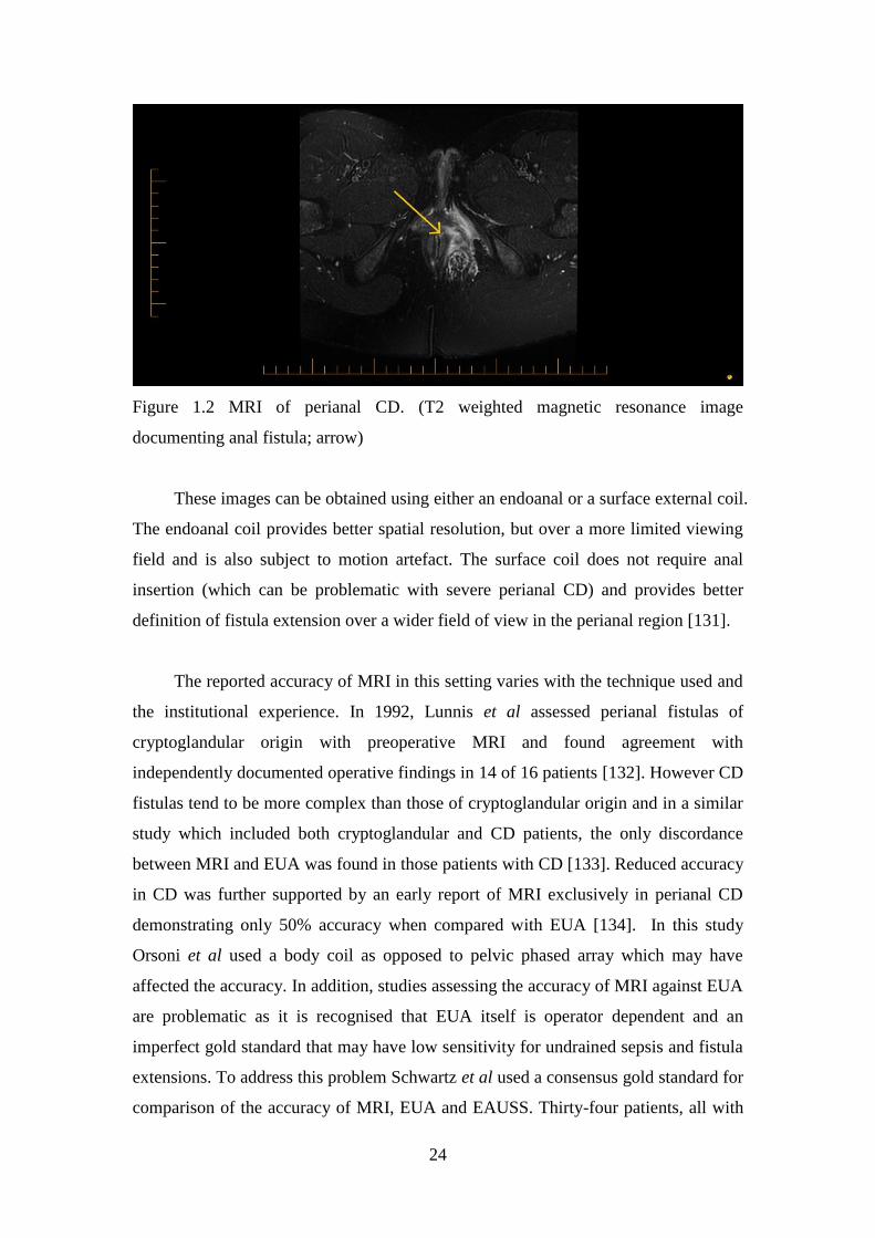
Active fistulas appear hyperintense on T2-weighted images (figure 1.2).
24
Figure 1.2 MRI of perianal CD. (T2 weighted magnetic resonance image
documenting anal fistula; arrow)
These images can be obtained using either an endoanal or a surface external coil.
The endoanal coil provides better spatial resolution, but over a more limited viewing
field and is also subject to motion artefact. The surface coil does not require anal
insertion (which can be problematic with severe perianal CD) and provides better
definition of fistula extension over a wider field of view in the perianal region [131].
The reported accuracy of MRI in this setting varies with the technique used and
the institutional experience. In 1992, Lunnis et al assessed perianal fistulas of
cryptoglandular origin with preoperative MRI and found agreement with
independently documented operative findings in 14 of 16 patients [132]. However CD
fistulas tend to be more complex than those of cryptoglandular origin and in a similar
study which included both cryptoglandular and CD patients, the only discordance
between MRI and EUA was found in those patients with CD [133]. Reduced accuracy
in CD was further supported by an early report of MRI exclusively in perianal CD
demonstrating only 50% accuracy when compared with EUA [134]. In this study
Orsoni et al used a body coil as opposed to pelvic phased array which may have
affected the accuracy. In addition, studies assessing the accuracy of MRI against EUA
are problematic as it is recognised that EUA itself is operator dependent and an
imperfect gold standard that may have low sensitivity for undrained sepsis and fistula
extensions. To address this problem Schwartz et al used a consensus gold standard for
comparison of the accuracy of MRI, EUA and EAUSS. Thirty-four patients, all with
25
CD, underwent all three modalities and discordance was resolved by review of the
findings of all the modalities together to reach the consensus gold standard. Using this
methodology, the accuracies of MRI, EAUSS and EUA were 87%, 91% and 91%
respectively. In addition any two of the modalities taken together resulted in an
accuracy of 100% [135]. While this study did, to some extent, strengthen the
reference used for comparison amongst these techniques, no clinical follow up was
included. Given the complexity of perianal CD fistulas, it may only become clear
which modality was correct when the outcome of treatment is known hence, the
results of this type of study can be further strengthened by including the clinical
outcome in the determination of the gold standard. This very approach was taken in a
study from St Mark‟s Hospital in the United Kingdom of 104 patients. After mean
clinical follow up of 23 months, an outcome based gold standard was derived to
compare clinical examination (not under anaesthetic) with MRI and EAUSS. This
study found a significant linear trend in the proportion of fistula tracks correctly
identified by clinical examination (61%), EAUSS (81%) and MRI (90%) [136]. While
this study suggests that MRI is accurate in defining the anatomy of fistulas, only nine
of the 104 patients had CD. Hence, to date the literature contains variable results
regarding the accuracy of MRI in perianal CD.
Given the cost and restricted availability of MRI in some areas, it is important to
determine not only its accuracy but also whether adding MRI to the diagnostic
algorithm in CD actually produces clinical management changes. This question has
been addressed by studies that initially blind the surgeon performing EUA to the MRI
result and after intraoperative revelation of the result, operative management changes
resulting from the MRI are documented. The first of these studies involved only
cryptoglandular fistulas in 34 patients. It demonstrated a relatively high discordance
of 50% between MRI and EUA and concluded that MRI caused significant change to
surgical management in 10% of patients [137]. With the increased complexity of CD
perianal fistulas it could be expected that MRI may have a more significant impact on
management in CD patients. Using similar methodology, Beets-Tan et al investigated
56 patients of which 15 had CD and found that MRI produced important information
that lead to additional surgical management in 21% overall. Of note, in the 15 CD
patients, 40% had surgical management altered [138].
26
In addition to its role in initial management of perianal CD, MRI is also
increasingly used to monitor response to therapy. As discussed in the following
section, modern biologic treatments have demonstrated considerable efficacy in
healing CD fistulas. However, serial MRI of these fistulas has demonstrated persistent
tracts despite apparent clinical healing [139, 140] suggesting a role for assessing
response to treatment and potentially guiding decision making regarding continuing or
changing biologic agents.
1.4.2 EAUSS in Perianal CD
EAUSS is performed using a specialised probe placed in the anal canal. The
standard endoanal ultrasound probe obtains axial 360˚ images and allows accurate
definition of the sphincter components, fistulas and abscesses. Modern probes contain
a mobile crystal which can obtain multiple axial images to produce three-dimensional
reconstructions giving enhanced views of the anal canal anatomy. Instillation of
hydrogen peroxide into fistula tracts may also enhance visualisation [141, 142]. The
proposed advantages of EAUSS over MRI are that it is simple and quick to perform
and relatively inexpensive. In addition, it can be used in the operating theatre to guide
surgery. However, the technique is highly operator dependent and the pathology may
limit its use, particularly in the case of painful perianal lesions or strictures preventing
probe insertion. As documented in the comparative studies discussed above [135,
136], the accuracy of EAUSS ranges from 81-91% and appears similar to that of MRI
in assessing and classifying CD fistulas.
As with MRI, EAUSS may have a role in monitoring response to therapy.
Ardizonne et al reported its use in 30 patients with perianal CD after treatment with
infliximab. Of the 15 fistulas that ceased to drain, only 5 disappeared on EAUSS and
closure on EAUSS was associated with a lower relapse rate [143]. Similarly,
Schwartz et al followed 21 patients after infliximab treatment with serial EAUSS.
Seven of 11 patients with no persistent fistula were taken off infliximab and no
recurrences occurred in this group (the other four patients without evidence of
persistent fistula were left on infliximab for luminal disease) [144].
27
1.4.3 Other Imaging Modalities
While computed tomography is often used to assess luminal and intra-
abdominal complications of CD, it has little application in assessment of perianal CD.
Detailed anatomic definition of the relationship of a fistula to the pelvic floor
structures is not possible to the same extent as in EAUSS and MRI, as the CT
attenuation of fistulas and the pelvic floor tissues is very similar. For this reason
sparse literature exists on its accuracy and the only comparative study of CT and
EAUSS in perianal CD demonstrated sensitivities of 24% and 82% respectively [145].
In the future CT fistulography using multi-detector imaging and contrast may offer
improved accuracy but will still carry the disadvantage of radiation exposure to
patients [131].
Fistulography was used historically to assess perianal fistulas however it had
several disadvantages including poor contrast penetration into secondary fistula
extensions and poor patient tolerability. The main downside compared with the more
modern techniques of EUASS and MRI was the inability to visualise the sphincter
mechanism and pelvic floor musculature and therefore accurately classify the fistula
relationship to these.
1.4.4 Summary: Assessment of Perianal CD
Magnetic resonance imaging and EAUSS have emerged as the imaging
techniques of choice in perianal CD and this is reflected in the most recent guidelines
published by the AGA. These recommend either EAUSS or MRI in all perianal CD
patients where surgery is the initial treatment strategy or in the presence of pain,
fluctuation or stricture [24]. However, as the preceding discussion demonstrates,
questions regarding the accuracy and utility of these modalities remain. While MRI
and EAUSS appear reasonably accurate, much of the literature to date applies to
cryptoglandular fistulas. The sparse literature available pertaining to more complex
CD fistulas suggests the concordance between EUA and imaging reduces. Chapter 4
addresses this question presenting the results of a clinical study comparing the
accuracy of MRI to EUA and its effect on operative management, including an
assessment of outcomes.
28
1.5 Management of Perianal CD
The current management of perianal CD involves combined medical and
surgical approaches and relies on close liaison between gastroenterologists and
surgeons. Generally, surgical conservatism is advocated as there is a high rate of
disease recurrence and poor wound healing. Inappropriate surgery can lead to
sphincter damage and incontinence and it has previously been stated that
“incontinence in CD is usually related to aggressive surgery, not progressive disease”
[146]. If medical and local surgical approaches fail, treatment will culminate in
proctectomy in a proportion of patients. Specific management depends on the nature
of the perianal lesion producing symptoms.
1.5.1 Management of perianal abscess and fistula-in-ano
In the setting of perianal sepsis, the first priority is adequate drainage of
abscesses including insertion of loose seton ligatures if fistulas are identified. With
sepsis adequately drained, attention is turned to fistula healing and both medical and
surgical approaches are available. The approach depends largely on the whether the
fistula is classified as simple or complex and whether rectal disease activity is present.
Surgical intervention in the presence of severe proctitis is associated with lower
success rates. Simple and complex fistulas require different surgical approaches as
discussed below and often, medical treatment is required in the first instance.
1.5.1.1 Medical Management of fistula-in-ano
1.5.1.1.1 Antibiotics
Antibiotics, most often metronidazole and/or ciprofloxacin, are widely used to
treat perianal CD sepsis. This is despite a relative paucity of randomised evidence. A
review of the literature published in 2003 documented several case series which
demonstrated reduced pain and tenderness and up to half of patients achieving
complete healing however relapse rates were high with dose reduction or cessation of
therapy[36]. Subsequent to this review, a randomised, double blind, placebo
29
controlled, pilot study was performed. In 25 patients, this demonstrated clinical
remission in 30%, 0% and 12.5% with ciprofloxacin, metronidazole and placebo
respectively [147]. Despite the lack of evidence, antibiotics remain a common
treatment due to perceived relative safety and lack of other therapeutic alternatives.
1.5.1.1.2 Immunomodulators
There is also a lack of randomised evidence pertaining to the use of azathioprine
and 6-Mercaptopurine in perianal CD with fistula closure as the primary end point. A
post-hoc meta-analysis of 5 studies considering fistula closure did demonstrate a 54%
rate of healing of fistulas in the treatment group compared with 21% with placebo
[148]. A single study also considered the combination of antibiotics and azathioprine
and found improved response when both treatments were used together [149].
1.5.1.1.3 Anti-TNFα antibody therapy
The introduction of the anti-TNFα antibody treatments infliximab and more
recently adalimumab have provided another option to attempt healing of perianal CD
fistulas and these agents are now central to the treatment algorithm. The efficacy of
infliximab was established in two randomised, placebo controlled trials. The first,
ACCENT I, demonstrated a 68% clinical response rate (defined as a 50% reduction in
the number of draining fistulas) and complete fistula healing in 55% [150]. The
second study, ACCENT II, considered maintenance therapy and found that in the 69%
of patients that responded at week 14, 39% maintained the response at 54 weeks
following 8 weekly dosing of infliximab [151].
While infliximab does appear to have medium term efficacy in healing perianal
CD fistulas, questions remain as to the necessary duration of therapy, the role of
combination therapy both with other medical agents and surgery and whether any
patient factors will predict response. These are all important issues given the expense
and potential short and long term toxicity of these agents including infusion reactions,
formation of human antichimeric antibodies, infectious complications and malignancy
[152, 153]. Evidence accumulated since the ACCENT trials suggests a high relapse
rate after cessation of therapy [154]. Combined medical therapy was investigated by a
30
post-hoc assessment of the ACCENT II data that did not demonstrate any benefit in
the one-third of patients who were administered both infliximab and
immunomodulators [155]. Combining surgery with infliximab infusion has the
potential advantage of ensuring all sepsis is identified and facilitating on going
drainage with seton placement and several small series report success with this
approach [156-159]. However, data from the ACCENT II study did not show any
evidence of increased perianal abscess formation in the infliximab treated group [160]
and EUA prior to infliximab therapy is not considered mandatory.
A number of factors have been investigated in an attempt to predict those
patients who will respond to infliximab, but the only association has been the
presence of proctitis predicting a poor response [139]. In those that do not respond to
infliximab, the alternative anti-TNFα antibody, adalimumab, has still been shown to
have some efficacy albeit reduced compared with first line response rates to
infliximab [161].
1.5.1.1.4 Other medical options
A number of other medical therapies have been trialled in small series including
tacrolimus [162-165] and cyclosporine [166-172] which have shown some success but
there use is restricted to patients who have failed other modalities. In addition there
are recent experimental reports of the local injection of infliximab into fistula tracts
[173, 174]. Several case reports found that hyperbaric oxygen therapy was associated
with fistula healing [175-178] suggesting further investigation into this modality is
warranted, however, the limited availability of hyperbaric chambers and duration of
therapy are likely to limit its widespread use.
1.5.1.2 Surgical management of fistula in ano
Successful surgical management requires adequate drainage of all abscesses,
accurate classification of the fistula as simple or complex and an assessment of
luminal disease, especially in the rectum. This assessment dictates the specific
operation performed.
31
1.5.1.2.1 Fistulotomy
For low fistulas involving minimal sphincter muscle, laying open of the fistula
is an option and the reported healing rates range from 8% to 100% with majority of
studies demonstrating healing rates of 80-100% [36]. However, there is also a broad
range of reported recurrence (6-60%) and incontinence (0-50%) [36]. Healing rates
are lower in the presence of macroscopic rectosigmoid inflammation hence
fistulotomy should be avoided in this situation.
1.5.1.2.2 Seton ligature
If fistulotomy is not appropriate, due to rectal inflammation or the presence of a
complex fistula, then a non-cutting seton ligature is the treatment of choice. Cutting
setons, which effectively cause a slow fistulotomy and are associated with non-
healing and incontinence, are not widely advocated in complex CD fistulas. A non-
cutting seton can be left in situ long term to prevent abscess formation, whilst proctitis
is controlled medically, then fistula healing can be attempted with medical treatment
or a definitive surgical procedure.
1.5.1.2.3 Rectal advancement flaps
An endorectal advancement flap involves raising a flap of rectal wall containing
the internal opening of the fistula, excising the mucosal opening, then suturing the
healthy mucosa over the fistula tract in the muscle. This should only be attempted in
the absence of rectal inflammation otherwise, failure rates are high. Even in the
absence of rectal inflammation success rates are only around 50% in CD fistulas,
compared with 70-80% in the setting of cryptoglalndular fistulas [179, 180].
1.5.1.2.4 Anal fistula plugs and fibrin glue
Recent developments in the treatment of cryptoglandular fistulas have included
the insertion of either fibrin glue or collagen plugs into the fistula tract. These
techniques have been extended to CD fistulas and have the advantage of avoiding
sphincter damage. An initial study of CD patients treated with collagen fistula plugs
32
demonstrated 80% healing at 10 months follow up [181]. However, more recent small
studies with both collagen fistula plugs and fibrin glue have shown healing rates in
CD patients of only 26.6% [182] and 38% [183] respectively. Further randomised
studies with larger numbers of CD patients would be necessary before advocating
these treatments.
1.5.1.2.5 Faecal diversion and proctectomy
The severity of perianal sepsis and the aforementioned difficulties in treatment
has seen proctectomy rates of 12-20% overall [34]. The presence of rectal
involvement with CD is associated with even higher rates of proctectomy [45]. In
severe perianal disease, faecal diversion with temporary ileostomy is occasionally
employed. While several studies confirm it is rare for the ileostomy to be reversed
[184, 185], it may allow the perianal disease to be brought under sufficient control to
facilitate proctectomy with improved perineal wound healing. Poor perineal wound
healing is a major complication of proctectomy in this setting with over one third of
patients unhealed at 6 months. Yamamoto et al documented that 23% of patients had
perineal wound sinuses after proctocolectomy and attributed this lower rate to the use
of defunctioning prior to proctectomy [186]. Other strategies to avoid perineal wound
problems include the use of gracilis transposition flaps [187] or performing a low
Hartmann‟s procedure as opposed to proctectomy [188].
1.5.2 Surgery for other perianal CD conditions
As documented in section 1.2, skin tags occur frequently in perianal CD and
excision is best avoided due to poor healing. In contrast to skin tags, symptomatic
haemorrhoids are rare in CD but when they do occur, operative management should
also be avoided on the basis of high rates of wound complications and anal stenosis,
which can potentially lead to proctectomy [36].
Typical CD anal fissures are painless and not necessarily associated with
increased anal sphincter tone. Surgical intervention is generally not advised as
spontaneous healing occurs in greater than 80% of patients [52] and the risk of
incontinence with sphincterotomy needs to be carefully considered in patients with a
33
chronic diarrhoeal illness. However, in selected patients with appearances more
typical of an idiopathic fissure in ano, that is; in the posterior midline, painful and
associated with anal spasm, lateral sphincterotomy can be performed. Glycerol
trinitrate or diltiazem ointments have efficacy in idiopathic fissures as does injection
of botulinum toxin into the anal sphincter. Little data exists on the efficacy of these
conservative measures in CD although they are simple measures that can be used
prior to sphincterotomy. Symptomatic anal strictures require dilation which often
needs to be repeated and some patients will go on to require proctectomy.
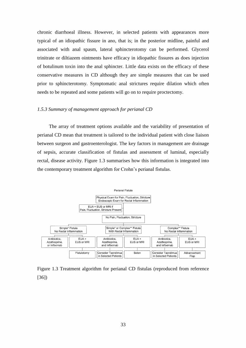
1.5.3 Summary of management approach for perianal CD
The array of treatment options available and the variability of presentation of
perianal CD mean that treatment is tailored to the individual patient with close liaison
between surgeon and gastroenterologist. The key factors in management are drainage
of sepsis, accurate classification of fistulas and assessment of luminal, especially
rectal, disease activity. Figure 1.3 summarises how this information is integrated into
the contemporary treatment algorithm for Crohn‟s perianal fistulas.
Figure 1.3 Treatment algorithm for perianal CD fistulas (reproduced from reference
[36])
34
1.5.4 Frequency and risk factors for perianal surgical intervention in CD
The foregoing discussion and resulting treatment algorithm demonstrate that
surgery is an integral part of treatment and therefore likely a frequent occurrence in
perianal CD patients. However, there is surprisingly little data documenting the
overall frequency of perianal surgical intervention (PSI), or the patient characteristics
that predict the requirement for this. The few studies that have documented rates of
PSI display a wide variation from 4% to 42% [18, 55, 56]. As with estimates of the
incidence of perianal CD overall, these studies may not represent the true rate of PSI
in the CD population as the subjects were drawn from referral centre cohorts where
severe disease is likely to be over represented. A better understanding of the true rate
of PSI, the operations performed and the patient factors associated with PSI is
necessary to aid in refinement of the treatment algorithm, particularly in the era of
biological therapy. In chapter 5 the results of a study investigating the rates of PSI and
factors associated with PSI from the Canterbury IBD Project are presented.
1.6 The cost of perianal Crohn’s disease
In section 1.1 it was emphasised that the incidence of CD is increasing
worldwide[3] and the peak age of onset is between 15 and 35 years [4]. The foregoing
discussion of management demonstrates that while advances in treatment have been
made, particularly with the introduction of modern biological medications, CD
remains incurable. A proportion of patients will endure recurrent, prolonged periods
of illness requiring extensive medical and surgical interventions during what would
otherwise be a highly productive time of life. Hence CD presents an increasingly
significant health problem not only in terms of morbidity, but also cost to the
individual patient and society.
The introduction of these modern biological treatments has been associated with
significant expense and has created a need for government agencies to consider the
economic impact of therapeutic alternatives. To achieve this, the cost of CD and its
societal burden requires further study. Previous studies have indicated that the
majority of the total cost associated with this disease relates to “extensive
interventions required by a small proportion of severely affected individuals” [189].
35
The fact that perianal CD is a marker of a more severe disease course, and that
perianal disease patients develop local perianal complications requiring frequent
intervention means this group is more likely to use significant health resources and
incur the largest costs as a result of the disease. A number of international studies
have considered the cost of inflammatory bowel disease overall [190-199], however,
to date, there has been no published research documenting the average patient cost of
IBD in New Zealand or Australia and no previous work specifically investigating the
perianal CD group. Chapter 6 presents the results of a study performed to assess the
cost of perianal CD from the societal point of view to provide baseline information on
which to guide future decisions regarding CD therapy.
36
Chapter 2
The Spectrum of Perianal CD
Abstract
Background
Perianal Crohn‟s disease represents a phenotype distinct from luminal Crohn‟s disease
(CD) and may follow a different course. To date, the only detailed classifications of
perianal CD arise from referral centre cohorts that do not reflect the spectrum of
disease in the population as a whole. This study aimed to document the frequency,
classification and time course of symptomatic perianal CD in a population based
cohort.
Methods
All patients with inflammatory bowel disease (IBD) in Canterbury, New Zealand,
were eligible for recruitment over a three year period. The clinical records of all CD
patients were reviewed and all symptomatic perianal CD was classified according to
the American Gastroenterological Society position statement.
Results
An estimated 91% of IBD patients in the region were recruited. 715 patients had CD
of which 190 (26.6%) had symptomatic perianal CD. The median age of patients with
perianal CD was 37 years (range 4-82 years) and 58.4 % were female. Median follow
up was 9 years (range 2 months to 45 years) from CD diagnosis. Onset of perianal CD
ranged from 18 years pre-CD diagnosis, to 33 post-CD diagnosis. Fistulas were the
most common lesion (50% of patients), followed by perianal abscesses (42.1%),
fissures (32.6%), skin tags (11.1%), strictures (7.4%) and haemorrhoids (1.6%). The
cumulative probability at 20 years of any perianal CD was 42.7% and of a perianal
fistula 28.3%.
Conclusions
This study provides the first detailed description of perianal CD in a population based
cohort and confirms that the onset of perianal symptoms relative to CD diagnosis is
highly variable.
37
2.1 Introduction
It is commonly accepted that perianal lesions complicate a significant
proportion of CD cases and that these patients represent an important subgroup in CD,
as they tend to have a more severe course. However, the discussion in section 1.2
highlighted the fact that the reported proportion of CD patients affected by perianal
complications varies widely. This appears to be the result of discrepancies in the
classification of perianal lesions included in the studies and also whether the studies
drew subjects from tertiary referral centre practices or the population as a whole.
Studies which include only anal fistulas and abscesses, not unexpectedly, report a
lower rate of perianal involvement than those which include other perianal lesions in
addition, such as skin tags, fissures and haemorrhoids. Likewise, population based
studies also produce lower estimates of the rate of perianal involvement as the more
severe CD cases and, therefore, those with a higher rate of perianal involvement are
over-represented in referral centre cohorts. Evidence continues to mount that perianal
disease may represent a distinct CD phenotype and research into clinical and genetic
risk factors for perianal disease is on-going. To allow meaningful comparison of these
factors, both within and amongst cohorts, accurate classification and standardised
definitions of perianal lesions are required. In 2005 the Canterbury Inflammatory
Bowel Disease Project completed data acquisition on IBD patients in the Canterbury
region as part of a PhD Thesis [200]. This resulted in the finding of an extremely high
incidence of CD within the Canterbury region by global standards [201]. The
existence of this well characterised population based cohort provided the opportunity
to study the perianal CD subgroup in detail.
2.2 Aims
The aims of this study were to;
- determine the rate of symptomatic perianal CD lesions in a population
based cohort
- classify these lesions according to the American Gastroenterological
Association Guidelines
38
- determine the temporal relationship of the onset of perianal disease to
CD diagnosis
2.3 Methods
This study was performed as part of the on-going Canterbury Inflammatory
Bowel Disease Project, a large population based study aimed at describing the
epidemiology of IBD in the Canterbury region and to study the clinical,
environmental and genetic risk factors for IBD. The methods used in data collection
in this project have been described in detail previously [200, 201] and are summarised
here.
The Canterbury region is located on the east coast of the South Island of New
Zealand and had a population of 464,700 in 2005 when recruitment for the study was
completed [202]. Ethnic diversity in Canterbury is limited, with approximately 90% of
the population being Caucasian (descended predominantly from migrants from the
United Kingdom and Ireland) and the remainder consisting of Maori (7.2%), and
other (mainly Asian) ethnicities. Young people less than 20 years of age make up
20.3% of the population while 13.8% are aged over 65 years [202].
Patients were initially recruited to the project as part of a PhD Thesis (RBG)
and a multifaceted approach to recruitment was used. Over 3 years (2003–2005)
surgeons and gastroenterologists referred all inflammatory bowel disease patients
from public and private specialist clinics. In addition, patients self-referred for
inclusion following advertising in public media, general practices, specialist clinics
and with the Crohn‟s and colitis support group. Validation of the completeness of
recruitment using representative general practices indicated 91 per cent of patients
with IBD in the Canterbury region were included in the study. IBD cases were
confirmed using standard diagnostic criteria [203]. Demographic information and
clinical data, together with IBD phenotype and IBD-related surgical procedures, were
extracted retrospectively from patient medical records. This information was then
entered into a custom-built Microsoft Access (Microsoft, Redmond, Washington,
USA) database. In 2009/2010 TE reviewed the CD patient cohort to classify perianal
cases. All patients with symptomatic perianal lesions noted in the medical record at
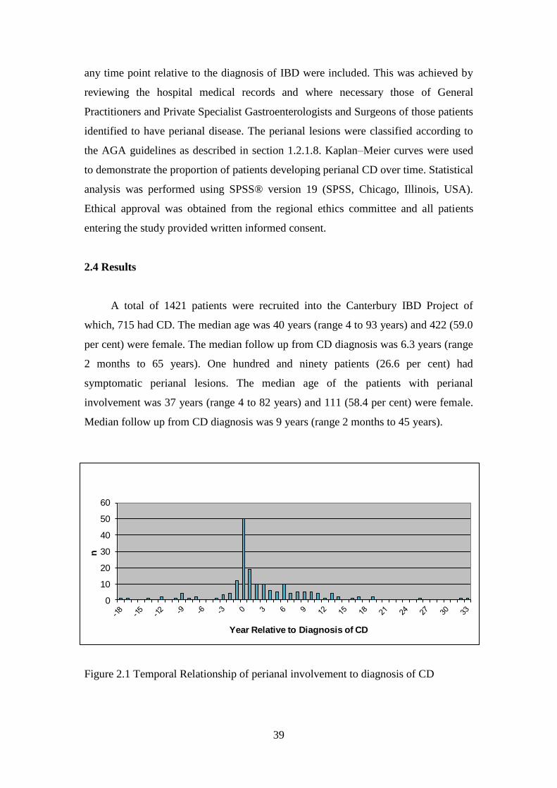
39
any time point relative to the diagnosis of IBD were included. This was achieved by
reviewing the hospital medical records and where necessary those of General
Practitioners and Private Specialist Gastroenterologists and Surgeons of those patients
identified to have perianal disease. The perianal lesions were classified according to
the AGA guidelines as described in section 1.2.1.8. Kaplan–Meier curves were used
to demonstrate the proportion of patients developing perianal CD over time. Statistical
analysis was performed using SPSS® version 19 (SPSS, Chicago, Illinois, USA).
Ethical approval was obtained from the regional ethics committee and all patients
entering the study provided written informed consent.
2.4 Results
A total of 1421 patients were recruited into the Canterbury IBD Project of
which, 715 had CD. The median age was 40 years (range 4 to 93 years) and 422 (59.0
per cent) were female. The median follow up from CD diagnosis was 6.3 years (range
2 months to 65 years). One hundred and ninety patients (26.6 per cent) had
symptomatic perianal lesions. The median age of the patients with perianal
involvement was 37 years (range 4 to 82 years) and 111 (58.4 per cent) were female.
Median follow up from CD diagnosis was 9 years (range 2 months to 45 years).
Figure 2.1 Temporal Relationship of perianal involvement to diagnosis of CD
0
10
20
30
40
50
60
n
Year Relative to Diagnosis of CD
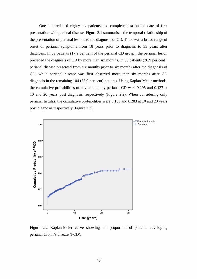
40
One hundred and eighty six patients had complete data on the date of first
presentation with perianal disease. Figure 2.1 summarises the temporal relationship of
the presentation of perianal lesions to the diagnosis of CD. There was a broad range of
onset of perianal symptoms from 18 years prior to diagnosis to 33 years after
diagnosis. In 32 patients (17.2 per cent of the perianal CD group), the perianal lesion
preceded the diagnosis of CD by more than six months. In 50 patients (26.9 per cent),
perianal disease presented from six months prior to six months after the diagnosis of
CD, while perianal disease was first observed more than six months after CD
diagnosis in the remaining 104 (55.9 per cent) patients. Using Kaplan-Meier methods,
the cumulative probabilities of developing any perianal CD were 0.295 and 0.427 at
10 and 20 years post diagnosis respectively (Figure 2.2). When considering only
perianal fistulas, the cumulative probabilities were 0.169 and 0.283 at 10 and 20 years
post diagnosis respectively (Figure 2.3).
Figure 2.2 Kaplan-Meier curve showing the proportion of patients developing
perianal Crohn‟s disease (PCD).
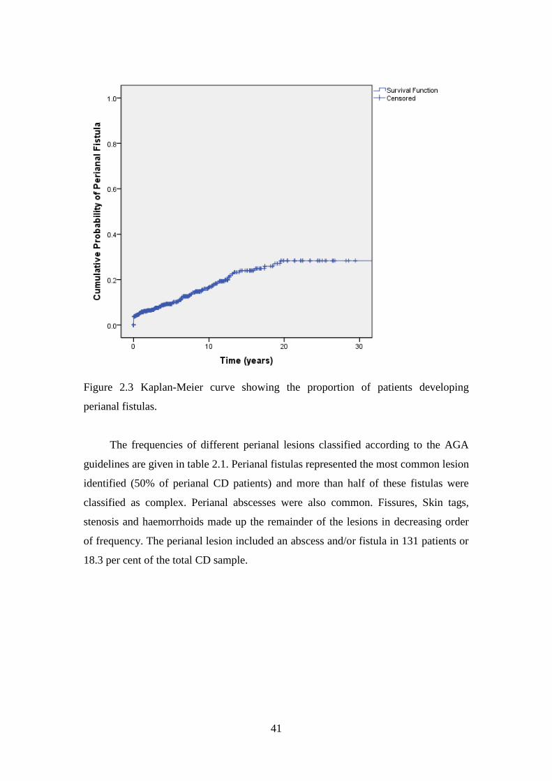
41
Figure 2.3 Kaplan-Meier curve showing the proportion of patients developing
perianal fistulas.
The frequencies of different perianal lesions classified according to the AGA
guidelines are given in table 2.1. Perianal fistulas represented the most common lesion
identified (50% of perianal CD patients) and more than half of these fistulas were
classified as complex. Perianal abscesses were also common. Fissures, Skin tags,
stenosis and haemorrhoids made up the remainder of the lesions in decreasing order
of frequency. The perianal lesion included an abscess and/or fistula in 131 patients or
18.3 per cent of the total CD sample.
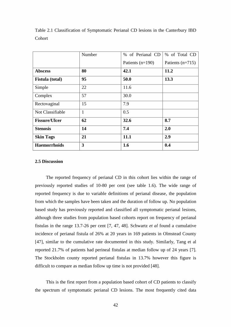
42
Table 2.1 Classification of Symptomatic Perianal CD lesions in the Canterbury IBD
Cohort
Number % of Perianal CD
Patients (n=190)
% of Total CD
Patients (n=715)
Abscess 80 42.1 11.2
Fistula (total) 95 50.0 13.3
Simple 22 11.6
Complex 57 30.0
Rectovaginal 15 7.9
Not Classifiable 1 0.5
Fissure/Ulcer 62 32.6 8.7
Stenosis 14 7.4 2.0
Skin Tags 21 11.1 2.9
Haemorrhoids 3 1.6 0.4
2.5 Discussion
The reported frequency of perianal CD in this cohort lies within the range of
previously reported studies of 10-80 per cent (see table 1.6). The wide range of
reported frequency is due to variable definitions of perianal disease, the population
from which the samples have been taken and the duration of follow up. No population
based study has previously reported and classified all symptomatic perianal lesions,
although three studies from population based cohorts report on frequency of perianal
fistulas in the range 13.7-26 per cent [7, 47, 48]. Schwartz et al found a cumulative
incidence of perianal fistula of 26% at 20 years in 169 patients in Olmstead County
[47], similar to the cumulative rate documented in this study. Similarly, Tang et al
reported 21.7% of patients had perineal fistulas at median follow up of 24 years [7].
The Stockholm county reported perianal fistulas in 13.7% however this figure is
difficult to compare as median follow up time is not provided [48].
This is the first report from a population based cohort of CD patients to classify
the spectrum of symptomatic perianal CD lesions. The most frequently cited data
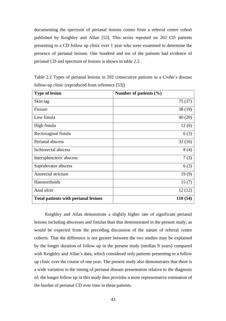
43
documenting the spectrum of perianal lesions comes from a referral centre cohort
published by Keighley and Allan [53]. This series reported on 202 CD patients
presenting to a CD follow up clinic over 1 year who were examined to determine the
presence of perianal lesions. One hundred and ten of the patients had evidence of
perianal CD and spectrum of lesions is shown in table 2.2 .
Table 2.2 Types of perianal lesions in 202 consecutive patients in a Crohn‟s disease
follow-up clinic (reproduced from reference [53])
Type of lesion Number of patients (%)
Skin tag 75 (37)
Fissure 38 (19)
Low fistula 40 (20)
High fistula 12 (6)
Rectovaginal fistula 6 (3)
Perianal abscess 32 (16)
Ischiorectal abscess 8 (4)
Intersphincteric abscess 7 (3)
Supralevator abscess 6 (3)
Anorectal stricture 19 (9)
Haemorrhoids 15 (7)
Anal ulcer 12 (12)
Total patients with perianal lesions 110 (54)
Keighley and Allan demonstrate a slightly higher rate of significant perianal
lesions including abscesses and fistulas than that demonstrated in the present study, as
would be expected from the preceding discussion of the nature of referral centre
cohorts. That the difference is not greater between the two studies may be explained
by the longer duration of follow up in the present study (median 9 years) compared
with Keighley and Allan‟s data, which considered only patients presenting to a follow
up clinic over the course of one year. The present study also demonstrates that there is
a wide variation in the timing of perianal disease presentation relative to the diagnosis
of; the longer follow up in this study thus provides a more representative estimation of
the burden of perianal CD over time in these patients.
44
The timing of perianal symptoms relative to the diagnosis of CD has been
explored in other cohorts with comparable results to those presented here. Williams et
al reviewed 1098 patients with CD presenting to the Lahey Clinic in Massachusetts
over a 10 year period. In the majority (64%) of the 242 patients with perianal CD, the
perianal manifestations developed after intestinal disease over a wide range of time
from 2 weeks to over 10 years. Hellers investigated perianal fistulae in a population
based cohort from Stockholm and found that in 20% the fistula preceded diagnosis by
more than six months, in 38% perianal fistulae occurred between six months prior and
three months after the diagnosis and in the remaining 40% occurred more than three
months after diagnosis. The present study similarly demonstrated that a significant
proportion of patients develop perianal symptoms prior to intestinal disease with 17%
presenting more than six months prior to a CD diagnosis and 32.2% prior to or at the
time of diagnosis of CD. In addition, the present study, with longer follow up,
documents an even more dramatic range of first perianal presentation from 18 years
prior to 32 years after the diagnosis of CD.
This study assumed that all perianal lesions that occurred in patients with an
eventual diagnosis of CD were related to CD. Many of the lesions associated with
perianal CD also occur commonly in the general population in the absence of CD.
Idiopathic anal fissure and cryptoglandular fistula-in-ano are two examples of such
conditions. Retrospectively differentiating these lesions was not possible; hence some
patients with perianal lesions not necessarily related to CD may have been included.
This dilemma is common to any retrospective study of CD and indeed remains an
issue in prospective studies and clinical practice itself. In practice any CD patients
with perianal lesions, whether truly CD related or idiopathic, require special
consideration. Different management approaches are required in these patients; hence
the inclusion of all lesions is justified.
The retrospective identification and classification of perianal CD lesions
employed in this study may be inaccurate in some cases. Patients who moved into, or
out of, the district may have had perianal lesions not included in this follow up.
Classification was performed using public and private hospital records including
inpatient and, outpatient notes, operation and radiology reports. Where classification
45
remained inconclusive despite these sources, treating gastroenterologists and surgeons
were contacted for clarification. Using this methodology, only one of 95 fistulas could
not be classified according to the AGA guidelines. In addition, the stringent capture-
recapture methodology used in assembling this cohort of patients resulted in inclusion
of an estimated 91% of IBD cases within the region. This amounts to true population
based data and, to the author‟s knowledge, the classification provided is the only
detailed classification of perianal CD to come from a population based cohort to date.
2.6 Conclusion
The reported rate and definition of perianal CD has varied widely in studies to
date. Using stringent methodology to obtain population based data; this study
estimated the rate of perianal fistulae at 16.9 per cent and all perianal lesions at 29.5
per cent at 10 years post diagnosis. While the onset of perianal symptoms in CD was
clustered around the time of diagnosis, the temporal relationship between perianal
symptoms and CD diagnosis was highly variable. This purely descriptive study was
undertaken to accurately document the true rate of perianal CD and highlight the
importance of a standardised definition of perianal CD, recognition of the relevance
of the population under investigation and the importance of adequate duration of
follow up.
46
Chapter 3
Risk Factors for Perianal CD
Abstract
Background
Perianal Crohn‟s disease (CD) affects around one quarter of CD patients and
represents a distinct disease phenotype. This study investigated a large population-
based cohort of inflammatory bowel disease (IBD) patients to identify clinical and
genetic risk factors for perianal CD.
Method
Data was collected in the Canterbury IBD database, estimated to include 91% of all
patients with IBD in Canterbury, New Zealand. Genotyping was performed for
selected loci previously demonstrated to be associated with CD. Patients with perianal
disease were then compared to both CD patients without perianal disease and healthy
controls to assess the presence of potential phenotypic, environmental and genetic risk
factors.
Results
Of the 715 CD patients in the database, 190 (26.5%) had perianal disease. 507
patients with genotype data available were analysed. Perianal disease was associated
with younger age at diagnosis (p<0.0001), complicated intestinal disease (p<0.0001)
and ileal disease location (p=0.002). There was no association with gender, ethnicity,
smoking or breast feeding. Genotype analysis revealed an association with the
Neutrophil Cytosolic Factor 4 (NCF4) gene compared with both non-perianal CD
patients (OR 1.47; 95% CI 1.08-1.99) and healthy controls (OR 1.47; 95%CI 1.10-
1.95). There was no association identified with other genes, including IBD5 (OR 0.91;
95% CI 0.69-1.20), TNFα (OR 1.04; 95%CI 0.56-1.85) and IRGM (OR 1.21; 95% CI
0.80-1.82).
Conclusion
This study suggests that younger age at diagnosis, complicated disease behaviour, and
ileal disease location are risk factors for perianal CD. In addition, this paper
represents the first report of an association of the NCF4 gene with perianal disease.
47
3.1 Introduction
Chapter two demonstrated that perianal manifestations occur in around one
quarter of CD patients and that these manifestations occur at a variable time relative
to the diagnosis of CD itself. However, why only a subset of CD patients develops
perianal manifestations and at time points so distant from the onset of luminal disease
activity, is unclear. Perianal CD bears pathologic resemblance to luminal CD with
common lesions such as stricturing and fistulas. However, it is evident from the
discussion in chapter one, that perianal and luminal fistulas occur independently,
suggesting that perianal CD represents a distinct disease phenotype [7, 9]. While the
aetiology of CD itself is incompletely understood, it is clear that both genetic and
environmental influences contribute [60-62]. Analysis of these factors reveals a
number of emerging associations in patient characteristics, environmental influences
and genotype as documented in chapter one. If perianal CD does in fact represent a
separate disease phenotype then it may be expected that patients with perianal CD
would have different characteristics to those CD patients without perianal lesions.
Characterising the genetic, clinical and environmental risk factors for perianal CD not
only has the potential to contribute to unravelling the pathogenesis of CD but also has
substantial clinical relevance. Perianal CD is a marker of luminal disease severity and
these patients require multiple surgical interventions and intensive medial therapy for
both the perianal and luminal manifestations of the disease. Therefore, identifying
those at risk of perianal disease has both prognostic and therapeutic implications.
3.2 Aims
The aim of this study is to identify environmental, clinical and genetic risk
factors for perianal CD.
3.3 Methods
This study was performed as part of the on-going Canterbury Inflammatory
Bowel Disease Project. The study population and methods for recruitment for this
project were described in section 2.3. Following recruitment, demographic
information and clinical data were extracted retrospectively from medical records.
48
The diagnosis of IBD was confirmed according to recognised and accepted criteria
[203]. The IBD phenotype was classified according to the Montreal classification (see
table 1.5) and symptomatic perianal lesions occurring at any time were recorded.
All patients recruited into the project underwent the Canterbury IBD
Questionnaire; a self-administered survey developed to determine the presence,
absence and timing of exposure to a number of environmental factors. Participants
were encouraged to corroborate answers with family members and Plunket Books to
minimise recall bias. The survey used categorical yes, no or unsure answers to
minimise information bias. Responses to this survey were used for data pertaining to
smoking and breast feeding.
Control subjects were identified at random from the Canterbury electoral roll
and details of their recruitment and characteristics have been published elsewhere [65].
None of the controls had IBD.
DNA was collected from peripheral blood of study participants using phenol
chloroform extraction and stored at -20˚C in Tris-EDTA buffer until analysis.
Genotyping for the NOD2 single nucleotide polymorphisms (SNPs), rs2066842
(802C>T, P268S), rs2066844 (2104C>T, R702W), rs2066845 (2722G>C, G908R)
rs2066847 (3020insC, 1007fs) was conducted using a previously described multiplex
allele-specific polymerase chain reaction (PCR) assay [204] Briefly, PCR primers
specific for the wildtype and variant allele of each SNP were divided across two PCRs
per sample. An additional pair of primers, specific for a 569bp region of the beta-2-
microglobulin (β2M) gene, was included in each reaction as an independent control
for DNA amplification. PCR products were resolved using 3% agarose
electrophoresis. Study participants were genotyped for all other risk loci using pre-
designed TaqMan® SNP Genotyping Assays (Applied Biosystems, Foster City, CA,
USA). TaqMan® assays were performed in 384-well plates following the
recommendations of the manufacturer and run on a Roche LightCycler 480 machine
(Roche Diagnostics Corporation, Indianapolis, IN, USA). The accuracy of the NOD2
multiplex allele-specific PCR and each TaqMan® assay was confirmed by repeat
analysis of 10% of samples. Concordance between original and repeat genotype calls
was 100%.
49
All collected data was entered into a custom-built Microsoft Access (Microsoft,
Redmond, Washington, USA) database. Statistical analysis was performed using the
R language for statistical computing Version 2.10.1 [205]. For the purposes of this
study, only those with complete genetic data were included. Patients were grouped
into those with perianal disease and those without. The presence of demographic,
clinical and phenotypic risk factors was compared using Χ2 tests without continuity
correction for count data and t-tests (unequal variance) for continuous variables. All
results were considered significant at p<0.05, however p values less than 0.1 are
shown in the tables. The Bonferroni correction for multiple testing of 13 SNPs (10
genes) adjusts the significance level to 0.0038 for these results, however this is a
conservative correction which does not account for lack of independence between the
SNPs tested. Genotypes were compared between both the CD cohort and healthy
controls and within the CD cohort between the groups with and without perianal CD.
Each genetic marker was tested for Hardy-Weinberg equilibrium (HWE) in both the
patient and control group before inclusion in the analysis (P value ≥0.05). The
significance of the difference in allele frequencies was compared using the Χ2 test.
Genetic association was tested under both allelic and genotypic models. The
genotypic model presented is the log additive model on the number of minor alleles,
tested by using the Likelihood ratio test against the null model. Multiple logistic
regression was then used to investigate the effect of NCF4 on perianal disease within
the CD cohort. Covariates fitted were gender, age over 40 years old, age over 30 years
old at diagnosis, family history of IBD, breast feeding history, smoking, disease
behaviour and location along with NCF4 genotype fitted as the number of T alleles
(the log additive genetic model).
3.4 Results
Of the 715 CD patients in the study, 507 patients had had blood samples
available for DNA extraction and were included in this analysis. The mean age was
45 years (standard deviation 17.9 years) and 320 (62.9 per cent) were female. The
mean follow up from CD diagnosis was 9 years (standard deviation 9.1 years). One
hundred and thirty seven patients (26.9 per cent) had symptomatic perianal lesions.
The mean age of the patients with perianal involvement was 41 years (standard
50
deviation 16.8 years) and 83 (60.6 per cent) were female. Mean follow up from CD
diagnosis was 10.9 years (standard deviation 9 years).
Clinical, phenotypic and environmental risk factors for the genotyped CD
patients with and without perianal disease are compared in table 3.1. The mean age at
diagnosis was 8.1 years younger for those with perianal CD than those without
(p<0.0001) and disease duration was also significantly longer in the perianal disease
group. Of those with perianal disease, 55.5 per cent had complicated intestinal
disease compared with 38.6 per cent in the group without perianal disease (p<0.0001).
The percentage of patients in the perianal disease group with ileal involvement was
higher (69.3 per cent) compared with the no perianal disease group (53.8 per cent,
p=0.002). The patients in the perianal disease group had higher rates of surgery and
immunomodulator use. There was no association found between perianal disease and
smoking status or breastfeeding history.
The genotypes of CD cases for the genes that were selected for testing for
association with perianal disease are shown in table 3.2 which also demonstrates the
data for the same genes in the healthy controls. Significant deviations in HWE were
observed for the SNPs IRGM rs13361189 and NOD2 rs2066847 in controls, but not
for other SNPs in these genes, hence rs13361189 and rs2066847 were excluded from
the analysis. In addition, significant deviations in HWE were observed for the SNPs
IL2_21 rs6822844, NOD2 rs2066844, NOD2 rs2066845, and IL23R rs11209026, in
cases but not controls. Such deviations are common in cases when the variant has a
significant effect on disease risk, hence these SNPs were included [206].
Minor allele frequencies (MAF) in the CD group were compared with those in
healthy controls. CD patients had significantly different MAF to healthy controls at
the following loci; CARD 8, IL2_21, IRGM, ATG16L1, NOD2 (rs2066844), IL23R
and IBD5 (rs2631367), with IL2_21 and ATG16L1 exceeding the Bonferroni
significance level (Table 2). Restricting analysis to patients with perianal disease
found the MAF of IL2_21, IRGM, NCF4, ATG16L1 and NOD2 (including all three
SNPs analysed) differed significantly between patients and healthy controls (Table
3.3).
51
MAF were then compared within the Crohn‟s disease cohort for the perianal
disease and no perianal disease groups. The only significant difference in MAF was a
higher frequency of the minor allele of the nuclear cytosolic factor 4 (NCF4) gene
(table 3.4), albeit not at the Bonferroni level.
The clinical and demographic factors: sex, age, smoking, diagnosis age, any
family history, breastfed, complicated disease and any ileal involvement were then
included in the additive genotypic model to produce the adjusted P values (tables 3.3
and 3.4) showing that they did not modify the association of NCF4 with perianal
disease above the 0.05 significance level.
Power calculations demonstrated that the study was adequately powered to
compare the associations of CARD8, ATG16L1, NOD2, IL21, NCF4 and IBD5
between the perianal disease group and both the no perianal disease group and healthy
controls as these genes had sufficiently high MAF. The sample was large enough to
detect MAF as low as 18 per cent against a MAF of 30 per cent in the perianal disease
group with 80% power. However for the genes with lower MAF, that is IL23R,
NALP3, TNFα and IRGM, there was insufficient power to conclude that the null
results supports no association.
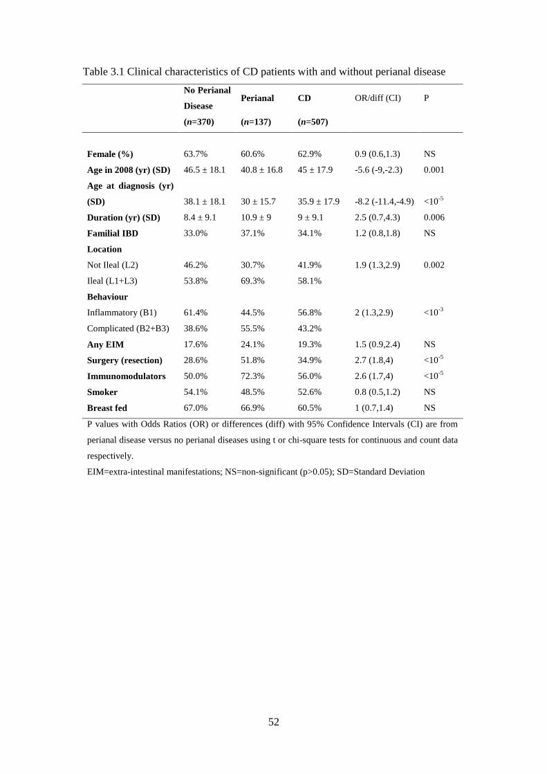
52
Table 3.1 Clinical characteristics of CD patients with and without perianal disease
No Perianal
Disease Perianal CD OR/diff (CI) P
(n=370) (n=137) (n=507)
Female (%) 63.7% 60.6% 62.9% 0.9 (0.6,1.3) NS
Age in 2008 (yr) (SD) 46.5 ± 18.1 40.8 ± 16.8 45 ± 17.9 -5.6 (-9,-2.3) 0.001
Age at diagnosis (yr)
(SD) 38.1 ± 18.1 30 ± 15.7 35.9 ± 17.9 -8.2 (-11.4,-4.9) <10-5
Duration (yr) (SD) 8.4 ± 9.1 10.9 ± 9 9 ± 9.1 2.5 (0.7,4.3) 0.006
Familial IBD 33.0% 37.1% 34.1% 1.2 (0.8,1.8) NS
Location
Not Ileal (L2) 46.2% 30.7% 41.9% 1.9 (1.3,2.9) 0.002
Ileal (L1+L3) 53.8% 69.3% 58.1%
Behaviour
Inflammatory (B1) 61.4% 44.5% 56.8% 2 (1.3,2.9) <10-3
Complicated (B2+B3) 38.6% 55.5% 43.2%
Any EIM 17.6% 24.1% 19.3% 1.5 (0.9,2.4) NS
Surgery (resection) 28.6% 51.8% 34.9% 2.7 (1.8,4) <10-5
Immunomodulators 50.0% 72.3% 56.0% 2.6 (1.7,4) <10-5
Smoker 54.1% 48.5% 52.6% 0.8 (0.5,1.2) NS
Breast fed 67.0% 66.9% 60.5% 1 (0.7,1.4) NS
P values with Odds Ratios (OR) or differences (diff) with 95% Confidence Intervals (CI) are from
perianal disease versus no perianal diseases using t or chi-square tests for continuous and count data
respectively.
EIM=extra-intestinal manifestations; NS=non-significant (p>0.05); SD=Standard Deviation
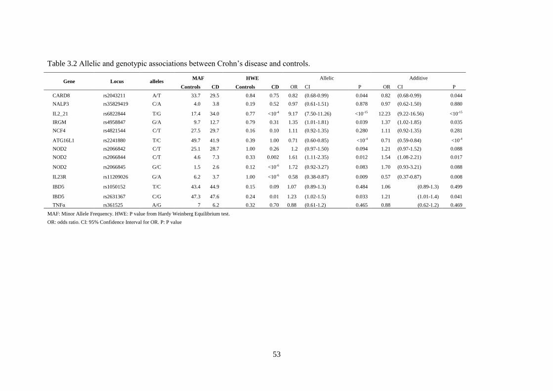
53
Table 3.2 Allelic and genotypic associations between Crohn‟s disease and controls.
Gene Locus alleles MAF HWE Allelic Additive
Controls CD Controls CD OR CI P OR CI P
CARD8 rs2043211 A/T 33.7 29.5 0.84 0.75 0.82 (0.68-0.99) 0.044 0.82 (0.68-0.99) 0.044
NALP3 rs35829419 C/A 4.0 3.8 0.19 0.52 0.97 (0.61-1.51) 0.878 0.97 (0.62-1.50) 0.880
IL2_21 rs6822844 T/G 17.4 34.0 0.77 <10-4 9.17 (7.50-11.26) <10-15 12.23 (9.22-16.56) <10-15
IRGM rs4958847 G/A 9.7 12.7 0.79 0.31 1.35 (1.01-1.81) 0.039 1.37 (1.02-1.85) 0.035
NCF4 rs4821544 C/T 27.5 29.7 0.16 0.10 1.11 (0.92-1.35) 0.280 1.11 (0.92-1.35) 0.281
ATG16L1 rs2241880 T/C 49.7 41.9 0.39 1.00 0.71 (0.60-0.85) <10-4 0.71 (0.59-0.84) <10-4
NOD2 rs2066842 C/T 25.1 28.7 1.00 0.26 1.2 (0.97-1.50) 0.094 1.21 (0.97-1.52) 0.088
NOD2 rs2066844 C/T 4.6 7.3 0.33 0.002 1.61 (1.11-2.35) 0.012 1.54 (1.08-2.21) 0.017
NOD2 rs2066845 G/C 1.5 2.6 0.12 <10-6 1.72 (0.92-3.27) 0.083 1.70 (0.93-3.21) 0.088
IL23R rs11209026 G/A 6.2 3.7 1.00 <10-6 0.58 (0.38-0.87) 0.009 0.57 (0.37-0.87) 0.008
IBD5 rs1050152 T/C 43.4 44.9 0.15 0.09 1.07 (0.89-1.3) 0.484 1.06 (0.89-1.3) 0.499
IBD5 rs2631367 C/G 47.3 47.6 0.24 0.01 1.23 (1.02-1.5) 0.033 1.21 (1.01-1.4) 0.041
TNFα rs361525 A/G 7 6.2 0.32 0.70 0.88 (0.61-1.2) 0.465 0.88 (0.62-1.2) 0.469
MAF: Minor Allele Frequency. HWE: P value from Hardy Weinberg Equilibrium test.
OR: odds ratio. CI: 95% Confidence Interval for OR. P: P value
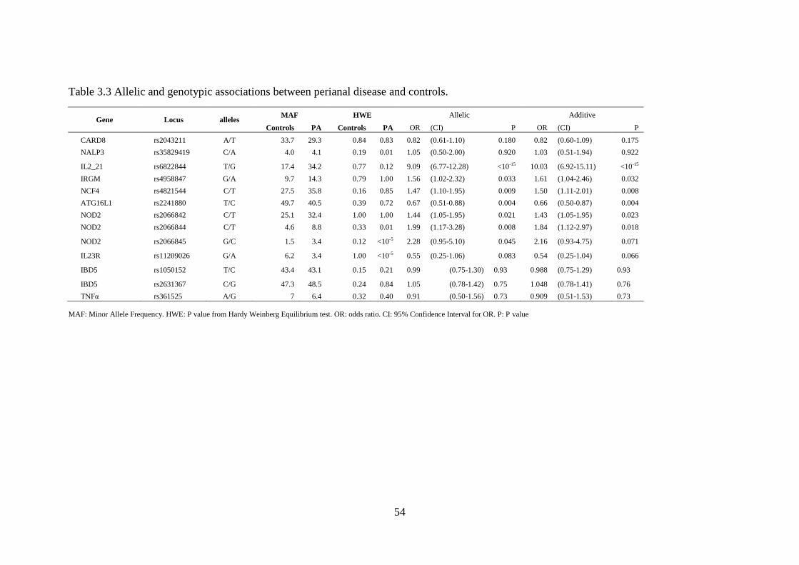
54
Table 3.3 Allelic and genotypic associations between perianal disease and controls.
Gene Locus alleles MAF HWE Allelic Additive
Controls PA Controls PA OR (CI) P OR (CI) P
CARD8 rs2043211 A/T 33.7 29.3 0.84 0.83 0.82 (0.61-1.10) 0.180 0.82 (0.60-1.09) 0.175
NALP3 rs35829419 C/A 4.0 4.1 0.19 0.01 1.05 (0.50-2.00) 0.920 1.03 (0.51-1.94) 0.922
IL2_21 rs6822844 T/G 17.4 34.2 0.77 0.12 9.09 (6.77-12.28) <10-15 10.03 (6.92-15.11) <10-15
IRGM rs4958847 G/A 9.7 14.3 0.79 1.00 1.56 (1.02-2.32) 0.033 1.61 (1.04-2.46) 0.032
NCF4 rs4821544 C/T 27.5 35.8 0.16 0.85 1.47 (1.10-1.95) 0.009 1.50 (1.11-2.01) 0.008
ATG16L1 rs2241880 T/C 49.7 40.5 0.39 0.72 0.67 (0.51-0.88) 0.004 0.66 (0.50-0.87) 0.004
NOD2 rs2066842 C/T 25.1 32.4 1.00 1.00 1.44 (1.05-1.95) 0.021 1.43 (1.05-1.95) 0.023
NOD2 rs2066844 C/T 4.6 8.8 0.33 0.01 1.99 (1.17-3.28) 0.008 1.84 (1.12-2.97) 0.018
NOD2 rs2066845 G/C 1.5 3.4 0.12 <10-5 2.28 (0.95-5.10) 0.045 2.16 (0.93-4.75) 0.071
IL23R rs11209026 G/A 6.2 3.4 1.00 <10-5 0.55 (0.25-1.06) 0.083 0.54 (0.25-1.04) 0.066
IBD5 rs1050152 T/C 43.4 43.1 0.15 0.21 0.99 (0.75-1.30) 0.93 0.988 (0.75-1.29) 0.93
IBD5 rs2631367 C/G 47.3 48.5 0.24 0.84 1.05 (0.78-1.42) 0.75 1.048 (0.78-1.41) 0.76
TNFα rs361525 A/G 7 6.4 0.32 0.40 0.91 (0.50-1.56) 0.73 0.909 (0.51-1.53) 0.73
MAF: Minor Allele Frequency. HWE: P value from Hardy Weinberg Equilibrium test. OR: odds ratio. CI: 95% Confidence Interval for OR. P: P value
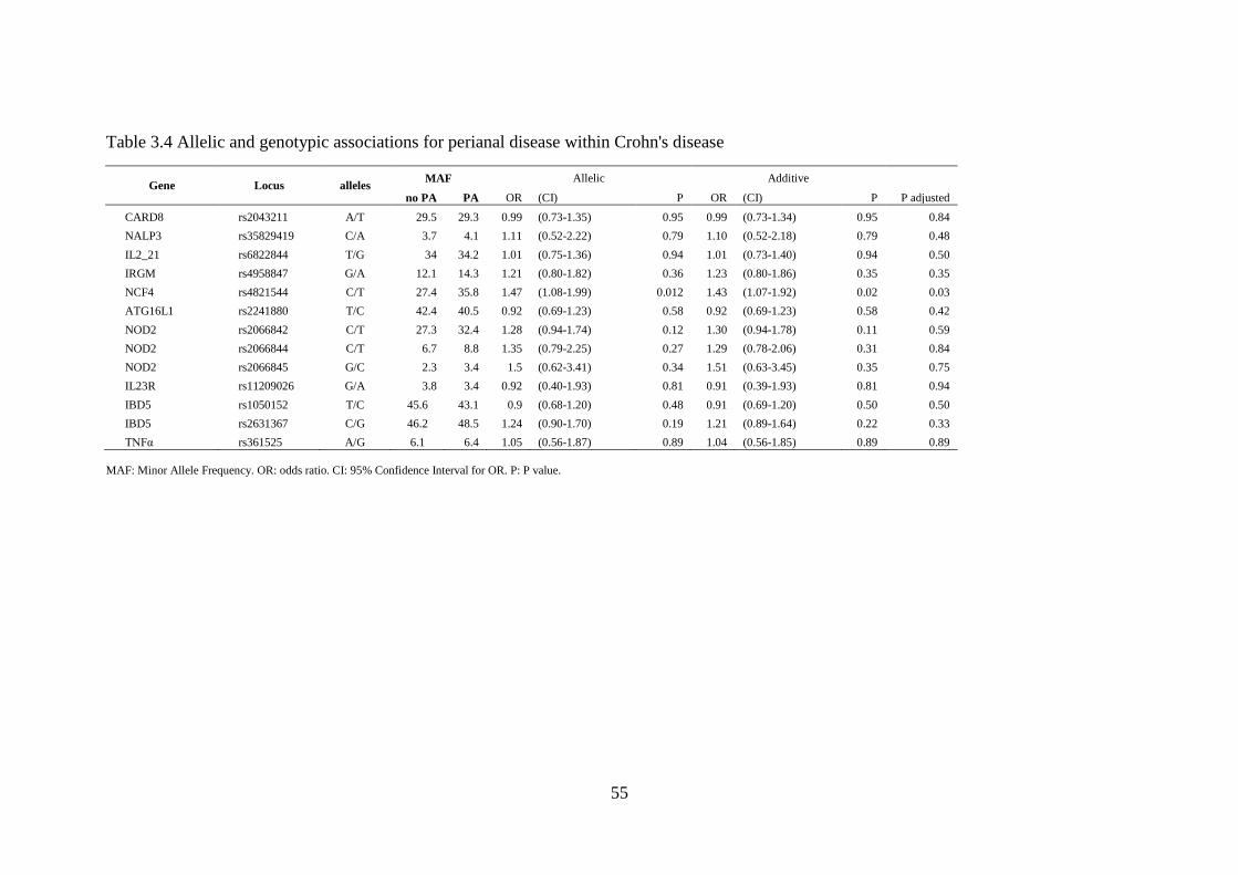
55
Table 3.4 Allelic and genotypic associations for perianal disease within Crohn's disease
Gene Locus alleles MAF Allelic Additive
no PA PA OR (CI) P OR (CI) P P adjusted
CARD8 rs2043211 A/T 29.5 29.3 0.99 (0.73-1.35) 0.95 0.99 (0.73-1.34) 0.95 0.84
NALP3 rs35829419 C/A 3.7 4.1 1.11 (0.52-2.22) 0.79 1.10 (0.52-2.18) 0.79 0.48
IL2_21 rs6822844 T/G 34 34.2 1.01 (0.75-1.36) 0.94 1.01 (0.73-1.40) 0.94 0.50
IRGM rs4958847 G/A 12.1 14.3 1.21 (0.80-1.82) 0.36 1.23 (0.80-1.86) 0.35 0.35
NCF4 rs4821544 C/T 27.4 35.8 1.47 (1.08-1.99) 0.012 1.43 (1.07-1.92) 0.02 0.03
ATG16L1 rs2241880 T/C 42.4 40.5 0.92 (0.69-1.23) 0.58 0.92 (0.69-1.23) 0.58 0.42
NOD2 rs2066842 C/T 27.3 32.4 1.28 (0.94-1.74) 0.12 1.30 (0.94-1.78) 0.11 0.59
NOD2 rs2066844 C/T 6.7 8.8 1.35 (0.79-2.25) 0.27 1.29 (0.78-2.06) 0.31 0.84
NOD2 rs2066845 G/C 2.3 3.4 1.5 (0.62-3.41) 0.34 1.51 (0.63-3.45) 0.35 0.75
IL23R rs11209026 G/A 3.8 3.4 0.92 (0.40-1.93) 0.81 0.91 (0.39-1.93) 0.81 0.94
IBD5 rs1050152 T/C 45.6 43.1 0.9 (0.68-1.20) 0.48 0.91 (0.69-1.20) 0.50 0.50
IBD5 rs2631367 C/G 46.2 48.5 1.24 (0.90-1.70) 0.19 1.21 (0.89-1.64) 0.22 0.33
TNFα rs361525 A/G 6.1 6.4 1.05 (0.56-1.87) 0.89 1.04 (0.56-1.85) 0.89 0.89
MAF: Minor Allele Frequency. OR: odds ratio. CI: 95% Confidence Interval for OR. P: P value.
56
3.5 Discussion
Recent evidence indicates that perianal CD represents a distinct disease
phenotype and the results of this study do suggest specific associations with perianal
CD patients compared with those patients without perianal involvement. Of the
clinical factors investigated, this study demonstrated associations with perianal CD
and young age at diagnosis, ileal disease location and complicated disease behaviour.
In addition, an association with the NCF4 gene was demonstrated.
One of the more notable findings of this study was that perianal involvement
was associated with ileal disease in this cohort. Traditionally it has been believed that
perianal CD is associated with distal intestinal involvement and this contention is
supported by a number of referral centre based cohorts [44, 49-51, 53, 55, 57]. A
possible explanation for the different association of perianal disease with intestinal
disease location in this study is the broader inclusion of other perianal lesions in
addition to fistulae. Several of the aforementioned cohorts included only perianal
fistulae, potentially altering the relationship to intestinal disease. The relationship may
also differ in this study compared to the aforementioned referral centre cohorts due to
the population based nature of the data. The evidence for the association of perianal
disease with colonic disease is less consistent in previously reported population based
data; one study supporting an association with colonic disease [7], another showing
the association only with ileocolonic location [47] and a third, the Swedish study,
failing to demonstrate a relationship [48]. The population based data presented here
likely represents a true relationship between ileal disease and perianal CD in this
cohort. The makeup of this population based cohort highlights this relationship, as it
contains a high proportion of mild colonic CD patients, who may not be as susceptible
to perianal disease. The rigorous methodology employed in the present study to
identify all CD patients combined with the long median follow up period of nine years
and high rate of colonoscopy in this cohort giving accurate documentation of the
extent of colonic disease [201] all support the contention of a genuine relationship
between ileal disease and perianal disease.
The finding that young age at diagnosis is associated with perianal disease is
consistent with the majority of previous studies from both referral centres [54, 58, 71]

57
and population based cohorts [7, 48]. However, two small studies found no
relationship with age and perianal CD [45, 55]. The present study also demonstrated
that perianal disease was associated with longer disease duration, consistent with the
data presented in chapter 2 showing that perianal disease can occur at any time, often
well after diagnosis of luminal CD. Those diagnosed at a young age had longer
duration of follow up, which may have contributed to the association of young age at
diagnosis. However, the fact the perianal patients were significantly younger at follow
up also supports the contention that perianal disease is more likely in those diagnosed
at a young age independent of disease duration.
The relationship of perianal disease with intestinal disease behaviour in the
present study is also interesting. Penetrating and stricturing intestinal disease were
both associated with perianal CD. Previous studies of disease behaviour and perianal
disease have yielded varying results. Smith et al [8] and Veloso et al [57] found that
perianal disease was not associated with penetrating intestinal disease. However, Tang
et al did find a strong relationship between perianal and luminal fistulas but also
observed that the two fistula types often occurred exclusively [7]. Of note, in the
present study, while perianal disease was associated with luminal fistulising disease,
85% of patients with perianal disease did not have penetrating intestinal disease. This
is consistent with Tang et al’s data and reinforces the theory that perianal disease
represents a different disease phenotype to intestinal penetrating disease.
Environmental factors undoubtedly play a role in CD pathogenesis; however
their influence on perianal disease has not been well documented. Prior to the present
study, no others had directly compared smoking rates in perianal and CD patients. In
this cohort there was no relationship between smoking and perianal disease. The
epidemiologic evidence for being breast fed is less consistent but it is likely to have a
protective effect against developing CD [65]. This study did not show any effect of
breast feeding on perianal disease. Any effect of these environmental factors is likely
to be modest, hence the possibility of type II error would need to be considered in a
study of this size.
Increasing evidence suggests that CD results from a dysregulated immune
response to as yet unidentified environmental factors in genetically susceptible hosts.
58
Genotype not only influences susceptibility for CD overall but also the expression of
certain CD phenotypes. Given that perianal disease appears to represent a separate CD
phenotype, one may expect different genetic susceptibility factors to be present in
these patients. After comparing the genotypes at a number of loci previously
associated with CD, this study found a statistically significant association with the
minor allele of neutrophil cytosolic factor 4 (NCF4) in patients with perianal disease.
This association was apparent when perianal disease patients were compared both
with healthy controls and with other CD patients without perianal involvement. In
contrast, the effects of the other loci significantly associated with perianal disease
(particularly IL21 and ATG16L1) did not persist when compared with CD patients
without perianal disease.
NCF4 is located on chromosome 22 and was first associated with CD in a
genome wide association study in 2007 [207]. However, the association of NCF4 with
CD has not been universally replicated and two recently published studies in large
European and Canadian cohorts failed to demonstrate an association [208, 209].
While the association with CD hasn‟t been consistent, the NCF4 gene does offer
biological plausibility as it has a role in phagocytosis and the antimicrobial response.
It encodes a protein known as p40phox which is one of the subunits of the
nicotinamide adenine dinucleotide (NADPH)-oxidase enzyme system [210]. This
system produces reactive oxygen species upon phagocytosis which are important for
mounting an effective antimicrobial response [211]. This provides an appealing link
to CD pathogenesis, with ineffective bacterial killing of phagocytised microbes
producing a prolonged immune activation. The association of NCF4 with ileal CD
had previously been demonstrated in this cohort of patients [212]. The present study
has now demonstrated a further association with perianal disease which maintained
statistical significance after adjusting for potential confounding factors including
intestinal disease location, behaviour, age and smoking.
This is the first study to demonstrate an association between the NCF4 locus
and perianal CD. As discussed in chapter 1.3.4.4, three other genetic loci had
previously been shown to be associated with perianal CD; IBD5, IRGM and TNFα,
none of which demonstrated a significant association in this cohort.
59
Of these, the IBD5 susceptibility locus on chromosome 5q31 has been most
investigated. Associations with this locus and CD have been widely replicated and
possible functional variants have been identified in the carnitine/organic cation
transporter genes OCTN1 and OCTN2 (SLC22A4 and SLC22A5). Of the six studies to
date investigating the IBD5 region for a relationship with perianal disease, four have
suggested an association [110, 114, 115, 117], while two others have not [74, 116].
No association was shown in this study and doubt remains as to the role of IBD5 in
perianal CD and to the possible causative variants.
IRGM was also investigated in this cohort and did not demonstrate a significant
association with perianal disease when compared with the non-perianal CD group. .
As discussed in section 1.3.4.4.2, IRGM has previously been associated with CD
susceptibility overall in a large genome-wide association study [118] and the fact that
it is an important autophagy gene also provides a plausible biological link to CD
pathogenesis. While IRGM did not show an association in the present study, the
potential pathogenic mechanism described and the association of NCF4 warrant
further investigation into the role of the autophagy genes in perianal disease and CD
overall.
The only other gene previously associated with perianal CD is TNFα. Section
1.3.4.4.3 highlighted that, while the proinflammatory cytokine TNFα has an
established role in CD pathogenesis, results of association studies for CD overall have
been varied [122]. In addition only one small study has shown an association with
perianal CD [213]. The present study failed to replicate this and further studies are
required to assess this association.
The variation in this and other previously reported studies of the factors
influencing perianal CD may result from differences in definitions and inclusion
criteria. This study included all perianal lesions classified according to AGA
guidelines whereas several previous studies considered only perianal fistulas. In
addition, given that the previous study of IRGM demonstrated only modest odds ratios
and it has a low MAF, the present study was inadequately powered to detect such a
modest difference. The present data is drawn from an ethnically homogenous
60
population made up of 97.8 per cent Caucasians and ethnic differences are another
important factor which may account for variations in study results.
There is now substantial evidence that perianal CD is associated with a more
severe luminal disease course [214] and this study supports that contention,
demonstrating a greater requirement for surgery and immunomodulator use in the
perianal disease group. Defining risk factors for CD behaviour and attempting to
predict the future course of the disease has taken on greater significance in the era of
increasingly effective medical therapies, in particular anti-TNFα agents. Many
physicians now advocate a “top-down” treatment strategy with early aggressive use of
medical therapy in an attempt to alter the natural history of CD[215]. The data from
this study suggests that patients diagnosed at a young age, those with ileal disease and
those with the minor allele of NCF4 SNP rs4821544 are more likely to develop
perianal disease and could be candidates for such “top-down” therapy.
3.6 Conclusion
This study, from a large, well characterised, population based CD cohort,
supports the hypothesis that perianal CD represents a distinct disease phenotype. It
has demonstrated associations with perianal disease and younger age at diagnosis,
complicated disease behaviour and ileal disease location. In addition, this paper
represents the first report of an association of the NCF4 gene with perianal disease.
Perianal CD patients endure a more severe disease course and further studies that
continue to clarify these risk factors will allow better targeting of therapy in the future.
61
Chapter 4
MRI in Perianal CD
Abstract
Background
In the setting of perianal CD, MRI is often used to establish the presence and anatomy
of fistulas and undrained sepsis prior to examination under anaesthetic (EUA).
Despite its inclusion in management guidelines, there is conflicting literature defining
the accuracy and utility of MRI. The objectives of this study were to determine the
concordance of MRI and EUA findings and to assess the impact of MRI on intra-
operative management.
Method
All patients undergoing EUA for perianal CD at Christchurch Hospital were eligible
for the study. EUA was performed by a colorectal surgeon blinded to the pre-
operative MRI pelvis result. Blinded findings were documented, after which the MRI
result was revealed intra-operatively. Subsequent alterations to findings or
management were recorded. Discordance was resolved and a gold standard for
comparison developed for each case by combining MRI, EUA and clinical outcome.
Results
Twenty patients completed the study protocol and were followed up for a median of
12 months (range 1-22 months). Unblinded EUA findings were abscess (7 patients),
fistula (12), ulcer or fissure (6), haemorrhoids (1) and carcinoma (1). EUA findings
were discordant with MRI in 7 patients (35%). These included 5 false positive and 2
additional true positive findings on MRI involving minor fistula classification changes.
The sensitivity and specificity of MRI was 100% and 50% respectively for
identification of fistulas and/or abscesses. Surgical management included abscess
drainage (7 patients), loose seton (8), and defunctioning ileostomy (2). In no case did
the MRI findings alter intra-operative management.
Conclusion
MRI is widely used prior to EUA for perianal CD. However, this study demonstrated
minimal impact of MRI on intra-operative decision making, despite moderate
discordance between MRI and EUA findings. The high sensitivity of MRI for fistulas

62
and abscesses suggests it is useful in excluding occult perianal sepsis and avoiding
unnecessary EUA.
63
4.1 Introduction
Due to the complex nature of perianal CD, simple clinical examination is often
inadequate or impossible and further more detailed assessment is required to
accurately define the anatomy of fistulas, the presence of undrained sepsis and to plan
appropriate treatment. The literature review presented in section 1.4 concluded that
MRI and EAUSS have emerged as the imaging techniques of choice to complement
EUA in the assessment of perianal CD. This is reflected in current management
guidelines which recommend MRI or EAUSS in all perianal CD patients where
surgery is the initial treatment strategy or in the presence of pain, fluctuation or
stricture [24]. Much of the available literature assessing the accuracy of MRI pertains
to cryptoglandular fistulas and it appears the accuracy of MRI decreases in the more
complex lesions often present in CD [133]. Furthermore, the effect of MRI on
management of perianal CD has not been well studied. Chapter 5 will demonstrate
that many perianal CD patients require multiple surgical interventions and therefore
have the potential to require multiple MRI scans. MRI is an expensive test with
restricted availability in some areas hence a full assessment of its accuracy and impact
on management is important.
4.2 Aims
The aims of this study were to determine the concordance of MRI and EUA
findings in perianal CD and to assess the impact of MRI on intra-operative
management.
4.3 Methods
Patients were recruited for the study between 1st September 2009 and 31
st
October 2010. All adult patients with perianal CD presenting to Christchurch hospital
over this period in whom the treating surgeon considered EUA necessary were
eligible for the study. The diagnosis of CD was confirmed using established
diagnostic criteria[203]. Exclusion criteria were contraindications to MRI (including
intra-orbital foreign body, implantable metal devices and a personal history of
claustrophobia), pregnancy and the inability to provide informed consent.
64
Following informed consent, patients enrolled in the trial underwent
preoperative clinical assessment which included classification of CD using the
Montreal system, recording of medications, C reactive protein level and white blood
cell count. Detailed perineal examination was performed, the presence of any perianal
lesions documented and the perianal disease activity index (PDAI) recorded. Routine
preoperative pelvic MRI was then performed on all patients. The MRI was performed
using a 1.5T Signa Excite HD, machine (GE Medical Systems, Milwaukee, USA)
with a surface phased array 8 channel coil. High resolution axial and coronal T2
weighted and T2 Fat Saturation sequences angled to the plane of the anal sphincter
were used. The resulting MR images were reported by radiologists experienced in the
technique.
Patients then underwent EUA performed by either a specialist colorectal
surgeon or colorectal fellow. The surgeon initially performed the EUA without
knowledge of the MRI findings. The blinded EUA findings and proposed
management were documented on a proforma, but no therapeutic manoeuvres were
undertaken at that stage. With the patient still under anaesthetic, the MR images and
radiologist report were revealed to the operating surgeon. Any discordance between
the blinded EUA and the MRI was documented. The EUA was then repeated and any
therapeutic intervention undertaken. Changes to the proposed management recorded
at the initial blinded EUA were documented. The case report form used to document
findings and operative management is provided in Appendix 2.
Patients were followed up at six weeks, six months and 1 year post EUA. At
each of these time points clinical data identical to that in the preoperative work up
was recorded.
Those cases where discordance between EUA and MRI existed and was not
resolved at EUA were reviewed by the study investigators. By combining the
outcome from clinical follow up, EUA and MRI findings, an outcome based gold
standard was derived using methodology employed in similar previous studies [135,
136].
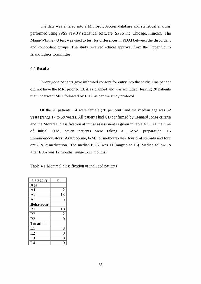
65
The data was entered into a Microsoft Access database and statistical analysis
performed using SPSS v19.0® statistical software (SPSS Inc. Chicago, Illinois). The
Mann-Whitney U test was used to test for differences in PDAI between the discordant
and concordant groups. The study received ethical approval from the Upper South
Island Ethics Committee.
4.4 Results
Twenty-one patients gave informed consent for entry into the study. One patient
did not have the MRI prior to EUA as planned and was excluded; leaving 20 patients
that underwent MRI followed by EUA as per the study protocol.
Of the 20 patients, 14 were female (70 per cent) and the median age was 32
years (range 17 to 59 years). All patients had CD confirmed by Lennard Jones criteria
and the Montreal classification at initial assessment is given in table 4.1. At the time
of initial EUA, seven patients were taking a 5-ASA preparation, 15
immunomodulators (Azathioprine, 6-MP or methotrexate), four oral steroids and four
anti-TNFα medication. The median PDAI was 11 (range 5 to 16). Median follow up
after EUA was 12 months (range 1-22 months).
Table 4.1 Montreal classification of included patients
Category n
Age
A1 2
A2 13
A3 5
Behaviour
B1 18
B2 2
B3 0
Location
L1 3
L2 9
L3 8
L4 0
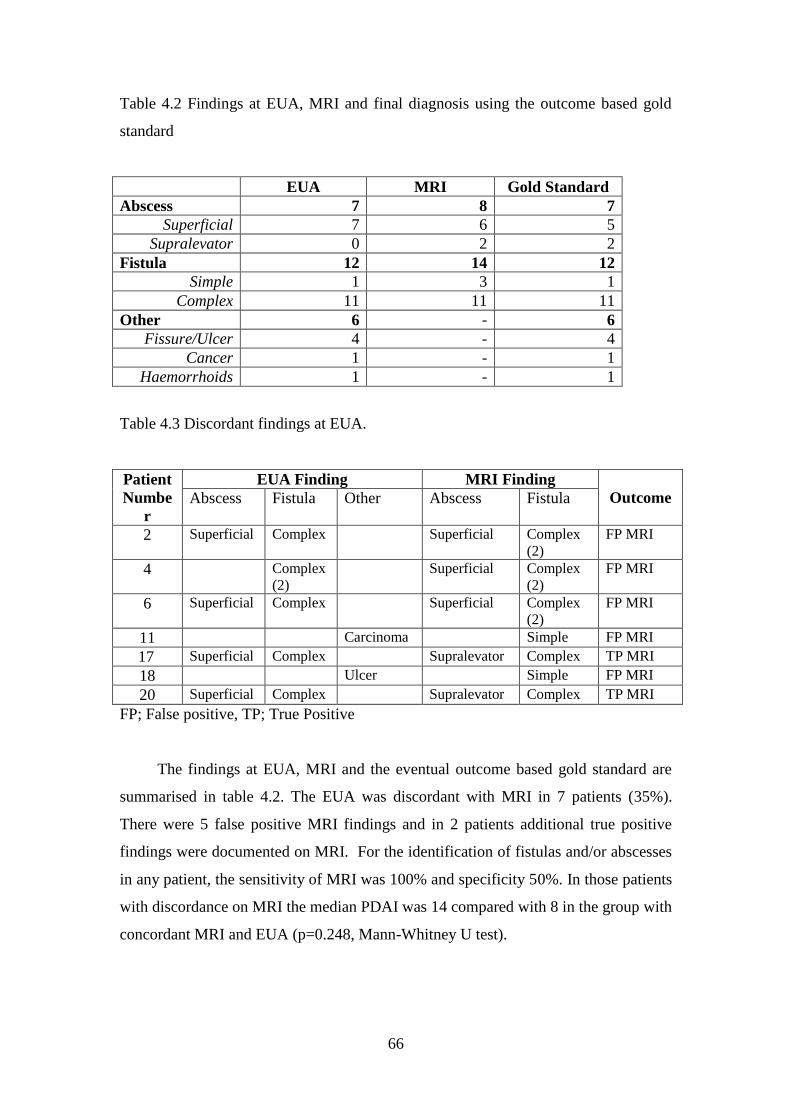
66
Table 4.2 Findings at EUA, MRI and final diagnosis using the outcome based gold
standard
EUA MRI Gold Standard
Abscess 7 8 7
Superficial 7 6 5
Supralevator 0 2 2
Fistula 12 14 12
Simple 1 3 1
Complex 11 11 11
Other 6 - 6
Fissure/Ulcer 4 - 4
Cancer 1 - 1
Haemorrhoids 1 - 1
Table 4.3 Discordant findings at EUA.
Patient
Numbe
r
EUA Finding MRI Finding
Outcome Abscess Fistula Other Abscess Fistula
2 Superficial Complex Superficial Complex
(2)
FP MRI
4 Complex
(2)
Superficial Complex
(2)
FP MRI
6 Superficial Complex Superficial Complex
(2)
FP MRI
11 Carcinoma Simple FP MRI
17 Superficial Complex Supralevator Complex TP MRI
18 Ulcer Simple FP MRI
20 Superficial Complex Supralevator Complex TP MRI
FP; False positive, TP; True Positive
The findings at EUA, MRI and the eventual outcome based gold standard are
summarised in table 4.2. The EUA was discordant with MRI in 7 patients (35%).
There were 5 false positive MRI findings and in 2 patients additional true positive
findings were documented on MRI. For the identification of fistulas and/or abscesses
in any patient, the sensitivity of MRI was 100% and specificity 50%. In those patients
with discordance on MRI the median PDAI was 14 compared with 8 in the group with
concordant MRI and EUA (p=0.248, Mann-Whitney U test).
67
The reasons for the discordance are detailed in table 4.3. MRI in two patients
(patients 2 and 6) suggested additional separate fistula tracts which could not be
identified at EUA. Patient 2 had a seton ligature placed in the fistula that was
identified and a defunctioning ileostomy because of severe sepsis. After follow up of
18 months, the patient was maintained on adalimumab, the seton had been removed
and the fistula had ceased draining. At no stage did the area of the second fistula
suggested on the MRI cause any clinical symptoms hence this was deemed a false
positive MRI finding. Similarly, patient 6 had a loose seton placed in the identified
fistula and at 17 months follow up had commenced adalimumab and the seton had
been removed. As the site of the other suggested fistula on MRI had not produced
symptoms, this was also deemed a false positive MRI finding. Two further MRI
scans were deemed false positive for the presence of fistulas. In patient 11, the MRI
was reported as a complex fistula however EUA revealed an unexpected low rectal
carcinoma without the presence of a fistula. In patient 18 an intersphincteric fistula
was reported however only an ulcer was evident at EUA. The ulcer was biopsied and
curreted. The patient was on maintenance adalimumab for ileocolic disease and at 7
months follow up the ulcer remained healed. The other false positive MRI (patient 4)
suggested a peri-coccygeal abscess however despite probing, curetting and laying
open sinus tracts, only a granulation tissue lined cavity was apparent and no pus was
drained. Two patients had false negative EUAs. In both patients 17 and 20, abscesses
were classified as superficial but the MRIs clearly showed supralevator extension.
Despite the anatomic misclassification by the operating surgeon, the planned drainage
manoeuvres with a combination of loose setons and malekot catheters were
considered adequate and further management was not instituted as a result of the MRI.
With follow up of 7 and 5 months respectively, the abscesses had clinically resolved
leaving draining fistulas in both patients.
Initial surgical management included abscess drainage in seven patients,
placement of loose seton ligatures in eight patients and defunctioning ileostomy in
two patients. In no patient did the MRI alter the intraoperative management. One
patient (patient 4) had an MRI demonstrating sacral osteomyelitis as a result of the
severe perianal sepsis which was not evident at EUA and resulted in institution of
long term antibiotic therapy. The patient is currently awaiting proctectomy and distal
sacrectomy.
68
4.5 Discussion
MRI is commonly used in perianal CD and included in management guidelines
as a mandatory part of the preoperative work up for many patients. This study has
demonstrated a moderate level of discordance between findings at MRI and EUA that
is within the previously reported range of 59-91% in studies including perianal CD
patients [133-136, 138].
The discordant findings included four false positive MRI findings of fistulas,
one false positive abscess and two additional true positive MRI findings where
abscesses were recognised at EUA but misclassified. Discordance between these
modalities can arise due to the operator dependence of both EUA and MRI. The MRIs
were reported by radiologists with over ten years‟ experience in the technique.
However, previous studies have documented that there is a level of both intra-
observer and inter-observer variation in MRI interpretation even amongst experienced
practitioners [138]. All reporting radiologists were aware of their participation in the
study at the time of reporting and the fact that the patient was to undergo surgical
exploration. This does have the potential to introduce a tendency to over reporting as
recognised in previous studies [138]. The fact that the major discordance was
produced by false positive MRI findings of fistulas suggests this phenomenon could
have contributed to the high sensitivity but relatively low specificity documented here.
While all EUAs in the present study were performed by either specialist
colorectal surgeons or fellows, it is recognised that, even in experienced hands, EUA
is not completely accurate. In fact previous studies have suggested a superior
accuracy of MRI to EUA [132]. The lack of a perfect gold standard can make the
interpretation of discordance and sensitivity and specificity calculations troublesome.
This problem was mitigated against in the present study by including clinical follow
up and using the clinical outcome to determine a reference based gold standard. This
methodology has previously been successfully employed in cryptoglandular fistulas
[136], however, this is the first report using it exclusively in perianal CD.
Previous reports have suggested the level of disagreement between MRI and
EUA increases in the presence of more complex sepsis. Schaefer et al only found
69
discordance in CD patients with complex sepsis and not in those with cryptoglandular
sepsis [133]. Beets-Tan et al found a statistically significantly greater level of
beneficial information provided by MRI in CD patients compared with non CD and
a lower level in those with simple fistulas (p<0.05). The population in this study
contained a spectrum of perianal disease ranging from no sepsis to complex sepsis, as
reflected by the PDAI range at initial assessment of five to 16. The patients with
discordance had a higher median PDAI than those without although this difference
was not statistically significant. This trend does support the findings of these previous
studies that discordance is found in more complex disease.
The most surprising finding of this study was the fact that, despite the
discordance identified, intraoperative management was not altered by the MRI
findings. Two previous studies have considered management change using similar
methodology to that employed here. The first, involving only cryptoglandular fistulas,
found intraoperative management was changed in 10% [137] and the other which
included 15 CD patients found MRI altered management in 21% overall [138]. The
lack of management change is partly explained by the fact that two thirds of the
discordant findings were false positive MRIs. The two additional true positive MRI
findings were abscesses that were classified incorrectly by the operating surgeon as
superficial rather than supralevator. Adequate drainage was performed, despite this
anatomic misconception, as was evidenced by resolution in both cases at clinical
follow up.
The low therapeutic impact of MRI may also be explained by the institutional
approach to the management of perianal CD. Of interest, no definitive fistula
treatments (laying open or mucosal advancement flap) were performed in this study
population, rather patients simply had abscess drainage with or without loose seton
placement. With sepsis controlled, patients were then considered for anti-TNFα
therapy with removal of seton following two induction doses. The key to surgical
treatment in this approach then becomes the identification and adequate drainage of
all sepsis, rather than its exact anatomic classification. While MRI may not always
directly alter management it may increase operator confidence that sepsis has been
adequately drained.
70
This study focused on the role of MRI in altering the immediate intra-operative
management. It should be noted that in this study in one patient, MRI did provide
additional information diagnosing sacral osteomyelitis and therefore altering the
subsequent medical and surgical approach. In addition, there are other situations
where MRI is useful in perianal CD. MRI is often used in the selection of patients for
possible surgical treatment and an MRI negative for sepsis in a patient with perianal
symptoms can avoid the need for an unnecessary general anaesthetic. The high
sensitivity of MRI for fistulas and abscesses demonstrated in this study supports its
utility for this purpose. In addition MRI is being increasingly used to monitor
response to anti-TNFα therapy and its role in this setting continues to evolve [139,
140].
The small numbers in this study limit the validity of its results. Using an
outcome derived gold standard demands adequate follow up to allow time for
undrained infection which may have been suggested by the MRI to declare itself
clinically. While the follow up in the study was relatively short with a median of 12
months, if reinfection from undrained sepsis was to occur, one would expect it to do
so within this time period. Recurrence beyond this time period could well be the result
of further CD activation or failure of deep tissue healing rather than inadequate sepsis
drainage.
4.6 Conclusion
Preoperative MRI in perianal CD did produce a number of findings discordant
with subsequent EUA. However, the nature of these findings did not produce
significant management changes at EUA performed by colorectal surgeons in this
study. Further larger studies of MRI in perianal CD are indicated to determine the
necessity of the mandatory use of imaging prior to planned operative treatment of
perianal CD as recommended by current guidelines. The high sensitivity for the
presence of fistulas or abscesses confirms the utility of MRI in ruling out occult
perianal sepsis and therefore avoiding unnecessary EUA.
71
Chapter 5
Perianal Surgical Intervention in CD
Abstract
Background
The aim of this study was describe the frequency and characteristics of perianal
surgical intervention (PSI) for Crohn‟s disease in a population-based cohort of
patients with inflammatory bowel disease (IBD).
Methods
A total of 1421 patients with IBD were recruited, representing approximately 91 per
cent of people with IBD in Canterbury, New Zealand. The clinical notes were
screened to confirm the diagnosis and extract clinical data, including details of PSIs.
Results
Some 649 patients with Crohn‟s disease were included in the analysis, of whom 119
(18·3 per cent) had at least one PSI. Of these, 61 (51·3 per cent) required further
procedures. Operations for perianal abscess and fistula accounted for 72·4 per cent of
interventions. PSI rates did not differ between the sexes (P = 0·218). Age less than 17
years (adjusted odds ratio (OR) 1·89 (95 per cent confidence interval 1·08 to 3·28))
and ileal disease (OR 1·76 (1·06 to 2·92)) were identified as predictors of PSI. As
disease duration increased, so did the proportion of patients with complicated
intestinal disease among those who had undergone PSI. The median time to first PSI
from diagnosis of Crohn‟s disease was 28 (interquartile range 7–82) months. Sex, age
at diagnosis and disease location did not influence the time to first PSI.
Conclusion
PSIs are frequent in patients with Crohn‟s disease, particularly those with ileal disease
and those diagnosed at a young age.
72
5.1 Introduction
The preceding chapters of this thesis support the notion that perianal disease is a
common and debilitating manifestation of Crohn‟s disease requiring intensive medical
and surgical interventions. Despite the increasing use and demonstrated efficacy of
anti-TNFα agents for perianal disease, surgery remains integral to the treatment
algorithm. Surgical Interventions include simple examination under anaesthetic,
abscess drainage, procedures for fissures and fistula and previous reports suggest that
12-20% of patients will go on to require proctectomy [34].
As reports of the frequency of perianal involvement overall in CD vary due to
the populations studied and the classification criteria employed (see discussion
chapter 2), so too does the reported frequency of perianal surgical intervention. The
few previous studies concerned with rates of perianal surgical interventions (PSI) in
patients with CD report rates from 4 to 42% [18, 55, 56]. As with overall incidence
data, these studies evaluating PSI may have reported an inaccurate incidence of PSI in
perianal CD patients overall by using single-centre, hospital-based data rather than
population-based data [55, 56, 216]. As discussed in chapter 2, hospital-based studies
come from referral centres, which are non-representative of the general population
because of greater numbers of complicated patients. In addition, the small amount of
available data regarding PSI has generally been from studies conducted in the 1980s
and 1990s. Thus, new medical treatments and surgical techniques are not well
represented in the literature [18, 55, 56, 216-218].
Chapter three discussed the inconsistency in the reported patient and disease
characteristics associated with perianal CD in previous literature. There is little
published regarding the association of these factors directly with PSI but age, gender
and the location and behaviour of the disease have all been associated variably with
perianal CD [219]. In particular, a number of studies report that perianal CD increases
in association with more distal intestinal involvement [7, 18, 45, 55, 72] however this
finding has not been universal [48] and data from this cohort presented in chapter
three suggested ileal disease, not colonic disease was associated with perianal
manifestations. Added to this local experience anecdotally has suggested a high rate
of PSI requirement in patients with ileal disease.
73
A better understanding of the true rate of PSI, the operations performed and the
patient factors associated with PSI is necessary to aid in refinement of the treatment
algorithm, particularly in the era of biological therapy.
5.2 Aims
This study aims to describe the frequency and types of PSI and also the patient
and phenotypic CD characteristics associated with PSI in a validated population-based
Crohn‟s disease cohort in Canterbury, New Zealand.
5.3 Methods
This study was performed as part of the on-going Canterbury Inflammatory
Bowel Disease Project. The study population and methods for recruitment for this
project were described in section 2.3.
Over a three-year period (2003-2005), 1421 IBD patients were recruited. This
number represents an estimated 91% of patients with IBD in the Canterbury region.
Demographic information and clinical data, together with IBD phenotype and IBD-
related surgical procedures (including PSI), were extracted retrospectively from
patient medical records. This information was then entered into a custom built
Microsoft Access database. IBD cases were confirmed using standard diagnostic
criteria [203].
The term „perianal disease‟, as employed in this study, encompasses patients
with CD who experienced one or more anal manifestations included in the AGA
guidelines classification described in section 1.2.1.8. Cases of CD were reviewed to
identify those with perianal disease and subsequently, patients who had undergone
one or more local PSI were selected from this group of patients. The type of operation
performed was recorded and grouped into one of five categories; abscess drainage,
procedure for anal fistula, examination under anaesthetic, procedure for anal fissure or
anal dilatation.
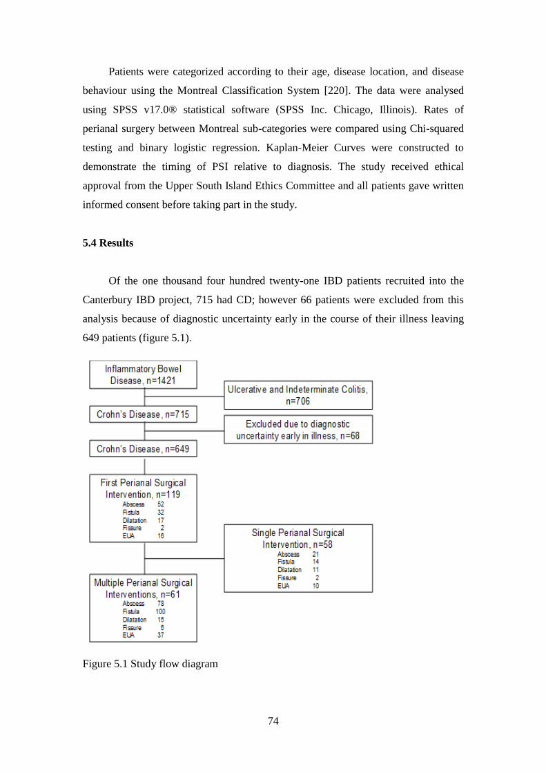
74
Patients were categorized according to their age, disease location, and disease
behaviour using the Montreal Classification System [220]. The data were analysed
using SPSS v17.0® statistical software (SPSS Inc. Chicago, Illinois). Rates of
perianal surgery between Montreal sub-categories were compared using Chi-squared
testing and binary logistic regression. Kaplan-Meier Curves were constructed to
demonstrate the timing of PSI relative to diagnosis. The study received ethical
approval from the Upper South Island Ethics Committee and all patients gave written
informed consent before taking part in the study.
5.4 Results
Of the one thousand four hundred twenty-one IBD patients recruited into the
Canterbury IBD project, 715 had CD; however 66 patients were excluded from this
analysis because of diagnostic uncertainty early in the course of their illness leaving
649 patients (figure 5.1).
Figure 5.1 Study flow diagram
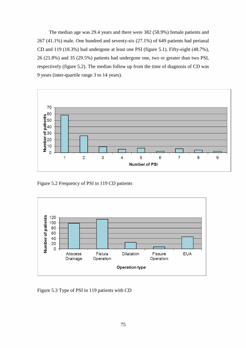
75
The median age was 29.4 years and there were 382 (58.9%) female patients and
267 (41.1%) male. One hundred and seventy-six (27.1%) of 649 patients had perianal
CD and 119 (18.3%) had undergone at least one PSI (figure 5.1). Fifty-eight (48.7%),
26 (21.8%) and 35 (29.5%) patients had undergone one, two or greater than two PSI,
respectively (figure 5.2). The median follow up from the time of diagnosis of CD was
9 years (inter-quartile range 3 to 14 years).
Figure 5.2 Frequency of PSI in 119 CD patients
Figure 5.3 Type of PSI in 119 patients with CD
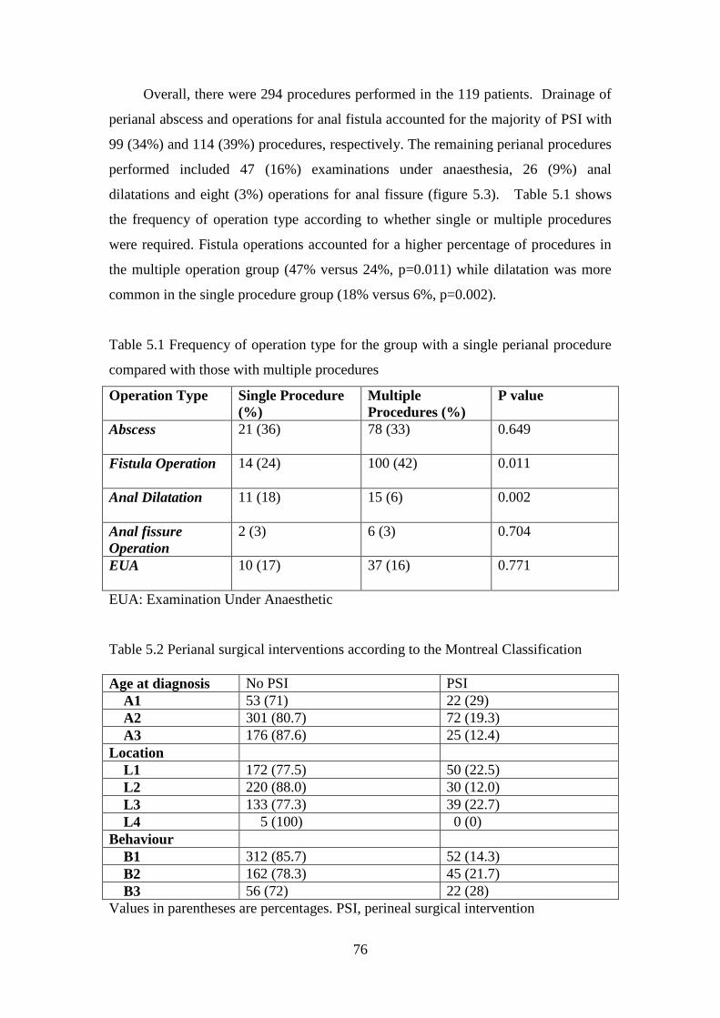
76
Overall, there were 294 procedures performed in the 119 patients. Drainage of
perianal abscess and operations for anal fistula accounted for the majority of PSI with
99 (34%) and 114 (39%) procedures, respectively. The remaining perianal procedures
performed included 47 (16%) examinations under anaesthesia, 26 (9%) anal
dilatations and eight (3%) operations for anal fissure (figure 5.3). Table 5.1 shows
the frequency of operation type according to whether single or multiple procedures
were required. Fistula operations accounted for a higher percentage of procedures in
the multiple operation group (47% versus 24%, p=0.011) while dilatation was more
common in the single procedure group (18% versus 6%, p=0.002).
Table 5.1 Frequency of operation type for the group with a single perianal procedure
compared with those with multiple procedures
Operation Type Single Procedure
(%)
Multiple
Procedures (%)
P value
Abscess 21 (36) 78 (33) 0.649
Fistula Operation 14 (24) 100 (42) 0.011
Anal Dilatation 11 (18) 15 (6) 0.002
Anal fissure
Operation
2 (3) 6 (3) 0.704
EUA 10 (17) 37 (16) 0.771
EUA: Examination Under Anaesthetic
Table 5.2 Perianal surgical interventions according to the Montreal Classification
Age at diagnosis No PSI PSI
A1 53 (71) 22 (29)
A2 301 (80.7) 72 (19.3)
A3 176 (87.6) 25 (12.4)
Location
L1 172 (77.5) 50 (22.5)
L2 220 (88.0) 30 (12.0)
L3 133 (77.3) 39 (22.7)
L4 5 (100) 0 (0)
Behaviour
B1 312 (85.7) 52 (14.3)
B2 162 (78.3) 45 (21.7)
B3 56 (72) 22 (28)
Values in parentheses are percentages. PSI, perineal surgical intervention
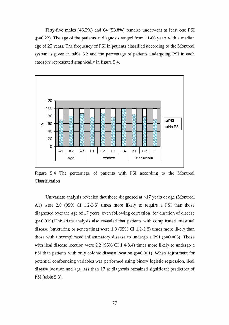
77
Fifty-five males (46.2%) and 64 (53.8%) females underwent at least one PSI
(p=0.22). The age of the patients at diagnosis ranged from 11-86 years with a median
age of 25 years. The frequency of PSI in patients classified according to the Montreal
system is given in table 5.2 and the percentage of patients undergoing PSI in each
category represented graphically in figure 5.4.
Figure 5.4 The percentage of patients with PSI according to the Montreal
Classification
Univariate analysis revealed that those diagnosed at <17 years of age (Montreal
A1) were 2.0 (95% CI 1.2-3.5) times more likely to require a PSI than those
diagnosed over the age of 17 years, even following correction for duration of disease
(p<0.009).Univariate analysis also revealed that patients with complicated intestinal
disease (stricturing or penetrating) were 1.8 (95% CI 1.2-2.8) times more likely than
those with uncomplicated inflammatory disease to undergo a PSI (p=0.003). Those
with ileal disease location were 2.2 (95% CI 1.4-3.4) times more likely to undergo a
PSI than patients with only colonic disease location (p=0.001). When adjustment for
potential confounding variables was performed using binary logistic regression, ileal
disease location and age less than 17 at diagnosis remained significant predictors of
PSI (table 5.3).
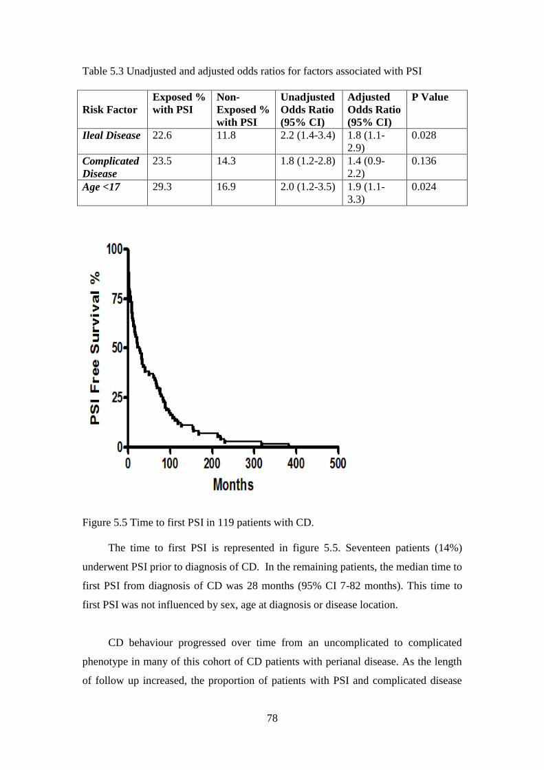
78
Table 5.3 Unadjusted and adjusted odds ratios for factors associated with PSI
Risk Factor
Exposed %
with PSI
Non-
Exposed %
with PSI
Unadjusted
Odds Ratio
(95% CI)
Adjusted
Odds Ratio
(95% CI)
P Value
Ileal Disease 22.6 11.8 2.2 (1.4-3.4) 1.8 (1.1-
2.9)
0.028
Complicated
Disease
23.5 14.3 1.8 (1.2-2.8) 1.4 (0.9-
2.2)
0.136
Age <17 29.3 16.9 2.0 (1.2-3.5) 1.9 (1.1-
3.3)
0.024
Figure 5.5 Time to first PSI in 119 patients with CD.
The time to first PSI is represented in figure 5.5. Seventeen patients (14%)
underwent PSI prior to diagnosis of CD. In the remaining patients, the median time to
first PSI from diagnosis of CD was 28 months (95% CI 7-82 months). This time to
first PSI was not influenced by sex, age at diagnosis or disease location.
CD behaviour progressed over time from an uncomplicated to complicated
phenotype in many of this cohort of CD patients with perianal disease. As the length
of follow up increased, the proportion of patients with PSI and complicated disease
79
increased such that within 5 years of diagnosis, 30% of the patients who underwent a
PSI had complicated disease. However, in the group that were greater than 10 years
post diagnosis, 80% of patients who had undergone a PSI had complicated disease
(p=0.0006). No evidence was found for a difference in the relationship between PSI
and age or disease location as the length of follow-up increased.
Thirty-five (29.4%) of the 119 patients who underwent PSI had more than 2
surgical interventions. Males were 2.8 (95% CI 1.3-6.1) times more likely to have
greater than 2 PSI than females (p=0.0085). Age, behaviour, or location of disease did
not influence the rate of recurrent PSI.
5.5 Discussion
This population-based study confirms that PSI is frequent in CD patients, with
18% of CD patients having had at least one PSI and of those, more than one half
required further procedures after a median of 9 years follow up.
Few studies have described the rate of PSI in CD overall, but the results from
this study are within the previously reported range of 4-42% [18, 55, 56]. Unlike the
population-based data presented here, previous studies have drawn data from clinic-
based cohorts from specialist centres, which may have a higher proportion of
complicated CD patients and therefore not represent the true rate of PSI in the CD
population. In addition, the overall rate of perianal disease, whether requiring surgical
intervention or not, varies widely from 10-80% due principally to the definitions used
and the types of lesions included. Diagnostic uncertainty may also alter the incidence
of perianal disease, with the inadvertent inclusion of ulcerative colitis patients as
Crohn‟s colitis potentially lowering reported incidence [16]. The rigorous
methodology employed in this study to classify the inflammatory bowel disease
population should minimise this effect [201].
Patients in this series diagnosed before 17 years of age were over two times
more likely to require a PSI than those diagnosed after age of 17 years. While no
other study has reported a higher rate of PSI in younger patients, a number have
suggested that perianal disease itself may be more frequent in younger patients.
80
Halme et al reported a series of 112 patients and found the average age of those with
perianal disease was 27.7 years compared 37.4 years in those without (p<0.01) [54].
Roberts et al described a cohort of CD patients over 50 years of age and concluded
they were less likely to present with perianal complications [71] and Cosnes et al
found fistulizing complications, including perianal disease, were more common in
those younger than 40 years [58]. Data presented in chapter 3 of this thesis, from this
cohort also suggests an association between the occurrence of perianal disease and
young age at diagnosis. However, the relationship of age to perianal disease is not
consistent across studies as both Platell et al [55] and Hellers et al [45] found no
difference in age in patients with or without perianal disease.
In this study, there was no significant difference in the proportion of females
undergoing at least one PSI compared with males. Males, however, were significantly
more likely than females to require multiple PSI. Once again, no previous studies
relate gender to risk of PSI directly and considering perianal disease itself, the results
concerning the impact of gender are conflicting. Sangwan et al and Hellers et al both
report that significantly more males than females in their studies had perianal CD [18,
45]. On the contrary, Bell [25] noted a slight female predominance for perianal
fistulae and Fry [72] reported more than twice as many females as males had perianal
CD. One of the primary concerns in surgical therapy of perianal CD is the
preservation of continence. As females have a higher risk of incontinence from factors
unrelated to CD, such as childbirth, surgeons may adopt a less aggressive surgical
approach to female CD patients which could contribute to the lower rate of multiple
interventions in females in this study.
As perianal disease itself was found to be associated with ileal disease in this
cohort (see chapter 3), PSI was more likely to be required in patients with ileal or
ileocolonic disease than those with colonic disease. Once again, no studies relate
disease location directly to the risk of PSI, however, as discussed in chapter 3,
traditionally it has been thought that the risk of perianal disease increases with more
distal intestinal involvement. Several clinic-based cohort studies that have considered
surgical intervention have supported this contention and reported increased perianal
complications with distal intestinal disease, in particular, rectal involvement [18, 45,
55, 72]. Presumably this would translate into an increase in PSI the more distal the
81
disease in these studies, although no data was given to confirm this. The reason for
increased PSI in the group with isolated ileal disease in the present study has a
number of possible explanations. The broader inclusion of other perianal lesions in
addition to fistulae in this study could alter the relationship between PSI and intestinal
disease location, however this seems unlikely because the majority of operations
performed were still for fistulising perianal disease. Using PSI as the endpoint rather
than perianal CD itself, may produce a higher rate amongst ileal CD patients if their
perianal CD produced more surgically significant lesions requiring intervention.
However the data presented in chapter three from this cohort demonstrates an
association between ileal disease and all perianal CD, not just those requiring surgical
intervention. A tempting explanation for this is the fact this data is from a population-
based cohort as compared with the aforementioned referral centre cohorts. Therefore,
this may represent a true relationship between ileal disease and perianal CD that is
highlighted in this cohort, as it contains a high proportion of mild colonic CD patients,
who may not be as susceptible to perianal disease. As discussed in sections 1.4 and
3.5, the evidence from population based cohorts is less consistent for an association of
perianal disease with colonic disease location and PD overall [7, 47, 48]. The rigorous
methodology employed in the present study to identify all CD patients combined with
the long median follow up period of nine years and high rate of colonoscopy in this
cohort giving accurate documentation of the extent of colonic disease [201] all
support the contention of a genuine relationship between ileal disease and PSI.
As described in section 1.3.4 and chapter 3, genetic factors have an influence on
the development of perianal CD, with a number of studies describing susceptibility
loci on chromosome 5 [110, 117, 121]. In addition an association with the NCF4
locus was identified in the present cohort (see chapter 3). The present study cohort has
a homogeneous ethnicity with Caucasians making up 97.8% [221]. Underlying
genetic differences in the populations studied may contribute to the reported
differences in incidence and the disease characteristics of those with perianal CD.
In addition to disease location, the relationship between disease behaviour and
PSI was considered. Univariable analysis based on most recent disease behaviour
classification suggested an association, however, the multivariable analysis showed
complicated intestinal disease behaviour did not predict the need for PSI overall. This
82
finding is consistent with recent studies suggesting that perianal and intestinal fistulae
are distinct phenotypic entities [8, 9]. These studies contributed to the Montreal
modification of the Vienna Classification to separate perianal disease from
penetrating intestinal disease by addition of a perianal modifier [70]. Of particular
note in one of these studies of 5491 patients, it was found that in colonic, but not ileal
disease, intestinal fistulae were associated with perianal disease [9]. Hence, it is not
surprising, given the aforementioned high proportion of PSI patients with ileal disease
in this cohort, that overall, complicated intestinal disease was not associated with PSI.
While disease behaviour was not significantly associated with PSI, the evolution
of disease behaviour over time following diagnosis in relation to PSI was of interest.
At diagnosis, 70% of PSI patients had uncomplicated (B1) intestinal disease.
However, later in the disease course, 80% of patients who had undergone a PSI had
complicated intestinal disease. It has previously been documented that CD behaviour
evolves over time with inflammatory disease, progressing to penetrating and
stricturing phenotypes [58], while the location of disease remains relatively stable
[222]. A previous report from this cohort of patients has demonstrated that perianal
disease itself is an independent predictor for progression to complicated intestinal
disease [5], hence it follows that a large proportion of those with a PSI will develop
complicated intestinal disease over time.
While this study was population-based, with careful phenotyping, this was
performed retrospectively and is dependent on the accuracy of the clinical records.
Another weakness is the possibility of asymptomatic disease progression affecting the
accuracy of disease classification.
5.5 Conclusion
Using a large, validated, population-based cohort, this study has confirmed that
PSI is frequent in CD patients. The requirement for PSI was associated with ileal
disease and young age at diagnosis. Over time post diagnosis, the proportion of PSI
patients with complicated intestinal disease increased. The bulk of the existing
literature on the subject contains studies published 10-20 years ago. While this more
recent data includes patients managed in the era of new medical treatments such as
anti-Tumour Necrosis Factor drugs, continuing evolution of medical management and

83
surgical techniques are likely to alter the frequency and characteristics of PSI in the
future.
84
Chapter 6
The Cost of Perianal CD
Abstract
Aims
The aim of this study was to determine the direct and indirect costs of perianal
Crohn‟s disease (CD) in patients in Canterbury in one year.
Methods
A retrospective cross-sectional analysis was performed. Perianal CD patients were
recruited over a three month period. Interviews were conducted to obtain information
regarding demographic, socioeconomic factors, and indirect costs. Hospital clinical
notes were reviewed to determine direct health care utilization and costs.
Results
Twenty-five patients were enrolled in the study. In one year the total costs per patient
for perianal CD were $20,366 with direct and indirect costs comprising $18,261 and
$2,105, respectively. Extrapolating these data across New Zealand, the total cost of
perianal CD in one year is approximately $49.5 million.
Conclusions
Perianal CD is a high cost disease with significant costs borne by patients and their
families. Expensive pharmaceuticals comprise a significant proportion of the costs:
increased access to these drugs might decrease hospital admissions and prevent work
absenteeism and loss of carer productivity.
85
6.1 Introduction
Section 1.1 noted that the incidence of CD is increasing worldwide [3] and the
peak age of onset is between 15 and 35 years [4]. CD remains incurable and a
proportion of patients will endure recurrent and prolonged periods of illness requiring
extensive medical and surgical interventions during what would otherwise be a highly
productive time of life. Hence CD presents an increasingly significant health problem
not only in terms of morbidity, but also in cost to the individual patient and society.
Previous studies have indicated that the majority of the total cost associated
with this disease relates to “extensive interventions required by a small proportion of
severely affected individuals” [189]. Several clinical markers of disease severity have
been documented including diagnosis at a young age and the presence of perianal
disease. In addition to being an independent marker of a more severe disease course,
perianal disease produces local perianal complications requiring frequent intervention
(see chapter 5) hence, this patient group is more likely to use significant health
resources and incur the largest costs as a result of the disease. A number of
international studies have considered the cost of inflammatory bowel disease overall
[190-199], however, to the authors‟ knowledge, there are no data documenting the
average patient cost of IBD in New Zealand or Australia and no previous work
specifically investigating the perianal CD group.
Evidence is accumulating that newer therapies, such as anti-tumour necrosis
factor alpha (anti-TNFα) antibodies, have significant efficacy in inducing and
maintaining remission and have particular roles in complicated CD, such as perianal
CD [150, 223]. However, these agents are expensive and predicting which patients
will benefit from their early use remains a challenge [224]. The introduction of these
modern biological treatments has created a need for government agencies to consider
the economic impact of therapeutic alternatives. To achieve this, the cost of CD and
its societal burden requires further study.
This is a cross-sectional retrospective study, which aimed to estimate the direct
and indirect costs of perianal CD in one year in Canterbury using patient-based data.
The costs of CD are borne not only by the taxpayer through government funded

86
healthcare but the patient and their family, therefore the cost perspective is
approached from a societal point of view.
6.2 Methods
This study was performed in Christchurch Hospital, a tertiary university hospital
serving a population of around 500,000. Ethical approval for the study was obtained
from the regional ethics committee. All patients with CD according to previously
documented diagnostic criteria [203] presenting to the institution during the period
November 2009 to February 2010 were eligible for entry into the study. Perianal
disease was defined as any symptomatic perianal lesion included in the American
Gastroenterological Association classification [24]. Patients were recruited through
Gastroenterology, Colorectal surgical clinics and hospital admissions.
After giving written informed consent, patients were submitted to a structured
interview to obtain information regarding demographic and socioeconomic factors,
work and school absenteeism, alternative health resource use and other related data
for the preceding twelve month period. Participants were also offered the opportunity
to nominate other costs that were not mentioned in the interview. Following the
structured interview, hospital clinical notes were reviewed to determine direct health
care utilization. This included hospital inpatient and outpatient visits and prescription
drug use.
For the purposes of this analysis the costs were classified as direct or indirect.
Direct costs included hospital (Emergency Department visits, laboratory tests,
radiological investigations, endoscopy, pharmaceuticals, inpatient care and operating
theatre costs) and outpatient (General Practice visits, specialist clinic visits, alternative
health professional visits, non-prescription medications, pharmaceuticals, laboratory
tests, District Nurse and Social Work services) associated costs. Indirect costs
included; lost productivity, travel, carers, tutors and additional phone or internet
requirements.
The costs of hospital resources were determined through the Costing
Department of the Canterbury District Health Board (CDHB) and the Ministry of
87
Health. Hospital costs were calculated using DRG codes assigned to the patient each
time they visited the hospital. A different code is given for each service required
during each visit. Based on the quantity used during the visit the cost is calculated for
each service used in a given visit. The cost department for the CDHB supplied the
authors with all the codes and costs accrued by the patients during the study period.
For primary care cost calculation, it was assumed that all patients were enrolled in a
primary health organisation (PHO). The New Zealand Government provides subsidies
to lower the cost of general practitioner (GP) visits for eligible people enrolled in a
PHO. The cost of GP services was estimated using the average cost of an appointment
by age as obtained from Pegasus Health PHO and the 2010 yearly capitation rates
provided by the Government. The capitation rates took into account whether or not
the patient had a high user health card (HUHC).
Pharmaceutical costs were calculated from the cost to the Pharmaceutical
Management Agency in New Zealand (PHARMAC) provided by their pharmaceutical
schedule accessed December 1st 2009. Additionally a 4% mark-up was added to
pharmaceutical costs plus a $5.80 dispensing fee attributable to pharmacists. The co-
payments paid by the patient are a transfer not a cost therefore these were not
included.
The human capital method as described by Drummond et al [225] was
employed in calculating indirect costs. Patients were asked the number of days they
had off work as either unpaid or annual leave related to CD. This was transferred into
hours off work and was multiplied by their gross hourly wage. For those patients not
in work their indirect costs are discussed descriptively as monetary values were not
able to be estimated. Government welfare payments received by patients not in work
were not included as they represent a transfer rather than a cost.
It is conservatively estimated that there are approximately 9000 individuals with
IBD in New Zealand, with perianal patients consisting of 27% based on data produced
in chapter 2 of this thesis. This estimation was used to extrapolate the data to obtain
values for the cost to society in New Zealand of perianal CD in one year.
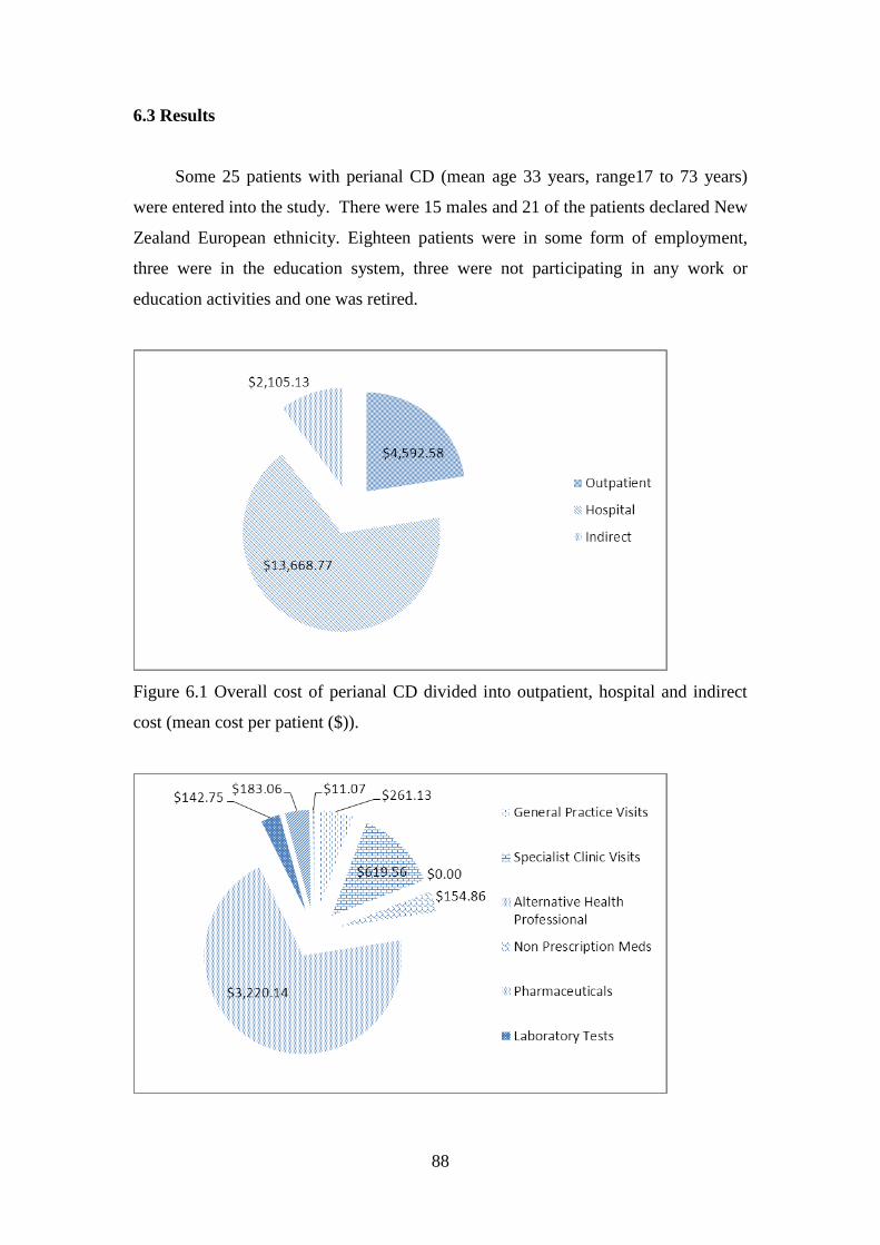
88
6.3 Results
Some 25 patients with perianal CD (mean age 33 years, range17 to 73 years)
were entered into the study. There were 15 males and 21 of the patients declared New
Zealand European ethnicity. Eighteen patients were in some form of employment,
three were in the education system, three were not participating in any work or
education activities and one was retired.
Figure 6.1 Overall cost of perianal CD divided into outpatient, hospital and indirect
cost (mean cost per patient ($)).
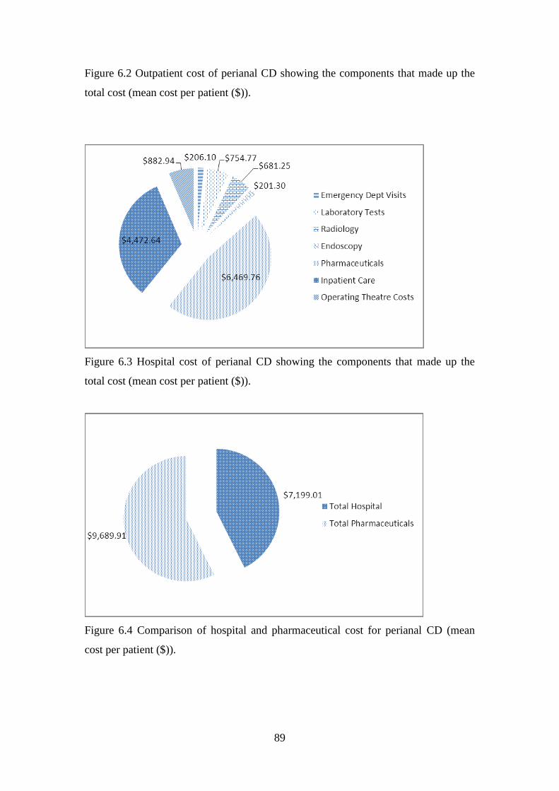
89
Figure 6.2 Outpatient cost of perianal CD showing the components that made up the
total cost (mean cost per patient ($)).
Figure 6.3 Hospital cost of perianal CD showing the components that made up the
total cost (mean cost per patient ($)).
Figure 6.4 Comparison of hospital and pharmaceutical cost for perianal CD (mean
cost per patient ($)).
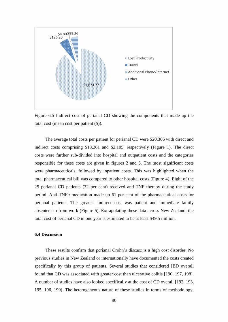
90
Figure 6.5 Indirect cost of perianal CD showing the components that made up the
total cost (mean cost per patient ($)).
The average total costs per patient for perianal CD were $20,366 with direct and
indirect costs comprising $18,261 and $2,105, respectively (Figure 1). The direct
costs were further sub-divided into hospital and outpatient costs and the categories
responsible for these costs are given in figures 2 and 3. The most significant costs
were pharmaceuticals, followed by inpatient costs. This was highlighted when the
total pharmaceutical bill was compared to other hospital costs (Figure 4). Eight of the
25 perianal CD patients (32 per cent) received anti-TNF therapy during the study
period. Anti-TNFα medication made up 61 per cent of the pharmaceutical costs for
perianal patients. The greatest indirect cost was patient and immediate family
absenteeism from work (Figure 5). Extrapolating these data across New Zealand, the
total cost of perianal CD in one year is estimated to be at least $49.5 million.
6.4 Discussion
These results confirm that perianal Crohn‟s disease is a high cost disorder. No
previous studies in New Zealand or internationally have documented the costs created
specifically by this group of patients. Several studies that considered IBD overall
found that CD was associated with greater cost than ulcerative colitis [190, 197, 198].
A number of studies have also looked specifically at the cost of CD overall [192, 193,
195, 196, 199]. The heterogeneous nature of these studies in terms of methodology,
91
varying costs in different health care systems and the differential effects of inflation
since the period of the study makes direct comparison with the present data difficult.
Juan et al [196] reported the annual cost per patient in a Spanish cohort in 2003
was €6,808 with €2,104 from direct costs and €4,704 from indirect costs [196].
Extrapolating data reported in 2009 by Mesterton et al the average annual cost of CD
in Swedish patients was €9,400 [199] (approximately 16,240 New Zealand dollars at
current exchange rates). Hence this group found comparable figures and also noted
that increased cost was predicted by increased severity as measured by the Harvey-
Bradshaw index. While the patients in the present study were not stratified for
severity, perianal disease itself is a predictor of severe disease and this may explain
the slightly higher direct costs reported here. This is supported by the previous finding
that the presence of fistulae doubled the costs of care [199]. Patients recruited from
tertiary referral centres, as in the present study, also tend to have more severe disease
and this too could have contributed to the higher direct costs reported in this study.
This fact and the small sample size in the present study mean some caution should be
exercised in interpreting the results of this study when extrapolated to the perianal and
population across New Zealand.
In contrast to the present analysis, both the previous European studies
determined a higher relative contribution from indirect compared with direct costs.
This may reflect the method of estimation of indirect costs. As the patients (or carers)
in the present study were asked to estimate the absenteeism over the previous year, an
element of recall bias may be present. In addition, no allowance for lost productivity
while at work due to the disease (presenteeism) was made in this study. The patients
were not asked if they were working part-time as a result of their disease. This
potentially could have increase the value of their lost productivity. There was an
economic recession for the majority of 2009 that could have contributed to the
reduced work hours; therefore it does not seem appropriate to assume working part-
time was a result of their illness. Altogether, these factors likely underestimate the
indirect costs associated with CD in the current study.
These issues highlight the fact that there is some controversy in the literature in
regards to the best approach to use when estimating indirect costs. The human capital-
92
cost method is recommended over other approaches by Liljas [226] and Johannesson
[227] for cost studies from a societal perspective. These authors propose it is the most
consistent with economic theory, therefore, indirect costs were estimated using the
human capital method in this study, consistent with the approach taken in other recent
studies [198, 199]. Despite this, the lower proportion of indirect costs found here
could indicate the total cost has been underestimated in the present study hence these
cost figures should be considered as a minimum.
Studies of this nature do not capture other effects of a chronic disease on
productivity because the estimation of indirect costs is calculated based on the
patients current gross wage rate. However, this does not take into account the wage
rate the patient could have realised had they not been diagnosed with CD. Given that
many patients with CD are diagnosed before or during the second or third decades of
life, their disease may have potential life-long impact on educational achievement,
career prospects and earning potential consequent to disrupted education and work.
Previous studies have considered the effect of CD on educational achievement [228,
229]. Of note while the cases in these two studies showed significantly greater
absenteeism than controls, decreased ability to present for exams and some degree of
discrimination from teachers, there was no difference in level of educational
achievement between cases and controls. Despite this, 47 per cent of respondents to a
survey conducted in Germany felt that CD had interfered with their career prospects
[198]. A separate study revealed 30 per cent of CD patients concealed their diagnosis
from their employers [228]. Hence more subtle loss of productivity costs are likely to
exist which have not been measured in this study.
In addition to both the direct and indirect costs discussed above, this study has
not included intangible elements associated with the burden of disease, or loss of
wellbeing, associated with CD. A more global assessment of the economic impact of
CD would include such costs. Recent economic theory has allowed integration of
these concepts into cost calculations. Using willingness to pay measures of mortality
and morbidity associated with disease, economists have developed estimates of the
Value of a Statistical Life. This can be used to attach a monetary value to the non-
financially derived Disability Adjusted Life Year concept and thereby derive a
financial cost associated with the burden of disease [230]. This was not attempted in
93
the present study where the aim was to produce estimates for future cost-benefit
analysis that will utilise the direct costs associated with CD.
The principal direct costs incurred were for inpatient care and pharmaceuticals.
In the adult group with perianal disease, the total pharmaceutical bill was greater than
the other hospital associated costs (Figure 4). In several early studies, hospital costs
were found to make up a higher proportion of the total cost of care for IBD than
pharmaceutical costs [191, 193, 231]. However, these three studies were performed
prior to the widespread use of anti-TNFα agents. As the present study was performed
in the era of biologic treatments, the proportionally increased pharmaceutical costs are
likely to represent a genuine finding and are consistent with the other most recent cost
study from Sweden [199]. Anti-TNFα therapy is expensive but it may prove cost-
effective if it leads to a reduction in hospitalisation and the high costs associated with
this. This study was not designed to assess this. However, Hay and Hay [232] have
previously created a model for the cost-effectiveness of expensive drug therapy in CD.
Their model demonstrated that if a new drug reduced other costs such as
hospitalisation by 20% then, despite a doubling of the pharmaceutical bill, the overall
cost of care would reduce by 13% [232].
Surgery was another significant direct cost associated with CD in the present
study. As patients undergoing surgery tend to more severely affected, direct
comparisons of efficacy with medical treatments for similar clinical states are limited.
However, Silverstein et al [192] found in a Markov analysis that despite higher costs
for surgery, post-surgical remission was longer than for patients treated medically.
These authors concluded that surgery may therefore be a more cost-effective option in
selected cases [192]. Laparoscopic surgery is now increasingly used in the treatment
of IBD. Short term advantages of laparoscopic surgery include improved pulmonary
function, decreased ileus, shorter hospital stay and improved cosmesis [233]. In the
longer term, there does not appear to be any difference in recurrence rates compared
with open surgery [234]. While operative time and intraoperative expenses are
increased, total hospital costs are reduced with the decreased length of stay [233].
Hence, any future cost-benefit analysis will need to allow for the impact of
laparoscopic surgery.

94
6.5 Conclusion
This study has confirmed that perianal CD patients consume significant health
resources. Prior to this, no studies in New Zealand have estimated the cost of IBD and
no international studies have estimated the indirect costs of perianal Crohn‟s disease.
With the advent of increasingly costly and effective medical therapies and evolving
surgical treatment, this research will provide valuable information for future cost-
effectiveness studies.
95
Chapter 7
Discussion
7.1 Overview
CD represents a significant health problem with an increasing incidence
worldwide [3] and a documented high incidence in New Zealand [201]. It is a protean
disorder with a range of different phenotypes included under the banner of CD. Of
these, perianal manifestations are common and a source of significant morbidity.
Despite the fact it is now more than 70 years since the description of a perianal fistula
in a patient with regional enteritis by Penner and Crohn [11], significant gaps exist in
our understanding of the epidemiology, aetiology and management of this phenotype
of CD.
This thesis has addressed a number of these aspects of perianal CD which are
summarised here in the context of existing knowledge and proposed directions for
future research. Consistent data on fundamental features of the disease such as the
incidence and type of perianal involvement in CD are lacking. While advances have
been made in the aetiology of CD overall, how these aetiological factors relate to the
perianal phenotype is poorly understood. The first part of this thesis (chapters 2 and 3)
addressed these issues. While CD remains incurable, advances in technology in both
diagnosis and management have led to a more complex and sophisticated treatment
algorithm. The second part of this thesis addressed aspects of diagnosis (chapter 4)
and management (chapter 5) and finally the health economic implications of this
chronic disorder were dealt with in chapter 6.
7.2 The spectrum of perianal CD
In studying perianal CD, the first hurdle is to assess and describe the extent of
the problem. Section 1.2 highlighted that existing data on the incidence of perianal
CD is highly variable due to the definitions of perianal disease employed and the
sources from which the study populations have been drawn. Due to the fact that
perianal disease is a predictor of severe luminal disease behaviour, its occurrence is
96
over represented in referral centre cohorts compared with population based data.
Despite the publication of several classification systems for perianal disease, a lack of
clinical utility meant none have gained widespread clinical use. This has resulted in
varying definitions used in previous studies and subsequent difficulty in comparing
results amongst institutions. The classification from the AGA technical review of
perianal CD was adopted for use in this thesis as it is clinically based and provides
clear definitions of the various perianal manifestations of CD [24].
Using this definition, chapter 2 provided the results of a descriptive study of
perianal CD in the Canterbury population, which found the rate of perianal
involvement was 26.6 per cent. To the author‟s knowledge, the sub-phenotypic
classification provided (see table 2.1) is the only existing classification of perianal
disease drawn from a population based cohort. This study also assessed the temporal
relationship of the onset of perianal manifestations to the diagnosis of CD and
supported existing data that it is highly variable.
Future research should employ consistent definitions of perianal CD with clear
sub-phenotypic classification. This is crucial to allow comparisons of cohorts when
considering the results of both descriptive studies assessing aetiological factors for
perianal disease, such as that presented in chapter 3, and interventional studies of
modern medical therapy.
7.3 Aetiology
The results from chapter 2 raise the questions as to why only a subset of patients
suffer perianal CD and at time points so distant to the onset of the luminal
manifestations of the disease. Section 1.2.9 highlighted the fact that perianal fistulas
occur independently of luminal fistulas and that perianal disease is now considered a
separate disease phenotype as recognised in the Montreal Classification of CD [70]. If
perianal CD does indeed represent a different disease phenotype, then it may be
expected that different aetiological factors contribute in this group of patients. Several
genetic and environmental factors have been recognised to contribute to the
pathogenesis of CD overall. Chapter 3 documented the results of a study investigating
whether any of these factors were specifically associated with perianal CD.
97
The study considered whether the phenotypic factors included in the Montreal
classification were associated with perianal disease. The findings of an association
with young age at diagnosis and complicated disease behaviour were consistent with
previous studies. Notably the finding that perianal disease was associated with ileal
disease involvement was contrary to a number of previous studies from referral centre
cohorts and likely reflects the different spectrum of disease represented in these
tertiary centre studies compared to the population based data collected in this study.
The strongest environmental associations with CD overall include smoking and
a history of being breast fed, however this study did not demonstrate any specific
association of these factors with perianal CD when compared to CD overall. To the
author‟s knowledge, this is the first study to investigate the relationship of
environmental risk factors to perianal disease. While no associations were found, the
issue deserves further study in larger cohorts particularly given the possibility of
under-powering discussed in section 3.5.
Heritability plays a significant role in CD aetiology as evidenced by the most
recent meta-analysis of GWAs which documented 71 distinct susceptibility loci for
CD [107]. Genotype-phenotype interaction studies indicate many of these genes are
associated with susceptibility to different phenotypes of CD. The perianal phenotype
has previously been associated with IBD5, IRGM and TNFα, all with varying strength
and consistency. This study investigated 13 SNPs previously associated with CD and
of these found a previously unreported association of perianal disease with the NCF4
genotype. In many of the loci associated with CD, functional variants are yet to be
identified however the product the NCF4 gene has a biologically plausible role in the
pathogenesis of CD. NCF4 encodes a crucial protein mediating the oxidative burst in
phagocytosis and alteration in its function may contribute to the abnormal innate
immune response to commensal bacteria hypothesised to underlie CD pathogenesis.
The other genes previously associated with perianal CD investigated in this study did
not show an association. However, it should be noted that the study was
underpowered to detect an association with IRGM and TNFα (see section 3.2). The
finding of a distinct susceptibility locus for perianal CD supports the hypothesis that
perianal CD is a separate phenotype with distinct risk factors and these associations
98
warrant further research. It is important such studies are powered to detect differences
in genotype frequencies which may involve combining cohorts from different regions.
This highlights the need for consistency in perianal CD classification discussed above.
Further defining the cohort at risk of perianal disease not only contributes to our
understanding of the pathogenesis of perianal CD but has substantial clinical
relevance. Given that perianal CD patients endure a severe disease course from both
the perianal and luminal manifestations, early identification of these patients allows
for a “top-down” treatment strategy in an attempt to alter the natural history of the CD.
Other potential contributors to the pathogenesis of perianal CD exist that were
not investigated in this thesis. The commensal gut microbiological flora likely plays a
role in CD pathogenesis. Few studies have considered the role of microbiological
flora in perianal CD and the only study which cultured aspirates from CD perianal
abscesses found a predominance of gram positive organisms [235] in contrast to
studies which have investigated cryptoglandular sepsis and found predominantly gram
negative enteric organisms [236, 237]. Further research comparing cultures from these
different forms of perianal sepsis may shed more light on the role bacteria play in
perianal CD. In addition to microbiological factors, immunological factors have also
been implicated, in particular abnormal myofibroblast function has been documented
in perianal CD fistulas [238]. A case report finding that human autologous fibroblasts
seeded onto biocompatible scaffolds healed a perianal CD fistula [239] also supports a
role for abnormal fibroblast function and suggests further research in this area is
warranted.
7.4 Assessment
Treatment of the perianal CD depends on the nature of the lesion and in the
modern era may involve expensive medical therapy or complex and potentially
morbid surgical intervention. Simple clinical examination is often insufficient to
allow treatment decisions to be made, hence further imaging and/or EUA are often
necessary. Modern imaging techniques such as EAUSS or MRI are now often used as
an adjunct to EUA and while there accuracy is relatively well documented in
cryptoglandular fistulas, there is a paucity of evidence attesting to their efficacy in the
more complex perianal sepsis of CD. Despite this lack of evidence, guidelines now
99
suggest the use of MRI or EAUSS in perianal CD patients undergoing surgery. The
results presented in chapter 4, which is the only study using this methodology
exclusively in CD patients, do not support the routine use of MRI with EUA. While a
relatively high rate of discordance was found between MRI and EUA results, this was
mainly related to minor fistula classification changes and did not produce
management changes at EUA. In addition to the other limitations discussed in section
4.5, this was a small study designed to fit within the time constraints of this thesis and
a larger study using similar methodology would be of value.
7.5 Management
Evidence suggests that perianal CD patients require intensive medical and
surgical intervention. Even in the era of anti-TNFα agents for perianal disease,
surgery remains integral to the treatment algorithm and 12-20% will go on to require
proctectomy. A lack of data exists documenting the rate of perianal surgical
intervention and the risk factors for this. The data that does exist comes from referral
centre cohorts so likely overestimates PSIs and distorts the risk factors. Hence, the
study presented in chapter 5 set out to determine the rate of PSI in the CD population
and to identify risk factors associated with this. The data confirmed that PSI remains a
frequent occurrence in the era of biologic treatments and multiple interventions are
often required. The risk factors identified here mirrored those of perianal disease itself.
Further study into risk factors (be they phenotypic, genetic, serologic or others) for
patients likely to undergo a complicated course requiring surgery is warranted
particularly as these patients may be then be identified for early escalation of medical
therapy.
7.6 Cost
To this point it has been demonstrated that CD has an increasing incidence,
relapsing nature and requirement for intensive, prolonged medical and surgical
management. These facts combined with the knowledge that perianal CD predicts a
severe disease course; all suggest that perianal CD is likely to be a high cost disorder.
With the advent of increasingly expensive therapies, an estimate of the cost of
perianal CD from the societal viewpoint is essential to aid in future decisions in
100
treatment funding. Chapter 6 provides the results of the first study looking specifically
at the costs associated with perianal CD patients and confirmed that these are
significant. The strength of the methodology was that it included both direct and
indirect treatment costs which both need to be considered for a disease such as CD
which affects patients during the productive years of life. This study provides
valuable baseline data for consideration when funding decisions are made in the
future.
7.7 Summary
Perianal CD represents a significant health problem in terms of morbidity and
cost but many unanswered questions remain with respect to its aetiology,
pathogenesis and optimal management. This thesis has provided the first detailed
classification of the disease in a population based cohort. It has supported the
hypothesis that perianal CD represents a distinct disease phenotype by documenting
distinct clinical and genetic associations with perianal disease. With regard to
investigation of perianal disease it has suggested that MRI is not an essential tool in
dictating operative management. Finally it has confirmed that perianal disease
requires frequent and costly interventions. Further research into the clinical and
genetic factors associated with perianal disease building on that presented here not
only has the potential to contribute to a greater understanding of the pathogenesis of
CD but also has significant potential therapeutic implications in the future.
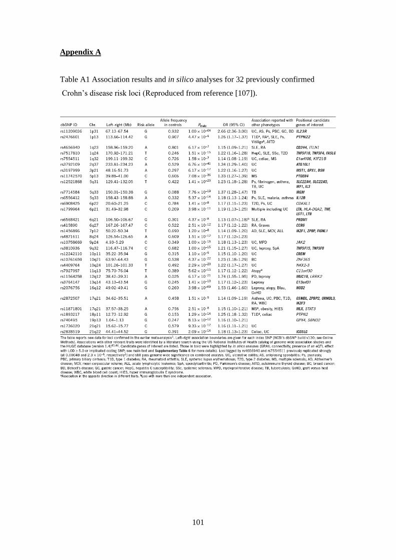
101
Appendix A
Table A1 Association results and in silico analyses for 32 previously confirmed
Crohn‟s disease risk loci (Reproduced from reference [107]).
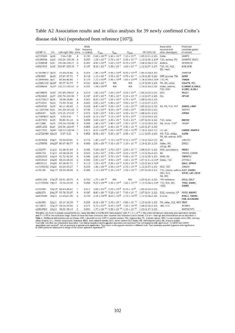
102
Table A2 Association results and in silico analyses for 39 newly confirmed Crohn‟s
disease risk loci (reproduced from reference [107]).

103
Appendix B
Case Report Form 3 for study investigating the role of MRI in perianal CD (see
chapter 4).
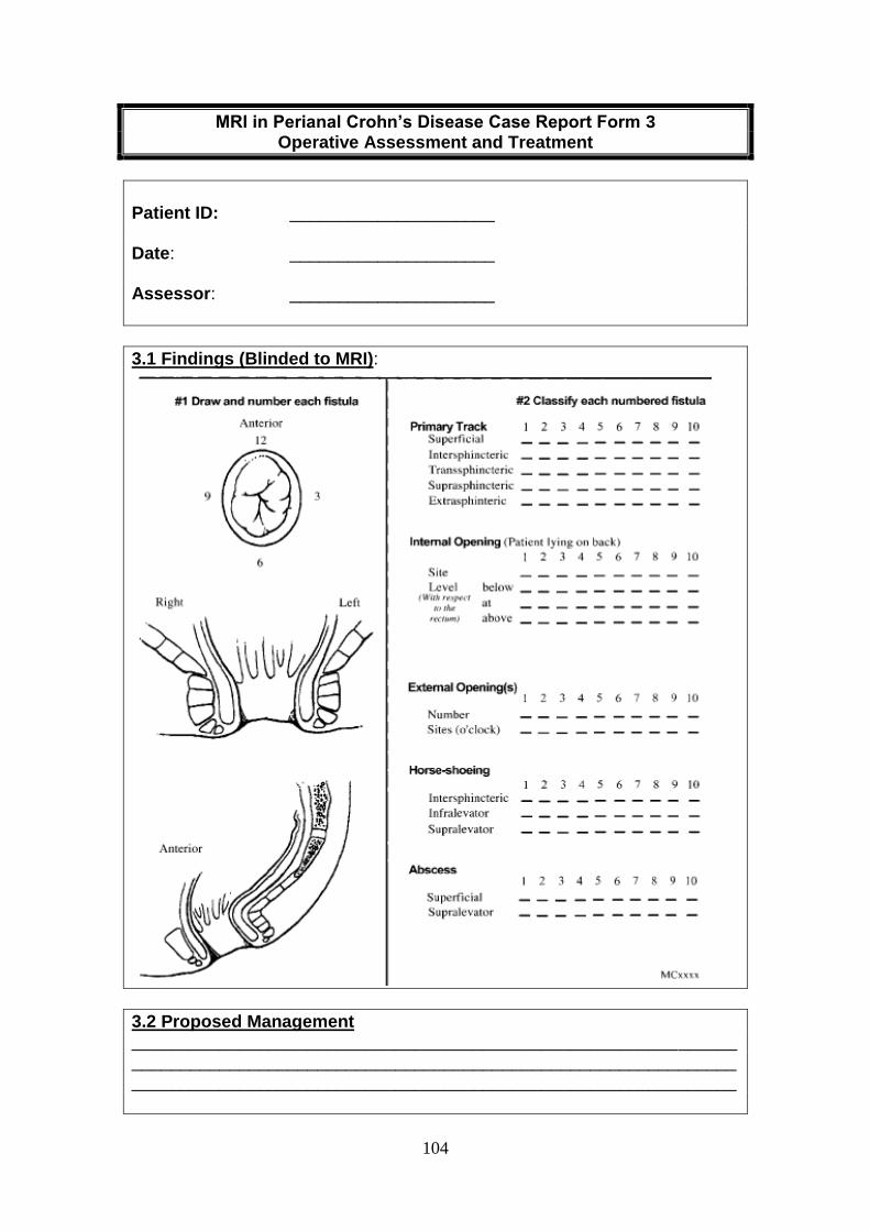
104
MRI in Perianal Crohn’s Disease Case Report Form 3 Operative Assessment and Treatment
Patient ID: _____________________ Date: _____________________ Assessor: _____________________
3.1 Findings (Blinded to MRI):
3.2 Proposed Management __________________________________________________________________________________________________________________________________________________________________________________________
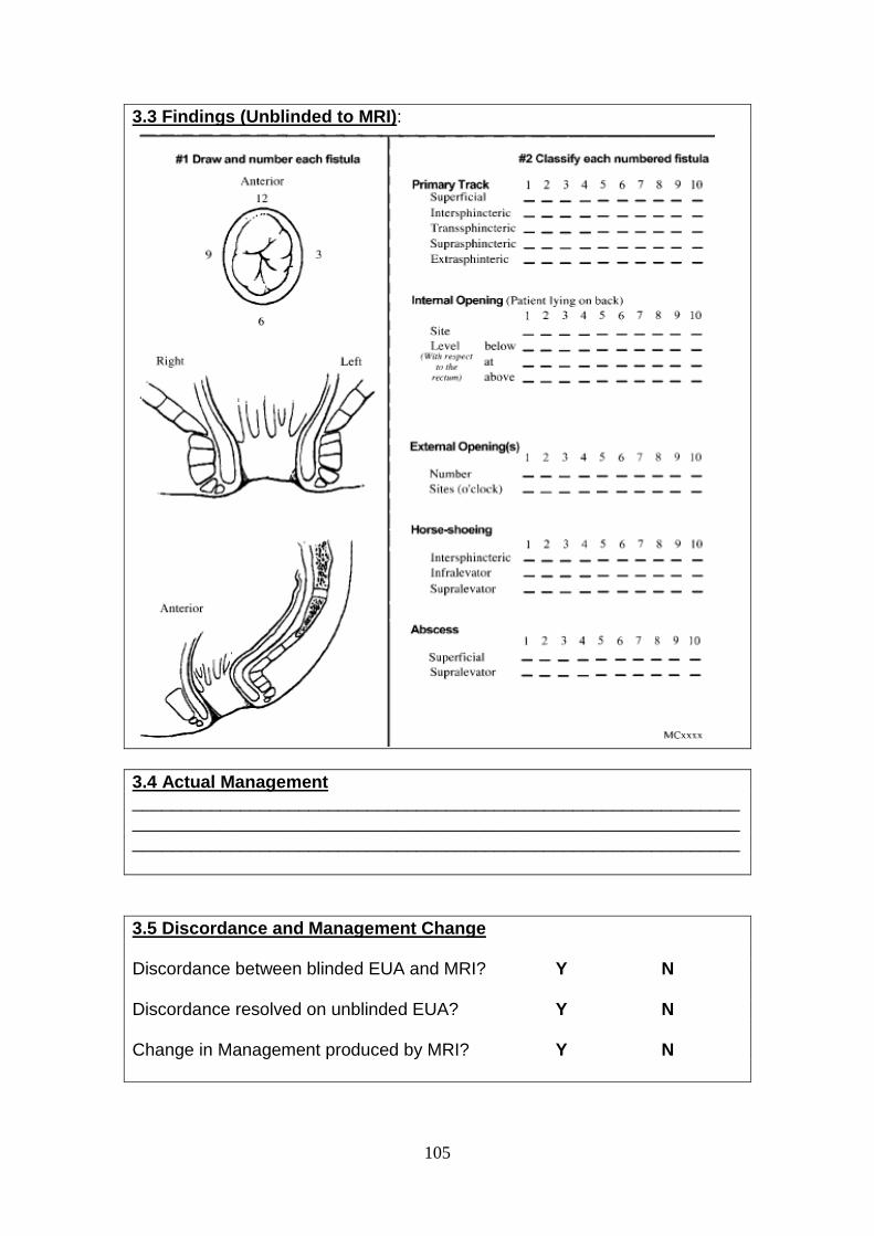
105
3.3 Findings (Unblinded to MRI):
3.4 Actual Management __________________________________________________________________________________________________________________________________________________________________________________________
3.5 Discordance and Management Change Discordance between blinded EUA and MRI? Y N Discordance resolved on unblinded EUA? Y N Change in Management produced by MRI? Y N
106
References
1. Ardizzone, S. and G.B. Porro, Perianal Crohn's disease: overview. Digestive
& Liver Disease, 2007. 39(10): p. 957-8.
2. Sartor, R.B., Pathogenesis and immune mechanisms of chronic inflammatory
bowel diseases. American Journal of Gastroenterology, 1997. 92(12 Suppl): p.
5S-11S.
3. Loftus, E.V., Jr., Clinical epidemiology of inflammatory bowel disease:
Incidence, prevalence, and environmental influences. Gastroenterology, 2004.
126(6): p. 1504-17.
4. Farrokhyar, F., E.T. Swarbrick, and E.J. Irvine, A critical review of
epidemiological studies in inflammatory bowel disease. Scand J Gastroenterol,
2001. 36(1): p. 2-15.
5. Tarrant, K.M., et al., Perianal disease predicts changes in Crohn's disease
phenotype-results of a population-based study of inflammatory bowel disease
phenotype. American Journal of Gastroenterology, 2008. 103(12): p. 3082-93.
6. Beaugerie, L., et al., Predictors of Crohn's disease. Gastroenterology, 2006.
130(3): p. 650-6.
7. Tang, L.Y., et al., Are perineal and luminal fistulas associated in Crohn's
disease? A population-based study. Clinical Gastroenterology & Hepatology,
2006. 4(9): p. 1130-4.
8. Smith, B.R., et al., Disease location, anti-Saccharomyces cerevisiae antibody,
and NOD2/CARD15 genotype influence the progression of disease behavior in
Crohn's disease. Inflammatory Bowel Diseases, 2004. 10(5): p. 521-8.
9. Sachar, D.B., et al., Is perianal Crohn's disease associated with intestinal
fistulization? American Journal of Gastroenterology, 2005. 100(7): p. 1547-9.
10. Penner, A. and B.B. Crohn, Perianal Fistulae as a Complication of Regional
Ileitis. Ann Surg, 1938. 108(5): p. 867-73.
11. Crohn, B.B., L. Ginzburg, and G.D. Oppenheimer, Landmark article Oct 15,
1932. Regional ileitis. A pathological and clinical entity. By Burril B. Crohn,
Leon Ginzburg, and Gordon D. Oppenheimer. JAMA, 1984. 251(1): p. 73-9.
12. Gabriel, W.B., Results of an Experimental and Histological Investigation into
Seventy-five Cases of Rectal Fistulae. Proc R Soc Med, 1921. 14(Surg Sect): p.
156-61.
13. Jeffery, P.J., A.G. Parks, and J.K. Ritchie, Treatment of haemorrhoids in
patients with inflammatory bowel disease. Lancet, 1977. 1(8021): p. 1084-5.
14. Sweeney, J.L., J.K. Ritchie, and R.J. Nicholls, Anal fissure in Crohn's disease.
Br J Surg, 1988. 75(1): p. 56-7.
15. Fleshner, P.R., et al., Anal fissure in Crohn's disease: a plea for aggressive
management. Dis Colon Rectum, 1995. 38(11): p. 1137-43.
16. Alexander-Williams, J., et al., Perianal Crohn's disease. World Journal of
Surgery, 1980. 4(2): p. 203-8.
17. Andersson, P., et al., Increased anal resting pressure and rectal sensitivity in
Crohn's disease. Diseases of the Colon & Rectum, 2003. 46(12): p. 1685-9.
18. Sangwan, Y.P., et al., Perianal Crohn's disease. Results of local surgical
treatment. Diseases of the Colon & Rectum, 1996. 39(5): p. 529-35.
19. Wolff, B.G., et al., Anorectal Crohn's disease. A long-term perspective. Dis
Colon Rectum, 1985. 28(10): p. 709-11.
107
20. Lockhart-Mummery, H.E., Anal lesions in Crohn's disease. Br J Surg, 1985.
72 Suppl: p. S95-6.
21. Solomon, M.J., Fistulae and abscesses in symptomatic perianal Crohn's
disease. International Journal of Colorectal Disease, 1996. 11(5): p. 222-6.
22. Vermeire, S., G. Van Assche, and P. Rutgeerts, Perianal Crohn's disease:
classification and clinical evaluation. Digestive & Liver Disease, 2007. 39(10):
p. 959-62.
23. Parks, A.G., P.H. Gordon, and J.D. Hardcastle, A classification of fistula-in-
ano. Br J Surg, 1976. 63(1): p. 1-12.
24. American Gastroenterological Association Clinical Practice, C., American
Gastroenterological Association medical position statement: perianal Crohn's
disease. Gastroenterology, 2003. 125(5): p. 1503-7.
25. Bell, S.J., et al., The clinical course of fistulating Crohn's disease. Alimentary
Pharmacology & Therapeutics, 2003. 17(9): p. 1145-51.
26. Galandiuk, S., et al., Perianal Crohn disease: predictors of need for
permanent diversion. Ann Surg, 2005. 241(5): p. 796-801; discussion 801-2.
27. Radcliffe, A.G., et al., Anovaginal and rectovaginal fistulas in Crohn's disease.
Dis Colon Rectum, 1988. 31(2): p. 94-9.
28. Linares, L., et al., Natural history and treatment of anorectal strictures
complicating Crohn's disease. Br J Surg, 1988. 75(7): p. 653-5.
29. Gillen, C.D., et al., Ulcerative colitis and Crohn's disease: a comparison of
the colorectal cancer risk in extensive colitis. Gut, 1994. 35(11): p. 1590-2.
30. Gillen, C.D., et al., Crohn's disease and colorectal cancer. Gut, 1994. 35(5): p.
651-5.
31. Nikias, G., et al., Crohn's disease and colorectal carcinoma: rectal cancer
complicating longstanding active perianal disease. American Journal of
Gastroenterology, 1995. 90(2): p. 216-9.
32. Slater, G., A. Greenstein, and A.H. Aufses, Jr., Anal carcinoma in patients
with Crohn's disease. Ann Surg, 1984. 199(3): p. 348-50.
33. Hughes, L.E., Surgical pathology and management of anorectal Crohn's
disease. J R Soc Med, 1978. 71(9): p. 644-51.
34. Singh, B., et al., Perianal Crohn's disease.[see comment]. British Journal of
Surgery, 2004. 91(7): p. 801-14.
35. Irvine, E.J., Usual therapy improves perianal Crohn's disease as measured by
a new disease activity index. McMaster IBD Study Group. Journal of Clinical
Gastroenterology, 1995. 20(1): p. 27-32.
36. Sandborn, W.J., et al., AGA technical review on perianal Crohn's disease.
Gastroenterology, 2003. 125(5): p. 1508-30.
37. Gasche, C., et al., A simple classification of Crohn's disease: report of the
Working Party for the World Congresses of Gastroenterology, Vienna 1998.
Inflammatory Bowel Diseases, 2000. 6(1): p. 8-15.
38. Klebl, F.H., et al., Association of perinuclear antineutrophil cytoplasmic
antibodies and anti-Saccharomyces cerevisiae antibodies with Vienna
classification subtypes of Crohn's disease. Inflammatory Bowel Diseases,
2003. 9(5): p. 302-7.
39. Linskens, R.K., et al., Genetic and serological markers to identify phenotypic
subgroups in a Dutch Crohn' s disease population. Digestive & Liver Disease,
2004. 36(1): p. 29-34.
108
40. Newman, B., et al., CARD15 and HLA DRB1 alleles influence susceptibility
and disease localization in Crohn's disease. American Journal of
Gastroenterology, 2004. 99(2): p. 306-15.
41. Heresbach, D., et al., NOD2/CARD15 gene polymorphisms in Crohn's disease:
a genotype- phenotype analysis. European Journal of Gastroenterology &
Hepatology, 2004. 16(1): p. 55-62.
42. Chow, D.K., et al., Changes in Crohn's disease phenotype over time in the
Chinese population: validation of the Montreal classification system.
Inflammatory Bowel Diseases, 2008. 14(4): p. 536-41.
43. McLeod, R.S., Management of fistula-in-ano: 1990 Roussel Lecture. Can J
Surg, 1991. 34(6): p. 581-5.
44. Williams, D.R., et al., Anal complications in Crohn's disease. Dis Colon
Rectum, 1981. 24(1): p. 22-4.
45. Hellers, G., et al., Occurrence and outcome after primary treatment of anal
fistulae in Crohn's disease. Gut, 1980. 21(6): p. 525-7.
46. Lapidus, A., et al., Clinical course of colorectal Crohn's disease: a 35-year
follow-up study of 507 patients. Gastroenterology, 1998. 114(6): p. 1151-60.
47. Schwartz, D.A., et al., The natural history of fistulizing Crohn's disease in
Olmsted County, Minnesota. Gastroenterology, 2002. 122(4): p. 875-80.
48. Lapidus, A. and A. Lapidus, Crohn's disease in Stockholm County during
1990-2001: an epidemiological update.[see comment]. World Journal of
Gastroenterology, 2006. 12(1): p. 75-81.
49. Fielding, J.F., Perianal lesions in Crohn's disease. J R Coll Surg Edinb, 1972.
17(1): p. 32-7.
50. Lockhart-Mummery, H.E., Symposium. Crohn's disease: anal lesions. Dis
Colon Rectum, 1975. 18(3): p. 200-2.
51. Rankin, G.B., et al., National Cooperative Crohn's Disease Study:
extraintestinal manifestations and perianal complications. Gastroenterology,
1979. 77(4 Pt 2): p. 914-20.
52. Buchmann, P., et al., Natural history of perianal Crohn's disease. Ten year
follow-up: a plea for conservatism. American Journal of Surgery, 1980.
140(5): p. 642-4.
53. Keighley, M.R. and R.N. Allan, Current status and influence of operation on
perianal Crohn's disease. International Journal of Colorectal Disease, 1986.
1(2): p. 104-7.
54. Halme, L. and A.P. Sainio, Factors related to frequency, type, and outcome of
anal fistulas in Crohn's disease. Dis Colon Rectum, 1995. 38(1): p. 55-9.
55. Platell, C., et al., Anal pathology in patients with Crohn's disease. Australian
& New Zealand Journal of Surgery, 1996. 66(1): p. 5-9.
56. Michelassi, F., et al., Surgical treatment of anorectal complications in Crohn's
disease. Surgery, 2000. 128(4): p. 597-603.
57. Veloso, F.T., et al., Clinical outcome of Crohn's disease: analysis according
to the vienna classification and clinical activity. Inflammatory Bowel Diseases,
2001. 7(4): p. 306-13.
58. Cosnes, J., et al., Long-term evolution of disease behavior of Crohn's disease.
Inflammatory Bowel Diseases, 2002. 8(4): p. 244-50.
59. Leong, R.W., J.Y. Lau, and J.J. Sung, The epidemiology and phenotype of
Crohn's disease in the Chinese population. Inflammatory Bowel Diseases,
2004. 10(5): p. 646-51.
109
60. Lees, C.W. and J. Satsangi, Genetics of inflammatory bowel disease:
implications for disease pathogenesis and natural history. Expert Rev
Gastroenterol Hepatol, 2009. 3(5): p. 513-34.
61. Podolsky, D.K., Inflammatory bowel disease. N Engl J Med, 2002. 347(6): p.
417-29.
62. Binder, V., Genetic epidemiology in inflammatory bowel disease. Dig Dis,
1998. 16(6): p. 351-5.
63. Cho, J.H., The genetics and immunopathogenesis of inflammatory bowel
disease. Nat Rev Immunol, 2008. 8(6): p. 458-66.
64. Klement, E., et al., Breastfeeding and risk of inflammatory bowel disease: a
systematic review with meta-analysis. Am J Clin Nutr, 2004. 80(5): p. 1342-52.
65. Gearry, R.B., et al., Population-based cases control study of inflammatory
bowel disease risk factors. J Gastroenterol Hepatol. 25(2): p. 325-33.
66. Gent, A.E., et al., Inflammatory bowel disease and domestic hygiene in
infancy. Lancet, 1994. 343(8900): p. 766-7.
67. Xavier, R.J. and D.K. Podolsky, Unravelling the pathogenesis of inflammatory
bowel disease. Nature, 2007. 448(7152): p. 427-34.
68. Taylor, G.A., IRG proteins: key mediators of interferon-regulated host
resistance to intracellular pathogens. Cell Microbiol, 2007. 9(5): p. 1099-107.
69. Atreya, R. and M.F. Neurath, Chemokines in inflammatory bowel diseases.
Dig Dis. 28(3): p. 386-94.
70. Silverberg, M.S., et al., Toward an integrated clinical, molecular and
serological classification of inflammatory bowel disease: Report of a Working
Party of the 2005 Montreal World Congress of Gastroenterology. Canadian
Journal of Gastroenterology, 2005. 19 Suppl A: p. 5-36.
71. Roberts, P.L., et al., Clinical course of Crohn's disease in older patients. A
retrospective study. Diseases of the Colon & Rectum, 1990. 33(6): p. 458-62.
72. Fry, R.D., et al., Techniques and results in the management of anal and
perianal Crohn's disease. Surg Gynecol Obstet, 1989. 168(1): p. 42-8.
73. Nguyen, G.C., et al., Inflammatory bowel disease characteristics among
African Americans, Hispanics, and non-Hispanic Whites: characterization of
a large North American cohort. American Journal of Gastroenterology, 2006.
101(5): p. 1012-23.
74. Karban, A., et al., Risk factors for perianal Crohn's disease: the role of
genotype, phenotype, and ethnicity. American Journal of Gastroenterology,
2007. 102(8): p. 1702-8.
75. Tobin, M.V., et al., Cigarette smoking and inflammatory bowel disease.
Gastroenterology, 1987. 93(2): p. 316-21.
76. Silverstein, M.D., et al., Cigarette smoking in Crohn's disease. American
Journal of Gastroenterology, 1989. 84(1): p. 31-3.
77. Somerville, K.W., et al., Smoking and Crohn's disease. Br Med J (Clin Res
Ed), 1984. 289(6450): p. 954-6.
78. Ekbom, A., et al., Perinatal risk factors for inflammatory bowel disease: a
case-control study. Am J Epidemiol, 1990. 132(6): p. 1111-9.
79. Godet, P.G., G.R. May, and L.R. Sutherland, Meta-analysis of the role of oral
contraceptive agents in inflammatory bowel disease. Gut, 1995. 37(5): p. 668-
73.
80. Koutroubakis, I.E., et al., Appendectomy, tonsillectomy, and risk of
inflammatory bowel disease: case-controlled study in Crete. Dis Colon
Rectum, 1999. 42(2): p. 225-30.
110
81. Wurzelmann, J.I., C.M. Lyles, and R.S. Sandler, Childhood infections and the
risk of inflammatory bowel disease. Digestive Diseases & Sciences, 1994.
39(3): p. 555-60.
82. Card, T., et al., Antibiotic use and the development of Crohn's disease. Gut,
2004. 53(2): p. 246-50.
83. Montgomery, S.M. and A. Ekbom, Epidemiology of inflammatory bowel
disease. Curr Opin Gastroenterol, 2002. 18(4): p. 416-20.
84. Baron, S., et al., Environmental risk factors in paediatric inflammatory bowel
diseases: a population based case control study. Gut, 2005. 54(3): p. 357-63.
85. Louis, E., et al., Early development of stricturing or penetrating pattern in
Crohn's disease is influenced by disease location, number of flares, and
smoking but not by NOD2/CARD15 genotype. Gut, 2003. 52(4): p. 552-7.
86. Picco, M.F. and T.M. Bayless, Tobacco consumption and disease duration are
associated with fistulizing and stricturing behaviors in the first 8 years of
Crohn's disease. American Journal of Gastroenterology, 2003. 98(2): p. 363-8.
87. Orholm, M., et al., Familial occurrence of inflammatory bowel disease. New
England Journal of Medicine, 1991. 324(2): p. 84-8.
88. Satsangi, J., et al., Genetics of inflammatory bowel disease. Gut, 1994. 35(5):
p. 696-700.
89. Probert, C.S., et al., Prevalence and family risk of ulcerative colitis and
Crohn's disease: an epidemiological study among Europeans and south
Asians in Leicestershire. Gut, 1993. 34(11): p. 1547-51.
90. Kuster, W., et al., The genetics of Crohn disease: complex segregation
analysis of a family study with 265 patients with Crohn disease and 5,387
relatives. Am J Med Genet, 1989. 32(1): p. 105-8.
91. Meucci, G., et al., Familial aggregation of inflammatory bowel disease in
northern Italy: a multicenter study. The Gruppo di Studio per le Malattie
Infiammatorie Intestinali (IBD Study Group). Gastroenterology, 1992. 103(2):
p. 514-9.
92. Bonen, D.K. and J.H. Cho, The genetics of inflammatory bowel disease.
Gastroenterology, 2003. 124(2): p. 521-36.
93. Thompson, N.P., et al., Genetics versus environment in inflammatory bowel
disease: results of a British twin study. BMJ, 1996. 312(7023): p. 95-6.
94. Halfvarson, J., et al., Inflammatory bowel disease in a Swedish twin cohort: a
long-term follow-up of concordance and clinical characteristics.
Gastroenterology, 2003. 124(7): p. 1767-73.
95. Orholm, M., et al., Concordance of inflammatory bowel disease among
Danish twins. Results of a nationwide study. Scand J Gastroenterol, 2000.
35(10): p. 1075-81.
96. Tysk, C., et al., Ulcerative colitis and Crohn's disease in an unselected
population of monozygotic and dizygotic twins. A study of heritability and the
influence of smoking. Gut, 1988. 29(7): p. 990-6.
97. Silverberg, M.S., Genetics of Inflammatory Bowel Disease, in The
Inflammatory Bowel Disease Year Book, C.N. Bernstein, Editor. 2010,
Remedica: Chicago. p. 1-20.
98. Ogura, Y., et al., A frameshift mutation in NOD2 associated with susceptibility
to Crohn's disease. Nature, 2001. 411(6837): p. 603-6.
99. Hugot, J.P., et al., Association of NOD2 leucine-rich repeat variants with
susceptibility to Crohn's disease. Nature, 2001. 411(6837): p. 599-603.
111
100. Zhang, H., et al., Genetics of inflammatory bowel disease: clues to
pathogenesis. British Medical Bulletin, 2008. 87: p. 17-30.
101. Economou, M., et al., Differential effects of NOD2 variants on Crohn's
disease risk and phenotype in diverse populations: a metaanalysis. American
Journal of Gastroenterology, 2004. 99(12): p. 2393-404.
102. Ahmad, T., et al., The molecular classification of the clinical manifestations of
Crohn's disease. Gastroenterology, 2002. 122(4): p. 854-66.
103. Brant, S.R., et al., Defining complex contributions of NOD2/CARD15 gene
mutations, age at onset, and tobacco use on Crohn's disease phenotypes.
Inflamm Bowel Dis, 2003. 9(5): p. 281-9.
104. Lesage, S., et al., CARD15/NOD2 mutational analysis and genotype-
phenotype correlation in 612 patients with inflammatory bowel disease. Am J
Hum Genet, 2002. 70(4): p. 845-57.
105. van Bodegraven, A.A., et al., Genetic variation in myosin IXB is associated
with ulcerative colitis. Gastroenterology, 2006. 131(6): p. 1768-74.
106. Vavassori, P., et al., CARD15 mutation analysis in an Italian population:
Leu1007fsinsC but neither Arg702Trp nor Gly908Arg mutations are
associated with Crohn's disease. Inflamm Bowel Dis, 2004. 10(2): p. 116-21.
107. Franke, A., et al., Genome-wide meta-analysis increases to 71 the number of
confirmed Crohn's disease susceptibility loci. Nat Genet. 42(12): p. 1118-25.
108. Negoro, K., et al., Analysis of the IBD5 locus and potential gene-gene
interactions in Crohn's disease. Gut, 2003. 52(4): p. 541-6.
109. Giallourakis, C., et al., IBD5 is a general risk factor for inflammatory bowel
disease: replication of association with Crohn disease and identification of a
novel association with ulcerative colitis. Am J Hum Genet, 2003. 73(1): p.
205-11.
110. Armuzzi, A., et al., Genotype-phenotype analysis of the Crohn's disease
susceptibility haplotype on chromosome 5q31. Gut, 2003. 52(8): p. 1133-9.
111. Hampe, J., et al., A genomewide analysis provides evidence for novel linkages
in inflammatory bowel disease in a large European cohort. Am J Hum Genet,
1999. 64(3): p. 808-16.
112. Peltekova, V.D., et al., Functional variants of OCTN cation transporter genes
are associated with Crohn disease. Nat Genet, 2004. 36(5): p. 471-5.
113. Roediger, W.E. and S. Nance, Metabolic induction of experimental ulcerative
colitis by inhibition of fatty acid oxidation. Br J Exp Pathol, 1986. 67(6): p.
773-82.
114. Vermeire, S., et al., Association of organic cation transporter risk haplotype
with perianal penetrating Crohn's disease but not with susceptibility to IBD.
Gastroenterology, 2005. 129(6): p. 1845-53.
115. Palmieri, O., et al., Variants of OCTN1-2 cation transporter genes are
associated with both Crohn's disease and ulcerative colitis. Alimentary
Pharmacology & Therapeutics, 2006. 23(4): p. 497-506.
116. Newman, B., et al., A risk haplotype in the Solute Carrier Family 22A4/22A5
gene cluster influences phenotypic expression of Crohn's disease.
Gastroenterology, 2005. 128(2): p. 260-9.
117. Onnie, C.M., et al., Diverse effects of the CARD15 and IBD5 loci on clinical
phenotype in 630 patients with Crohn's disease. European Journal of
Gastroenterology & Hepatology, 2008. 20(1): p. 37-45.
112
118. Parkes, M., et al., Sequence variants in the autophagy gene IRGM and
multiple other replicating loci contribute to Crohn's disease susceptibility.
Nature Genetics, 2007. 39(7): p. 830-2.
119. Singh, S.B., et al., Human IRGM induces autophagy to eliminate intracellular
mycobacteria. Science, 2006. 313(5792): p. 1438-41.
120. Rioux, J.D., et al., Genome-wide association study identifies new susceptibility
loci for Crohn disease and implicates autophagy in disease pathogenesis.
Nature Genetics, 2007. 39(5): p. 596-604.
121. Latiano, A., et al., Polymorphism of the IRGM gene might predispose to
fistulizing behavior in Crohn's disease. American Journal of Gastroenterology,
2009. 104(1): p. 110-6.
122. Han, Z., et al., Meta-analysis: polymorphisms in TNF-alpha gene promoter
and Crohn's disease. Alimentary Pharmacology & Therapeutics. 32(2): p.
159-70.
123. Wilson, A.G., et al., Effects of a polymorphism in the human tumor necrosis
factor alpha promoter on transcriptional activation. Proc Natl Acad Sci U S A,
1997. 94(7): p. 3195-9.
124. Koss, K., et al., Cytokine (TNF alpha, LT alpha and IL-10) polymorphisms in
inflammatory bowel diseases and normal controls: differential effects on
production and allele frequencies. Genes Immun, 2000. 1(3): p. 185-90.
125. Brinkman, B.M., et al., Relevance of the tumor necrosis factor alpha (TNF
alpha) -308 promoter polymorphism in TNF alpha gene regulation. J Inflamm,
1995. 46(1): p. 32-41.
126. van Heel, D.A., et al., Inflammatory bowel disease is associated with a TNF
polymorphism that affects an interaction between the OCT1 and NF(-kappa)B
transcription factors. Hum Mol Genet, 2002. 11(11): p. 1281-9.
127. Levine, A., et al., A polymorphism in the TNF-alpha promoter gene is
associated with pediatric onset and colonic location of Crohn's disease. Am J
Gastroenterol, 2005. 100(2): p. 407-13.
128. Vatay, A., et al., Relationship between the tumor necrosis factor alpha
polymorphism and the serum C-reactive protein levels in inflammatory bowel
disease. Immunogenetics, 2003. 55(4): p. 247-52.
129. Guerreiro, C.S., et al., Fatty acids, IL6, and TNFalpha polymorphisms: an
example of nutrigenetics in Crohn's disease. Am J Gastroenterol, 2009. 104(9):
p. 2241-9.
130. Sykora, J., et al., Cytokine tumor necrosis factor-alpha A promoter gene
polymorphism at position -308 G-->A and pediatric inflammatory bowel
disease: implications in ulcerative colitis and Crohn's disease. J Pediatr
Gastroenterol Nutr, 2006. 42(5): p. 479-87.
131. Ardizzone, S., et al., Imaging of perianal Crohn's disease. Digestive & Liver
Disease, 2007. 39(10): p. 970-8.
132. Lunniss, P.J., et al., Magnetic resonance imaging of anal fistulae. Lancet,
1992. 340(8816): p. 394-6.
133. Schaefer, O., et al., Assessment of anal fistulas with high-resolution
subtraction MR-fistulography: comparison with surgical findings. Journal of
Magnetic Resonance Imaging, 2004. 19(1): p. 91-8.
134. Orsoni, P., et al., Prospective comparison of endosonography, magnetic
resonance imaging and surgical findings in anorectal fistula and abscess
complicating Crohn's disease.[see comment]. British Journal of Surgery, 1999.
86(3): p. 360-4.
113
135. Schwartz, D.A., et al., A comparison of endoscopic ultrasound, magnetic
resonance imaging, and exam under anesthesia for evaluation of Crohn's
perianal fistulas. Gastroenterology, 2001. 121(5): p. 1064-72.
136. Buchanan, G.N., et al., Clinical examination, endosonography, and MR
imaging in preoperative assessment of fistula in ano: comparison with
outcome-based reference standard. Radiology, 2004. 233(3): p. 674-81.
137. Buchanan, G.N., et al., Magnetic resonance imaging for primary fistula in
ano.[see comment]. British Journal of Surgery, 2003. 90(7): p. 877-81.
138. Beets-Tan, R.G., et al., Preoperative MR imaging of anal fistulas: Does it
really help the surgeon? Radiology, 2001. 218(1): p. 75-84.
139. Ng, S.C., et al., Prospective evaluation of anti-tumor necrosis factor therapy
guided by magnetic resonance imaging for Crohn's perineal fistulas.
American Journal of Gastroenterology, 2009. 104(12): p. 2973-86.
140. Van Assche, G., et al., Magnetic resonance imaging of the effects of infliximab
on perianal fistulizing Crohn's disease. American Journal of Gastroenterology,
2003. 98(2): p. 332-9.
141. Poen, A.C., et al., Hydrogen peroxide-enhanced transanal ultrasound in the
assessment of fistula-in-ano. Diseases of the Colon & Rectum, 1998. 41(9): p.
1147-52.
142. Sloots, C.E., et al., Assessment and classification of fistula-in-ano in patients
with Crohn's disease by hydrogen peroxide enhanced transanal ultrasound.
International Journal of Colorectal Disease, 2001. 16(5): p. 292-7.
143. Ardizzone, S., et al., Perianal fistulae following infliximab treatment: clinical
and endosonographic outcome. Inflammatory Bowel Diseases, 2004. 10(2): p.
91-6.
144. Schwartz, D.A., et al., Use of endoscopic ultrasound to guide combination
medical and surgical therapy for patients with Crohn's perianal fistulas.
Inflammatory Bowel Diseases, 2005. 11(8): p. 727-32.
145. Schratter-Sehn, A.U., et al., Endoscopic ultrasonography versus computed
tomography in the differential diagnosis of perianorectal complications in
Crohn's disease. Endoscopy, 1993. 25(9): p. 582-6.
146. Frizelle, F.A., G.A. Santoro, and J.H. Pemberton, The management of perianal
Crohn's disease. International Journal of Colorectal Disease, 1996. 11(5): p.
227-37.
147. Thia, K.T., et al., Ciprofloxacin or metronidazole for the treatment of perianal
fistulas in patients with Crohn's disease: a randomized, double-blind, placebo-
controlled pilot study. Inflammatory Bowel Diseases, 2009. 15(1): p. 17-24.
148. Pearson, D.C., et al., Azathioprine and 6-mercaptopurine in Crohn disease. A
meta-analysis. Ann Intern Med, 1995. 123(2): p. 132-42.
149. Dejaco, C., et al., Antibiotics and azathioprine for the treatment of perianal
fistulas in Crohn's disease. Alimentary Pharmacology & Therapeutics, 2003.
18(11-12): p. 1113-20.
150. Present, D.H., et al., Infliximab for the treatment of fistulas in patients with
Crohn's disease. New England Journal of Medicine, 1999. 340(18): p. 1398-
405.
151. Sands, B.E., et al., Infliximab maintenance therapy for fistulizing Crohn's
disease. New England Journal of Medicine, 2004. 350(9): p. 876-85.
152. Lichtenstein, G.R., et al., Serious infections and mortality in association with
therapies for Crohn's disease: TREAT registry. Clinical Gastroenterology &
Hepatology, 2006. 4(5): p. 621-30.
114
153. Bongartz, T., et al., Anti-TNF antibody therapy in rheumatoid arthritis and the
risk of serious infections and malignancies: systematic review and meta-
analysis of rare harmful effects in randomized controlled trials. JAMA, 2006.
295(19): p. 2275-85.
154. Domenech, E., et al., Clinical evolution of luminal and perianal Crohn's
disease after inducing remission with infliximab: how long should patients be
treated? Alimentary Pharmacology & Therapeutics, 2005. 22(11-12): p. 1107-
13.
155. Lichtenstein, G.R., et al., Clinical trial: benefits and risks of
immunomodulators and maintenance infliximab for IBD-subgroup analyses
across four randomized trials. Alimentary Pharmacology & Therapeutics,
2009. 30(3): p. 210-26.
156. Topstad, D.R., et al., Combined seton placement, infliximab infusion, and
maintenance immunosuppressives improve healing rate in fistulizing anorectal
Crohn's disease: a single center experience. Dis Colon Rectum, 2003. 46(5): p.
577-83.
157. Guidi, L., et al., Combined therapy with infliximab and seton drainage for
perianal fistulizing Crohn's disease with anal endosonographic monitoring: a
single-centre experience. Techniques in Coloproctology, 2008. 12(2): p. 111-7.
158. Talbot, C., et al., Infliximab in the surgical management of complex fistulating
anal Crohn's disease. Colorectal Disease, 2005. 7(2): p. 164-8.
159. Regueiro, M. and H. Mardini, Treatment of perianal fistulizing Crohn's
disease with infliximab alone or as an adjunct to exam under anesthesia with
seton placement. Inflammatory Bowel Diseases, 2003. 9(2): p. 98-103.
160. Sands, B.E., et al., Maintenance infliximab does not result in increased
abscess development in fistulizing Crohn's disease: results from the ACCENT
II study. Alimentary Pharmacology & Therapeutics, 2006. 23(8): p. 1127-36.
161. Tozer, P.J., et al., Review article: medical, surgical and radiological
management of perianal Crohn's fistulas. Alimentary Pharmacology &
Therapeutics, 2011. 33(1): p. 5-22.
162. Sandborn, W.J., Preliminary report on the use of oral tacrolimus (FK506) in
the treatment of complicated proximal small bowel and fistulizing Crohn's
disease. Am J Gastroenterol, 1997. 92(5): p. 876-9.
163. Lowry, P.W., et al., Combination therapy with oral tacrolimus (FK506) and
azathioprine or 6-mercaptopurine for treatment-refractory Crohn's disease
perianal fistulae. Inflamm Bowel Dis, 1999. 5(4): p. 239-45.
164. Fellermann, K., et al., Steroid-unresponsive acute attacks of inflammatory
bowel disease: immunomodulation by tacrolimus (FK506). Am J
Gastroenterol, 1998. 93(10): p. 1860-6.
165. Ierardi, E., et al., Oral tacrolimus (FK 506) in Crohn's disease complicated by
fistulae of the perineum. J Clin Gastroenterol, 2000. 30(2): p. 200-2.
166. Lichtiger, S., Cyclosporine therapy in inflammatory bowel disease: open-label
experience. Mt Sinai J Med, 1990. 57(5): p. 315-9.
167. Present, D.H. and S. Lichtiger, Efficacy of cyclosporine in treatment of fistula
of Crohn's disease. Dig Dis Sci, 1994. 39(2): p. 374-80.
168. Hinterleitner, T.A., et al., Combination of cyclosporine, azathioprine and
prednisolone for perianal fistulas in Crohn's disease. Z Gastroenterol, 1997.
35(8): p. 603-8.
169. Gurudu, S.R., et al., Cyclosporine therapy in inflammatory bowel disease:
short-term and long-term results. J Clin Gastroenterol, 1999. 29(2): p. 151-4.
115
170. Egan, L.J., W.J. Sandborn, and W.J. Tremaine, Clinical outcome following
treatment of refractory inflammatory and fistulizing Crohn's disease with
intravenous cyclosporine. Am J Gastroenterol, 1998. 93(3): p. 442-8.
171. Hanauer, S.B. and M.B. Smith, Rapid closure of Crohn's disease fistulas with
continuous intravenous cyclosporin A. Am J Gastroenterol, 1993. 88(5): p.
646-9.
172. Fukushima, T., et al., Effects of cyclosporin A on active Crohn's disease.
Gastroenterol Jpn, 1989. 24(1): p. 12-5.
173. Poggioli, G., et al., Local injection of Infliximab for the treatment of perianal
Crohn's disease. Diseases of the Colon & Rectum, 2005. 48(4): p. 768-74.
174. Asteria, C.R., et al., Treatment of perianal fistulas in Crohn's disease by local
injection of antibody to TNF-alpha accounts for a favourable clinical response
in selected cases: a pilot study. Scand J Gastroenterol, 2006. 41(9): p. 1064-72.
175. Brady, C.E., 3rd, Hyperbaric oxygen and perineal Crohn's disease: a follow-
up. Gastroenterology, 1993. 105(4): p. 1264.
176. Nelson, E.W., Jr., D.E. Bright, and L.F. Villar, Closure of refractory perineal
Crohn's lesion. Integration of hyperbaric oxygen into case management.
Digestive Diseases & Sciences, 1990. 35(12): p. 1561-5.
177. Lavy, A., et al., Hyperbaric oxygen for perianal Crohn's disease. Journal of
Clinical Gastroenterology, 1994. 19(3): p. 202-5.
178. Colombel, J.F., et al., Hyperbaric oxygenation in severe perineal Crohn's
disease. Diseases of the Colon & Rectum, 1995. 38(6): p. 609-14.
179. Mizrahi, N., et al., Endorectal advancement flap: are there predictors of
failure? Dis Colon Rectum, 2002. 45(12): p. 1616-21.
180. Sonoda, T., et al., Outcomes of primary repair of anorectal and rectovaginal
fistulas using the endorectal advancement flap. Diseases of the Colon &
Rectum, 2002. 45(12): p. 1622-8.
181. O'Connor, L., et al., Efficacy of anal fistula plug in closure of Crohn's
anorectal fistulas. Dis Colon Rectum, 2006. 49(10): p. 1569-73.
182. Ky, A.J., et al., Collagen fistula plug for the treatment of anal fistulas. Dis
Colon Rectum, 2008. 51(6): p. 838-43.
183. Grimaud, J.C., et al., Fibrin glue is effective healing perianal fistulas in
patients with Crohn's disease. Gastroenterology, 2010. 138(7): p. 2275-81,
2281 e1.
184. Yamamoto, T., R.N. Allan, and M.R. Keighley, Effect of fecal diversion alone
on perianal Crohn's disease. World Journal of Surgery, 2000. 24(10): p. 1258-
62; discussion 1262-3.
185. Hong, M.K., et al., Faecal diversion in the management of perianal Crohn's
disease. Colorectal Dis, 2011. 13(2): p. 171-6.
186. Yamamoto, T., et al., Persistent perineal sinus after proctocolectomy for
Crohn's disease. Dis Colon Rectum, 1999. 42(1): p. 96-101.
187. Rius, J., et al., Gracilis transposition in complicated perianal fistula and
unhealed perineal wounds in Crohn's disease. Eur J Surg, 2000. 166(3): p.
218-22.
188. Sher, M.E., et al., Low Hartmann's procedure for severe anorectal Crohn's
disease. Dis Colon Rectum, 1992. 35(10): p. 975-80.
189. Bodger, K., Cost of illness of Crohn's disease. Pharmacoeconomics, 2002.
20(10): p. 639-52.
116
190. Odes, S., et al., Cost analysis and cost determinants in a European
inflammatory bowel disease inception cohort with 10 years of follow-up
evaluation. Gastroenterology, 2006. 131(3): p. 719-28.
191. Blomqvist, P. and A. Ekbom, Inflammatory bowel diseases: health care and
costs in Sweden in 1994. Scand J Gastroenterol, 1997. 32(11): p. 1134-9.
192. Silverstein, M.D., et al., Clinical course and costs of care for Crohn's disease:
Markov model analysis of a population-based cohort. Gastroenterology, 1999.
117(1): p. 49-57.
193. Feagan, B.G., et al., Annual cost of care for Crohn's disease: a payor
perspective. American Journal of Gastroenterology, 2000. 95(8): p. 1955-60.
194. Bernstein, C.N., et al., Direct hospital costs for patients with inflammatory
bowel disease in a Canadian tertiary care university hospital. American
Journal of Gastroenterology, 2000. 95(3): p. 677-83.
195. Cohen, R.D., et al., The cost of hospitalization in Crohn's disease. American
Journal of Gastroenterology, 2000. 95(2): p. 524-30.
196. Juan, J., et al., Burden of illness of Crohn's disease in Spain. Digestive &
Liver Disease, 2003. 35(12): p. 853-61.
197. Bassi, A., et al., Cost of illness of inflammatory bowel disease in the UK: a
single centre retrospective study. Gut, 2004. 53(10): p. 1471-8.
198. Stark, R., H.H. Konig, and R. Leidl, Costs of inflammatory bowel disease in
Germany. Pharmacoeconomics, 2006. 24(8): p. 797-814.
199. Mesterton, J., et al., Resource use and societal costs for Crohn's disease in
Sweden. Inflammatory Bowel Diseases, 2009. 15(12): p. 1882-1890.
200. Gearry, R., Inflammatory Bowel Disease in Canterbury, New Zealand. 2005,
University of Otago: Christchurch.
201. Gearry, R.B., et al., High incidence of Crohn's disease in Canterbury, New
Zealand: results of an epidemiologic study. Inflammatory Bowel Diseases,
2006. 12(10): p. 936-43.
202. Subnational population statistics: Statistics New Zealand 2005. 2005;
Available from: http://www.stats.govt.nz/products-and-services/info-
releases/subnat-pop-proj-info-releases.htm.
203. Lennard-Jones, J.E., Classification of inflammatory bowel disease. Scand J
Gastroenterol Suppl, 1989. 170: p. 2-6; discussion 16-9.
204. Roberts, R.L., et al., Rapid detection of common CARD15 variants in patients
with inflammatory bowel disease. Mol Diagn, 2004. 8(2): p. 101-5.
205. R Development Core Team. R: A language and environment for statistical
computing. 2010; Available from: http://www.R-project.org.
206. Little, J., et al., Strengthening the reporting of genetic association studies
(STREGA): an extension of the STROBE statement. Eur J Epidemiol, 2009.
24(1): p. 37-55.
207. Barrett, J.C., et al., Genome-wide association defines more than 30 distinct
susceptibility loci for Crohn's disease. Nature Genetics, 2008. 40(8): p. 955-62.
208. Glas, J., et al., rs224136 on chromosome 10q21.1 and variants in PHOX2B,
NCF4, and FAM92B are not major genetic risk factors for susceptibility to
Crohn's disease in the German population. Am J Gastroenterol, 2009. 104(3):
p. 665-72.
209. Amre, D.K., et al., NELL1, NCF4, and FAM92B genes are not major
susceptibility genes for Crohn's disease in canadian children and young adults.
Inflamm Bowel Dis.
117
210. Matute, J.D., et al., p40phox: the last NADPH oxidase subunit. Blood Cells
Mol Dis, 2005. 35(2): p. 291-302.
211. Ellson, C.D., et al., Neutrophils from p40phox-/- mice exhibit severe defects in
NADPH oxidase regulation and oxidant-dependent bacterial killing. J Exp
Med, 2006. 203(8): p. 1927-37.
212. Roberts, R.L., et al., Confirmation of association of IRGM and NCF4 with
ileal Crohn's disease in a population-based cohort. Genes Immun, 2008. 9(6):
p. 561-5.
213. Kim, T.H., et al., [Tumor necrosis factor-alpha and interleukin-10 gene
polymorphisms in Korean patients with inflammatory bowel disease]. Korean
J Gastroenterol, 2003. 42(5): p. 377-86.
214. Eglinton, T.W. and R.B. Gearry, Clinical factors predicting disease course in
Crohn's disease. Expert Rev Clin Immunol, 2010. 6(1): p. 41-5.
215. Baert, F., R. Caprilli, and E. Angelucci, Medical therapy for Crohn's disease:
top-down or step-up? Dig Dis, 2007. 25(3): p. 260-6.
216. Pescatori, M., et al., Management of perianal Crohn's disease. Results of a
multicenter study in Italy. Dis Colon Rectum, 1995. 38(2): p. 121-4.
217. Scott, H.J. and J.M. Northover, Evaluation of surgery for perianal Crohn's
fistulas. Diseases of the Colon & Rectum, 1996. 39(9): p. 1039-43.
218. Williamson, P.R., et al., Twenty-year review of the surgical management of
perianal Crohn's disease. Dis Colon Rectum, 1995. 38(4): p. 389-92.
219. Ingle, S.B. and E.V. Loftus, Jr., The natural history of perianal Crohn's
disease. Digestive & Liver Disease, 2007. 39(10): p. 963-9.
220. Satsangi, J., et al., The Montreal classification of inflammatory bowel disease:
controversies, consensus, and implications. Gut, 2006. 55(6): p. 749-53.
221. Gearry, R.B., et al., Effect of inflammatory bowel disease classification
changes on NOD2 genotype-phenotype associations in a population-based
cohort. Inflammatory Bowel Diseases, 2007. 13(10): p. 1220-7.
222. Louis, E., et al., Behaviour of Crohn's disease according to the Vienna
classification: changing pattern over the course of the disease. Gut, 2001.
49(6): p. 777-82.
223. Hanauer, S.B., et al., Maintenance infliximab for Crohn's disease: the
ACCENT I randomised trial. Lancet, 2002. 359(9317): p. 1541-9.
224. Eglinton, T.W. and R.B. Gearry, Clinical factors predicting disease course in
Crohn's disease. Expert Rev Clin Immunol. 6(1): p. 41-5.
225. Drummond, M.F., Methods for the economic evaluation of health care
programmes. 2nd ed. Oxford medical publications. 1997, Oxford ; New York:
Oxford University Press. 305 p.
226. Liljas, B., How to calculate indirect costs in economic evaluations.
Pharmacoeconomics, 1998. 13(1 Pt 1): p. 1-7.
227. Johannesson, M., Avoiding double-counting in pharmacoeconomic studies.
Pharmacoeconomics, 1997. 11(5): p. 385-8.
228. Mayberry, M.K., et al., Perceived discrimination in education and
employment by people with Crohn's disease: a case control study of
educational achievement and employment. Gut, 1992. 33(3): p. 312-4.
229. Ferguson, A., D.M. Sedgwick, and J. Drummond, Morbidity of juvenile onset
inflammatory bowel disease: effects on education and employment in early
adult life. Gut, 1994. 35(5): p. 665-8.
230. Access Economics, The Economic Cost of Crohn's Disease and Ulcerative
Colitis. 2007.
118
231. Hay, J.W. and A.R. Hay, Inflammatory bowel disease: costs-of-illness. Journal
of Clinical Gastroenterology, 1992. 14(4): p. 309-17.
232. Hay, A.R. and J.W. Hay, Inflammatory bowel disease: medical cost
algorithms. Journal of Clinical Gastroenterology, 1992. 14(4): p. 318-27.
233. Rosenthal, R.J., B. Bashankaev, and S.D. Wexner, Laparoscopic management
of inflammatory bowel disease. Dig Dis, 2009. 27(4): p. 560-4.
234. Eshuis, E.J., et al., Long-term outcomes following laparoscopically assisted
versus open ileocolic resection for Crohn's disease. Br J Surg, 2010. 97(4): p.
563-8.
235. West, R.L., et al., Perianal fistulas in Crohn's disease are predominantly
colonized by skin flora: implications for antibiotic treatment? Dig Dis Sci,
2005. 50(7): p. 1260-3.
236. Lunniss, P.J., et al., Histological and microbiological assessment of the role of
microorganisms in chronic anal fistula. Br J Surg, 1993. 80(8): p. 1072.
237. Seow-Choen, F., et al., Bacteriology of anal fistulae. Br J Surg, 1992. 79(1): p.
27-8.
238. Tozer, P.J., et al., Etiology of perianal Crohn's disease: Role of genetic,
microbiological, and immunological factors. Inflammatory Bowel Diseases,
2009.
239. Ascanelli, S., et al., Autologous fibroblasts transplant after infliximab
administration: a new approach in Crohn's perianal fistulas? Brief clinical
report. Int J Colorectal Dis, 2007. 22(9): p. 1135-6.


![Medical and surgical management of perianal Crohn’s … · fissures, ulcers, anorectal stricture, hemorrhoids, anal cancer) [7]. The true incidence of PCD is difficult to determine](https://img.pdfslide.us/doc/110x75/5b63ac187f8b9a6c178c4607/medical-and-surgical-management-of-perianal-crohns-fissures-ulcers-anorectal.jpg)