Embed Size (px)
Citation preview

SPECIAL ANNUAL ISSUE
Peri-insular hemispherotomy in paediatric epilepsy
Jean-Guy Villemure & Roy Thomas Daniel
Received: 28 February 2006 / Published online: 29 June 2006# Springer-Verlag 2006
AbstractObjects Outline the indications, investigation, surgicaltechnique, pitfalls, complications and benefits of peri-insular hemispherotomy (PIH) in the surgical treatment ofpaediatric epilepsy.Materials and methods This report is based on a consec-utive series of 43 children who underwent PIH. Sixtypercent were males; there were slightly more left-sidedsurgeries. Median interval between seizure onset andsurgery was 5 years. In more than half the cases, theanatomical substrate was congenital. There were fewcomplications: one death, one hydrocephalus and twoanatomically remote haemorrhages. Ninety percent of thepatients have remained in Engel’s class I epilepsy outcome.Conclusions There are clear indications for hemispherec-tomy in children. In some instances of incomplete deficit,timing of surgery remains a major concern. The lessinvasive approach to eliminate the influence of the diseasedhemisphere, in our opinion, is with disconnective techni-ques of hemispherectomy, and among the latter, peri-insularhemispherotomy provides, in our opinion, the best compli-cations–benefits ratio.
Keywords Peir-insular . Hemispherotomy . Epilepsysurgery . Hemispherectomy
Introduction
Functional hemispherectomy (FH) was introduced in the1970s as a surgical method aiming at achieving the sameseizure outcome obtained with anatomical hemispherec-tomy (AH), while avoiding the known potential complica-tions of the latter, namely superficial cerebral hemosiderosis[19, 27, 30, 31, 54]. FH consists in an anatomical subtotalremoval of the hemisphere but with complete disconnec-tion. It requires removal of the central region, including theparasagittal tissue, temporal lobectomy, complete callosot-omy, frontal and parieto-occipital lobotomies. The discon-nected frontal and parieto-occipital lobes are left in place,vascularized. The insular cortex is removed [32, 38].
The experience acquired with FH has allowed, on oneside, to confirm the expected benefits, i.e. seizure outcomecomparable to AH, and on the other side, has ledtechnically to smaller brain removal to achieve hemisphericdisconnection [49]. Peri-insular hemispherotomy (PIH)results from the conceptual and technical evolution of FHand represents the latest stage in its development [24, 25,44, 46].
While the first series of hemispherectomy carried out byDandy [10] was for the treatment of gliomas, the indicationfor this surgical method rapidly shifted toward the treatmentof refractory hemispheric epilepsy. The first reports on theuse of hemispherectomy for epilepsy were those ofMcKenzie in 1938 [26] concerning a child with infantilehemiplegia, and later on, of Krynauw in 1950 concerning aseries of 12 children with infantile hemiplegia [21]. We
Childs Nerv Syst (2006) 22:967–981DOI 10.1007/s00381-006-0134-3
J.-G. Villemure (*)Neurosurgery Service,Centre Hospitalier Universitaire Vaudois,Rue du Bugnon 46,1011 Lausanne, Switzerlande-mail: [email protected]
R. T. DanielNeurosurgery Service,Christian Medical College,Vellore, India

report our experience with peri-insular hemispherotomy ina consecutive series of 43 children.
Materials and methods
Disconnective hemispherectomy, i.e. FH and PIH, werecarried out in 84 patients for the treatment of refractoryhemispheric epilepsy (Fig. 23). There were 60 children(71%) and 24 adults. Of the paediatric population, 43underwent PIH (71%) and 17 underwent FH.
In this report, we describe our experience with peri-insular hemispherotomy in refractory paediatric hemispher-ic epilepsy. We discuss the indications, the anatomicalsubstrates, technical steps, pitfalls, benefits and complica-tions in a consecutive series of 43 paediatric patients(Table 1).
There were 25 males and 18 females. Age at surgeryranged from 1 to 15 years (mean: 8, median: 7.5). Therewere 19 surgeries on the right and 24 on the left. Theinterval between seizure onset and surgery varied from 1 to12 years (mean: 5, median: 4.5). The anatomical substratesresponsible for seizures were: infantile hemiplegia in 17,chronic encephalitis in 13, trauma in one, hemimegalence-phaly in three, non-hypertrophic migrational disorder infour, vascular insult in two, infection in two and anoxia inone (Table 2).
Indications
The indications to hemispherectomy concern a restrictedpopulation of epileptic patients who suffer from pharma-coresistant seizures. The brain insult has to be unilateral andwidespread throughout the hemisphere. The anatomicalsubstrates in such conditions are relatively already wellidentified and classified as hemispheric syndrome “remedi-able by surgery” [15]. The hemispheric disease has, in mostinstances, already created a “clinical hemispheric syn-drome” characterized by hemiplegia and hemianopsia. Thesurgical decision to proceed to hemispherectomy is basedon the critical evaluation of the following six parameters:seizures, neurological status, aetiologies, electroencepha-lography, imaging, neuropsychology [40].
Seizures
PIH is indicated for the treatment of refractory hemisphericepilepsy. The indication is the same, independent of thesurgical method of hemispherectomy utilized. The predom-inant seizure pattern is focal motor, but often, patientssuffer many seizure patterns, i.e. focal motor, partialcomplex, generalized, etc. The seizures have been docu-mented to be pharmacoresistant, i.e. without successfulcontrol despite numerous anticonvulsants. Seizures have afrequency of a few per day to thousands per year. Theseizures have a dramatic impact on the patient’s psychoso-cial development; frequentation of normal school isexceptional, learning disabilities is the rule. The seizurefrequency and/or severity may be such that regression onacquired development is noticed; this may be characterizedby loss of speech abilities and aggravation of concentrationor memory faculties. Abnormal behaviour, mainly hyper-activity and aggressiveness may be major elements of theclinical picture [21, 22]. Whether these represent seizuremanifestations or the result of an epileptic encephalopathyare possibilities [29].
Neurological examination
Depending on the pathological substrate responsible for theseizures, the hemispheric syndrome may be complete orincomplete and either stable or progressive. Classically, thepatient harbours a complete and stable hemispheric syn-drome characterized by a hemiplegia and a hemianopsia. Incertain conditions such as Rasmussen’s chronic encephalitisor extensive Sturge–Weber at an early stage, there may beincapacitating seizures and minimal, if any, objectiveneurological dysfunction. In these conditions, which areon one hand progressive and on the other hand responsiblefor severe seizure disorder before maximal deficits, thedecision to proceed to PIH, in these instances, is based on
Sex M: 25F: 18
Age at surgery(1–15 years)
Mean: 8Median: 7.5
Side Rt: 19Lt: 24
Interval(1–12 years)
Mean: 5Median: 4.5
Table 1 Summary of pa-tients data
IHSS 17Rasmussen’s 13Trauma 1Hemimegalencephaly 3Non-hypertrophicMigrational disorder 4vascular 2Infection 2Anoxia 1
Table 2 Anatomical substratein 43 cases of PIH
968 Childs Nerv Syst (2006) 22:967–981

the severity of the seizures, the rapidity of evolution of theunderlying condition and the lack of alternative medicaltreatments. Surgery, when carried out before maximaldeficit, will definitively aggravate the neurological status.Even though this is to be avoided when possible, PIH maybe necessary in these dynamic, naturally progressiveconditions, which will lead to severe hemispheric deficitwith time. This management issue is a matter of experienceand judgment and requires from the parents, as well, a goodcomprehension of the underlying disease [39, 51].
When assessing the motor function preoperatively todetermine its postoperative outcome, we have found thatthe motor deficit is usually not made worse whenpreoperatively, patients are unable to perform fingeropposition to the thumb, even if they are able to open andclose the hand. This is particularly observed in cases ofearly pathology, such as infantile hemiplegia, where thelesion might have preceded the final cortical organizationand allowed some plasticity mechanism to develop. Thesame interpretation is given to the inability to performrepeated foot tapping. Repeated alternating movement offoot taping and finger opposition to the thumb or individualfinger movements depend on cortical function. The pres-ence of gross voluntary movement, such as major jointmovements, walking, gross movement of the hand, do notrequire cortical participation and results, in part, fromsubcortical structures or ipsilateral motor participationthrough sprouting.
We have found in six cases who preoperatively demon-strated preservation of their visual field on formal testingthat the loss of visual field after hemispherectomy did nothave any clinical impact. We suspect that the preservedfield on formal testing in a severely damaged hemisphere isnot necessarily useful in daily activities. We consider thatthe aggravation of visual field by hemispherectomy, insomeone who otherwise meet the other criteria for surgery,is not, by itself, a contraindication to PIH [51].
The sensory examination is most often close to normaldespite severe anatomical hemispheric damage. However,slight to moderate deficits can de demonstrated in mostpatients when testing discrimination. Specific studies onsound perception have also documented decreased preci-sion in sound localization from the auditory field contra-lateral to surgery [28, 57]. Studies of visual field afterhemispherectomy have demonstrated persistent vision in anarrow meridian lateral to the midline of the contralateralfield, extending from 3 to 6° [53].
Etiologies
The anatomical substrates responsible for seizures are eithercongenital or acquired. Acquired conditions are trauma,infection, Rasmussen’s encephalitis; in these conditions, the
brain has had a period of normal development and normalfunctioning for various lengths of time. In acquiredconditions, one can assume that the hemispheric neurolog-ical deficit might be worse than in congenital lesion as thecompensatory mechanisms, named plasticity, did notintervene as early [3, 18, 55]. Congenital anatomicalsubstrates consist in prenatal vascular insult resulting fromcarotid or middle cerebral artery occlusion characteristic ofthe infantile hemiplegia, and extensive Sturge–Weber,hemimegalencephaly and non-hypertrophic diffuse hemi-spheric migrational disorder.
Anatomical substrates that are solely affecting onehemisphere are accompanied by better neurological func-tion and seizure outcome after PIH. Seizure outcome is alsonoted to be not as good in migrational disorder compared toother aetiologies, either congenital or acquired such astrauma or Rasmussen’s chronic encephalitis [12, 20, 50,51].
Electroencephalography
The electroencephalographic abnormalities on the affectedhemisphere are usually multifocal, diffuse and independent,reflecting the extent of the hemispheric involvement andthe severe epileptogenicity. Epileptic abnormalities from thegood hemisphere are often encountered; from a prognosticview, it is important to value these and determine if they aresecondary or independent. Their presence is not a contra-indication to PIH, as they may represent dependent orintermediate epileptogenicity, in which cases, the ultimateseizure outcome should be excellent; their presence remainsslightly unfavourable [8, 34, 52]. However, the abnormal-ities on the “good hemisphere” raise concern about anaetiology which could affect the brain bilaterally, questionsthe nature of the anatomical substrate and the presence ofsecondary epileptogenesis. It could be a contribution tounderstand the persistence of seizures after hemispherec-tomy [20].
Imaging
Magnetic resonance imaging, with the different sequences,i.e. T1, T2, flair and contrast, is the imaging modality ofchoice. It demonstrates the global anatomy, allows todetermine the degree of atrophy usually present andprovides information concerning the status of the whitematter, which may show abnormality before atrophy.Furthermore, it provides important information about theintegrity of the “normal” hemisphere. It may show specificchanges or show the evolution of lesions, confirming thediagnosis of infantile hemiplegia (Fig. 1), Sturge–Weber,migrational disorder (Fig. 2), Rasmussen’s encephalitis(Fig. 3). Computed tomography (CT) scan can demonstrate
Childs Nerv Syst (2006) 22:967–981 969

atrophy and some specific diagnostic features but has beenreplaced by magnetic resonance imaging (MRI). Rarely, anangiogram will add any significant information, and thus, isnot part of the routine imaging. The presence of atrophydemonstrated radiologically at the level of the cerebralpeduncle and medullary pyramid is also indicative of asevere cortical motor problem which should not beworsened by surgery (Fig. 4). In instances of few MRIabnormalities, functional imaging with positron emissiontomography (PET) scan can be contributing in demonstrat-ing extensive abnormal hypometabolic areas [9] (Fig. 5).
The MRI is also useful for the preparation of the surgicalstrategy. Ventricular size, configuration of the callosum,thickness of the brain, configuration of the insula–basalganglia complex should be understood before undertakingsurgery. Good understanding of the three dimensionalindividual brain anatomy will make PIH safer.
Neuropsychology
Neuropsychology determines the status of brain functioningbefore surgery and documents cognitive functions whichare usually below average; we have, however, encounteredinstances of normal IQ [22, 29]. Serial testings beforesurgery may document a temporal profile of cognitivedegradation secondary to progressing brain pathology suchas in Rasmussen’s encephalitis or the deleterious effects ofrepeated seizure and the development of an epilepticencephalopathy; regression in development quotient (DQ)is not unusual. In our experience, the demonstration ofsevere cognitive deficits reflects the involvement of bothhemisphere and raises concerns about seizure outcome;persistent seizures have been encountered more frequentlyin patients with severe mental impairment, operated on thebasis of clearly lateralizing electroencephalograms and
Fig. 1 a MRI axial T1, b coronalT2 large left porencephaly—infantilehemiplegia, from in utero vascularocclusion of left MCA
Fig. 2 MRI T2, a coronal, b axialenlarged right hemisphere, enlargedventricle, abnormal gyration, abnor-mal grey–white matter differentiation,characteristic of hemimegalencephaly
970 Childs Nerv Syst (2006) 22:967–981

imaging. In these instances, the objective of surgery mayshift from curative to palliative but still be very worthwhile[47].
Surgical method of peri-insular hemispherotomy
Peri-insular hemispherotomy is a surgical method offunctional hemispherectomy. It allows to disconnect thehemisphere through a peri-insular approach requiring onlyremoval of the fronto–parieto–temporal opercular cortices(Figs. 6 and 7).
a) skin and bone flap (Fig. 8)Surgery is carried out under general anaesthesia with
the patient supine, a cushion under the ipsilateralshoulder and the head turned contralateral to surgeryand kept horizontal; the head is either fixed in a pinclamp or rests on a soft horseshoe headrest and is taped.The skin and craniotomy flaps are planned to allowaccess around the insula. Taking into account the
atrophy and brain shift that may exist, the MRI isuseful in appreciating the projected skin and bone flaps.The flaps should extend anteriorly from the level of thecoronal suture, go posteriorly 3–4 cm behind the ex-ternal auditory canal, allowing them to reach theposterior insula. The flaps do not need to reach thefloor of the middle fossa but should reach the mid-convexity and be high enough to provide comfortableaccess to the suprasylvian circular sulcus, and eventu-ally, once in the ventricle, the corpus callosum.Preoperative analysis of the coronal and sagittal MRIhelps in defining the exact site and size of the flaps(Fig. 9). The dura is reflected either rostrally orcaudally. The brain exposure should provide at least2- to 2.5-cm exposure on either side of the sylvianfissure. This can vary according to the degree ofatrophy and whether the intraventricular portion of theoperation will be done through an already existing largeporencephaly, such as in middle cerebral artery (MCA)prenatal infarct; more exposure is useful in situationwhere little atrophy is present, such as in earlyencephalitis or hemimegalencephaly.
Fig. 3 Rasmussen’s chronic enceph-alitis a MRI, T1, coronal. Mildenlargement of the frontal horn, andsmaller right hemisphere, at an earlystage of the disease, b CT axial, withcontrast. Severe atrophy of the lefthemisphere in a late stage
Fig. 4 MRI, T1, axial (temporo-occipital). Severe atrophy of the lefthemisphere. Note the severe atrophy of the left cerebral peduncle
Fig. 5 a MRI, T1, axial. Mild atrophy of the right hemisphere inthe early stage of Rasmussen’s chronic encephalitis, b FDG-PET inthe same patient demonstrating diffuse right hemispheric glucosehypometabolism
Childs Nerv Syst (2006) 22:967–981 971

b) PIH has three major surgical stages, i.e. supra-insularwindow, infra-insular window, insula. Each has techni-cal steps that are now detailed. Magnification with theoperating microscope is useful. The illustrations referto a left PIH.
The supra-insular window stage
The aim of this stage is to transect the corona radiata fromthe frontal to parietal region and reach the ventricle toaccess the corpus callosum in its whole extent; this willallow to disconnect the whole of the suprasylvian portion ofthe hemisphere (Fig. 10).
Resection of the fronto-parietal opercular cortex
The first step is accomplished by resecting the frontal andparietal opercular cortex using a subpial resection techniquewith suction and biporal (SB) or ultrasonic aspirator (UA),depending on the tissue consistency (Fig. 11). This needs to
be done with preservation of arteries and veins reaching theconvexity, which prevents infarct of the disconnected brain.The resection of the fronto-parietal operculum will allowthe surgeon to visualize through layers of pia, the insularcortex and the vessels in the sylvian fissure. This exposureis extended rostrally to reach the circular sulcuscorresponding to the white matter of the corona radiatajust rostral to the insula. Once this has been carried outfrom the most anterior frontal part to the mid-parietalregion, the suprasylvian insular cortex is completelyexposed.
Transsection of the corona radiata
The second step consists in transsecting the corona radiataaiming at opening the lateral ventricle from the frontal hornto the trigone (Fig. 12). Using SB or the UA, the whitematter at the level of the circular sulcus is transacted in aplane perpendicular to the insula until the lateral ventricle isreached; thus, this incision is just rostral to the basal gangliaand thalamus. The opening in the ventricle is then extended
Fig. 6 MRI a coronal T2, observe the normal perisylvian anatomy,and the angle for the callosotomy (almost horizontal to the callosum),b coronal T1, note the disturbed superficial anatomy and thepossibility to do a parasagittal callosotomy directly (vertical to the
approach), c coronal T2, note the absence of insula and basal ganglia,which will modify the surgical stages, and exclude the insular stage ofPIH
Fig. 7 Anatomical preparation.a Coronal section illustrating thethree surgical stages of peri-insularhemispherotomy, b lateral viewillustrating the resected fronto–parieto–temporal opercularcortices, and the sylvian vessels
972 Childs Nerv Syst (2006) 22:967–981
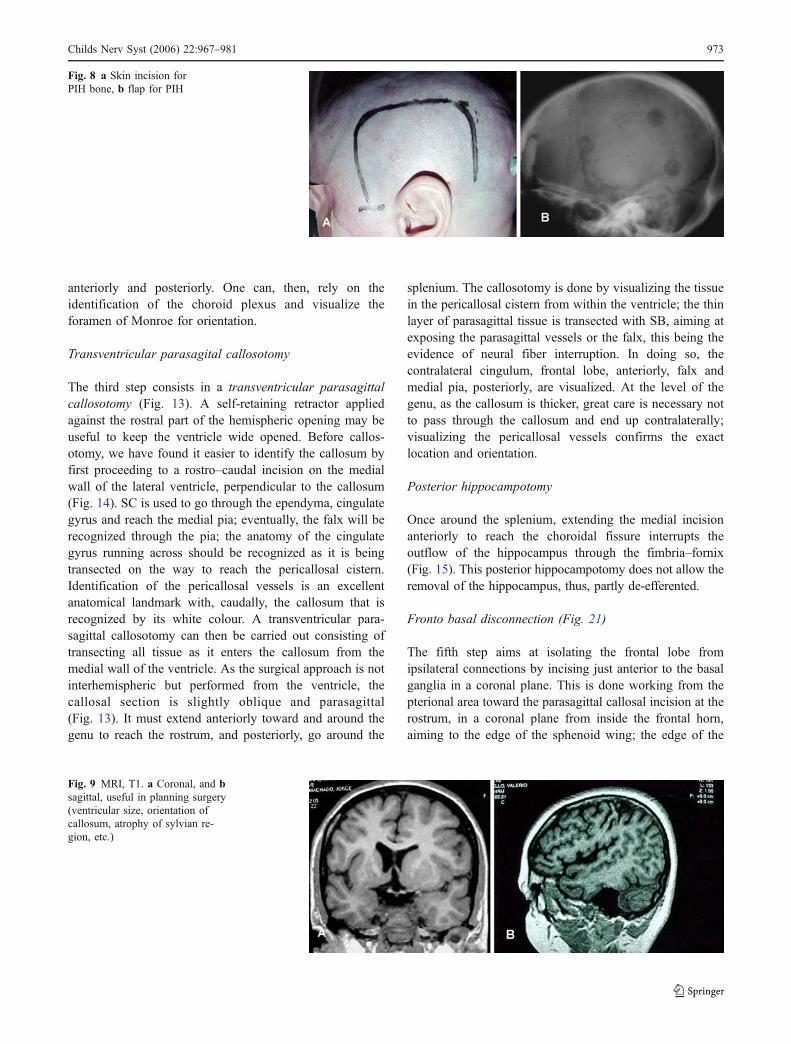
anteriorly and posteriorly. One can, then, rely on theidentification of the choroid plexus and visualize theforamen of Monroe for orientation.
Transventricular parasagital callosotomy
The third step consists in a transventricular parasagittalcallosotomy (Fig. 13). A self-retaining retractor appliedagainst the rostral part of the hemispheric opening may beuseful to keep the ventricle wide opened. Before callos-otomy, we have found it easier to identify the callosum byfirst proceeding to a rostro–caudal incision on the medialwall of the lateral ventricle, perpendicular to the callosum(Fig. 14). SC is used to go through the ependyma, cingulategyrus and reach the medial pia; eventually, the falx will berecognized through the pia; the anatomy of the cingulategyrus running across should be recognized as it is beingtransected on the way to reach the pericallosal cistern.Identification of the pericallosal vessels is an excellentanatomical landmark with, caudally, the callosum that isrecognized by its white colour. A transventricular para-sagittal callosotomy can then be carried out consisting oftransecting all tissue as it enters the callosum from themedial wall of the ventricle. As the surgical approach is notinterhemispheric but performed from the ventricle, thecallosal section is slightly oblique and parasagittal(Fig. 13). It must extend anteriorly toward and around thegenu to reach the rostrum, and posteriorly, go around the
splenium. The callosotomy is done by visualizing the tissuein the pericallosal cistern from within the ventricle; the thinlayer of parasagittal tissue is transected with SB, aiming atexposing the parasagittal vessels or the falx, this being theevidence of neural fiber interruption. In doing so, thecontralateral cingulum, frontal lobe, anteriorly, falx andmedial pia, posteriorly, are visualized. At the level of thegenu, as the callosum is thicker, great care is necessary notto pass through the callosum and end up contralaterally;visualizing the pericallosal vessels confirms the exactlocation and orientation.
Posterior hippocampotomy
Once around the splenium, extending the medial incisionanteriorly to reach the choroidal fissure interrupts theoutflow of the hippocampus through the fimbria–fornix(Fig. 15). This posterior hippocampotomy does not allow theremoval of the hippocampus, thus, partly de-efferented.
Fronto basal disconnection (Fig. 21)
The fifth step aims at isolating the frontal lobe fromipsilateral connections by incising just anterior to the basalganglia in a coronal plane. This is done working from thepterional area toward the parasagittal callosal incision at therostrum, in a coronal plane from inside the frontal horn,aiming to the edge of the sphenoid wing; the edge of the
Fig. 9 MRI, T1. a Coronal, and bsagittal, useful in planning surgery(ventricular size, orientation ofcallosum, atrophy of sylvian re-gion, etc.)
Fig. 8 a Skin incision forPIH bone, b flap for PIH
Childs Nerv Syst (2006) 22:967–981 973

sphenoid wing, seen from the pterional area and alsotranspially, is a good visual landmark for the posteriorextent of this frontal incision. The identification of theolfactory tract and gyrus rectus provides good anatomicallandmarks when reaching medially. Care is necessary not todamage the optic nerve. Preserving the basal pia adds safetyto this step.
The infra-insular window stage
The aim of this stage is to disconnect the whole temporallobe.
There are four steps to the infra-insular stage. Here, also,major cortical arteries and veins are to be preserved.
Resection of the temporal opercular cortex
The temporal operculum (T1) is removed in a subpialmatter exposing the insular cortex (Fig. 16). The extent ofremoval is as far back as the posterior insula. At that site,the temporal opercular removal should reach the supra-sylvian opercular removal. T1 removal is extended anteri-orly and medially to reach the uncus. This is done using CS
or UA. The insula is, thus, completely exposed whichallows access to the circular sulcus caudally.
Transsection of the temporal stem
At the level of the circular sulcus, the white matter istranssected to reach the temporal horn which is openedfrom its most anterior aspect to the trigone (Fig. 17). At thismoment, the whole lateral ventricle, infra- and suprasylvianare accessible around the insula.
Resection of the amygdala
The uncus and amygdala are excised by subpial aspiration(Fig. 18). The extent of supero-medial removal of theamygdala does not transgress an imaginary line between theroof of the temporal horn and the medial temporal pial bed.There are no visible anatomical boundaries outlining thesupero-medial aspect of the amygdala. The optic tract maybe visualized in the cistern if the medial resection ismaximal; this is a good anatomical landmark and representsthe limit of supero-medial removal of the amygdala. The
Fig. 10 Suprasylvian window—lateral
Fig. 11 Suprasylvian—coronal—opercular resection
Fig. 12 Suprasylvian—coronal—section of corona radiata
Fig. 13 Suprasylvian—coronal—parasagittal callosotomy
974 Childs Nerv Syst (2006) 22:967–981

risk of not recognizing this landmark is to end up in a planerostral to the choroidal fissure, medial to the temporal horn;this is to say in the basal ganglia. We recognize that theclinical consequence may not be the same when thishappens in PIH, unlike an anterior temporal lobectomy.
The anterior hippocampus
As the posterior hippocampotomy has previously beencarried out in the supra-insular window stage (step 4), thehippocampus does not need to be completely resected butthe disconnection completed; this is ensured by removingthe anterior hippocampus until the choroidal fissure isreached; there remains, then, no hippocampal efferents.
The insular stage
The insula is potentially epileptogenic; this stage aims ateliminating its influence [17, 23, 36, 37] (Figs. 19 Figs. 20Figs. 21). At this point, the insula is the only corticalepileptogenic structure still physiologically connected tothe hemisphere. It can either be resected by subpialaspiration or undermined by incising at the level of theclaustrum/extreme capsule, working from either side of the
insula, i.e. supra- and infrasylvian, at a depth of 5–7 mm. Inboth techniques, overlying arteries and veins have to bepreserved.
Following these three stages, the whole hemisphere isdisconnected from ipsilateral and contralateral neurologicalstructures and kept vascularized. Once the disconnection iscompleted, inspection of the surgical sites is carried out toinsure perfect hemostasis. Drains are left in the lateralventricle and in the subgaleal space. These drains are left towash out blood debris form the cerebrospinal fluid (CSF)space and to prevent epidural and subgaleal collection.Careful monitoring of the volume of CSF that is drained ismandatory, as overdrainage may be associated with venoushaemorrhage occurring topographically at a distance fromthe surgical site [14].
Bone flap and wound closures are carried out accordingto standard neurosurgical technique. The drains are re-moved within 48 h. Antibiotics are not routinely adminis-tered, and there are no indications to use corticosteroids.
Fig. 14 a Suprasylvian—later-al–medial vertical incisionapproaching the corpus cal-losum, b per-operative micro-photograph—transventricularparasagittal callosotomy incisionat the level of the genu of thecorpus callosum
Fig. 15 Suprasylvian—lateral—site of posterior callosotomy, aroundthe splenium, and posterior hippocampotomy Fig. 16 Infrasylvian—coronal—opercular resection
Childs Nerv Syst (2006) 22:967–981 975

Complications, technical pitfalls and their avoidancein paediatric peri-insular hemispherotomy
Preservation of cortical vessels
In a situation where anatomically, the hemisphere exhibitslittle, if any, atrophy despite the underlying pathology, greatcare is necessary to preserve as many arteries and veins aspossible [11, 43] (Fig. 22 and Table 3). If this is not doneproperly, brain swelling and hemispheric infarct secondaryto ischemia, from interfering with the arterial supply orvenous drainage, may occur, leading to intracranial hyper-tension, mass effect and death. This may be the case inRasmussen’s encephalitis before severe atrophy, in Sturge–Weber, in hemimegalencephaly or non-hypertrophic diffusehemispheric dysplasia where at the time of surgery, theremay be no or little atrophy or the volume of the hemispheremay even be larger than normal. We have encountered onesuch instance in an early chronic encephalitis adult patientwith hardly any atrophy, where many perisylvian arteries
and veins were sacrificed; this patient deteriorated rapidlyduring the third postoperative night and died of trans-tentorial herniation secondary to hemispheric swelling. Thesame caution applies for any anatomical substrate withminimal or absent brain atrophy. In instances of severeatrophy, however, such as in large porencephaly secondaryto prenatal vascular insult or advanced cases of Ramussen’sencephalitis, the issue is not of the same amplitude, as theenlarged ventricle which needs to be widely opened acts asa mechanical buffer in case of any brain swelling.Nevertheless, even in these instances, we make all effortsto preserve as many arteries and veins as possible.
Volume of CSF drainage
Two of our patients who showed postoperative neurologicaldeterioration were documented to have suffered distanthaemorrage from the surgical site. One patient did not wakeup from surgery, while the other deteriorated 48 h aftersurgery and was found to have remote haemorrhage in thecontralateral frontal lobe and in the cerebellum [14]. Thischild made an uneventful recovery. In this latter instance,the deterioration seems to have coincided with a suddenrapid drainage from the ventricular drain (Fig. 22). Wepostulate that the sudden drainage of a large volume ofventricular CSF modifies the intracranial dynamic, maycreate traction on veins and is responsible for theparenchymal haemorrhage as documented radiologically,and subsequently, for the neurological deterioration. Thisphenomenon can be prevented by setting the ventriculardrain at a slightly positive pressure, without vacuum andavoiding sudden drainage of a large amount of CSF.
Hydrocephalus
One child developed hydrocephalus manifesting at 1 monthafter surgery. This was successfully treated with a ventri-culo-peritoneal CSF shunt. The potential for hydrocephalusis present in any intracranial surgery. The risk forhydrocephalus after peri-insular hemispherotomy is theo-retically increased in cases of infection or trauma as boththese aetiologies may affect the patency of the subarachnoidspace, which can be further altered by surgery. In the singlepatient who needed a shunt, the aetiology was infantilehemiplegia seizure syndrome associated with neurofibro-matosis without specific features favouring hydrocephalus.
PIH before maximal deficits
In children who did not have maximal hemispheric deficitbefore surgery, their motor or visual field status postoper-atively were, as expected, definitively aggravated. Most ofthese patients suffered from progressive Rasmussen’s
Fig. 17 Infrasylvian—coronal—section of corona radiata (temporalstem)
Fig. 18 Infrasylvian—coronal—resection of amygdala and anteriorhippocampus
976 Childs Nerv Syst (2006) 22:967–981

chronic encephalitis, and surgery was carried out based onthe severity of the seizures, knowing that the natural historyof the disease would lead to maximal deficit anyway.Postoperatively, the recovery manifested by the return oftone and some gross motor function within 1 month. Nopatient demonstrated any discomfort or changes in habitsfrom the aggravation of their visual field.
Seizure outcome
Seizure outcome is expressed according to Engel’s classi-fication and based on a follow-up of 9 years. Of 37 childrenwith adequate follow-up, 34 have remained in Engel’sClass I since surgery (90%), while three are in class II. Infive children, we do not have adequate follow-up tocategorize their seizure outcome. Overall, all children have
been helped by surgery. This figure can be broken downaccording to aetiologies, which do influence the successrate of surgery. The best results have been obtained inchildren suffering from infantile hemiplegia (93%), sec-ondary to prenatal vascular occlusion of the carotid butmost often middle cerebral artery territory. Surgical out-come after PIH in Rasmussen’s encephalitis which also, inmost instances, is strictly a unilateral condition butacquired, is excellent (90%). Results obtained in unilateralhemispheric involvement where the anatomical substrate isone of hypertrophic or non-hypertrophic migrational dis-orders are not as good (80%), likely reflecting a differentphysiopathology of epileptogenicity; one could suspect thepresence of migrational abnormalities in the preservedhemisphere or the early development of an epilepticencephalopathy, already in utero (Table 4) [50].
Fig. 19 Anatomical preparationillustrating the incision under-mining the insula (a) anatomicalpreparation—axial, (b) MRIaxial T1
Fig. 20 Postoperative MRI T1.a sagittal, illustrating the peri-sylvian incision, b sagittal,illustrating the parasagittalcallosotomy and verticalmedian incision
Childs Nerv Syst (2006) 22:967–981 977

Discussion
All the techniques of hemispherectomy that have beendeveloped to replace anatomical hemispherectomy had acommon denominator which was to reduce the volume ofthe residual postoperative cavity, in view of eliminating theappearance of superficial cerebral hemosiderosis. Thiscomplication has not been reported after hemispherectomyover the last 30 years. However, the techniques have beenmodified in view of reducing the complication rate whileachieving the same seizure control.
Peri-insular hemispherotomy represents the latest mod-ification of functional hemispherectomy. It follows thesame surgical principle of “anatomical subtotal removal ofthe hemisphere and complete disconnection”. In PIH, theratio disconnection to excision is largest. As opposed to a
resective strategy consisting in anatomical hemispherec-tomy, hemidecortication or modified hemispherectomy, PIHis classified under disconnective strategy of hemispherec-tomy (Fig. 23). It resulted from the demonstration that thehemisphere could be disconnected, made nonfunctional,through very small removal of brain tissue [24, 25, 44, 46].The complications–benefits ratio have been clearly docu-mented and confirm that this surgical methodology ofhemispherectomy provides excellent seizure outcome, witha low incidence of side effects or complications. Theepilepsy outcome should be identical to any other methodof hemispherectomy, as physiologically, PIH eliminates theinfluence of the whole hemisphere; the outcome is not,then, only technique dependent but results, in a great part,from the anatomical substrate responsible for the epilepsy[20]. The nature and rate of complications of some othermethods of hemispherectomy are available in the literature[1, 2, 6, 7, 13, 16, 41, 42, 45, 48, 56]. When comparing therate and nature of complications, PIH provides a very lowincidence of complications. Some of the potential compli-cations of PIH have been avoided by understanding theircauses and modifying the technique; this is the case inproposing, to spare cortical vessels in view of preventingbrain swelling. Complications not specific to PIH, such asoverdrainage of CSF, should now be avoided by closemonitoring. The rate of hydrocephalus after PIH is thelowest encountered in such a large series. Specifically, wehave not encountered any hydrocephalus after PIH indiffuse migrational disorders, compared to other techniques.We believe that the minimal interference with the subarach-noid space remains the logic explanation for this low rate ofpostoperative hydrocephalus, as compared to other techni-ques of hemispherectomy that requires removal of half thesubarachnoid space. By comparison with techniques ofanatomical hemispherectomy or hemidecortication, inwhich the extent of surgery requires large exposure ofalmost the whole hemisphere, with a large skin and boneflaps, the exposure in PIH is relatively small whichcontributes to less blood loss, electrolytes and coagulationdisturbance and favours a shorter operative time; inchildren, these influence the operative and perioperativecourse and management [6].
Cognitive performance are, in most patients, belowaverage before surgery and often show regression second-ary to frequency of seizures. General brain disturbance mayresult from frequent seizures, but focal interference ofseizures can also be deleterious, particularly, towardlanguage development [5, 33, 35]. It is surprising to find
Death 1 (2.3%)Distant haemorrage 2 (4.6%)Hydrocephalus 1 (2.3%)
Table 3 Complications inperi-insular hemispherotomy(Nb: 43)
Fig. 21 Lateral view illustrating the peri-insular incision andpreservation of cortical vessels
Fig. 22 CT, axial, demonstrating haemorrhagic lesion in the rightfrontal lobe (contralateral to surgery) and blood over the cerebellum
978 Childs Nerv Syst (2006) 22:967–981

that despite complete seizure control, the postoperativeevolution of the developmental quotient does not follow astandard positive pattern [22, 29]. Without there being clearexplanations, it appears that some patients will show moreimprovement in cognitive functions than others. Even ifimprovement does not occur, deterioration is usuallystopped, and once seizures are controlled, the brain is inthe optimal condition to manifest its whole psychosocialdevelopment potential.
Patients with anatomical hemispheric dysfunction andfrequent seizures often creating bilateral hemisphericdysfunction harbour abnormal behaviour. As in manyinstances, the hyperactive and aggressive behaviourimproves rapidly after cessation of seizure, we canconclude that behaviour disturbances are secondary tobilateral brain disturbances resulting from seizures [21, 22].
The interval between seizure onset and control of seizuremay be an important variable. Though instinctively, onewould suspect a better outcome with an early treatment, itis, however, difficult to confirm that. The development of
secondary epileptogenesis and epileptic encephalopathymay be related to age at onset of seizure and intervalbefore treatment. We have not been able to demonstrate thatthis phenomenon exists, except in Rasmussen’s chronicencephalitis (adult and paediatric series), where thereappears to be a linear relation between seizure outcomesand interval; a shorter interval between seizure onset andsurgery is associated with a better seizure outcome [51].The development of an epileptic encephalopathy can resultfrom high seizure frequency and time interval betweenseizure onset and surgery.
It appears that the earliest PIH is carried out, themaximum will the benefit be. Benefits manifest throughseizure control and should provide the best environment forthe optimal psychosocial development of the child. Thereremains a subcategory of potential candidates for PIHwhere surgery may not be carried out right away, butdelayed, due to the absence of maximal deficit and possiblebenefits from other medical therapy. In Rasmussen’sencephalitis, a European consensus has defined some
Table 4 Complications and seizure outcome following PIH according to etiology
Engel’s Class
Aetiology Nb evaluable/total nb I II III IV Death Complications
IHSS 15/17 14 2 0 0 1 Remote bleed (2)Hydrocephalus (1)CE 12/13 11 1 0 0 0HM 2/3 2 0 0 0 0Migration 3/4 2 1 0 0 0Others 5/6 5 0 0 0 0
Fig. 23 Disconnective hemispherectomy
Childs Nerv Syst (2006) 22:967–981 979

guidelines for the management of these patients, with theuse of immunotherapy before surgery in certain patients [4].In most instances, however, PIH should be performed assoon as the situation for such surgery is met and brought tothe attention of the epilepsy surgeon.
Among the different surgical methods of hemispherec-tomy, peri-insular hemispherotomy appears to provide thelowest complication rate. Critical patient selection, clearthree-dimensional understanding of the patients’ brainanatomy and constant perioperative anticipation are keysto successful surgery.
References
1. Adams CBT (1983) Hemispherectomy: a modification. J NeurolNeurosurg Psychiatry 46:617–619
2. Beardsworth ED, Adams CBT (1988) Modified hemispherectomyfor epilepsy: early results in 10 cases. Br J Neurosurg 2:73–84
3. Bernasconi A, Bernasconi N, Lassonde M, Toussaint PJ, Meyer E,Reutens DC, Gotman J, Andermann F, Villemure JG (2000)Sensorimotor organization in patients who have undergone hemi-spherectomy: a study with (15)O-water PET and somatosensoryevoked potentials. Neuroreport 11(14):3085–3090
4. Bien CG, Granata T, Antozzi C, Cross JH, Dulac O, Kurthen M,Lassmann H, Mantegazza R, Villemure JG, Spreafico R, Elger CE(2005) Pathogenesis, diagnosis and treatment of Rasmussenencephalitis: a European consensus statement. Brain 128:454–471
5. Boatman D, Freeman J, Vining E, Pulsifer M et al (1999)Language recovery after left hemispherectomy in children withlate-onset seizures. Ann Neurol 46:579–586
6. Brian JE, Deshpande JK, McPherson RW (1990) Management ofcerebral hemsipherectomy in children. J Clin Anesth 2:91–95
7. Cabiese F, Jeni R, Landa R (1957) Fatal brain stem shift followinghemispherectomy. J Neurosurg 14:74–91
8. Carmant L, Kramer U, Riviello JJ, Helmers SL, Mikati MA,Madsen JR, Black PM, Lombroso CT, Holmes GL (1995) EEGprior to hemispherectomy: correlation with outcome and pathol-ogy. Electroencephalogr Clin Neurophysiol 94:265–270
9. Caplan R, Chugani HT, Messa C, Guthrie D, Sigman M, deTraversay J, Mundy P (1993) Hemispherectomy for intractableseizures: presurgical cerebral glucose metabolism and post-surgical non-verbal communication. Dev Med Child Neurol35:582–592
10. Dandy W (1928) Removal of right cerebral hemisphere for certaintumors with hemiplegia. JAMA 90:823–825
11. Daniel RT, Villemure JG (2003) Peri-insular hemispherotomy:potential pitfalls and complication avoidance. Stereotact FunctNeurosurg 80:22–27
12. Daniel RT, Broggi G, Farmer JP, Montes JL, Villemure JG (2003)Indicators of seizure outcome following hemispherectomy. Eur JNeurol 10(suppl 1):21
13. Davies KG, Maxwell RE, French LA (1993) Hemispherectomyfor intractable seizures: long-term results in 17 patients followedfor up to 38 years. J Neurosurg 78:733–740
14. De Ribaupierre S, Villemure JG, Chalaron M, Cotting J, Pollo C(2004) Contralateral frontal and cerebellar haemorrhages afterperi-insular hemispherotomy. Acta Neurochir 146:743–744
15. Engel J, Cascino GD, Shields WD (1997) Surgically remediablesyndromes. In: Engel J, Pedley TA (eds) Epilepsy: a comprehen-sive textbook. Lippincott-Raven, Philadelphia–New York, pp1687–1696
16. Falconer MA, Wilson PJE (1969) Complications related todelayed hemorrhage after hemispherectomy. J Neurosurg30:413–426
17. Freeman JM, Arroyo S, Vining EP, Breiter SN, Barker PB, PardoCA, Carson BS, Zuckerberg AL (1994) Insular seizures: a studyin Sutton’s law. Epilepsia 35(suppl 8):49
18. Graveline C, Mikulis D, Crawley AP, Hwang P (1998) Region-alized sensorimotor plasticity after hemispherectomy fMRI eval-uation. Pediatr Neurol 19:337–342
19. Griffith HB (1967) Cerebral hemispherectomy for infantilehemiplegia in the light of late results. Ann R Coll Surg Engl41:183–201
20. Holthausen H, May TW, Adams CBT, Andermann F, Comair Y,Delalande O, Duchowny M, freeman JM, Hoffman H, May P,Oppel F, Oxbury JM, Peacock WJ, Polkey C, Resnick T,Schramm J, Shewmon DA, Tuxhorn I, Vigevano F, VillemureJG, Wyllie E, Zaiwalla Z (1997) Seizures post-hemispherectomy.In: Tuxhorn I, Holthausen H, Boenigk H (eds) Paediatric epilepsysyndrome and their surgical treatment. John Libbey, London, pp749–773
21. Krynauw RA (1950) Infantile hemiplegia treated by removing onecerebral hemisphere. J Neurol Neurosurg Psychiatry 13:243–267
22. Lindsay J, Ounsted C, Richards P (1987) Hemispherectomy forchildhood epilepsy: a 36 years study. Dev Med Child Neurol29:592–600
23. Mascot C, Villemure JG, Andermann F, Rasmussen T (1990)Hemispherectomy and the insula. Can J Neurol Sci 17:236
24. Mascott C, Choi W, Rasmussen T, Villemure JG (1992) Theevolution of functional hemispherectomy at the MNI. Epilepsia 33(suppl 3):9
25. Mascot CR, Villemure JG (1993) Neuroanatomical principles ofperi-insular hemispherotomy: a novel variant of functional hemi-spherectomy for epilepsy. Epilepsia 34:101–102
26. McKenzie KG (1938) The present status of a patient who had theright cerebral hemisphere removed. JAMA 111:168
27. Oppenheimer DR, Griffith HB (1966) Persistent intracranialbleeding as a complication of hemispherectomy. J Neurol Neuro-surg Psychiatry 9:229–240
28. Poirier P, Lassonde M, Villemure JG, Geoffroy G, Lepore F(1994) Sound localization in hemispherectomized patients. Neuro-psychologia 32:541–553
29. Pulsifer MB, Brandt J, Salorio CF, Vining EP, Carson BS,Freeman JM (2004) The cognitive outcome of hemispherectomyin 71 children. Epilepsia 45:243–245
30. Ransohoff J, Hess W, in discussion Rasmussen T (1973) Post-operative superficial hemosiderosis of the brain, its diagnosis,treatment and prevention. Ann Neuro Assoc 98:133–137
31. Rasmussen T (1983) Hemispherectomy for seizures revisited. CanJ Neurol Sci 10:71–78
32. Rasmussen T, Villemure JG (1989) Cerebral hemispherectomy forseizures with hemiplegia. Clev Clin J Med 56(suppl 1):62–83
33. Rosenblatt B, Vernet O, Montes JL, Andermann F, Schwartz S,Taylor LB, Villemure JG, Farmer JP (1998) Continuous unilateralepileptiform discharge and language delay: effect of functionalhemispherectomy on language acquisition. Epilepsia 39:787–792
34. Smith SJM, Andennann F, Villemure JG, Rasmussen T, QuesneyLF (1991) Functional hemispherectomy: EEG findings, spikingfrom isolated brain postoperatively, and prediction of outcome.Neurology 41:1790–1794
35. Vargha-Khadem F, Mishkin M (1997) Speech and langageoutcome after hemispherectomy in childhood. In: Tuxhorn I,Holthausen H, Boenigk H (eds) Paediatric epilepsy syndromesand their surgical treatment. John Libbey, London, pp 774–784
36. Villemure JG, Mascott C, Andermann F, Rasmussen TB (1989) Isremoval of the insular cortex in hemispherectomy necessary?Epilepsia 30:728
980 Childs Nerv Syst (2006) 22:967–981

37. Villemure JG, Mascott C, Andermann F, Rasmussen T (1989)Hemispherectomy and the insula. Epilepsia 34:639–650
38. Villemure JG, Rasmussen T (1990) Functional hemispherectomy:methodology. J Epilepsy 3:177–182
39. Villemure JG, Andermann F, Rasmussen TB (1991) Hemispher-ectomy for the treatment of epilepsy due to chronic encephalitis.In: Andermann F (ed) Chronic encephalitis and epilepsy:Rasmussen’s syndrome. Butterworth-Heinemann, Boston, pp235–244
40. Villemure JG (1992) Hemispherectomy. In: Resor SR, Kutt H(eds) The medical treatment of epilepsy. Marcel Dekker, NewYork, pp 243–249
41. Villemure JG (1992) Hemispherectomy techniques. In: LudersHO (ed.) Epilepsy surgery. Raven, New York, pp 569–578
42. Villemure JG (1992) Anatomical to functional hemispherectomyfrom Krynauw to Rasmussen. Epilepsy Res (Suppl. 5):209–215
43. Villemure JG (1993) Hemispherectomy: techniques and compli-cations. In: Wyllie E (ed) The treatment of epilepsy: principles andpractice. Lea and Febiger, Philadelphia, pp 1116–1119
44. Villemure JG, Mascott C (1993) Hemispherotomy: the peri-insularapproach: technical aspects. Epilepsia 34(suppl 6):48
45. Villemure JG, Adams CBT, Hoffman HJ, Peacock WJ (1993)Hemispherectomy. In: Engel J (ed.) Surgical treatment of theepilepsies. Raven, New York, pp 511–518
46. Villemure JG, Mascott R (1995) Peri-insular hemispherectomy:surgical principles and anatomy. Neurosurgery 37:975–981
47. Villemure JG (1997) Hemispherectomy: a critical review. In: EdTuxhorn I, Holthausen H, Boenigk H (eds) Paediatric epilepsysyndrome and their surgical treatment. John Libbey, London, pp729–738
48. Villemure JG, Vernet O, Delalande O (2000) Hemisphericdisconnection: callosotomy and hemispherotomy. In: Cohadon F,
Dolenc VV, Lobo Antunes J, Pickard JD, Reulen HJ, Sindou M,Strong AJ, Tribolet N, Tulleken CAF, Vapalahti M (eds)Advances and technical standards in neurosurgery. Springer,Berlin Heidelberg New York, pp 25–78
49. Villemure JG (2001) Functional hemispherectomy: evolution oftechnique and results in 65 cases. In: Luders HO, Comair YG(eds) Epilepsy surgery. Lippincott Williams and Wilkins, Phila-delphia–New York, pp 733–739
50. Villemure JG, Meagher-Villemure K, Montes JL, Farmer JP,Broggi G (2003) Disconnective hemispherectomy for hemisphericdysplasia. Epileptic Disord 5(suppl 2):125–130
51. Villemure JG, Daniel RT, Farmer JP, Montes JL, Broggi G,Meagher-Villemure K (2004) Hemispherectomy (in chronicencephalitis). Epilepsia 45(suppl 3):19–20
52. Wennberg R, Quesney LF, Villemure JG (1998) ECoG findings inhemispherectomy. Electroencephalogr Clin Neurophysiol Suppl48:132–139
53. Wessinger CM, Fendrich R, Ptito A, Villemure J-G, GazzanigaMS (1996) Residual vision with awareness in the field contralat-eral to a parietal or complete functional hemispherectomy.Neuropsychologia 34:1129–1137
54. Wilson PJE (1970) Cerebral hemispherectomy for infantilehemiplegia. Brain 93:147–180
55. Wieser GH, Henke K, Zumsteg D, Taub E, Yonekawa Y, Buck A(1999) Activation of the left motor cortex during left legmovements after right central resection. J Neurol NeurosurgPsychiatry 67:487–491
56. Winston KR, Welch K, Adler JR, Erba G (1992) Cerebralhemicorticectomy for epilepsy. J Neurosurg 77:889–895
57. Zattore RJ, Ptito A, Villemure JG (1995) Preserved auditoryspatial localization following cerebral hemispherectomy. Brain118:879–889
Childs Nerv Syst (2006) 22:967–981 981