Embed Size (px)
Citation preview
Performing the Urinalysis
Laboratory Procedures
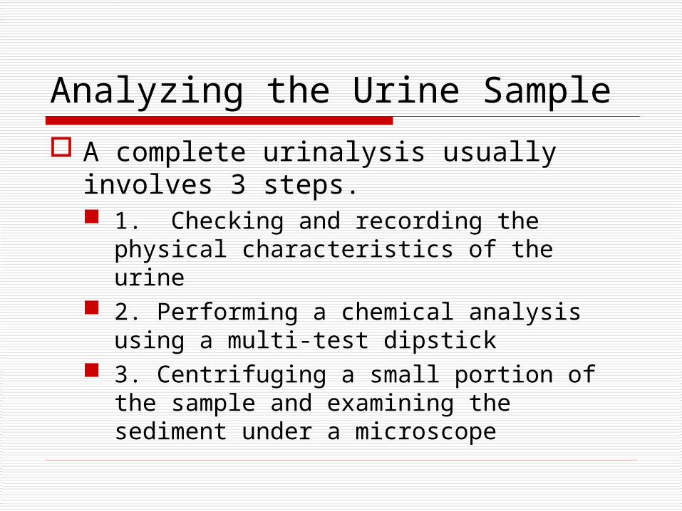
Analyzing the Urine Sample
A complete urinalysis usually involves 3 steps. 1. Checking and recording the physical
characteristics of the urine 2. Performing a chemical analysis using a
multi-test dipstick 3. Centrifuging a small portion of the
sample and examining the sediment under a microscope
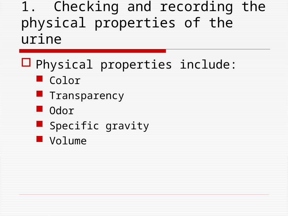
1. Checking and recording the physical properties of the urine
Physical properties include: Color Transparency Odor Specific gravity Volume
2. Chemical Analysis Many chemical tests can be performed on a small
quantity of urine by using a dipstick
Each pad on dipstick is designed to test for a particular substance in the urine.
When the urine comes in contacts with the reagents, a chemical reaction will cause a color change based on the amount of the substance in the urine.
Color is compared to chart, and approximate amount of substance in urine can be determined
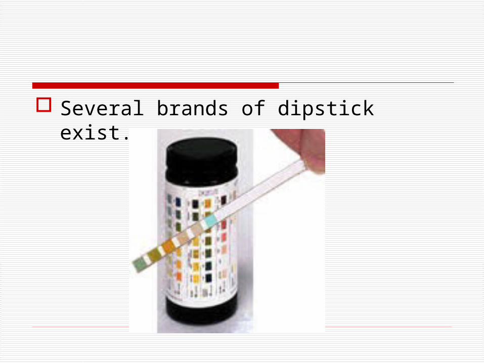
Several brands of dipstick exist.
Chemical analysis
Certain drugs and medications may interfere with chemical tests.
Be sure to know any medications animal is currently receiving when performing a urinalysis.
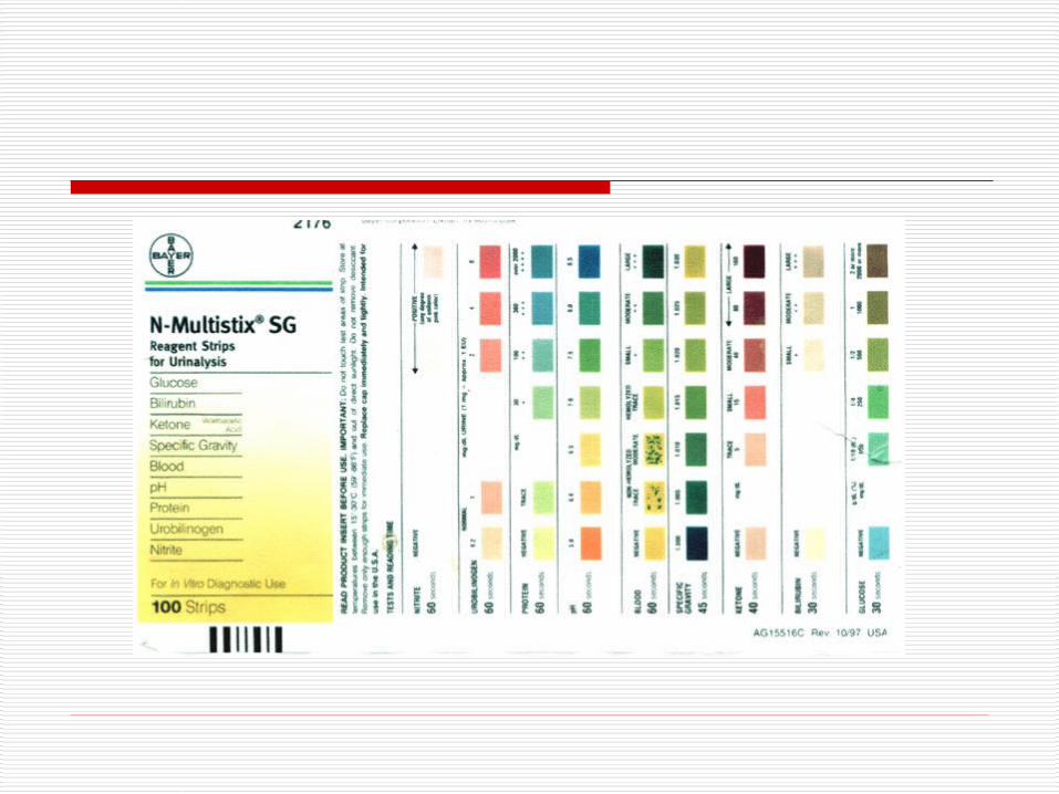
Components on the Dipstick Urine pH Protein Glucose Ketones Bilirubin Urobilinogen Blood Nitrites
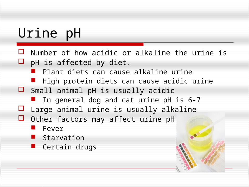
Urine pH Number of how acidic or alkaline the urine is pH is affected by diet.
Plant diets can cause alkaline urine High protein diets can cause acidic urine
Small animal pH is usually acidic In general dog and cat urine pH is 6-7
Large animal urine is usually alkaline Other factors may affect urine pH
Fever Starvation Certain drugs
Protein Healthy animals will usually not have any
protein in their urine although in some cases trace amounts can be found in concentrated dog and cat urine.
The urine protein level must be interpreted along with the Urine specific gravity.
Small amounts of protein are more significant in dilute or unconcentrated urine.
Protein may be lost in the urine due to glomerulopathies, inflammation, or hemorrhage.

Glucose If the sugar in the blood is
significantly higher than normal, some excess may be found in the urine.
Normal dog and cat urine should be negative for glucose.
In some cases if the urine is not run immediately, false glucose readings may occur.
Ketones Ketones are the substances formed in the
body during the breakdown of lipids. Normal pet urine should be negative for
ketones When excess amounts of ketones are
formed, their levels rise in the blood and then are released in the urine.
Can cause CNS depression and acidosis May result in ketonuria caused by:
Starvation Diabetes
Bilirubin Pigment made by the liver from dead or
dying red blood cells. Small amounts may sometimes be found in
healthy dogs. Dogs can conjugate bilirubin in their kidneys, so
small amount may be insignificant.
Bilirubin found in cat urine is a concern and can be a sign of liver disease, bile duct obstruction, or hemolysis.
Urobilinogen Compound formed from bilirubin by
intestinal tract. Normal dogs and cats have small amounts
of urobilinogen in their urine. Results from dipstick are not considered
very accurate and may be difficult to interpret.
Usually recorded as “normal” or “abnormal”.
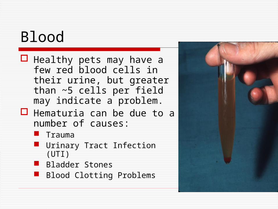
Blood Healthy pets may have a
few red blood cells in their urine, but greater than ~5 cells per field may indicate a problem.
Hematuria can be due to a number of causes: Trauma Urinary Tract Infection (UTI) Bladder Stones Blood Clotting Problems
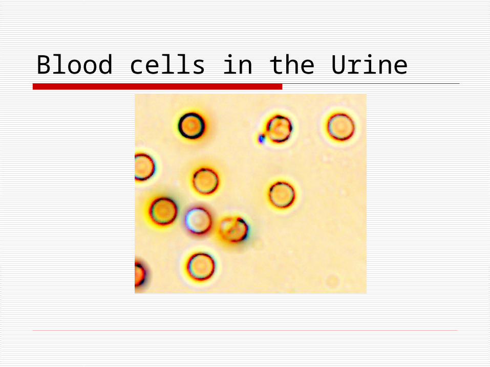
Blood cells in the Urine
Nitrites
May be produced by the bacteria present in some infections
Test often shows a “false negative” and is considered inaccurate in pets.
However, if positive, should examine sediment closely for bacteria.
Examining Urine Sediment After urine sediment is centrifuged
(generally about 5 minutes), the top portion of the liquid is poured off and the sediment is resuspended and examined microsopically.
Indications for sediment exam include: Provides additional information A form of cytology Must be interpreted with other clinical data,
including physical and chemical composition of the urine.
Urine Sediment Exam Procedure
1. Collect urine in a clean container 2. Throughly mix specimen and transfer 3-5
ml volume to a centrifuge tube 3. Centrifuge for 3-5 mins 4. Pour off supernatant 5. Leave approximately 0.5 ml of
supernatant 6. Resuspend urine sediment by tapping
tube or flicking it w/ your finger. 7. Examine a stained or unstained
sediment. (Or both!)
Potential Sediment Elements
White blood cells Red blood cells Lipid droplets Bacteria Crystal Casts

White blood cells
Larger than normal numbers of white blood cells may indicate inflammation from a bladder or kidney infection.
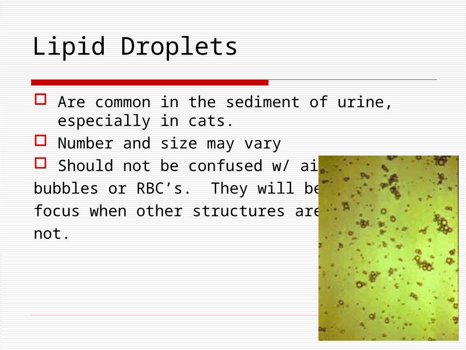
Lipid Droplets
Are common in the sediment of urine, especially in cats.
Number and size may vary Should not be confused w/ air bubbles or RBC’s. They will be infocus when other structures are not.
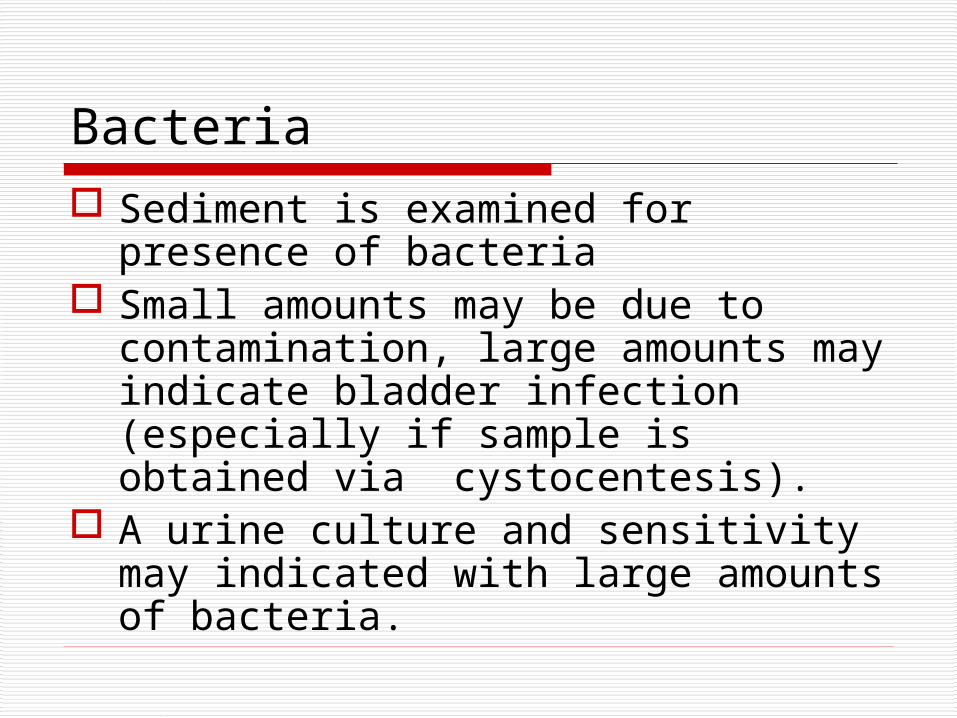
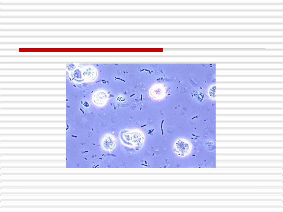
Bacteria Sediment is examined for presence of
bacteria Small amounts may be due to
contamination, large amounts may indicate bladder infection (especially if sample is obtained via cystocentesis).
A urine culture and sensitivity may indicated with large amounts of bacteria.
Crystals Made up of minerals and can
sometimes be found in the urine. Under certain conditions, crystals can
clump together to form bladder stones (uroliths).
The pH of the urine may influence the type of crystal development
Some animals and species are more predisposed to crystal and stone formation.
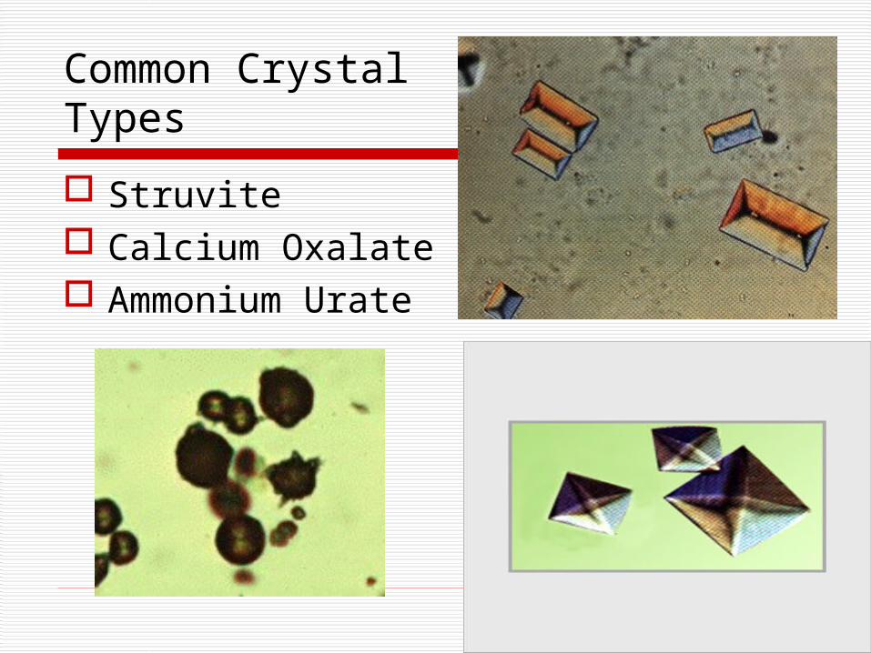
Common Crystal Types
Struvite Calcium Oxalate Ammonium Urate
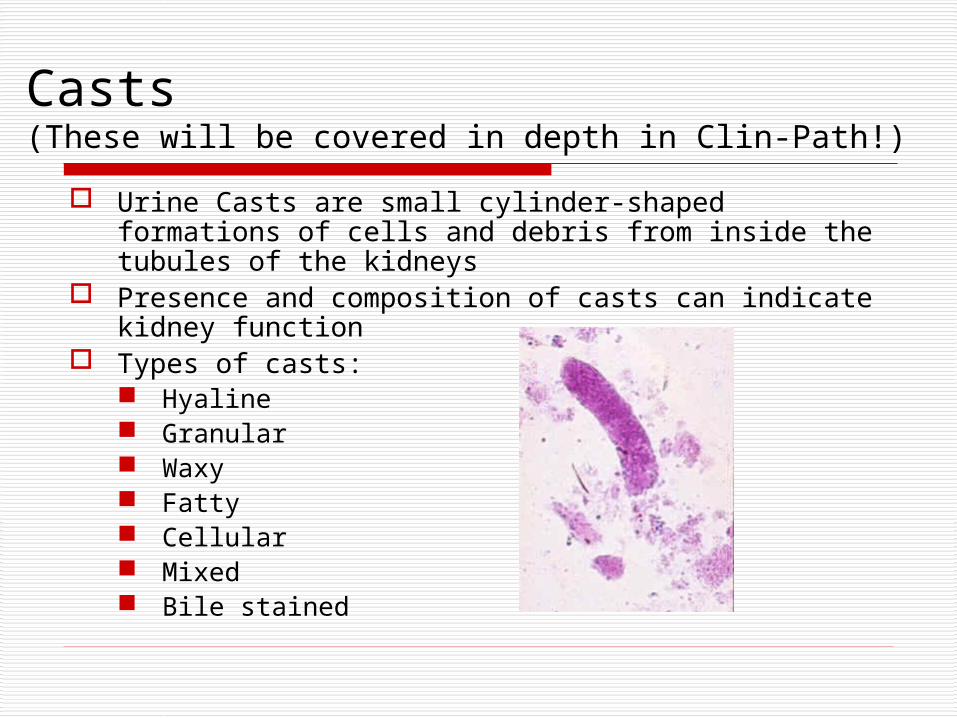
Casts (These will be covered in depth in Clin-Path!)
Urine Casts are small cylinder-shaped formations of cells and debris from inside the tubules of the kidneys
Presence and composition of casts can indicate kidney function
Types of casts: Hyaline Granular Waxy Fatty Cellular Mixed Bile stained
Specific Gravity
Measures the concentrating ability of the kidney.
Done with a refractometer There may be a spot on the dip-stick
for SG, however in animals, these are not valid results.

Specific Gravity “normals” p. 158 Lab bk
“Normal” values in dogs is between 1.001-1.060 “Normal” values in cats can range from 1.001 – 1.080 There is no set “normal” value in animals, however the
following guidelines should be used: SG: below 1.008 is said to indicate dilution (hyposthenuric) SG: 1.008-1.012 is said to be fixed or isosthenuric (same SG as
plasma) SG: 1.013 – 1.030 is considered normal if no dehydration
suspected. SG: above 1.025 implies renal tubule concentration ( in cats, this
can indicate renal disease)