Embed Size (px)
Citation preview

From the Department of Neuroscience, Karolinska Institutet, Stockholm, Sweden
Performance and trainability in paraplegics- motor function, shoulder muscle strength and sitting balance
before and after kayak ergometer training
Anna Bjerkefors
Stockholm 2006

Gårdsvägen 4, 169 70 Solna
Published and printed by
_____________________________________________________
2
Illustration: Yvonne Svensson
Correspondence to: Anna Bjerkefors Biomechanics and Motor Control Laboratory The Swedish School of Sport and Health Sciences (GIH) Box 5626 114 86 Stockholm Sweden Telephone: + 46 8 402 22 39 Fax: + 46 8 402 22 87 E-mail: [email protected]
© Anna Bjerkefors, 2006 ISBN 91-7140-981-5

_____________________________________________________
3
Till min älskade familj
- och till livet, kärleken och känslorna

_____________________________________________________
4

_____________________________________________________
5
ABSTRACTBackground Spinal cord injury (SCI) results in a complete or partial loss of motor and/or sensory function below the injury level. An SCI causes extensive functional impairment compelling many persons to wheelchair usage. Maintaining an adequate strength and control of trunk and shoulder muscles becomes essential, as the majority of every day tasks will be performed in a sitting position. Moreover, physical exercise is crucial to avoid risks connected with a sedentary life-style. Therefore, it becomes important to find suitable, effective, and attractive physical activities to retain and even improve motor functions achieved during rehabilitation. Ideally, such a training activity should be versatile and have the potential to improve several capacities beneficial to everyday life and thereby increase the independence of persons with SCI. Kayak paddling appears to fulfil several of the criteria for such an activity. Objectives The overall aims were to see if, and to what extent, a period of training on a modified kayak ergometer could influence functional performance as well as specific qualities, such as, shoulder muscle strength and sitting balance control in a group of post-rehabilitated persons with thoracic SCI. An additional aim was to understand more about the availability of the trunk muscles in a person with a clinically complete thoracic SCI and how the trunk muscles are used to maintain upright sitting in response to balance perturbations. Methods Ten adult post-rehabilitated persons with thoracic SCI performed 30 sessions of kayak ergometer training for a 10-week period, with progressively increased intensity and balance demand in the medio-lateral direction. Pre- and post-training measurements included performance in functional wheelchair tests, maximal voluntary shoulder muscle strength, and trunk stability in response to support-surface translations. Electromyographic (EMG) recordings from deep and superficial trunk muscles were obtained in a sub-sample of two subjects, one with a high thoracic SCI and one able-bodied person. Results There were significant improvements with training in functional performance, shoulder muscle strength, and the ability to maintain an upright sitting posture in response to balance perturbations in the group of persons with SCI. The EMG results revealed that the person with a high thoracic SCI, clinically classified as complete, was still able to activate trunk muscles below the injury, both in maximal voluntary efforts and in response to balance perturbations, but the response pattern differed from that of the able-bodied.Conclusions The improvements in test-performance observed with the kayak ergometer training in the persons with SCI should enhance their capacity to master similar challenges in everyday life, which, in turn, might lead to a greater independence. The pilot data on muscle activation highlight the importance of including examination of trunk muscle function in persons with thoracic SCI in relation to injury classification, prognosis, and training prescription.Key words Balance, electromyography, exercise, kinematics, motor skills, paraplegia, postural control, shoulder joint, spinal cord injury, wheelchair.

_____________________________________________________
6
SAMMANFATTNING
Bakgrund
Vid en ryggmärgsskada upphör helt eller delvis överföringen av nervsignaler i ryggmärgen, vilket medför motorisk och sensorisk funktionsnedsättning nedanför skadenivån. I Sverige skadas varje år cirka 150 personer så svårt att det leder till en ryggmärgsskada. Medelåldern vid skadetillfället är cirka 38 år, majoriteten är män (75-80 %) och den vanligaste orsaken är trafikolyckor, följd av fall- och dykolyckor. Ryggmärgsskador kan medföra tetraplegi eller paraplegi. Tetraplegi innebär att funktionsnedsättningen påverkar arm, bål och ben, medan paraplegi omfattar nedsatt funktion i enbart bål och ben. Skadan kan antingen vara komplett eller inkomplett, d.v.s. ha varierande grad av kvarvarande motorisk och/eller sensorisk funktion nedanför skadeområdet. Förutom att skadan påverkar känsel och muskelfunktion leder den till en rad andra problem, såsom blås- och tarmrubbning, smärta och spasticitet. Ett av målen med rehabiliteringen efter skada blir därför att minimera effekten av dessa problem. Det övergripande målet är att skapa förutsättningar för att varje individ får möjlighet att bli så självständig som möjlig. För personer som blivit rullstolsbrukare innebär det att optimera funktionen i bål och skuldror, eftersom merparten av vardagliga moment utförs i sittande. Andra viktiga fysiska faktorer är styrka, balans och uthållighet.
Behovet av regelbunden fysisk aktivitet för att bibehålla och/eller förbättra funktioner uppnådda under rehabiliteringsperioden kvarstår hela livet. Genom att utveckla effektiva och attraktiva träningsmetoder kan utövandet av fysisk aktivitet stimuleras. Denna träning bör, förutom att förbättra nämnda prestationsvariabler, också vara skonsam mot axlar och armar, då upprepad ensidig belastning vid exempelvis förflyttningar och rullstolskörning har visat sig leda till ökad förekomst av skulderbesvär hos personer med kronisk ryggmärgsskada. Kajakpaddling bör vara en lämplig aktivitet för att möta ovanstående krav. De specifika rörelserna som man utför vid kajakpaddling påverkar en stor del av överkroppens muskulatur. Dessutom ställer paddlingen krav på balans, styrka och uthållighet. Kajakpaddling har också visat sig lämplig för personer med ryggmärgsskada och den kan utövas på liknande villkor som för icke-skadade personer.
Paddling i kajakergometer är ett alternativ till paddling på öppet vatten och gör träningen oberoende av utomhusklimatet (dock missas naturupplevelsen). Träning i kajakergometer har visat sig ställa liknade fysiologiska krav som paddling på öppet vatten. En uppenbar skillnad är dock att ergometern, till skillnad från kajaken, vilar på ett stabilt underlag. En del i projektet var därför att utveckla en justerbar balansmodul till ergometern, vilket ger möjlighet att reglera och kontrollera tröghetsmomentet och därmed balanskravet i sidled under paddlingen.

_____________________________________________________
7
Syfte
Det övergripande syftet med avhandlingsarbetet var att utvärdera om regelbunden träning på kajakergometer resulterar i överföringseffekter på förmågan att utföra funktionella vardagliga moment i rullstolen samt specifika kvalitéer, såsom styrka i skuldermuskulaturen och bålstabilitet i sittande vid olika typer av balansstörningar, hos personer med ryggmärgsskada på bröstryggsnivå. Syftet var också att studera förmågan att aktivera bålmuskulaturen vid kliniskt komplett1 ryggmärgskada på bröstryggsnivå och undersöka hur bålmuskulaturen används för att stabilisera överkroppen vid olika balansstörningar.
Metod
Tio personer deltog i träningsstudien, 7 män och 3 kvinnor. Samtliga hade en ryggmärgsskada på bröstryggsnivå (T3-T12), hade avslutat sin rehabiliteringsperiod och var rullstolsbrukare. Medelåldern för gruppen var 38 år (24-60 år) och antal år efter skadan varierade från 3 till 26 (medianvärde: 12 år). Under 10 veckor genomförde gruppen 30 träningspass på kajakergometer, som utrustats med den specialutvecklade modulen med reglerbart balanskrav i sidled. Intensiteten och balanskravet ökades successivt under träningsperioden. Före och efter träningsperioden utfördes mätningar av prestationsförmågan i funktionella tester, maximal styrka i skuldermuskulaturen (i isokinetisk dynamometer) och stabilitet i överkroppen (tredimensionellt röreseutslag i bålen, mätt med ett optoelektroniskt rörelseregistreringssystem) efter balansstörningar åstadkomna genom hastiga förflyttningar av underlaget. Samtliga tester, förutom styrkemätningen, genomfördes i den egna rullstolen. Efter avslutad träningsperiod fick deltagarna skriftligt utvärdera upplevelsen av träningen med avseende på välbefinnande och funktionsförmåga i vardagliga moment. I den påföljande fallstudien gjordes mätningar av muskelaktivitet (via ytelektroder applicerade på huden och trådelektroder placerade i muskulaturen) i åtta bålmuskler, dels vid balansstörningar (som ovan), dels vid försök till maximala viljemässiga sammandragningar av respektive bålmuskel hos en person med ryggmärgsskada (vid bröstkota 3, kliniskt klassificerad som komplett) och en icke-skadad person.
1 I kliniken används ett internationellt framtaget testinstrument (International Standard of Neurological Classification of Spinal Cord Injury, Marino et al. 2003) för klassificering av graden av sensorisk och motorisk funktionsnedsättning efter en ryggmärgsskada samt bedömning av den neurologiska nivån på skadan. Test av känseln sker på 28 nyckelpunkter på armar, ben och bål medan den motoriska funktionen bedöms enbart i armar och ben. Den lägsta nivån med normal sensorik och motorik fastställer den neurologiska skadenivån. Eftersom bedömning av bålmuskulaturen saknas, blir den neurologiska skadenivån på bröstryggen klassificerad enbart efter den sensoriska nivån.
_____________________________________________________
8
Resultat
Träningen i kajakergometer ledde till positiva överföringseffekter på prestationen i funktionella rullstolstester, exempelvis rullstolskörning i uppförsbacke, förflyttning från rullstol till hög brits och ”sit-and-reach” tester, d.v.s. i förmåga att nå så långt som möjligt framför kroppen utan att tappa balansen. Träningen förbättrade också specifika kvalitéer, som den maximala styrkan i skuldermusklerna och stabiliteten i överkroppen vid balansstörningar. Resultaten från fallstudien visade att personen med hög ryggmärgskada i bröstryggen, klassificerad kliniskt som komplett, kunde aktivera även den bålmuskulatur som innerveras nedanför skadenivån, både vid maximal viljemässig sammandragning och som svar på oförberedda balansstörningar. Däremot skiljde sig aktiveringsmönstret vid balansstörningarna från den icke-skadades.
Slutsats
Baserat på resultaten från träningsstudien kan regelbunden paddling i kajakergometer rekommenderas som träningsform för personer med ryggmärgsskada på bröstryggsnivå. Träningen medförde att deltagarna kunde utföra svårare funktionella moment i rullstolen, vilket i sin tur kan leda till ökad självständighet i det dagliga livet. Kajakergometerträningen orsakade heller ingen skuldersmärta eller medförde några andra belastningsrelaterade problem. Deltagarna var också positiva till träningsformen och uppgav att de kände ett ökat välbefinnande efter avslutad träningsperiod. Den påvisade förmågan hos en person med hög ryggmärgsskada i bröstryggen att aktivera sina bukmuskler, pekar på vikten av att inkludera bålmuskulaturen vid klassificering av motorisk funktion och skadenivå. Härigenom ges också en bättre bas för diagnos, prognos och träningsrekommendation.
Nyckelord
Balans, elektromyografi, hållning, kinematik, kraftmoment, motoriska färdigheter, muskelstyrka, paraplegi, ryggmärgsskada, rullstol, skulderled, träning.

_____________________________________________________
9
CONTENTS
ABSTRACT 5
SAMMANFATTNING 6
LIST OF PAPERS 11
INTRODUCTION 13Spinal cord injury 13Physical activity and SCI 15
AIMS 18
METHODS 19Subjects 19Training tool and protocol 20Tests, equipment and protocols 22Ethical approval 29Statistical analysis 30
RESULTS 31The training (study I – III) 31The tests (study I - IV) 32
DISCUSSION 38Training effects 38Trunk muscle activation 46
CONCLUSIONS 48
TACK 49
REFERENCES 51
STUDIES I - IV

_____________________________________________________
10

_____________________________________________________
11
LIST OF PAPERS
This thesis is based on the following original papers, which are referred to in the text by their Roman numerals:
I Bjerkefors A. and Thorstensson A. Effects of kayak ergometer training on motor performance in paraplegics International Journal of Sports Medicine 2006: 27: 824-829.
II Bjerkefors A., Jansson A., and Thorstensson A. Shoulder muscle strength in paraplegics before and after kayak ergometer training European Journal of Applied Physiology 2006: 97: 613-618.
III Bjerkefors A., Carpenter M.G. and Thorstensson A. Dynamic trunk stability is improved in paraplegics following kayak ergometer training Accepted for publication in Scandinavian Journal of Medicine & Science in Sports.
IV Bjerkefors A., Carpenter M.G., Cresswell A.G. and Thorstensson A. Trunk muscle responses to balance perturbations in paraplegics Manuscript.
The published papers are reprinted with permission from their copyright holders, i.e., Georg Thieme Verlag for paper I, Springer-Verlag for paper II, and Blackwell Munsgaard for paper III.

_____________________________________________________
12

_____________________________________________________
13
INTRODUCTION
Spinal cord injury (SCI)
Spinal cord injury (SCI) results in a complete or partial loss of motor and/or sensory function below the injury level. The incidence of traumatic SCI in Sweden is about 10 cases per million inhabitants per year (Norrbrink Budh, 2004). The mean age at injury is 38 years; the majority (78 %) are men (Jackson et al., 2004), and traffic accidents are the primary cause of injury (Sekhon et al., 2001). Impairment of motor and sensory function in the arms, trunk and legs defines tetraplegia, whereas paraplegia is characterised by impairment or loss of motor and sensory function in the trunk and legs alone.
Neurological classification
In order to make a standard method of assessing the level and completeness of an SCI, an Impairment Scale and sensory and motor scores were developed by the American Spinal Injury Association (ASIA) in 1982. Since then the tests have been revised, most recently in 2003 by Marino et al. (2003) One of the goals of the manual was to ensure that both the measurement technique and the usage of the resulting data were consistent across practitioners and researchers.
Sensory and motor function
The sensory examination is completed through the testing of a key point in each of the 28 dermatomes on both sides of the body. Two aspects of sensation are examined at each point: sensitivity to pin prick and to light touch. Each sensation is separately scored on a three-point scale: 0 = absent, 1 = impaired and 2 = normal.
The motor examination involves bilateral manual muscle tests, graded on a six-point scale (range 0-5)2 of the upper and lower limbs, each consisting of five key muscles or muscle groups. Unfortunately, the assessment does not include the trunk muscles, which makes conclusions about motor connectivity to these muscles uncertain.
2 Muscle grading on a six-point scale according to the ASIA Motor Score (Marino et al., 2003). 0 = total paralysis, 1 = palpable or visible contraction, 2 = active movement, gravity eliminated, 3 = active movement, against gravity, 4 = active movement, against some resistance, and 5 = (normal) active movement, against full resistance.

_____________________________________________________
14
Neurological level
The neurological lesion level is defined as the most caudal level at which both motor and sensory modalities are intact on both sides of the body. In cases in which there is no key muscle for a segment the trunk that has sensory dermatomes intact, e.g. T2 – L1, the motor level is taken as that which corresponds to the sensory level. The lack of information about motor preservation to the trunk muscles will accordingly constitute a severe limitation to the classification of the neurological lesion level.
Completeness
The ASIA Impairment Scale is used in grading the degree of impairment, which is classified as complete or incomplete3. A complete injury means that there is on absence of sensory or motor function in the lowest sacral segments, and an incomplete injury has partial preservation of sensory and/or motor function below the neurological level and includes the lowest sacral segments.
Medical consequences
An SCI is associated with functional impairment compelling many persons to use wheelchairs, and is often complicated by medical problems, such as impaired bladder and bowel function (Dahlberg et al, 2004; Vallès et al., 2006), pain (Norrbrink Budh et al., 2003), spasticity (Sköld et al., 1999; Adams and Hicks, 2005) and sexual dysfunction (Westgren et al., 1997; Anderson et al., 2006). One of the primary goals in rehabilitation of patients with SCI is therefore to minimize the effects of this type of problems.
3The ASIA Impairment Scale (AIS, Marino et al., 2003): AIS A= complete injury, no sensory or
motor function is preserved in the sacral segments S4-S5, AIS B = sensory but no motor function below the neurological level and includes the sacral segments S4-S5, AIS C = motor function preserved below the neurological level, and more than half of the key muscles below the neurological level have a muscle grade less than 3, AIS D = motor function is preserved below the neurological level, and at least half of the key muscles below the neurological level have a muscle grade greater than or equal to 3, AIS E = normal.

_____________________________________________________
15
Physical activity and SCI
Rehabilitation
The overall goal of rehabilitation is that every individual with an SCI should be given an opportunity to achieve the highest degree of independence possible. For a person with paraplegia compelled to wheelchair usage, this means regaining as much of normal function and control of the trunk and shoulder muscles as possible, since the majority of every day tasks will be performed in a sitting position. Training of muscle strength of upper body muscles, balance control in sitting and aerobic capacity (endurance) therefore becomes a central part of the life of the person with paraplegia during the rehabilitation, that is, about 3-6 months after the injury, depending on whether the treatment involves surgery or is conservative.
Post-rehabilitation training
After termination of the rehabilitation the need for physical training still remains, to maintain the acquired abilities and avoid the risks involved with a sedentary life-style (Jacobs and Nash, 2004). Apart from improving physical capacities, participation in physical activities has numerous benefits, for example, it may help to reduce depression, improve family and social interaction and prolong life expectancy (Slater and Meade, 2004). When choosing an activity it becomes important to consider to what extent the particular type of training stimulates the above-mentioned parameters without, in itself, provoking overuse syndromes. Overuse syndromes are frequent in persons with chronic SCI (Curtis et al., 1999; van Drongelen et al., 2006), and have been related, among other things, to regular participation in sport activities, e.g. basketball, with frequent vigorous starts and stops (Curtis and Black, 1999).
Earlier studies investigating the effects of training on performance and function in persons with SCI are relatively few and, so far, only Grigorenko et al. (2004) have used kayak paddling as a training paradigm. In previous publications, trainability of this category of subjects with respect to different capacities has been demonstrated after various types of training. Improvements in cardio-respiratory function were reported after circuit training (Jacobs et al., 2001), wheelchair ergometer training (Tordi et al., 2001, Bougenot el al., 2003) and stimulation-assisted rowing (Wheeler et al., 2002). Gains in shoulder muscle strength were observed after training combining arm or wheelchair ergometer exercise and exercises with weights (Jacobs et al., 2001; Hicks et al., 2003), whereas training only on the ergometers appeared to have less effect on muscle strength (Davis and Shephard, 1990; Yim et al., 1993). Studies on training of balance control, in general, are few and none on persons with SCI appeared in a search of available databases. The pertinent question about the transferability of

_____________________________________________________
16
post-rehabilitation training effects to functional activities in daily life has not been dealt with specifically in the above mentioned studies. In a paper by Durán et al. (2001) a training program consisting of a variety of different exercises was shown to result in improvements in tests that were aimed at mimicking daily activities.
Kayak paddling appears to be an activity versatile enough to enable training effects on several of the above-mentioned functional capabilities and specific qualities. It occurs in sitting, which is a necessary prerequisite, and it involves most of the upper body musculature (Trevithick et al., 2006) in alternating three-dimensional movements (Plagenhof, 1979). The requirements for balance control are high due to the complex movements, the interplay between reaction forces in different directions, and the construction of the kayak. Furthermore, by varying the intensity of paddling, the activity can be directed mainly towards endurance or strength type of training (Tesch, 1983; Shephard, 1987; Fry and Morton, 1991).
Open-sea kayaking
Open sea kayaking has been promoted as one of several physical activities for persons with SCI by the Rekryteringsgruppen Active Rehabilitation.4 Special arrangements to make the kayak suitable for persons with SCI and to allow a progression with respect to independent paddling have been undertaken (cf. Fig. 1).
4Rekryteringsgruppen Active Rehabilitation is a Swedish non-profit organization (founded 1976)
working with sport activities for persons with SCI as a method to improve their performance and self-confidence and to help them succeed in their ambition to become independent in daily living. Many of the instructors have disabilities themselves, which enable them to both teach and serve as role models.
Figure 1
Kayaking on open sea with varying degrees of independence. Above: paddling with an able-bodied person in the rear position. Below: paddling alone with pontoons at the sides of the kayak to increase sideways stability.

_____________________________________________________
17
Based on these experiences, a training study was performed with regular open sea kayak training in a group of persons with thoracic SCI during an 8-week period (Grigorenko et al., 2004). After the cessation of the training period the participants declared positive subjective experiences of the training concerning, e.g., sitting balance control, shoulder muscle strength and endurance. Furthermore, the maximal aerobic power measured on a kayak ergometer was increased with 4% after the training period (Bjerkefors et al., 2005), but the objective evidence for balance improvements was meagre. One factor that may have contributed to the limited effects of training was the difficulty in controlling, and adjusting over time, the level of balance challenge during open-sea kayaking. A second factor may have been the restriction to static measures of balance recorded during quiet, undisturbed sitting. The authors speculate that training effects might be revealed with more challenging tests and increased variability of training intensity, e.g. balance demand.
Kayak ergometer training
Paddling on a kayak ergometer is an alternative to open sea kayaking. Commercially available kayak ergometers make it possible to perform paddling without being dependent on weather conditions. In addition, they provide means of controlling the training in detail, with feedback of training intensity, speed and distance. However, an obvious difference is that the kayak ergometer normally rests on a steady surface, which minimizes the otherwise considerable unsteadiness during kayaking on open sea. By adding an adjustable balance module to the ergometer, the balance demand can be individually adjusted and progressively increased as training advances.
With our previous study on open sea kayaking (Grigorenko et al., 2004) as a starting point, we decided to perform a training study on kayak ergometer. As part of the project a special module was constructed allowing a controlled variation of the sideways unsteadiness during paddling.

_____________________________________________________
18
AIMS
General aims of the thesis
to see if, and to what extent, a period of 10 weeks of training on a modified kayak ergometer could influence functional performance as well as specific qualities, such as muscle strength and balance control in a group of post-rehabilitated persons with thoracic SCI.
to understand more about the motor function of the trunk muscles in persons with SCI and how these muscles are used to maintain upright sitting in response to balance perturbations.
Specific aims of the four studies
to assess the effects of kayak ergometer training on the performance in functional tests carried out in wheelchair by persons with SCI. (Study I)
to investigate the effects of kayak ergometer training on shoulder muscle strength in persons with SCI, and to investigate if, and how, their shoulder strength before and after training differed from that of able-bodied persons. (Study II)
to determine whether kayak ergometer training in persons with SCI could influence postural responses to support-surface translations, consisting of both an unpredictable and a predictable balance perturbation. (Study III)
to investigate if lower trunk muscles could be activated during maximal efforts in a person with a high thoracic SCI, and to see if, and how, his activation pattern of upper and lower trunk muscles differed from that of an able-bodied person in response to unexpected support-surface translations. (Study IV)

_____________________________________________________
19
METHODS
Subjects
Persons with SCI were contacted by mail via Rekryteringsgruppen Active Rehabilitation and 10 healthy persons (7 M and 3 F; 38 ± 12 years, 1.76 ± 0.09 m and 70.8 ± 13.9 kg) with traumatic SCI volunteered to participate in study I, II, and III. A description of the subjects including a classification according to the ASIA (American Spinal Injury Association, Marino et al., 2003) is presented in Table 1.
Table 1 Characteristics of the subjects with SCI participating in studies I, II, III and IV
Note: *Subject 1 participated also in study IV. The sit height was defined as the distance between the seat of the chair and the top of the head. More information concerning the classification of the neurological lesion level and the sensory and motor score is given in the Introduction. AIS (American Spinal Injury Association Impairment Scale, Marino et al., 2003). The frequency of participating in physical activities reported by the subjects (hours per week).
In study II, a reference group of 10 able-bodied persons with similar body measures as the persons with SCI took part in the strength tests (7 M and 3 F; 35 ± 10 years, 1.77 ± 0.08 m and 76.5 ± 12.7 kg).
In study IV, two healthy habitually active men participated, one with a thoracic SCI (Subj 1 in Table 1), and one able-bodied. The age, height, mass, and sit height of the able-bodied subject were: 38 years, 1.91 m, 91 kg, and 1.01 m. His level of habitual physical activity was similar to that of the subject with SCI.
Subj Age / gender
Years post-injury
Sit height
(m)
Lesion level
Sensory pinprick / light touch
Motor score
AIS Physical activity (h/w)
1* 29 / M 3 0.97 T 3 37 / 44 50 A 3 – 5 2 42 / M 22 0.91 T 4 48 / 48 50 A 3 – 5 3 60 / M 7 0.89 T 6 52 / 52 50 A 1 – 2 4 38 / M 18 0.99 T 6 52 / 66 50 B 0 – 2 5 43 / M 20 0.95 T 9 48 / 48 50 A 0 – 2 6 26 / M 5 0.91 T 9 64 / 68 50 A 3 – 5 7 50 / F 26 0.94 T 9 88 / 90 71 C 0 8 36 / M 16 0.94 T 11 78 / 83 50 B 3 – 5 9 28 / F 7 0.81 T 12 76 / 77 50 A 0 – 2 10 24 / F 7 0.79 T 12 83 / 81 62 A 0 – 2

_____________________________________________________
20
Training tool and protocol
A commercially available kayak ergometer (Dansprint, Denmark) was modified with an additional balance module constructed and built within the realm of this project. This module made it possible to regulate the balance demand in the medio-lateral direction by changing the axis of rotation between 11 different positions (Fig. 2). To secure the sitting position, a special kayak seat with adjustable back and footrest was mounted onto the kayak ergometer (Fig. 2).
Figure 2 The kayak ergometer with the special seat and the custom-built balance module. Numbers 1 and 11 in the right picture indicate the most and the least stable position, respectively.
Subjects paddled three times a week during a 10-week-period; total number of sessions was 30 for each subject. A session lasted approximately 60 min and included a warm-up, interval training, and a cool-down. Kayak instructors supervised all sessions. During the first three sessions, subjects were taught paddling technique. After this familiarization the balance demand and the training intensity were progressively increased during the training period.
The kayak ergometer was equipped with a display, which provided information on the subject’s performance, such as, paddling distance (m), speed (m/s), average intensity (W), and stroke rate (strokes/min). This feedback was used to present a predetermined, individually adjusted training program to each subject and to document each training session (cf. Table 2, where an example of an individual training program is presented).
1
11

_____________________________________________________
21
Table 2 A training protocol for one person (Subj 2) during all sessions (1 – 30)
Note: “Balance demand” refers to the instability of the balance module in medio-lateral direction according to 11 different levels (see Fig. 2). (lo)=low intensity, (me)=medium intensity, (hi)=high intensity.
Type of training Balance demand
Watt (W)
Distance (m)
1 Technique 6 9 1700 2 Technique 6 13 2720 3 Technique 6 10 2302 4 3 x 200 m 6 15 2744 5 3 x 200 m, 1 x 300 m 6 15 2955 6 2 x 200 m, 1 x 300 m 6 15 2510 7 4 x 500 m 6 20 3392 8 6 x 500 m 6 21 4560 9 3500 m 6 21 4557 10 700 m, 3 x 300 m 6 26 3839 11 700, 600, 500, 400 m 6 27 3795 12 3 x 1000 m 6 25 4231 13 6 x 600 m 7 25 4886 14 600 m (me), 100 m (lo), 3 x 300 m (hi) 7 30 4200 15 400, 500, 600, 700, 800 m 7 33 4276 16 3000 m 7 25 4200 17 4 x 200, 3 x 300, 2 x 400, 1 x 500 m 7 28 5126 18 6 x 500 m 7 32 4205 19 3000 m 7 21 4229 20 400, 500, 600, 500, 400, 300 m 7 32 4300 21 10 x 300 m 7 30 4380 22 3 x [5 x 150 m (hi) and 50 m (lo)] 7 35 4297 23 1 x 500, 2 x 400, 3 x 300, 4 x 200 m 7 32 4304 24 1000, 800, 600, 400, 200 m 7 34 4226 25 2 x [6 x 90 m (hi) and 30 m (lo)] 8 32 4556 26 1600 m, 3 x 600 m 8 26 4150 27 9 x 250 m (hi) and 100 m (lo) 8 25 5104 28 5 x 400, 5 x 200 m 8 27 4513 29 200, 400, 600, 800, 600, 400, 200 m 8 37 4514 30 10 x 300 m 8 31 4172

_____________________________________________________
22
Tests, equipment and protocols
Table 3 summarizes the tests, equipment and protocols used in the four studies. In study I, II and III, the tests were performed before and after the training period. In study IV, the data were collected in one session for each subject.
Table 3 Tests, equipment and protocols in studies I-IV
Note: “Trials” refers to the number of trials per experimental situation. * In mounting a platform subjects had 5 trials at their disposal. In study I, III and IV, the subjects used their own wheelchairs, with identical adjustments, such as air pressure in the tires and rear-wheel position, and the same wheelchair cushion. To ensure the same sitting position in the before and after tests (study I and III), the distance between the knees (apex of patella) and the backrest of the wheelchair, as well as the position of the feet on the footrest, were kept the same in all tests.
Study Tests Equipment Measurements Trials
I
Sit-and-reach Forward (both hands) Forward (right hand) Forward (left hand) 45º rotated (right hand) 45º rotated (left hand)
Sit-and-reach device Distance (m) 2
I Transfer to a plank bed Plank bed, measuring tape Height (m) 5
I
Propelling wheelchair 5 m on the rear wheels Platform Figure-8 15 m, level surface 50 m, inclined surface
Cones, measuring tape, stop-watch, platforms at different heights
Time (s) Height (m)
2*
I Selecting alternatives Questionnaire Subjective experiences 1
II
Maximal voluntary concentric contractions Flexion, Extension Abduction, Adduction External rot., Internal rot.
Isokinetic dynamometer
Shoulder torque (Nm) at 3 specific angular positions: beginning, middle and end of the motion
4
III
Support-surface translations Forward, Backward Rightward, Leftward
Moveable platform, accelerometer, motion capture system
4 kinematic responses of trunk angular (º) and linear (m) displacement
10
IV
Support-surface translations Forward, Backward Rightward, Leftward
Moveable platform, accelerometer, motion capture syst., EMG: intra-muscular, surface
Response to platform acceleration of 8 trunk muscles, EMG pattern and onset times (s), trunk kinematics
5
IV
Maximal voluntary efforts Trunk: extension, flexion, flexion with leftward and rightward rotation
EMG: intra-muscular and surface
Activation of trunk muscles 2
IV
Muscle size Obliquus externus abd., Obliquus internus abd., Transversus abdominis
Ultrasound Thickness (m) 1

_____________________________________________________
23
Sit-and-reach (study I)
A special device was built for the sit-and-reach tests. A thin string with a moveable handle was attached to a board and a measuring tape was pasted onto the board below the string. A sharp pointer underneath the handle was used for exact reading of the distance reached. The distance between the wheelchair and the board, as well as the height of the board, was controlled in all tests. The subjects were instructed to hold the handle with thumbs and index fingers. The maximal distance was determined when the subjects reached their stability limit, i.e. when they could hold the end position without falling over.
Figure 3
Sit-and-reach test performed in a straightforward direction with both hands.
Figure 4
Sit-and-reach test performed in a straightforward direction with the left and the right hand separately.
Figure 5
Sit-and-reach test performed with each hand and the wheelchair rotated 45º to the contralateral side.

_____________________________________________________
24
Transfer to a plank bed (study I)
Propelling tests (study I)
Figure 7
Propelling the wheelchair 5 meters on the rear-wheels. The subjects started in a wheelie position with the rear wheel axle on the start line, and finished when the axle crossed the goal line.
Figure 8
Mounting a platform. The subjects started 1.5 m in front of the platform. The height of the platform was progressively increased by 1 cm until the subjects reached their maximal height within 5 trials.
The other wheelchair propelling tests were: five laps in a figure-8 around two cones placed 3 m apart, and propelling 15 m on a level surface and 50 m up a 3º incline (cf. Table 3).
Figure 6
The test was to transfer the body from the wheelchair to an adjustable plank bed. To perform a proper test the subjects had to sit stably and securely on the bed after the transfer.

_____________________________________________________
25
Figure 9
Subjects were seated in an experimental chair (Biodex) with the backrest tilted at 85º. To prevent trunk movement during testing, the upper body was secured with Velcro straps across the subject’s chest and pelvis. In all tests, subjects were applying torque to a handle. Wristmovements were prevented by a rigid brace. In shoulder flexion, extension, abduction and adduction the elbow was maintained in an extended position by an orthoplastic splint.
Questionnaire (study I)
A written questionnaire was distributed directly after the last training session. The subjects had to rate their subjective experiences of the training with respect to general well-being; ability to reach an object, transfer into a car, and propel over a curb; shoulder strength and upper body stability.
Shoulder muscle strength (study II)
Shoulder muscle strength (torque) measurements were performed using an isokinetic dynamometer (Biodex System 3, USA). Torque was assessed during maximal voluntary concentric contractions performed during six shoulder movements: flexion and extension (range of motion 65º), abduction and adduction (65º), and external and internal rotation (60º), with an angular velocity of 30º s-1 (Fig. 9). Position specific strength was assessed at three shoulder angles (at the beginning, middle and end of the range of motion) in the respective movements. Each position occurred during the isokinetic (constant angular velocity) phase of the movement, as illustrated in Fig. 10.
0
10
20
30
40 endmidbeg
Velocity (º·s-1)
Shou
lder
stre
ngth
(Nm
)
0
10
20
30
40
50
60
1 sTime
Figure 10
Representative recordings of shoulder muscle strength (torque in Nm, thick line) and angular velocity (o.s-1, thin line) from one subject during a single maximal voluntary concentric contraction in shoulder external rotation. Vertical lines denote the three shoulder angles at which the position specific torque was determined, i.e. at the beginning (beg), middle (mid) and end (end) of the range of motion.

_____________________________________________________
26
Balance perturbation (study III and IV)
In study III and IV, horizontal support-surface translations were presented randomly, either in the forward (FWD) or backward (BWD) direction, or in the rightward (RWD) or leftward (LWD) direction, while subjects sat in their own wheelchairs (Fig. 11). The platform perturbation consisted of an unpredictable initial acceleration followed by a constant velocity phase and a predictable deceleration (Fig. 12). The platform acceleration was sampled at 2 kHz with an accelerometer (Kistler, USA) fixed to the wooden platform (Fig. 11).
Trunk kinematics (study III and IV)
In study III, triads of infra-red light emitting diodes were attached at the following positions to define the trunk segment: 4 markers placed in a diamond shape, 2 over the spine at C7 and T7 level and 1 on the left and 1 on the right acromion (Fig. 11). An additional three markers were placed in a triangle on the backrest of the wheelchair (reference segment) (Fig. 11). Movement data were recorded using an optoelectronic motion-analysis system (Selspot II, Sweden) at a sampling frequency of 100 Hz. The movement data were used to calculate 3D angular and linear displacement of the trunk segment relative to the wheelchair.
In study IV, reflective markers were attached at the same anatomic positions as in study III to define the trunk segment, and movement data were recorded using a motion capture system (ProReflex, Sweden). The data were sampled at a frequency of 100 Hz and were used to calculate 3D trunk angular displacement relative to the wheelchair.
Figure 11
The wheels were firmly fixed to a wooden platform on top of a moving belt. Subjects were instructed to sit in a relaxed position, with folded arms, and focus on a visual target 2 m in front of them. Earphones were worn to remove any audio feedback from the motor or actions of the operator prior to the onset of the perturbation.
In response to each perturbation, subjects were advised to recover their sitting balance as quickly as possible without using their arms.

_____________________________________________________
27
Figure 12
Angular displacement curve to illustrate the kinematic variables analyzed. Four kinematic responses (I – IV) of angular and linear trunk displacement were investigated.
Zero time indicates onset of the support-surface translation. Hatched vertical bars denote duration of the acceleration and deceleration of the platform, with the constant velocity phase in-between.
Kinematic responses
As illustrated in Fig. 12, four kinematic responses to support-surface translation were calculated: (I) peak amplitude following platform acceleration onset, (II) trunk position 1 s after the end of platform acceleration, (III) peak amplitude following platform deceleration onset, and (IV) trunk position 1 s after the end of platform deceleration.
In study III, all four kinematic responses were investigated for trunk angular and linear displacement in the anterio-posterior (AP) direction during FWD and BWD translations, and in the AP and medio-lateral (ML) directions for LAT translations (LAT = mean of RWD and LWD data). Angular displacement about the longitudinal axis (trunk twisting = TW) was also measured for LAT translations.
In study IV, peak amplitude and time to peak amplitude following platform acceleration onset were investigated for trunk angular displacement in the AP direction during FWD and BWD translations. For RWD and LWD translation, trunk angular displacement was calculated in the AP, ML and TW direction, respectively.
Muscle activity (study IV)
Electromyography (EMG) was recorded unilaterally from four abdominal muscles using indwelling fine-wire electrodes, and from erector spinae and three upper trunk muscles with surface electrodes in response to sudden support-surface translations and in maximal voluntary contractions. Surface and intra-muscular EMG signals were sampled at 2 kHz.
0 1 2 3 4 5
-4
-2
0
2
4
6
8
10
time (s)
II
IV
IIII
angu
lar d
ispl
acem
ent (
0 )
duration of translation
dec
constant velocity
acc

_____________________________________________________
28
Intra-muscular EMG
Two fine-wires were placed in parallel into rectus abdominis (RA), obliquus externus abdominis (OE), obliquus internus abdominis (OI), and transversus abdominis (TrA) (cf. Fig. 13). The precise placement was achieved with online sonographic imaging (EnVisor, The Netherlands). Needles were carefully removed prior to recording, and sonographic imaging used to ensure that the wires had remained in their correct locations.
Surface EMG
Pairs of surface electrodes were attached with 2 cm inter-electrode separation on the skin over pectoralis major (PM) in the middle of the cranial part of the sternocostal head, trapezius (TZ) on the ascending part at the level of T7, latissimus dorsi (LD) 2 cm caudo-laterally to the inferior angle of the scapula, and erector spinae (ES) at T12 level. Two ground electrodes were placed on the left and right patella, respectively. Prior to electrode placement the skin was shaved, lightly abraded and cleaned with 98 % alcohol.
EMG responses to support-surface translations
The EMG signals were rectified and EMG latencies were calculated relative to the onset of platform acceleration for all eight muscles. The onsets were identified by first determining the baseline mean and standard deviation (SD) of the EMG signal over a 1-s-period before acceleration onset. EMG onsets were then identified as the instant after which the EMG signal exceeded one SD of the baseline mean, and remained above this threshold for at least 25 ms, while allowing for a drop below the threshold for no longer than 3 ms.
0.05 m
OE
OI
TrA
Figure 13
Ultrasound images of the ventro-lateral abdominal wall showing a cross-section of the obliquus externus (OE), obliquus internus (OI), and transversus abdominis (TrA). The high contrast line running obliquely from top right shows an insertion needle carrying a fine-wire electrode.

_____________________________________________________
29
EMG during maximal voluntary contractions
Maximal voluntary isometric contractions against resistance were performed aiming at involving each of the eight muscles investigated with EMG. The contractions were carried out when subjects sat upright in the wheelchair, and included attempted trunk extension, trunk flexion, and trunk flexion with leftward and rightward rotation. A maximum voluntary pressurisation of the abdominal cavity (”Valsalva manoeuvre”) was also performed.
Ultrasound imaging (study IV)
Thicknesses of the abdominal muscles, OE, OI and TrA, were measured from ultrasound images (EnVisor, The Netherlands). The right ventro-lateral abdominal wall was explored and measurements were made at the position where TrA was thickest (Fig. 14).
Figure 14 Ultrasound images showing obliquus externus (OE), obliquus internus (OI), and transversus abdominis (TrA) muscles taken from the subject with SCI (a) and the Control subject (b). The images were taken when subjects were lying in a supine position with knees slightly bent. The vertical lines denote the positions in which the measurements of muscle thickness were taken.
Ethical approval
All subjects were given both oral and written information about all aspects of the study and gave their written consent to participate. Approval was granted from the Ethical Committee of the Karolinska Institutet and the Regional Ethics Review Board in Stockholm, respectively.
0.04
m
OI
TrA
OE
a
0.04
m
OI
TrA
OE
b

_____________________________________________________
30
Statistical analysis
An overview of the statistical procedures employed in this thesis is presented in Table 4. In all cases the STATISTICA program, version 6 and 7 (StatSoft, USA) was used for the analysis. The level of significance was set at P < 0.05 and tendencies (study II) were identified at 0.05 P < 0.01. Shapiro-Wilk’s W test was applied to examine normality in the distribution of data.
Table 4 Overview of the statistical analyses performed in studies I - IV
Note: SD=standard deviation, CI=confidence interval. * If the data did not conform to the assumption of sphericity, the P values were Greenhouse-Geisser corrected. † Descriptive statistics were used to present individual data for the variables with multiple observations.
In study I, paired Student’s t-test and Wilcoxon Matched Pairs Test were used to compare before and after training data. In the test-retest comparisons, coefficient of variation, Pearson’s correlation coefficient and Student’s t-testwere used. Pearson’s correlation coefficient and Spearman rank order correlation were calculated between subject characteristics, and the before, after, and after-before values, as well as between the training effects and the before training values.
In study II, ANOVA was used to detect differences between before and after training values with the factors: training, movement and position. To estimate test-retest reliability intraclass correlations were calculated.
In study III, the differences between before and after training values were determined using ANOVA with the factors: training and direction of translation, or using paired Student’s t-test and Wilcoxon Matched Pairs Test. Pearson’s correlation coefficient and Spearman rank order correlation were calculated between subject characteristics and the kinematic responses, both for the values before training and the differences with training.
Study I Study II Study III Study IV†
Descriptive statistics Mean ± SD • • • • Mean ± 0.95 % CI • Mode value •
Analytic statistics t-test for dependent samples • • Wilcoxon Matched Pairs Test • • Two-way ANOVA* • Three-way ANOVA* • Pre-planned comparisons • • Pearson’s correlation coefficient • • Spearman rank order correlation • • Intraclass correlation (ICC 2,l) •

_____________________________________________________
31
RESULTS
The training (study I – III)
All 10 subjects with SCI completed the 30 sessions of kayak ergometer training as planned. The average balance demand, intensity and distance were significantly increased from the beginning to the end of the 10-week training period (Table 5).
Table 5 Individual balance demand, intensity and paddling distance at the start and end of the training period
NOTE. “Balance demand” refers to the instability of the balance module according to 11 different levels in the medio-lateral direction, with 1 representing the lowest demand. “First” refers to average values for the first 3 (sessions 4 to 6) and “last” for the last 3 (28 to 30) full training sessions, respectively. Bold figures denote a significant increase (p < 0.05) in mean values from the first to the last training sessions. W=Watts, m=meter, SD=standard deviation.
Balance demand Intensity (W) Distance (m) Subject first last first last first last 1 6 8 19 40 2678 4387 2 7 9 15 32 2736 4400 3 5 8 15 39 2389 4335 4 5 8 20 44 2807 3919 5 8 9 36 75 3289 4540 6 6 9 22 34 3200 4408 7 5 9 15 34 2626 4393 8 7 9 33 58 3046 6237 9 7 9 15 31 2385 4099
10 7 9 14 27 2870 3500 mean 6 9 20 41 2803 4422
SD 1 1 8 15 308 709

_____________________________________________________
32
The tests (study I – IV)
Subjective experiences (study I)
A majority of the subjects reported perceived improvements after the training. No one experienced any deterioration. The mode value, defined as the answer selected by most subjects, was highest, i.e. “large improvement”, for “general well-being”, and ”upper body stability” (Table 6).
Table 6 Subjective experiences rated after 10 weeks of kayak ergometer training
The figures in the columns represent the number of subjects selecting each alternative answer listed to the left. The bold figures represent the mode value.
General well-being
Reach an
object
Transfer into a car
Propel over a curb
Propeluphill
Shoulder strength
Upper body
stability
“Unchanged” 2 3 2 1 1 1
“Small improvement” 1 2 2 5 3 2 1
“Moderate improvement” 3 2 3 1 4 3 2
“Large improvement” 5 2 1 3 4
“Very large improvement” 1 2 1 1 2 1 1
“Don’t know” 1 1

_____________________________________________________
33
Sit-and-reach (study I)
There were significant improvements in all sit-and-reach tests after training (Fig. 15). The average increase was 4.8 cm (14 %). The largest improvement (23 %) was seen in the bilateral forward sit-and-reach test. The coefficient of variation for test-retest trials varied from 1.3 to 4.6 %.
-10 -5 0 5 10 15 20 25 30 35 40 45 50 55
*
*
*
*
**
Rotated (left hand)
Rotated (right hand)
Forward (left hand)
Forward (right hand)
Forward (both hands)
Differences (%)
After - before Retest - test
Figure 15 Mean values (and standard deviation) for the differences (%) between after-before training and retest-test values in sit-and-reach tests. * A significant increase (p<0.05) from before to after training and from test to retest, respectively.
Transfer and propelling tests (study I)
There were significant improvements in height of transfer from the wheelchair to the plank bed (3.3 ± 3.6 cm, 9.7 %), mounting a platform (1.0 ± 1.0 cm, 7.2 %), propelling 15 m on level (- 0.19 ± 0.20 s, 3.1 %) and 50 m up a 3° inclined surface (-1.06 ± 0.47 s, 5.6 %). In propelling the wheelchair on the rear wheels, there was a trend towards an improvement (- 9.8 %, P = 0.059) whereas no statistical difference with training was seen in the figure-8 test. The coefficient of variation for test-retest trials varied from 1.8 to 4.0 %.
_____________________________________________________
34
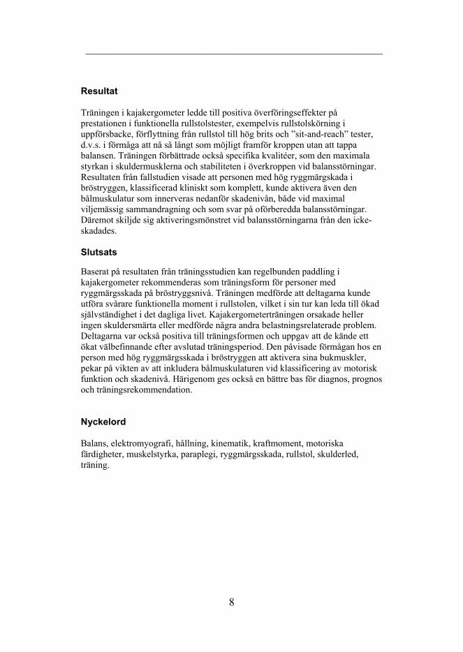
Shoulder muscle strength (study II)
There was a main effect (P = 0.023) of kayak ergometer training with increased shoulder muscle strength after training. The improvements were independent of shoulder movement, and occurred in the beginning and middle positions of the range of motion (Fig. 16).
A tendency (P = 0.081) towards lower shoulder muscle strength was observed in the SCI group compared to a matched reference group of able-bodied persons (Fig. 16).
The average intraclass correlation coefficient for test-retest torque values in the SCI group before training was 0.941 (95 % confidence interval: 0.928 – 0.954).
1520253035404550556065707580859095
a
ExtensionFlexion
3817.5- 3
Torq
ue (N
m)
1520253035404550556065707580859095
Shoulder position (0)
b
Abduction
Adduction
5837.517
1520253035404550556065707580859095
c
Torq
ue (N
m)
Shoulder position (0)
Before After Reference
External rotation
Internal rotation
3515- 5
Figure 16
Mean values of shoulder muscle strength (torque, Nm) in specific angular positions at a velocity of 30º s-1 for the group of persons with SCI before and after training and for the reference group, in a. shoulder flexion and extension, b. shoulder abduction and adduction, and c.shoulder external and internal rotation. Arrows indicate thedirection of movement. (Note that in paper II, least squares means were shown in Fig. 2 and used in the statistics.)

_____________________________________________________
35
Trunk stability (study III)
Significant improvements in postural stability were demonstrated after training with smaller angular and linear trunk displacements in anterio-posterior direction and in twisting in all four kinematic responses during FWD, BWD and LAT translations (Figs. 17a-b, d-e). No effects of training were seen in medio-lateral angular and linear trunk movement during LAT translations (Fig. 17c).
0 1 2 3 4-10
-5
0
5
Time (s)
d
post
antAP displacement LAT translation
0 1 2 3 4-5
0
5
10
c
Ang
ular
dis
plac
emen
t (0 )
ipsi
contra
ML displacement LAT translation
0 1 2 3 4-10
-5
0
5
e
counterclockwise
clockwise
Ang
ular
dis
plac
emen
t (0 )
Time (s)
TW displacement LAT translation
0 1 2 3 4-10
-5
0
5
a
Ang
ular
dis
plac
emen
t (0 )
post
ant
AP displacement FWD translation
0 1 2 3 4-5
0
5
10
b
post
ant
AP displacement BWD translation
Figure 17 Mean curves of trunk angular displacement in anterio-posterior (AP) direction during a. forward (FWD), and b. backward (BWD) translations, and in c. medio-lateral (ML), d. AP, and e. twisting (TW) direction during lateral (LAT) translations, versus time. The thin curves indicate the group mean before training and the thick curves after training (N=10). The red curves represent mean trunk displacement for one able-bodied subject (from study IV).

_____________________________________________________
36
0.0
1.8
1 s
TrA0
4 OI0.0
0.4 OE
0.0
0.9 TrA0
1 OI0.0
0.4 OE
Control
Abdominal muscle thickness (study IV)
The ultrasound measurements indicated similar thickness of each of the muscle layers of the ventro-lateral abdominal wall in the subject with SCI (OE: 13 mm, OI: 11 mm, and TrA: 6 mm) as in the Control subject (11, 15, and 7 mm, respectively) (Fig. 18, left).
Trunk muscle activation (study IV)
Activation ability during maximal voluntary efforts
The subject with SCI, classified clinically as complete (AIS A) at T3 level, was able to activate all his abdominal muscles, innervated from segments below the lesion level, in maximal voluntary efforts (Fig. 18, upper right). The EMG traces from the two subjects appeared similar from a qualitative point of view (Fig. 18, right), but with more frequent occurrence of individual spikes in the recording from the subject with SCI, which is most likely related to a lower level of activation in this subject.
Figure 18 Ultrasound images (left) and rectified EMG traces (right) from obliquus externus (OE), obliquus internus (OI), and transversus abdominis (TrA) in the subject with SCI (upper) and the able-bodied Control subject (lower). The EMG traces are from single voluntary contractions aiming at a maximal activation of that particular muscle.
0.04
m
OI
TrA
OE
0.04
m
OI
TrA
OE
SCI

_____________________________________________________
37
Activation responses to support-surface translations
The pattern and timing of muscle responses to support-surface translations differed between the two subjects (Figs. 19 a-d). Responses in the subject with SCI generally involved a larger number of muscles, with a more frequent engagement of upper trunk muscles, i.e. PM, TZ, and LD, and occurrence of coactivation of ventral and dorsal muscles, particularly in FWD and BWD translations (Figs. 19 a-b).
TrA
OI
OE
RA
ES
LD
TZ
PM
0.0 0.1 0.2 0.3 0.4 0.5
SCI Control
a FWD translation Figure 19 a
In forward (FWD) translation, the subject with SCI had an early activation of upper trunk muscles and an activation of dorsal before ventral lower trunk muscles. In contrast, the Control subject displayed an early and more selective activation of the abdominal muscles.
TrA
OI
OE
RA
ES
LD
TZ
PM
0.0 0.1 0.2 0.3 0.4 0.5
b BWD translation Figure 19 b
In backward (BWD) translation, dorsal muscles were activated with similar latencies in both subjects. In addition, there was a late activation of abdominal muscles (OE, OI and TrA) in the subject with SCI.
TrA
OI
OE
RA
ES
LD
TZ
PM
0.0 0.1 0.2 0.3 0.4 0.5Time (s)
c RWD translation
TrA
OI
OE
RA
ES
LD
TZ
PM
0.0 0.1 0.2 0.3 0.4 0.5Time (s)
d LWD translation
Figure 19 c and d In rightward (RWD) translation (c), the subject with SCI showed an early recruitment of the dorsal muscles, including those of the upper trunk, whereas the abdominal muscles had longer latencies. For the Control subject, on the other hand, the initial responses appeared specifically in the ES, OI and TrA muscles. In the leftward (LWD) translation (d), the muscles (recorded from the right side) showed more variable responses in both subjects.

_____________________________________________________
38
DISCUSSION
Training effects
Main findings of this thesis were that persons with long-standing thoracic spinal cord injury (SCI) were able to improve their shoulder muscle strength and postural stability after a 10-week period of kayak ergometer training. Also, more challenging functional tasks could be performed after the training, which, in turn, might lead to a greater independence in daily living. In addition, the training in the kayak ergometer, albeit intense, did not cause any shoulder pain or other problems. These findings, plus the positive subjective experience expressed by the participants, indicate that this type of training is an effective, and attractive, activity for persons with thoracic SCI.
As always, the effects, if any, of a period of physical training, are dependent on the proficiency of the participants, the characteristics of the training stimulus, and the properties of the tests used for evaluation.
Participants
The proficiency of the current subjects with SCI, as a group, is difficult to establish due to lack of comparable data. Interestingly, in the one test where their performance was compared to that of able-bodied subjects, the results showed shoulder strength values that were similar to those of the able-bodied.
As far as the individual variation within the SCI group is concerned, it may, at first glance, appear small, since they all had a thoracic SCI, were wheelchair users and had finished their rehabilitation period. However, when adding additional group descriptors, such as age, time post-injury, sensory and motor function scores, and level of physical activity, the heterogeneity of the group becomes apparent. The inter-individual variation within each parameter is large and the number of parameters to consider is high. Furthermore, they can interact in numerous ways. For example, an initial disadvantage, due to a high level of injury, may be counteracted by a long and physically active period post-injury. This complexity is likely to underlie the finding of few and low correlations between background parameters, including level of injury, and initial proficiency with respect to functional tests and trunk stability in response to balance perturbations. Correlations were present between initial performance and training effects for three of the five functional wheelchair tests, i.e. a large training effect was related to a low initial performance level, as would be expected from current training science (Kraemer et al., 2002). However, no

_____________________________________________________
39
correlations were found between changes with training and any of the background variables, e.g. level of the SCI.
Another limiting factor in the study is the relatively low number of subjects. This has to do with the special category of subjects as well as with the specific inclusion criteria. The number of persons with a clinically classified complete thoracic SCI in the Stockholm area is limited to about 150 and of these about 60 are included in the database of Rekryteringsgruppen, from which the subjects were recruited. In addition, the subjects had to make a commitment to devote time to this study for an extended period, including numerous test and training sessions, which might have deterred some of the potential participants. The subjects volunteering for the project were extraordinary positive and loyal to the project and the problem of lack of adherence to the protocol, which training studies often suffer from, did not exist in this project.
It would have been desirable with a control group of subjects with SCI, who were to take part in the tests and not in the training. However, it was decided early on not to attempt to recruit such a group based on the limited number of potential subjects and the additional difficulty in finding a group of persons with a sufficient match of critical variables with the experimental group. Extra care was instead taken to establish a stable pre-training level in the experimental group by having them go through the entire test protocol twice before the start of the training. A comparison of these two tests generally showed no statistical difference due to learning. Corresponding statistics was not carried out for the balance tests, but we regard it unlikely that there is a training/learning effect just due to repeated measurements, considering the fact that such a long time (10 weeks) elapsed between test occasions.
Even though the study was primarily aimed at investigating training effects, it would have been of interest to perform a more systematic comparison between the experimental group and a matched group of able-bodied subjects. This would have allowed exploring the possibility that the changes with training in the SCI group would go either in the direction of becoming more ”normal” or in the other direction, i.e. deferring more from the ”normal” by further establishing unique responses of persons with SCI. The former could be the case for compensations to balance perturbations (though only based on comparison with data from one able-bodied subject, cf. Fig 16 in the Summary) and the latter in the case of balance in quite sitting (Grigorenko et al., 2004). A comparison of strength performance between the SCI group and a reference group of able-bodied persons was considered feasible, since the tests were done in a special experimental set-up deemed equally unfamiliar to both subject categories. The functional tests, on the other hand, were designed for the subjects with SCI and they would have been difficult to perform for a group of able-bodied subjects not used to a wheelchair. The balance tests did not require wheelchair

_____________________________________________________
40
experience and could have included a reference group of able-bodied. Such a comparison was made in the case study, where invasive recordings of muscle activation were carried out on one occasion, i.e. training effects were not investigated.
Training
Kayak ergometer paddling was selected as the training paradigm for several reasons. Firstly, it meets the necessary criterion of being an activity possible to carry out in sitting. Secondly, it appears to involve most of the upper body musculature. This has been demonstrated for the muscles around the shoulder joint in able-bodied subjects (Trevithick et al., 2006). Corresponding data for the trunk muscles are lacking, but it is evident from a mechanical point of view that there has to be a link transferring the forces from the paddle via the shoulders to the kayak. Moreover, the paddling movement is complex with alternating three-dimensional upper body movements during the pull, lift and push phases. These movements have been described in able-bodied paddlers (Plagenhof, 1979). Although no study has yet been done comparing paddling technique between able-bodied persons and persons with SCI, the basic alternating paddle movement appears to be similar. Thirdly, the complex interplay between forces in different directions, and the construction of the kayak, placed marked demands on balance control and stability of the upper body, particularly in the medio-lateral direction. Lastly, by varying the intensity of paddling, the activity can be directed mainly towards endurance or strength type of training (Tesch, 1983; Shephard, 1987; Fry and Morton, 1991). Furthermore, kayaking on open sea has been shown to be a suitable and appreciated training activity for persons with thoracic SCI (Grigorenko et al., 2004).
Training indoors on a kayak ergometer is an alternative to open sea kayaking, having the advantage of not being weather-dependent. Kayak ergometers are commercially available and widely used among competitive kayakers and also for general training purposes. A great advantage with the ergometer is that the intensity of the activity is easily controlled. A display provides information on, for example, paddling distance, intensity and speed. This information adds motivation to the trainee as well. A direct comparison of movement and muscle activity patterns in the ergometer and on open sea has still to be performed. However, kayak ergometer training has been reported to be able to simulate open sea kayaking in terms of physiological demands, such as oxygen uptake and heart rate (van Someren et al., 2000). An obvious difference is, however, that the ergometer normally rests on a steady surface, which minimizes the otherwise considerable unsteadiness during kayaking on open sea. To compensate for this and make the training more realistic in terms of challenge to balance control, one of the first tasks in this thesis work was to modify a

_____________________________________________________
41
commercially available kayak ergometer with an adjustable balance demand in the medio-lateral direction. This module made it possible to individually adjust and progressively increase the balance demand for each subject during the training period. It also appeared to be suitable for this category of persons, since all of them could increase the level of difficulty, but none was able to reach the most unstable setting during the training period. Anecdotally, a world champion kayaker has tried the modified ergometer and deemed it a valuable tool worth incorporating in his own training.
All subjects in this study expressed subjective improvements on general well being after the intervention. This is in line with earlier studies showing positive effects of exercise in general on quality of life in persons with long-standing SCI (Ditor et al., 2003; Hicks et al., 2003). The kayak ergometer training was also easy to learn, and the social togetherness, which is often mentioned as an asset of physical training, was met by having the participants train in parallel on four similarly equipped ergometers. Another positive aspect of the training was that the extra load on the upper body induced by the rather intense training did not lead to any shoulder problems or other overload symptoms. Shoulder pain is a major problem for persons with long-standing SCI (e.g. Curtis et al., 1999; van Drongelen et al., 2006) and it has been related, among other things, to regular participation in sport activities, e.g. basketball, with frequent vigorous starts and stops (Curtis and Black, 1999). The absence of shoulder problems with kayak training could probably be ascribed to the individually adjusted progression of the training as well as the smooth character of the paddling movement itself. The majority of the trainees also reported experiences of improvements in functional tasks, such as reaching for an object and propelling the wheelchair uphill. These results are in line with previous findings from open sea kayak training in persons with SCI (Grigorenko et al., 2004), and were substantiated by the actual improvement in performance in most of the functional tests.
Due to the gradual improvement on the part of the participants, the overall training intensity and load could be progressively increased over the training period in all subjects. The outline of the training protocol was such that it contained sessions and periods of lesser or higher intensity, presumably stimulating both increases in endurance capacity and muscle strength.
No direct tests of endurance were performed in the current study, but the decreased time in the energetically demanding task of propelling 50 m on an inclined surface may be indicative of such an adaptation. In a previous study, with a similar protocol, but on open sea, it was shown that maximal aerobic power measured on a kayak ergometer increased with 4% after an 8-week-period of training in a group of persons with SCI (Bjerkefors et al., 2005). Different types of exercise programs have earlier been reported to improve the cardiorespiratory function in persons with SCI, e.g. circuit training (Jacobs et

_____________________________________________________
42
al., 2001), wheelchair ergometer training (Tordi et al., 2001; Bougenot el al., 2003), and stimulation-assisted rowing (Wheeler et al., 2002).
As far as muscle strength is concerned, the paddling regime apparently provided enough stimuli to improve shoulder muscle strength, as evidenced by the various standardized isokinetic strength tests applied. It also appeared to be sufficient to lead to strength gains in shoulder movements in all three planes. Other studies that have evaluated muscle strength after interventions with similar training equipment have reported that training only on an arm ergometer or on a wheelchair ergometer (Davis and Shephard, 1990; Yim et al., 1993) caused a minimal strength improvement, whereas studies that included also strength training exercises have been able to demonstrate marked strength gains (Jacobs et al., 2001; Hicks et al., 2003). The strength improvement measured here after kayak ergometer training, albeit relatively modest, indicates that the three-dimensional upper body movement during paddling contains parts of high enough muscle load to stimulate strength growth, which seems not to be the case for the mainly two-dimensional movements in arm and wheelchair ergometers. Measuring the strength of trunk muscles was attempted initially in the current project, but was abandoned due to difficulties in creating a standardized experimental situation where any strength output of these specific muscles could be assessed in a satisfactory way.
In addition to being able to progressively increase the intensity of the training, the trainees were able to cope with a gradually more demanding challenge to the sitting balance provided by the adjustable balance module. Since this instability was provoked primarily in the sideways direction, an improvement in balance and stability of the upper body was expected mainly in the medio-lateral direction. Moreover, movements in this direction were assumed to be least affected by the subjects’ regular wheelchair propulsion and thus more trainable. Contrary to expectations, no effects were observed in the medio-lateral kinematic responses to lateral support-surface translations. However, the posterior and twisting movements in lateral translations were diminished with training. A possible explanation for this might lie in the compensatory “techniques” used. Before training, the subjects appeared to fixate the upper body in a backward leaning position, utilizing the backrest to withstand the lateral perturbation, since this position was assumed after the initial acceleration and maintained throughout the rest of the translation. The backward movement occurred simultaneously with a trunk twisting towards the direction of translation. This combination of additional trunk movements might be a consequence of a limited ability to specifically perform lateral trunk flexion, i.e. to approach the balance limit in relation to the support surface in the sideways direction. To compensate for this, the trunk had to be twisted around the vertical axis, moving the upper body in the opposite direction and thus avoiding tipping over to the side. After training, there seems to be lesser need for this

_____________________________________________________
43
compensatory mechanism. Thus, even though the actual trunk movement to the side remained unchanged, the training appeared to improve the coordination of movements and allow for a more “pure” side-bending response without concomitant movements in other planes. The suggested movement strategy in response to sideways perturbations in persons with SCI was supported by the findings in the case study. The person with SCI had a movement pattern with more of twisting and posterior trunk movement, whereas the able-bodied person showed a more “pure” side-bending response.
The improvements with training on the responses to the initial unpredictable acceleration and the ensuing predictable deceleration indicate that the mechanisms involved, primarily muscle reflex responses and feed-forward control, are, to some extent, trainable by the specific training regime applied. It is, however, difficult to speculate about putative neural mechanisms. One possible reason for the increased trunk stability might be that the relatively demanding training could have provoked an increased neural drive in descending cortico-spinal pathways to postural trunk muscles. This increased drive might, in turn, induce activation of denervated and/or atrophied musculature. Such an improvement in neural communication between the brain and effector muscles has been reported after intense locomotion training in persons with an incomplete SCI (Thomas and Gorassini, 2005). In the current study, all subjects, except one, were classified as having complete SCI, and should therefore have an even more limited function in these neural pathways. Interestingly, several studies (e.g. Dimitrijevic et al., 1983; Sherwood et al., 1992) have demonstrated a “discomplete” syndrome in patients with thoracic SCI, clinically classified as complete. Study IV constitutes a beginning of studying motor strategies underlying the control of upper body stability and sitting balance in persons with thoracic SCI.
Tests
The tests used to evaluate the possible transfer effects of kayak ergometer training had different specific purposes and therefore different characteristics. The functional tests were chosen to mirror daily activities and thus they should have an implicit validity. The strength tests were specifically aimed at measuring a certain quality, namely muscle strength at the shoulder joint under highly standardized conditions. Also, the balance tests were carried out in a laboratory environment, but were selected to represent situations that can occur in daily life, such as when travelling in a bus or subway with sudden accelerations and decelerations. No other measures were taken to ensure the validity of the tests. The reliability of the tests was assessed with a test-retest approach for the functional tests and the strength measurements and found to be of acceptable magnitude. To minimize the possible influence of learning in the

_____________________________________________________
44
balance tests, the subjects were allowed a familiarization session one week prior to the first test occasion.
Due to the relatively high proficiency of our subjects, who were all in a post-rehabilitation stage, available functional tests that have been evaluated in terms of reliability and validity (Catz et al., 1997; Lynch et al., 1998; Kilkens et al., 2002 and 2004) had to be modified to increase the resolution of the outcome measure and avoid ceiling effects. These adjustments of the tests were obtainedafter pilot studies on persons with SCI with a similar range of performance levels as the experimental group. The functional tests were based on measurements of time, distance and height. Thus, no evaluation was done of the actual techniques involved in carrying out the tasks. Other studies have used scoring systems, which included a qualitative evaluation of the level of skill involved (Catz et al., 1997; Harvey et al., 1998; Kilkens et al., 2003; Kirby et al., 2004). In our material of post-rehabilitation subjects, it would have been essentially impossible to subjectively judge changes in the quality of performance as well as selecting appropriate levels of pass and fail. The sensitivity of the tests applied here proved to be enough to measure changes with training and difference between subjects and experimental and control groups. One exception was the sit-and-reach tests, where a ceiling effect could not be avoided for the three subjects with a low injury level. They could control the forward inclination of the trunk and thus only anatomical dimensions limited their reaching distance. Therefore, this type of sit-and-reach-test cannot be recommended for subjects with an SCI at or below the T11 level.
In the strength measurements, dynamic concentric (shortening) contractions were selected because of the intuitive similarity to the contraction type predominating in most muscles during paddling. Isokinetic testing, i.e. at a constant angular movement velocity, was chosen to provide standardization of the measurements of dynamic strength. Since paddling is a three-dimensional movement, it was decided to measure strength in reciprocal movements about all three axes. It is realized that the paddling movement is not restricted to movements around these axes and that it does not occur at constant angular velocities. However, the standardization was given priority in these measurements of a specific quality, namely muscle strength. The peak angular velocities during paddling were measured for movements around each of the three axes of rotation and ranged from 170 to 390 deg/s. Thus, the selected speed of 30 deg/s was included in the lower part of each speed range. The dynamometer used in this study has earlier been shown to provide accurate and reliable measures of torque and velocity (Drouin et al., 2004). However, we discovered that with a speed set at 180 deg/s, the machine could not reach the set velocity before the very end of the range of motion, thus limiting the part that was isokinetic to about 5 deg. This speed was therefore omitted from our protocol.

_____________________________________________________
45
The test paradigm used here to assess trunk stability via translations of the support-surface was recently introduced by Carpenter et al. (2005). It actually utilizes a motor-driven treadmill, earlier used for studies of locomotion (e.g. Thorstensson et al., 1982). The control of the acceleration and speed of the treadmill has proven to be of acceptable accuracy and reproducibility to serve also for balance studies (Carpenter et al., 2005). The initial studies were carried out on standing able-bodied persons (Carpenter et al., 2005; Tokuno et al., 2006). This is the first time that sitting has been studied. The experimental set-up made it possible to evaluate postural compensations to both an unpredictable acceleration and a predictable deceleration during a single trial. One limitation in the control was that the size of the two perturbations could not be made exactly the same. The magnitudes of the perturbations were selected in systematic pilot experiments to be manageable, yet challenging, for all subjects in the training group. However, due to the heterogeneity among the participants, the challenge could be close to the maximal for one and relatively “easy” for another. Using more individually adapted criteria for choosing the size of the perturbation, e.g. having it closer to each person’s balance limit, might have revealed more clear training effects. On the other hand, such tests could involve more risks to the subject. During the tests with balance perturbations, the movements of the upper body were recorded with a motion-capture system with markers placed on the skin over certain anatomical landmarks. One of the limitations adhering to such a method is the possibility of skin-slippage, i.e. movement of the skin and marker in relation to the underlying skeletal structures. No systematic evaluation of skin-slippage was done here, but such movements have been shown to be relatively small (<2 mm) over the spine (Nilsson, 1990). Unfortunately, the analyses in this study did not include movements of the head. A significant role of the head in postural control has previously been demonstrated in paraplegics in response to tilting of the support-surface (Bernard et al., 1994).
A variety of valid and reliable questionnaires for evaluating self-reported well being (e.g. The Swedish SF-36 Health Survey, Westgren and Levi, 1998) and functioning (e.g. SCIM, Catz et al., 1997) exist for persons with SCI. The basis for our decision not to use standardized questionnaires was that we wanted to evaluate primarily specific experiences related to our particular tests and training protocol. One of the drawbacks with this approach is that it limits the possibilities to make comparisons with other studies.

_____________________________________________________
46
Trunk muscle activation
Among the main findings was also the observation that a person with a clinically diagnosed complete SCI at the T3 level still was able to activate his trunk muscles below the lesion level, and that he had adopted a different pattern of muscle response to balance perturbation as compared to an able-bodied person.
Naturally, observations on a single person are limited, but still allow speculations about possible underlying mechanisms and implications for injury classification and training during and after rehabilitation in persons with thoracic SCI.
Possible neural mechanisms
Results from the electromyographic recordings showed that the person with SCI was able to activate trunk muscles below the injury, both in voluntary efforts and in response to balance perturbations. The ability to activate those muscles with motor nerves exiting from the spinal cord below the injury level could have different underlying mechanisms. One is that the SCI was incomplete and that remaining connections might have been reinforced during rehabilitation and usage during the post-rehabilitation period. Another possibility is that activation of these lower trunk muscles is induced indirectly by activation of other muscles, e.g. the upper trunk muscles. Contractions of upper trunk muscles, with intact innervation also after thoracic SCI, might lead to stretching and a subsequent reflex activation of the lower ones. There may also be additional triggers other than stretch, e.g. activation may be directed from heteronymous or even antagonistic muscle actions via neural connections adopted over time to compensate for the impairment caused by the SCI. Using neurophysiological techniques, it has been possible to demonstrate remaining sub-clinical motor function via neural pathways passing an SCI clinically classified as complete (Dimitrijevic et al., 1983; Sherwood et al., 1992). These experiments have mainly been designed to identify supraspinal influence on spinal reflexes in the legs. Rarely have the trunk muscles been investigated. In response to the balance perturbations the subject with SCI demonstrated less specific electromyographic responses than the able-bodied subject. Lower trunk muscles were activated also in this situation, but often in a different order and together with antagonist muscles and/or other normally non-postural upper trunk muscles. Evidently, even though lower muscles could be activated, their contribution to counteracting the movement induced by the perturbation was insufficient and had to be complemented by recruitment of additional muscles. A development of unique muscle activation patterns and new muscle synergies has previously been demonstrated in persons with thoracic SCI in sit-and-reach tests (Seelen et al., 1998; Potten et al., 1999).

_____________________________________________________
47
Implications for classification
The present finding of an ability to activate lower trunk muscles in a person with a high thoracic SCI points to a need to revise the current classification system for thoracic SCI to include motor tests also for trunk muscles. In clinical practice, the International Standard of Neurological Classification of Spinal Cord Injury (Marino et al., 2003) has become a standardized and routinely used assessment tool to classify motor and sensory function in persons with SCI. The motor examination involves bilateral manual muscle tests, graded on a 0-5 scale, of the upper and lower limbs, each consisting of five key muscles. Unfortunately, the assessment tool does not include the trunk muscles, which makes conclusions about motor connectivity to these muscles uncertain. The lack of information about motor preservation to the trunk muscles will also affect the classification of the neurological lesion level, which normally is based on the most caudal segment with normal motor and sensory function. Therefore, in persons with thoracic SCI, the classification of the level of the injury will be determined solely based on sensory function. The development a new instrument for injury classification should occur with close interaction between basic neurophysiological and clinical research to make it valid and reliable on the one hand and also user friendly on the other. Sensitivity enough to discern level of injury as well as progress with rehabilitation and training is highly desirable.
Implications for rehabilitation and training
Knowing if a person with a thoracic SCI has access to his/her lower trunk muscles is important for judging the potential for improvement and for prescribing optimal exercises in rehabilitation and for post-rehabilitation training. Since stabilization of the trunk is a prerequisite for adequate movements of the arms and head, trunk muscle activation is provoked by daily activities, but only if the trunk is unsupported. Exercises should therefore imply extra provocations of the active trunk stabilization system, directly or indirectly. Such training would potentially increase the ability of independent trunk stabilization and thereby have the beneficial effect of increasing the repertoire of tasks achievable manually. Ideally, the training exercises should be flexible enough to be applicable in different stages of rehabilitation as well as for progressive post-rehabilitation training. Training tools could be used, such as the kayak ergometer in the current project, but less complex tools and exercises without tools should be developed based on the present results and ideas and continued research. Finally, the present findings of remaining motor connectivity to lower trunk muscles should caution the clinician when using the diagnosis “complete” SCI and encourage the persons given such a diagnosis.

_____________________________________________________
48
CONCLUSIONS
in relation to the aims of the four studies:
I There were clear transfer effects of the kayak ergometer training onto performance in functional wheelchair tests in the persons with thoracic SCI. Since these tests mimicked some daily activities, the improvements should, in turn, lead to a better ability to carry out more challenging tasks of daily living, which might lead to a greater independence. This, and the positive subjective experience from the training expressed by the subjects, promotes kayaking as a suitable activity for persons with thoracic SCI.
II Kayak ergometer training resulted in a general increase of shoulder muscle strength in persons with SCI. There was a tendency towards lower shoulder muscle strength as compared to a reference group of able-bodied persons, which might indicate that post-rehabilitated persons with SCI have not managed to fully maintain their shoulder muscle strength. Evidently the current training regime contained loads high enough to induce gains in strength, but still did not cause any shoulder problems or other overload symptoms.
III In general, postural stability in response to support-surface translations was improved with kayak ergometer training, i.e. the same balance perturbation, be it predictable or unpredictable, caused smaller trunk displacements after training. This improvement seen in standardized laboratory tests, might, in turn, allow persons with long-standing SCI to better master similar challenges to balance perturbations in everyday life.
IV The results from the case study showed that a person with a high thoracic SCI, clinically classified as complete, was still able to activate trunk muscles below the injury, both in maximal voluntary efforts and in response to balance perturbations. In the latter case, the person with SCI displayed a different trunk muscle coordination pattern than an able-bodied subject. These pilot data highlight the importance of including examination of trunk muscle function in persons with thoracic SCI in relation to injury classification, prognosis, and training prescription.

_____________________________________________________
49
TACK!Alf Thorstensson, jag vill framföra mitt allra varmaste tack för allt du gett mig under dessa år som doktorand! Jag har med stor beundran sett hur du med briljant förmåga omvandlat mina spretiga texter och tankar till något tydligt och ”enkelt”. Du har med stor kunskap, en ängels tålamod och ett enormt engagemang inspirerat mig att göra mitt bästa! Jag vill också tacka för att du aldrig lämnat mig i sticket, utan alltid ställt upp till sista punkt! oavsett om det varit kväll eller helgdag och för att du med värme och humor försökt ”uppfostra” mig i hur man beter sig i den akademiska världen!
Mark Carpenter, it has always been a great pleasure to work with you, and it has been fun! Thanks a lot for all your positive support! You have taught my in a very friendly way how to use the equipments, to run the tests, to analyse the data, and have inspired me to work carefully and systematically! Hopefully we can continue to collaborate!
Andrew Cresswell, tack för all hjälp i sista studien och den kunskap du har förmedlat!
Anna Jansson, ett stort tack för all hjälp med styrkestudien, det var ett stort nöje att jobba med dig!
Tack till alla underbara kollegor i Tegelhögen/LTIV, ni är bäst! Alexander Ovendal, ”- hur var det nu med cosinus?” Andrezza Saldanha, we had a great evening at Brunnsviken! Carl Askling, grabben i rummet bredvid som med humor, värme och klarsynthet alltid stöttat! Många tack! Craig Tokuno, my “särbo” and technical support (THANKS!) down-under. Hans Rosdahl, ett stort varmt tack för att du inspirerade mig att börja forska, men var det nödvändigt att bada med mobil och skor?! Jan Seger, susar fram i naturen på cykel, i kajak eller på skridskor! Johnny Nilsson, inspirerar till utveckling och skratt! Karl Daggfeldt, killen med hjärnkoll på det mesta som en dag sakta upphävde alla fysiska lagar och lyfte från mitt golv (och jag gick på det!). Kjartan Halvorsen, tålmodigt har du förklarat svåra biomekaniska begrepp och med entusiasm inspirerat mig att gå vidare! Kristjan Oddsson, förmedlat välbehövlig massage. Lee Nolan, Queen of Biomechanics and President of Enable. Tjejen som kan det mesta, från att bygga bastu till att måla tavlor, å så har du dom tuffaste röda skorna! Maria Ekblom, med ett hjärta av guld och en briljant förmåga att förklara har du vid oräkneliga tillfällen ställt upp för mig! Du har varit en förebild för mig från första dan på labbet! Martin Eriksson, lycka till med fortsatta studier i Örebro! Mårten Fredriksson, always happy man! P-O Åstrand, med humor och skärpa levererar du dagens vits. Toni Arndt, ditt positiva humör och ditt skratt smittar! Härligt! Fd kollega och rumskamrat Anatoli Grigorenko, många tack för all hjälp när jag kom till labbet!
Tack till alla fantastiska arbetskamrater i Rekryteringsgruppen och ledare i Friluftsgruppen, hos er har jag mina rötter och mitt hjärta! Tack till Magnus Andersson, Anna Stannervik och Peter Fahlström för allt stöd genom åren!
Ett stort varmt TACK till alla försökspersoner i studierna! Utan er hade det aldrig blivit någon avhandling! Tack för att jag fick ert förtroende!
Micka Hahne, Gunnar Krantz, Ulla Löfquist (min bästa fotomodell!) och Mats-Erik Bjerkefors, ett varmt tack för all hjälp under träningsstudien!
Sten Ljungar och Gunnar Krantz, mina vapendragare och förebilder som rest runt och föreläst med mig, vilken tillgång ni är! Jag tackar och bockar!
Marie Alm, många tack för all hjälp under åren!!
Ett stort tack till alla arbetskamrater på GIH för inspirerande år hos er! Ett speciellt tack till vaktmästeriet, biblioteket och IT-avdelningen för förträfflig service!
Elisabeth Berg, för mycket värdefull hjälp med statistiken.
_____________________________________________________
50
Gunilla Hammarskiöld, Mickas och min ”projektmamma” som för många år sen hjälpte oss att formulera ansökningar till nya projekt. Ett stort tack till Susann Forsberg för allt stöd genom åren.
Benny Setterberg och Leif Wahlstedt, många, många tack från ”rivjärnet”, utan er hade det inte blivit några grejer! Det är alltid en fröjd att samarbeta med er!
Ninitha Maivorsdotter, tack för alla luncher på trappen som har vidgat mina vyer om forskningen och livet!
Ann-Cristin Åberg, Gunilla Elmgren Frykberg, Kerstin Wahman och Åsa Hedberg, tack för alla goda samtal och kloka tankar!
Anna Frohm-Grönqvist, Doktor Anna, sida vid sida har vi kämpat dag ut och dag in i Tegelhögen och stångats med biomekaniska begrepp, dataprogram och statistiska metoder. Men vi har också, sida vid sida, åkt skidor, navigerat i ösregn, seglat på öppet hav, men framför allt har vi pratat om livet, kärleken och känslorna! Jag hoppas att vi fortsätter med det i många år!
Micka Hahne, min starka modiga väninna, snart är det dags för oss att förverkligar våra drömmar om Friluftspraktiken! Tack Micka för att du finns och jag önskar dig allt gott livet har att bjuda på!
Karin Willis, ja, tänk om vi hade vetat att våra liv skulle vara så här, när vi för nästan 20 år sen gick och drömde om framtiden på väg mellan Åre och Duved i solnedgången!
Ulla Lundholm, systera mi som med en fantastisk förmåga ser livet från den ljusa sidan!
Ann-Marie Carlberg och Sigge, snart har vi mer tid för bus!
Tack till alla Semesterbästisar och Goamiddagsvänner som gör livet ljust och glatt!
Tack till alla ”extrabarn” som förgyller vår tillvaro med fniss, kladdkakor och underbart pianospel!
”Min” stora familj i Lima, Vidar, Sara, Ola, Annelie, Anki, Lena, Andreas, Tony, Emma, Ida, Ero, Hobbe och Zacke – som öppnat ert hem för en 08 och lärt mig massor om livet som att plocka potatis, hjortron, lingon och blåbär, valla skidor, fiska (hua), ro, plantera skog, köra skoter, men framför allt så fick ni mig att bli älgetarian istället för vegetarian!
Mamma och Pappa, mina fantastiska förebilder, som nyfiket njuter av livet, antingen på väg till Alperna ”med ett gäng pensionärer” eller sitter i en solig glänta på Omberg och dricker kaffe. Mitt allra varmaste tack till er båda för allt gott ni har gjort för mig och att ni alltid trott på mig, i alla lägen, å så tack för lakritssträngen, den finns alltid kvar. Ett varmt tack också till ”Berta med fästman” som gör vårt liv lite ”klarare”!
Magda och Johanna, mina underbara tjejer som med ert sprudlande sätt är LIVET och KÄRLEKEN! Det bästa som finns är att vara med er och ”snusa” på hästarna, sjunga duett i badrummet och hoppa från klipporna. Tack för alla gånger jag har fått njuta av livet tillsammans med er och för allt omtanke ni visat mig, ni är BÄST!
Min älskade Masse, utan DIG hade det jag aldrig klarar det! Mitt innerligaste tack för alla gånger du har ställt upp för mig, villkorslöst och för all den KÄRLEK du gett mig! Tack också för alla matlådor du har packat med nyttiga bönor och grönsaker som fått mina arbetskamrater vid lunchbordet att nyfiket titta och fråga: - Vad äter du? - Öh, rödbetslimpa?!
Till sist vill jag tacka för ekonomiskt stöd från Allmänna Arvsfonden, Centrum för Idrottsforskning, Hjälpmedelsinstitutet, Gymnastik- och Idrottshögskolan, Norrbacka Eugeniastiftelsen, Legitimerade Sjukgymnaster Riksförbund.

_____________________________________________________
51
REFERENCES
Adams, M.M. and Hicks, A.L. (2005) Spasticity after spinal cord injury. Spinal Cord 43, 577-86.
Anderson, K.D., Borisoff, J.F., Johnson, R.D., Stiens, S.A. and Elliott, S.L. (2006) Long-term effects of spinal cord injury on sexual function in men: implications for neuroplasticity. Spinal Cord (published online)
Bernard, P.L., Peruchon, E., Micallef, J.P., Hertog, C. and Rabischong, P. (1994) Balance and stabilization capability of paraplegic wheelchair athletes. J Rehabil Res Dev 31, 287-96.
Bjerkefors, A., Rosdahl, H., and Thorstensson, A. (2005) Kayak training in paraplegics – effects on endurance capacity and balance. Proceedings from the 4th International Scientific Conference on Kinesiology, Croatia, 780-2.
Bougenot, M.P., Tordi, N., Betik, A.C., Martin, X., Le Foll, D., Parratte, B., Lonsdorfer, J. and Rouillon, J.D. (2003) Effects of a wheelchair ergometer training programme on spinal cord-injured persons. Spinal Cord 41, 451-6.
Carpenter, M.G., Thorstensson, A. and Cresswell, A.G. (2005) Deceleration affects anticipatory and reactive components of triggered postural responses. Exp Brain Res 167, 433-45.
Catz, A., Itzkovich, M., Agranov, E., Ring, H. and Tamir, A. (1997) SCIM--spinal cord independence measure: a new disability scale for patients with spinal cord lesions. Spinal Cord 35, 850-6.
Curtis, K.A. and Black, K. (1999) Shoulder pain in female wheelchair basketball players. J Orthop Sports Phys Ther 29, 225-31.
Curtis, K.A., Drysdale, G.A., Lanza, R.D., Kolber, M., Vitolo, R.S. and West, R. (1999) Shoulder pain in wheelchair users with tetraplegia and paraplegia. Arch Phys Med Rehabil 80, 453-7.
Dahlberg, A., Perttila, I., Wuokko, E. and Ala-Opas, M. (2004) Bladder management in persons with spinal cord lesion. Spinal Cord 42, 694-8.
Davis, G.M. and Shephard, R.J. (1990) Strength training for wheelchair users. Br J Sports Med 24, 25-30.
Dimitrijevic, M.R., Faganel, J., Lehmkuhl, D. and Sherwood, A. (1983) Motor control in man after partial or complete spinal cord injury. Adv Neurol39, 915-26.
Ditor, D.S., Latimer, A.E., Ginis, K.A., Arbour, K.P., McCartney, N. and Hicks, A.L. (2003) Maintenance of exercise participation in individuals with spinal cord injury: effects on quality of life, stress and pain. Spinal Cord 41, 446-50.
Drouin, J.M., Valovich-mcLeod, T.C., Shultz, S.J., Gansneder, B.M. and Perrin, D.H. (2004) Reliability and validity of the Biodex system 3 pro isokinetic dynamometer velocity, torque and position measurements.

_____________________________________________________
52
Eur J Appl Physiol 91, 22-9. Duran, F.S., Lugo, L., Ramirez, L. and Eusse, E. (2001) Effects of an exercise
program on the rehabilitation of patients with spinal cord injury. Arch Phys Med Rehabil 82, 1349-54.
Fry, R.W. and Morton, A.R. (1991) Physiological and kinanthropometric attributes of elite flatwater kayakists. Med Sci Sports Exerc 23, 1297-301.
Grigorenko, A., Bjerkefors, A., Rosdahl, H., Hultling, C., Alm, M. and Thorstensson, A. (2004) Sitting balance and effects of kayak training in paraplegics. J Rehabil Med 36, 110-6.
Harvey, L.A., Batty, J. and Fahey, A. (1998) Reliability of a tool for assessing mobility in wheelchair-dependent paraplegics. Spinal Cord 36, 427-31.
Hicks, A.L., Martin, K.A., Ditor, D.S., Latimer, A.E., Craven, C., Bugaresti, J. and McCartney, N. (2003) Long-term exercise training in persons with spinal cord injury: effects on strength, arm ergometry performance and psychological well-being. Spinal Cord 41, 34-43.
Jackson, A.B., Dijkers, M., Devivo, M.J. and Poczatek, R.B. (2004) A demographic profile of new traumatic spinal cord injuries: change and stability over 30 years. Arch Phys Med Rehabil 85, 1740-8.
Jacobs, P.L. and Nash, M.S. (2004) Exercise recommendations for individuals with spinal cord injury. Sports Med 34, 727-51.
Jacobs, P.L., Nash, M.S. and Rusinowski, J.W. (2001) Circuit training provides cardiorespiratory and strength benefits in persons with paraplegia. Med Sci Sports Exerc 33, 711-7.
Kilkens, O.J., Dallmeijer, A.J., De Witte, L.P., Van Der Woude, L.H. and Post, M.W. (2004) The Wheelchair Circuit: Construct validity and responsiveness of a test to assess manual wheelchair mobility in persons with spinal cord injury. Arch Phys Med Rehabil 85, 424-31.
Kilkens, O.J., Post, M.W., Dallmeijer, A.J., Seelen, H.A. and van der Woude, L.H. (2003) Wheelchair skills tests: a systematic review. Clin Rehabil17, 418-30.
Kilkens, O.J., Post, M.W., van der Woude, L.H., Dallmeijer, A.J. and van den Heuvel, W.J. (2002) The wheelchair circuit: reliability of a test to assess mobility in persons with spinal cord injuries. Arch Phys Med Rehabil 83, 1783-8.
Kirby, R.L., Dupuis, D.J., Macphee, A.H., Coolen, A.L., Smith, C., Best, K.L., Newton, A.M., Mountain, A.D., Macleod, D.A. and Bonaparte, J.P. (2004) The wheelchair skills test (version 2.4): measurement properties. Arch Phys Med Rehabil 85, 794-804.
Kraemer, W.J., Adams, K., Cafarelli, E., Dudley, G.A., Dooly C., Feigenbaum, M.S., Fleck, S.J., Franklin, B., Fry A.C., Hoffman, J.R., Newton, R.U., Potteiger, J., Stone, M.H., Ratamess, N.A. and Triplett-McBride, T. (2002) Progression models in resistance training for healthy adults.

_____________________________________________________
53
Med Sci Sports Exerc 34, 364-80. Lynch, S.M., Leahy, P. and Barker, S.P. (1998) Reliability of measurements
obtained with a modified functional reach test in subjects with spinal cord injury. Phys Ther 78, 128-33.
Marino, R.J., Barros, T., Biering-Sorensen, F., Burns, S.P., Donovan, W.H., Graves, D.E., Haak, M., Hudson, L.M. and Priebe, M.M. (2003) International standards for neurological classification of spinal cord injury. J Spinal Cord Med 26 Suppl 1, S50-6.
Nilsson, J.E. (1990) On the adaptation to speed and mode of progression in human locomotion. Thesis, Karolinska Institutet, Stockholm, Sweden.
Norrbrink Budh, C. (2004) Pain following spinal cord injury. Thesis, Karolinska Institutet, Stockholm, Sweden.
Norrbrink Budh, C., Lund, I., Ertzgaard, P., Holtz, A., Hultling, C., Levi, R., Werhagen, L. and Lundeberg, T. (2003) Pain in a Swedish spinal cord injury population. Clin Rehabil 17, 685-90.
Plagenhof, S. (1979) Biomechanical analysis of Olympic flatwater kayaking and canoeing. Res Quart 50, 443-59.
Potten, Y.J., Seelen, H.A., Drukker, J., Reulen, J.P. and Drost, M.R. (1999) Postural muscle responses in the spinal cord injured persons during forward reaching. Ergonomics 42, 1200-15.
Seelen, H.A., Potten, Y.J., Drukker, J., Reulen, J.P. and Pons, C. (1998) Development of new muscle synergies in postural control in spinal cord injured subjects. J Electromyogr Kinesiol 8, 23-34.
Sekhon, L.H. and Fehlings, M.G. (2001) Epidemiology, demographics, and pathophysiology of acute spinal cord injury. Spine 26, S2-12.
Shephard, R.J. (1987) Science and medicine of canoeing and kayaking. Sports Medicine 4, 19-33.
Sherwood, A.M., Dimitrijevic, M.R. and McKay, W.B. (1992) Evidence of subclinical brain influence in clinically complete spinal cord injury: discomplete SCI. J Neurol Sci 110, 90-8.
Sköld, C., Levi, R. and Seiger, A. (1999) Spasticity after traumatic spinal cord injury: nature, severity, and location. Arch Phys Med Rehabil 80,1548-57.
Slater, D. and Meade, M.A. (2004) Participation in recreation and sports for persons with spinal cord injury: review and recommendations. Neuro Rehabilitation 19, 121-9.
Tesch, P.A. (1983) Physiological characteristics of elite kayak paddlers. Can J Appl Sport Sci 8, 87-91.
Thomas, S.L. and Gorassini, M.A. (2005) Increases in corticospinal tract function by treadmill training after incomplete spinal cord injury. JNeurophysiol 94, 2844-55.
Thorstensson, A., Carlson, H., Zomlefer, M.R. and Nilsson, J. (1982) Lumbar back muscle activity in relation to trunk movements during locomotion in man. Acta Physiol Scand 116, 13-20.

_____________________________________________________
54
Tokuno, C.D., Carpenter, M.G., Thorstensson, A. and Cresswell, A.G. (2006) The influence of natural body sway on neuromuscular responses to an unpredictable surface translation. Exp Brain Res 174, 19-28.
Tordi, N., Dugue, B., Klupzinski, D., Rasseneur, L., Rouillon, J.D. and Lonsdorfer, J. (2001) Interval training program on a wheelchair ergometer for paraplegic subjects. Spinal Cord 39, 532-7.
Trevithick, B.A., Ginn, K.A., Halaki, M. and Balnave, R. (2006) Shoulder muscle recruitment patterns during a kayak stroke performed on a paddling ergometer. J Electromyogr Kinesiol (published online).
Vallès, M., Vidal, J., Clavé, P. and Mearin, F. (2006) Bowel dysfunction in patients with motor complete spinal cord injury: clinical, neurological, and pathophysiological associations. Am J Gastroenterol 101, 2290-9.
van Drongelen, S., de Groot, S., Veeger, H.E., Angenot, E.L., Dallmeijer, A.J., Post, M.W. and van der Woude, L.H. (2006) Upper extremity musculoskeletal pain during and after rehabilitation in wheelchair-using persons with a spinal cord injury. Spinal Cord 44, 152-9.
van Someren, K.A., Phillips, G.R. and Palmer, G.S. (2000) Comparison of physiological responses to open water kayaking and kayak ergometry. Int J Sports Med 21, 200-4.
Westgren, N., Hultling, C., Levi, R., Seiger, A. and Westgren, M. (1997) Sexuality in women with traumatic spinal cord injury. Acta Obstet Gynecol Scand 76, 977-83.
Westgren, N. and Levi, R. (1998) Quality of life and traumatic spinal cord injury. Arch Phys Med Rehabil 79, 1433-9.
Wheeler, G.D., Andrews, B., Lederer, R., Davoodi, R., Natho, K., Weiss, C., Jeon, J., Bhambhani, Y. and Steadward, R.D. (2002) Functional electric stimulation-assisted rowing: Increasing cardiovascular fitness through functional electric stimulation rowing training in persons with spinal cord injury. Arch Phys Med Rehabil 83, 1093-9.
Yim, S.Y., Cho, K.J., Park, C.I., Yoon, T.S., Han, D.Y., Kim, S.K. and Lee, H.L. (1993) Effect of wheelchair ergometer training on spinal cord-injured paraplegics. Yonsei Med J 34, 278-86.