Embed Size (px)
Citation preview

BACTERIOLOGICAL REVIEWS, Dec. 1968, p. 425-464 Vol. 32, No. 4, Pt. 2Copyright @ 1968 American Society for Microbiology Printed in U.S.A.
Use of Bacteriolytic Enzymes in Determination ofWall Structure and Their Role in Cell Metabolism
JEAN-MARIE GHUYSENService de Bacteriologie, Universite de Liege, 32, Constitution, Liege, Belgium
INTRODUCTION............................................................... 426Lysozyme and Penicillin...................................................... 426Peptidoglycan in Gram-Positive Bacteria........................................ 426Peptidoglycan in Gram-Negative Bacteria....................................... 427Properties of Protoplasts and Spheroplasts...................................... 427Summation................................................................ 428
GENERAL STRucTURE OF THE BACTERLAL PEPTIDOGLYCAN NETWORK................ 428Glycan Strands.............................................................. 428Peptide Subunits .............. .............................................. 428Cross-linking Bridges......................................................... 429
ENZYMES THAT DEGRADE BACTERIAL PEProDGLycANs: NATURE OF THE HYDROLYZEDLINKAGES ................................................................ 430
Endo-N-Acetylmuramidases................................................... 430Endo-N-acetylglucosaminidases................................................ 432Streptomyces N-AcetylmuraMyl-L-Alanine Amidase.............................. 432Streptomyces KM Endopeptidase.............................................. 432Streptomyces SA Endopeptidase.............................................. 433Streptomyces ML Endopeptidase.............................................. 433Myxobacter AL I Protease................................................... 433Streptomyces MR Endopeptidase and Lysostaphin Endopeptidase................... 433Peptidase Preparations with Mixed Activities.................................... 433Summation................................................................. 434
STRUCTURE OF SEVERAL BACTERIAL PEPTIDoGLycANs AS REVEALED BY ENZYMATICDEGRADATIONS: GLYCAN MOIETY........................................... 434
Staphylococcus aureus........................................................ 434Micrococcus lysodeikticus .............. ...................................... 436Other Bacterial Peptidoglycans................................................ 436Molecular Size of the Glycan Moiety.......................................... 437Base-Catalyzed Lactyl Elimination from N-Acetylmuramic Acid.................... 437
STRuCTuRE OF SEVERAL BACTERIAL PEPTIDOGLYCANS AS REVEALED BY ENZYMATICDEGRADATIONS: PEPTIDE Mownr ............................................ 437
Peptidoglycans of Type I....................... 437Characterization of the NH2-(i)-meso-DAP in the link to glutamic acid........... 438Characterization ofthe -y-carboxyl group ofglutamic acid in the link to NH2-(L)-meso-DAP................................................................ 439
Characterization of the link meso-DAP-(L)-(D)-Ala............................ 439Characterization of the i-Ala-(D)-meso-DAP cross-linkages between peptide sub-
units................................................................ 439Peptidoglycans of Type II with Peptide Bridges of Glycine or L-Amino Acid Residues,
or Both ... ........................................................... 440Peptidoglycans of Type II with a D-Isoasparaginyl Bridge......................... 442Peptidoglycans of Type III................................................... 444Peptidoglycans of Type IV.................................................... 446Molecular Size of the Peptide Moieties......................................... 446
PPTIDOGLYCAN AS A TAXONOMIC CHARACTER................................... 447Gram-Negative Bacteria...................................................... 447Actinomycetes.............................................................. 447Gram-Positive Eubacteria .............. ...................................... 447
UNIFIED VIEW OF THE BACTERLAL PEPTDoGLycANs............................... 448LYTIC ENZYMES AS A MEANS FOR THE STUDY OF THE LINK BETWEEN THE PEPTDOGLYCAN
AND OTHER COMPONENTS IN WALLS OF GRAM-POSriTVE BACTERIA................ 452Micrococcus lysodeikticus.................................................... 453Streptococcus pyogenes...................................................... 453
425

JEAN-MARIE GHUYSEN
AUTOLYTIC ENZYMES AS BACTERIAL WALL CONSTITUENTS......................... 455Cosynthesis of the Wall Polymers........................................... 455Site of Synthesis of Cell Wall Material......................................... 455Biological Roles of Autolysins........................................... 455
CONCLUSION........................................... 457LrRATU RECrrm............................................................ 457
INTRODUCTIONSalton conducted an exhaustive survey of
bacterial cell wall biochemistry (137). However,the field has developed so rapidly that since thenat least 10 reviews have appeared, each of thememphasizing one aspect or another of the subject(38, 41, 85, 130, 132, 148, 155, 156, 177, 179). Afew essential facts will be recalled briefly at theoutset of this review.
Lysozyme and PenicillinMore than 40 years have elapsed since Fleming
published his first observations of bacterial lysisby two different agents: lysozyme (27, 29), anenzyme present in many animal tissues; andpenicillin (28), an antibiotic secreted by a mold(Penicillium). These studies prepared the way formajor investigatons which led to the discovery ofan impressive arsenal of antibacterial drugs.Chemotherapy underwent spectacular growthand culminated in the "antibiotic era," in whichwe are now living. Fleming's studies also catalyzedresearch into the structure and function ofenzymatically active proteins and into the struc-ture, function, and biosynthesis of the externalbacterial layers, both currently proceeding at themolecular level. As has been pointed out, thefundamental similarity of the sites of action ofpenicillin and lysozyme is quite remarkable (156).Each brings about a loss of integrity of a rigidmacromolecule which, underlying the outerbacterial cell envelope, confers to the bacteriatheir particular shape and allows them to liveunder hypotonic environmental conditions. Lyso-zyme and penicillin, however, act through entirelydifferent mechanisms. Lysozyme hydrolyzes aspecific structural linkage within this rigid layer,which consequently is solubilized. On the otherhand, penicillin inhibits the biosynthesis of thatsame layer, leaving it incomplete and in solubleform. That different mechanisms are involved inthe bacteriolytic activities of lysozyme and peni-cillin is evident from the conditions required foreach to effect lysis. Early in the 1950's, Weibullpresented a beautiful illustration of the lysozymeaction (176). A resting cell suspension of therod-shaped Bacillus megaterium was known to beentirely clarified by lysozyme. When the cell sus-pension, however, was made up in the presence
of a solute unable to permeate the cells, such assucrose, and at a concentration equivalent to theintracellular osmotic pressure (from 5 to 30 atm,according to the physiological state of the cell),the bacteria were not lysed by lysozyme but weretransformed into spherical bodies characterizedby a high osmotic fragility. Contrary to lysozyme,penicillin has no lytic action on resting cellsuspensions. To induce the loss of cellular integ-rity, it must act on actively growing bacteria (79).In the presence of penicillin, all processes involvedin cell expansion and division continue to takeplace, except that the cells are no longer capable ofsynthesizing a normal external rigid layer. As aresult, the external layers cannot withstand thehigh internal osmotic pressure, and the cellsbecome osmotically sensitive bodies.
Peptidoglycan in Gram-Positive Bacteria.
The location of the rigid component among thecell surface structures has been clearly establishedin gram-positive bacteria. Electron microscopy ofthin sections generally shows a thick (20 to 80 nm),rather amorphous structure (the wall) covering analternating electron-dense electron-transparentlayering, about 7.5 nm thick (the plasma mem-brane). The wall and plasma membrane areseparate, distinct structures. Both structures canbe readily obtained free of mutual contaminationand of other cellular components. Treatment ofgram-positive bacteria with lysozyme, or withanother suitable lytic enzyme, in the presence ofan osmotic stabilizer, usually results in theselective and complete solubilization of the wall.The "protoplasts" that are formed have only onesurface integument, the cytoplasmic membrane.Subsequent osmotic disruption of the isolated andpurified protoplasts yields homogeneous prep-arations of plasma membranes. On the otherhand, sonic or mechanical disruption of bacteria,followed by differential centrifugations, yieldshomogeneous preparations of bacterial walls. Inthe electron microscope, they appear as emptybags preserving the shape and the size of theoriginal cells. Proteins, polysaccharides, orteichoic acid (polyol-phosphate polymers) (i.e.,the specific determinants of the cells) usuallyrepresent about 50% of the dry weight of thewalls. Relatively mild hydrolytic agents, such as
426 BACTrERIOL. REV.

BACIERIOLYTIC ENZYMES
trichloroacetic acid or hot formamide, can removethese components from the insoluble rigid matrixof the cell wall. Analysis of this matrix establishedits chemical nature. It is composed of a fewamino acids and acetamido sugars, and it iscalled peptidoglycan. (The terms mucopeptide,glycopeptide, or murein, used by some authors,are all synonymous with peptidoglycan.) Es-sentially, this peptidoglycan is composed of sixdifferent compounds, present in equimolaramounts: two acetamido sugars, 2-acetamido-2-deoxy-D-glucose (N-acetylglucosamine) and 2-acetamido-2- deoxy- 3 -0 - (D - 1 - carboxyethyl) -D -
glucose (N-acetylmuramic acid), and four aminoacids, L-alanine (L-Ala), D-alanine (D-Ala),D-glutamic acid (D-Glu), and a diamino acid,L-lysine (L-Lys) or meso-diaminopimelic acid(meso-DAP). Glycine (Gly), L-serine (L-Ser),L-threonine (L-Thr), D-aspartic acid (D-Asp),additional L-Ala residues, and amide ammoniaare, in some species, also found as constituents ofthe bacterial wall peptidoglycan.
Peptidoglycan in Gram-Negative BacteriaIn this group of bacteria, the organization of the
outer cell layers is exceedingly complex. By carefulfixation and special staining techniques, Murrayand his colleagues (102) recently succeeded indemonstrating that the rigid peptidoglycan layerof Escherichia coli (2 to 3 nm thick) is sandwichedbetween the underlying plasma membrane and anouter multiple-track layer. This latter layer isalmost certainly composed of the lipoprotein andlipopolysaccharide complexes which are betterknown, in the case of E. coli and that of Sal-monella sp., as the 0-antigens or the bacterialendotoxins. Mechanical disruption of gram-negative bacteria always yields heterogeneouspreparations consisting of the external multiple-track layer, the rigid layer, and at least some of theinner plasma membrane, all firmly associated.These have been designated as bacterial "enve-lopes" (138). The isolation of the rigid peptido-glycan, which represents as little as 5 to 10% ofthe total weight of the envelopes, is a difficulttask. Of course, in this process, N-acetylmuramicacid and DAP, the diamino acid usually found inpeptidoglycans of gram-negative bacteria, serveas useful markers. Various techniques involvingtreatments with phenol and detergents have beenproposed to strip the other complexes from thepeptidoglycan. Weidel and his colleagues werethe first to succeed in isolating from E. coli a
"rigid layer" consisting of a peptidoglycansacculus with protein globules attached to it as amultitude of tuft-like appendages (177). Later on,it was shown that these globules could be removed
by protease treatment. The "rigid layer" inProteus mirabilis (53, 84) was also shown to havea similar structure. Unless modified by specialprocedures, such as the use of ethylenediamine-tetraacetic acid, the outer multiple-track layer ofthe envelopes functions as a "barrier" and pre-vents most lytic enzymes from reaching the under-lying peptidoglycan. The enzymatic breakdown ofthe peptidoglycan layer within the cell envelopedoes not result in the dissolution or disruption ofthe external multiple-track layer. The bacteria aretransformed into spherical bodies, called sphero-plasts (8), but the external lipoprotein and lipo-polysaccharide "plastic" layers remain presentand play some mechanical, protective role.Spheroplast formation can also be induced byinhibiting the biosynthesis of the peptidoglycanthrough the action of penicillin, for example.These spheroplasts retain a cell envelope, some-times including a fragile, balloon-shaped peptido-glycan sacculus, as has been shown in the case ofP. mirabilis (53). Consequently, spheroplasts areoften less osmotically fragile than true proto-plasts of gram-positive bacteria, but they aremore osmotically sensitive than the intact parentcells.
Properties of Protoplasts and SpheroplastsProtoplasts and spheroplasts possess many of
the physiological capabilities of the parentbacterial cells, including the capability of growth(i.e., increase of cell mass). This cell growth,however, may proceed in an unbalanced manner,characterized by alterations in the dynamics andintegration of the biosynthesis of macromoleculessuch as ribonucleic acid (RNA), deoxyribonucleicacid (DNA), proteins, and phospholipids (A. J.Isquith and G. D. Shockman, personal communi-cation). When the agent which has induced thetransformation into osmotically fragile bodies isremoved, reversion to normal bacterial forms issometimes observed, providing that the externalintegument of the cell still contains some pepti-doglycan fragments, which probably act asprimers. Usually, the osmotically fragile forms ofthe bacteria do not multiply as such. Lack of abil-ity to multiply is a general property of proto-plasts obtained by the enzymatic procedure. Ex-ceptions may occur, however. According to tworeports (76, 88), division of B. megaterium proto-plasts was observed when special nutrient brothswere used. More frequently, it has been shownthat a number of gram-positive and gram-nega-tive bacteria can be induced to grow and multiply,in the osmotically fragile form, by the influenceof penicillin in a high-salt medium. These or-ganisms are then designated as L forms. They
427VOL. 32, 1968

JEAN-MARIE GHUYSEN
lack an organized wall, or envelope, and are re-sistant to those antibiotics known to interferewith peptidoglycan synthesis. Some L forms, how-ever, continue to be able to make importantpeptidoglycan intermediates (14) when providedwith appropriate substrates and, thus, they muststill contain at least some of the enzymes in-volved in peptidoglycan synthesis. The study ofthe bacterial L forms is a field of both academicand medical interest, but it is not within the scopeof this paper. (For a thorough discussion of thisproblem, see 43.)
SummationThe bacterial peptidoglycan has excited much
interest among bacteriologists, biochemists,chemists, and pharmacologists of this generationfor several reasons. First, this peptidoglycanlayer which is essential for the survival of thebacteria in normal environments must be animmense macromolecule (177), much larger thanany within the cell, since that one moleculecompletely envelopes the cell. Yet, this macro-molecule results from the assembly of a very smallnumber of different compounds. Some of these,like the D-amino acids, DAP, and N-acetyl-muramic acid, have the fascinating property ofbeing unique to the microbial world and are notfound anywhere else in nature. Second, evidencehas been obtained that several antibiotics, notablythe penicillins, cephalosporins, vancomycin,bacitracin, etc., exert their antibacterial activitiesthrough the inhibition of the biosynthetic path-ways of this peptidoglycan macromolecule atspecific steps. Thus this discovery offered a newbasic, rational approach to the problem ofselective toxicity in chemotherapy.
GENERAL STRUCTURE OF THE BACTERIALPEPTIDOGLYCAN NETWORK
From an integration of the structural studies, ageneral agreement has emerged that the peptido-glycan polymer is a network composed of threeconstituents, glycan strands, peptide subunits,and peptide cross-linking bridges. A monolayerrepresentation of such a network is given in Fig. 1.A multilayered structure could be easily built upby interconnecting several superimposed glycansheets by means of peptides. As mentioned above,the peptidoglycan in walls of gram-negativebacteria has a thickness of about 2 to 3 nm; thus,it probably occurs as a monolayer. In contrast tothis, the peptidoglycan sheet in walls of gram-positive bacteria is much thicker and is probablyorganized as a multilayered network. Rigidity andinsolubility are properties solely of the intactnetwork, so that a loss of integrity resulting from
I I
FIG. 1. Schematic representation ofa peptidoglycanmonolayer. Glycan chains are composed of N-acetyl-glucosamine (G) and N-acetylmuramic acid (M).Vertical dots from M represent the peptide subunits.The horizontal dots represent the cross-linking peptidebridges (38; figure reprinted by permission).
the breakdown of either the glycan or the peptidemoieties brings about the solubilization of thewhole complex. The general structural features ofthe three peptidoglycan constituents will bepresented first.
Glycan StrandsIn all bacteria so far examined, the glycan
strands consist of alternating 83-1 ,4-linked N-acetylglucosamine and N-acetylmuramic acidresidues (Fig. 2). The only variation so far en-countered among bacteria is the possible pres-ence of O-acetyl substituents on C-6 of some ofthe N-acetylmuramic acid residues (vide infra).
Peptide SubunitsThe peptide subunits substitute through their
N-termini the D-lactic acid moiety of some of themuramic acids in the glycan (Fig. 2). Figure 3shows several peptide subunits for which thestructures have been established. In these sub-units, the y-carboxyl group of the glutamic acidresidue is linked to the next amino acid in thesequences. Depending upon the bacterial species,the a-carboxyl group of glutamic acid is eitherfree, or substituted by an amide or by an aminoacid residue. In Micrococcus lysodeikticus (Fig.3, type C), the substituting amino acid is usuallyGly (vide infra). This Gly can be replaced by
428 BAcTERioL. REV.

BACTERIOLYTIC ENZYMES
(a) (a) (b)
NHAc
CH-CH-CO CH-CH-CO3 1 ',,,,,,, (C) 3 |
Peptide peptideSubunit SubunA
(a) endo-N-acetylmuramidase; (b): endo-N-acetylglucosaminidase;
(c): N-acetylmuramyl-L-alanine amnidase.FIG. 2. A portion ofa glycan strand. (a), Site ofaction of endo-N-acetylmuramidase; (b), site of action ofendo-
N-acetylglucosaminidase; (c), site of action of N-acetylmuramyl-L-Ala amidase.
D-serine when M. lysodeikticus is grown in adefined medium in the presence of D-Ser (181).Similarly, the carboxyl group of meso-DAP notengaged in a peptide bond may be substituted byan amide group. Nomenclature involving meso-DAP-containing peptide subunits may be confus-ing. In order to specify which of the two asym-metric carbons of meso-DAP has the substitutedamino groups, the notation (L) or (D) writtenimmediately before meso-DAP has been suggested(9). It has also been proposed to write (L) or (D)immediately after meso-DAP in order to dis-tinguish between the carboxyl-substituted groups.This terminology is used throughout this review.The most common peptide subunits are the
peptides A, B, and C (Fig. 3). The peptide sub-units, D and E, (Fig. 3) contain neither L-Lys normeso-DAP. Moreover, the N-terminal amino acid(that is, the one which is joined to the glycanchains) is not an L-Ala residue. It is remarkablethat the peptide subunit D contains no diaminoacid residue. Peptide subunits with sequencesL-Ala-'y-D-Glu-diamino acid-D-Ala are knownwhich differ from peptides A, B, or C (Fig. 3) bythe fact that L-Lys or meso-DAP is replaced byanother diamino acid. Examples of unusualdiamino acids are the following: LL-DAP (inmany actinomycetes, vide infra), DD-DAP in aminor part of the peptidoglycan of B. megateriumand probably in some Micromonospora and
actinomycetes, vide infra; 2,4 diaminobutyricacid in Corynebacterium tritici (116); 2,6 dia-mino-3-hydroxypimelic acid in several Actino-plannaceae (112); L-ornithine (L-Orn) in M.radiodurans (182), Lactobacillus bifidus varPennsylvanicus (175), L. cellobiosus (121-123), andin Treponema reiteri (164); and hydroxylysine,known to replace L-Lys in Streptococcus faecalis,but only under conditions of lysine deprivation(150). Hydroxylysine was also found as a minorconstituent of the peptide of S. pyogenes (99).
Finally, variations in the peptide subunits canalso occur in the nature of the dicarboxylicamino acid residue. The sequence Gly-threo-3-hydroxy-Glu-L-Lys-D-Ala has been characterizedin Microbacterium lacticum (144, 145).
Cross-linking BridgesSeveral types of cross-linking bridges are
presented in Fig. 4-9. The bridges which cross-link the peptide subunits have identical locationsin bacterial peptidoglycans of types 1, II, and III(Fig. 4-7). They extend from the E-amino groupof L-Lys or from the amino group on the D-car-bon of meso-DAP (NH2-(D)-meso-DAP) of onepeptide subunit to the C-terminal D-Ala carboxylgroup of another peptide subunit. According tothe bacterial species, however, the bridges presentgreat variations in their chemical composition.In type I, the bridging results from direct peptide
VOL. 32, 1968 429

JEAN-MARIE GHUYSEN
Type A GLYCAN
L-Ala-D-Glu(L
(D)
Type B GLYCAN
L-Aa- D-Glu4 NH2L*L-Lys-.D-Aba
Type C GLYCANIL-Ala-D-Glu -Gly
X L. L-L 4-Aa
Type D GLYCANIGly-D-Glu
t- HomoSer-.D-AIa
Type E GLYCAN_IL-SerD-Glu
L-L-Orn-D-AlaFIG. 3. Five types ofpeptide subunits. Glu-NH2 =
isoglutamine. A. Peptide subunits in E. coli, B. mega-terium, and B. subtilis. In B. subtilis, the carboxylgroups on the D carbon of meso-DAP ofsome peptidesubunits are amidated. Vertical bar separating (L)and (D) represents diaminopimelic acid. B. Peptidesubunits in Staphylococcus aureus, M. roseus, Strepto-coccus pyogenes, A. crystallopoietes, S. faecalis, L.acidophilus, and L. casei. C. Peptide subunits in Sar-cina lutea, M. lysodeikticus, M. flavus, and M. citreus.D. Peptide subunits in C. poinsettiae, C. flacumfaciens,and C. betae. E. Peptide subunits in Butyribacteriumrettgeri.
Type I. E.coli B. megaterium B. subtilis
L-A--G---LuL- Ala--D-Glu CL( L) - D-Ala---
SL ()D-AlaI---L. (D)
(D)KM endop.
FIG. 4. Peptidoglycan type L. Direct cross-linkagebetween two peptide subunits A (Fig. 3). Arrow: siteof action of the KM endopeptidase.
bonds between peptide subunits (Fig. 4). Thistype of bonding seems to be frequent amongmeso-DAP-containing peptidoglycans. Thus, thebridges are D-Ala-(D)-meso-DAP. In type II,
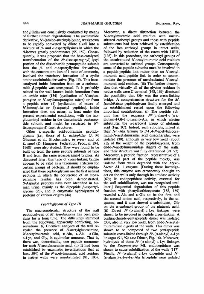
the bridges consist of several Gly or L-amino acidresidues, or both (Fig. 5), or are composed of oneD-amino acid, namely D-isoasparagine (Fig. 6).In type III, the bridges are formed by a "head totail" assembly of several peptides, each havingthe same sequence of amino acids as the peptidesubunit (Fig. 7). Two types of linkages, N'(D-alanyl)-L-Lys and D-alanyl-L-Ala, are involved inthe binding between the peptide subunits. In typeIV, in a few bacteria, the bridges which cross-link the peptide subunits extend from D-Glu ofone peptide subunit to the C-terminal D-Alacarboxyl group of another peptide subunit. Thesepeptide bridges are composed of one diamino acidresidue, D-Orn in the case of C. poinsettiae andsome other plant pathogenic Corynebacteria(115; Fig. 8), D-Lys and D-Orn in the case ofButyribacterium rettgeri (M. Guinand et al., Bio-chemistry, in press; Fig. 9). As shown in Fig. 8 and9, the D-Lys or the D-Orn residues are linkedthrough their e- or 5-amino groups, respectively,to the a-carboxyl group of glutamic acid, andthrough their a-amino groups to the carboxyl ter-minal D-Ala. A similar type of bridging probablyoccurs in Microbacterium lacticum (145), in whichcase a dipeptide Na-(Gly)-Lys would extend fromthe threo-3-hydroxyglutamic acid residue of onepeptide subunit to the D-Ala residue of anotherpeptide subunit.
ENZYMES THAT DEGRADE BACTERIAL PEPTIDO-GLYCANS: NATURE OF THE HYDROLYZED
LINKAGES
The establishment of the peptidoglycan struc-ture has necessitated the development of accurateanalytical and fractionation techniques and thediscovery of a series of different hydrolytic agentsof high specificity. Within the past 10 years,numerous bacteriolytic enzymes have beenisolated, which have proven to be just such tools.A description of these techniques and a survey ofthe lytic enzymes so far discovered and charac-terized have been given recently (41). The follow-ing enzymes, selected for their specific actionupon critical linkages, were particularly useful indismantling the bacterial peptidoglycans intomeaningful fragments (Table 1).
Endo-N-AcetylmuramidasesThese enzymes, such as egg-white lysozyme
(27), Streptomyces 32 (34, 36, 37) or F1 enzymes(21, 100), and Chalaropsis B enzyme (46, 47, 172),hydrolyze the glycosidic linkages between N-acetylmuramic acid and N-acetylglucosamine(Fig. 2), releasing fragments with N-acetyl-muramic acid residues at the reducing end.
430 BACTERIOL. REV.

BACTERIOLYTIC ENZYMES
TABLE 1. Enzymes that degrade bacterial peptidoglycansa
Susceptible linkagesEnzymes
Nature Location
Endo-N-MurNAcases 13-1,4-MurNAc-GINAc Link a, Fig. 2 GlycanEndo-N-GlNAcases 13-1,4-GINAc-MurNAc Link b, Fig. 2
Streptomyces amidase and D-Lactyl-L-Ala Link c, Fig. 2Myxobacter AL 1 protease D-Lactyl-L-Ser Link 2, Fig. 9
KM endopeptidase D-Ala-(D)-meso-DAP Fig. 4, Type I At the C-termi-nus of the pep-tide subunit
N _(D-Ala)-(D)-Lys Link 1, Fig. 9, Type IVN"-(D-Ala)-(D)-Orn Link 1, Fig. 9, Type IV
SA endopeptidase and Myxo- D-Ala-Gly Fig. 5, Type IIbacter AL 1 protease D-Ala-L-Ala Fig. 5, Type II
D-Ala-D-isoAsN Fig. 6, Type IIML endopeptidase NE-(D-Ala)-L-Lys Link 2, Fig. 7, Type IIIMyxobacter AL 1 protease D-Ala-L-Ala Link 1, Fig. 7, Type III
MR endopeptidase and Myxo- L-Ala-L-Ala and L-Ala- Fig. 5, (c,d), Type II In the interior ofbacter AL 1 protease L-Thr the peptide
bridgeLysostaphin and Myxobacter Gly-Gly Fig. 5, (e), Type IIAL 1 protease
a Endo-N-MurNAcase = endo-N-acetylmuramidase; Endo-N-GlNAcase = endo-N-acetylglucosami-nidase; MurNAc = N-acetylmuramic acid; GINAc = N-acetylglucosamine.
Type II. )(a): A. crystallopoietes; (b): 5 pyogenes;(c): M. roseus T4 - (d): M. roseus R 27 ; (e). S. aureus Copenhagen.
---G-M-G - -
L - Ala-D -
L-Ala-.D-Glu5NH2(3) XLL-Lys-aD-Ala--
(3r(-Gly-Gly-oGly-Gly-Glj~
Glu°~NH2 1rL-Ala-L-Ala-L- Ala-L-Thr(d) riLL-ys-D -la-_L-Ala--L-Ala--L-Ala(c) k
L- Lys-~D-Ala-.~
L-Ala-L A'a(b)(1 ) L-Ala(a)
'L
tttA
(1) SA endop
(2)
(2): Aminopeptidase (3) Myxobacter ALI
FIG. 5. Peptidoglycan type II. Peptide bridges composed of glycine or L-amino acid residues cross-linking twopeptide subunits B (Fig. 3). (1), Site of action of the SA endopeptidase; (2), site of action of aminopeptidase; (3),site of action of Myxobacter AL I endopeptidase. Walls ofStaphylococcus epidermidis belong to the same type II.The bridges are formed of Gly and L-Ser residues (D. J. Tipper, Federation Proc., p. 294, 1968). L-Ser is non-randomly located. Four kinds of bridges in the following proportions have been shown to occur: Gly-Gly-Gly-Gly--Gly (20%); Gly-Gly-L-Ser-Gly-Gly (55%); Gly-L-Ser-Gly-Gly-Gly (10%); and L-Ser-Gly-L-Ser-Gly-Gly (15%).
431VOL. 32, 1968

JEAN-MARIE GHUYSEN
Type II. S. faecalis * L.acidophilus . L. casei
_ - - G-M-G - - -
L-Ala-.D-GluZNH2gL.L-Lys-.D-AIa---L- Ala .D-OluG.NH2 I,.,..'P.....................
tLoL-Lys.D-A1a a. D-Asp .NH2
SA. endop.Myxo. ALI endop.
FIG. 6. Peptidoglycan type I. A D-isoasparaginylbridge cross-linking two peptide subunits B (Fig. 3).Arrow: site of action o0 the SA and Myxobacter AL Iendopeptidases.
Endo-N-AcetylglucosaminidasesThese enzymes, such as streptococcal muralysin
(3) and the glycosidase in lysostaphin (170),hydrolyze the glycosidic bonds between N-acetylglucosamine and N-acetylmuramic acid(Fig. 2), releasing fragments with N-acetyl-glucosamine at the reducing end.
Streptomyces N-Acetylmuramyl-L-AlanineAmidase
This enzyme (J. M. Ghuysen et al.,Biochemistry,in press; 34) hydrolyzes the bonds between theD-lactic acid residues of the glycan strands andthe N-terminal residues of the peptide subunits(Fig. 2, 7, 9). The N-terminal amino acid isusually L-Ala; thus, the enzyme is designated asN-acetylmuramyl-L-Ala amidase. However, thisamidase is also able to cleave other linkages atidentical locations in the peptidoglycan, such asthe N-acetylmuramyl-L-Ser linkages in walls ofB. rettgeri (Fig. 9). This enzyme has no significantactivity on intact peptidoglycans. To be func-
Type IV. C. poinsettiae; C. flaccumfaciens; C. betae
xGy-'D-Glu---2, ~~~~~~~~~~~~...............
D LHomoSer--D-Ala -D-Orn:.......Gly-D-Glu °
LHomoSer-D-Ala - - - -
FIG. 8. Peptidoglycan type IV. A D-ornithine bridgecross-linking two peptide subunits D (Fig. 3). Notethe absence of diamino acid in the peptide subunit.
tional, it requires that the glycan first be split.Thus, this amidase has no bacteriolytic action.
Streptomyces KM EndopeptidaseThisenzyme (J. M. Ghuysen et al., Biochemistry
in press) has been used to hydrolyze the cross-peptide linkages which serve as bridges betweenpeptide subunits in the meso-DAP-containingpeptidoglycans of E. coli and B. megaterium KM[J. Van Heijenoort et al., Biochemistry, in press;9 (Peptidoglycan type 1, Fig. 4)]. The bondsspecifically hydrolyzed are thus D-alanyl (D)-meso-DAP linkages. This enzyme also hydro-lyzes the Na-(D-alanyl)-D-Lys and Na-(D-alanyl)-D-Orn linkages in walls of B. rettgeri (M. Guin-and et al., Biochemistry, in press; Peptidoglycantype IV, Fig. 9). An endopeptidase whose speci-ficity must be identical to that of the StreptomycesKM endopeptidase is present in the autolyticsystem of E. coli (177).
Type III. M. lysodeikticus S. lutea M. flavus. M. citreus.
-- - G-M-G-M-G-M-G-M-G-M- --
(l--or (3;*-D ---G-M-G---(1) or (3) -e- ---(1)orL-o-_---o Lee-()'L 0-0_-e-£e o
'1) t ~L @-o- (2)*-)Q L-Aba--D-Glu-%Gs 1(1L@-° 9L L-Lys -D-Ala
(1): Myxobacter ALI (2): ML endop. (3): Streptomyces Amidase
FIG. 7. Peptidoglycan type III. Assembling ofpeptide subunits C (Fig. 3) in some Micrococcaceae. (1), Site ofaction of Myxobacter AL I endopeptidase (hydrolysis of D-alanyl-L-Ala linkages and of N-acetylmuramyl-L-Alalinkages); (2), site of action ofML endopeptidase (hydrolysis ofNe-(D-alanyl)-L-Lys linkages); (3), site of action-ofStreptomyces N-acetylmuramyl-L-Ala amidase.
432 BACTERIOL. REV.

BACTERIOLYTIC ENZYMES
Type IV B. rettgeri
---G-M-G--- (2)1 --
L- Ser-D-Glu -- - (1) ;1I :, D-Lys:iLKL-Orn-.D-AtaL-. or
D-Ornj
-- -G-M -G ---
L- Ser D-Glu
6L.L-Orn-.D-Ala(1) KM endop. (2) Streptomyces AmidaseFIG. 9. Peptidoglycan type IV. Two peptide subunits
E (Fig. 3) cross-linked by a D-Lys or a D-Orn bridge.Note that the diamino acid L-Orn in the peptide subunitis not used for peptide cross-linking. Arrows: (1) siteof action of the KM endopeptidase; (2) site of actionof Streptomyces N-acetylmuramyl-L-Ala amidase.In Butyribacterium rettgeri peptidoglycan, D-lys andiD-Orn bridges occur in the ratio 2:1.
Streptomyces SA EndopeptidaseThis enzyme acts on type II peptidoglycans
(32, 33, 39, 99, 117; Fig. 5, 6). It hydrolyzes thelinkages at the N termini of the peptide bridgesand at the C-terminus of the peptide subunits.Sensitive linkages are, for example, D-alanyl-Gly, D-alanyl-L-Ala; D-alanyl-D-iso-asparogine.When opened at their N termini, the peptidebridges which contain glycine or L-amino acidresidues, or both (Fig. 5), can be further de-graded (117) by an amino peptidase such as thatsecreted by Streptomyces strains (J. M. Ghuysenet al., Biochemistry, in press). As a result, theamino acids are sequentially liberated until theE-amino groups of lysine in the peptide subunits,to which the peptide bridges are linked at theirC termini, are exposed (Fig. 5).
Streptomyces ML EndopeptidaseThis enzyme is active on type III peptidoglycans
(Fig. 7; J. Campbell et al., Biochemistry, in press;31, 117). It specifically hydrolyzes NE-(D-alanyl)-L-Lysine linkages (link 2, Fig. 7), which serve aslinking groups between peptide subunits.
Myxobacter AL I ProteaseIn contrast to Streptomyces KM, SA, and ML
endopeptidases, all of which have restrictedspecificities and are, for example, unable todegrade casein, Myxobacter AL 1 enzyme (24) is apowerful protease, active on casein, and itpossesses a broad bacteriolytic spectrum. It has
several sites of action on the cell wall peptide.(i) Investigations of the mode of action of thisenzyme upon Staphylococcus aureus (59, 17)have revealed that it catalyzes hydrolysis of twolinkages within the peptide moiety (Fig. 5). N-terminal Gly and both COOH-terminal D-Alaand COOH-terminal Gly are liberated by thishydrolysis. The pentaglycine bridges are thusattacked at internal glycyl-Gly linkages as well asat their linkages to the D-alanyl termini of thepeptide subunits (links 3, Fig. 5). (ii) MyxobacterAL 1 enzyme, acting upon walls of Arthrobactercrystallopoietes (74, 75), M. lysodeikticus (31),and other Micrococcaceae (J. Campbell et al.,Biochemistry, in press), hydrolyzes D-alanyl-L-Ala linkages in bridges of type II, as does the SAendopeptidase (Fig. 5), and the "head to tail"D-alanyl-L-Ala linkages in the type III bridges(link 1, Fig. 7). (iii) Moreover, Myxobacter AL1 enzyme is capable of hydrolyzing D-alanyl-D-isoAsN linkages (Fig. 6), as does the SA endo-peptidase, and it has been used to study thepeptidoglycan in L. casei (D. Hungerer, Feder-ation Proc., p. 294, 1968). (iv) Finally, in allcases so far studied, Myxobacter AL 1 enzymealso hydrolyzes N-acetylmuramyl-L-Ala linkages(Fig. 2) at a slow but detectable rate (31, 71, 75,171). In this respect, the Myxobacter AL 1enzyme has a very useful characteristic: it dis-plays amidase activity on substrates with intactglycan chains. Thus, this enzyme provides a wayof isolating the polysaccharide moiety free of itssubstituent peptide, but retaining its in vivodegree of polymerization.
Streptomyces MR Endopeptidase andLysostaphin Endopeptidase
Lysostaphin endopeptidase (11, 170) is knownto split glycyl-Gly linkages at several placeswithin the pentaglycine bridges (type Hie, Fig. 5).Since this structure is limited to the Staphylococci,lysostaphin endopeptidase is specifically staph-ylolytic. Streptomyces MR endopeptidasedisrupts types I1c and Ild bridges (117; Fig. 5).It hydrolyzes the L-alanyl-L-Thr linkages in M.roseus R 27 and, at a slower rate, the L-alanyl-L-Ala linkages in M. roseus (Thr-).
Peptidase Preparations with Mixed ActivitiesThe Streptomyces L3 enzyme preparation,
when acting upon walls of C. diphtheriae (J. M.Ghuysen et al., Federation Proc., p. 410, 1966;65, 72, 96, 97), is mainly a bridge-splitting enzymewhich catalyzes the hydrolysis of D-alanyl-meso-DAP. In this latter case, however, it is not knownwhich amino group of meso-DAP of one peptidesubunit is involved in the linkage to D-Ala of
433VOL. 32) 1968

JEAN-MARIE GHUYSEN
another peptide subunit. The L3 enzyme prepara-tion also contains (i) N-acetylmuramyl-L-Alaamidase activity, (ii) an enzyme catalyzing thehydrolysis of an amide that substitutes one of thecarboxyl groups of meso-DAP, and (iii) possibly,a D-Ala carboxypeptidase.The Flavobacterium L-11 enzyme (65, 66) and
the Staphylococcus epidermidis ALE enzyme (158)exert their lytic actions upon walls of S. aureusthrough the activities of glycyl-Gly endopeptidase,N-acetylmuramyl-L-Ala amidase, and D-alanyl-Gly endopeptidase (in the case of the L-11enzyme).As observed with Myxobacter AL 1 enzyme,
the above L-3, L-11, and ALE enzyme prepara-tions hydrolyze more than one type of linkage.From the data published to date, it seems likelythat several enzymes are present in the L-3, L-11,and ALE preparations as they are obtained.
SummationThe foregoing enzymes permit the specific
hydrolysis of many important linkages in thewall peptidoglycans (Table 1). Endo-N-acetyl-muramidases and endo-N-acetylglucosaminidasesact on the glycan chains. N-acetyl muramyl-L-Alaamidases hydrolyze linkages at the junction be-tween the glycan and the peptide moieties, i.e.,linkages that are located at the N termini of thepeptide subunits. The endopeptidases may begrouped into three main types: (i) those whichhydrolyze various linkages, all of which involvethe C-terminal D-Ala of the peptide subunits;(with the exception of the following examples, allthe known endopeptidases fall in this group);(ii) Myxobacter AL 1 which, in some bacterialwalls, hydrolyzes linkages involving both C andN termini of the peptide subunits (Fig. 7); and(iii) lysostaphin and MR endopeptidase, whichhydrolyze peptide internal bonds of the peptidebridges involving neither the C nor the N terminiof the peptide subunits. With some bacterial walls(for example, those of S. aureus), MyxobacterAL 1 also has this latter type of activity (Fig. 5).Streptomyces sp. appear to be a very interestingsource of various peptidases active on bacterialpeptidoglycans. Streptomyces aminopeptidase(link 2, Fig. 5) and the Streptomyces KM, SA,ML, and MR endopeptidases are basic proteins.They have been purified and separated from eachother by chromatography on CM-cellulose, andtheir specific activities upon soluble, well-defined,bacterial wall degraded compounds have beenstudied (J. M. Ghuysen et al., Biochemistry, inpress).
STRUCTURE OF SEVERAL BACTERIAL PEPTIDO-GLYCANS AS REVEALED BY ENZYMATICDEGRADATIONS: GLYCAN MOIETY
The studies to be reported here deal with thewall peptidoglycan of some gram-positivebacteria. Intact cell walls were obtained bymechanical disruption of the cells followed bydifferential centrifugation. However, these wallpreparations were usually treated with trypsin toremove cytoplasmic contaminants and, in someinstances, protein constituents of the walls them-selves. The walls were not subjected to any chem-ical agent such as trichloroacetic acid or hotformamide. Although these treatments are widelyused and yield enriched peptidoglycan prepara-tions by removing nonstructural wall components(other polysaccharides and teichoic acids), theyalso cause random cleavage of some covalentlinkages within the peptidoglycan, as has beenshown by end group analysis. In addition, variouschemical modifications, such as formylation offree amino groups, can occur when hot formamideis used (114). Moreover, chemically strippedpeptidoglycans are not capable of giving anyinformation about the way in which the variousconstitutive polymers are held together within thebacterial walls, a question that ultimately must beresolved.
Staphylococcus aureus
Three types of glycan degradation procedurewere carried out with (i) endo-N-acetylmur-amidase (Chalaropsis B enzyme; Streptomyces 32or F1 enzymes), (ii) endo-N-acetylglucosaminidase(from lysostaphin), and (iii) Myxobacter AL 1enzyme. In the first procedure, after solubilizationof the wall by the N-acetylmuramidase (36,37), teichoic acids were removed on Ecteolacellulose at pH 5. The peptide substituentswere detached from the glycan fragments byStreptomyces N-acetylmuramyl-L-Ala amidase(Fig. 2), and the liberated peptides were removedon CM cellulose at pH 6.4. The resulting freeglycan fragments were separated into two com-ponents by preparative paper chromatographyfollowed by gel filtration. They were charac-terized (167) as being Disaccharide 1 (/3-1,4-N-acetylglucosaminyl-N-acetylmuramic acid) andDisaccharide II [/3-1 ,4-N-acetylglucosaminyl-N,6-O-diacetylmuramic acid; (Fig. 10)].
In the second procedure, a similar degradationsequence (170) yielded two disaccharides isomericwith Disaccharides I and II, Disaccharide III:(,B-1 ,4-N-acetylmuramyl-N-acetylglucosamine)
434 BACTERIOL. REV.

BACTERIOLYTIC ENZYMES
Disaccharide I
CH2OH H20H
0OH OH HHHH
H NHCOCH3 H OCH3
CH3-CH-C00H
Disaccharide III
Disaccharide II
CH20H CH O-CO-CH30 ~~~~0
H H OH
0
OHOH H HH H
H NHCOCH 0 H NHCOCH3 1 3
CH3-CH -COOH
Disaccharide IV
3 3I'A31--CH3-CH-COOH CH3-CH-COOHFIG.V10. Structure of disaccharides isolated from the peptidoglycan of Staphylococcus aureus Copenhagen.
Disaccharide I and disaccharide III have also been obtainedfrom M. lysodeikticus. Disaccharide I: N-acetylglu-cosaminyl-,f-1 ,4-N-acetylmuramic acid. Disaccharide II: N-acetylglucosaminyl-,3-1 ,4-N, 6-0-diacetylmuramicacid. Disaccharide III: N-acetylmuramyl-,6-1,4-N-acetylglucosamine. Disaccharide IV: N, 6-0-diacetylmuramyl-B-I ,4-N-acetylglucosamine.
and Disaccharide IV [,/-1,4-N-,6-0-diacetyl-muramyl-N-acetylglucosamine (Fig. 10)]. Theobserved yields of the disaccharides obtained bythe various degradation procedures were con-sistent with a wall structure in which the glycanmoiety is composed of linear strands of fl-1,4-linked N-acetylglucosamine pyranoside residues,(i.e., a chitin-like structure). Unlike chitin,however, every other sugar is substituted by a3-0-D-lactyl group. In S. aureus, about 50% ofthe N-acetylmuramic acid residues have a 6-0-acetyl group, but whether the distribution of thissubstituent is regular, random, or localized is notyet known. Since glycan degradation yields freedisaccharide units only if it is followed by N-acetylmuramyl-L-Ala amidase treatment, allN-acetylmuramic acid residues must, therefore, besubstituted by peptide subunits. For this reason,the S. aureus peptidoglycan is said to be a "tight"network.
In the third procedure, Myxobacter AL 1
enzyme was used to achieve, in a one-step opera-tion, the opening of the pentaglycine bridges(Fig. 5) and the hydrolysis of the N-acetylmur-amyl-L-Ala amidic linkages, thus yielding freeunaltered glycan strands (171). Appropriatefractionation of the degraded products gave riseto various glycan fractions, some of which werebound to the teichoic acid polymer. Analyses ofthe fractions showed the glycan moiety to bepolydisperse with an average chain length ofabout 12 disaccharide units, but containingchains with as few as 6 and as many as 50 disac-charide units. These figures were based on theestimation of the formaldehydogenic end groupsthat originate from the reducing N-acetyl-hexosamine termini of the glycan chains on re-duction with NaBH4. An average chain length of16 disaccharide units can also be deduced froman estimation of the free N-acetylglucosamineresidues which are liberated from cell walls aftercomplete hydrolysis of the linkages between N-
435VOL. 32, 1968

JEAN-MARIE GHUYSEN
acetylmuramic acid and N-acetylglucosamine bymeans of an endo-N-acetylmuramidase (36).This liberation of free N-acetylglucosamineresidues under these conditions (actual data,32 nmoles/mg of walls or per 500 nm equivalentsof disaccharide units) indicates that N-acetyl-glucosamine residues are located at the reducingends of the intact glycan chains. These reducingN-acetylglucosamine termini in the glycan resultvery probably from an endo-N-acetylglucosamini-dase activity of the staphylococcal autolyticsystem (D. J. Tipper, Bacteriol Proc., p. 48, 1968).
Micrococcus lysodeikticusSimilar techniques of degradation, fractiona-
tion, and characterization showed the M. lyso-deikticus glycan moiety to be identical with that ofS. aureus, with the following exceptions, however.(i) 0-acetyl substitution is absent in all but a fewstrains. (ii) Only 40% of the N-acetylmuramicacid residues are substituted by peptide subunits(81, 100). (iii) A small amount of the muramicacid residues are not N-acetylated (92). (iv) Split-ting of the glycan with the help of either lysozymeor Streptomyces FK endo-N-acetylmuramidase isincomplete and free, unsubstituted glycan frag-ments, from di- to octasaccharides, are produced.Disaccharide I (60, 81, 110, 139, 147), disac-charide III) (81; Fig. 10), and a tetrasaccharide(81; Fig. 11) were isolated in good yields, andtheir structures were thoroughly established. Thelysozyme-catalyzed hydrolysis of isolated walloligosaccharides has been studied in detail (15, 16)and interpreted on the basis of the three-dimen-sional model of lysozyme developed by Philipsand co-workers (5, 6). The results obtained showthat hydrolysis of the isolated tetrasaccharide toyield disaccharides proceeds chiefly via trans-glycosilation, leading to the formation of higheroligosaccharides. Hexa-, octa-, deca- and dodec-asaccharides are readily degraded by lysozyme toyield the corresponding di- and tetrasaccharides.The tetrasaccharide is degraded at a much lowerrate than the higher oligosaccharides because it isbound largely to lysozyme in a nonproductive
manner, which does not lead to bond scission.Owing to the partial peptide substitution of its-glycan moiety, the M. lysodeikticus peptidoglycanis referred to as a "loose" network to contrast itwith that of S. aureus.
Other Bacterial PeptidoglycansDisaccharides have also been isolated after
endo-N-acetylmuramidase degradation of walls orenvelopes of the following bacteria: M. roseus(99, 117), Sarcina lutea (J. Campbell et al., Bio-chemistry, in press), Staphylococcus epidermidisr(D. J. Tipper, Federation Proc., p. 294, 1968),L. acidophilus (J. Coyette and J. M. Ghuysen,unpublished data), L. casei (D. Hungerer, Fed-eration Proc., p. 294, 1968), Streptococcuspyogenes (98, 99), S. faecalis (149, 151), B.megaterium (9), B. licheniformis (93), Butyri-bacterium rettgeri (M. Guinand et al., Biochem-istry, in press), and E. coli (J. van Heijenoort etal., Biochemistry, in press). None of these disac-charides was submitted to the same exhaustivestructural investigations as were those of Staphy-lococcus aureus and M. lysodeikticus, but analyseswere made which provided the following data.(i) N-acetyglucosamine and N-acetylmuramicacid were the only two monosaccharides present.(ii) Acid hydrolysis, followed by quantitation ofglucosamine using the yeast D-glucosamine 6-phosphate N-acetylase, revealed that half of the:hexosamine residues were glucosamine. (iii)!Reduction of the disaccharide with NaBH4 de-stroyed all of the muramic acid, half of the totalhexosamines, and none of the glucosamine, thus.establishing that muramic acid is at the reducingend of the disaccharide. (iv) Susceptibility topig epididymis exo-j3-N-acetylglucosaminidase,which is specific for ,B glycosidic linkages, es-tablished the (3-anomery of the link. (v) Deter-mination of the molar extinction coefficient of thedisaccharide with the Morgan-Elson reaction(147) established the glycosidic linkage to be1:4, not 1:6. Hence the linkage (8-1,4 of thedisaccharide (disaccharide I, Fig. 10) has beenwell characterized in several cases. Consequently,
CH2OH CH2OH CH2OH CH2OH
OH H 0 H0 H OOH H 0 H
H NHAc H NHAc H NHAM H NHAC
CH-CH-COOH CH- CH-COOH3
FIG. 11. Structure of a tetrasaccharide isolated from the peptidoglycan of M. lysodeikticus:N-acetylgluco-saminyl-,f-1 ,4-N-acetylmuramyl-f-1 ,4-N-acetylglucosaminyl-g3-1 ,4-N-acetylmuramic acid.
436 BACr13RoL. REV.-
BACTERIOLYTIC ENZYMES
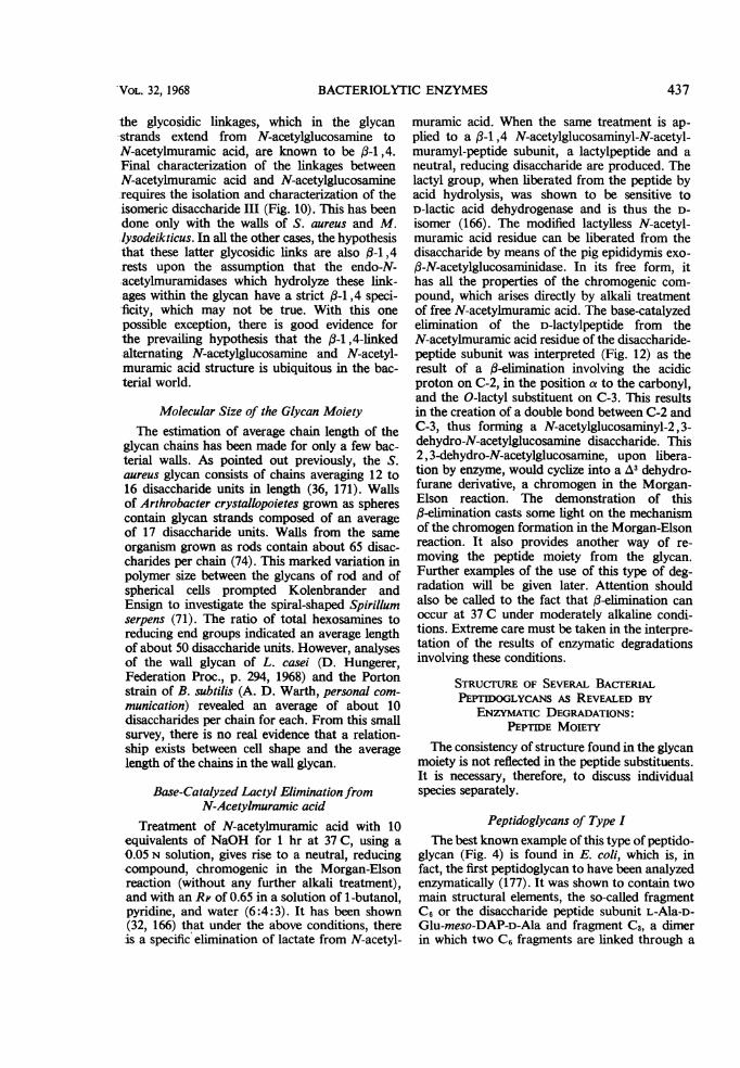
-the glycosidic linkages, which in the glycanstands extend from N-acetylglucosamine toN-acetylmuramic acid, are known to be p3-1,4.Final characterization of the linkages betweenN-acetylmuramic acid and N-acetylglucosaminerequires the isolation and characterization of theisomeric disaccharide III (Fig. 10). This has beendone only with the walls of S. aureus and M..lysodeikticus. In all the other cases, the hypothesisthat these latter glycosidic links are also p3-1 ,4rests upon the assumption that the endo-N-acetylmuramidases which hydrolyze these link-ages within the glycan have a strict (3-1,4 speci-ficity, which may not be true. With this onepossible exception, there is good evidence for-the prevailing hypothesis that the ,8-1,4-linkedalternating N-acetylglucosamine and N-acetyl-muramic acid structure is ubiquitous in the bac-terial world.
Molecular Size of the Glycan MoietyThe estimation of average chain length of the
glycan chains has been made for only a few bac-terial walls. As pointed out previously, the S.aureus glycan consists of chains averaging 12 to16 disaccharide units in length (36, 171). Wallsof Arthrobacter crystallopoietes grown as spherescontain glycan strands composed of an averageof 17 disaccharide units. Walls from the sameorganism grown as rods contain about 65 disac-charides per chain (74). This marked variation inpolymer size between the glycans of rod and ofspherical cells prompted Kolenbrander andEnsign to investigate the spiral-shaped Spirillumserpens (71). The ratio of total hexosamines toreducing end groups indicated an average lengthof about 50 disaccharide units. However, analysesof the wall glycan of L. casei (D. Hungerer,Federation Proc., p. 294, 1968) and the Portonstrain of B. subtilis (A. D. Warth, personal com-munication) revealed an average of about 10disaccharides per chain for each. From this smallsurvey, there is no real evidence that a relation-ship exists between cell shape and the averagelength of the chains in the wall glycan.
Base-Catalyzed Lactyl Elimination fromN-Acetylmuramic acid
Treatment of N-acetylmuramic acid with 10equivalents of NaOH for 1 hr at 37 C, using a0.05 N solution, gives rise to a neutral, reducingcompound, chromogenic in the Morgan-Elsonreaction (without any further alkali treatment),and with an RF of 0.65 in a solution of 1-butanol,pyridine, and water (6:4:3). It has been shown(32, 166) that under the above conditions, thereis a specific elimination of lactate from N-acetyl-
muramic acid. When the same treatment is ap-plied to a f3-1,4 N-acetylglucosaminyl-N-acetyl-muramyl-peptide subunit, a lactylpeptide and aneutral, reducing disaccharide are produced. Thelactyl group, when liberated from the peptide byacid hydrolysis, was shown to be sensitive toD-lactic acid dehydrogenase and is thus the D-isomer (166). The modified lactylless N-acetyl-muramic acid residue can be liberated from thedisaccharide by means of the pig epididymis exo-,B-N-acetylglucosaminidase. In its free form, ithas all the properties of the chromogenic com-pound, which arises directly by alkali treatmentof free N-acetylmuramic acid. The base-catalyzedelimination of the D-lactylpeptide from theN-acetylmuramic acid residue of the disaccharide-peptide subunit was interpreted (Fig. 12) as theresult of a (-elimination involving the acidicproton on C-2, in the position a to the carbonyl,and the O-lactyl substituent on C-3. This resultsin the creation of a double bond between C-2 andC-3, thus forming a N-acetylglucosaminyl-2,3-dehydro-N-acetylglucosamine disaccharide. This2, 3-dehydro-N-acetylglucosamine, upon libera-tion by enzyme, would cyclize into a A3 dehydro-furane derivative, a chromogen in the Morgan-Elson reaction. The demonstration of this(3-elimination casts some light on the mechanismof the chromogen formation in the Morgan-Elsonreaction. It also provides another way of re-moving the peptide moiety from the glycan.Further examples of the use of this type of deg-radation will be given later. Attention shouldalso be called to the fact that (-elimination canoccur at 37 C under moderately alkaline condi-tions. Extreme care must be taken in the interpre-tation of the results of enzymatic degradationsinvolving these conditions.
STRUCTURE OF SEVERAL BACTERIALPEPTIDOGLYCANS As REVEALED BY
ENZYMATIC DEGRADATIONS:PEPTIDE MOIETY
The consistency of structure found in the glycanmoiety is not reflected in the peptide substituents.It is necessary, therefore, to discuss individualspecies separately.
Peptidoglycans of Type IThe best known example of this type of peptido-
glycan (Fig. 4) is found in E. coli, which is, infact, the first peptidoglycan to have been analyzedenzymatically (177). It was shown to contain twomain structural elements, the so-called fragmentC6 or the disaccharide peptide subunit L-Ala-D-Glu-meso-DAP-D-Ala and fragment C3, a dimerin which two C6 fragments are linked through a
437'VOL. 32, 1968

JEAN-MARIE GHUYSEN
NHc - NHc
1 R-0 C-H,/1 \ ~~0 7 0.05 N NaOH
K H H-C oHO\Ht | ~~~~~370;lh
NHAc H - C -OH
AC H20H
-O-R + NHAc
CH20H H-C ',Occ'H6 ~~~~H-C
OH O -C7HO \J~H I
CHAc H-C-OH
IB CH2OH
CH20H0 OH
OH
HO XNHAc
C
K NHAc %
' H-C"~ -.C'II %HC>sC"
+ H-C-OH
H-C-OH
CH20H\D /
D
NHAc
IkH-C LCK/
I I_ OH
W[C -O
OH IH-C-OH
CH20H
Chromogen I
NHAc
k H-C C-H
borale 11 I
100', 7min IH-C-OH
ICH20H
Chromogen III
FIG. 12. ,3-Elimination of D-lactyl-peptide from disaccharide peptide. R, D-lactyl peptide residue; A, N-acetyl-glucosaminyl-j9-1,4-N-acetylmuramyl-peptide subunit; B, Prochromogen glycoside:N-acetylglucosaminyl-,6-1,4-2,3-dehydro-N-acetylglucosamine; Exo-,6-GkcNAcase:exo-13-N-acetylglucosaminidase; C and D, free N-acetyl-glucosamine andfree 2,3-dehydro-N-acetylglucosamine, respectively. During the course of its enzymatic liberation,2 ,3-dehydro-N-acetylglucosamine would cyclize into a A3-dehydrofuran derivative or chromogen L Transformationof chromogen I into a furan derivative such as chromogen III would result from further treatment with borate at100 C (32; figure reprinted by permission).
peptide linkage in which one amino group ofmeso-DAP is engaged. This pioneering work ofWeidel and his colleagues (177) contributedgreatly to our present concept of the wall peptido-glycan as an enormous, net-like bag-shapedmacromolecule. The E. coli autolytic system (109),which contains at least five enzymes (endopepti-dase, N-acetylmuramyl-L-Ala amidase, endo-N-acetylmuramidase, D-Ala carboxypeptidase, andexo-13-N-acetylglucosaminidase) specifically de-grading the wall peptidoglycan into smallfragments, as well as the demonstration of thegeneral structure assigned to the original peptido-glycan, have been described in several reviews(38, 41, 85, 156, 177). In this peptidoglycan,virtually all of the disaccharide units in the glycanchains are substituted by peptide subunits.Some of the peptide subunits occur as tripeptidesL-Ala-meso-DAP-D-Glu, the last D-Ala residuebeing lost as a result of the action of the autolyticD-Ala carboxypeptidase. The carboxyl groups ofGlu and of meso-DAP, which are not engagedin peptide linkages, have no amide substituents.
About 50% of the peptide subunits are cross-linked to form peptide dimers, the disaccharideunits from two adjacent glycan chains thus beingpaired (161). It is probable that there is a ran-dom distribution of the disaccharide peptide sub-unit and of its dimer throughout at least a majorpart of the network (Fig. 13). Only recently (J.van Heijenoort et al., Biochemistry, in press; 9,22) has a complete structure been assigned to thepeptidoglycan of E. coli, as well as to other meso-
DAP peptidoglycans from gram-positive bac-teria, such as that of B. megaterium KM. TheB. megaterium walls also present a low degree ofpeptide cross-linking. About 85% of the totalDAP residues are meso. Most of these peptidesoccur as uncross-linked tetrapeptide (L-Ala-D-Glu-meso-DAP-D-Ala) and tripeptide (L-Ala-D-Glu-meso-DAP) subunits. About 15% of thetotal DAP residues in these walls are DD. None ofthese latter residues exhibits free amino groups
(9).Characterization of the NH2-(L)-meso-DAP
in the link to glutamic acid. Making use of the
exo-q-GlcNAcase
37°, pH 4 a h
438 BACTERIOL. REV.-

BACrERIOLYTIC ENZYMES
M M M M
G | G/ GIM ML..UM M
G/ G G / I
MU-t.M MUz....MG( G G/ G/
/ / / /M M M M
G|G | G | G I
G I G/ G// / / /
MU zMU MM-L..M M-z-M/ / / /G G( G GFIG. 13. Schematic representation of a peptido-
glycan sheet ofE. coli. In this loose network, all oftheN-acetylmuramic acid residues are substituted eitherby peptide monomers (peptide subunits A; Fig. 3) orby peptide diners (Fig. 4).
fact that dinitrophenylation of amino acids oftenenhances their optical rotation, Diringer andJusic (22) determined the configuration of thatasymmetric carbon of the meso-DAP residuewhich, in the E. coil peptide subunit, has a freeamino group. The mono-dinitrophenyl (DNP)-meso-DAP obtained after dinitrophenylation andacid hydrolysis of the peptide subunit presented amolar optical rotation, [M]D, in glacial aceticacid equal to + 250 deg a 10%. Since syntheticdi-DNP-LL-DAP and synthetic mono-DNP-LL-DAP have [MmD equal to -444 and -231 deg,respectively (61), the above value of +250 degprovided evidence that, in the E. coli peptidesubunit, the free amino group of the meso-DAPresidues was on the asymmetric carbon having theD configuration. The same conclusion was reachedfor the meso-DAP subunits in B. megaterium KM(9). A disaccharide peptide subunit fraction (withno amide substituent, as in the case of E. coli),representing a major part of the total meso-DAPpeptide subunits, was isolated. After dinitro-phenylation and acid hydrolysis, the isolatedmonodinitrophenyl derivative of the meso-DAPresidues exhibited an [MID equal to +248 deg,i.e., a value identical, within the limits of experi-mental error, to that observed with the mono-DNP-DAP isolated from E. coil and to that ofthe synthetic mono-DNP-(D)-meso-DAP (9).Moreover, both the synthetic mono-DNP-(D)-meso-DAP and the natural mono-DNP-meso-DAP exhibited the same anomalous rotatorydispersions with a Cotton effect centered on 418
nm and, in the region 450 to 600 nm, both fol-lowed the simplified Drude equation with identicalK coefficients. These determinations proved thatin E. coil and in B. megaterium, the amino groupengaged in peptide linkage to glutamic acid islocated on the L-carbon of meso-DAP and,consequently, that the amino group used forcross-linking between peptide subunits must belocated on the D-carbon of meso-DAP.
Characterization of the 'y-carboxyl group ofglutamic acid in the link to NH2-(L)-meso-DAP.The tripeptide monomers L-Ala-D-Glu-(L)-meso-DAP were isolated from walls of both E. coliand B. megaterium KM. They were chroma-tographically and electrophoretically indis-tinguishable and inseparable from synthetictripeptide L-Ala-,y-D-Glu-(L)-meso-DAP underconditions which readily distinguish betweenthe two synthetic peptide isomers containingeither a- or 7-linked glutamic acid (J. vanHeijenoort et al., Biochemistry, in press). Char-acterization of the -y-monohydrazide of glutamicacid and the absence of the a derivative amongthe compounds arising by hydrazinolysis of thepeptide subunits of E. coli and B. megateriumKM also led to the conclusion that glutamic acidis linked to meso-DAP via the y-carboxyl group.An identical conclusion was independentlyreached by Diringer (21a) by hydrazinolysis ofthe E. coll monomer.
Characterization of the link meso-DAP-(L)-(D)-Ala. The tetrapeptide monomers with thesequence L-Ala-y-D-Glu-(L)-meso-DAP-D-Alawere also isolated from E. coil and from B.megaterium KM (J. Van Heijenoort et al., Bio-chemistry, in press). The location of the terminalD-Ala on the L-carbon of meso-DAP was provedby an Edman degradation. After one cycle ofthe degradation, N-terminal alanine was re-placed by N-terminal glutamic acid, thus demon-strating the sequence Ala-Glu. Concomitantly,most of the mono-N-terminal DAP groups dis-appeared, and no free alanine was liberated.Thus, the terminal D-Ala cannot be in a positionrelative to the free amino group, which, as shownabove, is on the D-carbon of meso-DAP; conse-quently, the terminal D-Ala must be located onthe L-carbon of meso-DAP. After the secondcycle of the degradation, N-terminal glutamicacid disappeared and was not replaced by anyother terminal amino group. The complete se-quence of the peptide subunit in E. coil and B.megaterium is thus L-Ala-'y-D-Glu-(L)-meso-DAP-(L)-D-Ala as it is shown in Fig. 3A.
Characterization of the D-Ala-(D)-meso-DAPcross-linkages between peptide subunits. Thebisdisaccharide peptide dimer from E. coli, i.e.,
439VOL. 32, 1968

JEAN-MARIE GHUYSEN
fragment C8 (177), was degraded into disac-charide peptide monomers with the help of thepurified Streptomyces KM endopeptidase (J.van Heijenoort et al., Biochemistry, in press).C- and N-terminal group analyses, before andafter degradation, together with the aforemen-tioned demonstration that the amino groupengaged in the link to glutamic acid is on the Lcarbon of meso-DAP, established that D-Ala-(D)-meso-DAP linkages are involved in peptidebridging (Fig. 4).The demonstration of the existence of the
same type of peptide bridging in walls of B.megaterium was attempted (J. van Heijenoortet al., Biochemistry, in press). Complicationsarose, however, due to the fact that about 15%of the total DAP residues are DD. DD-DAP resi-dues were identified as follows. A disaccharide-peptide monomer fraction containing solelymeso-DAP residues and a disaccharide-peptideoligomer fraction were isolated (9). After dinitro-phenylation and acid hydrolysis of the latterfraction, the meso-DAP residues were removedas mono-DNP-derivatives. The remaining DAPresidues were then bisdinitrophenylated andcharacterized as di-DNP-DD-DAP on the basisof the [M]D value equal to +426 deg. In J. vanHeijenoort's more recent study, the walls of B.megaterium were solubilized with the help of apurified KM endopeptidase. N- and C-terminalgroups analyses strongly suggested that the fewmeso-DAP-containing peptide subunits whichare cross-linked are actually engaged in D-Ala-(D)-meso-DAP linkages, as in the case of E. coli.However, the DD-DAP residues appeared to beinvolved in another type of peptide cross-linkage,the significance of which is not clear at the mo-ment. In summary, it has been now well es-tablished that the structure presented in Fig. 3and 4 are valid for the E. coil and for the majorpart of the B. megaterium peptidoglycans. In thelatter case, however, it is probable that D-Ala-(D)-meso-DAP peptide cross-linkages are not theonly important ones. The two tripeptides, L-Ala-Ty-D-Glu-(?)-meso-DAP and L-Ala-D-iso-gluta-minyl-(?)-meso-DAP, have been characterized(93) in walls of B. ficheniformis ATCC 9945(previously designated B. subtilis ATCC 9945).Finally, it has been reported recently (A. D.Warth and D. L. Strominger, Bacteriol. Proc.,p. 64, 1968) that the peptide subunits and thepeptide cross-linkages in the vegetative cell walland the spore cortex of B. subtilis have structuresidentical to those found in E. coli with the ex-ception, however, that in vegetative cells of B.subtilis, most of the peptide subunits have anamide substituent on the carboxyl group located
on the D-carbon of meso-DAP. Finally, in theC. diphtheriae peptidoglycan, both the D-Glu andthe meso-DAP residues are amide substituted(67).
Peptidoglycans of Type II with Peptide Bridges ofGlycine or L-Amino Acid Residues, or Both
The following sequential degradation has per-mitted the characterization of the peptide bridgesand the peptide subunits of this group of bacterialpeptidoglycans (99, 117). (Degradation of thewalls of M. roseus is given as an example in Fig.14.) The first step involves solubilization of thewalls by the SA endopeptidase. Equivalentamounts of C-terminal D-Ala, in all cases, andof N-terminal amino groups of either Gly orL-Ala, depending on the bacterium (Fig. 5), arereleased as a result of the opening of the peptidebridges at their N termini (hydrolysis of links1, Fig. 5, 14). The second step involves amino-peptidase degradation of the opened peptidebridges until the e-amino group of lysine of thepeptide subunits, to which the bridges are at-tached, are all exposed (hydrolysis of links 2,Fig. 5, 14). Quantitation of the number of freeamino acids liberated, per lysine residue, at theend of the process, and kinetics of the degradationpermitted the determination of the bridge se-quences. In the case of M. roseus R 27 cell walls,for example (Fig. 5, 14), first three L-Ala residuesand next, one L-Thr residue per lysine residuewere sequentially liberated. It was also observedthat the exposure of the e-amino group of lysineparalleled both the disappearance of N-terminalthreonine groups and the liberation of free L-Thrresidues. At the end of this second step of thedegradation, the peptide subunits are stripped ofthe peptide bridges but are still attached to in-tact glycan strands. The third step involves thedegradation of the glycan into N-acetylglu-cosaminyl-N-acetylmuramic acid disaccharidesthrough the action of an appropriate endo-N-acetylmuramidase (hydrolysis of links 3, Fig.14). The disaccharide peptide subunits can thenbe readily isolated from the other degradationproducts by gel filtration on Sephadex. The fourthstep involves cleavage of the isolated disac-charide-peptide subunit into free disaccharideand free peptide with the help of the N-acetyl-muramyl-L-Ala amidase (hydrolysis of links 4,Fig. 14). Thus, through controlled degradationsinvolving four successive enzymatic hydrolyses ofspecific linkages in the peptidoglycans, the pep-tide subunits can be readily obtained via theintermediate isolation of disaccharide-peptidesubunits. Again, it should be emphasized thatstructural studies of a peptidoglycan, on the
440 BAC-rERioL. REV.

BACIERIOLYTIC ENZYMES
G
AML-ALa-.D-4
G
(3).........s
/1MA- r% LILADL- AIa-.U - LAU-MM2
Glu-.NH L - Lys-eD-Ala-2 G
L-Lys -D-Ala-Aa-4L- Ala -L-ALa -L-Thr~~...M 1
L-Ala -.D - GluNH2~~~~~~~~~
L-Aa-.D-Glu-.NH2 p -LysD-Aa-
G L-Lys -D-Aa-uL-Ala .L-Ala _-L-Ala .L-Thr-J
*.~~~~~~~~~~~~~~~~~~~y......:OA/,........,.....,...............
G-M.
L-Ala -.D-Glu - -NH2I I. A
+ 3 L-Aa + 1 L-Thr
L-LY - UrL-A'
G + M * L-Aba + D-Glu-NH2LL-LLYS .D Ala
FIG. 14. Degradation sequence for the peptidoglycan of M. roseus R 27. A. Disruption of the L-Ala-L-Ala-L-Ala-L-Thr bridges and liberation of the disaccharide-peptide subunits. (1), site of action ofSA endopeptidase; (2),degradation of the opened bridges with aminopeptidase; (3), site ofaction ofendo-N-acetylmuramidase. B. Furtherdegradation ofthe disaccharide-peptide subunit. (4), Site ofaction ofN-acetylmuramyl-L-alanineamidase; (5), siteofaction ofaminopeptidase; (6), site ofaction ofexo-fl-N-acetylglucosaminidase.
basis of the fragments liberated by enzymaticdegradations, can only be attempted after thor-ough study of the kinetics to ensure that eachstep is carried to completion (156). If the enzy-matic hydrolysis at any step is stopped before allsusceptible linkages are broken, an exceedinglycomplex mixture of fragments can be anticipatedat the end of the sequential degradation. In
particular, complete wall solubilization does notnecessarily indicate that all sensitive linkageshave been hydrolyzed. The peptide subunits ob-tained after the sequential degradation describedabove have been fully characterized (Fig. 3B, 14)as tetrapeptide amide AT--(L-alanyl-D-isogluta-minyl)-L-lysyl-D-Ala (99). The characterizationof this structure was effected by the following
G.I/
441VOL. 32, 1968

JEAN-MARIE GHUYSEN
chemical and enzymatic procedures. (i) A de-termination was made of total amino acid com-position, ammonia amide, and C- and N-terminalamino acids. (ii) The L-alanyl-D-isoglutaminylsequence was demonstrated by degrading thetetrapeptide with aminopeptidase, which re-sulted in the liberation of one free L-Ala residueand the concomitant appearance of one N-termi-nal D-isoglutaminyl residue per molecule oftetrapeptide (hydrolysis of links 5, Fig. 14).(For substrate requirements of the Streptomycesaminopeptidase, see 99). (iii) The residual tri-peptide (Fig. 14) was isolated in the form of adi-DNP derivative which was chromatographi-cally indistinguishable and inseparable fromsynthetic di - DNP - isoglutaminyl - lysyl - ala-nine. The chromatographic system employed forthe comparison readily distinguishes betweenisomers containing a- or y-linked glutamic acid,or glutaminyl residues (10, 80). (iv) The presenceof an amide substituent was confirmed electro-phoretically and the occurrence of the isoglutam-inyl residue was demonstrated by chemical de-hydration and reduction, followed by acid hy-drolysis, according to the procedure of Resslerand Kashelikar (126). This produced y-amino-butyric acid, as one would predict if, indeed,the glutamic acid is in the iso form in the endoposition. No ornithine, which would be in-dicative of the presence of glutaminyl residues,was detected. (v) The involvement of the y-car-boxyl group of the glutamic acid in the peptidebond was shown by Edman degradation (168,169). The first cycle removed the N-terminalalanine, and the N-terminal glutamic acid ap-peared. After the second cycle of the degrada-tion, ammonia was liberated and N-terminalamino acids were no longer detectable, againdemonstrating that NH3 was a substituent ofthe a-carboxyl group of glutamic acid. The Ed-man degradation was initially carried out on acarbohydrate free polypeptide fraction (169).It provided the first proof for the occurrence of anisoglutaminyl residue in the wall peptide moiety,which observation was in agreement with theprevious demonstration (57) of the involvementof the y-linkage of D-GIu to the next amino acidin the nucleotide precursor of the wall. Degrada-tion by Myxobacter AL 1 enzyme of the walls ofS. aureus (59) and of A. crystallopoietes (171)also led to the identification of the same peptidesubunit and established the existence of a mono-L-Ala cross-linking bridge in the rod-shaped A.crystallopoietes (75). Similarly, the same peptidesubunits in Staphylococcus epidermidis strainTexas 26 were shown to be cross-linked by pep-tide bridges of Gly and L-Ser, with an average
composition of Gly4 L-Ser, (J. D. Tipper, Fede-ration Proc., p. 294, 1968). The location of theL-Ser residue was determined by Edman de-gradation (legend, Fig. 5).
Peptidoglycans of Type II with D-IsoasparaginylBridge
It has been known for some time that D-asparticacid is a constituent of cell walls of some species ofStreptococcus and of numerous species of Lacto-bacillus, where it occurs in amounts nearlyequivalent to that of L-Lys or D-Glu (20, 55, 56,151, 173). The hypothesis that the D-aspartic acidresidues are located in the wall peptidoglycans assubstituents of the e-amino groups of the L-Lysresidues was first proposed by Swallow andAbraham (159). It was based on the isolation ofa derivative of aminosuccinimide, Ne-(amino-succinyl)-lysine, from walls of L. brevis which hadbeen submitted to treatment with 11 N HC1 at80 C for 43 hr. On further treatment with dilutesodium hydroxide, the NE-(aminosuccinyl)-lysinewas converted into a mixture of predominantlyNe-(,3-aspartyl)-lysine and a minor amount ofNe-(a-aspartyl)-lysine. In later studies, the samecyclic peptide N'-(aminosuccinyl)-lysine wasfound in hydrolysates of many other D-asparticacid-containing bacterial walls (55, 56). How-ever, the above ring formation, as a result of acidtreatment and the subsequent interconversionof the aspartyllysine, made it impossible to de-termine whether the original sequence in thewall peptidoglycan consisted of an a- or ,3-aspartylpeptide. Recent investigations (32) conclusivelyestablished that, in S. faecalis ATCC 9790(reidentified as S. faecium var. durans by 0.Kandler, personal communication), D-isoas-paraginyl residues serve as bridges betweenpeptide monomers type B (Fig. 3, 6). This con-clusion arose from the results of a sequentialdegradation of the walls of S. faecalis essentiallyas described for S. aureus, M. roseus, and S.pyogenes with, however, the two followingmodifications. (i) Walls of S. faecalis, whenprepared from cells harvested in logarithmicphase of growth (log walls), contain a powerfulautolysin which has the specificity of an endo-N-acetyl-muramidase (151). It was observed thatthis enzyme can work in conjunction with theSA endopeptidase. Incubation of log wallswith the SA endopeptidase thus resulted in theappearance of terminal amino groups of iso-asparagine as well as cleavage of glycosidiclinkages (Fig. 6). (ii) Because of its D-configura-tion, the D-isoasparaginyl residues at the terminiof the bridges now opened could not be liberatedas free amino acids by further treatment with the
442 BACTERIOL. REV.

BACTERIOLYTIC ENZYMES
Streptomyces aminopeptidase, which is specificfor the L-configuration. Consequently, the singleincubation of S. faecalis log walls with the SAendopeptidase, followed by Sephadex filtration,resulted in the liberation and in the isolation ofdisaccharide-peptide monomer Na-(8-l ,4-N-acetylglucosaminyl-N-acetylmuramyl-L-alanyl-D-isoglutaminyl), Ne - (D - isoasparaginyl) - L -lysyl-D-Ala (Fig. 6).Amino acid and amide ammonia composition,
results of C- and N-terminal groups determina-tion, of degradation with N-acetylmuramyl-L-Alaamidase which liberated the disaccharide andexposed an N-terminal alanine of the peptidesubunit, and of subsequent degradation of the lib-erated peptide with aminopeptidase (which re-moved the N-terminal L-Ala residue and exposedan equivalent amount of N-terminal glutamicacid) were all consistent with the proposedstructure. The location of an isoasparaginylresidue as substituent of the NE-Lys residueemerged from a series of comprehensive de-terminations: (i) presence of two amide ammoniasper monomer unit; (ii) disappearance, throughEdman degradation, of the terminal amino
(5 moles)
group of aspartic acid without consequentexposure of any other terminal amino group(which is compatible with a peptide bond in-volving the 83-carbonyl group of aspartic acid),(iii) formation, by dehydration-reduction, ac-cording to Ressler and Kashelikar (126), ofB3-alanine (from the isoasparagine residue) and of'y-aminobutyric acid (from the isoglutamineresidue); (ornithine or a, y-diaminobutyricacid, which would be indicative of a glutaminylresidue or of an asparaginyl residue, were notdetected); (iv) absence of an aminosuccinimidering, as shown by infrared spectroscopy (char-acteristic bonds at 1,705 and 1,785 cm-'); and(v) deamidation of the isoasparagine residue andinterconversion of the peptide into a mixture ofNE,-(a- and ,B-aspartyl)-lysyl peptides (Fig. 15).This transformation was easily performed bysubmitting the disaccharide pentapeptide sub-unit to the alkaline conditions used for the base-catalyzed lactyl elimination from N-acetyl-muramic acid. The two peptides, Na-(D-lactyl-L-alanyl-D-isoglutaminyl) and NE-(a- and f3-D-aspartyl)-L-lysyl-D-Ala, were isolated by elec-trophoresis at pH 2, and the location of the a
H2N-CH-CONH2I .NH(CH2)4-CH-CO ---CHf CO |
isoasparaginyl - lysyl - peptide
4
H2N
5 X
*N-
H2N-CH-COOHCH-CO~ NH(CH2) CH-(
NH
N-((- aspartyl )- lysyl- peptid(4 moles )
-CH-
CH-
,aminos
co-N(CH2)CH -CO.NH
succinyl)-lysyl - peptide
+ 5 NH3
.-....4. H2N-CH CO'NH(CH2)-CH-CO
CHfCOOH 4 1NH
le Nit(cK-aspartyl )-lysyl- peptide
(1 mole )
Ft FIG. 15. Deamidation and interconversion ofNf-(isoasparaginyl)-lysylpeptide into NI-(-aspartyl)-lysyl peptideand N'-(a-aspartyl)-lysyl-peptide via the aminosuccinimide derivative N'-(aminosuccinyl)-lysyl-peptide.
443VOL. 32, 1968
JEAN-MARIE GHUYSEN
and ( links was conclusively confirmed by meansof further Edman degradations. The succinimidederivative, N1-(amino-succinyl) -lysine, was knownto be rapidly converted by dilute alkali into amixture of (- and a-aspartyllysines in which the,8-isomer greatly predominates (55, 159). Conse-quently, it was proposed that the base-catalyzedtransformation of the NI-(isoasparaginyl)-lysylportion of the disaccharide pentapeptide subunitinto the (3- and a-aspartyllysine derivatives,with the concomitant loss of 1 mole of ammonia,involved the transitory formation of a cyclicaminosuccinimide derivative (Fig. 15). This base-catalyzed imide formation from an a-carboxa-mide (-peptide was unexpected. It is probablyrelated to the well known imide formation froman amide ester (154) (cyclization of N-acylas-paragine or N-acylisoasparagine esters) or froma peptide ester (4) [cyclization of esters ofN-benzoyl-(a or (3)-aspartyl peptides]. Imideformation does not occur, at least under thepresent experimental conditions, with the iso-glutaminyl residue in the disaccharide pentapep-tide subunit and might be restricted to NE-(isoasparaginyl)-lysyl structures.Other D-aspartic acid-containing peptido-
glycans [i.e., those of L. acidophilus (J. MGhuysen et al., Biochemistry, in press) and ofL. casei (D. Hungerer, Federation Proc., p. 294,1968)] were also studied. They were found to bebuilt up from the same peptide subunits of typeB and from the same isoasparaginyl bridges. Asdiscussed later, this type of cross-linking bridgeappears to be valid as a taxonomic criterion forcertain groups of bacteria. It should be empha-sized that these peptidoglycans are the first naturalpeptides in which the occurrence of an isoas-paragine residue has been demonstrated.(3-Aspartyl peptides have been identified in hu-man urine, mainly as the dipeptide (3-aspartyl-glycine (23), and in enzymatic hydrolysates ofproteins of various origins (44).
Peptidoglycans of Type III
The macromolecular structure of the wallpeptidoglycan of M. lysodeikticus has been puz-zling for a long time. The difficulties stemmedfrom the following, apparently conflicting, ob-servations. (i) Chemical analyses of the wall re-vealed the presence of N-acetylglucosamine,N-acetylmuramic acid, D-Ala, L-Ala, D-Glu,L-Lys, and Gly, in equimolar amounts. That is,there was, theoretically, one peptide monomerfor each N-acetylmuramic acid. (ii) It had beenestablished by enzymatic investigations that atleast 50% of the N-acetylmuramic acid residuesin native walls were unsubstituted (81, 100).
Moreover, a direct distinction between theN-acetylmuramic acid residues with unsub-stituted carboxyl groups and those with peptidesubstituents had been achieved by esterificationof the free carboxyl groups in intact walls,followed by reduction of the esters with LiBH4(136). In this procedure, the carboxyl groups ofthe unsubstituted N-acetylmuramic acid residuesare converted to carbinol groups. Consequently,some of the peptide subunits must be involved ina peptide-peptide link rather than an N-acetyl-muramic acid-peptide link in order to accom-modate the presence of unsubstituted N-acetyl-muramic acid residues. (iii) The further observa-tion that virtually all of the glycine residues innative walls were C terminal (168, 169) dismissedthe possibility that Gly was the cross-linkingbridge. A comprehensive structure for the M.lysodeikticus peptidoglycan finally emerged andits establishment rested upon the followingimportant contributions. (i) The peptide sub-unit has the sequence Na-[L-alanyl-,y-(a-D-glutamyl-Gly)]-L-lysyl-D-Ala, in which glycinesubstitutes the a-carboxyl group of glutamicacid (Fig. 3C). Indeed, such subunits, linked attheir N-L-Ala termini to (-1,4-N-acetylglucosa-minyl-N-acetylmuramic acid disaccharides, wereisolated (30), although in very low yield (about2% of the weight of the peptidoglycan), fromendo-N-acetylmuramidase digests of the walls,and their structure was fully established (91, 92).Moreover, a peptide fraction, representative of asubstantial part of the peptide moiety, wasisolated from walls degraded with the Myxo-bacter AL 1 enzyme. [During these investiga-tions, this enzyme was erroneously thought toact on the walls only through its amidase activity(69); its endopeptidase activity, essential forthe wall solubilization, was not recognized untillater.] Sequential degradation of this peptidefraction with phenylisothiocyanate (168, 169)revealed L-Ala and D-Glu to be the first andthe second amino acid, respectively, in the se-quence, and it also showed a substituent, Glyon the a-carboxyl group of the glutamic acid.(ii) Direct NE-(D-alanyl)-L-Lys linkages wereshown to be involved in peptide cross-linking. Abisdisaccharide-pentapeptide dimer was isolated(30), also in very low yield, from endo-N-acetyl-muramidase digests of the walls. This dimer wasshown to be composed of two pentapeptidesubunits cross-linked through NE-(D-alanyl)-L-Lyslinkages (91, 92) (see Dimer, Fig. 16). Moreover,hydrolysis of those NE-(D-alanyl)-L-Lys linkagesby the Streptomyces ML endopeptidase wasshown to cause solubilization of the walls (117).Finally, NE-(D-alanyl)-L-Lys dipeptide and NE-(D-alanyl)-L-lysyl-D-Ala tripeptide were isolated
444 BACTERIOL. REV.

BACTERIOLYTIC ENZYMES
from partial acid hydrolysates of the walls andwere characterized (142). (iii) According to endgroup determinations, 60 to 80% of the lysineresidues in the walls had free e-amino groups;thus, only 40 to 20% of the total lysine residueswere involved in the above Ne-(D-alanyl)-L-Lyscross-linkages. In order to accommodate thepaucity of the N- and C-terminal alanine groupsin the walls, the existence of a second type ofpeptide bridges was postulated. It was proposedthat peptide subunits were also cross-linkedthrough D-alanyl-L-alanyl linkages, involvingthe C terminus of one subunit and the N terminusof another (35, 119). This thesis was strength-ened by the isolation and the characterization ofD-alanyl-L-Ala dipeptide from partial acid hy-drolysates of the walls (142). (iv) The existenceof the two types of cross-linkages was finallyproved by a series of enzymatic degradations ofthe walls (31), in the course of which use wasmade of the different specificities of the two endo-peptidases: the Streptomyces ML endopeptidasewhose activity is restricted to NE-(D-alanyl)-L-Lys linkages (Fig. 7, 16), and the MyxobacterAL 1 endopeptidase, whose activity is restrictedto D-alanyl-L-Ala linkages (Fig. 7, 16). Solubili-zation of the walls with ML endopeptidase,which hydrolyzes all the NE-(D-alanyl)-L-Lyslinkages, followed by disruption of the glycanand removal of the glycan debris by N-acetyl-muramyl-L-Ala amidase or by ,3-elimination,resulted in the isolation of a peptide monomer(Fig. 16) and of the peptide dimer (Ala -- Ala;Fig. 17) or of their D-lactyl derivatives (after1-elimination). Solubilization of the walls withMyxobacter AL 1 enzyme, which hydrolyzes allof the D-alanyl-L-Ala linkages as well as theN-acetylmuramyl-L-Ala linkages, followed byfractionation, permitted the isolation again of the
monomer (Fig. 16) and of the isomer peptidedimer (Ala -- Lys; Fig. 17). As would be ex-pected, the peptide dimer (Ala -- Ala) was sus-ceptible to Myxobacter AL 1 enzyme and thepeptide dimer (Ala -> Lys) was susceptible toML endopeptidase, both being quantitativelyconverted to monomer in each case.The observed yields of the peptide fragments
obtained by the various degradation proceduresconclusively established that the wall peptidemoiety was a polydisperse system (Fig. 16) inwhich 15% of the pentapeptide subunits occuras monomers, 4% as dimers (Ala -* Lys), 15%as trimers (in which a peptide monomer extendsbetween two other peptide monomers through aD-alanyl-L-Ala linkage on one side and a Nf-(D-alanyl)-L-Lys linkage on the other side), and60% as hexamers. This hexamer unit represents,of course, a statistical average of peptide oligo-mers having bridges of various sizes. Such a molardistribution explains the previous puzzling ob-servations. Large glycan fragments unsubstitutedby peptides can occur and still accommodatethe 1:1 ratio between the peptide and the di-saccharide subunits; 80% of the lysine residueshave their e-amino groups free (actual data,60 to 80%). There are virtually no terminalamino groups of L-Ala, and there is one C-terminal D-Ala per 4 peptide subunits (actualdata, the equivalent of 100 nm for 480 pepti-doglycan units). About 15% of the peptide sub-units are not accounted for by the above struc-tural model, and the possibility exists that stillother types of peptide cross-linking may alsooccur. A few Gly residues at the C terminus ofsome of the peptide subunits might be involvedin peptide cross-linkages with e-amino group oflysine (31, 174).The wall peptidoglycans of Sarcina lutea,
GmM-
I
-G-M-L endoI L-ooL-o- ft
ML endop.5 Monomers + 2 Dimers
-G-M-G- M--G-M- I
I *-0-0*~-@-o L @_
1 o-o ML endop.Myxo. ALI
+ 5 Trimers
+ 10 Hexamers as Represented in Fig. 7.FiG. 16. Molar distribution of peptide monomers, dimers, trimers, and hexamers in the peptidoglycan of M.
lysodeikticus. Arrows:peptide linkages specifically hydrolyzed either by ML endopeptidase or Myxobacter AL 1protease. In the course ofwall degradation, the N-acetylmuraMyl-L-Ala linkages are also hydrolyzed (Fig. 7).
VOL. 32, 1968 445
-G-m-I0-0-0
LO-0
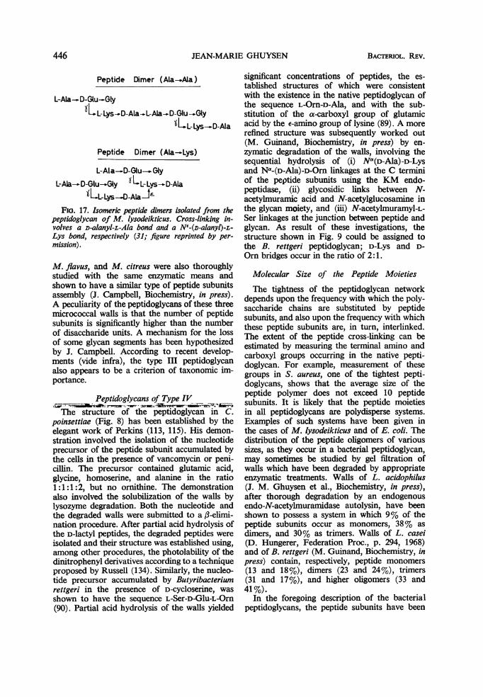
JEAN-MARIE GHUYSEN
Peptide Dimer (Ala-.Ala)
L-Ala-D-Clu-GlyXLL-Lys-D-AlaL-Ala_-.D-Glu-..Gly
ILL-Lys-D-A) a
Peptide Dimer (Ala-Lys)
L-Ala--D-Glu_--GlyL-Ala-D-GluGly XLL-Lys-D-Ala
KLLLys-D-Ala-J2FIo. 17. Isomeric peptide dimers isolated from the
peptidoglycan of M. lysodeikticus. Cross-linking in-volves a D-alanyl-L-Ala bond and a NE-(D-alanyl)-fiLys bond, respectively (31; figure reprinted by per-
mission).
M. flavus, and M. citreus were also thoroughlystudied with the same enzymatic means andshown to have a similar type of peptide subunitsassembly (J. Campbell, Biochemistry, in press).A peculiarity of the peptidoglycans of these threemicrococcal walls is that the number of peptidesubunits is significantly higher than the numberof disaccharide units. A mechanism for the lossof some glycan segments has been hypothesizedby J. Campbell. According to recent develop-ments (vide infra), the type III peptidoglycanalso appears to be a criterion of taxonomic im-portance.
Peptidoglycans of Type IVThe structure of the peptidoglycan in C.
poinsettias (Fig. 8) has been established by theelegant work of Perkins (113, 115). His demon-stration involved the isolation of the nucleotideprecursor of the peptide subunit accumulated bythe cells in the presence of vancomycin or peni-cillin. The precursor contained glutamic acid,glycine, homoserine, and alanine in the ratio1:1:1:2, but no ornithine. The demonstrationalso involved the solubilization of the walls bylysozyme degradation. Both the nucleotide andthe degraded walls were submitted to a 3-elimi-nation procedure. After partial acid hydrolysis ofthe D-lactyl peptides, the degraded peptides wereisolated and their structure was established using,among other procedures, the photolability of thedinitrophenyl derivatives according to a techniqueproposed by Russell (134). Similarly, the nucleo-tide precursor accumulated by Butyribacteriumrettgeri in the presence of D-cycloserine, wasshown to have the sequence L-Ser-D-Glu-L-Orn(90). Partial acid hydrolysis of the walls yielded
significant concentrations of peptides, the es-tablished structures of which were consistentwith the existence in the native peptidoglycan ofthe sequence L-Orn-D-Ala, and with the sub-stitution of the a-carboxyl group of glutamicacid by the e-amino group of lysine (89). A morerefined structure was subsequently worked out(M. Guinand, Biochemistry, in press) by en-zymatic degradation of the walls, involving thesequential hydrolysis of (i) Na(D-Ala)-D-Lysand Na-(D-Ala)-D-Orn linkages at the C terminiof the peptide subunits using the KM endo-peptidase, (ii) glycosidic links between N-acetylmuramic acid and N-acetylglucosamine inthe glycan moiety, and (iii) N-acetylmuramyl-L-Ser linkages at the junction between peptide andglycan. As result of these investigations, thestructure shown in Fig. 9 could be assigned tothe B. rettgeri peptidoglycan; D-Lys and D-Orn bridges occur in the ratio of 2:1.
Molecular Size of the Peptide Moieties
The tightness of the peptidoglycan networkdepends upon the frequency with which the poly-saccharide chains are substituted by peptidesubunits, and also upon the frequency with whichthese peptide subunits are, in turn, interlinked.The extent of the peptide cross-linking can beestimated by measuring the terminal amino andcarboxyl groups occurring in the native pepti-doglycan. For example, measurement of thesegroups in S. aureus, one of the tightest pepti-doglycans, shows that the average size of thepeptide polymer does not exceed 10 peptidesubunits. It is likely that the peptide moietiesin all peptidoglycans are polydisperse systems.Examples of such systems have been given inthe cases of M. lysodeikticus and of E. coll. Thedistribution of the peptide oligomers of varioussizes, as they occur in a bacterial peptidoglycan,may sometimes be studied by gel filtration ofwalls which have been degraded by appropriateenzymatic treatments. Walls of L. acidophilus(J. M. Ghuysen et al., Biochemistry, in press),after thorough degradation by an endogenousendo-N-acetylmuramidase autolysin, have beenshown to possess a system in which 9% of thepeptide subunits occur as monomers, 38% asdimers, and 30% as trimers. Walls of L. casei(D. Hungerer, Federation Proc., p. 294, 1968)and of B. rettgeri (M. Guinand, Biochemistry, inpress) contain, respectively, peptide monomers(13 and 18%), dimers (23 and 24%), trimers(31 and 17%), and higher oligomers (33 and41%).
In the foregoing description of the bacterialpeptidoglycans, the peptide subunits have been
446 BAcTERIOL. REV.

BACIERIOLYTICENZYMES4
presented as tetrapeptides having a D-Ala residueat their C termini. It should be understood thatthese sequences are typical of those subunitswhich are in the endo position in the peptidemoiety. The peptide subunits located at the C-terminus of a peptide polymer may be penta-peptides L-Ala-D-Glu-(L-Lys or meso-DAP)-D-Ala-D-Ala i.e., presenting the same sequencesfound in the peptidoglycan precursors), ortetrapeptides L-Ala-D-Glu-(L-Lys- or meso-DAP)-D-Ala, like those in endo-position, or tripeptidesL-Ala-D-Glu-(L-Lys or meso-DAP). These twolatter peptides may result from the elimination ofone or two D-Ala residues from the peptideprecursors at the time of assembly through theaction of specific carboxypeptidases. It followsthat a peptide monomer preparation obtainedafter endopeptidase treatment may be a mixtureof several compounds containing peptide sub-units which, in the native peptidoglycan, arelocated both in endo-position and at the Ctermini. Attention should also be called to thepeptide subunits which are located at the Ntermini of the peptide polymer. These are oftensubstituted by uncross-linked peptide bridges.In many cases, these open chains can be de-graded by aminopeptidase, which provides aconvenient way of determining the averagemolecular size of these units.
PEPrIDoGLYCAN AS A TAXONOMIC CHARACTER
Gram-Negative BacteriaIn spite of the fact that the walls of gram-nega-
tive bacteria contain a wide array of amino acids,so far as is known, the peptidoglycan containsonly those present in the peptide subunit withmeso-DAP as diamino acid. There is a generalbelief that the type of peptidoglycan presented inFig. 4 and 13 may be ubiquitous in this group ofeubacteria. Indirect evidence for cross-linkingbetween DAP and D-Ala has only been providedin a few cases, viz., Brucella abortus (83), Aero-bacter cloacae (62, 146), and Spirillum serpens(71). The extent of cross-linking seems to varyaccording to the species, and different techniqueshave revealed the existence primarily of peptidemonomers and dimers, with some trimers andtetramers also being present.
ActinomycetesIn 1957, Hoare and Work (52) made an
initial study of the distribution of the DAPstereoisomers among the actinomycetes. Meso-DAP was found in several species of Mycobac-terium and of Nocardia. Species of Actinomycesand Streptomyces, on the other hand, were shownto contain LL-DAP, meso-DAP being completely
absent or present only in trace amounts. Micro-monospora sp. contain both meso- and LL-DAP.In these studies, the characterization of LL- andmeso-DAP rested upon chromatographic separa-tion by the technique of Rhuland et al. (127), thespecific decarboxylation of the meso-isomer usinga meso-DAP decarboxylase (180), and thespecific enzymatic epimerization of LL-DAPinto meso-DAP. With three species of Micro-monospora, the meso-DAP spot obtained afterpaper chromatography in the solvent system ofRhuland et al. was found to be only partiallysensitive to meso-DAP decarboxylase. Since DD-DAP has the same mobility as meso-DAP inthe above chromatographic system, it was sug-gested that part of the DAP was in the DD formin the walls of these three microorganisms. Inmore recent studies (120, 160, 185), a tentativeclassification of various genera of actinomyceteswas proposed on the basis of the cell wall compo-sition as well as of morphology. (For a generalreview on this topic, see 85.) Several differenttypes of cell wall composition were observed,although these did not always correlate with theconventional taxonomic groups. The genus Acti-nomyces contains L-Lys alone, or both L-Lysand L-Orn. Streptomyces contain LL-DAP asso-ciated with Gly (103). Nocardia contain meso- orDD-DAP or both. Streptosporangium containmeso- or DD-DAP associated with a smalleramount of LL-DAP, or both. One group ofActinoplanaceae have meso- or DD-DAP asso-ciated with Gly and traces of LL-DAP, or both.Another group of Actinoplanaceae contain 2,6diamino-3-hydroxy-pimelic acid associated withglycine and traces of meso- or DD-DAP, orboth. The location of the glycine residues in theactinomycetal peptidoglycans is not yet known.
Gram-Positive EubacteriaL-lysine-containing peptidoglycans are typical
of the families of Micrococcaceae and Lacto-bacteriaceae and DAP-containing peptidoglycansare typical of the Bacillaceae, Propionobac-teriaceae, and Corynebacteriaceae families. Thetwo latter families are included in the orderEubacteriales according to the classification inBergey's manual (7). The nature of the diaminoacid, however, is not an absolute familial char-acteristic. For example, M. varians (137), L.inulinus, and L. plantarum (178) contain meso-DAP. Both L-Lys and meso-DAP occur in Plec-tridium tetani Mikhailov (Clostridium tetani; 163)and L-Lys alone occurs in A. crystallopoietes (74,75, 171). Among the DAP-containing peptido-glycans of gram-positive eubacteria, meso-DAPis the isomer most frequently encountered.
447VOL. 32, 1968

JEAN-MARIE GHUYSEN
However, LL-DAP has been found in a localstrain of Propionobacterium (2, 52) and in thefollowing strains (163): P. arabinosum NICB5958, Welchia perfringens BP 6K (C. welchii),C. botulinum Gunnison and Corynebacteriumanaerobium (Prevot 3103 and 3471). DD-DAPhas been identified in walls of B. megateriumKM (9).The structure of the DAP-containing peptido-
glycans among- the gram-positive bacteria islargely unknown. One may postulate that D-Ala-DAP linkages are also involved in peptide cross-linking (9, 35, 54), but exceptions will certainlybe found. It has already been postulated (119)that in the LL-DAP-containing peptidoglycanof C. welchii, some of the peptide subunits arelinked through D-Ala-L-Ala linkages in a "headto tail" sequence similar to that demonstratedin M. lysodeikticus peptidoglycan. This hypothesiswas not entirely confirmed, however, by veryrecent experiments (M. Leyh-Bouille and J. M.Ghuysen, unpublished data, and 0. Kandler,personal communication) the results of which in-dicate that Gly also serves as a bridge, extendingfrom LL-DAP to D-Ala. Walls of M. varians con-tain 3 to 4 glutamic acid residues per meso-DAPresidue. Solubilization of these walls by a stillunclassified endopeptidase from Streptomyces(M. Leyh-Bouille and J. M. Ghuysen, unpub-lished data) results in the appearance of N-terminal glutamic acid but not of N-terminalDAP, thus strongly suggesting that one orseveral glutamic acid residues serve as bridgesbetween peptide subunits. These investigationscarried out on C. welchil and M. varians verystrongly suggest that, in some DAP-containingpeptidoglycans, peptide bridges are mediatedthrough additional intervening amino acids, i.e.a structure analogous to that of the L-Lys-con-taining peptidoglycans type II (Fig. 5, 6).The peptidoglycans of many members of the
Micrococcaceae and Lactobacteriaceae familieshave been studied. In some investigations, thewalls were sequentially degraded with highlyspecific enzymes according to procedures previ-ously described. A second type of investigationinvolved, on the one hand, the characterizationof the peptidoglycan nucleotide precursors ac-cumulated in the presence of D-cycloserine,vancomycin, or penicillins and, on the otherhand, partial acid hydrolyses of the walls or ofchemically stripped peptidoglycans, followed byisolation and characterization of significantoligopeptides. The enzymatic procedure which,originally, was the only procedure available for acomprehensive study of the peptidoglycan struc-ture, is a time consuming process requiring en-zymnes which are frequently not readily available.
The chemical procedure is much faster and hasyielded valuable information. Using this lattermethod, about 50 species of Lactobacillus,Streptococcus, and Leuconostoc have been ex-amined by Kandler and his colleagues (63, 124,140, 141). Tables 2 and 3 represent a tentativeclassification of Micrococcaceae and Lacto-bacillaceae based on the chemistry of the pepti-doglycan. It is possible to distinguish four dis-tinct subgroups among the peptidoglycans typeII. In subgroup A, the bridges are composed ofL-amino acid residues. In subgroup B, the bridgesare composed of Gly residues. In subgroup C,the bridges are composed of Gly and L-aminoacid residues. In subgroup D, the bridges arecomposed of a single D-isoasparaginyl residue.In all cases, the peptide bridges extend from thee-amino group of L-Lys (or from one aminogroup of L-Orn, as in the case of M. radiodurans)to the carboxyl group of D-Ala.
UNIFIED VIEW OF THE BACrERIAL PEFTIDOGLYCANSThe mechanisms involved in the biosynthesis
of the peptidoglycan have been elegantly studiedin the cases of E. coli, S. aureus, and otherMicrococci. Since this subject has already beenreviewed several times (38, 157), only the es-sential points will be recalled here.
Uridine diphospho-N-acetylglucosamine andUDP N--(N-acetylmuramyl-L-alanyl---D-gluta-myl)-(L-lysyl or meso-diaminopimelyl)-D-ala-nyl-D-Ala are the nucleotide precursors.The reactions involved in their synthesis are allcatalyzed by soluble enzymes. Particularly, it isknown that the amino acids are added stepwise toUDP-N-acetylmuramic acid, each step beingunder the control of enzymes specific for boththe uridine nucleotide and the amino acid sub-strates. Each of the reactions is thus catalyzedby a distinct enzyme requiring adenosine tri-phosphate (ATP) and either Mg++ or Mn+. Thelast sequence, D-alanyl-D-Ala, however, is addedin one step to the UDP-N-acetylmuramyl-tripep-tide. D-Cycloserine is known to specificallyinhibit the synthesis of the dipeptide D-alanyl-D-Ala (107, 133). The synthesis of the nucleotideprecursors is followed by a complex reaction se-quence, which results in the formation of un-cross-linked peptidoglycan strands. This se-quence is catalyzed by particulate enzymeswhich have been identified as membrane frag-ments by electron microscopy (L. Matz andJ. L. Strominger, Bacteriol. Proc., p. 64, 1968).Three successive transfers are involved in thisreaction cycle. (i) From UDP-N-acetylmuramyl-pentapeptide, phospho-N-acetylmuramryl-penta-peptide is transferred to a membrane-boundpolyisoprenoid phosphate (51) resulting in forma-
448 BACTERIOL. REV.

BACTERIOLYTIC ENZYMES
TABLE 2. Peptidoglycans in micrococcaceae"
Genus and species Peptide bridges Chemical group
Sarcina lutea Polymerized peptide subunits III; Fig. 7Micrococcus lysodeikticus (31) Polymerized peptide subunitsM. flavusb Polymerized peptide subunitsM. citreusb Polymerized peptide subunits
M. roseus R27 (117) N"-(L-Ala-L-Ala-L-Ala-L-Thr)-L-Lys II (A); Fig. 5M. roseus Thr- (117) N-(L-Ala-L-Ala-L-Ala)-L-Lys
M. radioduransc N-(Gly-Gly)-L-Orn 11 (B); Fig. 5Staphylococcus aureus Copenhagen (117) N-- (Gly-Gly-Gly-Gly-Gly) -L-Lys
S. epidermidis NE,(Glyn; Sern)-L-Lysd 11 (C); Fig. 5NE-(G1y4 ; L-Ala)-L-Lys,
Gaffkya tetragena NE- (Gly.; Ser.)-L-Lysfa Peptide subunit. Group II (A, B, C): N-(L-Ala-D-isoGIN)-L-Lys-D-Ala. (In M. radiodurans, L-Lys
is replaced by L-Orn.) Group III: N--[L-Ala-y(a-D-Glu-Gly)]-L-Lys-D-Ala.b J. Campbell et al., Biochemistry, in press.c J. M. Ghuysen, unpublished data.d D. J. Tipper, Federation Proc., p. 294, 1968.a K. H. Schleifer et al., Arch. Mikrobiol., in press.f M. Lache and J. M. Ghuysen, unpublished data.
TABLE 3. Peptidoglycans in lactobacillaceaea
Genus and species Peptide bridges Chemical group
Streptococcus pyogenes group A (99) N'-(L-Ala-L-Ala)-L-Lys 11 (A); Fig 5S. thermophilis (viridans group) Ne-(L-Ala-L-Ala-L-Ala)-L-LysS. faecalis (enterococcus group) NE- (L-Ala-L-Ala-L-Ala) -L-LysS. cremoris 316a; 331a N1- (L-Ala-L-Thr) -L-Lys
S. Jaecalis ATCC 9790 (32)b N'-(D-isoAsN)-L-Lys 11 (D); Fig. 6S. lactis 211b; 212b (lactic group) Ne-(D-Asp; amide)-L-LysS. cremoris 71 (lactic group) NE-(D-Asp; amide)-L-LysStreptococcus serological group N (lactic N'-(D-Asp; amide)-L-Lysgroup)
Lactobacillus sp. Ne- (D-Asp; amide) -L-LysL. caseic NE-(D-isoAsN)-L-LysL. acidophilusd Ne-(D-isoAsN)-L-LysL. cellobiosus N-(D-Asp; amide)-L-Orn
L. coprophilus Nf-(L-Ala-L-Ala)-L-Lys 11 (A); Fig. 5L. viridescens spp. viridescens N-- (L-Ser-L-Ala) -L-LysL. viridescens spp. minor NE- (L-Ala-L-Ala-L-Ser) -L-LysL. mesenteroides Ne-(L-Ala-L-Ser) -L-LysLeuconostoc sp. N-- (L-Ala-L-Ala-L-Ser) -L-Lys or Ne- (L-
Ala-L-Ala)-L-Lys
a Peptide subunit = N(L-Ala-D-isoGlN)-L-Lys-D-Ala. References: 63, 124, 140, 141 (unless otherwisespecified).
b Recently identified as a S. faecium var. durans (0. Kandler, personal communication).c D. Hungerer, Federation Proc., p. 294, 1968.d J. M. Ghuysen, Biochemistry, in press.
tion of uracil monophosphate (UMP) andthe attachment of N-acetylmuramyl-pentapeptideto the lipid by means of a pyrophosphate bridge[Lipid-P-P-MurNAc(pentapeptide)]. (ii) Next,
N-acetylglucosamine is transferred by trans-glycosylation from UDP-N-acetylglucosamine tothe lipid-P-P-MurNAc(pentapeptide), with theliberation of UDP and the formation of /B-1,4-
449VOL. 32, 1968

JEAN-MARIE GHUYSEN
GlcNAc - MurNAc(pentapeptide) - P - P - lipid.(iii) This disaccharide pentapeptide is, in turn,transferred from the lipid intermediate to anendogenous acceptor, probably an incompletepeptidoglycan. The carrier is released as thepyrophosphate (P-P-lipid) which is subsequentlydephosphorylated, yielding the original polyiso-prenoid phosphate compound which can nowbegin another cycle. Vancomycin, ristocetin,and bacitracin interfere with the above reactioncycle. Bacitracin specifically inhibits the dephos-phorylation of the pyrophosphate lipid, whichappears as end product of the cycle, and thusprevents the lipid carrier from reentering thecycle (152). The precise mechanism by whichvancomycin and ristocetin inhibit the abovemechanism is not yet fully understood. Duringthe former reaction, and while the disaccharidepeptide subunit is bound to the lipid carrier, othermodifications in the pentapeptide moiety occur.(i) In S. aureus, the a-carboxyl group of glutamicacid is amidated (153) in the presence of ATPand ammonium ions. (ii) In M. lysodeikticus, thesame group is substituted by a Gly residue inthe presence of ATP (68). (iii) Bridging peptides,such as the pentaglycine in S. aureus, are addedto the -amino group of the L-Lys residue in thepeptide subunit. The glycine donor for the for-mation of the pentaglycine bridges is glycyl-tRNA (12, 108). Similarly, L-Thr (129) and L-Ala (128) have been shown to be activated asL-threonyl-tRNA or as L-alanyl-tRNA for in-corporation in the bridges of M. roseus R27 andA. crystallopoietes, respectively. Similar mech-anisms are involved in the incorporation of L-Serand Gly into interpeptide bridges in S. epidermi-dis (118). The above complex reaction cycle maybe visualized as a means of transporting the "dis-accharide-pentapeptide-peptide bridge" subunitin the form of a lipid intermediate from the intra-cellular site of synthesis to the extracellular site ofutilization (i.e., as a mechanism ofpassingthroughthe plasma membrane). The lipid moiety of theintermediate has been recently characterized(51) as a C55 isoprenoid alcohol. The existence,in S. aureus, of an ATP-dependent phospho-kinase which catalyzes the phosphorylation offree C55-isoprenoid alcohol has also been de-scribed (Y. Higashi et al., Federation Proc.,p. 294, 1968). C55 isoprenoid alcohol seems tohave a wide functional importance since it isalso involved in the biosynthesis of mannan inM. lysodeikticus (M. Sher and W. J. Lennarz,Federation Proc., p. 293, 1968) and of the 0-antigens of Salmonella (M. A. Cynkin and M. S.Osborn, Federation Proc., p. 293, 1968; 184).In this latter case, trisaccharide units (of man-nose, rhamnose, and galactose) linked through
pyrophosphate to the lipid (Man-Rh-Gal-P-P-lipid) are first polymerized forming O-antigenchains (Man-Rh-Gal).-P-P-lipid, which subse-quently are transferred to the "core" to formthe completed lipopolysaccharide (184). Muchremains to be done with regard to the exactmechanism through which the tRNA participatesin the cell wall synthesis by activating the aminoacids for their incorporation into the bridges.It seems clear, however, that ribosomes are notinvolved in this mechanism, which means thisprocess differs from "normal" protein synthesis.It should be noted that the incorporation ofamino acids into the peptide bridges can also bemediated by mechanisms other than those whichhave just been described. In L. viridescens, ofwhich the bridges are N'-(L-Ser-L-Ala)-L-Lys, theinitial amino acceptor for the tRNA-dependentincorporation of L-amino acids is apparentlynot the lipid intermediate (R. Plapp, FederationProc. p. 294, 1968), since a soluble enzymesystem was shown to be able to catalyze a directATP, Mg++ and tRNA-dependent incorpora-tion of either L-Ala or L-Ser into UDP-N-acetyl-muramyl-pentapeptide precursor. In S. faecalisand L. casei, of which the bridges are Ne-(D-isoAsN)-L-Lys, the lipid intermediate is the initialacceptor for D-aspartic acid incorporation;aminoacyl-tRNA, however, is not required andthe incorporation is not sensitive to ribonuclease(W. Standenbauer, Federation Proc., p. 294,1968).The bridge-closing reaction, the last step in
the synthesis of the peptidoglycan net, resultsfrom a transpeptidation in which the bondenergy of the terminal dipeptide (D-Ala-D-Ala)in the peptide subunit is utilized to effect theclosure of the peptide cross-link with the con-comitant release of the terminal D-Ala residue,i.e., the formation, for example, of a D-Ala-Glylinkage, in the case of S. aureus, or a D-Ala-(D)-meso-DAP, in the case of E. coli. In this trans-peptidation reaction, no exogenous energy sourceis necessary, and it can occur outside the cellmembrane at a site where ATP is not available.Penicillins and cephalosporins are specific in-hibitors of this transpeptidation. It has beenhypothesized that their actions depend upon astructural analogy with the D-alanyl-D-Ala pep-tide at the end of the peptide subunit (157,169). A direct demonstration of the transpeptida-tion reaction in E. coli has been given (58). Thehypothesis that this reaction is generally in-volved in the biosynthesis of the peptidoglycansrests upon indirect evidence, such as experimentsshowing that in the presence of penicillins, forexample, uncross-linked cell walls are formed.From the foregoing, it is becoming evident that
450 BACTERIOL. REV.

BACTERIOLYTIC ENZYMES
the biosynthetic mechanisms involved in theformation of the wall peptidoglycans are con-sistent throughout the microbial world. A four-stage mechanism can be proposed. First, allbacterial peptidoglycans are synthesized fromtwo nucleotide precursors, the UDP-N-acetyl-glucosamine and an UDP-N-acetylmuramylpentapeptide. The amino acid, located at theN-terminus of the pentapeptide, and the thirdresidue in the sequence may vary. Dependingupon the bacterial species, the three first aminoacids in the sequence are L-Ala-D-Glu-L-Lys,L-Ala-D-Glu-L-Orn, and L-Ala-D-Glu-meso, orLL- or DD-DAP, L-Ala-D-Glu-diaminobutyricacid, Gly-D-Glu-homoSer, L-Ser-D-Glu-L-Orn,etc. In all bacteria, however, the pentapeptidewould terminate in a D-alanyl-D-Ala dipeptideat the C terminus. In all of these examples, theD-Glu is linked to the next amino acid throughits y-carboxyl group. As pointed out above, eachof these different sequences would be deter-mined by individual soluble enzymes perform-ing the stepwise addition of each amino acid tothe UDP muramyl precursors.
Second, the assembly of the nucleotide pre-cursors into disaccharide peptide units and theirtransport through the plasma membrane wouldbe brought about, in all bacteria, with the helpof the polyisoprenoid phosphate carrier via thecyclic reaction described above.
Third, the pentapeptide subunits, while eitherin the form of nucleotide precursors or in theform of lipid intermediates, undergo variousmodifications. The most important modifica-tions are, of course, the incorporation of theamino acids which will serve as cross-peptidebridges. Amidation of carboxyl groups and in-corporation of other substituents, such as aGly residue in the pentapeptide of M. lysodeik-ticus, have also been described. Other modifica-tions of the pentapeptides would be, for examples,(i) the substitution of the a-carboxyl group ofglutamic acid by the E-amino group of a D-Lysresidue (in B. rettgeri) or by the 5-amino groupof a D-Orn residue (in C. poinsettiae and B.rettgeri), (ii) the hydroxylation of L-Lys (inS. faecalis, under special growth conditions), andthe hydroxylation of glutamic acid in Micro-bacterium lacticum (143).
Fourth, the final closure of the bridges betweenpeptide subunits might be, in all bacteria, theresult of a transpeptidation involving the D-alanyl-D-Ala dipeptide of one pentapeptidesubunit and a second peptide subunit as ac-ceptor with the concomitant loss of the terminalD-Ala. This acceptor is the NH2-(D)-meso-DAPof another peptide subunit (E. coli) or theamino terminus of the peptide bridge grouping,
such as Gly (S. aureus), L-Ala (S. pyogenes),D-isoasparagine (S. faecalis), a-NH2-D-Lys (B.rettgeri), or a-NH2-D-Orn (C. poinsettiae andB. rettgeri). Again, it should be understood thatthese bridge groupings may substitute either theL-Lys residue of the peptide subunit, as it is inmost cases, or the a-glutamic acid, as in B. rettgeriand C. poinsettiae.
Finally, attention should be called to the bio-synthesis of those peptides bridges, found inM. lysodeikticus and other related Micro-coccaceae, in which several peptide subunits arelinearly linked in "head to tail" sequencesthrough D-alanyl-L-Ala linkages (Fig. 7, 16). Theformation of such bridges might occur throughsuccessive alternating transpeptidation and ami-dase activities (31, 142), following the sequenceshown in Fig. 18. In reaction No. 1, a transpep-tidation, the penultimate D-Ala residue ofof peptide B is transferred to the lysine residue ofpeptide A with the concomitant loss of the ter-minal D-Ala residue. Reaction No. 2 is an amidaseaction in which the amide linkage at the N-L-Alaterminus of peptide B is hydrolyzed. ReactionNo. 3, a second transpeptidation, results in thepenultimate D-Ala residue of peptide C beingbound to the N-terminal L-Ala residue of pep-tide B with the expulsion of one D-Ala residue.Reactions 4 to 9 would follow in the indicatedorder, resulting ultimately in the formation ofthe peptide hexamer. In addition to the amidase,two different transpeptidases would be involved,the specificities of which depend upon the struc-ture of the amino acceptor (i.e., the e-aminogroup of L-Lys in reaction No. 1 and the amino
A B C D E F
*: L- Ala
(D- -D-Glu-Gly
Q: L -Lys
0 : D-Ala
FIG. 18. Proposed biosynthetic sequences of thepeptide hexamer unit ofM. lysodeikticus peptidoglycan(Fig. 7). Reactions 2, 4, 6, and 8 are amidasic hydroly-ses. Reactions 1, 3, 5, 7, and 9 are transpeptidations(31; figure reprinted by permission).
451VOL. 32, 1968

JEAN-MARIE GHUYSEN
group of L-Ala in reactions 3, 5, 7, and 9). Theabove mechanism has the result, of course, thaton the average the glycan strands have a lowextent of peptide substitution. Exposure of addi-tional receptor sites in the glycan through theaction of endo-N-acetylmuramidase autolysinscould result in the loss of some unsubstitutedglycan segments. This provides an explanation forthe observation (J. Campbell et al., Biochemistry,in press) that in some micrococcal peptidoglycanstype III (Fig. 7), the number of disaccharideunits is significantly lower than the number ofpeptide subunits.The biosynthetic reactions hereby postulated
may provide a way to unify our concept of thebacterial peptidoglycan. In spite of the extremevariations in chemical composition and instructural details present in bacterial peptidogly-cans, they all possess the same type of networkand appear to be synthesized by means ofanalogous mechanisms.
Lyric ENZYMES AS A MEANS FOR THE STUDY OFTHE LINK BETWEEN THE PEPTIDOGLYCAN
AND OTHER COMPONENTS IN WALLSOF GRAM-POSITIVE BACTERIA
Early electron microscopic examinations ofisolated cell walls of gram-positive bacteriashowed bag-shaped structures of rather uniformand homogeneous appearances. Immunologicalstudies, however, strongly suggested that therigid peptidoglycan network and the otherpolymers of the walls, most frequently proteins,polysaccharides, or teichoic acids (or both),were interlinked in a specific and organizedmanner. Besides the peptidoglycan, the S.pyogenes group A walls, for example, contain atleast two other polymers (73, 87), the group-specific C-polysaccharide and the type-specificM, R, and T proteins. The group C-polysac-charide contains a-1,3-linked L-rhamnose (25,48) and terminal N-acetylglucosamine linked torhamnose by a p3-1,3 bond (50). A portion ofthe rhamnose units, however, is also substitutedin the 2-position, and some of the glucosamineresidues may not be terminal (48). The structuresof the type-specific M protein, which is probablyrelated to virulence, and of the R and T proteinsare still largely unknown. Suitable proteases canremove these proteins from the walls withoutaffecting the viability of the microorganism orthe physical appearance of the walls. Furthertreatment of the walls with hot formamideeliminates, in turn, the group C polysaccharide,leaving, at the end of the process, an insolublepeptidoglycan which has the bag-shaped appear-ance of the original walls. It seems obvious thatsuch a sequential exposure of immunological
determinants must result from a distinct layeringof the chemical components. Using improvedsectioning, fixation, and staining techniques,direct demonstration of a layered structureamong walls of gram-positive bacteria has beenrecently achieved. The macromolecular arrange-ment of the surface of the walls of B. polymyxa(106) consists of a rectangular array of 7-nmglobules with a repeating interval of 10 nm.This outermost layer, mainly composed of pro-tein, is approximately 8 nm thick, and it is re-movable by hydrogen bond-breaking reagents orby trypsin. An underlying structure of 4-nmthickness is a polysaccharide complex whichappears to be covalently bound to the deepestlayer of 12 nm of thickness (probably the pepti-doglycan), from which it can be removed byhot formamide. Similarly, the walls of B. mega-terium, (105) have been shown to be composedof a rigid peptidoglycan layer of about 10 nmin thickness, surrounded by an elastic layer(12 nm) formed by bristles of teichoic acid. Thistype of architecture is perfectly consistent with thebiological roles that the teichoic acids play inthe bacterial cells. The immunological activityof the teichoic acid in S. aureus has been provedby a variety of techniques. It has been shown byhaptene inhibition studies, for example, that thea- and ,B-N-acetylglucosamine residues, whichsubstitute the polyribitol phosphate skeleton ofthe teichoic acid, are the specific antigenicdeterminants responsible for cell wall agglutina-tion (104). Moreover, evidence has also beengiven that some of the N-acetylglucosamineresidues on the teichoic acid are involved inphage fixation in S. aureus (18). Similar conclu-sions were reached by Young, who found thatphage fixation by B. subtilis requires that theteichoic acid be glucosylated (188, 189), and byGlaser et al. (42), who presented evidence thatphage ,u may recognize teichoic acid as part ofthe receptor site of B. subtilis. Another illustra-tion of a complex layering is that of the pig-mented walls of M. radiodurans (183). Theywere shown to be composed of (i) an outer-most network removable by trypsin; (ii) afragile soft layer, pink in color, containingcarotenoids, lipid, protein, and polysaccharideand presenting hexagonally packed subunits;(iii) a perforated rigid L-Orn-containing peptido-glycan layer. Other beautiful demonstrations ofdetailed organization of the various componentsin bacterial walls have been provided by Murray(101).From the foregoing, it is obvious that the
elucidation of the way in which the various con-stituent polymers are held together within thebacterial walls is a major, although still largely
452 BACTERIOL. REV.

BACrERIOLYTIC ENZYMES
unsolved, problem. Phosphodiester bonds areone means used by bacteria to link together therigid peptidoglycan and the other polymericstructures. The nonglycan polysaccharide in thewalls of L. casei, for example, is believed to bejoined through the reducing group of its terminalN-acetylglucosamine residue to a phosphategroup on the peptidoglycan (45, 70). Mostlikely, this is a phosphorylmuramic acid (1).The "atypical" teichoic acid present in the wallsof S. lactis is linked to the peptidoglycanvia phosphodiester bonds between the glyceroland C4 or C6 of muramic acid (13). In the case ofS. aureus strain Copenhagen, phosphodiesterbonds also cross-link the teichoic acid and thepeptidoglycan (40). Indirect evidence, based ontheestimation of formaldehydogenic end groups byperiodate oxidation, suggested that a phosphoryl-ated N-acetylglucosamine residue was the pepti-doglycan part of this link. This conclusion,however, cannot be regarded as definite, becausethe linking group was not actually isolated andcharacterized. According to a recent survey madeby Liu and Gotschlich (82), muramic acid 6-phosphate represents from 3 to 39% of all themuramic acid residues present in the walls ofM. lysodeikticus, Diplococcus pneumoniae, Myco-bacterium butyricum, S. aureus NYH 6, and ofvarious species of Streptococcus belonging togroups A, C, and E. Muramic acid phosphate wasalso detected in mycobacterial walls (64). Theimportance of muramic acid 6-phosphate in thewalls of gram-positive bacteria and actinomycetesis becoming evident. Further information con-cerning the location of these linking groups in twodifferent walls has been sought by the techniqueof specific enzymatic degradation.
Micrococcus lysodeikticusThe polysaccharide component of the M.
lysodeikticus walls is composed solely of equi-molar amounts of D-glucose and 2-acetamido 2-deoxymannuronic acid (111). In spite of thereports of Perkins (111) and of Prasad andLitwach (125) to the contrary, the presence oforganic phosphate, first detected by Salton(135), in amounts ranging from 0.09 to 0.13%of the weight of the walls, is now generally ac-cepted (82, 95). Walls of M. lysodeikticus, asprepared in my laboratory contain (per milli-gram dry weight) 26 nmoles of organicallybound phosphate, 260 nmoles of D-glucose, 480nmoles of disaccharide and peptide subunits,i.e., a ratio of 1:10:19. After complete degrada-tion of the wall peptidoglycan through sequen-tial action of ML endopeptidase, F1 endo-N-acetylmuramidase and N-acetylmuramyl-L-Ala
amidase, the polysaccharide polymer was iso-lated in the form of a complex presenting a ratiofor phosphate, glucose, disaccharide units, andpeptide units of 1:10: 2.5:1. Analytical data wereconsistent with a structure in which one peptidesubunit substitutes a tetra- or hexasaccharideresidue, itself linked to the glucose-containing-polysaccharide polymer through a phosphodiesterbond originating from the C-6 of one of the N-acetylmuramic acid residue. Another "poly-saccharide-glycopeptide fragments" complex ofthe same established composition and tentativestructure was isolated from walls which had beendegraded only with an endo-N-acetylmuramidase.Since the wall peptide moiety was not attackedin this latter type of degradation, one can postu-late that, in the intact walls, those glycan seg-ments which bear phosphate groups are notsubstituted by cross-linked peptide subunits.There is one phosphate group for every 18 di-saccharide units in intact walls. By analogy withchain length determinations carried out in thecase of S. aureus, this figure of 18 disaccharideunits may well represent the average chain lengthof the glycan strands in M. lysodeikticus. Sincephosphate bridging and peptide cross-linkingdo not occur on the same segments of the glycanmoiety, not many phosphate bridges can occurin a single glycan strand; therefore it was hypothe-tized that, on the average, each glycan strand of18 disaccharide units is joined through onephosphodiester bond to a segment, 10 glucoseand 10 aminomannuronic acid residues long, ofthe polysaccharide polymer.
Streptococcus pyogenesElucidation of the macromolecular structure of
the S. pyogenes cell wall has been attempted(98). Careful fractionations of the walls treatedwith trypsine and subsequently solubilized byF1 endo-N-acetylmuramidase yielded fractionsin which it was possible to distinguish betweentwo types of phosphate groups (Fig. 19). (i)About 20% of the organic phosphate present inthe walls (i.e., 50-nm equivalents/mg) occurs asN-acetylmuramic acid 6-phosphate residues.These groups join about 10% of the peptido-glycan subunits (50-nm equivalents/mg) to apolysaccharide polymer which is not the C-poly-saccharide. This polymer (complex S3, Fig. 19)was isolated as a disaccharide peptide monomersubunit linked through the phosphodiesterbridge to a complex containing, per peptidemonomer, five to six D-glucose residues, one glu-cosamine residue, and four to five unidentifiedhexosamine residues, possibly aminohexuronicacids. It was proposed that this polymer, referred
VOL. 32, 1968 453

JEAN-MARIE GHUYSEN
',et S ort c/t
FIG. 19. Proposed molecular arrangement of the four polymeric constituents of the walls ofStreptococcus pyo-genes type 14. Hydrolysis of the glycosidic linkages between N-acetylmuramic acid (Y) and N-acetylglucosamine(X) yields three main fractions. Complex 52 contains the group-specific C-polysaccharide linked to the peptido-glycan via tri-rhamose (R) groupings. Complex S3 contains the glucose-containing G-polysaccharide linked to thepeptidoglycan via phosphodiester bridges (P). The G-polysaccharide wouldbe the anchor pointsfor the type-specificM, R. and Tproteins. Glycan chains are bound by decapeptides resultingfrom the cross-linking oftwo peptide sub-units B (Fig. 3) through L-alanyl-L-Ala dipeptides (Fig. 5). Fraction 54 consists of unsubstituted disaccharide units-(about 20% of the total N-acetylhexosamine residues). (For more details, see 98; figure reprinted by permission.)
to as the G-polysaccharide, serves as an anchorpoint for the M-, R-, and T-type proteins. Thishypothesis has been strengthened by very recentinvestigations carried out by Barkulis et al.(Bacteriol. Proc., p. 110, 1968). It is noteworthythat, as with M. lysodeikticus, the phosphategroup joining the glycan and the G-polysac-charide is located on an N-acetylmuramic acidresidue bearing an unbridged peptide substituent.(ii) About 70% of the organic phosphate of thewalls (150 nm equivalents/mg) occurs as glycerolphosphate and glyceryl rhamnoside phosphate(49) in the interior of the C-polysaccharide. Thelinkage between the C-polysaccharide chains andthe peptidoglycan was visualized as consisting ofbridges of trirhamnose groupings, each bearingone p3-linked N-acetylglucosamine residue (Fig.19). In this case, each of these trirhamnose unitswould be bound at the reducing end, either di-rectly or through an as yet undetermined inter-mediate molecule, to the C-4 of an N-acetyl-muramic acid residue. After endo-N-acetyl-muramidase degradation of the walls, a complexhas been isolated (complex S2, Fig. 19), in whichthe C-polysaccharide is bound to N-acetyl-muramic acid residue. This N-acetylmuramic acidresidue was found to be substituted by one
tetrapeptide subunit which, in turn, was linkedvia a di-L-alanyl bridge to another disaccharidepeptide subunit.As a result of these findings (98), it was pro-
posed that a major part of the walls presented astructure of the type shown in Fig. 19. The N-acetylmuramic acid at the nonreducing end ofshort glycan strands are glycosidically linked tothe reducing ends of tri-a-l , 3-L-rhamnosyl frag-ments extending from the C-polysaccharidebulk. In about 10% of the glycan strands, someN-acetylmuramic acids are substituted on C-6by phosphate groups extending to the G-polysac-charide polymer. The remaining N-acetylmuramicacid residues are substituted by tetrapeptide sub-units cross-linked by L-alanyl-L-Ala dipeptides,so that neighboring glycan strands are cross-linked by decapeptide chains.
It should be understood, of course, that theproposed structure represents only one of thepossibilities. The phosphorylated N-acetylmur-amic acid residues need not be terminally located,and the chain length of five hexosamine residuesgiven for the glycan moiety may well be anaverage resulting from the actual presence of amixture of longer and shorter chains. Althoughas yet imprecise, the proposed structure offers aa
454 BACTERIOL. REV.

BACTERIOLYTIC ENZYMES
explanation at a molecular level for the com-plex architecture of the walls and, particularly,for the specific interconnection between con-stitutive polymers.AUTOLYTIC ENZYMES AS BACTERIAL WALL
CoNsTrruENTsWhereas the reactions involved in the bio-
synthesis of the peptidoglycan portion of thebacterial wall are understood at the molecularlevel, our concept of the mode of growth andreplication of the wall at the cellular level is stilllargely hypothetical. Three different aspects ofthis problem will be discussed briefly.
Cosynthesis of the Wall PolymersThe biosynthesis of the bacterial walls (i.e.,
the peptidoglycan and the other polymers) isan exceedingly complex reaction mechanism inwhich at least 30 and perhaps as many as 60enzymes are involved. If, for example, one con-siders the structure of the walls of S. pyogenes(Fig. 19), it would appear logical that biosyn-thesis and integration of each polymeric com-ponent occur simultaneously, if one is to arriveat such a structure in which all the polymers arecross-linked in a specific ordered manner. Someresults have been obtained which support thishypothesis. One approach has involved themarking of the walls of a living cell of S. pyogenesat a given time, by fluorescein-labeled antibodiesspecific for the wall type M protein or for thewall group C polysaccharide (17). By followingthe partition of the markers among daughtercells after division, the events involved in thewall replication appeared identical irrespective ofthe antibody used for the labeling; thus, it hasbeen possible to conclude that the synthesis ofthese two antigens is synchronized and occurs atidentical sites. It would be most important, how-ever, that identical experiments be repeated withfluorescent antibody specific to the wall pepti-doglycan matrix. Similarly, it has also beenobserved that application of any inhibitor ofpeptidoglycan synthesis leads to concomitantnhibition of teichoic acid synthesis (131),suggesting interrelationship between the forma-tion of the two polymers.
Site of Synthesis of Cell Wall MaterialAll bacteria do not replicate their cell wall
material in the same manner. Essentially twomechanisms have been shown to exist (17).In one type of replication, new wall material ap-pears localized in a specific area of the parentwall. Examples of this are S. pyogenes (17) andS. faecalis (149), wherein, as shown by thefluorescent antibody technique, peripheral wall
and cross-wall (septum) formation are initiatedat the same time along an equatorial ring, withthe result that the daughter cocci grow "back-to-back." Initiation of new bands of synthesisoccur before the old ones are complete, suggest-ing a necessity for initiation of new cleavagepoints. Similarly, it has also been reported byFitz-James and Hancock (26) that, in the presenceof penicillin, a fibrous material is formed at aspecific growing point of the cell wall of B.megaterium. As suggested by Strominger et al.(Federation Proc., p. 9-22, 1967), this disor-ganized fibrous material would represent thepeptidoglycan strands which continue to beformed in the presence of penicillin, but remainuncross-linked. The second type of replication,found in species of Salmonella (17) or of E. coil(177), is a diffuse process. As also shown by thefluorescent antibody technique or by following,microautoradiographically, the incorporation anddistribution of 'H-labeled DAP, new wall ma-terial appears to be continuously inserted intoexisting material at multiple sites along the surfaceof the wall. D-Methionine was shown by Larket al. (77) to induce the formation in Alcaligenesfaecalis of a fibrous material which, in contrastwith the situation observed in B. megateriumtreated with pencillin, was not localized, butoccurred over the entire bacterial surface. SinceD-methionine, or some other D-amino acids, iscapable, like penicillin, of reversing the transpep-tidation in the last step of the peptidoglycanbiosynthesis, it was also suggested that this fibrousmaterial was some uncross-linked peptidoglycan(J. L. Strominger et al., Federation Proc., p. 9-22, 1967).
Peripheral and cross-wall formation occur atthe same time. However, the two processes mustbe distinct and separately controlled in manyorganisms, since it is possible to selectively in-hibit cross-wall formation, by alteration of theenvironment, for example, with the resultantformation of greatly elongated septumless fila-ments. The demonstrated occurrence of meso-somes in close proximity to the developingsepta, plus the fact that they are absent in cellsgrowing under conditions where septum forma-tion is interrupted, has led several workers (138)to postulate that these organelles are involved inwall biosynthesis, possibly as a carrier forpeptidoglycan precursors. Much, however, re-mains to be done before the exact function ofmesosomes in this regard is established.
Biological Roles ofAutolysinsMany endogenous autolytic systems are fixed
in the walls themselves and must be regarded
455VOL. 32, 1968

JEAN-MARIE GHUYSEN
as an integral part of these layers. Autolysins,first thought solely a means of self-destruction,have excited considerable interest of late as pos-sible factors involved in wall biosynthesis, regula-tion, and in the permeation of large molecules.Several reasonable roles might be attributed tothese enzymes.Wall autolysins can be visualized as a means
of providing the peptidoglycan net with additionalreceptor sites to which newly synthesized disac-charide-peptide units could be transferred. Re-ceptor sites, both in the glycan and in the peptidemoieties, are needed. N-acetylglucosamine resi-dues at the nonreducing termini of glycan seg-ments (i.e., the expected sites for the transfer ofdisaccharides from the lipid intermediate) couldresult from the hydrolysis of 3-1,4-N-acetyl-muramyl-N-acetylglucosamine linkages. Aminotermini of peptide bridges or amino groups of thediamino acids of the peptide subunits if the peptidebridging is mediated through direct bonds betweenpeptide subunits, (i.e., the acceptors for the trans-peptidation reaction involving the D-alanyl-D-alanine dipeptides of newly synthesized penta-peptides) could be provided by cleavage oflinkages at the C termini of some peptide sub-units. According to this thesis, endo-N-acetyl-muramidases and many of the endopeptidaseswhich have been described are just such tools.Furthermore, and by analogy with the mechanismproposed for the hydrolytic action of lysozyme(16), the endo-N-acetylmuramidases might betransglycosilation agents playing an active rolein the growth of the glycans. In order to ensurean economic and safe enlargement of the net, astrict coordination of the machinery is required.This coordination would be a character of a well-balanced growth. One model for such a mecha-nism in E. coli has been proposed by Weidel andPelzer (177).Recent developments have provided evidence
for a preferential location of the autolysin en-zymes in some bacterial walls. The involvementof the autolysin of S. faecalis in wall growth hasbeen based upon several observations (149).This activity is only found in the wall fraction,and it is maximal during the period of exponen-tial growth. Furthermore, it was demonstrated byingenious radioactive labeling that newly syn-thesized wall material was the first to be dissolvedby this autolysin, suggesting that active enzymewas localized at the region of new wall synthesis(i.e., at the coccal equators). It has also beenshown that S.faecalis walls contain large amountsof latent autolysin, which, in vitro, is activatedby trypsin. A process whereby the latent autolysinis proteolytically activated at the future sites ofnew wall growth can readily be imagined. Further
evidence of the localization of the autolyticsystem at the site ofseptum formation comes fromthe earlier observation of Mitchell and Moyle(94) that osmotic shocking of S. aureus cells,allowed to autolyse in 1.2 M NaCl, gave rise tohemispherical halves of cell walls. More recently,Cripps and Work (19), working with a strain ofS. aureus, gave electron microscopic evidencethat at the time of lysis there was a localizedrupture of the developing septum and extrusionof intracellular high molecular weight material(RNA, DNA, proteins) from this point. It hasbeen proposed that this localized rupture couldbe the result of an activation of the autolyticsystem.The specificity of the wall autolysin in S.
faecalis and in S. aureus has been studied. InS. faecalis, the system has been characterized asbeing an endo-N-acetylmuramidase (149) and,hence, a reasonable role for this enzyme wouldbe to break the existing peptidoglycan net andto provide acceptor sites. In S. aureus, at leastpart of the wall autolytic system has been char-acterized by D. J. Tipper (Bacteriol Proc., p. 48,1968), who demonstrated the presence in theisolated walls of endo-N-acetylmuramyl-L-Alaamidase, endo-N-acetylglucosaminidase, andendopeptidase activities. Neither of the two firstenzymes would produce acceptors for enlargingthe peptidoglycan. They might however serve toseparate the two daughter cocci after septumformation is complete. Furthermore, as noted byTipper, if the glycan were in fact the only ac-ceptor for the synthesis of additional peptido-glycan material, the endo-N-acetylglucosamini-dase action which produces oligosaccharideswith N-acetylglucosamine-reducing terminiwould serve to fragment an otherwise endlesspolymer.Another possible role for wall autolysin is
based on recent investigations made by A. H.Dadd and R. J. L. Paulton (Proc. Soc. Gen.Microbiol., p. iii, 1968). Working with syn-chronized B. subtilis, they showed that incorpora-tion of cell wall material occurs solely at the timeof cell division. Between cell divisions, an in-crease of N-terminal DAP was observed, whichmust be the result of the activity of a wall endo-peptidase system. This suggests a loosening ofthe net, thus permitting the fixed amount of wallto stretch to accommodate the enlarging cyto-plasm. The presence of another "relaxation sys-tem" in the walls of competent B. subtilis hasalso been proposed (186, 187, 190). It has beenshown that those strains of B. subtilis whichare highly susceptible to genetic transformationcontain a higher autolytic activity than do thepoorly transformable strains. Here again, the
456 BACTEMIL. REV.

BACTERIOLYTIC ENZYMES
autolytic system which consists, in part at least,of an N-acetylmuramyl-L-Ala amidase, is asso-ciated with the walls and is probably covalentlylinked to the teichoic acid (W. C. Brown et al.,Bacteriol. Proc., p. 48, 1968). It has been sug-gested that this activity results in a relaxation ofthe peptidoglycan net, perhaps in a localizedarea, permitting the passage of DNA. Nothing isknown, however, regarding the mechanism in-volved in the repair of such gaps or how theserepair mechanisms are inhibited under the con-ditions of unbalanced growth required for maxi-mum competence, nor does it explain the bindingofDNA to competent cells or its passage throughthe cytoplasmic membrane. It has also beensuggested (F. E. Young, personal communica-tion) that this autolytic amidase may be in-volved in separation of cells during the finalstages of cleavage.
Bacterial wall or envelope enzymes are alsolikely to be involved in other regulation mecha-nisms. In E. coli, for example, two enzymes, orperhaps two different manifestations of theactivity of one enzyme (86), terminate the syn-thesis of the peptidoglycan (J. L. Stromingeret al., Federation Proc., p. 9-22, 1967): the trans-peptidase which, as described above, brings aboutthe closure of the bridges between two peptidesubunits, and a D-Ala carboxypeptidase whichremoves the D-Ala residues from the end of oneof these two peptide subunits so that oligomerslarger than dimers cannot be formed. D-Alacarboxypeptidase is one of the enzyme activitiesof the complex autolytic system of the E. colienvelope. It is possible that this enzyme is, infact, responsible for the limitation of the size ofthe polymer moiety. Similarly, the regulation ofthe size of those peptidoglycans that containpeptide bridges might be mediated by amino-peptidases, which would modify the acceptorsites needed for the closure of the bridges. Finally,autolytic N-acetylmuramyl-L-Ala amidases havebeen reported to be present in walls of an increas-ing number of bacterial species (162, 164). Thepossible involvement of this enzyme in the forma-tion of "head to tail" peptide sequences, such asthose demonstrated in M. lysodeikticus (31;Fig. 18) and other related Micrococcaceae (J.Campbell et al., Biochemistry, in press) may behypothesized.
CONCLUSION
In the period of the last 20 years, a new classof heteropolymers, the peptidoglycans in thebacterial walls, were discovered. Studies of theirstructure and biosynthesis have been developed ata comprehensive molecular level, opening a new
area of investigations. The coming years will see,we hope, the widening of our understanding ofthe tertiary structure and of the intimate func-tioning of this bacterial organelle as an integralpart of coordinated cellular systems.
ACKNOWLEDGMENTSThis investigation was supported by grant UR-E4-
10-2 from the U.S. Department of Agriculture underPublic Law 480.
I am grateful to the following persons for reading,criticizing, and discussing the manuscript: E. Bricas,Institut de Biochimie, Orsay, France; J. N. Camp-bell, Department of Microbiology, University ofAlberta, Edmonton, Canada; J. Coyette, Service deBacteriologie, Universit6 de Liege, Belgium; M. Guin-and, Service de Chimie Biologique, Universit6 deLyon, France; H. Heymann, Bacteriology Depart-ment, Ciba Pharmaceutical Co., Summit, N. J.;0. Kandler, Institut fur Angewandte Botanik,Technische Hochschule, Munich, Germany; M.Leyh-Bouille, Service de Bacteriologie, Universit6 deLiege, Belgium; E. Mufioz, Instituto de BiologiaCellular, Madrid, Spain; H. Perkins, Medical Re-search Council, Mill Hill, London, England; J. F.Petit, Institut de Biochimie, Orsay, France; K.Schleifer, Institut fur Angewandte Botanik, Tech-nische Hochschule, Munich, Germany; N. Sharon,The Weizmann Institute, Rehovoth, Israel; G. Shock-man, Department of Microbiology, Temple Univer-sity, Philadelphia, Pa.; R. Tinelli, Institut Pasteur,Paris, France; D. Tipper, Pharmacology Depart-ment, University of Wisconsin, Madison, Wis.;J. van Heijenoort, Institut de Biochimie, Orsay,France; and F. E. Young, Scripps Clinic and Re-search Foundation, La Jolla, California.
LimERATuRE CIT1. Agren, G., and C. H. De Verdier. 1958. The
isolation of protein-bound phosphorylmura-mic acid from Lactobacillus casei. ActaChem. Scand. 12:1927-1936.
2. Allsop, J., and E. Work. 1963. Cell walls ofPropionobacteriwn species: fractionation andcomposition. Biochem. J. 87:512-519.
3. Barkulis, S. S., C. Smith, J. J. Boltralik, and H.Heymann. 1964. Structure of streptococcalcell walls. IV. Purification and properties ofstreptococcal phage muralysin. J. Biol.Chem. 239:4027-4033.
4. Battersby, A. R., and J. C. Robinson. 1955.Studies on specific chemical fission of peptidelinks. Part I. The rearrangement of aspartyland glutamyl peptides. J. Chem. Soc., p. 259-269.
5. Blake, C. C. F., L. N. Johnson, G. A. Mair,A. C. T. North, D. C. Philips, and V. R.Sarma. 1967. Crystallographic studies of theactivity of hen egg-white lysozyme. Proc.Roy. Soc. (London) Ser. B 167:378-388.
6. Blake, C. C. F., G. A. Mair, A. C. T. North,D. C. Philips, and V. R. Sarma. 1967. On theconformation of the hen egg-white lysozyme
457VOL. 32, 1968

JEAN-MARIE GHUYSEN
molecule. Proc. Roy. Soc. (London) Ser. B167:365-378.
7. Breed, R. S., E. G. D. Murray, and N. R. Smith.1957. Bergey's manual of determinative bac-teriology. 7th ed. Williams & Wilkins Co.,Baltimore.
8. Brenner, J. F., A. Dark, P. Gerhardt, M. H.Jeynes, 0. Kandler, E. Kellenberger, E.Klieneberger-Nobel, K. McQuillen, M. Rubio-Huertos, M. J. R. Salton, R. E. Strange, J.Tomsick, and C. Weibull. 1958. Bacterialprotoplasts. Nature 181:1713.
9. Bricas, E., J. M. Ghuysen, and Ph. Dezelee.1967. The cell wall peptidoglycan of Bacillusmegaterium KM. I. Studies on the stereo-chemistry of a,a'-diaminopimelic acid. Bio-chemistry 6:2598-2607.
10. Bricas, E., P. Lefrancier, J. M. Ghuysen, E.Muiioz, L. Leyh-Bouille, J. F. Petit, and H.Heymann. 1967. Structure et synthese de lasubunitd peptidique de la paroi de trois bac-teries gram-positif, p. 286-292. In H. C. Beyer-man, A. Van de Linde, and W. Maassen vanDen Brink (ed.), Peptides. North HollandCo., Amsterdam.
11. Browder, H. P., W. A. Zygmunt, J. R. Young,and P. A. Tavormina. 1965. Lysostaphin:Mechanism of action. Biochem. Biophys.Res. Commun. 19:383-385.
12. Bumsted, R. M., J. L. Dahl, D. Soll, and J. L.Strominger. 1968. Biosynthesis of the peptido-glycan of bacterial cell walls. X. Furtherstudy of the glycyl transfer ribonucleic acidsactive in peptidoglycan synthesis in Staphy-lococcus aureus. J. Biol. Chem. 243:779-783.
13. Button, D., A. R. Archibald, and J. Baddiley.1966. The linkage between teichoic acid andglycosaminopeptide in the walls of a strainof Staphylococcus lactis. Biochem. J. 99:11c-14c.
14. Chatterjee, A. N., J. B. Ward, and H. R. Perkins.1967. Synthesis of mucopeptide by L-formmembranes. Nature 214:1311-1314.
15. Chipman, D. M., V. Grisaro, and N. Sharon.1967. The binding of oligosaccharides con-taining N-acetylglucosamine and N-acetyl-muramic acid to lysozyme: the specificityof binding subunits. J. Biol. Chem. 242:4388-4397.
16. Chipman, D. M., J. J. Pollock, and N. Sharon.1968. Lysozyme-catalyzed hydrolysis andtransglycosylation reactions of bacterial cellwall oligosaccharides. J. Biol. Chem. 243:487-496.
17. Cole, R. M. 1965. Symposium on the fine struc-ture and replication of bacteria and theirparts. III. Bacterial cell-wall replication fol-lowed by immunofluorescence. Bacteriol.Rev. 29:326-344.
18. Coyette, J., and J. M. Ghuysen. 1968. Structureof the cell wall of Staphylococcus aureus,strain Copenhagen. IX. Teichoic acid andphage adsorption. Biochemistry 7:2385-2389.
19. Cripps, R. E., and E. Work. 1967. The accumu-
lation of extracellular macromolecules byStaphylococcus aureus grown in the presenceof sodium chloride and glucose. J. Gen.Microbiol. 49:127-137.
20. Cummins, C. S., and H. Harris. 1956. The chem-ical composition of the cell wall in some gram-positive bacteria and its possible value as ataxonomic character. J. Gen. Microbiol.14:583-600.
21. Dierickx, L., and J. M. Ghuysen. 1962. Purifica-tion de la 03-(1-4)-N-acetylhexosaminidasesecretee par Streptomyces albus G et active surles parois de Micrococcus lysodeikticus. Bio-chim. Biophys. Acta 58:7-18.
21a. Diringer, H. 1968. Ober die Bindung der D-Glu-taminsaure im Murein von E. coli. Z. Natur-forsch. 23b: 883-884.
22. Diringer, H., and D. Jusic. 1966. Ulber die Bin-dung der meso-Diamino-pimelinsaure imMurein von E. coli. Z. Naturforsch. 21b:603-604.
23. Dorer, F. E., E. E. Haley, and D. L. Buchanan.1966. Quantitative studies of urinary f3-aspartyloligopeptides. Biochemistry 5:3236-3240.
24. Ensign, J. C., and R. S. Wolfe. 1966. Charac-terization of a small proteolytic enzyme whichlyses bacterial cell walls. J. Bacteriol. 91:524-534.
25. Estrada-Parra, S., M. Heidelberger, and P. A.Rebers. 1963. Immunochemical propertiesof the periodate-oxidized polysaccharide ofgroup A hemolytic Streptococcus. J. Biol.Chem. 238:510-512.
26. Fitz-James, P., and R. Hancock. 1965. Theinitial structural lesion of penicillin action inBacillus megaterium. J. Cell. Biol. 26:657-667.
27. Fleming, A. 1922. On a remarkable bacteriolyticelement found in tissues and secretions. Proc.Roy. Soc. (London) Ser. B 93:306-317.
28. Fleming, A. 1929. On the antibacterial actionof cultures of a penicillium, with special refer-ence to their use in the isolation of B. influen-zae. Brit. J. Exptl. Pathol. 10:226-236.
29. Fleming, A., and V. D. Allison. 1922. Observa-tions on a bacteriolytic substance ("Lyso-zyme") found in secretions and tissues. Brit.J. Exptl. Pathol. 3:252-260.
30. Ghuysen, J. M. 1961. Precisions sur la struc-ture des complexes disaccharide-peptidelibDres des parois de Micrococcus lysodeikticussous l'action des ,(1-4) N-acetylhexosamini-dases. Biochim. Biophys. Acta. 47:561-568.
31. Ghuysen, J. M., E. Bricas, M. Lache, and M.Leyh-Bouille. 1968. Structure of the cell wallsof Micrococcus lysodeikticus. III. Isolation of anew peptide dimer, Nq-[L-alanyl-y-(a-D-glutamyl - glycine)] - L - lysyl - D - alanyl - Na[L - alanyl - y - (a - D - glutamyl - glycine)] -L - lysyl-D-alanine. Biochemistry 7:1450-1460.
32. Ghuysen, J. M., E. Bricas, M. Leyh-Bouille,M. Lache, and G. D. Shockman. 1967. Thepeptide Na - (L - alanyl - D - isoglutaminyl) -
N' - (D - isoasparaginyl) - L - lysyl - D - alanineand the disaccharide N-acetylglucosaminyl-
458 BACTERIOL. REV

BACTERIOLYTIC ENZYMES
,-1,4-N-acetylmuramic acid in cell wallpeptidoglycan of Streptococcus faecalis strainATCC 9790. Biochemistry 6:2607-2619.
.33. Ghuysen, J. M., L. Dierickx, M. Leyh-Bouille,J. L. Strominger, E. Bricas, and C. Nicot.1965. Structure of the cell wall of Staphylo-coccus aureus strain Copenhagen. V. Isolationof peptidases active on the peptide moietyof the cell walls of some gram-positive bac-teria. Biochemistry 4:2237-2244.
34. Ghuysen, J. M., M. Leyh-Bouille, and L.Dierickx. 1962. Structure des parois deBacillus megaterium KM. I. Isolement del'amidase et d'un enzyme nouveau sdcretdspar Streptomyces albus G et actifs sur lesparois de Bacillus megaterium KM et deMicrococcus lysodeikticus. Biochim. Biophys.Acta 63:286-296.
35. Ghuysen, J. M., J. F. Petit, E. Mufioz, M. Leyh-Bouille, and L. Dierickx. 1966. Action desenzymes bact~riolytiques, autres que le lyso-zyme, sur le peptidoglycane des parois cellu-laires bacteriennes. Exposes Ann. Biochem.Med. 27:101-110.
36. Ghuysen, J. M., and J. L. Strominger. 1963.Structure of the cell wall of Staphylococcusaureus, strain Copenhagen. I. Preparation offragments by enzymatic hydrolysis. Biochem-istry 2:1110-1119.
37. Ghuysen, J. M., and J. L. Strominger. 1963.structure of the cell wall of Staphylococcusaureus, strain Copenhagen. II. Separation andstructure of disaccharides. Biochemistry 2:1119-1125.
38. Ghuysen, J. M., J. L. Strominger, and D. J.Tipper. 1968. Bacterial cell walls, p. 53-104.In M. Florkin and E. H. Stotz (ed.), Compre-hensive biochemistry, vol. 26A. AmericanElsevier Publishing Co., New York.
39. Ghuysen, J. M., D. J. Tipper, C. H. Birge, andJ. L. Strominger. 1965. Structure of the cellwall of Staphylococcus aureus, strain Copen-hagen. VI. The soluble glycopeptide and itssequential degradation by peptidases. Bio-chemistry 4:2244-2254.
40. Ghuysen, J. M., D. J. Tipper, and J. L. Stromin-ger. 1965. Structure of cell wall of Staphylococ-cus aureus, strain Copenhagen. IV. The tei-choic acid-glycopeptide complex. Biochemis-try 4:474-485.
41. Ghuysen, J. M., D. J. Tipper, and J. L. Stromin-ger. 1966. Enzymes that degrade bacterial cellwalls, p. 685-699. In S. P. Colowick and N. 0.Kaplan (ed.), Methods in enzymology.Academic Press, Inc., New York.
42. Glazer, L., H. Ionescu, and P. Schaeffer. 1966.Teichoic acids as components of a specificphage receptor in Bacillus subtilis. Biochim.Biophys. Acta 124:415-417.
43. Guze, L. B. editor, 1968. Microbial protoplasts,spheroplasts and L-forms. Williams & WilkinsCo., Baltimore.
44. Haley, E. E., B. J. Corcoran, F. E. Dorer, andD. L. Buchanan. 1966. ,B-aspartyl peptides in
enzymatic hydrolysates of protein. Biochem-istry 5:3229-3235.
45. Hall, E. A., and K. W. Knox. 1965. Propertiesof the polysaccharide and mucopeptide com-ponents of the cell wall of Lactobacillus casei.Biochem. J. 96:310-318.
46. Hash, J. H. 1963. Purification and properties ofstaphylolytic enzymes from Chalaropsis sp..Arch. Biochem. Biophys. 102:379-388.
47. Hash, J. H., and M. V. Rothlauf. 1967. TheN, O-diacetylmuramidase of Chalaropsisspecies. I. Purification and crystallization. J.Biol. Chem. 242:5586-5590.
48. Heymann, H., J. M. Manniello, and S. S. Barku-lis. 1963. Structure of streptococcal cell walls.I. Methylation study of C-polysaccharide.J. Biol. Chem. 238:502-509.
49. Heymann, H., J. M. Manniello, and S. S. Bar-kulis. 1967. Structure of streptococcal cellwalls. V. Phosphate esters in the walls ofgroup A Streptococcus pyogenes. Biochem.Biophys. Res. Commun. 26:486-491.
50. Heymann, H., J. M. Manniello, L. D. Zelezmick,and S. S. Barkulis. 1964. Structure of strep-tococcal cell walls. II. Group A biose andgroup A triose from C-polysaccharide. J.Biol. Chem. 230:1656-1663.
51. Higashi, Y., J. L. Strominger, and Ch. C. Swee-ley. 1967. Structure of a lipid intermediate incell wall peptidoglycan synthesis: a derivativeof a C65 isoprenoid alcohol. Proc. Natl.Acad. Sci. U.S. 57:1878-1884.
52. Hoare, D. S., and E. Work. 1957. The stereoiso-mers of a-e-diamino-pimelic acid. 2. Theirdistribution in the bacterial order Actino-mycetales and in certain Eubacteriales. Bio-chem. J. 65:441-447.
53. Hofschneider, P. H., and H. H. Martin. 1968.Diversity of surface layers in L-forms of Pro-teus mirabilis. J. Gen. Microbiol. 51:23-33.
54. Hughes, R. C. 1968. The cell wall of Bacilluslicheniformis NCIC 6346. Isolation of lowmolecular-weight fragments from the solublemucopeptide. Biochem. J. 106:49-59.
55. Ikawa, M. 1964. The configuration of asparticacid in cell walls of lactic acid bacteria andfactors affecting the racemization of asparticacid. Biochemistry 3:594-597.
56. Ikawa, M., and E. E. Snell. 1960. Cell wallcomposition of lactic acid bacteria. J. Biol.Chem. 235:1376-1382.
57. Ito, E., and J. L. Strominger. 1964. Enzymaticsynthesis of the peptide in bacterial uridinenucleotides. III. Purification and propertiesof L-lysine adding enzyme. J. Biol. Chem.239:210-214.
58. Izaki, K., M. Matsuhashi, and J. L. Strominger.1966. Glycopeptide transpeptidase and D-alanine carboxypeptidase: penicillin sensitiveenzymatic reactions. Proc. Natl. Acad. Sci.U.S. 55:656-663.
59. Jarvis, D., and J. L. Strominger. 1967. Structureof the cell wall of Staphylococcus aureus. VIII.Structure and chemical synthesis of the basic
459-VOL. 32, 1968

JEAN-MARIE GHUYSEN
peptides released by the Myxobacterium en-zyme. Biochemistry 6:2591-2598.
60. Jeanloz, R. W., N. Sharon, and H. M. Flowers.1963. The chemical structure of a disaccharideisolated from Micrococcus lysodeikticus cellwall. Biochem. Biophys. Res. Commun. 13:20-25.
61. Juii6, D., C. Roy, A. J. Schocker, and R. W.Watson. 1963. Preparation and properties ofisomeric 2,4-dinitrophenyl derivatives ofa-e-diaminopimelic acid. Can. J. Chem. 41:2715.
62. Jusic, D., C. Roy, and R. W. Watson. 1964.Sequence studies on bacterial cell wall pep-tides. Can. J. Biochem. 42:1553.
63. Kandler, O., R. Plapp, und W. Holzapfel. 1967.Die Aminosequenz des Serinhaltigen Mureinsvon L. viridescens und Leuconostoc. Biochim.Biophys. Acta 147:252-261.
64. Kanetsuna, F. 1968. Chemical analyses of myco-bacterial cell walls. Biochim. Biophys. Acta158:130-143.
65. Kato, K., T. Hirata, Y. Murayama, H. Sugi-naka, and S. Kotani. 1968. Studies on themode of action of Flavobacterium L-11 en-zyme on the cell walls of Staphylococcus aureusstrain Copenhagen. Identification of isolatedcell wall peptides. Biken's J. 11:1-12.
66. Kato, K., and J. L. Strominger. 1968. Structureof the cell wall of Staphylococcus aureus. IX.Mechanism of hydrolysis by the L-11 enzyme.Biochemistry 7:2754-2762.
67. Kato, K., J. L. Strominger, and S. Kotani.1968. Structure of the cell wall of Corynebac-terium diphteriae. I. Mechanism of hydrolysisby the L-3 enzyme and the structure of thepeptide. Biochemistry 7:2762-2773.
68. Katz, W., M. Matsuhashi, C. P. Dietrich, andJ. L. Strominger. 1967. Biosynthesis of thepeptidoglycans of bacterial cell walls. IV.Incorporation of glycine in Micrococcuslysodeikticus. J. Biol. Chem. 242:3207-3217.
69. Katz, W., and J. L. Strominger. 1967. Structureof the cell wall of Micrococcus lysodeikticus.II. Study of the structure of the peptidesproduced after lysis with the Myxobacteriumenzyme. Biochemistry 6:930-937.
70. Knox, K. W., and E. A. Hall. 1965. The linkagebetween the polysaccharide and mucopeptidecomponents of the cell wall of Lactobacilluscasei. Biochem. J. 96:302-309.
71. Kolenbrander, P. E., and J. C. Ensign. 1968.Isolation and chemical structure of the pepti-doglycan of Spirillum serpens cell walls. J.Bacteriol. 95:201-210.
72. Kotani, S., K. Kato, T. Matsubara, Y. Mori,M. Chimori, and H. Kishida. 1964. Changesin susceptibility of Staphylococcus aureus andCorynebacterium diphteriae cell walls to eggwhite lysozyme, the L3- and L11-enzymes,caused by trichloroacetic acid treatment. Bi-ken's J. 6:317-320.
73. Krause, R. M. 1963. Symposium on relation-ship of structure of microorganisms to their
immunological properties. IV. Antigenic andbiochemical composition of hemolytic strep-tococcal cell walls. Bacteriol. Rev. 27:369-380.
74. Krulwich, T. A., J. C. Ensign, D. J. Tipper,and J. L. Strominger. 1967. Sphere-rod mor-phogenesis in Arthrobacter crystallopoietes.I. Cell wall composition and polysaccharidesof the peptidoglycan. J. Bacteriol. 94:734-740.
75. Krulwich, T. A., J. C. Ensign, D. J. Tipper,and J. L. Strominger. 1967. Sphere-rod mor-phogenesis in Arthrobacter crystallopoietes.II. Peptides of the cell wall peptidoglycan.J. Bacteriol. 94:741-750.
76. Kusaka, I. 1967. Growth and division of proto-plasts of Bacillus megateriun and inhibitionof division by penicillin. J. Bacteriol. 94:884-888.
77. Lark, C., D. Bradley, and K. G. Lark. 1963.Further studies on the incorporation of D-methionine into the bacterial cell wall. Itsincorporation into the R-layer and the struc-tural consequences. Biochim. Biophys. Acta78:278-288.
78. Lechevalier, H. A., and M. P. Lechevalier. 1967.Biology of Actinomycetes. Ann. Rev. Micro-biol. 21:71-100.
79. Lederberg, J. 1957. Mechanism of action ofpenicillin. J. Bacteriol. 73:144.
80. Lefrancier, P., and E. Bricas. 1967. Synthesede la subunits peptidique du peptidoglycanede la paroi de trois bacteries gram-positifet de peptides de structure analogue. Bull.Soc. Chim. Biol. 49:1257-1271.
81. Leyh-Bouille, M., J. M. Ghuysen, D. J. Tipper,and J. L. Strominger. 1966. Structure of thecell wall of Micrococcus lysodeikticus. I.Study of the structure of the glycan. Biochem-istry 5:3079-3090.
82. Liu, T. Y., and E. C. Gotschlich. 1967. Muramicacid phosphate as a component of the muco-peptide of gram-positive bacteria. J. Biol.Chem. 242:471-476.
83. Mardarowicz, C. 1966. Isolierung und Charak-terisierung des Murein-Sacculus von Brucella.Z. Naturforsch. 21b:1006-1007.
84. Martin, H. H. 1964. Composition of the muco-polymer in cell walls of the unstable and stableL-forms of Proteus mirabilis. J. Gen. Micro-biol. 36:441.
85. Martin, H. H. 1966. Biochemistry of bacterialcell walls. Ann. Rev. Biochem. 35 (part 2):457-483.
86. Martin, H. H. 1967. Murein structure in cellwalls of normal bacteria and L-forms ofProteus mirabilis and the site of action ofpenicillin. Folia Microbiol. (Prague) 12:234-239.
87. McCarty, M., and S. L. Morse. 1964. Cell wallantigens of gram-positive bacteria. Advan.Immunol. 6:249-285.
88. McQuillen, K. 1955. Bacterial protoplasts:growth and division of protoplasts of Bacillusmegaterium. Biochim. Biophys. Acta 18:458-461.
460 BACTrERIOL. REVi.

BACITERIOLYTIC ENZYMES
89. Miller, I., R. Plapp, and 0. Kandler. 1966.The amino acid sequence of the serine con-taining murein of Butyribacterium rettgeri.Biochem. Biophys. Res. Commun. 25:415-420.
90. Miller, I., R. Plapp, and 0. Kandler. 1968.Isolierung und Identifizierung eines Serinhal-tigen UDP-Muramyl-Tripeptides aus Butyri-bacterium rettgeri. Z. Naturforsch. 23b:217-220.
91. Mirelman, D., and N. Sharon. 1966. Isolationand characterization of two disaccharide-peptides from lysozyme digests of Micrococ-cus lysodeikticus cell walls. Biochem. Bio-phys. Res. Commun. 24:237-243.
92. Mirelman, D., and N. Sharon. 1967. Isolationand study of the chemical structure of lowmolecular weight glycopeptides from Micro-coccus lysodeikticus cell walls. J. Biol. Chem.242:3414-3427.
93. Mirelman, D., and N. Sharon. 1968. Isolationand characterization of the disaccharideN-acetylglucosaminyl-16(1-4) N-acetylmuramicacid and two tripeptide derivatives of thisdisaccharide from lysozyme digests of Bacilluslicheniformis ATCC 9945 cell walls. J. Biol.Chem. 243:2279-2288.
94. Mitchell, P., and J. Moyle. 1957. Autolytic re-lease and osmotic properties of "protoplasts"from Staphylococcus aureus. J. Gen. Micro-biol. 16:184-194.
95. Montague, M. D., and J. D. Moulds. 1967.The phosphorus content of cell walls ofMicrococcus lysodeikticus. Biochim. Biophys.Acta 135:565-567.
96. Mori, Y., K. Kato, T. Matsuhara, and S.Kotani. 1960. Lysis of the cell walls of Coryne-bacterium diphteriae by extracellular enzymesof Streptomyces sp. I. Production and con-centration of the lytic factor. Biken's J. 3:139-149.
97. Mori, Y., and S. Kotani. 1962. Lysis of the cellwalls of Corynebacteriwn diphteriae by extra-cellular enzymes of Streptomyces sp. II.Production of "protoplasts" by treatmentwith a purified cell wall lytic enzyme. Biken'sJ. 5:55-56.
98. Mufnoz, E., J. M. Ghuysen, and H. Heymann.1967. Cell walls of Streptococcus pyogenesType 14. C polysaccharide-peptidoglycan andG polysaccharide-peptidoglycan complexes.Biochemistry 6:3659-3670.
99. Mufioz, E., J. M. Ghuysen, M. Leyh-Bouille,J. F. Petit, H. Heymann, E. Bricas, and P.Lefrancier. 1966. The peptide subunit NA- (L-alanyl-D-isoglutaminyl)-L-lysyl-D-alanine in cellwall peptidoglycans of Staphylococcus aureusstrain Copenhagen, Micrococcus aureus R 27and Streptococcus pyogenes group A, type 14.Biochemistry 5:3748-3764.
100. Mufioz, E., J. M. Ghuysen, M. Leyh-Bouille,J. F. Petit, and R. Tinelli. 1966. Structuralvariations in bacterial cell wall peptidoglycansstudied with Streptomyces F, endo-N-acetyl-muramidase. Biochemistry 5:3091-3098.
101. Murray, R. G. E. 1968. Bacterial cell wallanatomy in relation to the formation ofspheroplasts and protoplasts, p. 1-19. In L. B.Guze (ed.), Microbial protoplasts, spheroplastsand L-forms. Williams & Wilkins Co. Balti-more.
102. Murray, R. G. E., P. Steed, and H. E. Elson.1965. The location of the mucopeptide insections of the cell wall of Escherichia coliand other gram-negative bacteria. Can. J.Microbiol. 11:547-560.
103. Nakamura, T., G. Tamura, and K. Arima. 1967.Structure of the cell walls of Streptomyces.J. Ferment. Technol. 45:869-878.
104. Nathenson, S. G., N. Ishimoto, J. S. Anderson,and J. L. Strominger. 1966. Enzymatic syn-thesis and immunochemistry of a- and ,B-N-acetylglucosaminyl-ribitol linkages in teichoicacids from several strains of Staphylococcusaureus. J. Biol. Chem. 241:651-658.
105. Nermut, M. V. 1967. The ultrastructure of thecell wall of Bacillus megateriwn. J. Gen.Microbiol. 49:503-512.
106. Nermut, M. V., and R. G. E. Murray. 1967.Ultrastructure of the cell wall of Bacilluspolymyxa. J. Bacteriol. 93:1949-1965.
107. Neuhaus, F. C., and J. L. Lynch. 1964. Theenzymatic synthesis of D-alanyl-D-alamine.III. On the inhibition of D-alanyl-D-alaninesynthetase by the antibiotic D-cycloserine.Biochemistry 3:471-480.
108. Nitomporn Bunrugang, J. L. Dahl, and J. LStrominger. 1968. Biosynthesis of the pepti-doglycan of bacterial cell walls. IX. Purifica-tion and properties of the glycyl transferribonucleic acids synthetase from Staphylo-coccus aureus. J. Biol. Chem. 243:773-779.
109. Pelzer, H. 1963. Mucopeptidhydrolasen in Es-cherichia coli B. L. Nachweis und Wirkungs-spezifitit. Z. Naturforsch. 18b:950-956.
110. Perkins, H. R. 1960. The structure of a disac-charide liberated by lysozyme from the cellwalls of Micrococcus lysodeikticus. Biochem.J. 74:182-186.
111. Perkins, H. R. 1963. A polymer containingglucose and aminohexuronic acid isolatedfrom the cell walls of Micrococcus lysodeikti-cus. Biochem. J. 86:475-483.
112. Perkins, H. R. 1965. 2,6-Diamino-3-hydroxy-pimelic acid in microbial cell wall mucopep-tide. Nature 208:872-873.
113. Perkins, H. R. 1965. Homoserine in the cellwalls of plant pathogenic Corynebacteria.Biochem. J. 97:3c-Sc.
114. Perkins, H. R. 1965. The action of hot forma-mide on bacterial cell walls. Biochem. J.95:876-882.
115. Perkins, H. R. 1967. The use of photolysis ofdinitrophenyl-peptides in structural studieson the cell-wall mucopeptide of Corynebac-terium poinsettiae. Biochem. J. 102:29c-32c.
116. Perkins, H. R., and C. S. Cummins. 1964.Ornithine and 2,4-diaminobutyric acid as
461VOL. 32, 1968

JEAN-MARIE GHUYSEN
components of the cell walls of plant patho-genic Corynebacteria. Nature 201:1105-1109.
117. Petit, J. F., E. Mufioz, and J. M. Ghuysen.1966. Peptide cross-links in bacterial cellwall peptidoglycans studied with specificendopeptidases from Streptomyces albus G.Biochemistry 5:2764-2776.
118. Petit, J. F., J. L. Strominger, and D. Sill. 1968.Biosynthesis of the peptidoglycan of bacterialcell walls. VII. Incorporation of serine andglycine into interpeptide bridges in Staphylo-coccus epidermidis. J. Biol. Chem. 243:757-768.
119. Pickering, B. T. 1966. Components of the cellwall of Clostridium welchii (Type A). Biochem.J. 100:430-440.
120. Pine, L., and C. J. Boone. 1967. Comparativecell wall analyses of morphological formswithin the genus Actinomyces. J. Bacteriol.94:875-883.
121. Plapp, R., and 0. Kandler. 1967. Isolation of anornithine-containing cell wall precursor ofLactobacillus cellobiosus. Biochem. Biophys.Res. Commun. 28:141-145.
122. Plapp, R., and 0. Kandler. 1967. Identificationof L-ornithine and -aminosuccinyl ornithinein cell wall hydrolysates of Lactobacilluscellobiosus. Nature 213:803-804.
123. Plapp, R., and 0. Kandler. 1967. Die Amino-sauresequenz des Asparaginsaure enthalten-den Mureins von Lactobacillus coryneformisund Lactobacillus cellobiosus. Z. Natur-forsch. 22b:1062-1067.
124. Plapp, R., K. H. Schleifer, and 0. Kandler.1967. The amino acid sequence of the mureinsof lactic acid bacteria. Folia Microbiol.(Prague) 12:206-213.
125. Prasad, A., and G. Litwack. 1965. Growth andbiochemical characteristics of Micrococcuslysodeikticus sensitive or resistant to lysozyme.Biochemistry 4:496-501.
126. Ressler, C., and D. V. Kashelikar. 1966. Iden-tification of asparaginyl and glutaminylresidues in endo position in peptides by de-hydration-reduction. J. Am. Chem. Soc. 88:2025-2035.
127. Rhuland, L. E., E. Work, R. F. Denman, andD. S. Hoare. 1955. The behaviour of theisomers of a,e-diaminopimelic acid on paperchromatograms. J. Am. Chem. Soc. 77:4844-4846.
128. Roberts, W. S. L., J. F. Petit, and J. L. Stromin-ger. 1968. Biosynthesis of the peptidoglycanof bacterial cell walls. VIII. Specificity in theutilization of L-alanyl transfer ribonucleicacid for interpeptide bridge synthesis in Ar-throbacter crystallopoietes. J. Biol. Chem.243:768-773.
129. Roberts, W. S. L., J. S. Strominger, and D.Stll. 1968. Biosynthesis of the peptidoglycanof bacterial cell walls. VI. Incorporation ofL-threonine into interpeptide bridges inMicrococcus roseus. J. Biol. Chem. 243:749-757.
130. Rogers, H. J. 1965. The outer layers of bacteria:the biosynthesis of structure. Symp. Soc.Gen. Microbiol. 15:186-219.
131. Rogers, H. J., and A. J. Garrett. 1965. Theinterrelationship between mucopeptide andribitol teichoic acid formation as shown bythe effect of inhibitors. Biochem. J. 96:231-243.
132. Rogers, H. J., and H. R. Perkins. 1968. Cellwalls and membranes. Spon's BiochemicalMonographs. E. and F. N. Spon Ltd., Lon-don.
133. Roze, U., and J. L. Strominger. 1966. Alanineracemase from Staphylococcus aureus; con-formation of its substrates and its inhibitor,D-cycloserine. Mol. Pharmacol. 2:92-94.
134. Russell, D. W. 1963. Studies on the photo-chemical behaviour of 2,4-dinitrophenylderivatives of some amino acids and pep-tides. Biochem. J. 87:1-4.
135. Salton, M. R. J. 1953. Studies on the bacterialcell wall. IV. The composition of the cellwalls of some gram-positive and gram-negative bacteria. Biochim. Biophys. Acta 10:512-523.
136. Salton, M. R. J. 1961. The structure of sub-strates for lysozyme and related enzymes.Proc. 2nd Symposium on Fleming's lysozyme.Milan. Vol. II, p. 1, xii.
137. Salton, M. R. J. 1964. The bacterial cell wall.American Elsevier Publishing Co., NewYork.
138. Salton, M. R. J. 1967. Structure and function ofbacterial cell membranes. Ann. Rev. Micro-biol. 21:417-442.
139. Salton, M. R. J., and J. M. Ghuysen. 1960.Acetylhexosamine compounds enzymaticallyreleased from Micrococcus lysodeikticus cellwalls. III. The structure of di- and tetra-saccharides released from cell walls by lyso-zyme and Streptomyces F, enzyme. Biochim.Biophys. Acta 45:353-363.
140. Schleifer, K. H., and 0. Kandler. 1967. Zurchemischen Zusammensetzung der Zellwandder Streptokokken. I. Die Aminosauirese-quenz des Mureins von Str. thermophilus undStr. faecalis. Arch. Mikrobiol. 57:335-365.
141. Schleifer, K. H., and 0. Kandler. 1967. Zurchemischen Zusammensetzung der Zellwandder Streptokokken. II. Die Aminosaurese-quenz des Mureins von Streptococcus lactisund cremoris. Arch. Mikrobiol. 57:365-381.
142. Schleifer, K. H., and 0. Kandler. 1967. Micro-coccus lysodeikticus: a new type of cross-linkage of the murein. Biochem. Biophys.Res. Commun. 28:965-971.
143. Schleifer, K. H., R. Plapp, and 0. Kandler.1967. The amino acid sequence of a glycine-containing cell wall precursor of Microbac-terium lacticum. Biochem. Biophys. Res.Commun. 26:492-496.
144. Schleifer, K. H., R. Plapp, and 0. Kandler.1967. Identification of threo-3-hydroxyglu-tamic acid in the cell wall of Microbacterium
462 BACrERIOL. RLEV.

BACIERIOLYTIC ENZYMES
lacticwn. Biochem. Biophys. Res. Commun.28:566-570.
145. Schleifer, K. H., R. Plapp, and 0. Kandler.1968. Die Aminosauresequenz des Mureinsvon Microbacterium lacticum. Biochim. Bio-phys. Acta 154:573-582.
146. Schocher, A. J., S. T. Bayley, and R. W. Watson.1962. Composition of purified mucopeptidefrom the wall of Aerobacter cloacae. Can. J.Microbiol. 8:89-98.
147. Sharon, N., T. Osawa, H. M. Flowers, and R.W. Jeanloz. 1966. Isolation and study of thechemical structure of a disaccharide fromMicrococcus lysodeikticus cell walls. J. Biol.Chem. 241:223-230.
148. Shockman, G. D. 1965. Symposium on thefine structure and replication of bacteria andtheir parts. IV. Unbalanced cell-wall syn-thesis: autolysis and cell-wall thickening.Bacteriol. Rev. 29:345-358.
149. Shockman, G. D., H. M. Pooley, and J. S.Thompson. 1967. Autolytic enzyme system ofStreptococcus faecalis. III. Localization ofthe autolysin at the sites of cell wall synthesis.J. Bacteriol. 94:1525-1530.
150. Shockman, G. D., J. S. Thompson, and M. J.Conover. 1965. Replacement of lysine byhydroxylysine and its effects on cell lysis inStreptococcus faecalis. J. Bacteriol. 90:575-588.
151. Shockman, G. D., J. S. Thompson, and M. J.Conover. 1967. The autolytic enzyme systemof Streptococcus faecalis. II. Partial charac-terization of the autolysin and its substrate.Biochemistry 6:1054-1065.
152. Siewert, G., and J. L. Strominger. 1967. Bacitra-cin: an inhibitor of the dephosphorylation oflipid pyrophosphate, an intermediate in bio-synthesis of the peptidoglycan of bacterialcell walls. Proc. Natl. Acad. Sci. U.S. 57:767-773.
153. Siewert, G., and J. L. Strominger. 1968. Biosyn-thesis of the peptidoglycan of bacterial cellwalls. XI. Formation of the isoglutamineamide group in the cell walls of Staphylococcusaureus. J. Biol. Chem. 243:783-791.
154. Sondheimer, E., and R. W. Holley. 1957. Imidesfrom asparagine and glutamine. II. a-amino-glutarimide. J. Am. Chem. Soc. 79:3767-3770.
155. Stolp, H., and H. P. Starr. 1965. Bacteriolysis.Ann. Rev. Microbiol. 19:79-104.
156. Strominger, J. L., and J. M. Ghuysen. 1967.Mechanisms of enzymatic bacteriolysis. Sci-ence 156:213-221.
157. Strominger, J. L., and D. J. Tipper. 1965. Bac-terial cell wall synthesis and structure in rela-tion to the mechanism of action of penicillinsand other antibacterial agents. Am. J. Med.39:708-721.
158. Suginaka, H., S. Kotani, K. Kato, S. Kashiba,and T. Amano. 1968. Action of a staphylolyticenzyme (ALE) of a strain of Staphylococcusepidermidis. Biken's J. 11:13-24.
159. Swallow, D. L., and E. P. Abraham. 1958.Formation of e-(amino-succinyl)-lysine frome-aspartyl-lysine from bacitracin A, and fromthe cell walls of Lactobacilli. Biochem. J.70:364-373.
160. Szaniszlo, P. J., and H. Gooder. 1967. Cellwall composition in relation to the taxonomyof some Actinoplanaceae. J. Bacteriol. 94:2037-2047.
161. Takebe, I. 1965. Extent of cross linkage in themurein sacculus of Escherichia coli B cellwall. Biochim. Biophys. Acta 101:124-126.
162. Tinelli, R. 1965. Etude de l'autolyse des paroisde Listeria monocytogenes. Compt. Rend.261:4265.
163. Tinelli, R. 1966. Etude de la composition duglycopeptide des parois de bacteries gram-positives par une microtechnique de chroma-tographie sur couche mince. Bull. Soc. Chim.Biol. 48:182-185.
164. Tinelli, R. 1968. Mise en evidence d'enzymesautolytiques dans les parois de differentesSporulales. Compt. Rend. 266:1792.
165. Tinelli, R., and J. Pillot. 1966. Etude de lacomposition du glycopeptide de Treponemareiteri. Compt. Rend. 263:739.
166. Tipper, D. J. 1968. Alkali-catalyzed eliminationof D-lactic acid from muramic acid and itsderivatives and the determination of muramicacid. Biochemistry 7:1441-1449.
167. Tipper, D. J., J. M. Ghuysen, and J. L. Stro-minger. 1965. Structure of the cell wall ofStaphylococcus aureus, strain Copenhagen.III. Further studies of the disaccharides.Biochemistry 4:468-473.
168. Tipper, D. J., W. Katz, J. L. Strominger, andJ. M. Ghuysen. 1967. Substituents on thear-carboxyl group of D-glutamic acid in thepeptidoglycan of several bacterial cell walls.Biochemistry 6:921-929.
169. Tipper, D. J., and J. L. Strominger. 1965. Mech-anism of action of penicillins: a proposalbased on their structural similarity to acyl-D-alanyl-D-alanine. Proc. Natl. Acad. Sci.U.S. 54:1133-1141.
170. Tipper, D. J., and J. L. Strominger. 1966. Iso-lation of 4-0-,j-N-acetylmuramyl-N-acetyl-glucosamine and 40-,8--N, 6-0-diacetylmura-myl-N-acetylglucosamine and the structureof the cell wall polysaccharide of Staphylo-coccus aureus. Biochem. Biophys. Res. Com-mun. 22:48-56.
171. Tipper, D. J., J. L. Strominger, and J. C. En-sign. 1967. Structure of the cell wall of Staphy-lococcus aureus, strain Copenhagen. VII.Mode of action of the bacteriolytic peptidasefrom Myxobacter and the isolation of intactcell wall polysaccharides. Biochemistry 6:906-920.
172. Tipper, D. J., J. L. Strominger, and J. M.Ghuysen. 1964. Staphylolytic enzyme fromChalaropsis: mechanism of action. Science146:781-782.
173. Toennies, G., B. Bakay, and G. D. Shockman.
463VOL. 32, 1968

JEAN-MARIE GHUYSEN
1959. Bacterial composition and growthphase. J. Biol. Chem. 234:3269-3275.
174. Tsai, C. S., D. R. Whitaker, L. Jurdtek, andD. C. Gillespie. 1965. Lytic enzymes of Soran-gium sp. Action of the a- and ,8-lytic proteaseson two bacterial mucopeptides. Can. J.Biochem. 43:1971-1983.
175. Veerkamp, J. H., R. Lambert, and Y. Saito.1965. The composition of the cell wall ofLactobacillus bifidus var. pennsylvanicus.Arch. Biochem. Biophys. 112:120-125.
176. Weibull, C. 1953. The isolation of protoplastsfrom Bacillus megaterium by controlledtreatment with lysozyme. J. Bacteriol. 66:688-695.
177. Weidel, W., and H. Pelzer. 1964. Bagshapedmacromolecules: a new outlook on bacterialcell walls. Advan. Enzymol. 26:193-232.
178. Weiss, N., R. Plapp, and 0. Kandler. 1967.Die Aminosafiresequenz des DAP-haltigenMureins von Lactobacillus plantarum undLactobacillus inulinus. Arch. Mikrobiol.58:313-323.
179. Welsch, M. 1966. Bacteriolyse et parois cellu-laires bacteriennes. Pathol. Microbiol. 29:571-601.
180. White, P. J., B. Kelly, A Suffling, and E. Work.1964. Variation of activity of bacterial diami-nopimelate decarboxylase under differentconditions of growth. Biochem. J. 91:600-610.
181. Whitney, J. G., and E. A. Grula. 1968. Amajor attachment site for D-serine in the cell
wall mucopeptide of Micrococcus lysodeikti-cus. Biochim. Biophys. Acta 158:124-129.
182. Work, E. 1964. Amino acids of walls of Micro-coccus radiodurans. Nature 201:110-115.
183. Work, E., and H. Griffiths. 1968. Morphologyand chemistry of cell walls of Micrococcusradiodurans. J. Bacteriol. 95:641-657.
184. Wright, A., M. Dankert, P. Fennessey, andP. W. Robbins. 1967. Characterization of apolyisoprenoid compound functional in 0-antigen biosynthesis. Proc. NatI. Acad. Sci.U.S. 57:1798-1803.
185. Yamaguchi, T. 1965. Comparison of the cell-wall composition of morphologically dis-tinct actinomycetes. J. Bacteriol. 89:444-453.
186. Young, F. E. 1966. Fractionation and partialcharacterization of the products of autolysisof cell walls of Bacillus subtilis. J. Bacteriol.92:839-846.
187. Young, F. E. 1967. Competence in Bacillussubtilis transformation system. Nature 213:773-775.
188. Young, F. E. 1967. Requirement of glycosylatedteichoic acid for adsorption of phage inBacillus subtilis 168. Proc. Natl. Acad. Sci.U.S. 58:2377-2384.
189. Young, F. E., and L. Arias. 1967. Biosynthesisof the N-acyl-galactosamine in cell walls ofBacillus subtilis. J. Bacteriol. 94:1783-1784.
190. Young, F. E., D. J. Tipper, and J. L. Strominger.1964. Autolysis of cell walls of Bacillus sub-tilis. Mechanism and possible relationshipto competence. J. Biol. Chem. 239:3600-3602.
464 BACTERIOL. REV.