Embed Size (px)
Citation preview
615
0007 -4888/12/1525�0615 © 2012 Springer Science+Business Media, Inc.
BIOGERONTOLOGY
Peptidergic Regulation of Expression of Genes Encoding Antioxidant and Anti-Infl ammatory ProteinsV. Kh. Khavinson1,2, N. S. Lin’kova1, A. V. Dudkov1, V. O. Polyakova1, and I. M. Kvetnoi1
Translated from Byulleten’ Eksperimental’noi Biologii i Meditsiny, Vol. 152, No. 11, pp. 548-551, November, 2011Original article submitted July 1, 2010
Geroprotective peptide T-34 regulates the expression of mRNA for various genes. The develop-ment of gastric ulcer is associated with morphological and molecular changes resulting from modulation of the synthesis of antioxidant and anti-infl ammatory proteins. Peptide T-34 norma-lizes the synthesis of these proteins by regulating the expression of the corresponding genes.
Key Words: geroprotective peptide; gene expression; antioxidant proteins; anti-infl ammatory proteins
1St. Petersburg Institute of Bioregulation and Gerontology, North-
Western Division of the Russian Academy of Medical Sciences; 2I. P.
Pavlov Institute of Physiology, Russian Academy of Sciences, St. Pe-
tersburg, Russia. Address for correspondence: [email protected].
N. S. Lin’kova
Geroprotective peptides are characterized by stimula-tory effects manifesting both at the level of organs and systems and at the molecular genetic level [1,3,5]. It is demonstrated that peptide preparations of the thymus exhibit immunomodulatory and anti-infl ammatory ef-fects, pineal peptides exhibit a pronounced antioxidant effect and are used in the treatment of not only age-associated diseases, but also in stress exposure of dif-ferent kind [4,6,10,11]. However, the mechanisms of antioxidant and anti-infl ammatory effects of peptide bioregulators at the genome level remain not quite clear.
We studied peptide regulation of the genome on the model of gastric ulcer. This model was chosen because gastric ulcer is paralleled by disorders in the functioning of antioxidant and anti-infl ammatory system proteins [9].
MATERIALS AND METHODS
The study was carried out on male Sprague-Dawley rats (n=32; 180-220 g) divided into 4 groups, 8 per
group: 1) intact rats; 2) ulcer+saline; 3) ulcer+T-34 peptide (Glu-Asp-Gly); and 4) ulcer+clarithromycin (antibiotic).
Ulcer was induced by three (at 4-h intervals) intra gastric doses (25 mg/100 g) of cystamine-HCl (Aldrich, Milwannee, WI). The ulcer (28.0±3.5 mm2) emerged at the interface between the antral and fun-dal portions of the stomach 12 h after the last dose. Simultaneously with the fi rst cystamine-HCl dose the animals received intragastrically Helicobacter pylori culture (Curtin Matheson Scientifi c Inc.), strain Cag J117, 100 bacterial cells.
Peptide T-34 was injected subcutaneously (0.5 μg in 0.5 ml saline) over 5 days after ulcer appearance. Clarithromycin (Abbott) was injected intramuscularly (10 mg in 1 ml saline) for 5 days starting from the moment of ulcer development.
The material for analysis was collected from the ulcer edge on day 7 (for Western blotting and RT-PCR) and on days 7 and 21 for morphological studies.
Constitutive and inducible NO synthases (cNOS and iNOS), HSP70 heat shock protein, and NF-κBp65 transcription factor were selected as the signal mo-lecules, whose expression refl ected biochemical disor-ders in the gastric wall and reparative changes during
Bulletin of Experimental Biology and Medicine, Vol. 152, No. 5, March, 2012
616
the development of ulcer destruction [5,9]. The expres-sion of these factors was studied by Western blotting. The specimens were electrophoresed in monomeric gel plates on nitrocellulose with 0.45-μ pores (Millipore) using sponges with abrasive coating (Scotch-Brite).
The electrophoretic chamber with a nitrocellulose sheet contained 0.7% acetic acid, the voltage gradient of 6 V/cm was maintained for 1 h. The sheets were treated with 3% BSA in saline (0.9% NaCl/10 mM Tris-HCl, pH 7.4) for 1 h at 40oC, washed in saline, incubated with antibodies (BioRad), washed 5 times for 30 min, and incubated with the second antibo dies. Labeled spots were visualized using fl uorescein iso-thiocyanate conjugated with horseradish peroxidase (1:100, Fluka) for 30 min at ambient temperature, after which the spots were photographed in far UV light through a yellow fi lter.
According to some reports, the effects of peptide geroprotectors manifest at the level of gene expression, and therefore we used RT-PCR [2,7,8]. The expression of matrix RNA (mRNA) was evaluated for antioxi-dant enzyme superoxide dismutase (SOD), TNF-α, and cycloxygenase (Cox-2). RT-PCR was carried out in 25 μl mixture containing buffer (40 mM Tris-HCl, pH 8.0, 2.5 mM MgCl2, 25 mM KCl), 20 fmol DNA, 1 U activated Taq-DNA polymerase, 0.5 pmol of each primer labeled with the corresponding donor and ac-ceptor fl uorochrome. RT-PCR was carried out in an iCycler iQ DNA amplifi er (BioRad): denaturation of double-stranded DNA at 94oC for 30 sec (1 cycle), primer annealing and elongation at 45oC for 10 sec with fl uorescence registration, and denaturation of the target product at 80oC for 10 sec (a total of 25 cycles). Electrophoretic control of RT-PCR products was car-ried out in 8% PAAG in Tris-acetate buffer (pH 7.8)
under non-denaturing conditions at voltage gradient of 4 V per 1 cm gel length in a vertical chamber for 4 h. The gel was stained with ethidium bromide and photographed on a Gel Camera System (UVP, Inc.).
Fragments of the gastric wall collected from the ulcer edge for morphological studies were fi xed in 10% neutral formalin (pH 7.2), dehydrated in a Leica TP1020 automated station, and embedded in paraffi n. Paraffi n sections (5 μ) were mounted on slides coated with poly-L-lysine fi lm (Sigma). For visual examina-tion, the sections were stained with hematoxylin and eosin and with picrofuchsin after van Gieson.
RESULTS
Morphological and molecular studies showed that pathological changes characterizing the development of gastric ulcer were directly associated with expres-sion of antioxidant and anti-infl ammatory proteins.
In group 2, the ulcer on day 7 remained as large as on day 1 after induction. Pronounced perifocal edema was seen; the ulcer walls were coated with fi brinous necrotic deposit with signs of chronic infl ammation and small erosions. A wide leukocytic necrotic layer was found on the ulcer bottom. Granulation tissue with numerous thin-walled blood vessels and predominat-ing fi broblasts were found under it. Collagen fi brils with partially orderly orientation and few vessels were found in deeper layers of the granulation tissue. Pro-nounced diffuse infl ammatory infi ltration with pre-dominating neutrophils was seen in granulation tissue adjacent to the necrotic zone and in deeper layers.
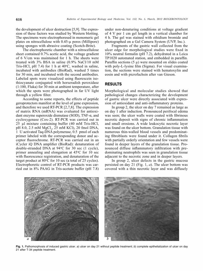
In group 2, ulcer defects in the gastric mucosa persisted on day 21 (Fig. 1, a). The ulcer bottom was covered with a thin necrotic layer and was diffusely
Fig. 1. Pathomorphosis of induced gastric ulcer. a) ulcer on day 21 without peptide treatment; b) complete epithelialization of ulcer on day 21 after T-34 peptide treatment.
Bulletin of Experimental Biology and Medicine, Vol. 152, No. 5, March, 2012 BIOGERONTOLOGY
617
infi ltrated with leukocytes. Maturing granulation tis-sue in the sublying layer contained partially orderly oriented collagen fi brils and was moderately infi ltrated with histiocytes, lymphocytes, and neutrophils.
In group 3 (injections of T-34 peptide), the area and depth of ulcer defect decreased on day 7; the ulcer bottom was free from detritus and infl ammation round the ulcer reduced. Histological studies of the gastric ulcer showed a thinner leukocytic necrotic layer. The sublying layer consisted of maturing granulation tissue with numerous vessels and moderately pronounced diffuse leukocytic infi ltration (mainly histiocytes, lym-phocytes, neutrophils). Cyst-like dilated glands with epitheliocyte proliferation were detected in the ulcer edges.
No ulcer defects were detected on day 21 after T-34 peptide treatment (Fig. 1, b). The gastric mucosa was evenly epithelialized and was pink. The ulcer de-fect epithelialized. No ulcerative defects were found in the mucosa. There were just zones with glandular cavities lined with proliferating epithelium. Fibrous tissue with orderly oriented collagen fi brils and slight infl ammatory lymphoid histiocytic infi ltration were found in the sublying stroma. Injections of the antibi-otic (clarithromycin; group 4) led to the same effects on days 7 and 21 as treatment with T-34 peptide.
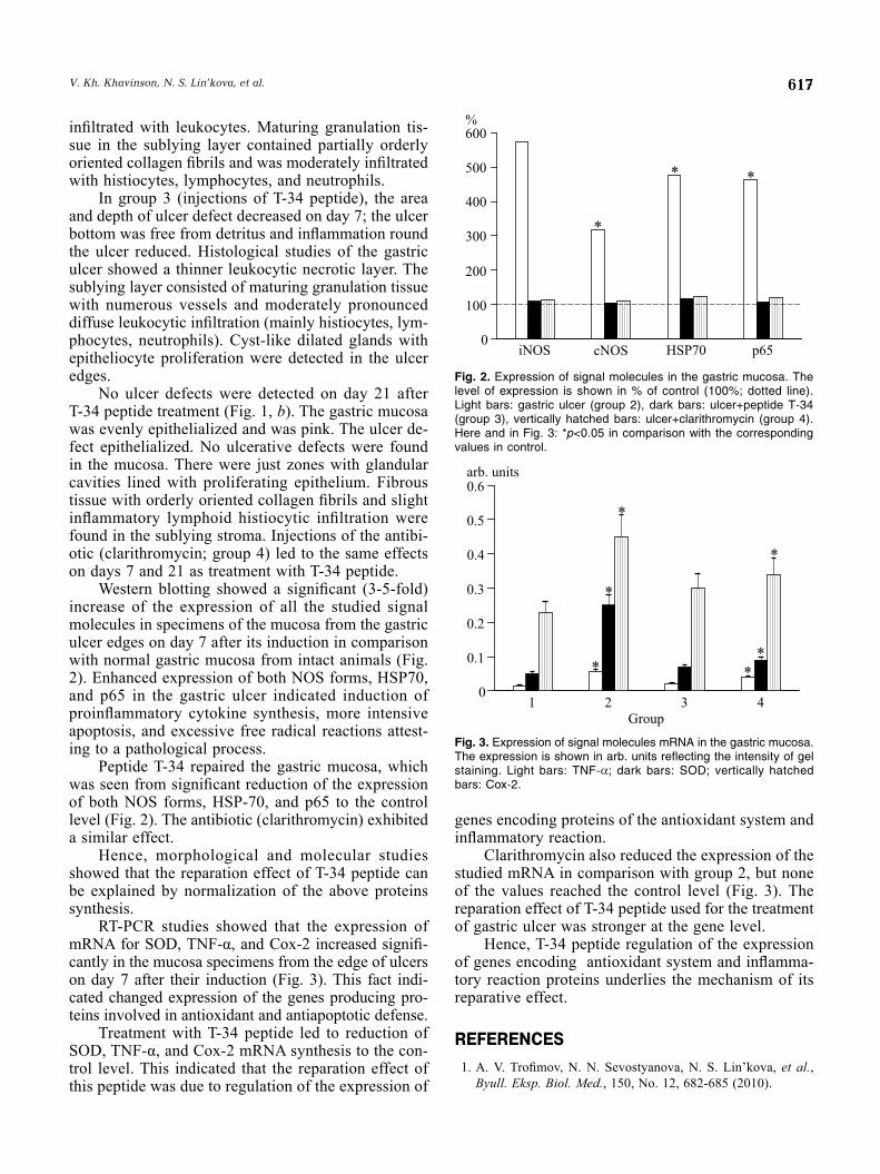
Western blotting showed a signifi cant (3-5-fold) increase of the expression of all the studied signal molecules in specimens of the mucosa from the gastric ulcer edges on day 7 after its induction in comparison with normal gastric mucosa from intact animals (Fig. 2). Enhanced expression of both NOS forms, HSP70, and p65 in the gastric ulcer indicated induction of proinfl ammatory cytokine synthesis, more intensive apoptosis, and excessive free radical reactions attest-ing to a pathological process.
Peptide T-34 repaired the gastric mucosa, which was seen from signifi cant reduction of the expression of both NOS forms, HSP-70, and p65 to the control level (Fig. 2). The antibiotic (clarithromycin) exhibited a similar effect.
Hence, morphological and molecular studies showed that the reparation effect of T-34 peptide can be explained by normalization of the above proteins synthesis.
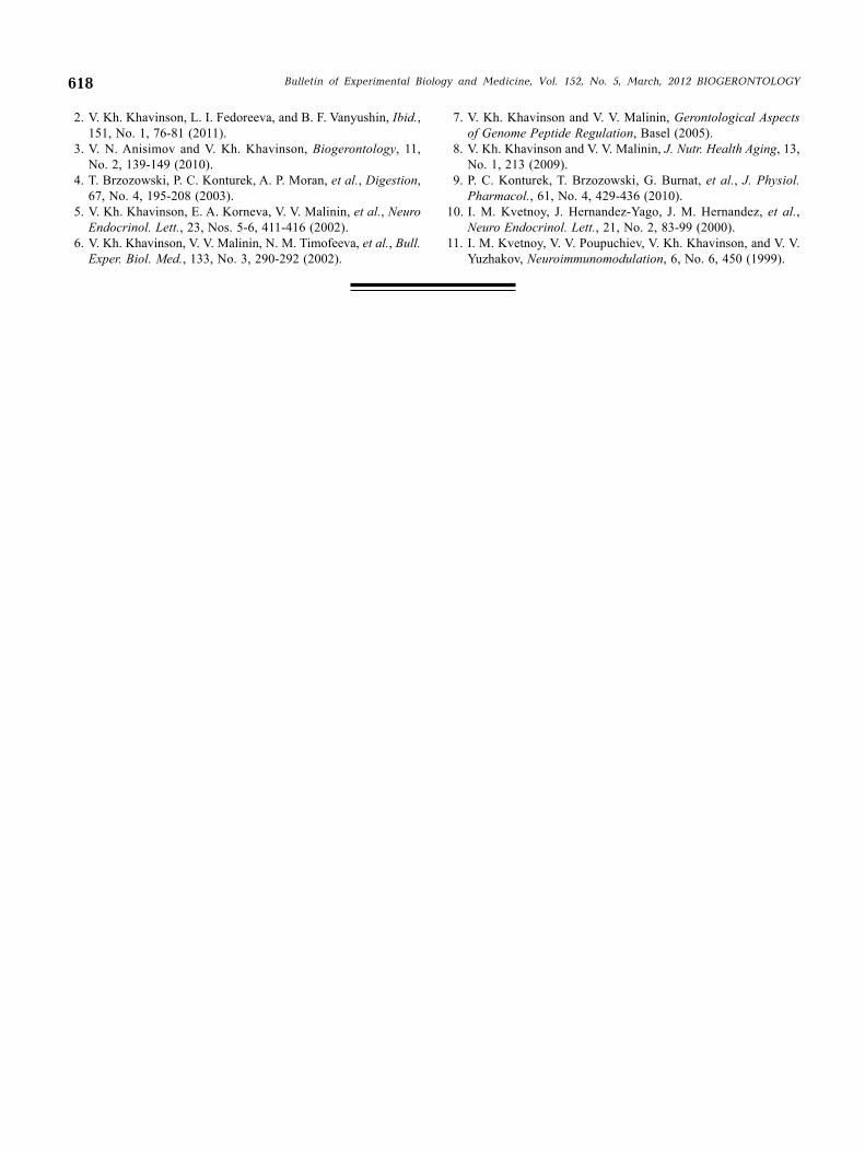
RT-PCR studies showed that the expression of mRNA for SOD, TNF-α, and Cox-2 increased signifi -cantly in the mucosa specimens from the edge of ulcers on day 7 after their induction (Fig. 3). This fact indi-cated changed expression of the genes producing pro-teins involved in antioxidant and antiapoptotic defense.
Treatment with T-34 peptide led to reduction of SOD, TNF-α, and Cox-2 mRNA synthesis to the con-trol level. This indicated that the reparation effect of this peptide was due to regulation of the expression of
genes encoding proteins of the antioxidant system and infl ammatory reaction.
Clarithromycin also reduced the expression of the studied mRNA in comparison with group 2, but none of the values reached the control level (Fig. 3). The reparation effect of T-34 peptide used for the treatment of gastric ulcer was stronger at the gene level.
Hence, T-34 peptide regulation of the expression of genes encoding antioxidant system and infl amma-tory reaction proteins underlies the mechanism of its reparative effect.
REFERENCES
1. A. V. Trofi mov, N. N. Sevostyanova, N. S. Lin’kova, et al., Byull. Eksp. Biol. Med., 150, No. 12, 682-685 (2010).
Fig. 2. Expression of signal molecules in the gastric mucosa. The level of expression is shown in % of control (100%; dotted line). Light bars: gastric ulcer (group 2), dark bars: ulcer+peptide T-34 (group 3), vertically hatched bars: ulcer+clarithromycin (group 4). Here and in Fig. 3: *p<0.05 in comparison with the corresponding values in control.
Fig. 3. Expression of signal molecules mRNA in the gastric mucosa. The expression is shown in arb. units reflecting the intensity of gel staining. Light bars: TNF-; dark bars: SOD; vertically hatched bars: Cox-2.
V. Kh. Khavinson, N. S. Lin’kova, et al.
618
2. V. Kh. Khavinson, L. I. Fedoreeva, and B. F. Vanyushin, Ibid., 151, No. 1, 76-81 (2011).
3. V. N. Anisimov and V. Kh. Khavinson, Biogerontology, 11, No. 2, 139-149 (2010).
4. T. Brzozowski, P. C. Konturek, A. P. Moran, et al., Digestion, 67, No. 4, 195-208 (2003).
5. V. Kh. Khavinson, E. A. Korneva, V. V. Malinin, et al., Neuro Endocrinol. Lett., 23, Nos. 5-6, 411-416 (2002).
6. V. Kh. Khavinson, V. V. Malinin, N. M. Timofeeva, et al., Bull. Exper. Biol. Med., 133, No. 3, 290-292 (2002).
7. V. Kh. Khavinson and V. V. Malinin, Gerontological Aspects of Genome Peptide Regulation, Basel (2005).
8. V. Kh. Khavinson and V. V. Malinin, J. Nutr. Health Aging, 13, No. 1, 213 (2009).
9. P. C. Konturek, T. Brzozowski, G. Burnat, et al., J. Physiol. Pharmacol., 61, No. 4, 429-436 (2010).
10. I. M. Kvetnoy, J. Hernandez-Yago, J. M. Hernandez, et al., Neuro Endocrinol. Lett., 21, No. 2, 83-99 (2000).
11. I. M. Kvetnoy, V. V. Poupuchiev, V. Kh. Khavinson, and V. V. Yuzhakov, Neuroimmunomodulation, 6, No. 6, 450 (1999).
Bulletin of Experimental Biology and Medicine, Vol. 152, No. 5, March, 2012 BIOGERONTOLOGY