Embed Size (px)
Citation preview
Submitted 20 March 2013Accepted 6 May 2013Published 21 May 2013
Corresponding authorClaus O. Wilke,[email protected]
Academic editorEmanuele Paci
Additional Information andDeclarations can be found onpage 9
DOI 10.7717/peerj.80
Copyright2013 Tien et al.
Distributed underCreative Commons CC-BY 3.0
OPEN ACCESS
PeptideBuilder: A simple Python libraryto generate model peptidesMatthew Z. Tien1, Dariya K. Sydykova2, Austin G. Meyer2,3 andClaus O. Wilke2
1 Department of Biochemistry & Molecular Biology, The University of Chicago, Chicago, IL,USA
2 Section of Integrative Biology, Institute for Cellular and Molecular Biology, and Center forComputational Biology and Bioinformatics, The University of Texas at Austin, Austin, TX, USA
3 School of Medicine, Texas Tech University Health Sciences Center, Lubbock, TX, USA
ABSTRACTWe present a simple Python library to construct models of polypeptides from scratch.The intended use case is the generation of peptide models with pre-specified back-bone angles. For example, using our library, one can generate a model of a set ofamino acids in a specific conformation using just a few lines of python code. We donot provide any tools for energy minimization or rotamer packing, since powerfultools are available for these purposes. Instead, we provide a simple Python interfacethat enables one to add residues to a peptide chain in any desired conformation. Bondangles and bond lengths can be manipulated if so desired, and reasonable values areused by default.
Subjects Biochemistry, Biophysics, Computational Biology, Computational ScienceKeywords Protein structure, Molecular modeling, Computational biology, Model peptides
INTRODUCTIONResearchers working in structural biology and related fields frequently have to create,
manipulate, or analyze protein crystal structures. To aid this work, many different
software tools have been developed. Examples include visualization (Schrodinger, 2013),
mutagenesis (Schrodinger, 2013; Leaver-Fay et al., 2011), high-throughput computational
analysis (Hamelryck & Manderick, 2003; Grant et al., 2006), ab-initio protein folding and
protein design (Leaver-Fay et al., 2011), and homology modeling and threading (Eswar et
al., 2006; Zhang, 2008). In comparison, a relatively simple task, the ab-initio creation of a
protein structure in a desired conformation, has received little attention. It is possible to
perform this task in PyRosetta (Chaudhury, Lyskov & Gray, 2010; Gray et al., 2013), but that
approach incurs the overhead of the entire Rosetta protein modeling package (Leaver-Fay
et al., 2011). One can also construct peptides manually in some graphical molecular
modeling packages, such as Swiss-PdbViewer (Guex & Peitsch, 1997). Finally, the Rose
lab has developed Ribosome (Srinivasan, 2013), a small program with the express purpose
of creating model peptides. However, Ribosome is implemented in Fortran, an outdated
programming language that integrates poorly with modern bioinformatics pipelines.
For a recent analysis by our group, we wanted to systematically enumerate GLY-X-GLY
tripeptides in all allowed conformations (Tien et al., 2012). After review of the available
How to cite this article Tien et al. (2013), PeptideBuilder: A simple Python library to generate model peptides. PeerJ 1:e80;DOI 10.7717/peerj.80

software packages, we determined that there was a need for a lightweight library,
implemented in a modern programming language, that would allow us to construct
arbitrary peptides in any desired conformation. We decided to write this library in the
language Python (Python Sofware Foundation, 2013), as this language is widely used in
scientific computing. Specifically, many tools suitable for computational biology and
bioinformatics are available (Cock et al., 2009), including tools to read, manipulate, and
write PDB (Protein Data Bank) files (Hamelryck & Manderick, 2003). This effort resulted in
the Python library PeptideBuilder, which we describe here. The library consists of two
Python files comprising a total of approximately 2000 lines of code. Both files are provided
as Supplemental Information 1. The entire PeptideBuilder package is also available online
at https://github.com/mtien/PeptideBuilder.
CONCEPTUAL OVERVIEWThe key function our library provides is to add a residue at the C terminus of an existing
polypeptide model, using arbitrary backbone angles. Our library also allows a user to
generate an individual amino acid residue and place it into an otherwise empty model. In
combination, these two functions enable the construction of arbitrary polypeptide chains.
The generated models are stored as structure objects using the PDB module of Biopython
(Bio.PDB, Hamelryck & Manderick 2003). The seemless integration with Biopython’s PDB
module means that we can leverage a wide range of existing functionality, such as writing
structures to PDB files or measuring distances between atoms.
Adding a residue to an existing polypeptide chain involves two separate steps. First,
we have to establish the desired geometric arrangement of all atoms in the residue to be
added. This means we have to determine all bond lengths and angles. In practice, we will
usually want to specify the dihedral backbone angles φ and ψ , and possibly the rotamers,
whereas all other bond lengths and angles should be set to reasonable defaults for the
amino acid under consideration. Once we have determined the desired geometry, we
have to calculate the actual position of all atoms in 3-space and then add the atoms to the
structure object. The exact calculations required to convert bond lengths and angles into
3D atom coordinates are given in the supporting text of Tien et al. (2012). Our library
places all heavy atoms for each residue, but it does not place hydrogens.
We obtained default values for bond lengths and angles by measuring these quantities
in a large collection of published crystal structures and recording the average for each
quantity, as described (Tien et al., 2012). We set the default for the backbone dihedral
angles to the extended conformation (φ = −120◦, ψ = 140◦, ω = 180◦). We based the
default rotamer angles of each individual amino acid on the rotamer library of Shapovalov
& Dunbrack (2011). For each amino acid, the rotamer library provided the frequency of
each combination of rotamer angles given the backbone conformation. We analyzed this
library at the extended backbone conformation (φ =−120◦,ψ = 140◦) and used the most
likely rotamer conformation at that backbone conformation as default. For amino acids
with multiple χ1 or χ2 angles (such as Ile or Leu) we used the most likely and the second
most likely angles from the rotamer library.
Tien et al. (2013), PeerJ, DOI 10.7717/peerj.80 2/10
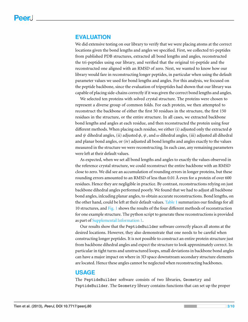
EVALUATIONWe did extensive testing on our library to verify that we were placing atoms at the correct
locations given the bond lengths and angles we specified. First, we collected tri-peptides
from published PDB structures, extracted all bond lengths and angles, reconstructed
the tri-peptides using our library, and verified that the original tri-peptide and the
reconstructed one aligned with an RMSD of zero. Next, we wanted to know how our
library would fare in reconstructing longer peptides, in particular when using the default
parameter values we used for bond lengths and angles. For this analysis, we focused on
the peptide backbone, since the evaluation of tripeptides had shown that our library was
capable of placing side-chains correctly if it was given the correct bond lengths and angles.
We selected ten proteins with solved crystal structure. The proteins were chosen to
represent a diverse group of common folds. For each protein, we then attempted to
reconstruct the backbone of either the first 50 residues in the structure, the first 150
residues in the structure, or the entire structure. In all cases, we extracted backbone
bond lengths and angles at each residue, and then reconstructed the protein using four
different methods. When placing each residue, we either (i) adjusted only the extracted φ
andψ dihedral angles, (ii) adjusted φ,ψ , and ω dihedral angles, (iii) adjusted all dihedral
and planar bond angles, or (iv) adjusted all bond lengths and angles exactly to the values
measured in the structure we were reconstructing. In each case, any remaining parameters
were left at their default values.
As expected, when we set all bond lengths and angles to exactly the values observed in
the reference crystal structure, we could reconstruct the entire backbone with an RMSD
close to zero. We did see an accumulation of rounding errors in longer proteins, but these
rounding errors amounted to an RMSD of less than 0.01 A even for a protein of over 600
residues. Hence they are negligible in practice. By contrast, reconstructions relying on just
backbone dihedral angles performed poorly. We found that we had to adjust all backbone
bond angles, inlcuding planar angles, to obtain accurate reconstructions. Bond lengths, on
the other hand, could be left at their default values. Table 1 summarizes our findings for all
10 structures, and Fig. 1 shows the results of the four different methods of reconstruction
for one example structure. The python script to generate these reconstructions is provided
as part of Supplemental Information 1.
Our results show that the PeptideBuilder software correctly places all atoms at the
desired locations. However, they also demonstrate that one needs to be careful when
constructing longer peptides. It is not possible to construct an entire protein structure just
from backbone dihedral angles and expect the structure to look approximately correct. In
particular in tight turns and unstructured loops, small deviations in backbone bond angles
can have a major impact on where in 3D space downstream secondary structure elements
are located. Hence these angles cannot be neglected when reconstructing backbones.
USAGEThe PeptideBuilder software consists of two libraries, Geometry and
PeptideBuilder. The Geometry library contains functions that can set up the proper
Tien et al. (2013), PeerJ, DOI 10.7717/peerj.80 3/10
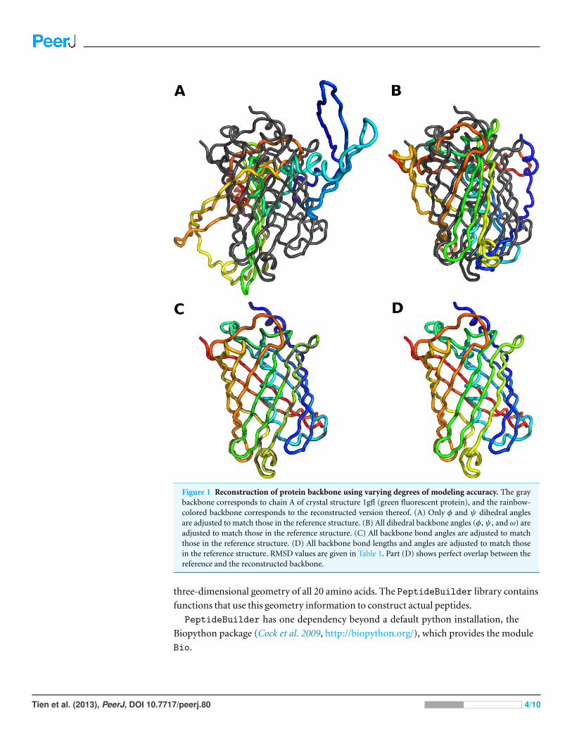
Figure 1 Reconstruction of protein backbone using varying degrees of modeling accuracy. The graybackbone corresponds to chain A of crystal structure 1gfl (green fluorescent protein), and the rainbow-colored backbone corresponds to the reconstructed version thereof. (A) Only φ and ψ dihedral anglesare adjusted to match those in the reference structure. (B) All dihedral backbone angles (φ,ψ , and ω) areadjusted to match those in the reference structure. (C) All backbone bond angles are adjusted to matchthose in the reference structure. (D) All backbone bond lengths and angles are adjusted to match thosein the reference structure. RMSD values are given in Table 1. Part (D) shows perfect overlap between thereference and the reconstructed backbone.
three-dimensional geometry of all 20 amino acids. The PeptideBuilder library contains
functions that use this geometry information to construct actual peptides.
PeptideBuilder has one dependency beyond a default python installation, the
Biopython package (Cock et al. 2009, http://biopython.org/), which provides the module
Bio.
Tien et al. (2013), PeerJ, DOI 10.7717/peerj.80 4/10
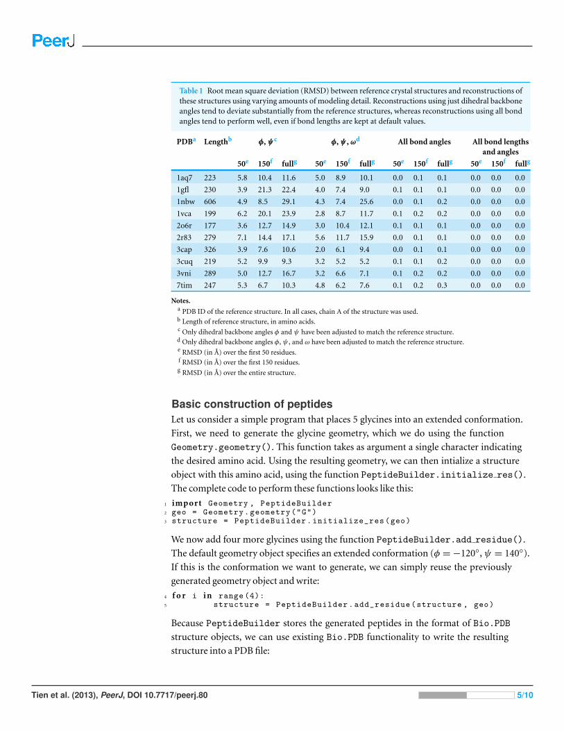
Table 1 Root mean square deviation (RMSD) between reference crystal structures and reconstructions ofthese structures using varying amounts of modeling detail. Reconstructions using just dihedral backboneangles tend to deviate substantially from the reference structures, whereas reconstructions using all bondangles tend to perform well, even if bond lengths are kept at default values.
PDBa Lengthb φ,ψc φ,ψ , ωd All bond angles All bond lengthsand angles
50e 150f fullg 50e 150f fullg 50e 150f fullg 50e 150f fullg
1aq7 223 5.8 10.4 11.6 5.0 8.9 10.1 0.0 0.1 0.1 0.0 0.0 0.0
1gfl 230 3.9 21.3 22.4 4.0 7.4 9.0 0.1 0.1 0.1 0.0 0.0 0.0
1nbw 606 4.9 8.5 29.1 4.3 7.4 25.6 0.0 0.1 0.2 0.0 0.0 0.0
1vca 199 6.2 20.1 23.9 2.8 8.7 11.7 0.1 0.2 0.2 0.0 0.0 0.0
2o6r 177 3.6 12.7 14.9 3.0 10.4 12.1 0.1 0.1 0.1 0.0 0.0 0.0
2r83 279 7.1 14.4 17.1 5.6 11.7 15.9 0.0 0.1 0.1 0.0 0.0 0.0
3cap 326 3.9 7.6 10.6 2.0 6.1 9.4 0.0 0.1 0.1 0.0 0.0 0.0
3cuq 219 5.2 9.9 9.3 3.2 5.2 5.2 0.1 0.1 0.2 0.0 0.0 0.0
3vni 289 5.0 12.7 16.7 3.2 6.6 7.1 0.1 0.2 0.2 0.0 0.0 0.0
7tim 247 5.3 6.7 10.3 4.8 6.2 7.6 0.1 0.2 0.3 0.0 0.0 0.0
Notes.a PDB ID of the reference structure. In all cases, chain A of the structure was used.b Length of reference structure, in amino acids.c Only dihedral backbone angles φ and ψ have been adjusted to match the reference structure.d Only dihedral backbone angles φ, ψ , and ω have been adjusted to match the reference structure.e RMSD (in A) over the first 50 residues.f RMSD (in A) over the first 150 residues.g RMSD (in A) over the entire structure.
Basic construction of peptidesLet us consider a simple program that places 5 glycines into an extended conformation.
First, we need to generate the glycine geometry, which we do using the function
Geometry.geometry(). This function takes as argument a single character indicating
the desired amino acid. Using the resulting geometry, we can then intialize a structure
object with this amino acid, using the function PeptideBuilder.initialize res().
The complete code to perform these functions looks like this:
1 import Geometry , PeptideBuilder2 geo = Geometry.geometry("G")3 structure = PeptideBuilder.initialize_res(geo)
We now add four more glycines using the function PeptideBuilder.add residue().
The default geometry object specifies an extended conformation (φ =−120◦,ψ = 140◦).
If this is the conformation we want to generate, we can simply reuse the previously
generated geometry object and write:
4 f o r i i n range (4):5 structure = PeptideBuilder.add_residue(structure , geo)
Because PeptideBuilder stores the generated peptides in the format of Bio.PDB
structure objects, we can use existing Bio.PDB functionality to write the resulting
structure into a PDB file:
Tien et al. (2013), PeerJ, DOI 10.7717/peerj.80 5/10

Table 2 Overview of functions provided by PeptideBuilder.
Function name Description
add residue Adds a single residue to a structure.
initialize res Creates a new structure containing a single amino acid.
make structure Builds an entire peptide with specified amino acid sequenceand backbone angles.
make extended structure Builds an entire peptide in the extended conformation.
make structure from geos Builds an entire peptide from a list of geometry objects.
6 import Bio.PDB # import Biopython ’s PDB module.7 out = Bio.PDB.PDBIO()8 out.set_structure(structure)9 out.save( "example.pdb" )
If we want to generate a peptide that is not in the extended conformation, we have to adjust
the backbone dihedral angles accordingly. For example, we could place the five glycines
into an alpha helix by setting φ = −60◦ and ψ = −40◦. We do this by manipulating the
phi and psi im1 members of the geometry object. (We are not actually specifying the
ψ angle of the residue to be added, but the corresponding angle of the previous residue,
ψi−1. Hence the member name psi im1. See the Detailed adjustment of residue geometry
Section for details.) The code example looks as follows:
1 geo = Geometry.geometry("G")2 geo.phi=-603 geo.psi_im1 =-404 structure = PeptideBuilder.initialize_res(geo)
5 f o r i i n range (4):6 structure = PeptideBuilder.add_residue(structure , geo)
Several convenience functions exist that simplify common tasks. For example, if we simply
want to add a residue at specific backbone angles, we can use an overloaded version of
the function add residue() that takes as arguments the structure to which the residue
should be added, the amino acid in single-character code, and the φ andψi−1 angles:
1 # add an arginine , setting phi=-60 and psi_im1 =-402 structure = PeptideBuilder.add_residue(structure , "R", -60, -40)
If we want to place an arbitrary sequence of amino acids into an extended structure, we can
use the function make extended structure(), which takes as its sole argument a string
holding the desired amino-acid sequence:
1 # construct a peptide corresponding to the2 # sequence "MGGLTR" in extended conformation3 structure = PeptideBuilder.make_extended_structure("MGGLTR")
Table 2 summarizes all functions provided by PeptideBuilder. All these functions are
documented in the source code using standard Python self-documentation methods.
Detailed adjustment of residue geometryGeometry objects contain all the bond lengths, bond angles, and dihedral angles necessary
to specify a given amino acid. These parameters are stored as member variables, and can be
Tien et al. (2013), PeerJ, DOI 10.7717/peerj.80 6/10
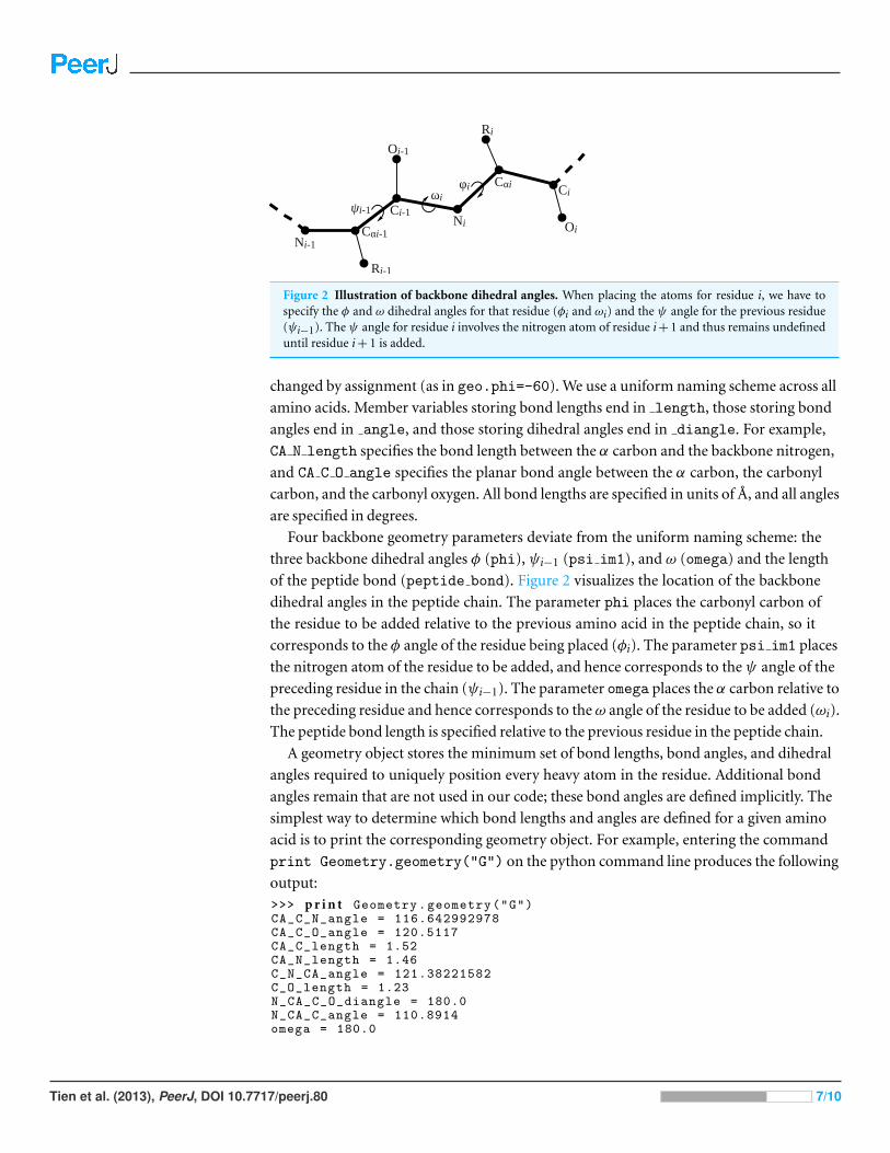
Figure 2 Illustration of backbone dihedral angles. When placing the atoms for residue i, we have tospecify the φ and ω dihedral angles for that residue (φi and ωi) and the ψ angle for the previous residue(ψi−1). The ψ angle for residue i involves the nitrogen atom of residue i+ 1 and thus remains undefineduntil residue i+ 1 is added.
changed by assignment (as in geo.phi=-60). We use a uniform naming scheme across all
amino acids. Member variables storing bond lengths end in length, those storing bond
angles end in angle, and those storing dihedral angles end in diangle. For example,
CA N length specifies the bond length between the α carbon and the backbone nitrogen,
and CA C O angle specifies the planar bond angle between the α carbon, the carbonyl
carbon, and the carbonyl oxygen. All bond lengths are specified in units of A, and all angles
are specified in degrees.
Four backbone geometry parameters deviate from the uniform naming scheme: the
three backbone dihedral angles φ (phi), ψi−1 (psi im1), and ω (omega) and the length
of the peptide bond (peptide bond). Figure 2 visualizes the location of the backbone
dihedral angles in the peptide chain. The parameter phi places the carbonyl carbon of
the residue to be added relative to the previous amino acid in the peptide chain, so it
corresponds to the φ angle of the residue being placed (φi). The parameter psi im1 places
the nitrogen atom of the residue to be added, and hence corresponds to theψ angle of the
preceding residue in the chain (ψi−1). The parameter omega places the α carbon relative to
the preceding residue and hence corresponds to theω angle of the residue to be added (ωi).
The peptide bond length is specified relative to the previous residue in the peptide chain.
A geometry object stores the minimum set of bond lengths, bond angles, and dihedral
angles required to uniquely position every heavy atom in the residue. Additional bond
angles remain that are not used in our code; these bond angles are defined implicitly. The
simplest way to determine which bond lengths and angles are defined for a given amino
acid is to print the corresponding geometry object. For example, entering the command
print Geometry.geometry("G") on the python command line produces the following
output:>>> p r i n t Geometry.geometry("G")CA_C_N_angle = 116.642992978CA_C_O_angle = 120.5117CA_C_length = 1.52CA_N_length = 1.46C_N_CA_angle = 121.38221582C_O_length = 1.23N_CA_C_O_diangle = 180.0N_CA_C_angle = 110.8914omega = 180.0
Tien et al. (2013), PeerJ, DOI 10.7717/peerj.80 7/10

peptide_bond = 1.33phi = -120psi_im1 = 140residue_name = G
The following code prints out the default geometries for all amino acids:
1 f o r aa i n "ACDEFGHIKLMNPQRSTVWY":2 p r i n t Geometry.geometry(aa)
We can construct modified geometries simply by assigning new values to the appropriate
member variables. For example, the following code constructs a Gly for which some bond
lengths and angles deviate slightly from the default values:
1 geo = Geometry.geometry("G")2 geo.phi=-1193 geo.psi_im1 =1414 geo.omega =179.05 geo.peptide_bond =1.36 geo.C_O_length =1.207 geo.CA_C_O_angle =121.68 geo.N_CA_C_O_diangle= 181.0
For amino acids whose side chains require specification of rotamer conformations,
there are two ways to specify them. First, we can set rotamers by directly assigning the
appropriate values to the correct dihedral angles:
1 geo = Geometry.geometry("L")2 geo.N_CA_CB_CG_diangle =-60.03 geo.CA_CB_CG_CD1_diangle =-80.24 geo.CA_CB_CG_CD2_diangle =181.0
Second, we can set all rotamer angles at once, using the member function
inputRotamers():
1 geo = Geometry.geometry("L")2 geo.inputRotamers ([-60.0, -80.2, 181.1])
In this function call, the angles are listed in order of standard biochemical convention, χ1,
χ2, χ3, and so on, for however many χ angles the amino-acid side chain has.
CONCLUSIONWe have developed a Python library to construct model peptides. Our design goals were to
make the library simple, lightweight, and easy-to-use. Using our library, one can construct
model peptides in only a few lines of Python code, as long as default bond lengths and
angles are acceptable. At the same time, all bond-length and bond-angle parameters are
user-accessible and can be modified if so desired. We have verified that our library places
atoms correctly. As part of this verification effort, we have found that with increasing
peptide length it becomes increasingly important to adjust bond angles appropriately to
reconstruct biophysically accurate protein structures.
ACKNOWLEDGEMENTSWe thank Jeffrey Gray for helpful comments on this work.
Tien et al. (2013), PeerJ, DOI 10.7717/peerj.80 8/10
ADDITIONAL INFORMATION AND DECLARATIONS
FundingThis work was supported by NIH grant R01 GM088344 to COW. The funders had no role
in study design, data collection and analysis, decision to publish, or preparation of the
manuscript.
Grant DisclosuresThe following grant information was disclosed by the authors:
NIH: R01 GM088344.
Competing InterestsThe authors declare no competing interests.
Author Contributions• Matthew Z. Tien and Claus O. Wilke conceived and designed the experiments,
performed the experiments, analyzed the data, contributed reagents/materials/analysis
tools, wrote the paper.
• Dariya K. Sydykova and Austin G. Meyer conceived and designed the experiments,
contributed reagents/materials/analysis tools, wrote the paper.
Supplemental InformationSupplemental information for this article can be found online at http://dx.doi.org/
10.7717/peerj.80.
REFERENCESChaudhury S, Lyskov S, Gray JJ. 2010. PyRosetta: a script-based interface for implementing
molecular modeling algorithms using Rosetta. Bioinformatics 26:689–691 DOI ./bioin-formatics/btq.
Cock PJ, Antao T, Chang JT, Chapman BA, Cox CJ, Dalke A, Friedberg I, Hamelryck T,Kauff F, Wilczynski B, de Hoon MJ. 2009. Biopython: freely available Python tools forcomputational molecular biology and bioinformatics. Bioinformatics 25:1422–1423DOI ./bioinformatics/btp.
Eswar N, Marti-Renom MA, Webb B, Madhusudhan MS, Eramian D, Shen M, Pieper U, Sali A.2006. Comparative protein structure modeling with MODELLER. Current Protocols inBioinformatics 15(Supplement):5.6.1–5.6.30 DOI ./.bis.
Grant BJ, Rodrigues APC, ElSawy KM, McCammon JA, Caves LSD. 2006. Bio3D: An Rpackage for the comparative analysis of protein structures. Bioinformatics 22:2695–2696DOI ./bioinformatics/btl.
Gray JJ, Chaudhury S, Lyskov S, Labonte JW. 2013. The PyRosetta interactive platform for proteinstructure prediction and design: a set of educational modules, 2nd edition. Baltimore: CreateSpaceIndependent Publishing Platform.
Guex N, Peitsch MC. 1997. SWISS-MODEL and the Swiss-PdbViewer: an environment forcomparative protein modeling. Electrophoresis 18:2714–2723 DOI ./elps..
Tien et al. (2013), PeerJ, DOI 10.7717/peerj.80 9/10

Hamelryck T, Manderick B. 2003. PDB file parser and structure class implemented in Python.Bioinformatics 19:2308–2310 DOI ./bioinformatics/btg.
Leaver-Fay A, Tyka M, Lewis SM, Lange OF, Thompson J, Jacak R, Kaufman K, Renfrew DP,Smith CA, Sheffler W, Davis IW, Cooper S, Treuille A, Mandell DJ, Richter F, Ban YEA,Fleishman SJ, Corn JE, Kim DE, Lyskov S, Berrondo M, Mentzer S, Popovic Z, Havranek JJ,Karanicolas J, Das R, Meiler J, Kortemme T, Gray JJ, Kuhlman B, Baker D, Bradley P. 2011.ROSETTA3: an object-oriented software suite for the simulation and design of macromolecules.Methods in Enzymology 487:545–574 DOI ./B----.-.
Python Sofware Foundation. Available at http://www.python.org (accessed 19 March 2013).
Schrodinger L. The pymol molecular graphics system. Available at http://www.pymol.org/ (accessed19 March 2013).
Shapovalov MV, Dunbrack RL. 2011. A smoothed backbone-dependent rotamer library forproteins derived from adaptive kernel density estimates and regressions. Structure 19:844–858DOI ./j.str....
Srinivasan R. Ribosome – program to build coordinates for peptides from sequence. Available athttp://roselab.jhu.edu/∼raj/Manuals/ribosome.html (accessed 19 March 2013).
Tien MZ, Meyer AG, Spielman SJ, Wilke CO. 2012. Maximum allowed solvent accessibilites ofresidues in proteins. arXiv preprint arXiv:1211.4251 [q-bio.BM].
Zhang Y. 2008. I-TASSER server for protein 3D structure prediction. BMC Bioinformatics 9:40DOI ./---.
Tien et al. (2013), PeerJ, DOI 10.7717/peerj.80 10/10