Embed Size (px)
Citation preview

Peptic UlcerPeptic UlcerPeptic UlcerPeptic Ulcer Mrs Nancy Allen is a 79-year-old pensioner who has been sent to the Emergency Dept that morning by
her general practitioner because of upper abdominal pain and black stools over the last 2 days.
History of Presenting Illness Burning epigastric pain 1.5 to 3 hrs after meals
���� Wakes patient up at night - exacerbated by fasting - improved with meals - improved with antacids
- BLACK STOOLS
Differential Diagnoses Biliary Colic Cholelithiasis Gastritis, Acute Gastritis, Chronic Gastroesophageal Reflux Disease Mesenteric Artery Ischemia Myocardial Ischemia Pancreatic Cancer
Pancreatitis, Acute Pancreatitis, Chronic Drug-induced dyspepsia Duodenitis Functional (nonulcer) dyspepsia Gastric infections Infiltrative diseases of the stomach
Pertinent Findings on History only 20-25% of patients with
symptoms suggestive of peptic
ulceration are found on
investigation to have a peptic ulcer. Findings on Examination : few and non-specific - Epigastric tenderness - Guaiac-positive blood in stool resulting from occult blood loss - Melena resulting from acute or subacute gastrointestinal bleeding - Succussion splash resulting from partial or complete gastric outlet obstruction
- SOMETIMES: stigma of chronic liver disease, eg. distended abdomen, ecchymoses, jaundice
There is no symptom complex that can
adequately differentiate gastric from
duodenal ulcers and nonulcer dyspepsia
GASTRITIS is made
WORSE WITH FOOD
Peptic Ulcer may be felt as a gnawing pain in the chest, back, mid-abdomen, or either upper quadrant
Melena = at least 60 mls of uper Gi bleeding
Really need at least 150 – 200 for the “black tarry”
Takes 8 hours to turn black
80% of pancreatitis
presents at 1 am (?…)
The important causes of
upper GI bleeding
- Duodenal ulcer
- Gastric ulcer
- Gastric erosions
- Ulcerative esophagitis
- Esophagogastric varices
- Mallory-Weiss tear
- Carcinoma, lymphoma
- Angiodysplasia
Nausea Vomiting, Dyspepsia, - belching, - bloating, - distention, - fatty food intolerance Heartburn Chest discomfort Anorexia, weight loss Hematemesis
Red Blood + Clots? = per rectum (massive duodenal bleed)
Cardiovascular effects eg. - tachycardia, palpitations - weak pulse - SYMPTOMS OF SHOCK
Anaemia (indicates chronicity)
ALCOHOL
TOBACCO
NSAIDs
And COCAINE
HISTORICALLY chronic peptic-ulcer sounding intermittent food-relieved epigastric pain… …SUDDEN TURN FOR WORSE: SEVERE CONSTANT AND GENERALISED
WITH RIGID ABDOMEN AND TACHYCARDIA ���� !! Perforated Ulcer !!
May be Embarrassing: Could all that blood be
coming from the patient’s
chronically bleeding NOSE??
put together by Alex Yartsev: Sorry if i used your imagesor data and forgot to reference you. Tell me who you are.

Tests and Investigations Full Blood Count To make sure they are not anaemic
Coagulation assay To make sure that you can operate on them
Chest X-ray Looking for gastric abnormality Also looking to explain any cardiovascular
side-findings Serum Biochemistry UREA is elevated in Helicobacter Pylori infections EXHALED UREA breath tests
Urea is ingested; in the presence of urease produced by H pylori,
labeled carbon dioxide (heavy isotope, carbon-13, or radioactive
isotope, carbon-14) is produced in the stomach, absorbed into the
bloodstream, diffused into the lungs, and exhaled.
GASTROSCOPY and/or ENDOSCOPY with subsequent BIOPSY
PLUS: Rapid urease tests for H. Pylori The presence of H pylori in gastric mucosal biopsy specimens is detected by testing for
the bacterial product urease. Three kits (CLOtest, Hpfast, Pyloritek) are commercially
available, each containing a combination of a urea substrate and a pH sensitive
indicator. One or more gastric biopsy specimens are placed in the rapid urease test kit. If
H pylori are present, bacterial urease converts urea to ammonia, which changes pH and
produces a color change.
How is this diagnosis made? Via BIOPSY + HISTOLOGY Reveal many H.Pylori organisms, with infiltration by marcophages + neurophils
MUST CHECK for dysplasia, metaplasia, neoplasia
INDICATIONS FOR IMMEDIATE ENDOSCOPY Those over 45 years of age with new onset of dyspeptic/ulcer symptoms or with other symptoms suggesting malignancy, such as - fever, - weight loss, - early satiety, - vomiting,
Follow-up endoscopy should be performed at least 4 weeks
after cessation of all anti-Helicobacter drugs
IS THE BLEEDING CONTINUING?
Mesenteric angiography may point to the site of ulcer
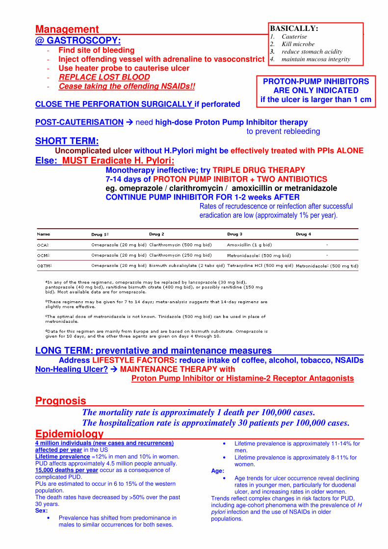
Management @ GASTROSCOPY:
- Find site of bleeding - Inject offending vessel with adrenaline to vasoconstrict - Use heater probe to cauterise ulcer - REPLACE LOST BLOOD - Cease taking the offending NSAIDs!!
CLOSE THE PERFORATION SURGICALLY if perforated POST-CAUTERISATION ���� need high-dose Proton Pump Inhibitor therapy
to prevent rebleeding
SHORT TERM: Uncomplicated ulcer without H.Pylori might be effectively treated with PPIs ALONE
Else: MUST Eradicate H. Pylori: Monotherapy ineffective; try TRIPLE DRUG THERAPY 7-14 days of PROTON PUMP INIBITOR + TWO ANTIBIOTICS eg. omeprazole / clarithromycin / amoxicillin or metranidazole CONTINUE PUMP INHIBITOR FOR 1-2 weeks AFTER
Rates of recrudescence or reinfection after successful eradication are low (approximately 1% per year).
LONG TERM: preventative and maintenance measures Address LIFESTYLE FACTORS: reduce intake of coffee, alcohol, tobacco, NSAIDs Non-Healing Ulcer? ���� MAINTENANCE THERAPY with
Proton Pump Inhibitor or Histamine-2 Receptor Antagonists
Prognosis
The mortality rate is approximately 1 death per 100,000 cases.
The hospitalization rate is approximately 30 patients per 100,000 cases.
Epidemiology 4 million individuals (new cases and recurrences) affected per year in the US Lifetime prevalence =12% in men and 10% in women. PUD affects approximately 4.5 million people annually. 15,000 deaths per year occur as a consequence of complicated PUD. PUs are estimated to occur in 6 to 15% of the western population. The death rates have decreased by >50% over the past 30 years. Sex:
• Prevalence has shifted from predominance in males to similar occurrences for both sexes.
• Lifetime prevalence is approximately 11-14% for men.
• Lifetime prevalence is approximately 8-11% for women.
Age:
• Age trends for ulcer occurrence reveal declining rates in younger men, particularly for duodenal ulcer, and increasing rates in older women.
Trends reflect complex changes in risk factors for PUD, including age-cohort phenomena with the prevalence of H pylori infection and the use of NSAIDs in older populations.
PROTON-PUMP INHIBITORS ARE ONLY INDICATED
if the ulcer is larger than 1 cm
BASICALLY: 1. Cauterise
2. Kill microbe
3. reduce stomach acidity
4. maintain mucosa integrity

Pharmacology of Gastric Secretion Therapies
Antacids Very brutally neutralise gastric acid
Magnesium hydroxide, aluminium hydroxide, sodium bicarbonate (!! CO2 Belching !!) Histamine 2 Receptor Antagonists
Since gastric parietal cells are stimulated by histamine to produce acid via histamine binding to the H2 receptor, at concentrations which are not enough to stimulate blood vessels (this histamine comes from adjacent “mast cell-like cells”) (there is a slow basal secretion of histamine) cimetidine, ranitidine, famotidine and nizatidine
Proton Pump Inhibitors inactivate H+/K+ATPase; thus no protons are pumped into the lumen and thus pH does not decrease. PPIs do not eradicate H.Pylori, but they do suppress its growth omeprazole, lansoprazole, pantoprazole, rabeprazole, esomeprazole
Cytoprotective Mucosa-loving Drugs: Sucralfate = a complex of aluminium hydroxide and sulfated sucrose
In presence of acid will release aluminium Released aluminium acquires a strong negative charge and binds to +ve groups in proteins, glycoproteins, etc. The result is thick mucus which limits the transit of H+ Its like a band-aid for your mucosa. Allows the ulcer to re-epithelialise
NEEDS ACID!! DO NOT GIVE WITH ANTACIDS!!
Carbenoxolone: natural liquorice root product. Promotes mucus. Prostaglandins by nature inhibit gastric acid secretion. Eg. misoprostol Bismuth Chelate: is toxic to H. Pylori and seems to coat the ulcer walls. !! may cause black tongue and faeces, nausea, vomiting, and encephalopathy.
Magnesium salts cause diarrhoea Aluminium salts cause constipation

Secretory cells of the stomach include :
- - - - - - - -
Parietal cell
Stem cell
Mucous neck cell
Superficial epithelial cell
Chief cell
Endocrine cell

- - - -
Gastrointestinal disturbances These are the commonest adverse effects of NSAIDs, rel.risk = 3 to 5 times that of non-NSAID users. 20% of long-term NSAID users will have evidence of ulcers. 30% of all cases of massive gastrointestinal haemorrhage in the elderly are from NSAIDs. misoprostol + famotidine = reduces the incidence of NSAID induced ulceration.
Renal effects NSAIDs block the synthesis of PGI2 and PGE2. (PGI2 and PGE2enhance glomerular filtration and inhibit the tubular reabsorption of sodium). Thus, NSAIDs can cause sodium retention and consequent oedema.
Drug interactions All NSAIDs have the potential to interfere with platelet function The renal clearance of lithium is decreased by some NSAIDs Some NSAIDs reduce the renal clearance of methotrexate
Risk factors in the elderly Crap liver = increased plasma half-liife of NSAIDs The elderly are more likely to be taking other medications for concurrent disease so the risks of drug interaction are greater in this group.
Speaking of the elderly… key word for the barrier: multidimensional holisitic approach Common Comorbidities in the elderly and their functional implications Degenerative arthritis contributes to 25 % of all physical disability in the elderly. Give physio instead of NSAIDs. Chronic Airflow Limitation Dementia occurs in approximately 15% of elderly over 75 years. Visual and hearing impairment . 5 % of elderly over 65, and 25 % over 85 have visual impairment. Around 5 % of elderly over 65 and 50 % over 85 have hearing impairment. Proper medication use .. Adverse drug reactions account for 5 to 10 % of hospital admissions. Social isolation : 40% of elderly females over 65 live alone; 55% of over 80 year olds. The current elderly population comes from a generation that do not question medical opinion. BEWARE

mucus�
GASTRIC SECRETION and its neurohormonal control
The stomach lumen � Parietal Cell
Epithelial Cells: bicarbonate cells
Mucous Cell: pepsinogen 2 cell
D Cell: Somatostatin Cell
G Cell: Gastrin Cell
Mast-like
cell
Muscarinic receptor
Histamine 2 receptor
Gastrin receptor GASTRIN
Acetylcholine
HISTAMINE Induced by gastrin
Calcium
cAMP
ATP H+ / K+ ATPase H+
activates
K+
K+
Cl-
INHIBITORY G protein
Suppresses adenylate cyclase
Prostaglandins somatostatin
HCl Low pH (1.0)
Chief Cell: pepsinogen 1 cell
Vitamin B12 Initially binds to R proteins from saliva (when freed from food by acid and pepsin)
Absorbable complex forms in the duodenum when proteins are degraded by pancreatic enzymes � then its on to he terminal ileum for active transport
Intrinsic factor
Gastric releasing peptide: bombesin = released in response to food from vagus nerve endings.
VAGAL STIMULATION ALSO INHIBITS
D CELLS
Gastrin
released
Amino acids and peptides in the lumen
SOMATOSTATIN Inhibits gastrin secretion
H. Pylori inhibits somatostatin secretion; THUS
� lots of gastrin and thus LOTS OF ACID
Entero-
-chromaffin cell
Gastrin exerts a TROPHIC EFFECT On parietalcells
Pepsinogen converted to PEPSIN by HCl
Pepsinogen 1
Degrades some proteins into peptides and amino acids
CHOLECYSTOKININ
Secretin
MUCUS continuously secreted
Bicarbonate: more @ pH <3.0
Food-stimulated acid secretion : the physiologic stimulus for acid secretion if food. Traditionally, food-stimulated acid secretion has been divided into three phases.
1. Cephalic phase acid secretion in response to the thought, sight and smell, taste is mediated by the vagus nerve. Vagal stimulation which may be elicited by sham feeding directly activates the parietal cells through muscarinic receptors and indirectly stimulates the G cells through GRP causing a small increase in gastrin.
2. Gastric phase of acid secretion occurs when food reaches the stomach and has two
components: i) a physical component caused by distention of the stomach which stimulates modest acid secretion directly and ii) the more important component is the effect of food, primarily amino acids, in stimulating G cells to release gastrin. Gastrin release accounts for up to 90% of the gastric phase of acid secretion.
3. Intestinal phase begins with the entry of chyme into small intestine. The primary
stimulatory factors are distention, proteins and products of protein digestion. Circulating amino acids may have a role in stimulating acid secretion without elevating gastrin levels. Under normal conditions, the intestinal phase only accounts for a small proportion of the acid secretory response to a meal.
This chloride channel is the CYSTIC FIBROSIS PROTEIN !


Pathophysiology of Vomiting
Basic Sciences : the epithelium Take home message: H2O follows Solutes: THUS
If you eat something that you cannot absorb, you will cause DIARRHOEA
Cholera toxin lock the crypt cells of the colon into
their SECRETORY PHENOTYPE (i.e aquaporins are all deployed and H2O issues forth) thus � gallons and gallons of watery diarrhoea
Any crypt cell
Na+
H2O AQ5 AQ3
Cystic Fibrosis Transport Protein
Cl-
Na+ K+ 2Cl- COTRANSPORTER
BICARBONATE SECRETING EPITHELIA: Duodenum, pancreas, stomach, bile ducts
Sodium is used to co-transport glucose, amino acids: Taking advantage of high lumen concentration
THUS: Mutant SGLT-1 = diarrhoea (cant absorb glucose or Na+)


Microbiology H. pylori is a gram-negative, spiral, flagellate bacillus = is noninvasive, living in gastric mucus motile in the mucous environment, urease protects it against acid by catalyzing urea hydrolysis to produce
� buffering ammonia. H. pylori is microaerophilic and slow-growing and requires complex growth media.
Humans are the only important reservoir of H. pylori. colonization is particularly common in childhood institutions.
The two major disease-associated H. pylori virulence factors described so far are
- a vacuolating cytotoxin, VacA,
- a group of genes termed the cag pathogenicity island (cag PaI).
=genes that confer enhanced virulence on H. pylori strains, at least partly by inducing epithelial cells to
produce proinflammatory cytokines.
UPPER GI MOTILITY upper oesophageal sphincter receives tonic excitatory innervation, � relaxes transiently to receive the bolus � constricts by reflex BEHIND the bolus � this wave sweeps down the oesophagus The lower oesophageal sphincter is always tonically contracted it also receives excitatory cholinergic innervation. When a swallowing effort is made, non-adrenergic, noncholinergic inhibitory nerves to the lower oesophageal sphincter cause it to relax and admit the bolus into the stomach food arrives in the stomach� � stretch receptors in the wall are activated via a vago-vagal reflex, the smooth muscle of the proximal stomach relaxes (adaptive relaxation) Within a few minutes of the start of a meal, regular peristaltic contractions (at a rate of three per minute) develop in the distal stomach; these contractions mix and grind the solid food and transport it to the pylorus. The muscle of the pylorus contracts in such a way that only liquids and tiny (approx. 2mm) food particles can pass through, provided there is a pressure gradient between the stomach and duodenum; duodenal chemoreceptors prevent chyme emptying too rapidly. After a mixed solid - liquid meal, the liquid component leaves the stomach at an approximately exponential rate,
with a mean half-emptying time of about 20 minutes. The solid component empties, after a lag phase of about 5-20 minutes,
in linear fashion, with a mean half-emptying time of about 90 minutes.
primary peristalsis is trigered by voluntary swallowing
secondary peristalsis is triggered by distension
Liquid fastest ; ~30 min
Fat slowest, ~2-3 hrs

FUNCTIONS OF THE GI Saliva:
- Lubricant; protects teeth (a mucoprotein) - High levels of Ca++, phosphate� KEEPS TEETH FROM DISSOLVING - 50-60 mmol/L of bicarbonate to neutralise bacterial acid - has some enzyme (eg. amylase) but no realdigestion happens here.
- AMYLASE is really there to dissolve the polysaccharides so that bacteria cant use them for food (and swallowing will flush them away)
- ALSO HAS LYSOZYME: kills bacteria by dissolving their walls
- GROWTH FACTORS to keep the oral mucosa healthy - Salivary secretion is under both sympathetic
and parasympathetic control: - PARA SYMPATHETIC= thin watery saliva
- SYMPATHETIC = thick gooey saliva
@ cephalic phase, both are secreted -
Oesophagus: Upper 2/3rds are under CNS control Lower 1.3rd are internal nervous network Downstream distension causes upstream contraction ITS NOT REAL PERISTALSIS: “I cant believe its not peristalis” ���� because oesophagus requires spinal reflexes to contract
Stomach: why all the acid? Barely any digestive properties… BUT IT KEEPS THE GERMS OUT Pepsin is the main worker, (pepsinogen���� activated by contact with low pH)
����endoprotease, breaks up proteins into petides to stimulate lots of GI stuff eg. gastrin secretion
STOMACH ONLY CARES ABOUT STOMACH: produces self-preserving MUCUS which is a barrier H+ tries to get in, HCO3- tries to get out, they meet half-way and neutralise each other = MUCOSAL pH GRADIENT!! pH 1 @ lumen becomes pH 8 @ epithelium!! REGULATED BY:
GASTRIN (for acid) Cholecystokinin (reduces emptying rate) + many other Enteroendocrine hormones
Small Intestine: DUODENUM = a REACTION VESSEL: Gastric contents + HCO3- to neutralise + Enzymes + BILE
(HCO3 comes from bile duct and pancreatic juices)
All those pancreatic enzymes are secreted as PRECURSORS which are then activated by ENTEROPEPTIDASES @ DUODENAL BRUSH BORDER … this way, the pancreas does not digest itself
PLUS: brush border enzymes finish the job of digesting the small fragments of peptides Cryps secrete, villi absorb.
As the cells migrate from the stem cells of the crypt up into the villus, they acquire brush-border enzymes.
BOWEL: Boring job of storing faeces. Kept mainly in the caecum or rectum. When triggered by eating, caecum content is pushed to the rectum.
MAINLY ABSORBS H2O and SALT
Like distal tubule, the colon removes ALL SALT before releasing its contents
Phase Triggers:
Cephalic: sight, smell of food
Gastric: food @ stomach
Intestinal: food @ upper gut
Gut activity is regulated by its contents
The CLOSER to the duodenum, The more ACTIVE the transport

PATHOGENESIS:
Enter the Helicobacter
“coccoid form” survives extremes until it is INGESTED
Specially sheathed flagella enables motility in thick mucus
@ ANTRUM, in the mucus: secretes
Platelet Activating Factor (PAF) + Phospholipase, Protease, Catalase, Mucinase
IN LOW pH: pH- gated UREA CHANNEL (URE-1) opens THUS: secretion of UREASE commences Urease breaks down urea back into AMMONIA and CO2 (which is exhaled � hence effectiveness of the CO2 breath test)
THUS: pH RISES due to the ammonia
Enzymes break down glycoprotein mucus; pH drops (no more mucus- facilitated bicarbonate gradient)
THUS: acid + bacteria may reach the tender juicy epithelium!
CREATES A FAVOURABLE ENVIRONMENT: H.Pylori Proliferates
@ EPITHELIAL SURFACE: binds toi epitjhelium via Bab A (Lewis B bloodgroop antigen) HOP family of proteins MHC class 2
BUT DOES NOT INVADE!!
INSERTS Vac-A VACUOLATING CYTOTOXIN � which is an Anion-Selective Voltage-Gated Channel � THUS: nutritious Anions and Bicarbonate released!!
APOPTOSIS @ epithelium
EROSION
INSERTS CAG Pathogenicity Gene Island � translocates into cell �is phosphorylated � binds to SHP-2 tyrosine kinase � INDUCES CYTOKINE SECRETION:
- TNF alpha - IL-2 - IL-8� activation of neutrophils, THUS � reactive O2 species - IL-1 beta - IL-18 - IL-12, thus specific TH2 response, B cells produce H+/K+ ATPase antibodies
LOW Affinity LPS
Activates macrophages and complement
BLEEDING
THROMBOSIS Thus ISCHAEMIA @ STOMACH WALL