Embed Size (px)
Citation preview

Departments of Gastroenterology and Gastrointestinal SurgeryAbdominal Center, Helsinki University Hospital
Doctoral Program in Clinical ResearchUniversity of Helsinki
Finland
PEPTIC ULCER DISEASEINCIDENCE, ASSOCIATED MORBIDITY
AND MORTALITY
Hanna Malmi
ACADEMIC DISSERTATION
To be presented, with the permission of the Faculty of Medicine of the University of Helsinki, for public examination in lecture room 3,
Biomedicum I, on 16 March 2018, at 12 noon.
Helsinki 2018

Supervisors Professor Martti A. Färkkilä, M.D., Ph.D. Department of Gastroenterology Helsinki University Hospital University of Helsinki Helsinki, Finland
Docent Lauri J. Virta, M.D., Ph.D. Research Department The Social Insurance Institution of Finland Turku, Finland
Reviewers Professor Markku Voutilainen, M.D., Ph.D. Department of Gastroenterology Turku University Hospital University of Turku Turku, Finland Professor Jyrki Mäkelä, M.D., Ph.D. Department of Surgery/Gastrointestinal Surgery Oulu University Hospital University of Oulu Oulu, Finland
Opponent Docent, Associate Professor Markku Heikkinen, M.D., Ph.D. Department of Internal Medicine Kuopio University Hospital University of Eastern Finland Kuopio, Finland
ISBN 978-951-51-4059-3 (paperback)ISBN 978-951-51-4060-9 (PDF)
http://ethesis.helsinki.fi
Unigrafia OyHelsinki 2018

3
4
ABSTRACT
The incidence and complications of peptic ulcer disease (PUD) have declined during the last two decades in Western countries. While the prevalence of Helicobacter pylori (H.pylori) infection has declined, the use of non-steroidal anti-inflammatory drugs (NSAIDs) has become a more significant risk factor for PUD. In addition, the proportion of non-helico-non-NSAID ulcers among PUD patients has increased. Other known risk factors for PUD and its complications are previous ulcer, older age, smoking and comorbidity. Despite the awareness of risk factors and the use of H.pylori infection eradication therapy and proton-pump inhibitors (PPIs), mortality associated with PUD has not declined as supposed.
The aims of this thesis were to evaluate time trends in the incidence of PUD and its complications, the significance of PUD among patients hospitalised due to acute gastrointestinal bleeding (GIB), risk factors for PUD recurrence and mortality, and survival of these patients.
In the retrospective part of the study, register data on patients hospitalised and diagnosed with PUD in the capital area of Finland during 2000-2008 was collected to analyse incidence rates of PUD and its complications, associated risk factors, recurrence of PUD, and mortality associated with PUD. In the prospective part of the study, data on patients admitted for acute oesophagogastroduodenoscopy (OEGD) during 2012-2014 was collected to analyse the significance of PUD and its differential diagnoses in those patients. In addition to endoscopy reports, data on smoking, alcohol use, obesity, comorbidity, and medication including over-the-counter products were collected to analyse risk factors for mortality and survival of PUD patients.
The incidence rate of PUD declined from 121/100 000 in 2000-2002, to 79/100 000 in 2006-2008. The incidence of complications, i.e. bleeding and perforation, also declined during the same time. The first-year cumulative incidence of recurrent ulcers was 13%. The number of different drugs used, prior to the first ulcer, was associated with the recurrence compared to the PUD patients with no drug use. No statistically significant difference appeared in the one-year standardised mortality ratio (SMR) of PUD patients during 2000-2008. The 30-day all-cause mortality was 4%, and the one-year mortality was 12%. The main causes of deaths were cardiovascular diseases and malignancies. PUD was regarded as the main cause of death for one third of the patients who died within 30 days, but it explained less than 15% of mortality within one year. The long-term survival of the PUD patients was significantly poorer than that for the age- and gender-matched background population. The use of statins prior the hospitalisation for PUD was associated

5
with a significant reduction in the overall mortality, whereas the prior use of PPIs did not affect the one-year survival.
PUD was still the most common source (23%) of bleeding in patients admitted for OEDG due to acute bleeding symptoms during the years 2012-2014. The short-term 30-day mortality among PUD patients was low at 0.7% in this cohort, but the one-year mortality was 13%. Of all PUD patients, 35% had major stigmata of bleeding (Forrest Ia-IIb ulcers) in OEDG. The use of bleeding-related drugs did not differ among the patients with major or minor stigmata of bleeding. Comorbidity was associated with decreased survival, whereas stigmata of bleeding, older age, smoking or amount of alcohol use did not have an effect on survival. Obesity was associated with better survival among PUD patients.
Of all patients referred for acute OEGD due to acute bleeding symptoms, no cause for bleeding was diagnosed in 19% of the patients. In further examinations undertaken, the most common finding was diverticular disease of the colon, which was considered as the most probable source of bleeding. The source of bleeding after further examinations remained unknown in 24% of patients. However, none of these patients with negative OEGD for bleeding symptoms died during the hospitalisation, and the one-year mortality was 6%, mainly explained by severe comorbidity.
Despite the declining incidence of PUD and its complications among hospitalised patients, PUD is still the most common cause for acute upper gastrointestinal bleeding leading to hospitalisation. In addition, mortality associated with PUD is remarkable with no change in the beginning of the 21st century in Finland.

6
CONTENTS
Abstract ...............................................................................................................................4Contents ..............................................................................................................................6List of original publications ...............................................................................................8Abbreviations ......................................................................................................................9
1 INTRODUCTION ...................................................................................................10
2 REVIEW OF THE LITERATURE .............................................................................112.1 Incidence and prevalence .................................................................................. 112.2 Etiology and risk factors ....................................................................................12
2.2.1 Helicobacter pylori infection ............................................................ 122.2.2 Non-steroidal anti-inflammatory drugs .......................................... 132.2.3 Other associated drugs ..................................................................... 172.2.4 Age and living habits .........................................................................182.2.5 Comorbidity .......................................................................................192.2.6 Rare diseases ..................................................................................... 192.2.7 Idiopathic ulcer ................................................................................ 20
2.3 Pathophysiology ................................................................................................ 202.3.1 H.Pylori induced ulcer ..................................................................... 202.3.2 NSAID induced ulcer ........................................................................21
2.4 Clinical manifestation ....................................................................................... 222.4.1 Symptoms ..........................................................................................222.4.2 Location .............................................................................................23
2.5 Diagnosis.............................................................................................................232.5.1 Oesophagogastroduodenoscopy (OEGD) .......................................23
2.5.1.1 Symptoms leading to acute OEGD ...........................................232.5.2 Diagnosis of perforated peptic ulcer ................................................242.5.3 Gastric biopsy specimens and tests for h.Pylori infection..............24
2.6 Differential diagnosis ........................................................................................ 242.6.1 Causes for upper gastrointestinal bleeding .....................................242.6.2 Patients presenting with acute upper gastrointestinal bleeding with no finding in OEGD ..................................................252.6.3 Malignant ulcers ................................................................................25
2.7 Complications .................................................................................................... 262.7.1 Incidence of complications ...............................................................262.7.2 Bleeding ulcer ....................................................................................262.7.3 Perforated ulcer ................................................................................ 282.7.3 Other rare complications ................................................................. 28
2.8 Management .......................................................................................................292.8.1 Gastroprotective agents ....................................................................292.8.2 Eradication therapy for H.Pylori...................................................... 312.8.3 Endoscopic therapy ...........................................................................332.8.4 Surgery ...............................................................................................352.8.5 Transarterial angioembolisation (TAE) ..........................................36

7
2.9 Prognosis ............................................................................................................362.9.1 Recurrence .........................................................................................362.9.2 Mortality ............................................................................................37
2.9.2.1 Short-term mortality ...................................................................372.9.2.2 Long-term mortality ...................................................................38
2.9.3 Risk factors for mortality ................................................................. 382.9.4 Causes of death ................................................................................. 40
3 AIMS OF THE STUDY ............................................................................................41
4 PATIENTS AND METHODS .................................................................................. 424.1 Identification of patients (I-IV) .........................................................................424.2 Definition of incidence (I)..................................................................................444.3 National Cause of Death Register (Ii) and Mortality (II-IV) ..........................454.4 Prescreption/Ddrug Purchase Register of the Finnish Social Insurance Institution (I-II) ...............................................................................454.5 Data collection for the prospective study part (III-IV)....................................454.6 Statistical analysis ..............................................................................................464.7 Ethical considerations........................................................................................47
5 RESULTS ............................................................................................................... 485.1 Patient characteristics (I-IV) .............................................................................485.2 Incidence of PUD and its complications in hospitalised patients and recurrence (I) ..............................................................................................485.3 PUD associated mortality and causes of death (II) .........................................525.4 Mortality and associated risk factors of patients hospitalised with PUD diagnosed in acute OEGD (III) .......................................................585.5 Outcomes of patients with acute bleeding symptoms but negative OEGD (IV) ....................................................................................59
6 DISCUSSION .........................................................................................................626.1 Acute gastrointestinal bleeding ........................................................................ 626.2 Incidence of PUD and its complications ......................................................... 636.3 Mortality associated with PUD .........................................................................656.4 Risk factors for PUD associated mortality ...................................................... 666.5 Outcomes of patients with acute bleeding symptoms but negative OEGD .. 686.6 Strengths and limitations of the study ............................................................. 69
7 CONCLUSIONS ..................................................................................................... 71
Acknowledgements ..........................................................................................................73References ......................................................................................................................... 75Original publications ........................................................................................................90

8
LIST OF ORIGINAL PUBLICATIONS
This thesis is based on the following original publications, which are referred to in the text by their Roman numerals:
I Malmi H, Kautiainen H, Virta LJ, Färkkilä N, Koskenpato J, Färkkilä MA. Incidence and complications of peptic ulcer disease requiring hospitalisation have markedly decreased in Finland. Aliment Pharmacol Ther 2014; 39: 496-506.
II Malmi H, Kautiainen H, Virta LJ, Färkkilä MA. Increased short- and long-term mortality in 8146 hospitalised peptic ulcer patients. Aliment Pharmacol Ther 2016; 44: 234-245.
III Malmi H, Kautiainen H, Virta LJ, Färkkilä MA. Outcomes of patients hospitalized with peptic ulcer disease diagnosed in acute upper endoscopy. Eur J Gastroenterol Hepatol 2017; 29: 1251-1257.
IV Malmi H, Kautiainen H, Virta LJ, Färkkilä MA. Prognosis of patients presenting with acute gastrointestinal bleeding symptoms with negative oesophagogastroduodenoscopy. [Submitted]
The publications have been reprinted with the permission of their copyright.
In addition, some unpublished material is presented.
9
ABBREVIATIONS
ACE Angiotensin-converting enzymeASA Acetylsalicylic acidATC Anatomical Therapeutic Chemical Classification SystemCI Confidence intervalCIF Cumulative incidence functionCOX-1 Cyclo-oxygenase-1COX-2 Cyclo-oxygenase-2DDD Defined daily doseDU Duodenal ulcerFIMEA Finnish Medicines AgencyGI GastrointestinalGIB Gastrointestinal bleedingGPA Gastroprotective agentsGU Gastric ulcerH2 Histamine-2HR Hazard ratioICD-10 The International Classification of Diseases, 10th editionINR International Normalized RatioIR Incidence rateIRR Incidence rate ratioNOAC Non-vitamin K antagonist oral anticoagulantNSAID Nonsteroidal anti-inflammatory drugOEGD OesophagogastroduodenoscopyOR Odds ratioOTC Over-the-counterPPI Proton pump inhibitorPPU Perforated peptic ulcerPPV Positive predictive valuePUB Peptic ulcer bleedingPUD Peptic ulcer diseaseRR (1) Rate ratio, (2) Relative riskSII The Finnish Social Insurance InstitutionSSRI Selective serotonin reuptake inhibitorTAE Transarterial embolisationTHL The National Institute for Health and Welfare

10
1 INTRODUCTION
Peptic ulcer disease (PUD) has been the most common source of bleeding diagnosed among patients admitted to a hospital for acute upper gastrointestinal bleeding (GIB). Patients presenting with perforated peptic ulcer (PPU) is another severe complication of PUD requiring hospitalisation.
The incidence of PUD and its complications have declined over the last two decades in the Western world. The Helicobacter pylori (H.pylori) infection and the use of non-steroidal anti-inflammatory drugs (NSAIDs) are essential factors in the pathogenesis and recurrence of PUD. Besides, older age, comorbidity, and the concomitant use of certain drugs increases the risk of PUD and its complications. Despite the use of eradication therapy for the H.pylori infection and proton-pump inhibitors (PPIs), and advanced endoscopic therapy possibilities available during recent decades, mortality associated with PUD has not decreased concurrently with the incidence.
This thesis includes the retrospective and prospective study part evaluating PUD leading to hospitalisation in the beginning of the 21st century in the capital area of Finland. The aims of the retrospective register-based part of the study were to evaluate time trends in the incidence of PUD and its major complications, bleeding and perforation. The risk factors for PUD recurrence and mortality were analysed, as well as the causes of death. The survival of hospitalised PUD patients was compared with the age- and gender-matched background population.
In the prospective observational study part, the prevalence of PUD among the patients referred for acute oesophagogastroduodenoscopy (OEGD) was evaluated. Among the PUD patients possible risk factors including age, gender, living habits, comorbidity, medication including over-the-counter products, and bleeding stigmata, for the short- and long-term mortality were analysed. The patients with acute GIB symptoms with negative OEGD presented nearly one fifth of the all prospective cohort patients, and therefore were analysed more particularly to evaluate these patients´ characteristics, further examinations undertaken to diagnose the non-upper-GI source of bleeding, and the short- and long-term mortality of these patients.

11
2 REVIEW OF THE LITERATURE
2.1 INCIDENCE AND PREVALENCE
The number of patients with asymptomatic PUD is unknown. Many patients with uncomplicated PUD are treated empirically without an endoscopically confirmed diagnosis, which affects epidemiological studies carried out and explains fluctuation in reported results. According to the systematic review of studies published 1997-2006, the incidence rate of uncomplicated PUD was 90 (95% CI: 78-104) per 100 000 for all patients and 71 (61-82) for hospitalised patients (Lin et al. 2011). The incidence rate of uncomplicated PUD in a population-based cohort declined from 110 to 52 per 100 000 inhabitants per year during 1997-2005 in the UK (Cai et al. 2009).
The incidence of PUD leading to hospitalisation has also declined during recent decades (Post et al. 2006, Wang et al. 2010, Lin et al. 2011, Leow et al. 2016), with few studies reporting the overall incidence of all uncomplicated and complicated PUD requiring hospitalisation varying from 55 to 165 per 100 000 (Table 1). In the Netherlands during 1980-2003, the incidence of hospitalisations for PUD decreased significantly mainly due to a decrease in the hospitalisation rate for patients with uncomplicated PUD (Post et al. 2006).
Table 1. Incidence rates of both uncomplicated and complicated peptic ulcer disease among patients requiring hospitalisation per 100 000 adult inhabitants per year.
12
2 REVIEW OF THE LITERATURE
2.1 INCIDENCE AND PREVALENCE
The number of patients with asymptomatic PUD is unknown. Many patients with uncomplicated PUD are treated empirically without an endoscopically confirmed diagnosis, which affects epidemiological studies carried out and explains fluctuation in reported results. According to the systematic review of studies published 1997-2006, the incidence rate of uncomplicated PUD was 90 (95% CI: 78-104) per 100 000 for all patients and 71 (61-82) for hospitalised patients (Lin et al. 2011). The incidence rate of uncomplicated PUD in a population-based cohort declined from 110 to 52 per 100 000 inhabitants per year during 1997-2005 in the UK (Cai et al. 2009). The incidence of PUD leading to hospitalisation has also declined during recent decades (Post et al. 2006, Wang et al. 2010, Lin et al. 2011, Leow et al. 2016), with few studies reporting the overall incidence of all uncomplicated and complicated PUD requiring hospitalisation varying from 55 to 165 per 100 000 (Table 1). In the Netherlands during 1980-2003, the incidence of hospitalisations for PUD decreased significantly mainly due to a decrease in the hospitalisation rate for patients with uncomplicated PUD (Post et al. 2006). Table 1. Incidence rates of both uncomplicated and complicated peptic
ulcer disease among patients requiring hospitalisation per 100 000 adult inhabitants per year.
Incidence Country Study year
Lewis et al. 2002 165 USA 1999
Pérez-Aisa et al. 2005
142 Spain 2000
Feinstein et al. 2010 57 USA 2005 Åhsberg et al. 2011 55 Sweden 2005
The incidence rate of PUD was higher among men and increased by age (Pérez-Aisa et al. 2005, Kang et al. 2006, Lassen et al. 2006, Post et al. 2006, Cai et al. 2009, Feinstein et al. 2010). The incidence of duodenal ulcers was higher than that of gastric ulcers in Spain (Pérez-Aisa et al. 2005) as also seen in other countries (Kang et al. 2006, Post et al. 2006). Contradictory results have been reported from the USA, where the hospitalisation rate for gastric ulcers was significantly higher (Feinstein et al. 2010), as well as from
The incidence rate of PUD was higher among men and increased by age (Pérez-Aisa et al. 2005, Kang et al. 2006, Lassen et al. 2006, Post et al. 2006, Cai et al. 2009, Feinstein et al. 2010). The incidence of duodenal ulcers was higher than that of gastric ulcers in Spain (Pérez-Aisa et al. 2005) as also seen in other countries (Kang et al. 2006, Post et al. 2006). Contradictory results have been reported from the USA, where the hospitalisation rate for gastric ulcers was significantly higher

12
(Feinstein et al. 2010), as well as from Denmark with a slightly higher incidence of uncomplicated gastric ulcers (Lassen et al. 2006).
The prevalence of endoscopically confirmed PUD was 4.1% in the general adult population in Sweden in the Kalixanda cross-sectional study during 1998-2001 (Aro et al. 2006). Of these patients, 81% reported PUD-related symptoms, whereas the others were asymptomatic. In other population-based studies from Europe and the USA, the one-year prevalence of PUD has ranged from 0.1% to 1.5% (Sung et al. 2009). However, in a recent study from Asia, the prevalence of asymptomatic PUD diagnosed endoscopically in the Taiwanese population was 9.4% (Wang et al. 2011). In another study from China, the prevalence of PUD among a randomly selected cohort of adults was 17% with most patients (72%) being asymptomatic (Li et al. 2010).
2.2 ETIOLOGY AND RISK FACTORS
The H.pylori infection and the use of NSAIDs also covering the use of acetylsalicylic acid (ASA) are the two main factors in the aetiology of peptic ulcer disease (Yeomans 2011). These are also independent risk factors for PUD complications. Other associated risk factors for ulcer formation and its complications have been reported. However, as much as one fifth of PUD cases are H.pylori and NSAID negative (Charpignon et al 2013).
2.2.1 HELICOBACTER PYLORI INFECTION
The association between the H.pylori infection and peptic ulcer formation was discovered in the early 1980s (Marshall and Warren 1984). The prevalence of H.pylori infection has declined in the Western world during the last decades (Pérez-Aisa et al. 2005, McJunkin et al. 2011, Leow et al. 2016), although its prevalence is higher in lower socioeconomic conditions (Suerbaum and Michetti 2002). More than a half of the world´s population is estimated to be infected (Hooi et al. 2017). Besides being an important factor in ulcer formation, the H.pylori infection increases the risk of peptic ulcer bleeding (PUB) (Udd et al. 2007, Nagata et al. 2015, Sostres et al. 2015).
The seroprevalence of the H.pylori infection in the general population in younger birth cohorts in Finland was significantly lower in 1994 compared to that in 1974 (Kosunen et al. 1997). The population-based voluntary “screen-and-treat” study among young adults aged 15-40 was carried out in Vammala, Finland, where 12% of all the participants were seropositive in 1996, and 91% of them showed a diagnostic fall of antibody titers within half a year (Rautelin and Kosunen 2004). In a subcohort of participants aged 15 years the seroprevalence of H.pylori infection

13
was low and decreased from 3 to 2% during 1997-2000, and a decrease from 27% to 12% occurred among the participants aged 45 years during the same time (Salomaa-Räsänen et al 2010). According to the Maastricht V/Florence consensus Report on H.Pylori infection, the test-and-treat strategy is nowadays recommended for young adults with persistent dyspepsia in primary care (Malfertheiner et al. 2017). The test-and-treat policy is applied to dyspepsia patients up to 55 years in many developed countries, if no alarming symptoms such as weight loss or anemia emerge (Malfertheiner et al. 2009, Lanas and Chan 2017).
The prevalence of H.pylori infection in PUD patient cohorts has varied from as low as 9% in the USA (Parasa et al. 2013), to 60% in France (Charpignon et al. 2013). Approximately 80% of infected individuals are symptomless (Sachs et al. 2011), and the life-time risk of developing PUD varies from 3% to 25% (Suerbaum and Michetti 2002, Maltferheiner et al. 2009). Both the virulence of H.pylori and characteristics of an infected individual affect PUD development (Rautelin and Kosunen 2004). The reasons for PUD to develop in only some infected individuals are not known completely. Patients with H.pylori antral gastritis and high acid output are more prone to develop duodenal ulcers.
2.2.2 NON-STEROIDAL ANTI-INFLAMMATORY DRUGS
The use of NSAIDs is becoming a more significant risk factor for PUD. The use of NSAIDs in Finland has increased significantly from the 1990s (Figure 1), as well as in other countries (Higham et al. 2002, Bardhan et al. 2004, Pérez-Aisa et al. 2005, Kang et al. 2006). In developed countries, one fourth of elderly patients use NSAIDs (Barat et al. 2000, Turunen et al. 2005, Sayer et al. 2010). An adverse effect of NSAIDs is one of the most common causes of drug side-effects requiring hospital admission, especially among the elderly (Sostres et al. 2009), with notable economic costs (Laine 2003). In one Finnish survey, 12% of adults aged 60 to 74 used prescribed NSAIDs daily, and in the same group, 9% often took at the same time both prescribed and over-the-counter (OTC) NSAIDs (Turunen et al. 2005).
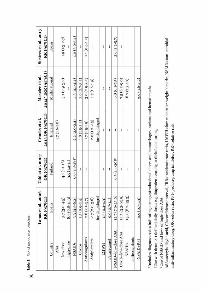
The risk of PUB among non-aspirin NSAID users is influenced by an individual NSAID used. The risk was reported to be at its lowest with the use of aceclofenac (Relative risk (RR) 2.6; 95%CI 1.5-4.6), diclofenac (3.1; 2.3-4.2) or ibuprofen (4.1; 3.1-5.3) compared to ketorolac (14.4; 5.2-39.9) (Lanas et al. 2006). The concomitant use of NSAID and ASA increased the risk of PUB as well as the use of other bleeding-related drugs (Table 2).

14
15
The risk of PUB among non-aspirin NSAID users is influenced by an individual NSAID used. The risk was reported to be at its lowest with the use of
Figure 1. Consumption of anti-inflammatory analgesics (M01A) in Finland during 1990-2015 (Finnish Medicines Agency and Social Insurance Institution 2016) by permission from FIMEA/Tinna Voipio.

15
The use of low-dose ASA alone has been shown to increase the risk of acute upper GIB (Hallas et al. 2006, García Rodrígues et al. 2011). The risk of PUB in H.pylori positive patients who use NSAIDs is markedly increased (RR 8.0; 5.0-13) as well as among patients who use only low-dose ASA (3.5; 2.6-6.1) compared t0 patients who are H.pylori negative and do not use NSAID/ASA (Sostres et al. 2015). The use of NSAIDs also increases the risk of ulcer perforation (Svanes 2000, Søreide et al. 2014, Chung and Shelat 2017).
The cyclo-oxygenase-2 (COX-2) selective inhibitor, celecoxib, was introduced to the market in 1999. The COX-2 selective inhibitors do not disturb the cyclo-oxygenase-1 (COX-1) enzyme involved in gastrointestinal (GI) mucosal protection and platelet function, while inflammation and pain mediated by prostaglandin synthesis dependent on the COX-2 enzyme are deprived. In a control-case study from Spain during 2001-2004, the use of any coxib was not associated with PUB (RR 1.5; 95%CI 0.9-2.4), whereas the use of rofecoxib slightly increased the risk of PUB (2.1; 1.1-4.0) (Lanas et al. 2006). According to Cochrane´s systematic review of 69 studies included, the risk of gastroduodenal ulcer complications was significantly lower (RR 0.39; 0.31-0.50) in COX-2 users than in nonselective NSAID users (Rostom et al. 2007). However, the rising concern about the increased risk of cardiovascular side-effects among COX-2 selective NSAID users (Mukherjee et al. 2001) eventually led to the withdrawal of rofecoxib from the market in 2004 followed by valdecoxib the next year.
In more recent analysis, the risk of cardiovascular events among COX-2 users and traditional NSAID users showed no difference; except in patients who used naproxen, for whom no excess of cardiovascular risk was shown (Kearney et al. 2006). In a very recent study from Hong Kong, patients who were hospitalised for PUB and required both ASA and NSAID therapy for comorbidities were randomised for further medication of celecoxib+esomeprazol or naproxen+esomeprazol to compare the occurrence of rebleeding (Chan et al. 2017). The cumulative incidence of rebleeding was significantly higher in the naproxen users with no difference in cardiovascular events.
16
Tabl
e 2.
R
isk
of p
eptic
ulc
er b
leed
ing.
17
Tabl
e 2.
R
isk
of p
epti
c ul
cer
blee
ding
.
L
anas
et
al. 2
00
6 R
R (
95%
CI)
U
dd
et
al. 2
00
7 O
R (
95%
CI)
C
rook
s et
al.
20
13 O
R (
95%
CI)
M
ascl
ee e
t al
. 20
14*
IRR
(9
5%C
I)
Sos
tres
et
al. 2
015
R
R (
95%
CI)
C
ount
ry
Spai
n Fi
nlan
d E
ngla
nd
Mul
tina
tion
al
Spai
n A
SA
low
-dos
e hi
gh-d
ose
3.
7 (3
.0-4
.5)
8.7
(6.8
-11.
3)
4.
1 (1
.1-1
6)
3.5
(1.2
-11)
1.7
(1.6
-1.8
)
3.1
(2.9
-3.2
) --
1.
9 (1
.3-2
.7)
--
NSA
IDs
5.3
(4.5
-6.2
) 6
.6 (1
.8-2
8)†
2.2
(2.0
-2.4
) 4.
3 (4
.1-4
.4)
4.0
(3.0
-5.4
) C
oxib
s 1.
5 (0
.9-2
.4)
--
1.8
(1.5
-2.2
) 2.
9 (2
.7-3
.2)
--
Ant
icoa
gula
nts
2.8
(2.1
-3.7
) --
1.
7 (1
.5-1
.9)
3.0
(2.9
-3.2
) 1.
1 (0
.9-1
.2)
Ant
ipla
tele
ts
2.7
(2.0
-3.6
) fo
r cl
opid
ogre
l --
2.
0 (1
.7-2
.5)
for
clop
idog
rel
1.7
(1.6
-1.9
) --
LMW
H
1.3
(0.4
-4.3
) --
--
--
--
Pa
race
tam
ol
0.9
(0.7
-1.1
) --
--
--
--
N
SAID
+lo
w-d
ose
ASA
12
.7 (7
.0-2
3.0)
6.
5 (1
.4-3
0)‡
--
6.8
(6.1
-7.5
) 2.
6 (1
.2-5
.7)
Cox
ib+
low
-dos
e A
SA
14.5
(3.3
-63.
9)
--
--
7.5
(6.2
-9.0
) --
N
SAID
+
anti
coag
ulan
ts
19.3
(8.2
-45.
3)
--
--
8.7
(7.3
-10)
NSA
ID+
PPI
0.9
(0.7
-1.3
) --
--
3.
9 (3
.6-4
.2)
*I
nclu
des
diag
nose
cod
es in
dica
ting
acu
te g
astr
oduo
dena
l ulc
ers
and
hem
orrh
ages
, mel
ena
and
hem
atem
esis
†Use
wit
h do
ses ≥
1 d
efin
ed d
aily
dos
es e
.g. i
bupr
ofen
120
0mg
or d
iclo
fena
c 10
0mg
‡Use
of N
SAID
and
low
- or
high
-dos
e A
SA
ASA
=ac
etyl
salic
ylic
aci
d, C
I=co
nfid
ence
inte
rval
, IR
R=
inci
denc
e ra
te r
atio
, LM
WH
=lo
w-m
olec
ular
-wei
ght h
epar
in, N
SAID
=no
n-st
eroi
dal
anti
-inf
lam
mat
ory
drug
, OR
=od
ds r
atio
, PPI
=pr
oton
-pum
p in
hibi
tor,
RR
=re
lati
ve r
isk

17
2.2.3 OTHER ASSOCIATED DRUGS
The use of selective serotonin reuptake inhibitors (SSRIs) was associated with only a slightly increased risk of PUB (or hemorrhagic gastritis) in a Danish population-based case-control study (Odds ratio OR 1.7; 1.01-5.1) (Dall et al. 2009). In that study, the risk of bleeding was clearly higher among SSRI users if they used NSAIDs (8.0; 4.8-13) or NSAIDs and ASA (28; 7.6-103) concomitantly. The exact mechanism of a SSRI affecting the risk of bleeding is unknown, but possible mechanisms are their ability to inhibit platelet aggregation and induce gastric acid secretion (Jiang et al. 2015, Laursen et al. 2017a). In a large multinational study, the risk of upper GIB among patients on SSRI monotherapy was increased (RR 2.1; 95%CI 1.9-2.2) with an increased risk of up to 7.0 (6.0-8.1) with a combination therapy of NSAIDs (Masclee et al. 2014). The risk of PUB was also reported to be increased in England among SSRI users after adjusting for all known risk factors for bleeding (OR 1.5; 1.3-1.7) (Crooks et al. 2013). Similar results were shown in a recent systematic review on the use of SSRI and the risk of upper GIB, where the risk of upper GIB was slightly increased among SSRI users (OR 1.6; 1.4-1.8), and the risk of bleeding was increased with concurrent use of NSAIDs (3.7; 3.0-4.7) or antiplatelet drugs (2.5; 1.7-3.6) (Jiang et al. 2015). However, the use of SSRI was not associated with endoscopy-refractory bleeding, rebleeding rate or short-term mortality in PUB patients in Denmark (Laursen et al. 2017a).
In one Spanish study, the use of oral corticosteroids was not associated with an increased risk of PUB (Lanas et al. 2006). On the contrary in England, the use of oral corticosteroids was associated with PUB when all the known risk factors for bleeding were adjusted (OR 1.3; 1.2-1.5) (Crooks et al. 2013). Similarly, the use of corticosteroids was associated with an increased risk of upper GIB (RR 4.1; 3.8-4.3) (Masclee et al. 2014). In that study, the use of NSAIDs with corticosteroids was associated with a clearly elevated risk of bleeding (13; 11-15). In another study from Taiwan, the short-term use of glucocorticoids was associated with an increase risk of PUB (Tseng et al. 2015). The risk of PUB was higher with higher doses or with concomitant use of NSAIDs. Based on most published studies, the use of corticosteroids is generally regarded as a risk factor for PUD and its complications. However, based on a recently published systematic review, the risk of gastrointestinal bleedings and perforations was increased only among hospitalised patients (Narum et al. 2014).
In a large multinational study, the risk of acute upper GIB was increased with the monotherapy use of calcium channel blockers (RR 1.6; 95%CI 1.5-1.6), nitrates (2.6; 2.4-2.7) and aldosterone antagonists (3.3; 3.1-3.5) (Masclee et al. 2014). The use of NSAIDs or coxibs concomitantly with those drugs increased the risk significantly with the highest in patients using combination of an aldosterone antagonist and NSAID (11; 8.6-14). However, in another hospital-based case-control study of only PUB patients, no association between PUB and the use of angiotensin converting
18
enzyme (ACE) inhibitors, angiotensin II receptor blockers, calcium channel blockers or α/β-blockers occurred (Nagata et al. 2015).
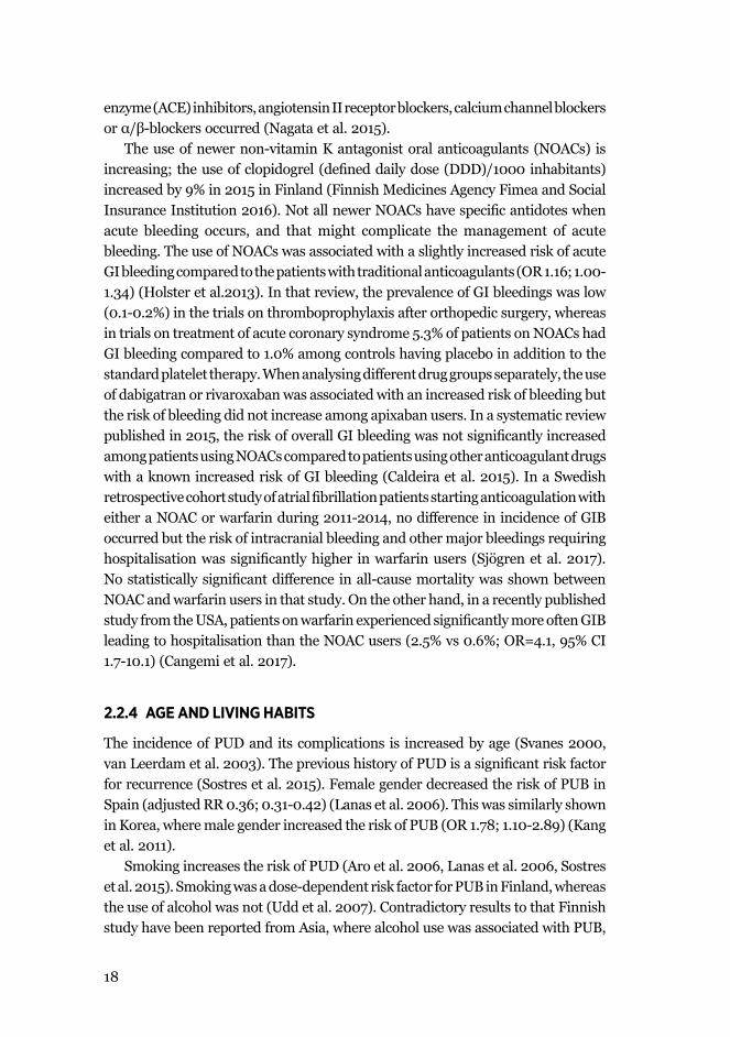
The use of newer non-vitamin K antagonist oral anticoagulants (NOACs) is increasing; the use of clopidogrel (defined daily dose (DDD)/1000 inhabitants) increased by 9% in 2015 in Finland (Finnish Medicines Agency Fimea and Social Insurance Institution 2016). Not all newer NOACs have specific antidotes when acute bleeding occurs, and that might complicate the management of acute bleeding. The use of NOACs was associated with a slightly increased risk of acute GI bleeding compared to the patients with traditional anticoagulants (OR 1.16; 1.00-1.34) (Holster et al.2013). In that review, the prevalence of GI bleedings was low (0.1-0.2%) in the trials on thromboprophylaxis after orthopedic surgery, whereas in trials on treatment of acute coronary syndrome 5.3% of patients on NOACs had GI bleeding compared to 1.0% among controls having placebo in addition to the standard platelet therapy. When analysing different drug groups separately, the use of dabigatran or rivaroxaban was associated with an increased risk of bleeding but the risk of bleeding did not increase among apixaban users. In a systematic review published in 2015, the risk of overall GI bleeding was not significantly increased among patients using NOACs compared to patients using other anticoagulant drugs with a known increased risk of GI bleeding (Caldeira et al. 2015). In a Swedish retrospective cohort study of atrial fibrillation patients starting anticoagulation with either a NOAC or warfarin during 2011-2014, no difference in incidence of GIB occurred but the risk of intracranial bleeding and other major bleedings requiring hospitalisation was significantly higher in warfarin users (Sjögren et al. 2017). No statistically significant difference in all-cause mortality was shown between NOAC and warfarin users in that study. On the other hand, in a recently published study from the USA, patients on warfarin experienced significantly more often GIB leading to hospitalisation than the NOAC users (2.5% vs 0.6%; OR=4.1, 95% CI 1.7-10.1) (Cangemi et al. 2017).
2.2.4 AGE AND LIVING HABITS
The incidence of PUD and its complications is increased by age (Svanes 2000, van Leerdam et al. 2003). The previous history of PUD is a significant risk factor for recurrence (Sostres et al. 2015). Female gender decreased the risk of PUB in Spain (adjusted RR 0.36; 0.31-0.42) (Lanas et al. 2006). This was similarly shown in Korea, where male gender increased the risk of PUB (OR 1.78; 1.10-2.89) (Kang et al. 2011).
Smoking increases the risk of PUD (Aro et al. 2006, Lanas et al. 2006, Sostres et al. 2015). Smoking was a dose-dependent risk factor for PUB in Finland, whereas the use of alcohol was not (Udd et al. 2007). Contradictory results to that Finnish study have been reported from Asia, where alcohol use was associated with PUB,

19
but no association was found between smoking and PUB (Kang et al. 2011, Nagata et al. 2015). Smoking has also been shown to be a risk factor for ulcer perforation (Svanes 2000). Obesity was associated with an increased risk of uncomplicated gastric ulcer in a Swedish population-based study (Aro et al. 2006).
In recent studies, psychological stress and dramatic changes in living conditions, such as experiencing a life-threatening earthquake or accommodation in a refugee shelter, is also associated with formation of PUD (Malfertheiner et al. 2009, Yamanaka et al. 2013, Kanno et al. 2015, Levenstein et al. 2015). The use of a gastroprotective agents, commonly PPIs, as stress ulcer prophylaxis in all critically ill patients in the intensive care units have been standard care until recently, with concerns about its necessity for all patients or possible associated harms (nosocomial pneumonia, Clostridium difficile infection or cardiovascular events) (Marker et al. 2017).
2.2.5 COMORBIDITY
Comorbidity (e.g. rheumatoid arthritis, liver cirrhosis, renal insufficiency) is associated with occurrence of PUD complications and their recurrence (Lau et al. 2011) and is an independent risk factor for PUB (Crooks et al. 2013, Nagata et al. 2015). Comorbidity can be defined as the existence of a certain comorbidity, e.g. congestive heart failure, or using the well-validated Charlson comorbidity index (CCI) (Charlson et al. 1987). In a Danish observational study, PUB patients had a significantly higher level of comorbidities compared to the age- and gender-matched control cohort (mean CCI 0.92 vs o.49, p<0.001) (Laursen et al. 2015a).
2.2.6 RARE DISEASES
A gastric or duodenal ulcer can be a clinical manifestation for some rare diseases. An anastomotic peptic ulcer after subtotal gastric resection is one manifestation of PUD and is also referred to as a marginal ulcer. With multiple ulcers or an ulcer occurring in the more distal duodenum than the bulbus, Zollinger-Ellison syndrome (acid hypersecretory syndrome), Crohn´s disease, ischemia or underlying malignancy should be considered (Maltferheiner et al. 2009). At least for patients suffering a recurrent ulcer with no common risk factors, the possibility of rare disease should be excluded.
Other rare specific causes of PUD are eosinophilic gastroduodenitis, systemic mastocytosis, radiation damage, viral infections (cytomegalovirus and herpes simplex, particularly in immunosuppressed patients), colonisation of stomach with Helicobacer heilmanii and Cameron ulcer (gastric ulcer where a hiatus hernia passes through the diaphragmatic hiatus) (Maltferheimer et al. 2009). Some patients have hyperfunctional antral G-cells leading to acid hypersecretion and ulcer formation. Additionally, mechanical obstruction (e.g. batteries) can lead to

20
an ulcer formation. Ingested batteries may also induce ulcer formation by leaking the battery contents or generating an external electric current. Abuse of crack cocaine can also cause peptic ulcer formation emerging as peptic ulcer perforation (Schuster et al. 2007).
2.2.7 IDIOPATHIC ULCER
The peptic ulcer is considered idiopathic, when no risk factor for an ulcer formation exists. The pathogenic mechanisms associated with the development of an idiopathic ulcer are unknown (Lanas and Chan 2017).
2.3 PATHOPHYSIOLOGY
PUD develops when the protective mechanisms of the gastrointestinal mucosa are disturbed. Several different exogenous and endogenous mechanisms affect the secretion of hydrochloric acid, pepsin and mucus. Secretion of gastrin, histamine and acetylcholine stimulates the secretion of hydrochloric acid, whereas somatostatin and secretin are inhibitors. Sense of food activates secretion of hydrochloric acid by stimulating the vagus nerve. Hydrochloric acid converts pepsinogen to pepsin. Mucus and bicarbonate are secreted from the mucus cells to prevent damage in the gastric epithelium.
Peptic ulcers occur mainly in the stomach or proximal duodenum. Peptic ulcer can be defined as a break in the mucosa of ≥5mm in diameter covered with fibrin, and it invades the muscularis mucosa according to the pathological criterion (Kumar et al. 1997, Hsu et al. 2001, Maltferheiner et al. 2009). The 5mm criterion is arbitrary, but is usually used in clinical trials and endoscopy reports. The smaller lesions are usually called erosions. Naturally, the medium pH level of the stomach is 1.4 varying from <1.o to 5.0 regarding to ingestion of food and fasting (Sachs et al. 2011). Besides the H.pylori infection and the use of NSAIDs, a decrease in circulation can lead to an impairment of the mucosal barrier. After the impairment of the mucosal barrier, hydrochloric acid and pepsin induce ulcer formation. However, the development of different types of peptic ulcers is not completely understood (Lanas and Chan 2017).
2.3.1 H.PYLORI INDUCED ULCER
H.pylori is able to acclimate in the highly acid environment of the stomach (Sachs et al. 2011). With the help of urease in the bacterial cytoplasm, it manages to maintain a cytoplasmic pH high enough to survive in an acidic environment. The ability of motility and binding tightly to gastric epithelial cells also helps colonisation

21
(Suerbaum and Michetti 2002). After binding to the epithelial cells, H.pylori causes gastric inflammation by activation of leukocytes, interleukins, and systemic and mucosal humoral responses. Epithelial injury can also occur from reactive oxygen and nitrogen species by activated neutrophils. This leads to the eruption of the mucosal barrier that allows acid and pepsins to invade through the epithelial cell to cause an ulcer (Figure 2). The usual site of infection is in the antrum of stomach, but under the PPI treatment with an increase in the pH of the antrum, the organism is found more probably in the fundus. Patients with corpus-predominant gastritis and with a lower level of acid produce are more prone to gastric ulcers. The H.pylori infection in the antrum leads to inhibition of somatostatin secretion form the D cells. That causes an increase in acid secretion, inducing gastric metaplasia in the bulb of duodenum and enables the colonisation of H.pylori there.
Normal gastric mucosa
Helicobacter pylori infection
Antral-predominantgastritis
Corpus-predominantatrophic gastritis
Individually high level ofacid production
Individually low level of acid production
Duodenal ulcer Gastric ulcer
Somatostatin↓Gastrin↑Acid secretion↑
Asymptomatic H.pylori infection
Nonatrophic pangastritis
H.pylori virulence + Host(genetic, ethnic, environmental, sosioeconomic) factors→Inflammation reactions
Gastric metaplasia induodenum
Figure 2. Pathogenesis of Helicobacter pylori positive ulcers.
2.3.2 NSAID INDUCED ULCER The mechanisms of an NSAID induced ulcer can be divided into topical injury and systemic mechanisms (Wolfe et al. 1999, Chan 2005, Musumba et al. 2009). The systemic effects include suppression of gastric prostaglandins synthesis through cyclooxygenase inhibition, which is considered to be the main mechanism of mucosal damage (Lanas and Chan 2017), and prostaglandin-independent effects

22
including upregulating of mucosal pro-inflammatory mediators. Prostaglandin inhibition leads to decreases in mucus and bicarbonate secretion, mucosal blood flow and epithelial proliferation, which leads to degradation of mucosal resistance on endogenous and exogenous factors. NSAIDs can directly affect gastric epithelial cells causing general damage or dysfunction via multiple cellular and molecular mechanisms that might lead to necrosis and apoptosis (Musumba et al. 2009). The consistency of the gastric mucosal barrier is dependent on continuous generation of prostaglandin E2 and prostacyclin requiring COX-1 and COX-2. The use of NSAIDS induces ulcer formation by COX inhibition. The use of COX-2 selective NSAIDs, i.e. coxibs, decreases the rate of endoscopic ulcers as compared to traditional NSAIDs (Laine 2003). The COX-2 derived prostaglandins, however, play an important role in ulcer healing (Musumba et el. 2011). The use of ASA induces mucosal damage mainly through topical mechanisms (Lanas and Chan 2017). The mucosal injury is caused by acidic effects of ASA and other NSAIDs (Wolfe et al. 1999). The use of enteric-coated ASA prevents topical injury, but even small doses of ASA suppress prostaglandin synthesis. Therefore, no difference in the risk of major upper GI bleedings occurs between the users of enteric-coated or buffered ASA compared to the users of plain ASA (Laine 2003).
2.4 CLINICAL MANIFESTATION
2.4.1 SYMPTOMS
Symptoms of peptic ulcers vary in individual patients not forgetting that some patients are symptomless. Upper abdominal pain is the most common symptom. Besides pain, patients can suffer from epigastric burning, postprandial fullness, bloating, nausea and vomiting. None of the symptoms is pathognomonic for PUD. Patients with duodenal ulcers typically feel hungry or have nocturnal abdominal pain, whereas postprandial abdominal pain, nausea, vomiting and weight loss are associated with gastric ulcers (Lanas and Chan 2017).
Elderly patients with PUD are frequently symptomless or have only mild symptoms, and the first manifestation of complicated peptic ulcer may lead to hospitalisation with marked morbidity and mortality. The long-lasting use of NSAIDs for chronic pain, impaired protective mechanisms of the gastrointestinal mucosa, and comorbidities are factors that make the elderly more vulnerable to PUD formation (Zullo et al. 2007).
Melena and hematemesis are common symptoms among PUB patients. Occasionally, only a drop in the hemoglobin level causes dizziness or tachycardia that leads to a PUB diagnosis. The classic triad of sudden onset of abdominal pain, tachycardia and abdominal rigidity or signs of peritonitis is representative for

23
perforation of peptic ulcer (PPU). After perforation, gastric juice and gas enters the peritoneal cavity leading to chemical peritonitis. In the following hours, chemical peritonitis progresses to bacterial peritonitis due to gut flora.
2.4.2 LOCATION
Of all peptic ulcers, most appear either in the first part of the duodenum or in the stomach, in a ratio of about 4:1 (Kumar et al. 1997). Another rare location of a peptic ulcer is appearing in the Meckel´s diverticulum.
The prevalence of duodenal ulcers among PUB patients has been higher than that of gastric ulcers (Pérez-Aisa et al. 2005, Lanas et al. 2006, Bardhan and Royston 2008, Sadic et al. 2009, Bakkevold 2010, Sung et al. 2010, Rosenstock et al. 2013), but contradictory results have emerged (Smith and Stabile 2005, Åhsberg et al. 2011, de Groot et al. 2014). The prevalence of gastric ulcers was 53% in H.pylori –negative idiopathic ulcer cohort, whereas among H.pylori –positive ulcer cohort 63% had duodenal/gastroduodenal ulcers in Taiwan (Wong et al. 2009). However, the peptic ulcer cases were evenly distributed in a Spanish PUB cohort 2006-2012 (Sostres et al. 2015).
2.5 DIAGNOSIS
2.5.1 OESOPHAGOGASTRODUODENOSCOPY (OEGD)
The diagnosis of PUD can be made endoscopically, when the location, appearance and size of an ulcer is defined. Therefore, OEGD is the gold standard for diagnosis of PUD. Patients with upper GIB symptoms should be referred for endoscopy within 24h after admission (Barkun et al. 2010, Gralnek et al. 2015). However, the proportion of patients with acute upper GIB experiencing OEGD in the recommended time frame has varied from 50% in the UK in 2007 (Hearnshaw et al. 2010) to 76% in Canada during 1999-2002 (Barkun et al. 2004) and 81% in Italy 2007-2008 (Marmo et al. 2010).
2.5.1.1 Symptoms leading to acute oegd
Patients are admitted to acute OEDG during the hospitalisation for various symptoms. Symptoms of acute bleeding, i.e. hematemesis or melena, are the most common indications, but patients with acute anemia, severe dysphagia, epigastric pain or suspicion of a foreign body are also referred for acute OEGD.

24
2.5.2 DIAGNOSIS OF PERFORATED PEPTIC ULCER
Patients with acute PPU are usually admitted to emergency departments with severe epigastric pain. Other symptoms as severe dyspepsia, abdominal distension, nausea, fever, tachycardia and hypotension may occur (Chung and Shelat 2017). Traditionally, free air under the diaphragm on an erect chest X-ray with upper abdominal pain symptoms suggests a diagnosis of PPU. However, in 25-40% of patients with PPU free air on the X-ray is not revealed (Grassi et al. 2004, Thorsen et al. 2011, Anbalakan et al. 2015). The gold standard nowadays is a computed tomography (CT) scan with a diagnostic accuracy as high as 98% (Thorsen et al. 2011, Kim et al. 2014). In addition, with the help of a CT scan and serum amylase, acute pancreatitis can be ruled out among patients with severe upper abdominal pain.
2.5.3 GASTRIC BIOPSY SPECIMENS AND TESTS FOR H.PYLORI INFECTION
During OEGD, biopsies are usually taken to exclude malignant ulcers and to demonstrate the H.pylori infection among patients with gastritis. In addition to histology biopsies, H.pylori infection can be diagnosed by non-invasive methods such as serology, urea breath test and stool antigen test (Rautelin and Kosunen 2004). The sensitivity of serological tests on H.pylori IgG antibodies based on enzyme immunoassay varies from 97% to 100% with specificity of 95-99% in Finland. The sensitivity and specificity of the urea breath test is more than 90% (Suerbaum and Michetti 2002).
2.6 DIFFERENTIAL DIAGNOSIS
2.6.1 CAUSES FOR UPPER GASTROINTESTINAL BLEEDING
Acute GI bleeding is one of the most common medical emergencies leading to hospitalisation with notable costs (Peery et al. 2015). The overall incidence of upper GI bleeding has varied from 45 per 100 000 inhabitants per year to 160 per 100 000 (Vreeburg et al. 1997, Paspatis et al. 2000, Åhsberg et al. 2010a, Button et al. 2011). Most patients are referred for acute OEDG for diagnosis (Table 3).

25
Table 3. Etiology (%) of acute upper GI bleeding.
27
Table 3. Etiology (%) of acute upper GI bleeding.
Etiology Vreeburg et
al. (1997) %
Leerdam et
al. (2003) %
Loperfido et
al. (2009) %
Nahon et al.
(2012) %
Miilunpohja et
al. (2017) %
PUD 39 46 53 31 50
Erosive 6 -- 10 7 --
Mallory-Weiss 5 -- 3 7 8
Variceal
bleeding
8 7 12 21 8
Oesophagitis 7 -- 4 13 17
Gastroduodenal
lesions*
-- 20 -- -- --
Neoplasm 3 5 5 3 4
Other 8 8 8 13 --
No finding 24 14 5 5 13
*Includes oesophagitis, Mallory-Weiss, erosions, gastroduodenitis
2.6.2 PATIENTS PRESENTING WITH ACUTE UPPER GASTROINTESTINAL BLEEDING WITH NO FINDING IN OEGD
The proportion of patients with acute GI bleeding symptoms without a known source in OEGD has varied from 5% (Nahon et al. 2012) to 32% (Sengupta et al. 2016) in previous studies. The most common sources for acute lower GI bleeding in the previous studies were diverticulosis, ischemic colitis, hemorrhoids and colon cancer (Longstreth 1997, Arroja et al. 2011, Hreinsson et al. 2013). In those lower GI bleeding studies, no source for bleeding was diagnosed in 8-12% of patients.
2.6.3 MALIGNANT ULCERS A bleeding ulcer can be the first symptom of gastric cancer. The H.pylori infection is a risk factor for both PUD and gastric cancer (Tsuda et al. 2017). In a large cohort study of hospitalised gastric ulcer patients in Sweden during 1965-1983, the risk for gastric cancer was nearly 10-fold during the first three years of follow-up, and it remained two-fold during the follow-up as long as 24 years for the same patients (Hansson et al. 1996). In a Taiwanese cohort study during 1997-2004, the risk for gastric cancer development was increased in gastric ulcer patients compared to duodenal ulcer patients (HR 2.9; 2.1-3.9) (Wu et al. 2009). According to that study, other independent risk factors for gastric cancer development were older age, male gender and
2.6.2 PATIENTS PRESENTING WITH ACUTE UPPER GASTROINTESTINAL BLEEDING WITH NO FINDING IN OEGD
The proportion of patients with acute GI bleeding symptoms without a known source in OEGD has varied from 5% (Nahon et al. 2012) to 32% (Sengupta et al. 2016) in previous studies. The most common sources for acute lower GI bleeding in the previous studies were diverticulosis, ischemic colitis, hemorrhoids and colon cancer (Longstreth 1997, Arroja et al. 2011, Hreinsson et al. 2013). In those lower GI bleeding studies, no source for bleeding was diagnosed in 8-12% of patients.
2.6.3 MALIGNANT ULCERS
A bleeding ulcer can be the first symptom of gastric cancer. The H.pylori infection is a risk factor for both PUD and gastric cancer (Tsuda et al. 2017). In a large cohort study of hospitalised gastric ulcer patients in Sweden during 1965-1983, the risk for gastric cancer was nearly 10-fold during the first three years of follow-up, and it remained two-fold during the follow-up as long as 24 years for the same patients (Hansson et al. 1996). In a Taiwanese cohort study during 1997-2004, the risk for gastric cancer development was increased in gastric ulcer patients compared to duodenal ulcer patients (HR 2.9; 2.1-3.9) (Wu et al. 2009). According to that study, other independent risk factors for gastric cancer development were older age, male gender and presence of complicated peptic ulcer at the index hospitalisation. The frequent use of ASA/NSAIDs and early H.pylori eradication
26
therapy was associated with a decreased risk of cancer. The H.pylori infection is also a known risk factor for mucosa-associated lymphoid tissue (MALT) lymphoma (Rautelin and Kosunen 2004). Therefore, proper healing of a gastric ulcer should be confirmed endoscopically after appropriate PPI therapy and H.pylori infection eradication therapy given when necessary. The reported proportion of patients having malignant gastric ulcer perforation varies from 10% to 16% (Ergul and Gozetlik 2009).
2.7 COMPLICATIONS
2.7.1 INCIDENCE OF COMPLICATIONS
The proportion of patients suffering complicated PUD differs in published studies. The annual number of hospital admission for complicated PUD (hemorrhage, perforation and stenosis) in Finland increased significantly from 38 per 100 000 in 1972-1976 to 69 per 100 000 in 1992-1996 (Paimela et al. 2002). The incidence of bleeding peptic ulcers remained stable in Denmark 1993-2002, while the incidence of perforated ulcers decreased (Lassen et al. 2006). The incidence of all PUD complications decreased from 109 to 81 per 100 000 in Spain during 1990-2000 (Pérez-Aisa et al. 2005). The decreasing trend for PUD complications is shown studies published over the last ten years (Kang et al. 2006, Sadic et al. 2009, Lanas et al. 2011, Laine et al. 2012).
2.7.2 BLEEDING ULCER
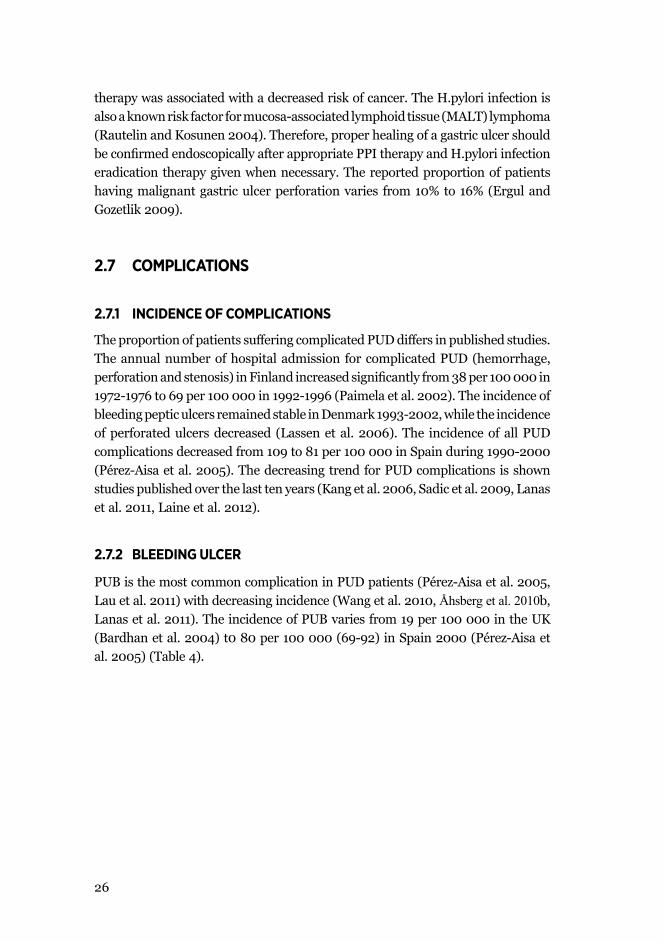
PUB is the most common complication in PUD patients (Pérez-Aisa et al. 2005, Lau et al. 2011) with decreasing incidence (Wang et al. 2010, Åhsberg et al. 2010b, Lanas et al. 2011). The incidence of PUB varies from 19 per 100 000 in the UK (Bardhan et al. 2004) t0 80 per 100 000 (69-92) in Spain 2000 (Pérez-Aisa et al. 2005) (Table 4).
27
Table 4. Incidence of peptic ulcer bleeding.
Reference Country Year(s) Incidence per 100 000 inhabitants
Bardhan et al. 2004 United Kingdom 1995-2000 19Ramsoekh et al. 2005 the Netherlands 2000 22Åhsberg et al. 2010 Sweden 2004 32Laine et al. 2012 USA 2009 32Bakkevold 2010 Norway 2007-2008 45Ohmann et al. 2005 Germany 1999-2000 49Soplepmann et al. 1997 Estonia 1992-1993 57Lassen et al. 2006 Denmark 2002 57Pérez-Aisa et al. 2005 Spain 2000 80
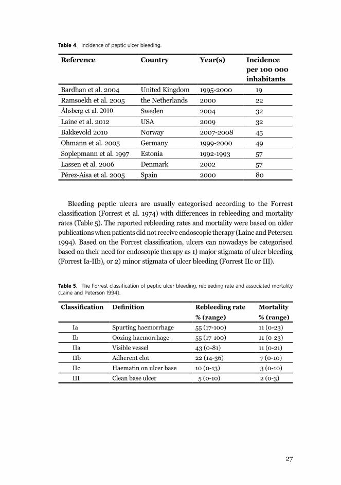
Bleeding peptic ulcers are usually categorised according to the Forrest classification (Forrest et al. 1974) with differences in rebleeding and mortality rates (Table 5). The reported rebleeding rates and mortality were based on older publications when patients did not receive endoscopic therapy (Laine and Petersen 1994). Based on the Forrest classification, ulcers can nowadays be categorised based on their need for endoscopic therapy as 1) major stigmata of ulcer bleeding (Forrest Ia-IIb), or 2) minor stigmata of ulcer bleeding (Forrest IIc or III).
Table 5. The Forrest classification of peptic ulcer bleeding, rebleeding rate and associated mortality (Laine and Peterson 1994).
Classification Definition Rebleeding rate
% (range)
Mortality
% (range)
Ia Spurting haemorrhage 55 (17-100) 11 (0-23)
Ib Oozing haemorrhage 55 (17-100) 11 (0-23)
IIa Visible vessel 43 (0-81) 11 (0-21)
IIb Adherent clot 22 (14-36) 7 (0-10)
IIc Haematin on ulcer base 10 (0-13) 3 (0-10)
III Clean base ulcer 5 (0-10) 2 (0-3)

28
In a recent published study, the rebleeding rates of peptic ulcers by Forrest classification after successful endoscopic hemostasis but with no PPI therapy were 23% in Ia ulcers, 5% in Ib, 11% in IIa, and 18% in IIb, respectively, suggesting that Ib ulcers after initial endoscopic management should not be categorised as major stigmata of hemorrhage (Jensen et al. 2017). In another study from the Netherlands during 2009-2012, the overall rebleeding rate was quite high (19%) varying from 59% among Forrest Ia ulcers to 7% in Forrest III ulcers (de Groot et al. 2014). In that study, only 70-74% of patients were treated with dual therapy in endoscopy. Based on a systemic review of 28 studies, the recurrent rate of all bleeding ulcers after successful initial endoscopic hemostasis has varied from 0-38%, being on average at 10% (Lau et al. 2011). In a recently published study from Finland, 4.4% of PUB patients hospitalised during 2000-2015 needed a secondary procedure for bleeding, and 1.0% were admitted to prophylactic transcatheter arterial embolisation (TAE) (Nykänen et al. 2017).
2.7.3 PERFORATED ULCER
The incidence of peptic ulcer perforations was 8 per 100 000 (5-13) in Spain 2000 (Pérez-Aisa et al. 2005) as well as in Sweden 2005 (Åhsberg et al. 2011). The hospitalisation rates of patients with PPU decreased significantly from 17 to 12 per 100 000 in Denmark during 1996-2oo4 (Christensen et al. 2007). The estimated annual incidence of peptic ulcer perforation is 3.8-14 per 100 000 individuals (Lau et al. 2011). The annual operation rate for PPU was 3.6 per 100 000 in the Northern Finland in 2000, with no significant change during 1979-2000 (Mäkelä et al. 2002). Recently, a relative increase in PPU among elderly women has occurred, while the incidence of duodenal ulcer perforations among young men has decreased (Svanes 2000).
2.7.3 OTHER RARE COMPLICATIONS
Rare complications of PUD are pylorus obstruction and penetration or fistula to another organ e.g. pancreas. The incidence of gastric outlet obstruction decreased from 6.8 per 100 000 (95% CI: 4.0-10.9) in 1985 to 1.7 per 100 000 (0.5-4.3) in 2000 in Spain (Pérez-Aisa et al. 2005). Of all PUD hospitalisation in the United States in 2006, 2.9% of patients had obstruction (Wang et al. 2010). After introduction of H.pylori eradication therapy and PPIs, other benign and malignant diseases than PUD usually cause gastric outlet obstruction (Johnson and Ellis 1990, Rana et al. 2011). PUD is an underlying cause of only up to 8% of patients presenting with gastric outlet obstruction (Napolitano 2009).

29
2.8 MANAGEMENT
Patients presenting with acute peptic ulcer bleeding or perforation should be assessed promptly and resuscitated before definitive management. International guidelines recommend a strict policy for blood transfusions with recommendation of a hemoglobin target level of 70 g/l (Barkun et al. 2010). Mortality among PUB patients with restrictive strategy showed a trend toward better survival (HR 0.70; 0.26-1.25), although it was not statistically significant, as was seen when all the patients admitted with severe acute upper GIB were analysed in Spain (0.55; 0.33-0.92) (Villanueva et al. 2013). However, in that Spanish study, patients with massive exsanguinating bleeding, or severe comorbidity as an acute coronary syndrome, stroke or symptomatic peripheral vascular disease were excluded, and one third of the patients had cirrhosis. On the other hand, no differences in outcomes occurred in the UK when strictive and liberal transfusion policies were compared among upper GIB patients in the TRIGGER study that also excluded unstable patients at admission (Jairath et al. 2015). Coagulopathy and acute bleeding is a challenging clinical problem. The exact target level of the International Normalized Ratio (INR) of coagulation has not been defined, and it should depend on the individual patient´s indication for anticoagulation or existence of disease causing coagulopathy (Lau et al. 2013).
The use of risk stratification scores is highly recommended (Barkun et al. 2010, Jairath and Barkun 2012). The pre- and post-endoscopic Rockall scores, the Glasgow Blatcford score, or a newer AIMS65 score are suitable for PUB patients for dividing patients into low- and high-risk categories when predicting rebleeding or mortality (Rockall et al. 1996, Blatchford et al. 2000, Saltzman et al. 2011). The Boey´s score is commonly used among PPU patients (Boey and Wong 1987) to predict mortality based on a patient´s comorbidity, pre-operative shock and time from the onset of abdominal pain. The American Society of Anesthesiology score ASA is also useful in risk stratification for patients undergoing surgery (Saklad 1941, Wolters et al. 1996).
Gastroprotective agents (GPAs) and endoscopic treatments are essential factors in PUD patient treatment, whereas surgery is needed in refractory bleeding or perforated cases. The utilisation of interventional radiology in PUB is emerging especially among fragile patients not suitable for surgery.
2.8.1 GASTROPROTECTIVE AGENTS
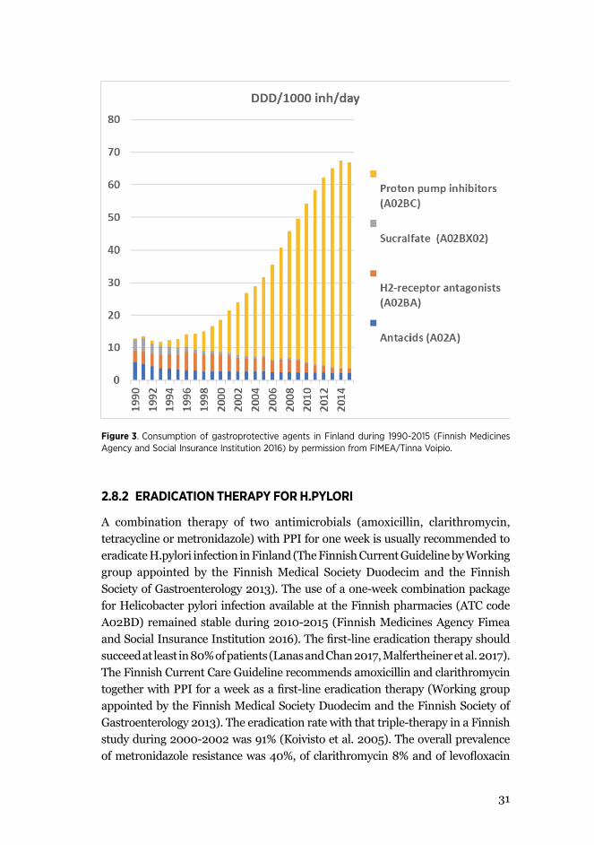
The use of GPAs, mainly proton pump inhibitors (PPIs), has increased during the last decades in Finland (Figure 3) and other countries (Pérez-Aisa et al. 2005, Kantor et al. 2015). The introduction of histamine-2 (H2) –receptor antagonists occurred in 1980 and of PPIs (omeprazole) in 1988 in Finland. The PPIs are used to

30
provide neutral gastric pH, which enables a favorable milieu for clot formation. The use of other GPAs as H2-receptor antagonists, antacids, misoprostol, sucralfate, and alginic acids is nowadays diminutive compared to that of PPIs (Finnish Medicines Agency and Social Insurance Institution 2016).
The use of PPIs is associated with a significant reduced risk of bleeding as a PUD complication [RR 0.3; 95%CI 0.3-0.4 (Lanas et al. 2006), RR 0.4; 0.3-0.6 (Sostres et al. 2015)]. The use of PPI with ASA or NSAIDs also reduced the risk of bleeding (Lanas et al. 2007). However, the monotherapy use of GPA was associated with a slightly increased risk of acute upper GIB in a multinational European study (IRR 1.6; 95%CI 1.6-1.7) (Masclee et al. 2014), indicating that patients at risk of bleeding are identified. In those patients with possible many known risk factors for bleeding, the use of GPA is not efficient enough for prevention.
The international guidelines recommend continuous intravenous PPI-treatment following the initial intravenous bolus for patients with high-risk stigmata of ulcer bleeding for the next 72 hours after the index endoscopy (Barkun et al. 2010, Gralnek et al. 2015). However, in a recent meta-analysis, no difference in rebleeding or mortality rate occurred among patients on oral or intravenous PPI (Tsoi et al. 2013). A common clinical practise for years has been to change intravenous PPI to an oral one when the patient is able to eat and the possibility of re-endoscopy or surgery has faded. After the initial hospitalisation, PPIs are used to heal the ulcer completely and to prevent the recurrence. The duration of PPI treatment is usually 4-8 weeks by clinical decision. According to a meta-analysis, the mean ulcer healing rate in H.pylori positive ulcers was 91% after a 7-day eradication therapy with PPI compared to 92% when PPI was used for 2-4 weeks more (Gisbert and Pajares 2005). On the other hand, patients at high-risk for NSAID induced recurrent ulcers should continue PPI use if NSAIDs are needed for comorbidity; that also outweighs the possible risks associated with long-term use of PPI (Freedberg et al. 2017).
31
33
2.8.2 ERADICATION THERAPY FOR H.PYLORI A combination therapy of two antimicrobials (amoxicillin, clarithromycin, tetracycline or metronidazole) with PPI for one week is usually recommended to eradicate H.pylori infection in Finland (The Finnish Current Guideline by Working group appointed by the Finnish Medical Society Duodecim and the Finnish Society of Gastroenterology 2013). The use of a one-week combination package for Helicobacter pylori infection available at the Finnish pharmacies (ATC code A02BD) remained stable during 2010-2015 (Finnish Medicines Agency Fimea and Social Insurance Institution 2016). The first-line eradication therapy should succeed at least in 80% of patients (Lanas and Chan 2017, Malfertheiner et al. 2017). The Finnish Current Care Guideline recommends amoxicillin and clarithromycin together with PPI for a week as a first-line eradication therapy (Working group appointed by the Finnish Medical Society Duodecim and the Finnish Society of Gastroenterology 2013). The eradication rate with that triple-therapy in a Finnish study during 2000-2002 was 91% (Koivisto et al. 2005). The overall prevalence of metronidazole resistance was 40%, of clarithromycin 8% and of levofloxacin 7% in Finland during 2000-2008
Figure 3. Consumption of gastroprotective agents in Finland during 1990-2015 (Finnish Medicines Agency and Social Insurance Institution 2016) by permission from FIMEA/Tinna Voipio.
2.8.2 ERADICATION THERAPY FOR H.PYLORI
A combination therapy of two antimicrobials (amoxicillin, clarithromycin, tetracycline or metronidazole) with PPI for one week is usually recommended to eradicate H.pylori infection in Finland (The Finnish Current Guideline by Working group appointed by the Finnish Medical Society Duodecim and the Finnish Society of Gastroenterology 2013). The use of a one-week combination package for Helicobacter pylori infection available at the Finnish pharmacies (ATC code A02BD) remained stable during 2010-2015 (Finnish Medicines Agency Fimea and Social Insurance Institution 2016). The first-line eradication therapy should succeed at least in 80% of patients (Lanas and Chan 2017, Malfertheiner et al. 2017). The Finnish Current Care Guideline recommends amoxicillin and clarithromycin together with PPI for a week as a first-line eradication therapy (Working group appointed by the Finnish Medical Society Duodecim and the Finnish Society of Gastroenterology 2013). The eradication rate with that triple-therapy in a Finnish study during 2000-2002 was 91% (Koivisto et al. 2005). The overall prevalence of metronidazole resistance was 40%, of clarithromycin 8% and of levofloxacin

32
7% in Finland during 2000-2008 (Kostamo et al. 2011). However, metronidazole resistance in vitro does not always lead to a treatment failure.
For the growing prevalence of antibiotic resistance worldwide, a longer 10-day- or two-week therapy is nowadays recommended (Lanas and Chan 2017, Malfertheiner et al. 2017). That will probably also change recommendations in Finland. The antimicrobial resistance of H.pylori to clarithromycin is over 20% in most European countries; except in the Northern Europe where the prevalence of resistance is less than 10%. Therefore, the choice of first-line therapy in countries with a high level of antimicrobial resistance should be based on susceptibility tests, or at least on local recommendations based on resistance rates (Figure 4). The use of susceptibility tests in H.pylori infection after failed first-line eradication therapy could be beneficial.
Clarithromycin resistance
< 15% ≥15% or not available
PPIx2Amoxicillin 1gx2Clarithromycin 500mgx2for 7 days
PPIx2Amoxicillin 1gx2Levofloxacillin 500mgx2for 7 days
H.pylori urea breath test or serology
H.Pylori positive
PPIx2, Bismuth 125mg 2x2 or 1x4, Tetracycline 250mgx4,Metronidatzole 400mgx3 for 10 days
Figure 4. The recommendation for choosing Helicobacter pylori eradication therapy by clarithromycin resistance (Modified after Malfertheiner et al. 2017).
The rate of success in eradication therapy can be increased by doubling the PPI dose, extending therapy duration to a maximum of 14 days, or using a quadruple non-bismuth- or bismuth-based therapy (PPI + amoxicillin + clarithromycin + metronidazole or PPI + bismuth + tetracycline + metronidazole). Bismuth is available in Finland with a special permit from FIMEA. The sequential therapy of a 5-day dual therapy with a PPI and amoxicillin followed by a 5-day triple

33
therapy with a PPI, clarithromycin and tinidazole/metronidazole is not anymore recommended (Lanas and Chan 2017). In challenging cases with multiple therapy failures, rifabutin-based therapy for 10 days leads to eradications rates of 60-70%. However, rifabutin is not recommended for use in Finland. New promising results with a potassium-competitive acid blocker vonoprazan -based triple therapy (amoxicillin and clarithromycin/metronidazole) have reported from Japan with an eradication rate of 85-97% (Tanabe et al. 2017).
The success of eradication therapy should be controlled. The gold standard for post-treatment test is nowadays the urea breath test that should be used not until an interval period of four weeks after eradication therapy in order to avoid false results. A stool antigen test can also be used, but not earlier than after an interval period of 8 weeks. Patients with a complicated gastric ulcer, or in some complicated duodenal ulcers cases by clinical decision, are admitted for a control endoscopy, and the eradication of H.pylori can be confirmed by biopsies taken. The successful eradication of H.pylori infection is essential to decrease the risk of recurrence of PUD and its complications. However, according to Cochrane systematic review, the role of H.pylori eradication therapy in acute gastric ulcer healing compare to ulcer healing therapy only is controversial (Ford et al. 2016). On the other hand, the risk of gastric cancer development was significantly decreased among hospitalised PUD patients who received H.pylori eradication therapy within the following year compared to the patients who received their therapy later (Wu et al. 2009).
2.8.3 ENDOSCOPIC THERAPY
The endoscopic therapy is recommended for ulcers with active bleeding or with a non-bleeding visible vessel or an adherent clot (Forrest Ia-IIb) for their risk of recurrent bleeding. The removal of an adherent clot (IIb) in search of an artery is suggested in some studies, and only when it is present the endoscopic therapy should be given (Lau et al. 2013, Lu et al. 2014). Among patients with haematin on ulcer base (IIc) or a clean base ulcer (III, see Figure) endoscopic therapy is not needed.
According to the international guidelines the dual therapy with epinephrine injection is recommended for reducing the risk of rebleeding, surgery and mortality (Barkun et al. 2010, Gralnek et al. 2015). The endoscopic treatment can be traditionally divided into injection, thermal and mechanical methods (Table 6). Recently, novel endoscopic topical hemostatic powders have come onto market (Lu et al. 2014). However, the proportion of patients receiving dual therapy for major stigmata of hemorrhages has been reported in some national audits to be as low as 34% in Canada, 35% in Italy and 38% in the UK (Barkun et al. 2004, Hearnshaw et al. 2010, Marmo et al. 2010).

34
Figure 5. Peptic ulcer with a clean fibrin base (Forrest III) (Picture from Martti Färkkilä, Helsinki University Hospital, Finland).
Table 6. Endoscopic treatment possibilities for bleeding peptic ulcers (Luet al. 2014).
Injection Thermal Mechanical
EpinephrineHypertonic salineSclerosants Polidocanol Ethanolamine Absolute alcohol Sodium tetradecyl sulfateTissue adhesives Cyanoacrylate Thrombin Fibrin
Contact electrocoagulation (=thermocoagulation) Monopolar Bipolar MultipolarNoncontact thermal therapy Argon plasma coagulation
EndoclipsOver-the-scope clips

35
2.8.4 SURGERY
The need for elective surgery has dramatically decreased due to efficient conservative treatments (Paimela et al. 2002). However, the need for emergency surgery for perforated ulcers and refractory rebleedings still exists. The need for emergency surgery due to PUB in Sweden decreased significantly over the years 1987-2004, while utilisation of endoscopic treatments increased (Sadic et al. 2009), as seen in other Western countries (Lau et al. 2013, Rosenstock et al. 2013). In a recently published study from the Netherlands, only 1.3% of PUB patients needed surgery (de Groot et al. 2014).
Superiority of open or laparoscopic surgery in acute PPU cases is unsettled. In the Cochrane systematic review of three randomised clinical trials on PPU patients with repair utilising an omentum patch or fibrin sealant, no statistically significant difference in complications or mortality occurred between the open and laparoscopic technique (Sanabria et al. 2013). In a retrospective study from Norway during 2003-2009, the laparoscopic surgery rate increased significantly over the study years (Thorsen et al. 2011). In that study, the operation time for open surgery was shorter than for laparoscopic surgery with no statistically significant differences in morbidity or mortality. Based on literature reviews, among patients with shock on admission, a delay in management (over 24h), age over 70 years, high American Society of Anesthesiologists (ASA) grade, and high Boey score are risk factors that advocate choice for open surgery (Lunevicius and Morkevicius 2005, Bertleff and Lange 2010). However, after laparoscopic operations patients experience less pain, and the hospital stay is shorter with a trend toward less comorbidity, infections and mortality (Byrge et al. 2013).
One rare entity is perforation of a marginal ulcer of gastrojejunostomy anastomosis located usually on the jejunal site. The incidence of all marginal ulcers varies from 1% up to 16% (Chung and Chelat 2017). The incidence of marginal ulcers seems to be increasing due to the popularity of Roux-en-Y gastric bypass for metabolic syndrome (Altieri et al. 2017, Schulman et al. 2017). The risk of a marginal ulcer is significantly higher among patients with the H.pylori infection (OR 11; 6.5-18) compared to H.pylori negative patients undergoing bariatric surgery (Schulman et al. 2017). In a retrospective single-center study from Singapore during 2008-2012, 2.7% (9/332) of patients operated for PPU had a marginal ulcer (Natarajan et al. 2016). Most of them had primary closure or omental patch repair with no postoperative complications or need for re-surgery.
In some PPU cases with spontaneous sealing, conservative management including nasogastric suction, intravenous liquids, antibiotics and repeated clinical assessment can be used. Patients with conservative management should not be hemodynamically unstable, and improvements in daily clinical outcomes should be observed during the hospitalisation. According to the previous studies, up to 40-80% of ulcer perforations will seal spontaneously with conservative management
36
with no significant differences in morbidity and mortality (Chung and Shelat 2017). However, the choice of conservative treatment should be based on individual evaluation because evidence is scarce (Søreide et al. 2014).
2.8.5 TRANSARTERIAL ANGIOEMBOLISATION (TAE)
Management by an interventional radiologist in specialised centers is an option for surgery in patients with severe and recurrent bleedings who do not respond to endoscopic therapy. No prospective randomised studies comparing surgery and TAE exist. TAE is associated with higher rebleeding rates with no significant difference in mortality when comparing to surgery, but patients admitted to TAE were significantly older and might not have been candidates for surgery due to comorbidity (Beggs et al. 2014, Kyaw et al. 2014). In a Finnish retrospective study, patients admitted to TAE had less postoperative complications, but no difference in rebleeding rate or mortality occurred (Nykänen et al. 2017), whereas the utilisation of TAE among endoscopy-refractory PUB patients in Denmark was associated with a lower 30-day mortality compared to surgical haemostasis (Laursen et al. 2015b). In the Netherlands during 2009-2012, 3.8% of all PUB patients were admitted to TAE with a significantly higher proportion of patients having a bleeding duodenal ulcer (de Groot et al. 2014).
2.9 PROGNOSIS
2.9.1 RECURRENCE
It is essential to confirm that patients with refractory PUD have had appropriate standard therapy for PUD and eradication of the H.pylori infection, if needed, has occurred. Patient compliance to medication and re-evaluation of possible risk factors including underlying illnesses, e.g. Zollinger-Ellison syndrome, is also recommended.
Prior history of PUD was an independent risk factor for asymptomatic PUD in the Taiwanese population (OR 2.0; 95%CI:1-3-2.9) (Wang et al. 2011). In addition, a history of peptic ulcer was a risk factor for PUB compared to patients suffering from non-bleeding peptic ulcers (OR 2.5; 1.4-4.5) (Kang et al. 2011). The risk of rebleeding and recurrence is higher among patients with idiopathic ulcers (Wong et al. 2009, Chung et al. 2015).
Peptic ulcers, particularly duodenal ulcers caused by the H.pylori infection, have a high rate of ulcer recurrence if the H.pylori infection persists (Gisbert and Pajares 2003, Yeomans 2011). After H.pylori infection eradication, the annual recurrence rate was only 1.9% in Japan (Miwa et al. 2004). In that study, the recurrence rate of

37
gastric ulcers was significantly higher than that of duodenal ulcers. A significantly larger proportion of patients with gastric ulcer recurrence were smokers, used alcohol and NSAIDs than patients with duodenal ulcer recurrence. The recurrence of PUD also occurs significantly more often after duodenal ulcer perforation if the H.pylori infection is not eradicated (El-Nakeeb et al. 2009).
2.9.2 MORTALITY
In a large population-based cohort study, the 30-day standardised mortality rate (SMR) was 12 (9.6-14), during the next 11 months 4.0 (3.6-4.4), and during the following years up to 10 years 2.5 (2.3-2.7) for patients with uncomplicated PUD, and 37 (33-31), 5.1 (4.6-5.6), and 2.6 (2.4-2.8) for complicated PUD (Lassen et al. 2006). The annual mortality rate from PUD slightly decreased from 7.7 to 6.0 per 100 000 in the USA during 1992-1999 without statistically significant difference (p=0.058) (Lewis et al. 2002). On the contrary, the in-hospital mortality of both uncomplicated and complicated PUD patients in the United States between 1993 and 2006 decreased from 3.8% in 1993 to 2.7% in 2006 (P<0.001) (Wang et al. 2010). The overall mortality rate for PUD based on death records declined in Scotland during 1982-2002 (Kang et al. 2006). At the same time, the in-hospital mortality of patients with uncomplicated and complicated gastric ulcers decreased, but mortality for duodenal ulcers patients increased.
The overall mortality associated with PUB has ranged from 5 to 15% (Lau et al. 2013, Holster and Kuipers 2012), whereas mortality associated with PPU has varied from 1.3% to 30% being higher among elderly patients (Chung and Shelat 2017). The 30-day overall mortality after emergency surgery for PUD complication, perforation or hemorrhage, varied from 4.9% to 10.9% in Finland during 1997-1999 (Paimela et al. 2004).
2.9.2.1 Short-term mortality
The short-term mortality usually applies for in-hospital mortality, i.e. case-fatality mortality, or 30-day mortality. The case-fatality mortality from PUB was 12% in the UK in 1993 (Rockall et al. 1995), including mortality of 2-16% for emergency admission patients and 23-38% for in-hospital patients. In more recently published studies, mortality from PUB varies between 3-11% (Table 7). No change in mortality from PUB occurred in Denmark (2004-2011) (Rosenstock et al. 2013) or in Sweden (1984-2004) (Åhsberg et al. 2010). However, a slight decreasing trend in 28-day mortality for PUB patients was observed in England during 1999-2007 (adjusted OR 0.96; 0.93-0.99) (Crooks et al. 2011).

38
Table 7. The short-term mortality from peptic ulcer bleeding.
Country In-hospital mortality rate (%)
30-day mortality rate (%)
van Leerman et al. 2003
The Netherlands 14 --
Lanas et al. 2005 Spain 3.3 (gastric ulcers)3.5 (duodenal ulcers)
--
Sadic et al. 2009 Sweden -- 3.4
Åhsberg et al. 2010b* and 2011†
Sweden 5.0* 6.2†
Sung et al. 2010 Hong Kong 5.0 6.2
Nahon et al. 2012 France 5.3 (gastric ulcers)6.0 (duodenal ulcers)
--
Rosenstock et al. 2013
Denmark -- 11
de Groot et al. 2014 the Netherlands 3.8
2.9.2.2 Long-term mortality
Data on long-term mortality associated with PUD is scarce. The long-term survival of patients with acute PUB in the United Kingdom in 1991-1995 with a mean follow-up of nearly 3 years was significantly reduced compared to the general population (Ruigómez et al. 2000). This was seen as well among PUB patients in Scotland 1988-1995 (Kubba et al. 1997). In a more recent study from Denmark, the long-term mortality with the median follow-up time of 9.7 years was significantly higher among PUB patients (Laursen et al. 2015a).
In Germany, the 5-year mortality of patients with PPU treated surgically in 1986-1995 was 32% (Imhof et al. 2008). The two-year mortality of PPU patients in the PULP trial in Denmark varied from 40 to 44% (Møller et al. 2013). The long-term (7-year) all-cause mortality in Hong Kong among PUB patients varied from 27% in H.pylori-positive up to 57% in H.pylori-negative idiopathic ulcers (Wong et al. 2009).
2.9.3 RISK FACTORS FOR MORTALITY
Mortality of PUD patients can be caused by PUD complication or exacerbation of comorbidity while hospitalised; hospital resources may also affect outcomes. Older age and comorbidities are risk factors for mortality, as well as hemodynamic instability on admission (Sung et al. 2010, Åhsberg et al. 2010, Lau et al. 2011, Laursen et al. 2015a, Imhof et al. 2008). Among patients with PPU, treatment

39
delay is strongly associated with mortality (Svanes 2000, Mäkelä et al. 2002, Kocer et al. 2007 Lau et al. 2011, Søreide et al. 2014, Chung and Shelat 2017). Metabolic acidosis, acute renal failure, hypoalbuminemia, smoking, female gender, resection surgery and being underweight are also risk factors for mortality among PPU patients (Møller et al. 2010, Chung and Shelat 2017).
The stigmata of bleeding among PUB patients did not influence mortality in Spain 2006-2009, but the patients with high-risk stigmata suffered more often from rebleeding and were referred for surgery (Lanas et al. 2014). On the contrary, stigmata of bleeding were associated with the rebleeding rate and mortality in a French multicenter study (Nahon et al. 2012). The risk of death was at its highest among patients with Forrest Ia (OR 4.1; 1.1-16) and IIb ulcers (5.2; 2.3-12) compared to Forrest III ulcers. The risk of rebleeding was 5-19 times higher among patients with high stigmata of bleeding, but only 12% of them had had dual treatment for high-risk stigmata of bleedings at the index endoscopy. Similarly, major stigmata of bleeding were associated with the 30-day mortality in the USA (OR 3.0, 1.3-7.2) (Camus et al. 2016). In that study, the size of the ulcer was also a risk factor for mortality and rebleeding. The risk of mortality was significantly higher among patients who started to bleed while hospitalised compared to that of patients admitted to hospital due to bleeding symptoms (Loperfido el al. 2009, Åhsberg et al. 2010b, Hearnshaw et al. 2011, Camus et al. 2016)
According to a recent systemic review, the risk of death was significantly higher in PUB patients with comorbidity than in those without (RR 4.4; 2.5-8.0) (Leontiadis et al. 2013). The risk of death was increased by the number of comorbidities. Patients with hepatic, renal or malignant disease were at higher risk of death than those patients having diabetes, cardiovascular or respiratory disease. In a Danish nationwide cohort study on PUB patients, the risk of death within 90 days after hospitalisation was significantly higher (2.28; 1.94-2.68) in patients with liver cirrhosis compared to those patients without (Holland-Bill et al. 2015). On the contrary, no difference in the 30-day mortality was seen in France when comparing PUB patients with or without cirrhosis (Rudler et al. 2012).
The use of ASA decreased the risk of death in a Swedish study cohort of PUB patients (OR 0.12; 0.01-0.67), whereas the use of bleeding promoting drugs did not affect mortality (Åhsberg et al. 2010). Therefore, the use of low-dose ASA as a secondary prophylaxis for cardiovascular diseases is recommended to resume soon after hemostasis to prevent mortality from thromboembolic events (Laine and Jensen 2012, Lau et al. 2013). The use of corticosteroids increased the risk of mortality among PUB patients in Denmark (RR 2.88; 1.78-4.67) (Laursen et al. 2015). The increased level of mortality among corticosteroid users was also observed among PPU patients (Christensen et al. 2006).

40
2.9.4 CAUSES OF DEATH
In a large prospective cohort study from Hong Kong, the majority (80%) of PUB patients deceased within 30 days died of non-bleeding related causes of death (Sung et al. 2010). Terminal malignancy was the most common cause of death in 34% of the deceased patients followed by multiorgan failure and pulmonary disease. Of the patients who died of bleeding related complications, 30% had uncontrolled bleeding or rebleeding, one fourth died within 48h after endoscopy without any other cause indicating failure of hemostasis, and 30% died for surgical complication or within 1 month after surgery. The reported causes of death in the PUB patient cohort in Denmark were as follows: 34% cardiovascular, 16% malignancy, 10% infection and 1.3% PUB with no difference in the main causes of death compared to the age- and gender-matched controls; except 0nly 0.3% of controls died of PUB (P=0.047) (Laursen et al. 2015).
In a retrospective register-based Swedish study during 1987-2007, the risk of acute myocardial infarction or stroke was significantly higher among patients with no previous history of cardiovascular diseases after hospitalisation for PUD than that for the general population (Sadr-Azodi et al. 2011). In a French cohort including all upper gastrointestinal bleeding patients (one third of PUB patients), 29% died of bleeding-related causes, 18% of hepatic failure, 13% of sepsis/infection and 11% of cardio-respiratory failure (Nahon et al. 2012). These observations indicate that mortality associated with PUD is more often explained rather by comorbidity than the PUD disease or its complication, but both have an effect on the patient´s outcome.
In a retrospective study of PPU patients from Turkey during 2001-2004, the 30-day mortality was 8.5%, and main causes of death were as follows: myocardial failure 39%, sepsis 38%, pneumonia/acute respiratory distress syndrome 17%, and renal failure 4% (Kocer et al. 2007). Of all the patients deceased, 87% were over 65 years old. The mortality rate after PPU surgery in Northern Finland during 1979-2000 was 14% with abdominal sepsis being the most common cause of death followed by cardiovascular events (Mäkelä et al. 2002).

41
3 AIMS OF THE STUDY
The main aims of this thesis were to evaluate:
IThe time trends of incidence rates of PUD and its complications in hospitalised patients during 2000-2008
IIPUD associated mortality and causes of death
IIIRisk factors for PUD mortality
IVSignificance of PUD among hospitalised patients admitted for acute OEGD

42
4 PATIENTS AND METHODS
4.1 IDENTIFICATION OF PATIENTS (I-IV)
The retrospective register study (I and II) was conducted as an epidemiological cohort study during 2000 to 2008. The data came from the Helsinki and Uusimaa Hospital register containing all hospital admissions and discharge diagnoses from the whole district. Adult (≥18 years) patients, hospitalised and diagnosed with PUD, were identified from the register in the capital region of Finland, within a catchment area of 1.2 million people. Only patients living permanently in the capital region were included. Data retrieval was based on the International Classification of Diseases, 10th edition (ICD-10) codes for PUD (Table 8).
The number of diagnoses settled for each hospitalisation can vary from one to five, with the first one being the primary diagnose and the possible following ones secondary diagnoses. In our study, the retrieval code could have been any of five possible diagnoses of a patient during hospitalisation, with the majority (85%) of patients having PUD as their primary diagnosis. Cases were regarded in this manner, as we assumed that some may have been overlooked if we included only those with PUD listed as their primary diagnosis. All hospitalisations regardless of the duration of stay were included. Those patients hospitalised due to PUD within 3 months before the study period started were excluded. The recurrent PUD case was considered when there were more than three months between the first and the successive hospitalisation for PUD during the study period for neither to miss real new ulcer cases nor to analyse old cases based on the hospital register.

43
Table 8. The International Classification of Diseases, 10th edition, codes for data retrieval of patients hospitalised with PUD.
K25 Gastric ulcer
K25.0 Acute with haemorrhageK25.1 Acute with perforationK25.2 Acute with both haemorrhage and perforationK25.3 Acute without haemorrhage or perforationK25.4 Chronic or unspecified with haemorrhageK25.5 Chronic or unspecified with perforationK25.6 Chronic or unspecified with both haemorrhage and perforationK25.7 Chronic without haemorrhage or perforationK25.9 Unspecified as acute or chronic, without haemorrhage or perforation
K26 Duodenal ulcer
K26.0 Acute with haemorrhageK26.1 Acute with perforationK26.2 Acute with both haemorrhage and perforationK26.3 Acute without haemorrhage or perforationK26.4 Chronic or unspecified with haemorrhageK26.5 Chronic or unspecified with perforationK26.6 Chronic or unspecified with both haemorrhage and perforationK26.7 Chronic without haemorrhage or perforationK26.9 Unspecified as acute or chronic, without haemorrhage or perforation
K27 Peptic ulcer, site unspecific
K27.0 Acute with haemorrhageK27.1 Acute with perforationK27.2 Acute with both haemorrhage and perforationK27.3 Acute without haemorrhage or perforationK27.4 Chronic or unspecified with haemorrhageK27.5 Chronic or unspecified with perforationK27.6 Chronic or unspecified with both haemorrhage and perforationK27.7 Chronic without haemorrhage or perforationK27.9 Unspecified as acute or chronic, without haemorrhage or perfo-ration
K28 Gastro-jejunal ulcer
K28.0 Acute with haemorrhageK28.1 Acute with perforationK28.2 Acute with both haemorrhage and perforationK28.3 Acute without haemorrhage or perforationK28.4 Chronic or unspecified with haemorrhageK28.5 Chronic or unspecified with perforationK28.6 Chronic or unspecified with both haemorrhage and perforationK28.7 Chronic without haemorrhage or perforationK28.9 Unspecified as acute or chronic, without haemorrhage or perforation

44
For the prospective observational study (III and IV), all consecutive adult patients (N=1580 cases) admitted for acute upper endoscopy (oesophagogastroduodenoscopy =OEGD) were recruited in two hospitals (Meilahti and Jorvi hospitals) in the capital area of Finland during two years (from March 2012 to April 2014). The definition of acute upper endoscopy was the need for acute OEGD during the hospitalisation based on clinical decision (e.g. hematemesis, melena, acute anaemia, severe dysphagia). The exclusion criteria were 1) patients unable to provide written informed consent (e.g. due to severe dementia, language problems) (N=145 cases), 2) refusal to participate in the study (76), 3) patients who were out-of-citizens (183), 4) patients who were not reached (e.g. due to day case endoscopies, inappropriate information from the endoscopy unit to the research nurses) (392), 5) No common language for including to the study (22). Re-endoscopy was regarded as an endoscopy made within one month of the previous one due to the same indication.
4.2 DEFINITION OF INCIDENCE (I)
The annual incidence rates per 100 000 at-risk adults (≥18 years) were calculated by dividing the number of new PUD cases requiring hospitalisation by the total number of population during each calendar year between 2000 and 2008. The same patient could have been a new PUD case if hospitalised for PUD after three months since the previous hospitalisation. For the time trend analysis, the study cohort was divided into three subcohorts by three time periods (2000-2002, 2003-2005, and 2006-2008). The cumulative incidence of recurrence of PUD requiring hospitalisation was expressed as percentage and calculated by dividing the number diagnosed recurrence cases by all ulcers at risk.
The population sizes for calculation of incidence rates came from the Official Statistics of Finland. During the study period, the adult population of the capital region of Finland increased 9% (from 1 085 597 to 1 184 360 inhabitants), which was taken into consideration in statistical calculations. At the end of 2008, the entire Finnish population totalled 5.33 million.

45
4.3 NATIONAL CAUSE OF DEATH REGISTER (II) AND MORTALITY (II-IV)
The retrospective data was linked with Statistics Finland’s national cause of death data by unique personal identity codes assigned to every Finn. Mortality and main causes of death were recorded by the end of 2009.
The long-term prognosis of PUD patients (II) was compared to that of the background population (relative survival ratio) matched with regard to age, gender, and calendar period.
For the survival analysis of patients in the prospective study part (III-IV), patients were followed until 31st October 2015. Mortality and possible causes of deaths were available in hospital patient charts.
4.4 PRESCREPTION/DRUG PURCHASE REGISTER OF THE FINNISH SOCIAL INSURANCE INSTITUTION (I-II)
Data on patients hospitalised with PUD was linked with the nationwide Drug Purchase Register of the Finnish Social Insurance Institution (SII) to provide detailed information on prescribed and reimbursed medical product purchases of each individual patient. Over-the-counter (OTC) purchases are not registered in the SII system.
4.5 DATA COLLECTION FOR THE PROSPECTIVE STUDY PART (III-IV)
In addition to data obtained from the hospital patient charts (including endoscopy reports and laboratory tests), the study nurses personally interviewed the patients who were recruited with a written informed consent. Data on lifestyle habits (current smoking, alcohol use), weight and height to calculate body mass index (=BMI), use of medications including over-the-counter products, and symptoms prior the endoscopy was collected. Alcohol abuse was determined by using the Alcohol Use Disorders Identification Test (Reinert and Allen 2002).

46
4.6 STATISTICAL ANALYSIS
The descriptive statistics include the means and standard deviations (SDs) for continuous variables and numbers and percentages for categorical variables. The statistical significance of differences in baseline characteristics was assessed using the t-test for continuous variables and the chi-square or Fisher´s exact test for categorical variables (I-III). In addition, the permutation test and Fisher-Freeman-Halton test were used in statistical comparison between the groups when appropriate (IV). The normality of variables was evaluated by the Shapiro-Wilk W test (IV).
All the statistical analyses for incidences were first made for all cases, and then related to gender, age groups, and the three time periods (2000-2002, 2003-2005, and 2006-2008) (I). The 95% confidence intervals (95% CI) for the incidence rates per 100 000 were calculated assuming a Poisson distribution. The standardised estimates of rate ratios (RR) for drug purchases between the PUD patients and the age- and gender-adjusted general population were calculated by using Poisson or negative binomial regression models when appropriate. The cumulative incidence function (CIF) (95%CI) was used to describe of recurrent ulcers with Grays´s test. Fine and Gray competing risks proportional hazards models was used to calculate the age- and gender-adjusted hazard (sHR) for different drugs (I-II).
The Kaplan-Meier estimation served to illustrate information on the cumulative proportions of survival (II-III). The Cox proportional hazard model served to estimate the adjusted risk for death between the PUD patient groups (II), and the multivariable model was used to determine the independent effect of multiple risk factors on the hazard for risk of death among PUD patients (III). The proportionality assumption was tested using Schoenfeld residuals, and it was found to be satisfied by this test. In analysing obesity as a risk factor for death, we also assessed non-linear trends of risk using restricted cubic-spline Cox regression with three knots (10, 50 and 90 percentiles) of the Body Mass Index (III).
The ratio of observed to expected number of deaths, the Standardised Mortality Ratio (SMR) for all-cause deaths, was calculated using subject-years methods with 95% CIs, assuming a Poisson distribution (II). SMR is expressed as either a ratio of percentage quantifying the increase or decrease in mortality. Probabilities of survival in an age- and gender-matched sample of the general population were calculated from data of the Official Statistics of Finland. The relative survival was calculated as the ratio of the observed survival rate of PUD patients to the expected survival rate in the population matched with regard to age, gender, and calendar-period by the Ederer II method (Ederer and Heise 1959).
All statistical analyses were performed with Stata (versions 12.0/1 or 14.0/1) (StataCorp, Collage Station, TX, USA). Statistician Hannu Kautiainen was involved in the study design and statistical analysis.

47
4.7 ETHICAL CONSIDERATIONS
Ethical approval for the retrospective, register-based study was unnecessary, and no informed consent was required in accordance with the Finnish regulations for register-based studies. We used only encrypted register data and did not contact the always-unidentifiable study subjects. However, the study protocol of the retrospective part of the thesis was accepted by the coordinating ethical committee of Helsinki University Hospital (404/13/03/00/2009). The study protocol of the prospective part of the thesis was also accepted by the coordinating ethical committee of Helsinki University Hospital (HUS310/13/03/01/11). Written informed consent was obligatory for gaining access to the patient data.

48
5 RESULTS
5.1 PATIENT CHARACTERISTICS (I-IV)
In the retrospective register-based study part (I and II), a total of 9951 peptic ulcer cases leading to hospitalisation were diagnosed among 8146 individual patients (4330 men and 3816 women). The mean (±SD) age of adults at the time of PUD diagnosis was 60 (±14) years for men and 66 (±15) years for women, respectively (p < 0.001).
In the prospective study part (III and IV), 762 out of 1580 acute upper endoscopies performed during the study period were included with written informed consent. Of all endoscopy cases, 85% (N=649 individual patients) were primary endoscopies. The mean age of adult patients at the time of primary endoscopy was 61 (±16) years. Of them 57% were men.
The number of patients with PUD diagnosed in acute OEGD was 147 (III). The mean age of all PUD patients was 66 (±15) and 57% of them were men. The mean age of patients with GI bleeding symptoms but negative OEGD was 66 (±16) and 47% were men (IV).
5.2 INCIDENCE OF PUD AND ITS COMPLICATIONS IN HOSPITALISED PATIENTS AND RECURRENCE (I)
During the whole study period, the crude mean annual incidence of hospitalised PUD was 97 per 100 000. During 2000-2008, divided into three time intervals, the crude mean annual incidence of all peptic ulcer cases decreased from 121 per 100 000 (95% CI: 117-125), to 79 (76-82) in 2006-2008 [the periodic incidence rate ratio (IRR) = 0.62 (0.58-0.64), p<0.001 after adjusting for age and gender]. The number of cases with diagnosis for uncomplicated and complicated peptic ulcers with unspecified site (ICD-10 code group K27) or gastrojejunal ulcers (K28) was low (N=541) during the whole study years, and thus they were not analysed as subgroups. The annual incidence of PUD was significantly higher in men than in women (p<0.001, after adjusting for age) during the all three time intervals. The annual incidence of PUD increased by age (Figure 6). The incidences of both gastric and duodenal ulcers decreased in both genders (Table 9), as well as incidences of complications (Table 10). However, the decrease in incidence of complications was mainly due to a decrease in PUB cases. Of all PUD cases, 49% had a complication. The incidence of complications increased markedly by age.

49
Table 9. Mean annual incidence rates (IR) per 100 000 of peptic ulcers during the three periods of 2000-08 in Finland according to gender and anatomical location.
2000-2002IR (95%CI)
2003-2005IR (95%CI)
2006-2008IR (95%CI)
Women GU DU Total
71 (67-75) 31 (28-34)102 (97-107)
57 (53-60)24 (22-27)81 (77-85)
46 (43-50)20 (18-22)66 (62-70)
Men GU DU Total
78 (73-82) 51 (48-55)129 (123-135)
62 (58-66) 42 (38-45)103 (98-108)
55 (52-59)28 (25-30)83 (79-87)
All GU DU Total
74 (71-77) 40 (38-43)115 (111-118)
59 (56-62)32 (30-34)91 (88-95)
51 (48-53)23 (22-25)74 (71-77)
GU=gastric ulcer; DU=duodenal ulcer
The cumulative incidence of recurrent ulcers during the first year after the primary ulcer was 13.1% (95% CI: 12.4-13.9). Use of several different drugs elevated the risk for recurrent ulcer compared to PUD patients with no use of drugs one year before the first ulcer (p for linearity = 0.014 after age and gender adjusted). The use of corticosteroids for systemic use, PPIs, coxibs, and blood glucose lowering drugs excluding insulins elevated the risk for PUD recurrence in univariate analysis, but the use of hormone antagonists and related agents was associated with a decreased risk (Table 11). In multivariate analysis, only the use of PPIs was associated with an increased risk of PUD recurrence (sHR=1.08; 95%CI: 1.05-1.11, p<0.001 after age and gender adjustment). Differences in purchases of various drug groups one year before the diagnosis of peptic ulcer were observed between the PUD patients and the age- and gender-matched background population, generally PUD patients had more often purchased different drugs indicating comorbidity (Table 4 in the original paper I).

50
Table 10. Mean annual incidence rates (IR) per 100 000 of severe PUD complications during the three periods 2000-08 in Finland according to gender and anatomical location.
2000-2002IR (95%CI)
2003-2005IR (95%CI)
2006-2008IR (95%CI)
Total 53 (51-56) 49 (47-51) 37 (35-39)
Gastric ulcer Total Women Hemorrhage Perforation Hemorrhage and perforation Men Hemorrhage Perforation Hemorrhage and perforation
36 (34-38)
22 (20-25)9 (7-11)5 (4-6)
30 (27-33)7 (5-8)5 (4-6)
32 (30-34)
21 (19-24)4 (3-5)5 (3-6)
26 (24-29)7 (6-8)2 (2-3)
24(23-26)
13 (12-15)6 (5-7)1 (0-1)
21 (19-23)7 (6-8)2 (1-3)
Duodenal ulcer Total Women Hemorrhage Perforation Hemorrhage and perforation Men Hemorrhage Perforation Hemorrhage and perforation
17 (16-19)
8 (7-10)4 (3-5)<1 (<1-1)
19 (17-22)3 (2-4)2 (1-3)
17 (16-19)
9 (8-11)3 (2-4)1 (<1-2)
18 (16-20)5 (4-6)1 (<1-2)
12 (11-13)
7 (5-8)3 (2-4)<1 (<1-1)
11 (9-13)3 (2-4)1 (<1-2)

51
53
The cumulative incidence of recurrent ulcers during the first year after the primary ulcer was 13.1% (95% CI: 12.4-13.9). Use of several different drugs elevated the risk for recurrent ulcer compared to PUD patients with no use of drugs one year before the first ulcer (p for linearity = 0.014 after age and gender adjusted). The use of corticosteroids for systemic use, PPIs, coxibs, and blood glucose lowering drugs excluding insulins elevated the risk for PUD recurrence in univariate analysis, but the use of hormone antagonists and related agents was associated with a decreased risk (Table 11). In multivariate analysis, only the use of PPIs was associated with an increased risk of PUD recurrence (sHR=1.08; 95%CI: 1.05-1.11, p<0.001 after age and
Figure 6. Age and gender specific annual incidence of peptic ulcer disease with 95% confidence intervals during 2000-2008 in Finland.
Table 11. Drug groups purchased one year before the first ulcer with significant impact on the risk of peptic ulcer recurrence in univariate analysis.
Drug group (ATC code) sHR* pCorticosteroids for systemic use (H02A) 1.31 (1.07 to 1.59) 0.008Proton pump inhibitors (A02BC) 1.30 (1.15 to 1.47) <0.001Coxibs (M01AH) 1.23 (1.01 to 1.50) 0.039Blood glucose lowering drugs, excluding insulins (A10B)
1.23 (1.01 to 1.49) 0.037
Hormone antagonists and related agents (L02B)
0.13 (0.02 to 0.90) 0.039
* Age and gender adjusted hazard risk (Fine and Gray competing risks proportional hazards model)

52
5.3 PUD ASSOCIATED MORTALITY AND CAUSES OF DEATH (II)
The mortality of PUD patients was based on a mean follow-up time of 4.9 years. The number of deaths observed among PUD patients during 2000-2008 was 2 798 with SMR being 2.53 (95% CI: 2.44-2.63). SMR varied from 1.79 to 2.92 regarding the ulcer site and complication among the PUD patients (Table 12). No statistically significant change in the one-year SMR occurred between the three time periods during 2000-2008 (Figure 7).
Table 12. Standardised mortality ratio (SMR) in the study´s peptic ulcer population during 2000-2008 by ulcer site and complication.
Number of cases
Number of person years
Observed numbers of death
SMR
Gastric Uncomplicated Bleeding/Perforated
5 0082 4112 597
24 03912 43711 603
1 842 7561 086
2.59 (2.47-2.71)2.23 (2.08-2.39)2.92 (2.75-3.10)
Duodenal Uncomplicated Bleeding/Perforated
2 6751 3321 343
13 936 7 635 6 300
816 339 477
2.45 (2.29-2.63)1.97 (1.77-2.19)2.97 (2.72-3.25)
Other Uncomplicated Bleeding/Perforated
463 429 34
2 2252 073 152
140 126 14
2.30 (1.95-2.72)2.38 (2.00-2.83)1.79 (1.06-3.01)
ALL Uncomplicated Bleeding/Perforated
8 1464 1723 974
40 20022 14518 055
2 7981 2211 577
2.53 (2.44-2.63)2.16 (2.05-2.29)2.92 (2.78-3.07)
0 2 4 6
2000-02
2003-05
2006-08
MenWomen
SMR
Figure 7. The one-year standardised mortality ratios (SMRs) with 95% CIs in three-year study periods by gender.

53
Patients with uncomplicated PUD survived significantly better than patients with complicated disease (p<0.001). The overall long-term survival of patients with gastric ulcer was worse than that of patients with duodenal or other subgroup (gastrojejunal or unspecific site ulcers) ulcers (p=0.030). The short-term survival of patients with PPU was worse than that of bleeding or uncomplicated ulcer patients; at six months the risk of death (hazard ratio) was 2.06 (adjusted for age, gender and ulcer site) (95% CI: 1.68-2.54) for perforated and 1.32 (95% CI: 1.11-1.58) for bleeding ulcers compared to the patients with uncomplicated PUD. On the long run, survival of patients with bleeding ulcers turned out to be worse than that of PPU patients.
Of all PUD patients in the cohort study, 300 (3.7%) died within 30 days and 958 (11.8%) within one year. PUD was regarded as a main cause of death in one third of patients who died within 30 days (Figure 8). For one-year mortality, the relative ratio of PUD as a main cause of death was only less than 15% (Figure 9). Malignancies turned out to be the main cause of mortality followed by cardiovascular diseases.
0 10 20 30 40 50 60 70 80 90 100
2006-08
2003-05
2000-02
Main causes of death ≤30 days
Cardiovascular Malignancy Other GI PUD
Figure 8. Main causes (%) of death within 30 days according to three study periods.
The short- and long-term survival of perforated duodenal ulcer patients differed between genders, survivals were significantly worse among women (Table 13). Survival at one year among women with perforated gastric ulcer was also significantly impaired compared to men. When comparing the overall survival of PUD patients to the survival of the age- and gender-adjusted general population, the relative survival of PUD patients worsened continuously during the whole follow-up of nine years (Figure 4 in the original paper II).

54
0 10 20 30 40 50 60 70 80 90 100
2006-08
2003-05
2000-02
Main causes of death ≤1 year
Cardiovascular Malignancy Other GI PUD
Figure 9. Main causes (%) of death within one year according to three study periods.
The previous (one year before the ulcer) purchases of different drugs and risk of mortality were analysed among the survived and deceased PUD patients. The risk of one-year death was increased among patients using oral corticosteroids and diuretics, whereas the use of lipid modifying agents, practically statins, did decrease the risk of death (Table 14). The use of PPI, H2-receptor antagonist, or combinations for eradication of H.pylori was not associated with the one-year risk of death in the univariate age- and gender-adjusted analysis. No difference in the survival of patients using different pain killers occurred, nearly without exceptions. The previous purchase of oxicams decreased the risk of death in patients with complicated gastric ulcers.
The use of lipid modifying agents before the index hospitalisation for PUD was associated with a decreased risk of 5-year mortality in all PUD patients (Table 15). The use of oral corticosteroids, diuretics and antidepressants including SSRIs was associated with a worse long-term survival. The use of PPIs was associated with a decreased risk of 5-year mortality among patients with complicated duodenal ulcers (HR0.72; 0.56-0.93).

55
Table 13. 30
-day, 1-year, and 5-year survival rates (%) of peptic ulcer study patients by gender.
Ulcer site
30-d
ay survival (9
5% C
I)1-year su
rvival (95%
CI)
5-year survival (9
5% C
I)
Wom
enM
enW
omen
Men
Wom
enM
en
Gastric
Uncom
plicated
Bleeding
Perforated
96.3 (95.4-96.9)
98.5 (97.7-99.0)
95.6 (94.0-96.8)
90.6 (87.0-93.2)
96.4 (95.6-97.1)
98.2 (97.2-98.8)
96.0 (94.7-97.1)
94.0 (91.2-96.0)
87.5 (86.1-88.7)
91.0 (89.4-92.5)
86.7 (84.2-88.8)
76.9 (72.1-81.0)
88.2 (86.8-89.4)
90.6 (88.7-92.2)
86.8 (84.5-88.7)
86.5 (82.7-89.6)
68.8 (66.8-70.7)
75.3 (72.7-77.6)
61.4 (57.8-64.7)
63.8 (58.2-68.8)
67.5 (65.4-69.4)
71.4 (68.4-74.2)
62.5 (59.2-65.6)
70.5 (65.4-75.1)
Duodenal
Uncom
plicated
Bleeding
Perforated
95.0 (93.5-96.2)
99.0 (97.7-99.5)
94.4 (91.4-96.3)
83.0 (75.9-88.2)
97.1 (96.2-97.8)
99.1 (98.1-99.6)
96.2 (94.4-97.4)
93.1 (88.4-95.9)
86.3 (84.1-88.2)
91.9 (89.3-93.8)
83.1 (78.7-86.6)
74.2 (66.3-80.5)
90.5 (88.9-91.8)
93.9 (91.9-95.4)
87.7 (84.9-90.0)
86.7 (81.0-90.8)
71.3 (68.3-74.0)
78.5 (74.7-81.8)
67.1 (61.6-71.9)
54.2 (45.2-62.3)
75.6 (73.3-77.7)
79.7 (76.5-82.5)
70.5 (66.7-74.1)
77.1 (70.1-82.6)
Other
Uncom
plicated
Com
plicated
97.7 (94.6-99.1)
97.7 (94.3-99.0)
100
95.9 (92.5-97.8)
95.5 (91.8-97.5)
100
91.8 (87.3-94.8)
92.8 (88.3-95.6)
75.0 (40.8-91.2)
87.6 (82.8-91.2)
87.3 (82.2-91.2)
90.9 (68.3-97.7)
74.1 (67.0-79.9)
75.0 (67.6-80.9)
58.3 (27.0-80.1)
71.9 (65.4-77.4)
72.9 (66.1-78.5)
64.1 (38.5-81.2)
ALL
Uncom
plicated
Com
plicated
96.0 (95.3-96.3)
98.5 (97.9-99.0)
92.9 (91.5-94.0)
96.6 (96.1-97.1)
98.2 (97.5-98.7)
95.2 (94.3-96.0)
87.4 (86.3-88.4)
91.4 (90.2-92.6)
82.4 (80.5-84.1)
89.0 (88.0-89.9)
91.4 (90.1-92.6)
86.7 (85.3-88.1)
69.8 (68.2-71.3)
76.2 (74.2-78.0)
62.0 (59.5-64.4)
70.7 (69.3-72.1)
74.6 (72.6-76.6)
67.2 (65.1-69.2)

56
Table 14. The purchase/use of different drugs in multivariate forward stepwise Hazard regression (HR) models with 95% confidence intervals (95% CI) for one-year risk of death from any cause.
Gastric ulcers Duodenal ulcers
Drug group (ATC code)
Uncomplicated HR (95% CI)*
ComplicatedHR (95% CI)*
Uncomplicated HR (95% CI)*
ComplicatedHR (95% CI)*
Insulins and analogues (A10A)
3.20 (1.46-7.00)
Antithrombotic agents (B01A)
1.43 (1.00-2.03) 2.05 (1.19-3.55)
Diuretics (C03) 2.00 (1.50-2.67) 1.50 (1.21-1.87) 1.69 (1.03-2.74) 1.84 (1.36-2.48)
Lipid modifying agents plain (C10A)
0.63 (0.22-0.91) 0.70 (0.51-0.94) 0.16 (0.06-0.45) 0.50 (0.31-0.80)
Corticosteroids for systemic use (H02A)
1.84 (1.27-2.66) 2.30 (1.69-3.13) 2.45 (1.57-3.83)
Thyroid preparations (H03A)
0.44 (0.22-0.60) 0.46 (0.21-0.98)
Oxicams (M01AC) 0.39 (0.20-0.75)*Forward selection. Only those variables shown that entered the age- and gender-adjusted model.

57
Table 15. The purchase/use of drugs in multivariate forward stepwise Hazard regression (HR) models with 95% confidence intervals (95% CI) for 5-year risk of death from any cause.
Gastric ulcers Duodenal ulcers
Drug group (ATC code)
Uncomplicated HR (95% CI)*
ComplicatedHR (95% CI)*
Uncomplicated HR (95% CI)*
ComplicatedHR (95% CI)*
Drugs for PUD and GORD(A02B except for A02BC)
0.62 (0.42-0.92)
Proton pump inhibitors (A02BC)
0.72 (0.56-0.93)
Insulins and analogues (A10A)
2.53 (1.52-4.21) 2.23 (1.45-3.43)
Antithrombotic agents (B01A)
1.55 (1.09-2.19)
Cardiac glycosides (C01A)
1.30 (1.03-1.65)
Diuretics (C03) 1.92 (1.61-2.29) 1.47 (1.26-1.71) 1.83 (1.36-2.46) 1.67 (1.33-2.08)
Lipid modifying agents plain (C10A)
0.64 (0.51-0.80) 0.76 (0.63-0.92) 0.47 (0.31-0.70) 0.50 (0.35-0.69)
Corticosteroids for systemic use (H02A)
1.61 (1.26-2.06) 1.79 (1.42-2.26) 2.03 (1.39-2.96)
Acetic acid derivatives and related (M01AB) substances
1.38 (1.01-1.88)
Other analgesics and antipyretics, incl. acetylsalicylic acid (N02B)
1.42 (1.05-1.91)
Antidepressants (N06A)
1.42 (1.15-1.74) 1.53 (1.29-1.81) 1.42 (1.00-2.01)
*Forward selection. Only those variables shown that entered the age- and gender-adjusted model.

58
5.4 MORTALITY AND ASSOCIATED RISK FACTORS OF PATIENTS HOSPITALISED WITH PUD DIAGNOSED IN ACUTE OEGD (III)
Of all acute upper endoscopies performed during 2012-2014, 762 out of 1580 (48.2%) were included into the study analysis. Of all endoscopy cases included, 85.2% (N=649 individual patients) were primary endoscopies, and 14.8% (113) were re-endoscopies. PUD was the most common diagnosis occurring in 147 (23%) individual patients with melena being the leading symptom for OEGD indication.
Of all PUD patients, 35% had major stigmata of bleeding (Forrest Ia-IIb) at the time of endoscopy. No statistically significant differences in gender, age, BMI, smoking, drinking habits, comorbidity (Charlson comorbidity index), or drug use occurred between patients with minor or major stigmata of bleeding. Patients with major bleeding stigmata were treated with dual therapy using epinephrine injections and fibrin glue in 83%, clips in 4%, and both fibrin glue and clips in 6%, whereas 8% of patients had no endoscopic therapy.
The 30-day mortality was 0.7% (95%CI: 0.01-4.7). None of the PUD patients died during hospitalisation. The one-year mortality was 12.9% (8.4-19.5) and the two-year mortality 19.4% (13.8-26.8). No statistically significant difference in mortality appeared between the patients categorised into the low- and high-risk Forrest classes [HR=1.45 (0.54 to 3.89), p=0.45, post-hoc power 59%]; after adjusted for age, gender, and Charlson class HR=1.25 (0.57 to 2.74), p=0.57). The risk of death was increased among patients with ≥2 comorbidities (Table 16). Underweight did not affect the risk of death, but obesity (BMI≥30) was associated with a significantly decreased risk of death.
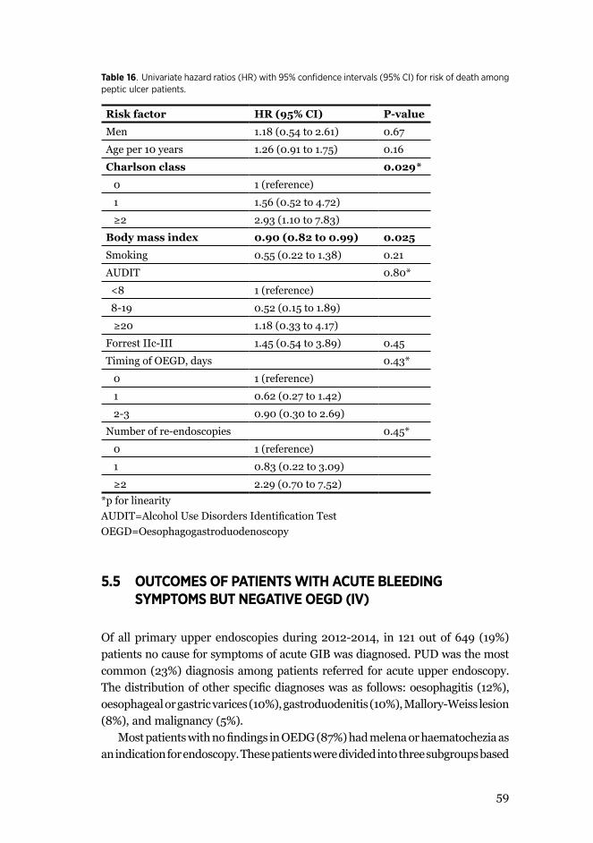
59
Table 16. Univariate hazard ratios (HR) with 95% confidence intervals (95% CI) for risk of death among peptic ulcer patients.
Risk factor HR (95% CI) P-value
Men 1.18 (0.54 to 2.61) 0.67
Age per 10 years 1.26 (0.91 to 1.75) 0.16
Charlson class 0.029*
0 1 (reference)
1 1.56 (0.52 to 4.72)
≥2 2.93 (1.10 to 7.83)
Body mass index 0.90 (0.82 to 0.99) 0.025
Smoking 0.55 (0.22 to 1.38) 0.21
AUDIT 0.80*
<8 1 (reference)
8-19 0.52 (0.15 to 1.89)
≥20 1.18 (0.33 to 4.17)
Forrest IIc-III 1.45 (0.54 to 3.89) 0.45
Timing of OEGD, days 0.43*
0 1 (reference)
1 0.62 (0.27 to 1.42)
2-3 0.90 (0.30 to 2.69)
Number of re-endoscopies 0.45*
0 1 (reference)
1 0.83 (0.22 to 3.09)
≥2 2.29 (0.70 to 7.52)*p for linearityAUDIT=Alcohol Use Disorders Identification TestOEGD=Oesophagogastroduodenoscopy
5.5 OUTCOMES OF PATIENTS WITH ACUTE BLEEDING SYMPTOMS BUT NEGATIVE OEGD (IV)
Of all primary upper endoscopies during 2012-2014, in 121 out of 649 (19%) patients no cause for symptoms of acute GIB was diagnosed. PUD was the most common (23%) diagnosis among patients referred for acute upper endoscopy. The distribution of other specific diagnoses was as follows: oesophagitis (12%), oesophageal or gastric varices (10%), gastroduodenitis (10%), Mallory-Weiss lesion (8%), and malignancy (5%).
Most patients with no findings in OEDG (87%) had melena or haematochezia as an indication for endoscopy. These patients were divided into three subgroups based

60
on comorbidity: no comorbidity, one comorbidity, or serious/several comorbidities. Patients with more comorbidity (Charlson class ≥2) were older (mean age 74 years, SD ±11) than the patients with no comorbidity (54 ±17) (p<0.001). No difference in BMI, smoking or alcohol consumption occurred regarding comorbidity. Of all patients, 60% used at least one bleeding-related drug regardless of comorbidity. Patients with comorbidity (≥1 comorbidity) used bleeding-related drugs more often than the patients with no comorbidity (p=0.012). The further examinations undertaken were based on clinical decisions (Table 17). The distribution of findings in further examinations is showed in Table 18. None of the patients (N=121) died within 30 days. The one-year mortality was 5.8% (95CI%: 2.4 -11.6).
Table 17. The further examinations undertaken among patients with a negative OEGD.
102 Colonoscopies (89 after negative OEGD, 13 recently 0-2 years prior)
With other examinations
20 CT-angiographies
13 Capsule endoscopies
3 Virtual CT colonoscopies
2 Double-balloon enteroscopies
2 Surgical operations
2 Re-OEGD
1 Proctoscopy
5 Refused for colonoscopy
1 Died before scheduled elective colonoscopy
1 Colonoscopy referral not done for unknown reason
3 Colonoscopy referral to occupational health care or another department
6 No further examinations on clinical judgment

61
Table 18. Findings in further examinations that most probably explain previous acute gastrointestinal bleeding symptoms among patients with negative OEGD by Charlson class.
Charlson=0N=42 (%)
Charlson=1N=39 (%)
Charlson≥2N=40 (%)
P-value
Diverticular disease of colon
10 (24) 20 (51) 17 (43) 0.033
No possible source ofbleeding identified
14 (33) 7 (18) 8 (20) 0.23
Colonic angioectasia 4 (10) 1 (3) 3 (7) 0.50
Cancer 1 (2) 1 (3) 3 (7) 0.53
Adverse effect of drug therapy
1 (2) 1 (3) 2 (5) 0.84
Hemorrhoids 2 (5) 0 (0) 2 (5) 0.54
Other 8 (19) 3 (8) 3 (7) 0.21
Data not available 2 (5) 6 (15) 2 (5) …

62
6 DISCUSSION
6.1 ACUTE GASTROINTESTINAL BLEEDING
One fourth of the patients referred for OEDG for acute GIB symptoms had a peptic ulcer in our study cohort during 2012-2014 (III) being the most common source of bleeding as seen in many other studies (Vreeburg et al. 1997, Leerdam et. al 2003, Loperfido et al. 2009, Nahon et al. 2012, Miilunpohja et al. 2017). Acute GIB is a universal medical emergency leading to hospitalisation including both upper and lower GIBs. Acute upper GIB originates from anywhere proximal to the Ligament of Treiz, whereas bleedings from other sources of the GI tract are traditionally and in most studies defined as lower GIB. After introduction of capsule endoscopy and double-balloon endoscopy, the definition of middle gastrointestinal bleeding occurring between the papilla Vateri and ileocecal valve in the small bowel has been proposed (Ell and May 2006, Raju et al. 2007).
The incidence of acute upper GIB is decreasing according to the published data (van Leerdam et al. 2003, Lanas et al. 2009, Loperfido et al. 2009) being 45-160 per 100 000 inhabitants per year (Vreeburg et al. 1997, Paspatis et al. 2000, Åhsberg et al. 2010a, Button et al. 2011). On the other hand, the incidence of acute lower GIB seems to be increasing (Lanas et al. 2009, Lanas et al. 2011) varying from 21 to 87 per 100 000 inhabitants per year in previous studies with the highest level reported from Iceland in 2010 (Longstreth 1997, Hreinsson et al. 2013). However, contradictory results have been reported from the USA during 2001-2009, where the incidence of both acute and lower GIB decreased by 15-23% during the study years (Laine et al. 2012).
The incidence of PUB decreased in Finland during 2000-2008 (I) as reported in other studies (Wang et al. 2010, Åhsberg et al. 2010b, Lanas et al. 2011, Laine et al. 2012). However, the incidence of PUB varying 9-87 per 100 000 is still remarkable comparing to that of reported incidences of all acute lower GIB (Bardhan et al. 2004, Pérez-Aisa et al. 2005, Lassen et al. 2006, Åhsberg et al. 2010). In addition, most lower GIBs stop spontaneously. One possible explanation for a downward trend in PUB is the decreasing prevalence of the H.pylori infection in Finland and other Western countries (Kosunen et al. 1997, Pérez-Aisa et al. 2005). However, one third of PUB patients tested positive for the H.pylori infection in our prospective study cohort during 2012-2014 (III).
The impact of newer anticoagulants introduced during the last ten years on the incidence of acute GIBs is not settled. According to a recent meta-analysis, the risk of major GIB with NOACs is, after initial concerns, at the same level as with the older vitamin K antagonists (Caldeira et al. 2015). As seen in Sweden

63
during 1984-2004, patients with acute upper and lower GIBs in the beginning of the 21st century were older, had more comorbidity and more often used bleeding related drugs (Åhsberg et al. 2010a). Older age and comorbidity are independent risk factors for morbidity and mortality associated with both upper and lower GIBs (Strate et al. 2008, Åhsberg et al. 2010a, Crooks et al. 2013). Therefore, the incidence of acute GIB might again increase in the future.
6.2 INCIDENCE OF PUD AND ITS COMPLICATIONS
The incidence of PUD and its complications leading to hospitalisation decreased significantly from 121 to 79 per 100 000 during 2000-2008 in Finland (I). The decrease in the incidence was seen in all age groups and in both genders. The incidence of PUD was higher among men and increased by age as shown in other previous studies (Pérez-Aisa et al. 2005, Kang et al. 2006, Lassen et al. 2006, Post et al. 2006, Cai et al. 2009, Feinstein et al. 2010). The incidence of both uncomplicated and complicated PUD cases decreased. That is partly contrary to the Danish results, where the incidence of uncomplicated PUD and PPU decreased, but that of bleeding ulcers remained stable during 1993-2002 (Lassen et al. 2006). In our retrospective study, the mean annual incidence rate of both uncomplicated and complicated PUD hospitalisation was 97 per 100 000, being in line with other studies reporting that incidence varies from 55 to 165 per 100 000 (Lewis et al. 2002, Pérez-Aisa et al. 2005, Feinstein et al. 2010, Åhsberg et al. 2011).
Previously, the incidence of hospital admission for complicated PUD was analysed in Finland 1972-1999 with a 79% increase in admissions (Paimela et al. 2002). In that previous Finnish study, the increase in complicated PUD hospitalisation was mainly due to bleeding gastric ulcers in elderly women. A significant decrease in the incidence of bleeding ulcers was seen in our cohort, except the incidence of duodenal ulcer bleeding remained stable among women (I). However, a decreasing trend of all PUB cases as seen in our study has also been shown in other countries (Wang et al. 2010, Åhsberg et al. 2010b, Lanas et al. 2011).
The decreasing trend in all PUD complications was mainly due to a decrease in all bleeding ulcers during 2000-2008. The incidence of PPU remained stable during the study years, being 6-7 per 100 000 for gastric ulcer perforations in 2006-2008 and 3 per 100 000 for duodenal ulcer perforations (I). As well, no change in the annual operation rate for PPU was seen in the Northern Finland during 1979-2000 (Mäkelä et al. 2002). On the other hand, based on data from the National Research and Development Centre for Welfare and Health, operations for PPU increased significantly in the whole Finland during 1987-1995, but remained stable during the years 1996-2000 (Paimela et al. 2004). Our results are in line

64
with the reported incidence of PPU varying from 4 to 14 per 100 000 (Pérez-Aisa et al. 2005, Åhsberg et al. 2011, Lau et al. 2011).
The proportion of gastric-ulcer patients was nearly twofold than that of duodenal-ulcer patients in the retrospective study cohort (I). This might be a consequence of the decreasing incidence of the H.pylori infection, whereas gastric mucosa is more vulnerable to NSAIDs. The prevalence between duodenal and gastric ulcers has varied in previous studies, the majority of them having more duodenal ulcers especially in studies with PUB patients (Pérez-Aisa et al. 2005, Lanas et al. 2006, Bardhan and Royston 2008, Sadic et al. 2009, Sung et al. 2010, Rosenstock et al. 2013). In our prospective cohort of bleeding ulcers, the proportion of gastric ulcers was slightly higher than that of duodenal ulcers (III). Similar results have been reported from Sweden, where hospitalisation rates for gastric ulcer bleeding were higher than for duodenal ulcer bleeding during 1987-2005 (Åhsberg et al. 2011) and as well as from the Netherlands during 2009-2012 (de Groot et al. 2014). The incidence of uncomplicated gastric ulcer in Denmark was also slightly higher than that of uncomplicated duodenal ulcers (Lassen et al. 2006). In that study, the distribution of complicated ulcers was not reported, but the proportion of possible NSAID-induced ulcers increased significantly from being 39% in 1993 to 53% in 2002 (p<0.01).
The cumulative one-year recurrence (≥3 months after the index hospitalisation) rate was 13% in our study cohort (I). The use of multiple drugs significantly increased the risk of recurrence. In a USA-based study, a significant increase in polypharmacy among adults is seen during the last 10 years, being at its highest among adults 65 years or older (Kantor et al. 2015). In our study, patients with recurrent PUD were neither analysed separately by initial site and complication of PUD nor by manifestation of a recurrent ulcer. In a large Danish population-based cohort study, 33% of PUB patients had a recurrent complication within 3 months and 14% of PPU patients (Lassen et al. 2006). In the long run (median follow-up varied from 2.7-3.7 years), 9% of PUB patients had a recurrent complication compared to 7% of PPU patients in that study. During the follow-up (median of 4.0 years) starting after three months after the initial hospitalisation with their first uncomplicated PUD, 3% of patients experienced PUD complication. The recurrence rate after PUB has varied between 0-38% with regard to the definition of recurrence time being on average 10% and that after PPU varying from 0.4% to 25%, with 12% being average. Very small numbers of recurrence have been reported in studies including H.pylori positive ulcer patients with successful eradication therapy after the initial hospitalisation (Miwa et al. 2004, Gisbert et al. 2012). After an ulcer episode, risk factors such as H.pylori infection and inappropriate use of NSAIDs are possible to eliminate, whereas the effect of increasing age and comorbidities as risk factors for recurrence, morbidity and mortality are not. The use of PPI before hospitalisation for PUD was associated with an increased risk of PUD recurrence

65
in our study cohort (I), suggesting that the patients at high-risk for ulcer formation are recognised.
6.3 MORTALITY ASSOCIATED WITH PUD
The short-term 30-day mortality associated with PUD was 3.7% in the retrospective cohort study (II) and 0.7% in the prospective study (III). The difference in the short-term mortality is most probably explained by the exclusion of unstable patients at the admission in the prospective cohort study. Hemodynamic instability at admission is an independent risk factor for mortality (Lau et al. 2011). Patients who were unable to give written informed consent due to severe dementia, e.g., were also excluded in the latter cohort (III); that might have led to exclusion of older patients with comorbidities affecting mortality. In the prospective study cohort 92% of patients with high-risk stigmata of ulcer bleeding had endoscopic dual therapy compared to only 12% in a French cohort with a higher in-hospital mortality of 5.8% (Nahon et al. 2012) or 38% in the UK audit cohort with mortality of 8.9% (Hearnshaw et al. 2010). Besides good adherence to the recommended dual therapy for high-risk ulcers in our study cohort, none of the patients needed surgery or TAE. These factors also explain the low short-term mortality rate in our cohort.
The mean 30-day mortality associated with PUD hospitalisation in Finland during 1995-1999 based on death certificates of patients who died of PUD or its surgery was 3.7% (Paimela et al. 2004). The 30-day mortality in our retrospective study cohort (II) was at the same level, but only one third of mortality that occurred within 30 days was explained by PUD as a main cause of death. So presumably, the all-cause mortality associated with PUD has decreased from the 1990s, although no decrease occurred during 2000-2008.
The in-hospital mortality for all PUD patients in the USA was 2.7% in 2006 (Wang et al. 2010). In that USA-based study, 86% of patients experienced a complicated peptic ulcer compared to 49% in our study cohort (I). The 30-day mortality was not analysed in the USA, but one might expect it to be at about same level as seen in our study. The 30-day mortality for uncomplicated PUD in Denmark 1993-2002 was 2.6% and for complicated PUD 11%. Most studies have reported mortality from complicated PUD. The short-term mortality (in-hospital or 30-day) associated with PUB has varied from 3.4% to 14% (van Leerdam et al. 2003, Lanas et al. 2005, Sadic et al. 2009, Åhsberg et al. 2010b and 2011, Sung et al. 2010, Nahon et al. 2012, Rosenstock et al. 2013). The all-cause 30-day mortality among PUB patients in our study varied from 3.8% to 5.6% in the retrospective study, being at the same level as in other countries.

66
The short-term mortality after PPU was about 10% in our study cohort varying from 6% among gastric ulcer perforations in men to 17% among duodenal ulcer perforations in women. Patients with PPU were not analysed regarding to operative or conservative treatment after PPU diagnosis. The short-term mortality after PPU surgery has varied from 7.5% (Paimela et al. 2004) to 14% (Mäkelä et al. 2002) in previous reports from Finland. According to a recent literature review, mortality associated with PPU varies from 1.3% to 30% (Chung and Shelat 2017).
However, no difference in one-year mortality emerged between the PUD patients in the retrospective (11.8%) and prospective (12.9%) study cohorts (II and III). According to other studies, excess mortality among PUD patients in the long run is explained by comorbidity (Wong et al. 2009, Holland-Bill et al. 2015, Laursen et al. 2015a, Liang et al. 2016). Malignancies and cardiovascular diseases explained over 50% of deaths that occurred during the first year after PUD hospitalisation in our study. The impaired survival among the patients once hospitalised for PUD compared to the age-and gender-adjusted population was also shown to worsen continuously during the follow-up time. Therefore, hospitalisation for PUD might be considered as a warning sign with a demand for an evaluation of individual patient´s overall health status.
Interestingly, patients on statin therapy before the first ulcer had a better short- and long-term survival in our cohort. The most probable explanation for our finding of reduced short- and long-term mortality in PUD patients on statins is their effect on overall mortality, which was demonstrated in a recent large meta-analysis showing a significant reduction in all-cause mortality among patients with statin therapy (Cholesterol Treatment Trialists’ (CTT) Collaboration 2015). Furthermore, among elderly people with no known history of vascular events, the use of statins or fibrates was also associated with a 30% decrease in the incidence of stroke (Alpérovitch et al. 2015). On the other hand, in a Taiwanese population-based cohort study, the risk of developing PUD was significantly lower among the statin users, suggesting that statins might act as potential gastroprotective agents by inhibiting neutrophil activity, reducing oxidative stress and maintaining vascular integrity (Feng et al. 2015).
6.4 RISK FACTORS FOR PUD ASSOCIATED MORTALITY
Comorbidity was associated with a significant increase in the risk of death in the prospective study (III), as well as the use of oral corticosteroids and diuretics, indicating cardiovascular comorbidities among PUD patients in the retrospective study cohort (II). In addition, most patients died of non-PUD-related causes in the retrospective study cohort as seen in other studies, indicating the impact of

67
comorbidity in survival of hospitalised PUD patients (Imhof et al. 2008, Sung et al. 2010, Åhsberg et al. 2010, Lau et al. 2011, Laursen et al. 2015a).
In our prospective cohort, increasing age was not associated with mortality, probably due to the exclusion of some patients (III). The annual incidence rate of PUD increased by age during the retrospective study years 2000-2008 (I). The association between increasing age and mortality in PUD patients is shown, however, in many other studies (Soplepmann et al. 1997, van Leerdam et al. 2003, Imhof et al. 2008, Sung et al. 2010, Åhsberg et al. 2010, Laursen et al. 2015a).
Obesity was shown to decrease the risk of mortality among PUB patients compared to normal or underweight patients (III). Obesity, in the population level, is associated with morbidity and mortality (Masters et al. 2013). However, the “obesity paradox” meaning better survival has been reported to occur among patients admitted to intensive care units, for an emergency abdominal operation or hospitalised for cardiac failure or ischemic stroke (Casas-Vara et al. 2012, Kim et al. 2012, Pickkers et al. 2013, Benjamin et al. 2017). On the other hand, obese trauma patients had more complications and worse survival that the normal weight patients in a large USA-based retrospective cohort study (Glance et al. 2014). In a Danish study on patients with PPU, obesity did not affect survival, whereas underweight was associated with poorer survival after surgery (Buck and Møller 2014). The pathophysiology of obesity with decreased risk of mortality among hospitalised patients is unclear. One explanation could be that obese patients are in good nutritional status compared to those patients with normal BMI but already suffering poorer nutritional stage caused by severe comorbidity.
The timing of endoscopy did not have an effect on survival in our prospective cohort study (III). Our result is similar to that seen in Denmark, where the timing of endoscopy was not associated with mortality in stable PUB patients with an ASA score of 1-2 (Laursen et al. 2017b). However, in our prospective PUB cohort, patients with more comorbidity were also included if they were hemodynamically stable enough to give the written informed consent. In the Danish study, patients who were unstable at admission or were both unstable and had more comorbidity (ASA 3-5) had better survival if they experienced an upper endoscopy 6-24 (unstable patients) or 12-36 (unstable patients with comorbidity) hours after admission, indicating the importance of resuscitation and managing comorbidities (Laursen et al. 2017b). This was also stated in the international recommendations for performing early endoscopy within 24 hours with no additional benefit from more urgent procedures (Barkun et al. 2010, Gralnek et al. 2015). However, patients who are already at hemodynamic shock at the time of admission have a worse survival (Lau et al. 2011). Results of the studies on the “weekend effect” (patients who are hospitalised out of office hours) on mortality are controversial, but no such effect was shown to occur in the most recent prospective international study (Murray et al. 2017). The “weekend effect” was not analysed in our study cohorts.

68
The rebleeding rate before the modern era of endoscopic therapies was high, being up to 55% after PUB with major stigmata of hemorrhage with mortality of 11% (Laine and Petersen 1994). The major stigmata of bleeding are considered as a risk factor for mortality (Lau et al. 2011, Camus et al. 2016). However, no difference in mortality between the high- and low-risk stigmata patients in Spain 2006-2009 was observed (Lanas et al. 2014). That is in line with results from our prospective study 2012-2014 (III), suggesting the good quality of endoscopic therapy. Similarly shown in the Netherlands 2009-2012, no correlation between Forrest classification and mortality occurred (de Groot et al. 2014). Patients who start to bleed while hospitalised for another cause are at increased risk of mortality compared to patients admitted to hospital for bleeding-related symptoms (Åhberg et al. 2010b, Hearnshaw et al. 2011, Camus et al. 2016). The proportion of in-hospital bleeders was not known in our study cohorts.
Among PPU patients, a delay in surgery of more than 24 hours after admission is associated with a significantly poorer outcome (Svanes 2000, Mäkelä et al. 2002, Kocer et al. 2007 Lau et al. 2011, Søreide et al. 2014, Chung and Shelat 2017). In our retrospective register-based study with no access to patient charts, it was not possible to analyse if a delay in surgery or decision to treat a PPU patient conservatively affected the survival of PPU patients (II). These factors besides comorbidity might explain the significantly worse survival among women with perforated duodenal ulcers compared to that of men.
6.5 OUTCOMES OF PATIENTS WITH ACUTE BLEEDING SYMPTOMS BUT NEGATIVE OEGD
The proportion of patients with acute bleeding symptoms referred for OEDG but no diagnosed source of bleeding in the upper GI tract was 19% in the prospective study cohort (IV). In other studies, the reported proportion of patients with no diagnosis has varied from 5% to 28% with no explanation of underlying cause for bleeding symptoms (Vreeburg et al. 1997, van Leerdam et al. 2003, Lanas et al. 2005, Lanas et al. 2009, Loperfido et al. 2009, Hearnshaw et al. 2011, Nahon et al. 2012, Miilunpohja et al. 2017). The reported in-hospital mortality has varied between 6-16%, which is clearly at a higher level than in our study. This difference is most probably explained by the exclusion of the most vulnerable and unstable patients.
The diverticular disease of the colon was regarded as the most probable source of bleeding in one third of patients (IV). That is also seen in studies on lower GI bleedings, explaining approximately one third of bleedings (Longstreth 1997, Gayer et al. 2009, Hreinsson et al. 2013). The diverticular disease of the colon as a bleeding source was more common in patients with comorbidities in our study cohort; these

69
patients also used more often bleeding-related drugs. The use of NSAIDs, ASA or calcium channel blockers was associated with diverticular bleedings according to a recent systematic review (Kvasnoysky et al. 2014).
As being an observational cohort study, further examinations were undertaken by clinical decision during the hospitalisation (IV). Therefore, the source of bleeding remained unknown for 24% patients. The proportion of those patients could have been smaller if all possible further examination methods including, e.g., capsule enteroscopy or double-balloon enteroscopy were utilised during the index hospitalisation. On the other hand, the proportion of patients with no diagnosis has varied from 3-12% in lower GI tract studies (Longstreth 1997, Gayer et al. 2009, Hreinsson et al. 2013). Besides, the costs of all the possible further examinations and hospitalisation days would have been remarkable when considering the very low mortality of this cohort.
Mortality among these patients was very low: none of the patients deceased within 30 days, and seven (5.8%) died within a year mainly due to chronic diseases (alcoholic cirrhosis, lung cancer, aortic stenosis, Alzheimer´s disease) (IV). Therefore, a clinical decision not to utilise all the available examination methods seems to be reasonable. In addition, further examinations after OEGD can be undertaken as elective procedures in most patients without lengthening the initial hospital stay. As seen in a study from the USA, urgent colonoscopy identified better the definite bleeding source among patients with acute lower GIB (OR 2.6; 95%CI 1.1-6.2), but without differences in outcomes (Green et al. 2005).
6.6 STRENGTHS AND LIMITATIONS OF THE STUDY
In the retrospective register-based study part (I and II), data on hospitalised PUD patients only might reduce the number of false-positive cases compared to studies reporting physician-diagnosed PUD in primary care. A recent analysis of 32 different studies comparing The Finnish Hospital Discharge Register data to external information showed that more than 95% of discharges could be identified from the register (Sund 2012). Positive predictive value (PPV) for common diagnoses ranged between 75-99%, suggesting that the completeness and accuracy in the register seem to vary from satisfactory to very good. In addition, based on systematic review of study methods, our study design most probably underestimates the incidence for uncomplicated PUD (Lin et al. 2011).
The retrospective study part also has some limitations. The study was conducted as a retrospective epidemiological study based on hospital records of patients requiring hospitalisation for their PUD and on hospitalised patients who have had their ulcer disease diagnosed during the hospital visit. Our epidemiological study describes trends and associations with PUD, but can´t define causation

70
of the disease. The major limitation of the study was unavailability of data on comorbidities and living habits. Besides, the H.pylori status and changes in drug therapy after the ulcer were not known. Therefore, the relative risks of drug therapy affecting survival after hospitalisation may not be clinically relevant in the long run.
The prospective study part (III and IV) was conducted as an observational cohort study, where all the included patients had upper endoscopy and were individually interviewed by a trained study nurse. In addition, access to the hospital records was available in order to obtain all information on risk factors for the short- and long-term mortality. For the patients with negative OEGD (IV), results of previous and further examination could be evaluated for the analysis.
However, written informed consent was mandatory, leading to a quite high number of patients excluded from the analysis. That might have led to underestimation of mortality and to patient selection bias when interpreting the study results. Among PUD patients (III) no statistically significant difference in mortality appeared between the patients categorised into the low- and high-risk Forrest classes, but the result of post-hoc power calculation was 59% suggesting that in a larger cohort a difference could possibly have emerged. Although PUD was the most common diagnosis in the primary endoscopy, the number of individual patients with PUD was only 147. The number of individual patients without diagnosis at the primary endoscopy was 121 (IV). As being an observational study, no possibilities to interfere with clinical judgments on patient care were allowed by the study group.

71
7 CONCLUSIONS
IThe incidence of PUD and its complications requiring hospitalisation decreased significantly in the capital area of Finland during 2000-2008. The one-year recurrence rate of PUD was quite high at 13%, although the recurrence was defined when the first and the successive hospitalisation occurred after more than three months to exclude hospitalisation for ulcer healing controls. The PUD patients used drugs more often than the age- and gender-adjusted background population, suggesting more comorbidity. The use of several drugs increased the risk of recurrence. The use of PPI was also associated with a slight increase in recurrence.
IIDuring the study period 2000-2008 no difference in SMR occurred among the patients hospitalised for PUD. The short-term 30-day mortality associated with PUD was 3.7%, and the long-term one-year mortality was 11.8%. The short- and long-term survival of women with a perforated duodenal ulcer was significantly impaired compared to that of men. The overall survival compared to the age- and gender-matched background population was worse among PUD patients, and it decreased constantly up to a follow-up of nine years. PUD explained one third of mortality occurring within 30 days. PUD was considered as a main cause death only in less than 15% deaths occurring within one year, whereas malignancies and cardiovascular were the main causes of death in over 50% of cases. The previous use of statins was associated with a significant decrease in all-cause mortality among all PUD patients.
IIIPUD is still the most common source of bleeding in hospitalised patients referred for acute OEDG during 2012-2014. The short-term mortality among PUD patients was low 0.7%, but 12.9% of patients died within one year. Comorbidity was associated with an increase risk of death, whereas obesity (BMI>30) was associated with better survival. However, age, gender, smoking or drinking habits, stigmata of ulcer haemorrhage (low- or high-risk stigmata), timing of endoscopy or number of re-endoscopies did not affect on survival.
IVOf all patients admitted for acute OEGD during hospitalisation in 2012-2014, no reason for bleeding symptoms was diagnosed in 19% of patients. In further examinations, the diverticular disease of the colon was regarded as the most

72
probable source of previous bleeding symptoms in one third of patients. Further examinations were undertaken by clinical decision; therefore, the source of bleeding remained unidentified in 24% of these patients. No patient died within 30 days, and the one-year mortality was 5.8%.
Further perspectives
Although the incidence of PUD is decreasing, the management of patients presenting with complicated PUD is becoming more challenging. The results of medical and interventional treatments for peptic ulcers are good. The utilisation of angioembolisation in fragile PUB patients, possibly unfit for surgery, is emerging. However, mortality associated with PUD among the elderly population is remarkable. Therefore, recognising PUD before its complication occurs is essential, as well as the management of a patient´s comorbidity while hospitalised for PUD. The growing antimicrobial resistance to H.pylori eradication therapy, as seen in other Western countries, is also worrying and can lead to a plateau or even an increase in PUD occurrence. Patients starting long-lasting NSAID therapy should be tested for the H.pylori infection. In addition, patient adherence to GPAs should be evaluated in patients with an increased risk of PUD.

73
ACKNOWLEDGEMENTS
This study was carried out at the Department of Gastroenterology and Gastrointestinal Surgery of the Abdominal Center at Helsinki University Hospital and University of Helsinki during 2010-2018. Financial support from the Mary and Georg C. Ehrnrooth Fund, the Professor Martti I.Turunen Fund, the Finnish Medical Foundation, and MSD Finland for Clinical Research Institute Helsinki University Central Hospital Ltd (HYKS-institute) for peptic ulcer study is gratefully acknowledged.
First and foremost, I thank Professor Martti Färkkilä for being a positive and encouraging supervisor without ignoring life outside the research and clinical work. I really appreciate that you have always been available for questions and guided me through the whole thesis process. I wish to express gratitude to Docent Lauri Virta for joining this thesis process and for your excellent supervision particularly in understanding research methods and statistics. I have really enjoyed our multidisciplinary team work!
Thank you Docent Leena Halme for introducing me to Professor Martti Färkkilä and for providing the opportunity to carry out this study beside clinical work. I thank Professor Pauli Puolakkainen for creating an enthusiastic atmosphere towards the science and research at the Department of Gastrointestinal Surgery.
I am thankful for all my co-authors. Statistician Hannu Kautiainen was an enormous help for explaining and performing the statistics. Jari Koskenpato is thanked for giving good advice and support when the thesis project started. Niilo Färkkilä´s knowledge and assistance was important in the beginning of the project. The study nurses Sari Karesvuori, Pirkko Tuukkala, Virpi Pelkonen and Paula Karlsson gave their invaluable effort in recruiting and interviewing patients for the prospective study part of this thesis. Additionally, I wish to warmly thank Luanne Siliämaa for the English language editing.
I respectfully thank the official reviewers of this thesis, Professors Jyrki Mäkelä and Markku Voutilainen, for your time and effort and the extremely valuable comments.
I express my warmest thanks to all my colleagues at the Department of Gastrointestinal Surgery. I also thank my superiors, Esko Kemppainen and Jukka Sirèn, for providing me with research facilities and a comprehensive clinical education during my specialisation. Especially, I thank colleagues Piia Pulkkinen and Minna Räsänen for your friendship, valuable conversations and sincere help when needed. Colleagues, nurses and other supporting staff especially at the Jorvi K5 ward are thanked for supporting me in the academic jungle, and I am very grateful to have the possibility to work with you in relevant clinical work. I thank

74
Jaana Laamanen for friendship and supervising me at work: you are an amazing clinician! I also thank my non-surgeon colleagues for expanding my knowledge in medical fields and other aspects of life.
And to my friends and relatives, my sincere thanks for helping and supporting me. Thank you, Anu, for your friendship, helpfulness and everything we have shared over the years. I thank Kati for long-lasting friendship and conversations outside the box; how unpredictable can life be? My dear friends Saikkis, Teija, Johanna G, Kristiina, Meeri, Susu, Maija, Birgitta, Jonna, Henrika, Suvi, Johanna A, and Satu, thank you for many incredible moments spent together. Words cannot express my gratitude and love to my parents Eija and Antero for supporting me and my family whenever needed! I also want to thank Pirjo and Risto for being such amazing grandparents and for your practical invaluable help in our daily life. I also want to thank my brother-in-law, Tuomo, for your kindness and especially being there for Eero!
Most of all, I want to thank my beloved family: Timo and our son Eero. You are the most important people to share my life with!
Helsinki, March 2018
Hanna

75
REFERENCES
Alpérovitch A, Kurth T, Bertrand M, Ancelin ML, Helmer C, Debette S, Tzourio C. 2015. Primary prevention with lipid lowering drugs and long term risk of vascular events in older people: population based cohort study. BMJ 350:h2335.
Altieri MS, Pryor A, Yang J, Yin D, Docimo S, Bates A, Talamini M, Spaniolas K. 2017. The natural history of perforated marginal ulcers after gastric bypass. Surg Endosc [epub ahead of print].
Anbalakan K, Chua D, Pandya GJ, Shelat VG. 2015. Five year experience in managemen of perforated peptic ulcer and validation of common mortality risk prediction models – are existing models sufficient? A retrospective cohort study. Int J Surg 14:38-44.
Aro P, Storskrubb T, Ronkainen J, Bolling-Sternevald E, Engstrang L, Vieth M, Stolte M, Talley NJ, Agréus L. 2006. Peptic Ulcer Disease in a General Adult Population. The Kalixanda Study: A Random Population-based Study. Am J Epidemiol 163:1025-1034.
Arroja B, Cremers I, Ramos R, Cardoso C, Rego AC, Caldeira A, Eliseu L, Silva JD, Gloria L, Rosa I, Perdosa J. 2011. Acute lower gastrointestinal bleeding management in Portugal: a multicentric prospective 1-year survey. Eur J Gastroenterol Hepatol 23:317-322.
Bakkevold KE. 2010. Time trends in incidence of peptic ulcer bleeding and associated risk factors in Norway 1985-2008. Clin Exp Gastroenterol 3:71-77.
Barat I, Andreasen F, Damsgaard EMS. 2000. The consumption of drugs by 75yearold individuals living in their own homes. Eur J Clin Pharmacol 56:501-509.
Barkun AN, Sabbah S, Enns R, Armstrong D, Gregor J, Fedorak RNN, Rahme E, Toubouti Y, Martel M, Chiba N, Fallone CA, and the RUGBE Investigators. 2004. The Canadian Registry on Nonvariceal Upper Gastrointestinal Bleeding and Endoscopy (RUGBE): Endoscopic Hemostasis and Proton Pump Inhibition are Associated with Improved Outcomes in a Real-Life Setting. Am J Gastroenterol 99:1238-1246.
Barkun AN, Bardou M, Kuipers EJ, Sung J, Hunt RH, Martel M, Sinclair P, for the International Consensus Upper Gastrointestinal Bleeding Conference Group. 2010. International Consensus Recommendations on the Management of Patients With Nonvariceal Upper Gastrointestinal Bleeding. Ann Intern Med 152:101-113.
Bardhan KD, Williamson M, Royston C, Lyon C. 2004. Admission rates for peptic ulcer in the Trent region, UK, 1972-200o; Changing pattern, changing disease? Dig Liv Dis 36:577-588.
Bardhan KD, Royston C. 2008. Time, change and peptic ulcer disease in Rotherham, UK. Dig Dis Sci 40:540-546.
Beggs AD, Dilworth MP, Powell SL, Atherton H, Griffiths EA. 2014. A systematic review of transarterial embolisation versus emergency surgery in treatment of major nonvariceal upper gastrointestinal bleeding. Clin Exp Gastroenterol 7:94-104.

76
Benjamin ER, Dilektasli E, Haltmeier T, Beale E, Inaba K, Demetriades D. 2017. The effects of body mass index on complications and mortality after emergency abdominal operations: The obesity paradox. Am J Surg 214:899-903.
Bertleff MJOE, Lange JF. 2010. Laparoscopic correction of perforated peptic ulcer: first choice? A review of literature. Surg Endosc 24:1231-1239.
Blatchford O, Murray WR, Blatchford M. 2000. A risk score to predict need for treatment for upper gastrointestinal hemorrhage. Lancet 356:1318-1321.
Boey J, Choi SKY, Alagaratnam TT, Poon A. 1987. Risk Stratification in Perforated Duodenal Ulcers. A Prospective Validation of Predictive Factors. Ann Surg 205:22-26.
Buck DL, Møller MH. 2014. Influence of body mass index on mortality after surgery for perforated peptic ulcer. BJS 101:993-999.
Byrke N, Barton RG, Enniss TM, Nirula R. 2013. Laparoscopic versus open repair of perforated gastroduodenal ulcer: a National Surgical Quality Improvement Program Analysis. Am J Surg 206:957-962.
Cai S, García-Rodríguez LA, Massó-González EL, Hernández-Díaz S. 2009. Uncomplicated peptic ulcer in the UK: trends from 1997 to 2005. Aliment Pharmacol Ther 30:1039-1048.
Caldeira D, Barra M, Ferreira A, Rocha A, Augusto A, Pinto FJ, Costa S, Ferreira JJ. 2015. Systematic review with meta-analysis: the risk of major gastrointestinal bleeding with non-vitamin K antagonist oral anticoagulants. Aliment Pharmacol Ther 42:1239-1249.
Camus M, Jensen DM, Kovacs TO, Jensen ME, Markovic D, Gornbein J. 2016. Independent risk factors of 30-day outcomes in 1264 patients with peptic ulcer bleeding in the USA: large ulcers do worse. Aliment Pharmacol Ther 43:1080-1089.
Cangemi DJ, Krill T, Weideman R, Cipher DJ, Spechler SJ, Feagins LJ. 2017. A Comparison of the Rate of Gastrointestinal Bleeding in Patients Taking Non-Vitamin K Antagonist Oral Anticoagulants or Warfarin. Am J Gastroenterol 112:734-739.
Casas-Vara A, Santolaria F, Fernández-Bereciartúa A, González-Reimers E, García-Ochoa A, Martínez-Riera A. 2012. The obesity paradox in elderly patients with heart failure: Analysis of nutritional status. Nutrition 28:616-622.
Chan FKL. 2005. NSAID-Induced Peptic Ulcers and Helicobacter pylori Infection, Implications for Patient Management. Drug Ssafety 28(4):287-300.
Chan FKL, Ching JYL, Tse YK, Lam K, Wong GLH, Ng SC, Lee V, Au KWL, Cheong PK, Suen BY, Chan H, Kee KM, Lo A, Wong VWS, Wu JCY, Kyaw MH. 2017. Gastrointestinal safety of celecoxib versus naproxen in patients with cardiothrombotic disease and arthritis after upper gastrointestinal bleeding (CONCERN): an industry-independent, double-blind, double-dummy, randomised trial. Lancet 389:2375-2382.
Charlson Charlson ME, Pompei P, Ales KL, MacKenzie CR. 1987. A new method of classifying prognostic comorbidity in longitudinal studies: development and validation. J Chronic Dis 40:373-383.

77
Charpignon C, Lesgourgues B, Pariente A, Nahon S, Pelaquier A, Gatineau-Sailliant G, Roucayrol A-M, Courillon-Mallet A, The Group de l´Observatoire National des Ulcéres de l´Association Nationale des HépatoGastroentérologues des Hôpitaux Généraux (ANGH). 2013. Peptic ulcer disease: one in five is related to neither Helicobacter pylori nor aspirin/NSAID intake. Aliment Pharmacol Ther 38:946-954.
Cholesterol Treatment Trialists’ (CTT) Collaboration, Fulcher J, O’Connell R, Voysey M, Emberson J, Blackwell L, Mihaylova B, Simes J, Collins R, Kirby A, Colhoun H, Braunwald E, La Rosa J, Pedersen TR, Tonkin A, Davis B, Sleight P, Franzosi MG, Baigent C, Keech A. 2015. Efficacy and safety of LDL-lowering therapy among men and women: meta-analysis of individual data from 174,000 paricipants in 27 randomised trials. Lancet 385:1397-1405.
Christensen S, Riis A, Nørgaard M, Thomsen RW, Tønnesen EM, Larsson A Sørensen HT. 2006. Perforated peptic ulcer: use of pre-admission oral glucocorticoids and 30-day mortality. Aliment Pharmacol Ther 23:45-52.
Christensen S, Riis A, Nørgaard M, Thomsen RW, Sørensen HT. 2007. Introdiction of newer selective cyclo-oxygenase-2 inhibitors and rates of hospitalization with bleeding and perforated peptic ulcer. Aliment Pharmacol Ther 25:907-912.
Chung KT, Shelat VG. 2017. Perforated peptic ulcer – an update. World J Gastrointest Surg 9(1):1-12.
Chung WC, Jeon EJ, Kim DB, Sung HJ, Kim Y-J, Lim ES, Kim M, Oh JH. 2015. Clinical characteristics of Helicobacter pylori –negative drug-negative peptic ulcer bleeding. World J Gastroenterol 21:8636-8643.
Crooks CJ, West J, Card TR. 2011. Reductions in 28-Day Mortality Following Hospital Admission for Upper Gastrointestinal Hemorrhage. Gastroenterology 141:62-70.
Crooks CJ, West J, Card TR. 2013. Comorbidities Affect Risk of Nonvariceal Upper Gastrointestinal Bleeding. Gastroenterology 144:1384-1393.
Dall M, Schaffalitzky de Muckadell OB, Lassen AT, Hansen JM, Hallas J. 2009. An Association Between Selective Serotonin Reuptake Inhibitor Use and Serious Upper Gastrointestinal Bleeding. Clin Gastroenterol Hepatol 7:1314-1321.
Ederer F, Heise H. 1959. Instructions to IBM 650 programmers in processing survival computations. Methodological note no.10. Bethesda, MD: End Results Evaluation Section, National Cancer Institute.
El-Nakeeb A, Fikry A, El-Hamed TMA, Fouda EY, Awady SE, Youssef T, Sherief D, Farid M. 2009. Effect of Helicobacter pylori eradication on ulcer recurrence after simple clousure of perforated duodenal ulcer. Int J Surg 7:126-129.
Ergul E, Gozetlik EO. 2009. Emergency spontaneous gastric perforations: ulcer versus cancer. Langenbecks Arch Surg 394:643-646.
Feinstein LB, Holman RC, Christensen KLY, Steiner CA, Swerdlow DL. 2010. Trends in Hospitalizations for Peptic Ulcer Disease, United States, 1998-2005. Emerg Inf Dis 16:1410-1418.

78
Feng A, Chuang E, Wu S-H, Wang J-C, Chang S-N, Lin C-L, Kao C-H. 2015. The effect of statins on the occurrence of peptic ulcer. Eur J Intern Med 26:731-735.
Finnish Medicines Agency Fimea and Social Insurance Institution. 2016. Finnish statistics on medicines 2015. Available at http://www.kela.fi/tilastojulkaisut_suomen-laaketilasto.
Ford AC, Gurusamu KS, Delaney B, Forman D, Moayyedi P. 2016. Eradication therapy for peptic ulcer disease in Helicobacter pylori -positive people. Cochrane Database of Systamatic Reviews 4:CD003840.
Forrest JA, Finlayson ND, Shearman DJ. 1974. Endoscopy in gastrointestinal bleeding. Lancet 2:394-397.
Freedberg DE, Kim LS, Yang Y-X. 2017. The Risks and Benefits of Long-term use of Proton Pump Inhibitors: Expert Review and Best Practice Advice From the American Gastroenterological Association. Gastroenterology 152:706-715.
García Rodrígues LA, Lin KJ, Hernández-Díaz S, Johansson S. 2011. Risk of Upper Gastrointestinal Bleeding With Low-Dose Acetylsalicylic Acid Alone and in Combination With Chlopidogrel and Other Medications. Cirucaltion 123:1108-1115.
Gayer C, Chino A, Lucas C, Tokioka S, Yamasaki T, Edelman DA, Sugawa C. 2009. Acute lower gastrointestinal bleeding in 1,112 patients admittedto an urban emergency medical center. Surgery 146:600-606.
Gisbert JP, Pajares JM. 2003. Helicobacter pylori Infection and Perforated Peptic Ulcer Prevalence of the Infection and Role Antimicrobial Treatment. Helicobacter 8:159-167.
Gisbert JP, Pajares JM. 2005. Systematic review and meta-analysis: is 1-week proton pump inhibitor –based triple therapy sufficient to heal peptic ulcer? Aliment Pharmacol Ther 21:795-804.
Gisbert JP, Calvet X, Cosme A, Almela P, Feu F, Bory F, Santolaria S, Aznárez R, Castro M, Fernández N, García-Grávalos R, Benages A, Cañete N, Montoro M, Borda F, Pérez-Aisa A, Piqué JM, on behalf of the H.Pylori Study Group of the Asociación Española de Gastroenterología (Spanish Gastroenterology Association). 2012. Long-Term Follow-Up of 1,000 Patients Cured of Helicobacter pylori Infection Following an Epidose of Peptic Ulcer Bleeding. Am J Gastroenterol 107:1197-1204.
Glance LG, Li Y, Osler TM, Mukamel DB, Dick AW. 2014. Impact of Obesity on Mortality and Complications in Trauma Patients. Ann Surg 259:576-581.
Gralnek IM, Dumonceau J-M, Kuipers JE, Lanas A, Sanders DS, Kurien M, Rotandano G, Hucl T, Dinis-Ribeiro M, Marmo R, Racz I, Arezzo A, Hoffmann RT, Lesur G, de Franchis R, Aabakken L, Veitch A, Radaelli F, Salgueiro P, Cardoso R, Maia L, Zullo A, Cipoletta L, Hassan C. 2015. Diagnosis and management of acute nonvariceal upper gastrointestinal hemorrhage: European Society of Gastrointestinal Endoscopy (ESGE) Guideline. Endoscopy 47:1-46.
Grassi R, Romano S, Pinto A, Romano L. 2004. Gastro-duodenal perforations: conventional plain film, US and CT findings in 166 consecutive patients. Eur J Radiol 24:1386-1393.
79
Green BT, Rockey DC, Portwood G, Tarnasky PR, Guarisco S, Branch M, Leung J, Jowell P. 2005. Urgent Colonoscopy for Evaluation and Management of Acute Lower Gastrointestinal Hemorrhage. A Randomized Controlled Trial. Am J Gastroenterol 100:2395-2402.
deGroot NL, van Oijen MGH, Kessels K, Hemmink M, Weusten BLAM, Timmer R, Hazen WL, van Lelyveld N, Vermeijden RR, Curvers WL, Baak BC, Verburg R, Bosman JH, de Wijkerslooth LRH, de Rooij J, Venneman NG, Pennigs M, van Hee K, Scheffer BCH, van Eijk RL, Meiland R, Siersema PD, Bredenoord AJ. 2014. Reassessment of the predictive value of the Forrest classification for peptic ulcer rebleeding and mortality: can classification be simplified? Endosc 46:46-52.
Hallas J, Dall M, Andries A, Andersen BS, Aalykke C, Hansen J, Andersen M, Lassen AT. 2006. Use of single and combined antithrombotic therapy and risk of serious upper gastrointestinal bleeding: population based control-case study. BMJ 333:726-730.
Hearnshaw SA, Logan RF, Lowe D, Travis SPL, Murphy MF, Palmer KR. 2010. Use of endoscopy for management of acute upper gastrointestinal bleeding in the UK: results of a nationwide audit. Gut 59:1022-1029.
Hearnshaw SA, Logan RF, Lowe D, Travis SPL, Murphy MF, Palmer KR. 2011. Acute gastrointestinal bleeding in the UK: patient characteristics, diagnoses and outcomes in the 2007 UK audit. Gut 60:1327-1335.
Hsu P-I, Lai K-H, Tseng H-H, Lo G-H, Lo C-C, Lin C-K, Cheng S-J, Chan H-H, Ku M-K, Peng N-J, Chien E-J, Chen W, Hsu P-N. 2001. Eradication of Helicobacter pylori prevents ulcer development in patients with ulcer-like functional dyspepsia. Aliment Pharmacol Ther 15:195-201.
Holland-Bill L, Christiansen CF, Gammelager H, Mortensen RN, Pedersen L, Sørensen HT. 2015. Chronic liver disease and 90-day mortality in 21 359 patients following peptic ulcer bleeding – a Nationwide Cohort Study. Aliment Pharmacol Ther 41:564-572.
Holster IL, Kuipers EJ. 2012. Management of acute nonvariceal upper gastrointestinal bleeding: Current policies and future perpectives. World J Gastroenterol 18(11):1202-1297.
Holster IL, Valkhoff VE, Kuipers EJ, Tjwa ETTL. 2013. New Oral anticoagulans Increase Risk for Gastrointestinal Bleeding: A Systematic Review and Meta-analysis. Gastroenterology 145:105-112.
Hooi JKY, Lai WY, Ng WK, Suen MMY, Underwood FE, Tanyingoh D, Malfertheiner P, Graham DY, Wong VWS, Wu JCY, Chan FKL, Sung JJY, Kaplan GG, Ng SC. 2017. Global Prevalence of Helicobacter pylori Infection: Systematic Review and Meta-Analysis. Gastroenterology 153:420-429.
Hreinsson JP, Guðmundsson S, Kalaitzakis E, Björnsson ES. 2013. Lower gastrointestinal bleeding: incidence, etiology, and outcomes in a population-based setting. Eur J Gastroenterol Hepatol 25:37-43.
Imhof M, Epstein S, Ohmann C, Roher HD. 2008. Duration of Survival after Peptic Ulcer Perforation. Word J Surg 32:408-412.

80
Jairath V, Barkun AN. 2012. Improving outcomes from acute upper gastrointestinal bleeding. Gut 61:1246-1249.
Jairath V, Kahan BC, Gray A, Doré CJ, Mora A, James MW, Stanley AJ, Everett SM, Bailey AA, Dallal H, Greenaway J, Le Jeune I, Darwent M, Church N, Reckless I, Hodge R, Dyer C, Meredith S, Llewelyn C, Palmer KR, Logan FR, Travis SP, Walsh TS, Murphy MF. 2015. Restrictive versus liberal blood transfusion for acute upper gastrointestinal bleeding (TRIGGER): a pragmatic, open-label, cluster randomised feasibility trial. Lancet 386:137-144.
Jensen DM, Eklund S, Persson T, Ahlbom H, Stuart R, Barkun AN, Kuipers EJ, Mössner J, Lau JM, Sung JJ, Kilhamn J, Lind T. 2017. Reassessment of Rebleeding Risk of Forrest I B (Oozing) Peptic Ulcer Bleeding in a Large International Randomized Trial. Am J Gastro 112:441-446.
Jiang H-Y, Chen H-Z, Hu X-J, Yu Z-H, Yang W, Deng M, Zhang Y-H, Ruan B. 2015. Use of Selective Serotonine Reuptake Inhibitors and Risk of Upper Gastrointestinal Bleeding: A Systemic Review and Meta-analysis. Clin Gastroenterol Hepatol 13:42-50.
Johnson CD, Ellis H. 1990. Gastric outlet obstruction now predicts malignancy. Br J Surg 77:1023-1024.
McJunkin B, Sissoko M, Levien J, Upschurch J, Ahmed A. 2011. Dramatic Decline in Prevalence of Helicobacter pylori and Peptic Ulcer Disease in an Endoscopy-referral Population. Am J Med 124:260-264.
Kang JM, Kim N, Lee BH, Park HK, Jo HJ, Shin CM, Lee SH, Park YS, Hwang JH, Kim JW, Jeong S-H, Lee DH, Jung HC, Song IS. 2011. Risk factors for peptic ulcer bleeding in terms of Helicobacter pylori, NSAIDs, and antiplatelet agenst. Scand J Gastroenterol 46:1295-1301.
Kang JY, Elders A, Majeed A, Maxwell JD, Bardhans KD. 2006. Recent trends in hospital admissions and mortality rates for peptic ulcer in Scotland 1982-2002. Aliment Pharmacol Ther 24:65-79.
Kanno T, Iijima K, Koike T, Abe Y, Shimada N, Hoshi T, Sano N, Ohyauchi M, Ito H, Atsumi T, Konishi H, Asonuma S, Shimosegawa T. 2015. Accommodation in a refugee shelter as a risk factor for peptic ulcer bleeding after the Great East Japan Earthquake: a case-control study of 320 patients. J Gastroenterol 50:31-40.
Kantor ED, Rehm CD, Haas JS, Chan AT, Giovannucci EL. 2015. Trends in Prescription Drug Use Among Adults in the United States From 1999-2012. JAMA 314:1818-1831.
Kvasnovsky CL, Papagrigoriadis S, Bjarnason I. Increased diverticular complications with nonsteroidal antiinflammatory drugs and other medications: a systematic review and meta-analysis. Colorectal Dis 16:189-196.
Kearney PM, Baigent C, Godwin J, Halls H, Emberson JR, Patrono C. 2006. Do selective cyclo-oxygenase-2 inhibitors and traditional non-steroidal anti-inflammatory drugs increase the risk of atherothrombosis? Meta-analysis of randomised trials. BMJ 332:1302-1308.

81
Kim BJ, Lee S-H, Jung K-H, Yu K-H, Lee B-C, Roh J-K, for the Korean Stroke Registry investigators. 2012. Dynamics of obesity paradox after stroke, related to time from onset, age, and causes of death. Neurology 79:856-863.
Kim HC, Yang DM, Kim SW, Park SJ. 2014. Gastrointestinal tract perforations: conventional plain film, US and CT findings in 166 consecutive patients. Eur J Radiol 50:30-36.
Kocer B, Surmeli S, Solak C, Unal B, Bozkurt B, Yildirim O, Dolapci M, Cengiz O. 2007. Factors affecting mortality and morbidity in patients with peptic ulcer perforation. J Gastroenterol Hepatol 22:565-570.
Koivisto TT, Rautelin HI, Voutilainen ME, Heikkinen MT, Koskenpato JP, Färkkilä MA. 2005. First-line eradication therapy for Helicobacter pylori in primary health care based on antibiotic resistance: results of three eradication regimens. Aliment Pharmacol Ther 21:773-782.
Kostamo P, Veijola L, Oksanen A, Sarna S, Rautelin H. 2011. Recent trends in primary antimicrobial resistance of Helicobacter pylori in Finland. Int J Antimicrob Agents 37:22-25.
Kosunen TU, Aromaa A, Knekt P, Salomaa A, Rautelin H, Lohi P, Heinonen OP. 1997. Helicobacter antibodies in 1973 and 1994 in the adult population of Vammala, Finland. Epidemiol Infect 119:29-34.
Kubba AK, Choudari C, Rajgopal C, Ghosh S, Palmer KR. 1997. Reduced long-term survival following major peptic ulcer haemorrhage. Br J Surg 84:265-268.
Kumar V, Cotran RS, Robbins SL. 1997. In book Basic Pathology 6th edition. Sauders Company.
Kyaw M, Tse Y, Ang D, Ang TL, Lau J. 2014. Embolization versus surgery for peptic ulcer bleeding after failed endoscopic hemostasis: a meta-analysis. Endosc Intern Open 2:E6-E14.
Laine L, Petersen WL. 1994. Bleeding peptic ulcer. N Engl J Med 331(11):717-727.
Laine L. 2003. Gastrointestinal Effects of NSAIDs and Coxibs. J Pain Symptom Manage S25:S32-S40.
Laine L, Yang H, Chang S-C, Datto C. 2012. Trends for Indicence of Hospitalizations and Death due to GI Complications in the United States From 2001 to 2009. Am J Gastroenterol 107:1190-1195.
Laine L, Jensen DM. 2012. Management of Patients With Ulcer Bleeding. Am J Gastroenterol 107:345-360.
Lanas A, Perez-Aisa MA, Feu F, Ponce J, Saperas E, Santolaria S, Rodrigo L, Balanzo J, Bajador E, Almela P, Navarro JM, Carballo F, Castro M, Quintero E, Investigators of the Asociacion Espanola de Gastroenterologia (AEG). 2005. A nationwide study of mortality associated with hospital admission due to severe gastrointestinal events and those associated with nonsteroidal antiinflammatory drug use. Am J Gastroenterol 100:1685-1693.

82
Lanas A, García-Rodríguez LA, Arroyo MT, Comollón F, Feu F, González-Pérez A, Zapata E, Bástida G, Rodrigo L, Santolaria S, Güell M, de Argila CM, Quintero E, Borda F, Piqué JM, on behalf of the Investigators of the Asociación Española de Gastroenterología (AEG). 2006. Risk of upper gastrointestinal ulcer bleeding associated with selective cyclo-oxygenase-2 inhibitors, traditional non-aspirin non-steroidal anti-inflammatory drugs, aspirin and combinations. Gut 55:1731-1738.
Lanas A, García-Rodríguez LA, Arroyo MT, Bujanda L, Comollón F, Forné M, Aleman S, Nicolas D, Feu F, González-Pérez A, Borda A, Castro M, Poveda MJ, Arenas J. 2007. Effects of antisecretory drugs and nitrates on the risk of ulcer bleeding associated with nonsteroidal anti-inflammatory drugs, antiplatelet agents, and anticoagulants. Am J Gastroenterol 102:507-515.
Lanas A, García-Rodríguez LA, Polo-Tomás M, Ponce M, Alonso-Abreu I, Perez-Aisa MA, Perez-Gisbert J, Bujanda L, Castro M, Muñoz M, Rodrigo L, Calvet X, Del-Pino D, Garcia S. 2009. Time Trends and Impact of Upper and Lower Gastrointestinal Bleeding and Perforation in Clinical Practise. Am J Gastroenterol 104:1633-1641.
Lanas A, García-Rodríguez LA, Polo-Tomás M, Ponce M, Quintero E, Perez-Aisa M, Gisbert JP, Bujanda L, Castro M, Muñoz M, Del-Pino MD, Garcia S, Calvet X. 2011. The changing face of hospitalisation due to gastrointestinal bleeding and perforation. Aliment Pharmacol Ther 33:585-591.
Lanas A, Carrerra-Lasfuentes P, García-Rodríguez LA, García S, Arroyo-Villarino MT, Ponce J, Bujanda L, Calleja JL, Polo-Tomas M, Calvet X, Feu F, Perez-Aisa M. 2014. Outcomes of peptic ulcer bleedinf following treatment with proton pump inhibitors in routine clinical practice: 935 patients with high- or low-risk stigmata. Scand J Gastroenterol 49:1181-1190.
Lanas A, Chan FKL. 2017. Peptic ulcer disease. Lancet 390:613-624.
Lassen A, Hallas J, Schaffalitzky de Muckadell OB. 2006. Complicated and Uncomplicated Peptic Ulcers in a Danish County 1993-2002: A Population-Based Cohort Study. Am J Gastroenterol 101:945-953.
Lau JYW, Sung J, Hill C, Henderson C, Howden CW, Metz DC. 2011. Systematic Review of the Epidemiology of Complicatetd Peptic Ulcer Disease: Incidence, Recurrence, Risk Factors and Mortality. Digestion 83:102-113.
Lau JYW, Barkun A, Fan D-M, Kuipers EJ, Yang Y-S, Chan FKL. 2013. Challenges in the management of acute peptic ulcer bleeding. Lancet 381:2033-2043.
Laursen SB, Hansen JM, Hallas J, Schaffalitzky de Muckadell OB. 2015a. The excess long-term mortality in peptic ulcer bleeding in explained by nonspecific comorbidity. Scand J Gastroenterol 50:145-152.
Laursen SB, Jakobsen M, Nielsen MM, Hovendal C, Schaffalitzky de Muckadell OB. 2015b. Transcatheter arterial embolization is the first line therapy of choice in peptic ulcer bleeding not responding to endoscopic therapy. Scand J Gastroenterol 50:264-271.
Laursen SB, Leontiadis GI, Stanley AJ, Hallas J, Schaffalitzky de Muckadell OB. 2017a. The use of selective serotonin receptor inhibitors (SSRIs) is not associated with increased risk of endoscopy-refractory bleeding, rebleeding or mortality in peptic ulcer bleeding. Aliment Pharmacol Ther 46:355-363.
83
Laursen SB, Leontidas GI, Stanley AJ, Møller MH, Hansem JM, Schaffalitzky de Muckadell OB. 2017b. Relationship between timing of endoscopy and mortality in patients with peptic ulcer bleeding: a nationwide cohort study. Gastrointest Endosc 85:936-944.
van Leerdam ME, Vreeburg EM, Rauws EAJ, Geraedts AAM, Tijssen JGP, Reitsma JB, Tytgat GNJ. 2003. Acute Upper GI Bleeding: Did Anything Change. Am J Gastroenterol 98:1494-1499.
Leontiadis GI, Molloy-Bland M, Moayyedi P, Howden CW. 2013. Effect of Comorbidity on Mortality in Patients With Peptic Ulcer Bleeding: Systematic Review and Meta-Analysis. Am J Gastroenterol 2013 108:331-345.
Leow AH-R, Lim Y-Y, Liew W-C, Goh K-L. 2016. Time trends in upper gastrointestinal diseases and Helicobacter pylori infection in a multiracial Asian population – a 20-year experience over three time periods. Aliment Pharmacol Ther 43:831-837.
Levenstein S, Rosenstock S, Jacobsen RK, Jorgensen T. Psychological stress increases risk for peptic ulcer, regardless of Helicobacter pylori infection or use of nonsteroidal anti-inflammatory drugs. 2015. Clin Gastroenterol Hepatol 13:498-506.
Lewis JD, Bilker WB, Brensinger C, Farrar JT, Strom BL. 2002. Hospitalization and Mortality Rates From Peptic Ulcer Disease and GI Bleedinf in the 1990s: Relationship to Sales of Nonsteroidal Anti-Inflammatory Drugs and Acid Suppression Medications. Am J Gastroenterol 97:2540-2549.
Li Z, Zou D, Ma X, Chen J, Shi X, Gong Y, Man X, Gao L, Zhao Y, Wang R, Yan X, Dent J, Sung JJ, Wernersson B, Johansson S, Liu W, He J. 2010. Epidemiology of Peptic Ulcer Disease: Endoscopic Results of the Systemic Investigation of Gastrointestinal Disease in China. Am J Gastroenterol 105:2570-2577.
Liang C-M, Hsu C-N, Tai W-C, Yang S-C, Wu C-K, Shih C-W, Ku M-K, Yuan L-T, Wang J-W, Tseng K-L, Sun W-C, Hung T-H, Nguang S-H, Hsu P-I, Wu D-C, Chuah S-K, on behalf of Taiwan Acid-Related Disease (TARD) Study Group. 2016. Risk factors influencing the outcome of peptic ulcer bleeding in chronic kidney disease after initial endoscopic hemostasis. A nationwide cohort study. Medicine 95:36(e4795).
Lin KJ, García Rodríguez LA, Hernández-Díaz S. 2011. Systematic review of peptic ulcer disease incidence rates: do studies without validation provide reliable estimates? Pharmacoepid Drug Saf 20:718-28.
Longstreth GF. 1997. Epidemiology and outcome of patients hospitalized with acute lower gastrointestinal hemorrhage: a population-based study. Am J Gastroenterol 92:419-424.
Loperfido S, Baldo V, Piovesana E, Bellina L, Rossi K, Groppo M, Caroli A, Bò ND, Monica F, Fabris L, Salvat HH, Bassa N, Okolicsanyi L. 2009. Changing trends in acute upper-GI bleeding: a population-based study. Gastrointest Endosc 70:212-224.
Lu Y, Chen Y-I, Barkun A. 2014. Endoscopic Management of Acute Peptic Ulcer Bleeding. Gastroenterol Clin N Am 43:677-705.
Lunevicius R, Morkevicius M. 2005. Management Strategies, Early Results, Benefits, and Risk Factors of Laparoscopic Repair of Perforated Peptic Ulcer. World J Surg 29:1299-1310.

84
Malfertheiner P, Chan FKL, McColl KEL. 2009. Peptic ulcer disease. Lancet 374:1449-1461.
Malfertheiner P, Megraud F, O´Morain C, Gispert JP, Kuipers EJ, Axon ATR, Bazzoli F, Gasbarrini A, Atherton J, Graham DY, Hunt R, Moayyedi P, Rokkas T, Rugge M, Selgrad M, Suerbaum S, Sugano K, El-Omar EM, on behalf of the European Helicobacter and Microbiota Study Group and Consensus panel. 2017. Management of Helicobacter pylori infection – the Maastricht V/Florence Consensus Report. Gut 66:6-30.
Marker S, Krag M, Møller MH. 2017. What´s new with stress ulcer prophylaxis in the ICU? Intensive Care Med 43:1132-1134.
Marmo R, Koch M, Cipoletta L, Capurso L, Grossi E, Cestari R, Bianco MA, Paldolfo N, Dezi A, Casetti T, Lorenzini I, Germani U, Imperiali G, Stroppa I, Barberani F, Boschetto S, Gigliozzi A, Gatto G, Peri V, Buzzi A, Allegretta L, Tronci S, Michetti P, Romagnoli P, Piubello W, Ferri B, Fornari F, Del Piano M, Pagliarulo M, Di Mitri R, Trallori G, Bagnoli S, Frosini G, Macchiarelli R, Sorrentini I, Pietrini L, De Stefano S, Ceglia T, Chiozzino G, Salvagnini M, Di Muzio D, Rotandano G, for the Italian registry on upper gastrointestinal bleeding (Progetto Nazionale Emorragie Digestive – PNED 2). 2010. Predicting Mortality in Non-Variceal Upper Gastrointestinal Bleeders: Validation of the Italian PNED Score and Prospective Comparison With the Rockall Score. Am J Gastroenterol 105:1284-1291.
Marshall BJ, Warren JR. 1984. Unidenified curved bacilli in the stomach of patients with gastritis and peptic ulceration. Lancet 1:1311-1315.
Masclee GMC, Valkhoff VE, Coloma PM, de Ridder M, Romio S, Schuemie MJ, Herings R, Gini R, Mazzaglia G, Picelli G, Scotti L, Pedersen L, Kuipers EJ, van der Lei J, Sturkenboom MCJM. 2014. Risk of Upper Gastrointestinal Bleeding From Different Drug Combinations. Gastroenterol 147:784-792.
Masters RK, Reither EN, Powers DA, Yang YC, Burger AE, Link BG. 2013. The Impact of Obesity on US Mortality Levels: The Importance of Age and Cohort Factors in Population Estimates. Am J Public Health 103:1895-1901.
Miwa H, Sakaki N, Sugano K, Sekine H, Higuchi H, Uemura N, Kato M, Murakami K, Kato C, Shiotani A, Ohkusa T, Takagi A, Aoyama N, Haruma K, Okazaki K, Kusugami K, Suzuki M, Joh T, Azuma T, Yanaka A, Suzuki H, Hashimoto H, Kawai TSugiyama T. 2004. Recurrent peptic ulcers in patients following successful Helicobacter pylori eradication: a multicenter study of 4940 patients. Helicobacter 9:9-16.
Møller MH, Adamsen S, Thomsen RW, Møller AM. 2010. Preoperative prognostic factors for mortality in peptic ulcer perforation: a systematic review. Scand J Gastroenterol 45:785-805.
Møller MH, Vester-Andersen M, Thomsen RW. 2013. Long-term mortality following peptic ulcer perforation in the PULP trial. A nationwide follow-up study. Scand J Gastroenterol 48: 168-75.
Mukherjee D, Nissen SE, Topol EJ. 2001. Risk of Cardiovascular Events Associated with Selective COX-2 Inhibitors. JAMA 286: 954-959.

85
Murray IA, Dalton HR, Stanley AJ, Ngu JH, Maybin B, Eid M, Madsen KG, Abazi R, Ashraf H, Abdelrahim M, Lissmann R, Herrod J, Khor CJL, Ong HS, Koay DSC, Chin YK, Laursen SB. 2017. International prospective observational study of upper gastrointestinal haemorrhage: Does weekend admission affect outcome? United Eur Gastroenterol J 5:1028-1089.
Musumba C, Pritchard DM, Pirmohamed M. 2009. Review article: cellular and molecular mechanisms of NSAID-induced peptic ulcers. Aliment Pharmacol Ther 30:517-531.
Mäkelä JT, Kiviniemi H, Ohtonen P, Laitinen SO. 2002. Factors That Predict Morbidity and Mortality in Patients with Perforated Peptic Ulcers. Eur J Surg 168:446-451.
Nagata N, Niikura R, Sekine K, Sakurai T, Shimbo T, Kishida Y, Tanaka S, Aoki T, Okubo H, Watanabe K, Yokoi C, Akiyama J, Yanase M, Mizokami M, Uemura N. 2015. Risk of peptic ulcer bleeding associated with Helicobacter pylori infection, nonsteroidal anti-inflammatory drugs, low-dose aspirin, and antihypertensive drugs: A case-control study. J Gastroenterol Hepatol 30:292-298.
Nahon S, Hagège H, Latrive JP, Rosa I, Nalet B, Bour B, Faroux R, Gower P, Arpurt JP, Denis J, Henrion J, Rémy AJ, Pariente A, and the Groupe des Hémorragies Digestives Hautes de l´ANGH. 2012. Epidemiological and prognostic factors involved in upper gastrointestinal bleeding: results of a French prospective multicenter study. Endoscopy 44:998-1006.
Napolitano L. 2009. Refractory Peptic Ulcer Disease. Gastroenterol Clin N Am 38:267-288.
Narum S, Westergren T, Klemp M. 2014. Corticosteroids and risk of gastrointestinal bleeding: a systematic review and meta-analysis. BMJ Open 15;4(5):e004587.
Natarajan SK, Chua D, Anbalakan K, Shelat VG. 2017. Marginal ulcer perforation: a sigle center experience. Eur J Trauma Emerg Surg 43:717-722.
Nykänen T, Peltola E, Kylänpää L, Marianne U. 2017. Bleeding gastric and duodenal ulcers: case-control study comparing angioembolization and surgery. Scand J Gastroenterol 52:523-530.
Ohmann C, Imhof M, Ruppert C, Janzik U, Vogt C, Frieling T, Becker K, Neumann F, Faust S, Heiler K, Haas K, Jurisch R, Wenzel E-G, Normann S, Bachmann O, Delgadillo J, Seidel F, Franke C, Lüthen R, Yang Q, Reinhold C. 2005. Time-trends in the epidemiology of peptic ulcer bleeding. Scand J Gastroenterol 40:914-920.
Paimela H, Paimela L, Myllykangas-Luosujärvi R, Kivilaakso E. 2002. Current Features of Peptic Ulcer Disease in Finland: Incidence of Surgery, Hospital Admissions and Mortality for the Disease During the Past Twenty-five Years. Scand J Gastroenterol 37:399-403.
Paimela H, Oksala NKJ, Kivilaakso E. 2004. Surgery for Peptic Ulcer Today. A Study on the Incidence, Methods and Mortality in Surgery for Peptic Ulcer in Finland between 1987 and 1999. Dig Surg 21:185-191.
Parasa S, Navaneethan U, Sridhar ARM, Venkatesh PG, Olden K. 2013. End-stage renal disease is associated with worse outcomes in hospitalized patients with peptic ulcer bleeding. Gastrointest Endosc 77:609-616

86
Paspatis GA, Matrella E, Kapsoritakis A, Leontithis C, Papanikolaou N, Chlouverakis GJ, Kouroumalis E. 2000. An epidemiological study of acute upper gastrointestinal bleeding in Crete, Greece. Eur J Gastoenterol Hepatol 12:1215-1220.
Peery AF, Crockett SD, Barritt AS, Dellon ES, Eluri S, Gangarosa LM, Jensen ET, Lund JL, Pasricha S, Runge T, Schmidt M, Shaheen NJ, Sandler RS. 2015. Burden of Gastrointestinal, Liver, and Pancreatic Diseases in the United Staes. Gastroenterology 149:1731-1741.
Pérez-Aisa MA, del Pino D, Siles M, Lanas A. 2005. Clinical trens in ulcer diagnosis in a population with high prevalence of Helicobacter pylori infection. Aliment Pharmacol Ther 21:65-72.
Pickkers P, de Keizer N, Dusseljee J, Weerheijm D, van der Hoeven JG, Peek N. 2013. Body Mass Index Is Associated With Hospital Mortality in Critically Ill Patients: An Observational Cohort Study. Crit Care Med 41:1878-1883.
Post PN, Kuipers EJ, Meijer GA. 2006. Declining incidence of peptic ulcer but nor of its complications: a nation-wide study in the Netherlands. Aliment Pharmacol Ther 23:1587-1593.
Ramsoekh D, van Leerdam ME, Rauws EA, Tytgat GN. 2005. Outcome of peptic ulcer bleeding, non-steroidal anti-inflammatory drug use, and Helicobacter pylori infection. Clin Gastroenterol Hepatol 3:859-864.
Rana SS, Bhasin DK, Chandail VS, Gupta R, Nada R, Kang M, Nagi B, Singh R, Singh K. 2011. Endoscopic balloon dilatation without fluoroscopy for treating gastric outlet obstruction because of benign etiologies. Surg Endosc 25:1579-1584.
Reinert FD, Allen JP. 2002. The Alcohol Use Disorders Identification Test (AUDIT): A Review of Recent Research. Alcohol Clin Exp Res 26:272-279.
Rockall TA, Logan RFA, Devlin HB, Northfield TC. 1995. Incidence of and mortality from acute upper gastrointestinal haemorrhage in the United Kingdom. BMJ 311:222-226.
Rockall TA, Logan RFA, Devlin HB, Northfielg. 1996. Risk assessment after acute upper gastrointestinal hemorrhage. Gut 38:316-321.
Rosenstock SJ, Møller MH, Larsson H, johnsen SP, Madsen AH, Bendix J, Adamsen S, Jensen AG, Zimmermann-Nielsen E, Nielsen A-S, Kallehave F, Oxholm D, Skarbye M, Jølving LR, Jørgensen HS, Schaffalitzky de Muckadell OB, Thomsen RW. 2013. Improving Quality of Care in Peptic Ulcer Bleeding: Nationwide Cohort Study of 13,498 Consecutive Patients in the Danish Clinical Register of Emergency Surgery. Am J Gastroenterol 108: 1449-1457.
Rostom A, Muir K, Dubé C, Jolicoeur E, Boucher M, Joyce J, Tugwell P, Wells GW. 2007. Gastrointestinal Safety of Cyclooxygenase-2 Inhibitors: A Cochrane Systematic Review. Clin Gastroenterol Hepatol 5:818-828.
Rudler M, Rousseau G, Benosman H, Massard J, Deforges L, Lebray P, Poynard T, Thabut D. 2012. Peptic ulcer bleeding in patients with or without cirrhosis: differen disease but the same prognosis? Aliment Pharmacol Ther 36:166-172.

87
Ruigómez A, García Rodríguez LA, Hasselgren G, Johansson S, Wallander MA. 2000. Overall mortality among patients surviving an episode of peptic ulcer bleeding. J Epidemiol Community Health 54:130-133
Sachs G, Scott DR, Wen Y. 2011. Gastric Infecion by Helicobacter pylori. Curr Gastroenterol Rep 13:540-546.
Sadic J, Borgström A, Manjer J, Toth E, Lindell G. 2009. Bleeding peptic ulcer – time trends in incidence, treatment and mortality in Sweden. Aliment Pharmacol Ther 30:392-398.
Sadr-Azodi O, Sundquist J, Ji J, Lundell L, Sundquist K. 2011. Risk of acute myocardial infarction and stroke after discharge from in-hospital care due to complicated and uncomplicated peptic ulcer disease. Eur J Gastroenterol Hepatol 23:461-466
Saklad M. 1941. Grading of patients for surgical procedures. Anesthesiology 2:281-284.
Salomaa-Räsenen A, Kosunen TU, Aromaa ARJ, Knekt P, Sarna S, Rautelin H. 2010. A “Screen-And-Treat” Approach for Helicobacter pylori Infection: A Population-Based Study in Vammala, Finland. Helicobacter 15:28-37.
Saltzman JR, Tabak YP, Hyett BH, Sun X, Travis AC, Johannes RS. 2011. A simple risk score accurately predicts in-hospital mortality, length of stay, and cost in acute upper GI bleeding. Gastrointest Endosc 74(6):1215-1224.
Sanabria A, Villegas MI, Morale Uribe CH. 2013. Laparoscopic repair for perforated peptic ulcer disease. Cochrane Database of Systematic Reviews Issue 2. Art.No.:CD004778.
Sayer GP, Britt H, Horn F, Bhasale A, McGeechan K, Charles J, Miller GC, Hull B, Scahill S. 2000. Measures of health and health care delivery in general practice in Australia. Australian Institute of Health andWelfare. Available at www.aihw.gov.au.
Schulman A, Abougergi MS, Thompson CC. 2017. H.Pylori as a Predictor of Marginal Ulceration: A Nationwide Analysis. Obesity 25:522-526.
Schuster KM, Feuer WJ, Barquist ER. 2007. Outcomes of Cocaine-Induced Gastric Perforations Repaired with an Omental Patch. J Gastrointest Surg 11:1560-1563.
Sengupta N, Tapper EB, Patwardhan VR, Ketwaroo GA, Thaker AM, Leffler associated with recurrent bleeding after discharge for patients hospitalized with upper gastrointestinal bleeding. Endoscopy 48:9-15.
Sjögren V, Byström B, Renlund H, Svensson PJ, Oldgren J, Norrving B, Själander A. 2017. Non-vitamin K oral anticoagulants are non-inferior for stroke prevention but cause fewer major bleedings than well-managed warfarin: A retrospective register study. PLos ONE 12(7):e0181000.
Smith BR, Stabile BE. 2005. Emerging trends in peptic ulcer disease and damage control surgery in the H.pylori era. Am Surg 71:797-801.
Soplepmann J, Peetsalu A, Peetsalu M, Tein A, Juhola M. 1997. Peptic ulcer hemorrhage in Tartu County, Estonia: Epidemiology and Mortality Risk Factors. Scand J Gastroenterol 32:1195-1200.
Sostres C, Gargallo C, Lanas A. 2009. Drug-related damage of the ageing gastrointestinal tract. Best Pract Research Clin Gastroenterol 23:849-860.
88
Sostres C, Carrera-Lasfuentes P, Benito R, Roncales P, Arruebo M, Arroyo MT, Bujanda L, García-Rodríguez LA, Lanas A. 2015. Peptic Ulcer Bleeding Risk. The Role of Helicobacter Pylori Infection in NSAID/Low-dose Aspirin Users. Am J Gastroenterol 110:684-689.
Søreide K, Thorsen K, Søreide JA. 2014. Strategies to improve the outcome of emergency surgery for perforated peptic ulcer. Br J Surg 101:e51-e64.
Strate LL, Ayanian JZ, Kotler G, Syngal S. 2008. Risk Factors for Mortality in Lower Gastrointestinal Bleeding. Clin Gastroenterol Hepatol 6:1004-1010.
Suerbaum S, Michetti P. 2002. Helicobacter pylori infection. N Engl J Med 347:1175-1186.
Sund R. 2012. Quality of the Finnish Hospital Discharge Register: A systemic review. Scand J Public Health 40:505-15.
Sung JJY, Kuipers EJ, El-Serag HB. 2009. Systemic review: the global incidence and prevalence of peptic ulcer study. Aliment Pharmacol Ther 29:938-946.
Sung JJY, Tsoi KKF, Ma TKW, Yung M-Y, Lau JYW, Chiu PWY. 2010. Causes of Mortality in Patients With Peptic Ulcer Bleeding: A Prospective Cohort Study of 10,428 Cases. Am J Gastroenterol 105:84-89.
Svanes. 2000. Trends in Perforated Peptic Ulcer: Incidence, Etiology, Treatment, and Prognosis. World J Surgery 24:277-283.
Tanabe H, Ando K, Sato K, Ito T, Goto M, Sato T, Fujinaki A, Kawamoto T, Utsumi T, Yanagawa N, Ichiishi E, Otake T, Kohgo Y, Nomura Y, Ueno N, Sugano H, Kashima S, Moriichi K, Fujiya M, Okumura T. 2017. Efficacy of Vonoprazan-Based Triple Therapy for Helicobacter pylori Eradication: A Multicenter Study and a Review of the Literature. Dig Dis Sci 62:3069-3076.
Thorsen K, Glomsaker TB, von Meer A, Søreide K, Søreide JA. 2011. Trends in Diagnosis and Surgical Management of Patients with Perforated Peptic Ulcer. J Gastrointest Surg 15:1329-1335.
Tseng C-L, Chen Y-T, Huang C-J, Luo J-C, Peng Y-L, Huang D-F, Hou M-C, Lin H-C, Lee F-Y. 2015. Short-term use of glucocorticoids and risk of peptic ulcer bleeding: a nationwide population-based case-crossover study. Aliment Pharmacol Ther 42:599-606.
Tsoi KKF, Hirai HW, Sung JJY. 2013. Meta-analysis: comparison of oral vs. intravenous proton pump inhibitors in patients with peptic ulcer bleeding. Aliment Pharmacol Ther 38:721-728.
Tsuda M, Asaka M, Kato M, Matsushima R, Fujimoro K, Akino K, Kikuchi S, Lin Y, Sakamoto N. 2017. Effect on Helicobacter pylori eradication therapy against gastric cancer in Japan. Helicobacter 22:e12415.
Turunen JHO, Mäntyselkä PT, Kumpusalo EA, Ahonen RS. 2005. Frequent analgesic use at population level: Prevalence and patterns of use. Pain 115:374-81.
Udd M, Miettinen P, Palmu A, Heikkinen M, Janatuinen E, Pasanen P, Tarvainen R, Mustonen H, Julkunen R. 2007. Analysis of the risk factors and their combinations in acute gastroduodenal ulcer bleeding: A case-control study. Scand J Gastroenterol 42:1395-1403.

89
Villanueva C, Colomo A, Bosch A, Concepción M, Hernandez-Gea V, Aracil C, Graupera I, Poca M, Alvarez-Urturi C, Gordillo J, Guarner-Argante C, Santaló M, Muñiz E, Guarner C. Transfusion Strategies for Acute Upper Gastrointestinal Bleeding. N Engl J Med 368:11-21.
Wang F-W, Tu M-S, Mar G-Y, Chuang H-Y, Yu H-C, Cheng L-C, Hsu P-I. 2011. Prevalence and risk factors of asymptomatic peptic ulcer disease in Taiwan. World J Gastroenterol 17(9): 1199-1203.
Wang YR, Richter JE, Dempsey DT. 2010. Trends and Outcomes of Hospitalizations for Peptic Ulcer Disease in the United States, 1993 to 2006. Ann Surg 251:51-58.
Wolfe MM, Lichtenstein DR, Singh G. 1999. Gastrointestinal toxicity of nonsteroidal anti-inflammatory drugs. N Eng J Med 340:1888-1899.
Wolters U, Wolf T, Stützer H, Schröder T. 1996. ASA classification and perioperative variables as predictors of postoperative mortality. Br J Anaesth 77:217-222.
Wong GL-H, Wong VW-S, Chan Y, Ching JY-L, Au K, Hui AJ, Lai LH, Chow DK-L, Siu DK-F, Lui Y-N, Wu JC-Y, To K-F, Hung LC-T, Chan HL-Y, Sung JJ-Y, Chan FK-L. 2009. High Incidence of Mortality and Recurrent Bleeding in Patients With Helicobater pylori-Negative Idiopathic Bleeding Ulcers. Gastroenterol 137:525-531.
Working group appointed by the Finnish Medical Society Duodecim and the Finnish Society of Gastroenterology (Höckerstedt K, Ashorn M, Heikkinen M, Karvonen A-L, Koskenpato J, Laukkala T, Niemelä S, Rasmussen M, Scheinin T, Sipilä R, Voutilainen M). 2013. www.kaypahoito.fi.
Wu C-Y, Kuo KN, Wu M-S, Chen Y-J, Wang C-B, Lin J-T. 2009. Early Helicobacter pylori Eradication Decreases Risk of Gastric Cancer in Patients With Peptic Ulcer Disease. Gastroenterology 137:1641-1648.
Yamanaka K, Miyatani H, Yoshida Y, Asabe S, Yoshida T, Nakano M, Obara S, Endo H. 2013. Hemorrhagic gastric and duodenal ulcers after the Great East Japan earthquake disaster. World J Gastroenterol 19:7426-7432.
Yeomans ND. 2011. The ulcer sleuths: The search for the cause of peptic ulcers. J Gastroenterol Hepatol 26 (Suppl.1):35-41.
Zullo A, Hassan C, Campo SMA, Morini S. 2007. Bleeding Peptic Ulcer in the Elderly – Risk Factors and Prevention Strategies. Drugs Aging 24:815-828.
Åhsberg K, Höglund P, Kim W-H, Staël von Holstein C. 2010a. Impact of aspirin, NSAIDs, warfarin, corticosteroids and SSRIs on the site and outcome of non-variceal upper and lower gastrointestinal bleeding. Scand J Gastroenterol 45:1404-1415.
Åhsberg K , Höglund P, Staël von Holstein C. 2010b. Mortality from peptic ulcer bleeding: the impact of comorbidity and the use of drugs that promote bleeding. Aliment Pharmacol Ther 32:801-810.
Åhsberg K, Ye W, Lu Y, Zheng Z, Staël von Holstein C. 2011. Hospitalisation of and mortality from bleeding peptic ulcer in Sweden: a nationwide time-trend analysis. Aliment Pharmacol Ther 33:578-584.