Embed Size (px)
Citation preview
PEP-19, an Intrinsically Disordered Regulator ofCalmodulin Signaling*□S
Received for publication, October 21, 2008, and in revised form, December 18, 2008 Published, JBC Papers in Press, December 23, 2008, DOI 10.1074/jbc.M808067200
Quinn K. Kleerekoper and John A. Putkey1
From the Department of Biochemistry and Molecular Biology and the Structural Biology Center, University of Texas, HoustonMedical School, Houston, Texas 77030
PEP-19 is a small calmodulin (CaM)-binding protein thatgreatly increases the rates of association and dissociation ofCa2� fromtheC-domainofCaM, aneffect that ismediatedby anacidic/IQ sequence in PEP-19. We show here using NMR thatPEP-19 is an intrinsically disordered protein, but with residualstructure localized to its acidic/IQmotif. We also show that thekon and koff rates for binding PEP-19 to apo-CaM are at least50-fold slower than for binding to Ca2�-CaM. These data indi-cate that intrinsic disorder confers plasticity that allows PEP-19to bind to either apo- or Ca2�-CaM via different structuralmodes, and that complex formation may be facilitated by con-formational selection of residual structure in the acidic/IQsequence.
PEP-19 (purkinje cell protein 4, pcp4) and RC32 (neurogra-nin, Ng) are small IQ-motif calmodulin (CaM)-binding pro-teins originally identified in the central nervous system (forreviews see Refs. 1–4). They are present at high concentrations(up to 40–50 �M), but have no known activity other than bind-ing toCaM in the presence or absence of Ca2�.We showed thatPEP-19 and RC3 have unique effects on Ca2� binding to theC-domain of CaM. Specifically, PEP-19 greatly increases theCa2� binding kon and koff rates, but does not alter Ca2� bindingaffinity (5), whereas RC3 increases koff but has a lesser effect onthe kon thereby decreasing the affinity of Ca2� binding to theC-domain of CaM (6). These data point to PEP-19 and RC3 asmodulators or regulators of CaM signaling that act at the levelof Ca2� binding to CaM.
PEP-19 is of particular interest because its expression is notrestricted to the central nervous system, and its pattern ofexpression suggests a link to Ca2� metabolism. For example,
the NCBI Gene Expression Omnibus shows PEP-19 isexpressed in neuroendocrine organs such as prostate, uterus,and kidney, which require high Ca2� for secretory and trans-port functions. PEP-19 is induced in lactating breast (7) andduring osteogenic differentiation of bonemarrow stemcells (8),which also have a need for high Ca2� levels.
A protective role for PEP-19 against high Ca2� levels is sug-gested by overexpression of PEP-19, which inhibits apoptosis inPC-12 cells (9), and protects HEK293T cells against death dueto Ca2� overload (10). Indeed, a general protective role forPEP-19 was put forth previously based on the fact that purkinjecells of the cerebellum and granule-cell neurons in the dentategyrus, which have high levels of PEP-19, are largely spared fromthe effects of Alzheimer disease, whereas PEP-19 negative cellsare severely affected (11). Conversely, cell types that are mostaffected by Huntington disease exhibit a significant loss ofexpression of PEP-19 (11).A protective role for PEP-19 against the high Ca2� in normal
or pathogenic conditions emphasizes the importance of char-acterizing its biophysical properties and its interaction withCaM.We show here that PEP-19 is intrinsically disordered, butwith residual structure localized to the acidic/IQ region thatregulates Ca2� binding to CaM.Moreover, this region remainspartially structured evenwhen bound toCaM.These data pointto structural disorder as a dominant feature of PEP-19 thatcompliments the conformational plasticity of CaM to allowbinding in the presence and absence of Ca2�, and facile transi-tion of the CaM�PEP-19 complex from apo to Ca2�-boundstates.
EXPERIMENTAL PROCEDURES
Recombinant Proteins and Peptides—Recombinant CaMandPEP19 were cloned, expressed, and purified as described previ-ously (5, 12, 13). Labeling CaM(T110C) with IAEDANS waspreviously reported (13, 14). PEP-19 with a C-terminal Gly-Cysextension was labeled with DDPM as described previously (15).Circular Dichroism Spectroscopy—CD measurements were
performed on anAviv 62ADS spectropolarimeter. Free protein,or protein mixtures at 1:1 molar ratio, were scanned at a con-centration of 20 �M in a buffer containing 10mMMES, 100mM
KCl, at pH 6.3 in the presence of either 1 mM EGTA or 1 mM
CaCl2 in a quartz cuvette with a path length of 1 mm. Wave-length scans were performed at 25 °C, except experimentsinvolving PEP-19 were performed at 25, 50, and 70 °C. Record-ing timewas 10 s in 1-nmsteps in the range of 190–260nmwiththe data averaged over 3 experiments. The Savitzky-Golay
* This work was supported, in whole or in part, by National Institutes of HealthGrants GM069611 and NS038310. This work was also supported by RobertA. Welch Foundation Grant AU1144. The costs of publication of this articlewere defrayed in part by the payment of page charges. This article musttherefore be hereby marked “advertisement” in accordance with 18 U.S.C.Section 1734 solely to indicate this fact.
□S The on-line version of this article (available at http://www.jbc.org) containssupplemental Fig. S1, Tables S1–S3, and Models 1 and 2.
1 To whom correspondence should be addressed. Tel.: 713-500-6061; Fax:713-500-0652; E-mail: [email protected].
2 The abbreviations used are: RC3, neurogranin; CaM, calmodulin; Ca2�-CaM,Ca2�-bound calmodulin; FRET, fluorescence resonance energy transfer;MOPS, 4-morpholinepropanesulfonic acid; NOESY, nuclear Overhausereffect spectroscopy; IAEDANS, 5-((((2-iodoacetyl)amino)ethyl)amino)-naphthalene-1-sulfonic acid; DDPM, N-(4-dimethylamino-3,5-dinitrophe-nyl)maleimide; MES, 4-morpholineethanesulfonic acid; TFE, trifluoroetha-nol; HSQC, heteronuclear single quantrum coherence.
THE JOURNAL OF BIOLOGICAL CHEMISTRY VOL. 284, NO. 12, pp. 7455–7464, March 20, 2009© 2009 by The American Society for Biochemistry and Molecular Biology, Inc. Printed in the U.S.A.
MARCH 20, 2009 • VOLUME 284 • NUMBER 12 JOURNAL OF BIOLOGICAL CHEMISTRY 7455
by guest on May 9, 2019
http://ww
w.jbc.org/
Dow
nloaded from
smoothing algorithm (16) was applied to the curve derivedfrom the averaged data.NMRMethodology—Backbone assignment of [13C,15N]PEP-
19 was carried out using a Varian Inova 800 MHz and BrukerDRX 800 MHz spectrometers using three-dimensionalHNCACB, CBCA(CO)NH, HN(CA)CO, and HNCO experi-ments at 298 K. For experiments requiring decalcified proteins,proteins and buffers were passed over a calcium sponge(Molecular Probes). The decalcified proteins was lyophilizedand resuspended to 0.5 mM in decalcified buffer containing 10mM imidazole, pH 6.3, 100 mM KCl, 1 mM EGTA, and 5% D2O.For Ca2� samples, 1 mM EGTA was replaced by 5 mM CaCl2 inthe buffer. Experiments involving CaM or CaM�PEP-19 com-plex were performed on a Bruker DRX 600 MHz spectrometerequipped with a 5-mmTXI CryoProbe at 298 K. The data wereprocessed using FELIX 2004 software. Resonance assignmentsfor apo-CaM were made by comparison with literature reports(17) and confirmed by three-dimensional CBCA(CO)NH,CBCAHN, HNCO, HN(CA)CO, and 15N-edited NOESYexperiments (18). Titration of [15N]apo-CaM with PEP-19 wasmade using a 10 mM stock solution of PEP-19 and a 0.5 mMsample of apo-CaM. PEP-19 was added in 1/8 molar eq to theNMR sample. Correction in protein concentration was takeninto effect during data analysis. The average amide chemicalshift change was calculated using the following formula,
�� avg � ����H�2 � ���N/5�2
2(Eq. 1)
where��H� change in 1H chemical shift; and��n� change in15N chemical shift.Equilibrium Binding of PEP-19 to CaM—A FRET-based
assay to measure equilibrium binding of PEP-19 to CaM wasdescribed previously (15). Briefly, a solution of 0.5 �M IAE-DANS-labeledCaM(T110C)was prepared in 20mMMOPS, pH7.5, 100 mM KCl, and 1 mM dithiothreitol with either 0.1 mMCaCl2 or 0.1 mM EGTA. Concentrated stock solutions ofDDPM-labeled PEP-19 in the same solution were then used totitrateCaM.The final increase in volumewas less than 10%.Weassessed potential nonspecific FRET effects using DDPM cou-pled to free Cys, and found a linear 5% decrease in fluorescencefrom donor-labeled CaM per increment of 25 �M Cys-DDPM.The FRET effect between donor-labeled CaM and acceptor-labeled PEP-19 was corrected for this nonspecific effect, andthe upper concentration of DDPM-labeled PEP-19 was limitedto 50 �M. Dissociation constants (Kd) were derived by fittingtitration curves to the following equation,
S � Si � (Sf � Si)�(L�Ct�Kd)��(L�Ct�Kd)2�4CtL
2Ct�
(Eq. 2)
where S � fluorescence signal at a given titration point; Si �initial signal in the absence of ligand; Sf � final signal in thepresence of excess ligand; L � total ligand added at a giventitration point; Ct � total CaM concentration; and Kd � disso-ciation constant. The equationwas used to fit plots of S versus Lwith Si, Sf, and Kd as fitted variables.
Stopped-flow Measurements—Stopped-flow fluorescenceexperiments were performed at 23 °C using an Applied Photo-physics Ltd. (Leatherhead, UK) model SX.18 MV sequentialstopped-flow spectrofluorimeter with a 150 watt Xe/Hg lamp,and a dead time of 1.7 ms. All solutions contained a base bufferof 20 mM MOPS, pH 7.5, 100 mM KCl. The concentrations ofother reagents in stopped-flow mixing solutions A and B aredefined in the figure legends. The final concentration of thesereagents in the optical chamber was one-half of these values,because the mixing ratio was 1:1.ComputationalModel—Amodel for cooperative Ca2� bind-
ing to the C-domain of CaMwas reported previously (15). Thiswas used to determine microscopic Ca2� binding constants inthe presence of PEP-19 for use in a model that predicts theeffect of PEP-19 on the rate of association of the C-domain ofCaM with a high affinity CaM target protein in response to anincrease in free Ca2�. Details of the model are provided undersupplemental materials.
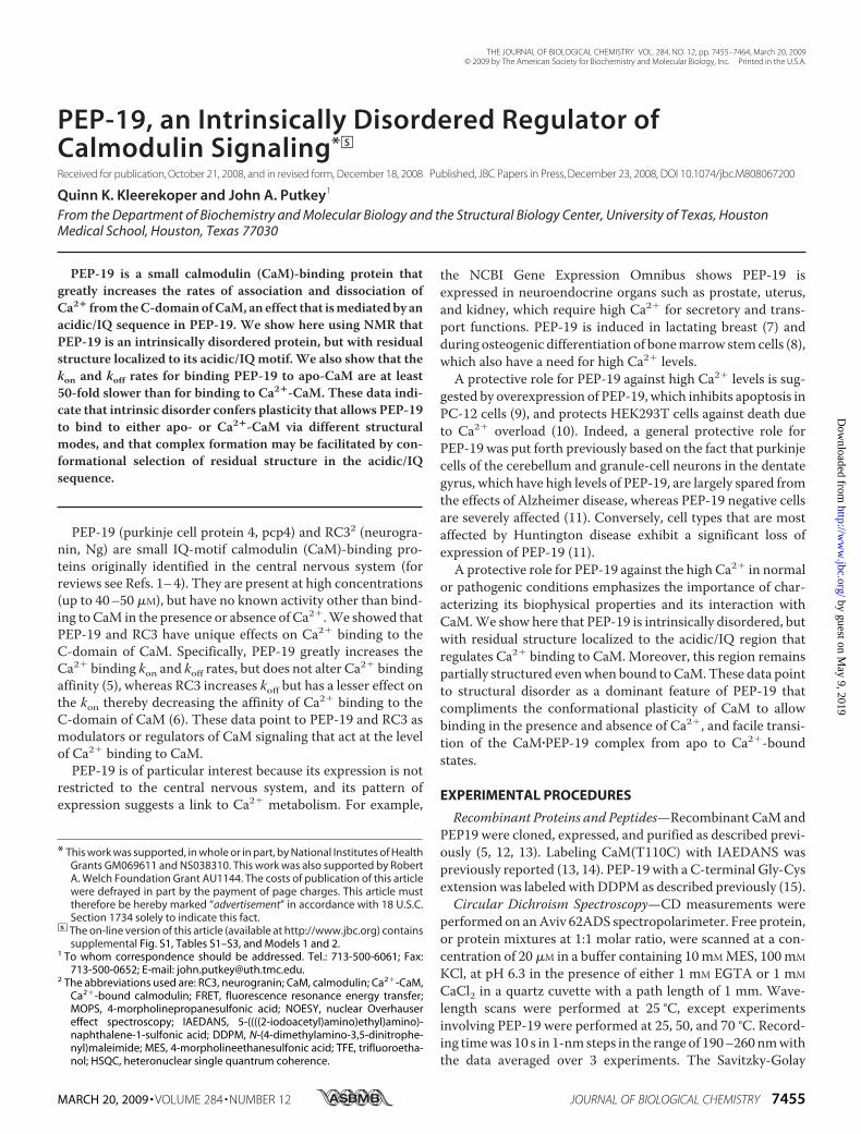
FIGURE 1. PEP-19 is intrinsically disordered. Panel A shows the far UV CDspectra for PEP-19 in the presence of 1 mM EDTA (closed diamonds), 1 mM
CaCl2 (open diamonds), or in the presence of 30% TFE (closed triangles). PanelB shows the 1H-15N HSQC spectrum of PEP-19 at 298 K in a buffer containing10 mM imidazole, 100 mM KCl at pH 6.8. The lack of chemical shift dispersionand narrow line widths seen for the amide protons are characteristic of intrin-sically disordered proteins.
PEP-19, A Regulator of Calmodulin Signaling
7456 JOURNAL OF BIOLOGICAL CHEMISTRY VOLUME 284 • NUMBER 12 • MARCH 20, 2009
by guest on May 9, 2019
http://ww
w.jbc.org/
Dow
nloaded from
RESULTS
Circular Dichroism and NMR Show PEP-19 Is an IntrinsicallyUnstructured Protein—CDspectroscopywas used to characterizethe secondary structure in PEP-19 as a function of CaCl2, temper-ature, and trifluoroethanol (TFE). Fig. 1A shows that the far UVspectrum of PEP-19 is characteristic of a random-coil with alarge negative ellipticity at 200 nm in the absence or presence ofCaCl2. Themean residual ellipticity at 222 nm indicates that 3%of the residues are helical (19) at 25 °C. Increasing the temper-ature to 70 °C did not change the random coil nature of theprotein (data not shown). CD spectrum collected in the pres-ence of the helix-promoting solvent TFE indicates substantial�-helix content from the negative bands at 208 and 222 nm. Amaximal helical content of about 50% was estimated from CDspectra collected in 40% TFE. Thus, the CD data shows thatPEP-19 is highly disordered and unstructured but has thepotential to form regions of �-helix.Fig. 1B shows the two-dimensional 1H-15N HSQC spectrum
of uniformly 15N-labeled PEP-19. All protein backbone amides
are clearly visible, but chemicalshifts are restricted to a window of0.8 ppm in the 1H dimension, andthe resonances show very narrowline widths. Both of these observa-tions are hallmarks of proteins thatlack stable long-range interactions,and indicate that PEP-19 is com-posed of unstructured or highlyflexible regions. This is similar tothat seen for RC3 (20). Additionally,the 6 Gly resonances in PEP-19 (cir-cled in the spectrum) have chemicalshifts that are characteristic of pro-teins denatured in 8 M urea (21),which also supports the conclusionthat PEP-19 is disordered.Primary Sequence Analysis Indi-
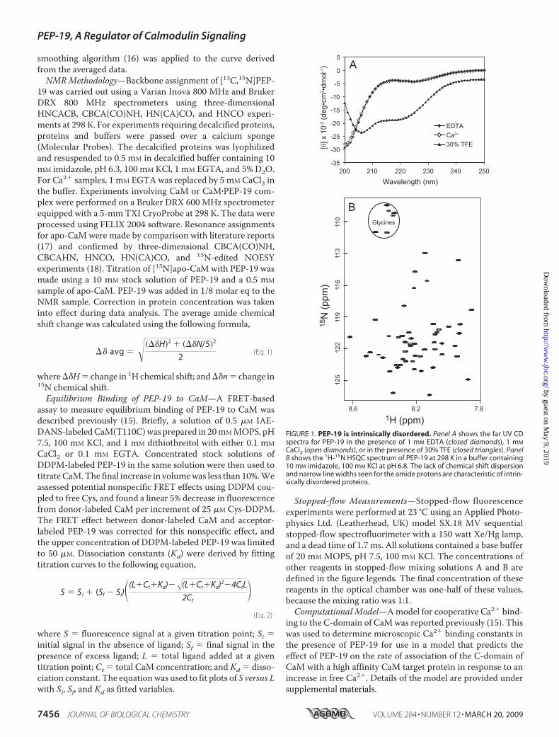
cates PEP-19 Has Residual HelicalContent—We used a combinationof structure prediction algorithmsto identify potential variations inorder/disorder along the primarysequence of PEP-19 (22, 23). Fig. 2Ashows that both PONDR VLS1(open circles) and RONN (closed cir-cles) predict PEP-19 to be disor-dered, but its C-terminal half is dis-ordered to a lesser extent than theN-terminal half. The sequence ofPEP-19 was next analyzed usingAGADIR (24, 25) to predict local-ized regions of residual helicalstructure as shown in Fig. 2B. AGA-DIR predicts that PEP-19 has anoverall helical content of less than1% under conditions used in theNMR experiments, which is punc-tuated by two distinct regions of
residual helicity. One region spans residues 38 to 54, includingpart of the IQ motif. The second region shows lower helicityand is upstream between residues 20 to 30. Fig. 2C shows thatTFE has the greatest effect on amides for those residues pre-dicted in Fig. 2B to have residual helical structure.We next used a variety of NMR experiments to identify
regions of residual structure in PEP-19 under native conditions.Secondary shifts, which are deviations of 13C� and 13C� chem-ical shifts from random coil values, can be used as indicators ofsecondary structure due to their dependence on backbonedihedral angles. Relative to random coils values, 13C� reso-nances are typically shifted downfield �3.1 ppm for residues instable �-helices, and shifted upfield (�1.5 ppm) in stable�-sheets (26–28). The 13C� resonances are less sensitive to hel-ical environments, but are shifted downfield by about 2.5 ppmin � sheets. Residues in less ordered regions of proteins willhave smaller deviations from random coil values as a result ofrapid conformational averaging. Positive 13C� secondary shifts,and weaker negative 13C� secondary shifts for Glu38 to Lys52 in
FIGURE 2. Sequence analysis predicts residual structure in PEP-19. Panel A shows plots of disorder proba-bility per residue for PEP-19 predicted from PONDR VLS1 (open circles) and RONN (closed circles). Panel B showsan AGADIR prediction of the fractional �-helical population in PEP-19. Panel C shows the average weightedchange in backbone amide chemical shift perturbation observed upon addition of 15% TFE to PEP-19. Chem-ical shift differences were calculated as described under “Experimental Procedures.”
PEP-19, A Regulator of Calmodulin Signaling
MARCH 20, 2009 • VOLUME 284 • NUMBER 12 JOURNAL OF BIOLOGICAL CHEMISTRY 7457
by guest on May 9, 2019
http://ww
w.jbc.org/
Dow
nloaded from
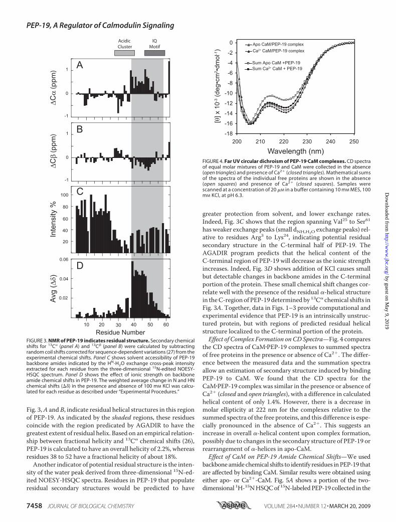
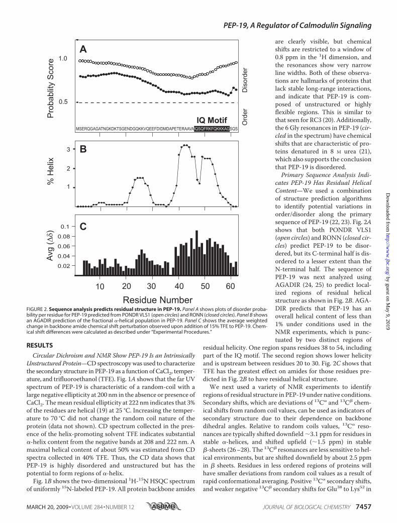
Fig. 3,A and B, indicate residual helical structures in this regionof PEP-19. As indicated by the shaded regions, these residuescoincide with the region predicated by AGADIR to have thegreatest extent of residual helix. Based on an empirical relation-ship between fractional helicity and 13C� chemical shifts (26),PEP-19 is calculated to have an overall helicity of 2.2%, whereasresidues 38 to 52 have a fractional helicity of about 18%.Another indicator of potential residual structure is the inten-
sity of the water peak derived from three-dimensional 15N-ed-ited NOESY-HSQC spectra. Residues in PEP-19 that populateresidual secondary structures would be predicted to have
greater protection from solvent, and lower exchange rates.Indeed, Fig. 3C shows that the region spanning Val25 to Ser61has weaker exchange peaks (small dNH,H2O exchange peaks) rel-ative to residues Arg3 to Lys24, indicating potential residualsecondary structure in the C-terminal half of PEP-19. TheAGADIR program predicts that the helical content of theC-terminal region of PEP-19 will decrease as the ionic strengthincreases. Indeed, Fig. 3D shows addition of KCl causes smallbut detectable changes in backbone amides in the C-terminalportion of the protein. These small chemical shift changes cor-relate well with the presence of the residual �-helical structurein theC-region of PEP-19 determinedby 13C� chemical shifts inFig. 3A. Together, data in Figs. 1–3 provide computational andexperimental evidence that PEP-19 is an intrinsically unstruc-tured protein, but with regions of predicted residual helicalstructure localized to the C-terminal portion of the protein.Effect of Complex Formation onCDSpectra—Fig. 4 compares
the CD spectra of CaM�PEP-19 complexes to summed spectraof free proteins in the presence or absence of Ca2�. The differ-ence between the measured data and the summation spectraallow an estimation of secondary structure induced by bindingPEP-19 to CaM. We found that the CD spectra for theCaM�PEP-19 complex was similar in the presence or absence ofCa2� (closed and open triangles), with a difference in calculatedhelical content of only 1.4%. However, there is a decrease inmolar ellipticity at 222 nm for the complexes relative to thesummed spectra of the free proteins, and this difference is espe-cially pronounced in the absence of Ca2�. This suggests anincrease in overall �-helical content upon complex formation,possibly due to changes in the secondary structure of PEP-19 orrearrangement of �-helices in apo-CaM.Effect of CaM on PEP-19 Amide Chemical Shifts—We used
backboneamidechemical shifts to identify residues inPEP-19 thatare affected by binding CaM. Similar results were obtained usingeither apo- or Ca2�-CaM. Fig. 5A shows a portion of the two-dimensional 1H-15NHSQCof 15N-labeledPEP-19 collected in the
FIGURE 3. NMR of PEP-19 indicates residual structure. Secondary chemicalshifts for 13C� (panel A) and 13C� (panel B) were calculated by subtractingrandom coil shifts corrected for sequence-dependent variations (27) from theexperimental chemical shifts. Panel C shows solvent accessibility of PEP-19backbone amides indicated by the HN-H2O exchange cross-peak intensityextracted for each residue from the three-dimensional 15N-edited NOESY-HSQC spectrum. Panel D shows the effect of ionic strength on backboneamide chemical shifts in PEP-19. The weighted average change in N and HNchemical shifts (��) in the presence and absence of 100 mM KCl was calcu-lated for each residue as described under “Experimental Procedures.”
FIGURE 4. Far UV circular dichroism of PEP-19�CaM complexes. CD spectraof equal molar mixtures of PEP-19 and CaM were collected in the absence(open triangles) and presence of Ca2� (closed triangles). Mathematical sumsof the spectra of the individual free proteins are shown in the absence(open squares) and presence of Ca2� (closed squares). Samples werescanned at a concentration of 20 �M in a buffer containing 10 mM MES, 100mM KCl, at pH 6.3.
PEP-19, A Regulator of Calmodulin Signaling
7458 JOURNAL OF BIOLOGICAL CHEMISTRY VOLUME 284 • NUMBER 12 • MARCH 20, 2009
by guest on May 9, 2019
http://ww
w.jbc.org/
Dow
nloaded from
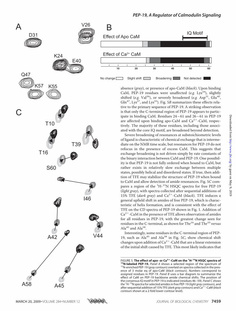
absence (gray), or presence of apo-CaM (black). Upon bindingCaM, PEP-19 residues were unaffected (e.g. Lys24), slightlyshifted (e.g. Val26), or severely broadened (e.g. Asp31, Glu40,Gln47, Lys57, and Lys55). Fig. 5B summarizes these effects rela-tive to the primary sequence of PEP-19. A striking observationis that only the C-terminal region of PEP-19 appears to partic-ipate in binding CaM. Residues 24–61 and 26–61 in PEP-19are affected upon binding apo-CaM and Ca2�-CaM, respec-tively. The majority of these residues, including those associ-ated with the core IQ motif, are broadened beyond detection.Severe broadening of resonances at substoichiometric levels
of ligand is characteristic of chemical exchange that is interme-diate on the NMR time scale, but resonances for PEP-19 do notrefocus in the presence of excess CaM. This suggests thatexchange broadening is not driven simply by rate constants ofthe binary interaction between CaM and PEP-19. One possibil-ity is that PEP-19 is not fully ordered when bound to CaM, butrather exists in relatively slow exchange between multiplestates, possibly helical and disordered states. If true, then addi-tion of TFE may stabilize the structure of PEP-19 when boundto CaM and allow detection of amide resonances. Fig. 5C com-pares a region of the 1H-15N HSQC spectra for free PEP-19(light gray), with spectra collected after sequential additions of15% TFE (dark gray) and Ca2�-CaM (black). TFE induces ageneral upfield shift in amides of free PEP-19, which is charac-teristic of helix formation, and is consistent with the effect ofTFE on the CD spectra of PEP-19 shown in Fig. 1. Addition ofCa2�-CaM in the presence of TFE allows observation of amidesfor all residues in PEP-19, with the greatest change seen forresidues in the C-terminal, as shown for Thr16 and Thr10 versusAla45 and Ala58.Interestingly, some residues in the C-terminal region of PEP-
19, such as Ala45 and Ala58 in Fig. 5C, show chemical shiftchanges upon addition of Ca2�-CaM that are a linear extensionof the initial shift caused by TFE. This most likely indicates that
FIGURE 5. The effect of apo- or Ca2�-CaM on the 1H-15N HSQC spectra of15N-labeled PEP-19. Panel A shows a selected region of the spectrum of15N-enriched PEP-19 (gray contours) overlaid on spectra collected in the pres-ence of 3 molar eq of apo-CaM (black contours). Numbers correspond toassigned residues in PEP-19. Panel B uses a bar diagram to summarize theeffect of CaM on PEP-19 backbone amide chemical shifts. The position ofthe consensus IQ motif in PEP-19 is indicated (residues 46 –59). Panel C showsthe 1H-15N spectra for selected amides in free PEP-19 (light gray contours), andafter sequential addition of 15% TFE (dark gray contours) and Ca2�-CaM (blackcontours shown at a 2-fold lower contour level).
PEP-19, A Regulator of Calmodulin Signaling
MARCH 20, 2009 • VOLUME 284 • NUMBER 12 JOURNAL OF BIOLOGICAL CHEMISTRY 7459
by guest on May 9, 2019
http://ww
w.jbc.org/
Dow
nloaded from
Ca2�-CaM causes a further increase of the percent helical con-tent of this region. In contrast, residues such as Ile46 of the IQdipeptide, show a distinct bidirectional change in chemical shiftupon sequential addition of TFE and Ca2�-CaM, which likelyindicates a direct interaction of these residues with CaM.Effect of PEP-19 on CaM Amide Chemical Shifts—We previ-
ously reported that PEP-19 bound primarily to theC-domain ofCa2�-CaM and that amide chemical shifts showed characteris-tics of fast exchange on the NMR time scale (5). The rate ofdissociation of PEP-19 from Ca2�-CaM was as least 154 s�1
based on the largest 1H change.Similar to Ca2�-CaM, amide chemical shifts for residues in
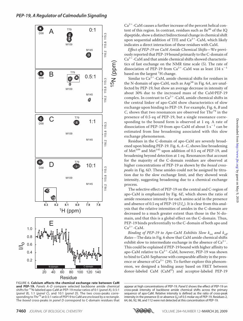
the N-domain of apo-CaM, such as Asp58 in Fig. 6A, are unaf-fected by PEP-19, but show an average decrease in intensity ofabout 30% due to the increased mass of the CaM�PEP-19complex. In contrast to Ca2�-CaM, amide chemical shifts inthe central linker of apo-CaM show characteristics of slowexchange upon binding to PEP-19. For example, Fig. 6, B andC, shows that two resonances are observed for Thr79 in thepresence of 0.5 eq of PEP-19, but a single resonance corre-sponding to the bound form is observed at 1 eq. A rate ofdissociation of PEP-19 from apo-CaM of about 5 s�1 can beestimated from line broadening associated with this slowexchange phenomenon.Residues in the C-domain of apo-CaM are severely broad-
ened upon binding PEP-19. Fig. 6, A–C, shows line broadeningof Met109 and Met145 upon addition of 0.5 eq of PEP-19, andbroadening beyond detection at 1 eq. Resonances that accountfor the majority of the C-domain residues are observed athigher concentrations of PEP-19 as shown by the boxed cross-peaks in Fig. 6D. These amides could not be assigned by titra-tion due to the slow exchange limit, and they showed weakintensity, suggesting broadening due to a chemical exchangeprocess.The selective effect of PEP-19 on the central and C-region of
apo-CaM is emphasized by Fig. 6E, which shows the ratio ofamide resonance intensity for each amino acid in the presenceand absence of 0.5 eq of PEP-19 (I/Io). It is clear from this anal-ysis that the relative intensities of amides in the C-domain aredecreased to a much greater extent than those in the N-do-main, and that this is a global effect on the C-domain. Thus,PEP-19 binds preferentially to the C-domain of both apo andCa2�-CaM.Binding of PEP-19 to Apo-CaM Exhibits Slow kon and koff
Rates—The data in Fig. 6 show that CaM amide chemical shiftsexhibit slow to intermediate exchange in the absence of Ca2�.This could be explained if PEP-19 bound with higher affinity toapo-CaM relative to Ca2�-CaM, however, PEP-19 was shownto bind to CaM-Sepharose with comparable affinity in the pres-ence or absence of Ca2� (29). To further explore this phenom-enon, we designed a binding assay based on FRET betweendonor-labeled CaM (CaMD) and acceptor-labeled PEP-19
FIGURE 6. Calcium affects the chemical exchange rate between CaMand PEP-19. Panels A–D compare selected backbone amide chemicalshifts for 15N-labeled apo-CaM at PEP-19 molar ratios of 0:1 (panel A), 0.5:1(panel B), 1:1 (panel C), and 10:1 (panel D). The two cross-peaks corre-sponding to Thr79 at 0.5:1 ratio of PEP19 to CaM are enclosed by a rectangle.The boxed cross-peaks in panel D correspond to C-domain residues that
appear at high concentrations of PEP-19. Panel E shows the effect of PEP-19 oncross-peak intensity of backbone amide chemical shifts across the primarysequence of apo-CaM. Relative intensity is defined as the ratio of cross-peakintensity in the presence (I) or absence (I0) of 0.5 molar eq of PEP-19. Residues 2,64, 66, 92, 98, and 112 were not detected at this concentration of PEP-19.
PEP-19, A Regulator of Calmodulin Signaling
7460 JOURNAL OF BIOLOGICAL CHEMISTRY VOLUME 284 • NUMBER 12 • MARCH 20, 2009
by guest on May 9, 2019
http://ww
w.jbc.org/
Dow
nloaded from
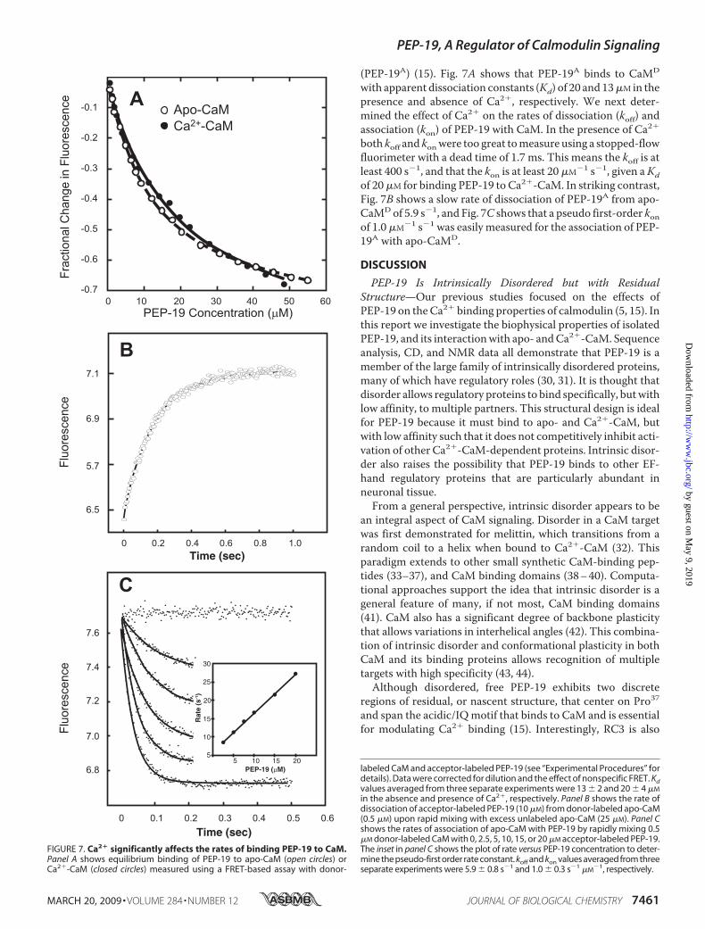
(PEP-19A) (15). Fig. 7A shows that PEP-19A binds to CaMD
with apparent dissociation constants (Kd) of 20 and 13�M in thepresence and absence of Ca2�, respectively. We next deter-mined the effect of Ca2� on the rates of dissociation (koff) andassociation (kon) of PEP-19 with CaM. In the presence of Ca2�
both koff and konwere too great tomeasure using a stopped-flowfluorimeter with a dead time of 1.7 ms. This means the koff is atleast 400 s�1, and that the kon is at least 20 �M�1 s�1, given aKdof 20�M for binding PEP-19 to Ca2�-CaM. In striking contrast,Fig. 7B shows a slow rate of dissociation of PEP-19A from apo-CaMDof 5.9 s�1, and Fig. 7C shows that a pseudo first-order konof 1.0 �M�1 s�1 was easily measured for the association of PEP-19A with apo-CaMD.
DISCUSSION
PEP-19 Is Intrinsically Disordered but with ResidualStructure—Our previous studies focused on the effects ofPEP-19 on theCa2� binding properties of calmodulin (5, 15). Inthis report we investigate the biophysical properties of isolatedPEP-19, and its interactionwith apo- andCa2�-CaM. Sequenceanalysis, CD, and NMR data all demonstrate that PEP-19 is amember of the large family of intrinsically disordered proteins,many of which have regulatory roles (30, 31). It is thought thatdisorder allows regulatory proteins to bind specifically, butwithlow affinity, to multiple partners. This structural design is idealfor PEP-19 because it must bind to apo- and Ca2�-CaM, butwith low affinity such that it does not competitively inhibit acti-vation of other Ca2�-CaM-dependent proteins. Intrinsic disor-der also raises the possibility that PEP-19 binds to other EF-hand regulatory proteins that are particularly abundant inneuronal tissue.From a general perspective, intrinsic disorder appears to be
an integral aspect of CaM signaling. Disorder in a CaM targetwas first demonstrated for melittin, which transitions from arandom coil to a helix when bound to Ca2�-CaM (32). Thisparadigm extends to other small synthetic CaM-binding pep-tides (33–37), and CaM binding domains (38–40). Computa-tional approaches support the idea that intrinsic disorder is ageneral feature of many, if not most, CaM binding domains(41). CaM also has a significant degree of backbone plasticitythat allows variations in interhelical angles (42). This combina-tion of intrinsic disorder and conformational plasticity in bothCaM and its binding proteins allows recognition of multipletargets with high specificity (43, 44).Although disordered, free PEP-19 exhibits two discrete
regions of residual, or nascent structure, that center on Pro37and span the acidic/IQmotif that binds to CaM and is essentialfor modulating Ca2� binding (15). Interestingly, RC3 is also
FIGURE 7. Ca2� significantly affects the rates of binding PEP-19 to CaM.Panel A shows equilibrium binding of PEP-19 to apo-CaM (open circles) orCa2�-CaM (closed circles) measured using a FRET-based assay with donor-
labeled CaM and acceptor-labeled PEP-19 (see “Experimental Procedures” fordetails). Data were corrected for dilution and the effect of nonspecific FRET. Kdvalues averaged from three separate experiments were 13 � 2 and 20 � 4 �M
in the absence and presence of Ca2�, respectively. Panel B shows the rate ofdissociation of acceptor-labeled PEP-19 (10 �M) from donor-labeled apo-CaM(0.5 �M) upon rapid mixing with excess unlabeled apo-CaM (25 �M). Panel Cshows the rates of association of apo-CaM with PEP-19 by rapidly mixing 0.5�M donor-labeled CaM with 0, 2.5, 5, 10, 15, or 20 �M acceptor-labeled PEP-19.The inset in panel C shows the plot of rate versus PEP-19 concentration to deter-mine the pseudo-first order rate constant. koff and kon values averaged from threeseparate experiments were 5.9 � 0.8 s�1 and 1.0 � 0.3 s�1 �M
�1, respectively.
PEP-19, A Regulator of Calmodulin Signaling
MARCH 20, 2009 • VOLUME 284 • NUMBER 12 JOURNAL OF BIOLOGICAL CHEMISTRY 7461
by guest on May 9, 2019
http://ww
w.jbc.org/
Dow
nloaded from
disordered, but exhibits residual structure in the region thatbinds to CaM (20).We propose that the residual structure playsa role in initiating binding of PEP-19 to CaM by selection of apopulation of PEP-19 conformers that display helical structure.Support for such a mechanism was recently reported for theintrinsically disordered �-subunit of phosphodiesterase 6. Inthis example, the free �-subunit exhibits a transient structurethat resembles the structure of the protein when bound to the�-subunit of transducin (45).PEP-19 Appears Partially Structured When Bound to CaM—
The effects of CaM on PEP-19 backbone amide chemical shiftsare very similar in both the presence and absence of Ca2�.Under both conditions, only the regions of residual structure inthe C-terminal half of PEP-19 are affected upon binding toCaM, with the majority of residues undergoing a chemicalexchange process that broadens the amide chemical shiftsbeyond detection. Interestingly, PEP-19 resonances remainbroadened beyond detection at high concentrations of CaM,and significant broadening is seen even in the presence of TFE.This is consistent with a high degree of conformationalexchange in CaM-bound PEP-19.At least two processes could contribute to conformational
exchange of bound PEP-19. The first is that weak binding ofPEP-19 to the C-domain does not provide a sufficient decreasein free energy to stabilize a defined conformation of PEP-19. Asecond potential source of amide broadening is transient inter-actions with the N-domain that are sensed as a chemicalexchange process by bound PEP-19 due to its highly adaptableintrinsically disordered nature. The first mechanism is sup-ported by a recent crystal structure showing that Tyr1675 andPhe1676 anchor the IQ peptide from the human voltage-dependent Ca2� channel (CaV1.2, Swiss_Prot Q13936) to theC-domain of Ca2�-CaM (46, 47), and that increased disorder inthe bound peptide is observed if Phe1676 is changed to Ala (46,47). Similarly, disorder in CaM-bound PEP-19 may existbecause its acidic/IQ motif lacks a corresponding stabilizingTyr.Conformational exchange in CaM-bound PEP-19 may have
at least two functional advantages. The first is to allow transi-tion of the PEP-19�CaM complex from apo to Ca2�-boundforms. The second is that rapid exchange of bound PEP-19between multiple conformations may increase rates of Ca2�
association and dissociation in the C-terminal Ca2� bindingloops of CaMvia allosteric coupling and conformational gating.It has been proposed that intrinsic disorder enhances allostericeffects (48), andwe showed that allosteric coupling between theacidic and IQ region of PEP-19 and between PEP-19 andCaM are necessary to modulate Ca2� binding to CaM (15). Ithas also been demonstrated that rates of ligand binding andrelease can be gated by intermolecular conformationalexchange (49), and the rates of conformational exchange inthe C-domain of CaM mutant correlate with the Ca2� off-rate (50). Together these observations support a mechanismin which conformational exchange of CaM-bound PEP-19exerts an allosteric effect that gates, or regulates the rates ofassociation and dissociation of Ca2�.Kinetics of Binding PEP-19 to CaM—Although the effects of
CaM on the amides of PEP-19 are very similar in the presence
and absence of Ca2�, this is not the case for the effect of PEP-19on amides of Ca2�-CaM versus apo-CaM. Amide chemicalshifts of Ca2�-CaM show characteristics of fast exchange onthe NMR time scale when titrated with PEP-19 (5). In contrast,binding of PEP-19 causes severe broadening of amidesthroughout the C-domain of apo-CaM even though PEP-19binds with similar affinity to both apo- and Ca2�-CaM. Weshow here that this is due, at least in part, to low koff and konrates for binding PEP-19 to apo-CaM. Thus, under the condi-tions used for the experiments in Fig. 6, the kex would be 30 s�1
and 1000 s�1 when apo- and Ca2�-CaM, respectively, arehalf-saturated with PEP-19.The underlying structural basis for greatly different rates of
binding PEP-19 to apo versus Ca2�-CaM likely resides in theCa2�-dependent structural dynamics of the C-domain. TheCa2�-bound C-domain is generally well structured, with adefined hydrophobic core and restricted backbone dynamics(42, 51). In contrast, the apo C-domain has an ill-defined aro-matic hydrophobic core (51, 52), multiple thermal meltingtransitions (53), with regions of intrinsic disorder (51, 54), and ahigh degree of backbone conformational entropy that allowsglobal conformation exchange between at least two conforma-tions (51, 55, 56). Thus, the slow rate of association of PEP-19with CaM in the absence of Ca2� may be due, in part, to a lowprobability of presentation of complimentary transient struc-tures in disordered PEP-19 and the poorly ordered apo C-do-main. The slow rate of dissociation may reflect an intrinsicallystable apo-CaM�PEP-19 complex, whereas the lowoverall affin-ity is driven by the slow association rate.Computational Model for Effect of PEP-19 on CaM/Target
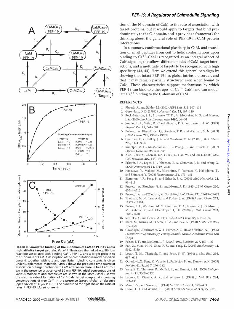
Binding—The data in Fig. 7 suggest that slow release of apo-CaM from PEP-19 could be a rate-limiting step in the transferof CaM to high affinity target proteins upon elevation of Ca2�
levels. However, PEP-19 greatly increases the intrinsically slowkon rate for Ca2� binding to the C-domain of CaM, and releaseof Ca2�-CaM from PEP-19 is also very fast. These factors maycompensate for the slow release of apo-CaM from PEP-19,making this a minor pathway in the transition of apo-CaM totarget-bound Ca2�-CaM.
We tested these concepts using a variation of the computa-tional model described previously (15). The scheme shown inFig. 8A incorporates cooperativeCa2�binding to theC-domainof CaM, together with binding of PEP-19 to all forms of theC-domain, and binding a high affinity target to Ca2�-saturatedC-domain. These reactions and corresponding rate constantswere incorporated into a computational model to simulate theeffect of PEP-19 on the rate of association of the C-domain ofCaMwith high affinity target proteins (see supplemental mate-rials for details).The simulations shown in Fig. 8B predict that PEP-19
increases the rate of Ca2�-dependent association of targetproteins with the C-domain of CaM. Fig. 8C shows that theeffect of PEP-19 is greater at lower Ca2� levels. Thus, theslow rate of release of apo-CaM from PEP-19 shown in Fig. 7would not inhibit the association of Ca2�-CaM with targetproteins due to compensatory effects of PEP-19 on the rate-limiting kinetics of Ca2� binding to the C-domain. It must benoted that simulation does not take into account contribu-
PEP-19, A Regulator of Calmodulin Signaling
7462 JOURNAL OF BIOLOGICAL CHEMISTRY VOLUME 284 • NUMBER 12 • MARCH 20, 2009
by guest on May 9, 2019
http://ww
w.jbc.org/
Dow
nloaded from
tion of the N-domain of CaM to the rate of association withtarget proteins, but it would apply to targets that bind pre-dominately to the C-domain, and it provides a framework forthinking about the general role of PEP-19 in CaM-proteininteractions.In summary, conformational plasticity in CaM, and transi-
tion of small peptides from coil to helix conformations uponbinding to Ca2�-CaM is recognized as an integral aspect ofCaM signaling that allows differentmodes of CaM-target inter-actions, and a multitude of targets to be recognized with highspecificity (43, 44). Here we extend this general paradigm byshowing that intact PEP-19 has global intrinsic disorder, andthat it may remain partially structured even when bound toCaM. These characteristics support mechanisms by whichPEP-19 can bind to either apo- or Ca2�-CaM, and can modu-late Ca2� binding to the C-domain of CaM.
REFERENCES1. Rhoads, A., and Bahler, M. (2002) FEBS Lett. 513, 107–1132. Gerendasy, D. D. (1999) J. Neurosci. Res. 58, 107–1193. Reck-Peterson, S. L., Provance, W. D., Jr., Mooseker, M. S., and Mercer,
J. A. (2000) Biochim. Biophys. Acta 1496, 36–514. Jurado, L. A., Sethu, P., Chockalingam, P. S., and Jarrett, H. W. (1999)
Physiol. Rev. 79, 661–6815. Putkey, J. A., Kleerekoper, Q., Gaertner, T. R., andWaxham, M. N. (2003)
J. Biol. Chem. 278, 49667–496706. Gaertner, T. R., Putkey, J. A., and Waxham, M. N. (2004) J. Biol. Chem.
279, 9374–93827. Rudolph, M. C., McManaman, J. L., Phang, T., and Russell, T. (2007)
Physiol. Genomics 28, 323–3368. Xiao, J., Wu, Y., Chen, R., Lin, Y., Wu, L., Tian,W., and Liu, L. (2008)Mol.
Cell. Biochem. 309, 143–1509. Erhardt, J. A., Legos, J. J., Johanson, R. A., Slemmon, J. R., and Wang, X.
(2000) Neuroreport 11, 3719–372310. Kanazawa, Y., Makino, M., Morishima, Y., Yamada, K., Nabeshima, T.,
and Shirakaki, Y. (2008) Neuroscience 154, 473–48111. Slemmon, J. R., Feng, B., and Erhardt, J. A. (2001) Mol. Neurobiol. 22,
99–11312. Putkey, J. A., Slaughter, G. R., and Means, A. R. (1985) J. Biol. Chem. 260,
4704–471213. Putkey, J. A., andWaxham,M. N. (1996) J. Biol. Chem. 271, 29619–2962314. Waxham, M. N., Tsai, A.-L., and Putkey, J. A. (1998) J. Biol. Chem. 273,
17579–1758415. Putkey, J. A., Waxham, M. N., Gaertner, T. A., Brewer, K. J., Goldsmith,
M., Kubota, Y., and Kleerekoper, Q. K. (2008) J. Biol. Chem. 283,1401–1410
16. Savitzky, A., and Golay, M. J. E. (1964) Anal. Chem. 36, 1627–163917. Ikura, M., Krinks, M., Tochia, D. A., and Bax, A. (1990) FEBS Lett. 266,
155–15818. Cavanagh, J., Fairbrother,W. J., Palmer, A. G., III, and Skelton, N. J. (1996)
Protein NMR Spectroscopy: Principles and Practice, Academic Press, SanDiego
19. Pelton, J. T., and McLean, L. R. (2000) Anal. Biochem. 277, 167–17620. Ran, X., Miao, H. H., Sheu, F. S., and Yang, D. (2003) Biochemistry 42,
5142–515021. Logan, T. M., Theriault, Y., and Fesik, S. W. (1994) J. Mol. Biol. 236,
637–64822. Obradovic, Z., Peng, K., Vucetic, S., Radivojac, P., andDunker, A. K. (2005)
Proteins 61, Suppl. 7, 176–18223. Yang, Z. R., Thomson, R., McNeil, P., and Esnouf, R. M. (2005) Bioinfor-
matics 21, 3369–337624. Lacroix, E., Viguera, A. R., and Serrano, L. (1998) J. Mol. Biol. 284,
173–19125. Munoz, V., and Serrano, L. (1994) Nat. Struct. Biol. 1, 399–40926. Dyson, H. J., and Wright, P. E. (2001)Methods Enzymol. 339, 258–270
FIGURE 8. Simulated binding of the C-domain of CaM to PEP-19 and ahigh affinity target protein. Panel A illustrates the linked equilibriumreactions associated with binding Ca2�, PEP-19, and a target protein tothe C-domain of CaM. A description of the computational model based onpanel A, together with rate and equilibrium binding constants, is givenunder supplemental materials. Panel B shows the predicted time course ofassociation of target protein with CaM after an increase in free Ca2� to 1�M in the presence or absence of 30 mM PEP-19. Initial concentrations ofvarious molecules and complexes are shown in the inset. Panel C showsthe maximal rate of formation of Ca2�-CaM�Target complex at increasingconcentrations of free Ca2� in the presence (closed circles) or absence(open circles) of 30 �M PEP-19. The ordinate on the right shows the ratio ofrates � PEP-19 (closed squares).
PEP-19, A Regulator of Calmodulin Signaling
MARCH 20, 2009 • VOLUME 284 • NUMBER 12 JOURNAL OF BIOLOGICAL CHEMISTRY 7463
by guest on May 9, 2019
http://ww
w.jbc.org/
Dow
nloaded from
27. Schwarzinger, S., Kroon, G. J., Foss, T. R., Chung, J., Wright, P. E., andDyson, H. J. (2001) J. Am. Chem. Soc. 123, 2970–2978
28. Wishart, D. S., and Case, D. A. (2001)Method Enzymol. 338, 3–3429. Slemmon, J. R., Morgan, J. I., Fullerton, S. M., Danho, W., Hilbush, B. S.,
and Wengenack, T. M. (1996) J. Biol. Chem. 271, 15911–1591730. Dunker, A. K., Cortese,M. S., Romero, P., Iakoucheva, L.M., and Uversky,
V. N. (2005) FEBS J. 272, 5129–514831. Uversky, V. N., Oldfield, C. J., and Dunker, A. K. (2008) Annu. Rev. Bio-
phys. 37, 215–24632. Seeholzer, S. H., Cohn, M., Putkey, J. A., Means, A. R., and Crespi, H. L.
(1986) Proc. Natl. Acad. Sci. U. S. A. 83, 3634–363833. Osawa, M., Tokumitsu, H., Swindells, M. B., Kurihara, H., Orita, M., Shi-
banuma, T., Furuya, T., and Ikura, M. (1999)Nat. Struct. Biol. 6, 819–82434. Precheur, B.,Munier, H.,Mispelter, J., Barzu,O., andCraescu, C. T. (1992)
Biochemistry 31, 229–23635. Craescu, C. T., Bouhss, A.,Mispelter, J., Diesis, E., Popescu,A., Chiriac,M.,
and Barzu, O. (1995) J. Biol. Chem. 270, 7088–709636. Kranz, J. K., Flynn, P. F., Fuentes, E. J., andWand, A. J. (2002) Biochemistry
41, 2599–260837. Gerendasy, D. D., Herron, S. R., Jennings, P. A., and Sutcliffe, J. G. (1995)
J. Biol. Chem. 270, 6741–675038. Permyakov, S. E., Millett, I. S., Doniach, S., Permyakov, E. A., and Uversky,
V. N. (2003) Proteins 53, 855–86239. Zhou, N., Yuan, T., Mak, A. S., and Vogel, H. J. (1997) Biochemistry 36,
2817–282540. Tapp, H., Al-Nagger, I. M., Yarmola, E. G., Harrison, A., Shaw, G., Edison,
A. S., and Bubb, M. R. (2005) J. Biol. Chem. 280, 9946–995641. Radivojac, P., Vucetic, S., O’Connor, T. R., Uversky, V. N., Obradovic, Z.,
and Dunker, A. K. (2006) Proteins 63, 398–41042. Chou, J. J., Li, S., Klee, C. B., and Bax, A. (2001) Nat. Struct. Biol. 8,
990–99743. Hoeflich, K. P., and Ikura, M. (2002) Cell 108, 739–74244. Vetter, S. W., and Leclerc, E. (2003) Eur. J. Biochem. 270, 404–41445. Song, J., Guo, L. W., Muradov, H., Artemyev, N. O., Ruoho, A. E., and
Markley, J. L. (2008) Proc. Natl. Acad. Sci. U. S. A. 105, 1505–151046. Fallon, J. L., Halling, D. B., Hamilton, S. L., and Quiocho, F. A. (2005)
Structure 13, 1881–188647. Van Petegem, F., Chatelain, F. C., and Minor, D. L., Jr. (2005) Nat. Struct.
Mol. Biol. 12, 1108–111548. Shmukler, B. E., Bond, C. T., Wilhelm, S., Bruening-Wright, A., Maylie, J.,
Adelman, J. P., andAlper, S. L. (2001)Biochim. Biophys. Acta 1518, 36–4649. Palmer, A. G., III (2004) Chem. Rev. 104, 3623–364050. Evenas, J., Malmendal, A., and Akke, M. (2001) Structure 9, 185–19551. Kuboniwa,H.,Nico, T., Grzesiek, S., Ren,H., Klee, C. B., andBax, A. (1995)
Nat. Struct. Biol. 2, 768–77652. Zhang, M., Tanaka, T., and Ikura, M. (1995) Nat. Struct. Biol. 2, 258–26753. Tsalkova, T. N., and Privalov, P. L. (1985) J. Mol. Biol. 181, 533–54454. Lundstrom, P.,Mulder, F. A. A., and Akke,M. (2005) Proc. Natl. Acad. Sci.
U. S. A. 102, 16984–1698955. Rabl, C. R., Martin, S. R., Neumann, E., and Bayley, P. M. (2002) Biophys.
Chem. 101–102, 553–56456. Chen, Y.-G., and Hummer, G. (2007) J. Am. Chem. Soc. 129, 2414–2415
PEP-19, A Regulator of Calmodulin Signaling
7464 JOURNAL OF BIOLOGICAL CHEMISTRY VOLUME 284 • NUMBER 12 • MARCH 20, 2009
by guest on May 9, 2019
http://ww
w.jbc.org/
Dow
nloaded from
Quinn K. Kleerekoper and John A. PutkeyPEP-19, an Intrinsically Disordered Regulator of Calmodulin Signaling
doi: 10.1074/jbc.M808067200 originally published online December 23, 20082009, 284:7455-7464.J. Biol. Chem.
10.1074/jbc.M808067200Access the most updated version of this article at doi:
Alerts:
When a correction for this article is posted•
When this article is cited•
to choose from all of JBC's e-mail alertsClick here
Supplemental material:
http://www.jbc.org/content/suppl/2008/12/23/M808067200.DC1
http://www.jbc.org/content/284/12/7455.full.html#ref-list-1
This article cites 55 references, 12 of which can be accessed free at
by guest on May 9, 2019
http://ww
w.jbc.org/
Dow
nloaded from