Embed Size (px)
Citation preview

PENTABLOCK COPOLYMER BASED CONTROLLED RELEASE FORMULATIONS
OF SMALL AND MACROMOLECULES FOR OPHTHALMIC APPLICATIONS
A DISSERTATION IN
Pharmaceutical Sciences
and
Chemistry
Presented to the Faculty of the University
of Missouri - Kansas City in partial fulfillment of
the requirements for the degree
DOCTOR OF PHILOSOPHY
by
VIRAL M. TAMBOLI
B. Pharm., Hemchandracharya North Gujarat University, India, 2006
Kansas City, Missouri
2012


iii
PENTABLOCK COPOLYMER BASED CONTROLLED RELEASE FORMULATIONS
OF SMALL AND MACROMOLECULES FOR OPHTHALMIC APPLICATIONS
Viral M. Tamboli, Candidate for the Doctor of Philosophy degree
University of Missouri Kansas City, 2012
ABSTRACT
Pentablock copolymers comprised of multi-polymer blocks such as polyethylene
glycol (PEG), polycaprolactone (PCL) and poly-lactide (PLA) were developed for
fabrication of nanoparticles and thermosensitive hydrogel formulations for long term delivery
of small and macromolecules. Different pentablock copolymer compositions were evaluated
to optimize drug release profile from nanoparticles and thermosensitive gel formulations. Our
composite approach i.e. pentablock copolymers based nanoparticles suspended in
thermosensitive gel provided sustained zero-order delivery of encapsulated therapeutic
agents without producing any significant burst effect.
Different compositions of pentablock copolymers (polylatide- polycaprolactone-
polyethylene glycol- polycaprolactone- polylatide) (PLA-PCL-PEG-PCL-PLA) and (PEG-
PCL-PLA-PCL-PEG) were synthesized and characterized to prepare nanoparticle and
thermosensitive hydrogel formulations, respectively. The effect of poly (L-lactide) (PLLA) or
poly (D, L-lactide) (PDLLA) incorporation on crystallinity of pentablock copolymers and in
vitro release profile of triamcinolone acetonide (selected as model drug) from nanoparticles was
also evaluated. Pentablock polymer with proper ratio PDLLA/PCL was amorphous in nature
whereas PLLA containing polymer has semicrystalline nature. Release of triamcinolone
acetonide from nanoparticles was significantly affected by crystallinity of the copolymers.
Burst release of triamcinolone acetonide from nanoparticles was significantly minimized
with incorporation of proper ratio of PDLLA in the existing triblock (PCL-PEG-PCL)

iv
copolymer. Moreover, pentablock copolymer based nanoparticles exhibited continuous
release of triamcinolone acetonide for longer duration. The release profile of various steroids
commonly utilized for chronic ocular diseases was also evaluated with optimized pentablock
polymer. We found that steroids with different log P values did not exhibited significant
difference in release profile. These results could be attributed with the fact that drug release
from the nanoparticles is mainly diffusion mediated process and small molecules can easily
diffuse out from the pore the polymer matrix. However, we found that pentablock copolymer
based nanoparticles can be utilized to achieve continuous near zero-order delivery of small
molecules from nanoparticles without any burst effect. Further, release profile of timolol
from composite formulations was evaluated for glaucoma therapy. We observed that
composite approach could provide sustain release of timolol for longer duration. Successful
accomplishment of this project may lead to application of this strategy for the treatment of
other chronic ocular diseases such as age related macular degeneration and diabetic macular
edema. Treatment of these diseases requires frequent intravitreal injections to maintain
therapeutic levels of antibodies at retina/choroid. Frequent administrations can cause
potential complications like endophthalmitis, retinal detachment and retinal hemorrhage.
Sustained intraocular therapeutic drug concentration can be achieved by suspending the
therapeutic macromolecules in thermosensitive hydrogel. Considering this we have
characterized the release kinetics of various macromolecules from pentablock copolymer
based thermosensitive hydrogel. We observed that release kinetics of macromolecules from
thermosensitive hydrogel was depended on the size of a molecule. Our studies indicate that
pentablock copolymer based delivery systems can provide sustained drug release profile for
longer duration, and thereby eliminate the need for repeated intravitreal injections.

v
APPROVAL PAGE
The faculty listed below, appointed by the Dean of the School of Graduate Studies have
examined a dissertation titled “Pentablock Copolymers Based Controlled Release
Formulations of Small and Macromolecules for Ophthalmic Applications” presented by Viral
M. Tamboli, candidate for the Doctor of Philosophy degree, and certify that in their opinion
it is worthy of acceptance.
Supervisory Committee
Ashim K. Mitra, Ph.D., Committee Chair
Department of Pharmaceutical Sciences
Chi Lee, Ph.D.
Department of Pharmaceutical Sciences
Kun Cheng, Ph.D.
Department of Pharmaceutical Sciences
Kenneth S. Schmitz, Ph.D.
Department of Chemistry
Andrew Holder, Ph.D.
Department of Chemistry

vi
CONTENTS
ABSTRACT ............................................................................................................................. iii
LIST OF ILLUSTRATIONS .................................................................................................. vii
LIST OF TABLES ................................................................................................................. xii
ACKNOWLEDGEMENTS ................................................................................................... xiii
Chapters
1. LITERATURE REVIEW ......................................................................................................1
Mechanism and barriers of ocular drug absorption ...................................................................1
Biodegradable polymers for ocular drug delivery .....................................................................8
Controlled release carriers for ocular drug delivery ................................................................31
2. INTRODUCTION ...............................................................................................................50
Statement of the problem .........................................................................................................50
Objectives ................................................................................................................................53
3. NOVEL PENTABLOCK COPOLYMER (PLA-PCL-PEG-PCL-PLA)
BASED NANOPARTICLES FOR CONTROLLED DRUG DELIVERY: EFFECT OF
COPOLYMER COMPOSITIONS ON POLYMER CRYSTALLINITY AND DRUG
RELEASE PROFILE FROM NANOPARTICLES.................................................................55
Rationale ..................................................................................................................................55
Materials and methods .............................................................................................................58
Results and discussion .............................................................................................................64
Conclusions ..............................................................................................................................96
4. A NOVEL PENTABLOCK COPOLYMER BASED COMPOSITE DRUG DELIVERY
SYSTEM FOR TIMOLOL MALEATE DELIVERY .............................................................97
Rationale ..................................................................................................................................97
Materials and methods ...........................................................................................................100

vii
Results and discussion ...........................................................................................................126
Conclusions ............................................................................................................................131
5. HYDROLYTIC AND ENZYMATIC DEGRADATION OF PENTABLOCK
POLYMERS ..........................................................................................................................132
Rationale ................................................................................................................................132
Materials and methods ...........................................................................................................132
Result and discussion .............................................................................................................134
Conclusions ............................................................................................................................144
6. PENTABLOCK COPOLYMER BASED THERMOSENSITIVE HYDROGEL FOR
MACRO MOLECULE DELIVERY .....................................................................................146
Rationale ................................................................................................................................146
Materials and methods ...........................................................................................................149
Results and discussion ...........................................................................................................154
Conclusions ............................................................................................................................171
7. SUMMARY AND RECOMMENDATIONS....................................................................172
Summary ................................................................................................................................172
Recommendations ..................................................................................................................176
APPENDIX ............................................................................................................................177
REFERENCES ......................................................................................................................181
VITA ......................................................................................................................................207

viii
LIST OF ILLUSTRATIONS
Figure Page
1.1 Anatomical Structure of the eye ..........................................................................................2
1.2 Corneal barriers ....................................................................................................................4
1.3 The blood retinal barrier ......................................................................................................5
1.4 Schematic representations of polymer degradation mechanisms ......................................10
1.5 Structures of different biodegradable polymers .................................................................13
1.6 Chemical structures of different polymers of polyester class ............................................18
1.7 Chemical structures of different Poly (ortho esters) ..........................................................27
1.8 Schematic representation of basic structures and different types of Liposomes ...............39
3.1 Synthetic scheme of pentablock copolymer (PLA-PCL-PEG-PCL-PLA) ........................66
3.2 1H-NMR spectra of PCL-PEG-PCL copolymer in CDCl3 ................................................70
3.3 1H-NMR spectra of PLA-PCL-PEG-PCL-PLA copolymer in CDCl3 ...............................71
3.4 FTIR spectra of copolymers (A) P-2 (B) P-3 (C) P-5 ........................................................73
3.5A X-ray diffraction diagrams of polymers P-1, P-4, P-6 ....................................................76
3.5B X-ray diffraction diagrams of polymers P-2, P-3, P-5 ....................................................77
3.6 DSC thermograms of polymers (A) First heating (B) Second heating ..............................78
3.7 Particle size distributions ...................................................................................................81
3.8 SEM image of nanoparticles prepared from pentablock polymers ....................................85
3.9 Figure 3.9 Release of triamcinolone acetonide from A , B , and C
triblock copolymers nanoparticles in PBS buffer (pH 7.4) at 37 °C. The values are
represented as mean ± standard deviation of n=3 ....................................................................89
a
b c
ix
3.10 Release of triamcinolone acetonide from P-2 , P-3 and P-5
copolymers nanoparticles in PBS buffer (pH 7.4) at 37 °C. The values are represented as
mean ± standard deviation of n=3 ............................................................................................90
3.11 Release of triamcinolone acetonide from P-2 , P-4 and P-6
copolymers nanoparticles in PBS buffer (pH 7.4) at 37 °C. The values are represented as
mean ± standard deviation of n=3 ............................................................................................91
Figure 3.12 Release of triamcinolone acetonide from P-6 nanoparticles alone and P-6
nanoparticles suspended in gel copolymers nanoparticles in PBS buffer (pH 7.4) at 37
°C. The values are represented as mean ± standard deviation of n=3 .....................................92
4.1 Synthetic scheme of PLA-PCL- PEG -PCL-PLA ...........................................................102
4.2 Synthetic scheme of PEG-PCL-PLA-PCL-PEG .............................................................103
4.3 1H-NMR spectra of PLA-PCL-PEG-PCL-PLA copolymer in CDCl3 .............................106
4.4 1H-NMR spectra of PEG-PCL-PLA-PCL-PEG copolymer in CDCl3 .............................107
4.5 FT-IR spectra (A) PLA-PCL-PEG-PCL-PLA (B) PEG-PCL-PLA-PCL-PEG ...............108
4.6 Sol-gel transition study of P-3 ........................................................................................110
4.7 Particle size distribution for P-1 ......................................................................................112
4.8 XRD analysis of nanoparticles loaded with timolol ........................................................115
4.9 Release of timolol from P-1 and P-2 copolymers nanoparticles in PBS buffer (pH
7.4) at 37 °C. The values are represented as mean ± standard deviation of n=3 ...................118
4.10 Release of timolol from P-1 Nanoparticles alone and P-1 nanoparticles in gel
copolymers nanoparticles in PBS buffer (pH 7.4) at 37 °C. The values are represented as
mean ± standard deviation of n=3 ..........................................................................................119
x
4.11 Cell viability studies (MTS assay) on P-1 and P-2 nanoparticles. The values are
represented as mean ± standard deviation of n=6. .................................................................123
4.12 Cell cytotoxicity studies (LDH assay) on P-1 and P-2 nanoparticles. The values are
represented as mean ± standard deviation of n=6. .................................................................124
4.13 Cell viability studies (MTS assay) on P-3 hydrogel. The values are represented as mean
± standard deviation of n=6. ..................................................................................................125
5.1 XRD analysis of PCL2500-PEG2000-PCL2500 after hydrolytic degradation .......................135
5.2 XRD analysis of PLLA2500-PCL2500-PEG2000-PCL2500-PLLA2500 after hydrolytic
degradation .............................................................................................................................136
5.3 XRD analysis of PDLLA2500-PCL2500-PEG2000-PCL2500-PDLLA2500 after hydrolytic
degradation .............................................................................................................................137
5.4 XRD analysis of PLLA2500-PCL7500-PEG1000-PCL7500-PLLA2500 after enzymatic
degradation .............................................................................................................................138
5.5 XRD analysis of PLLA2500-PCL2500-PEG2000-PCL2500-PLLA2500 after enzymatic
degradation .............................................................................................................................139
5.6 XRD analysis of PDLLA2500-PCL2500-PEG2000-PCL2500-PDLLA2500 after enzymatic
degradation .............................................................................................................................140
5.7 XRD analysis of PEG550-PCL825-PLA550-PCL825-PLA550 after enzymatic degradation ..141
6.1 Synthetic scheme of PEG-PCL-PLA-PCL-PEG .............................................................156
6.2 1HNMR spectra of P-2 .....................................................................................................157
6.3 FTIR spectrum of P-1 ......................................................................................................158
6.4 Phase diagram of thermosensitive hydrogel formulations of P-1 and P-2 .......................162

xi
6.5 Lysozyme release from two different thermosensitive gels PB-1 and PB-2 (20 wt %) at
37 °C. The values are represented as mean ± standard deviation of n=3 (p< 0.05) ..............164
6.6 Lysozyme release from PB-2 thermosensitive gel at two different concentrations at 37
°C. The values are represented as mean ± standard deviation of n=3 (p< 0.05) ...................165
6.7 Release profile of FITC-Dextran of different Mw was suspended in thermosensitive gel
PB-2 (20 wt %) at 37 °C. The values are represented as mean ± standard deviation of n=3 (p<
0.05) .......................................................................................................................................166
6.8 CD spectra of released lysozyme sample from P-1 formulation .....................................170

xii
LIST OF TABLES
Table Page
1.1 Polymeric drug delivery systems used in ophthalmic research .........................................15
1.2 Physico-chemical properties of polymers ..........................................................................20
1.3 Size of different types of liposomes...................................................................................40
1.4 Application of liposomes for the delivery of various drug molecules ...............................46
3.1 Characterization of triblock polymers ...............................................................................68
3.2 Characterization of pentablock copolymers .......................................................................69
3.3 Characterization of triblock nanoparticles .........................................................................83
3.4 Characterization of pentablock nanoparticles ....................................................................84
3.5 Kinetic parameters for drug release from triblock nanoparticles .......................................94
3.6 Kinetic parameters for drug release from pentablock nanoparticles .................................95
4.1 Characterization of copolymers .......................................................................................104
4.2 Characterization of nanoparticles ....................................................................................114
4.3 Kinetic parameters for drug release .................................................................................120
5.1 Degradation of PLA2500-PCL7500-PEG1000-PCL7500-PLA2500 ...........................................142
5.2 Degradation of PLLA2500-PCL2500-PEG2000-PCL2500-PLLA2500 ......................................142
5.3 Degradation of PDLLA2500-PCL2500-PEG2000-PCL2500-PDLLA2500 ................................142
5.4 Degradation of PEG550-PCL825-PLA550-PCL825-PLA550 ..................................................142
6.1 Molecular weight characterization of polymers ..............................................................159
6.2 Rheological measurement ................................................................................................160
6.3 Biological activity of lysozyme in the released samples .................................................169

xiii
ACKNOWLEDGEMENTS
I would like to express my sincere regards to everyone contributed in the successful
completion of my research work. I am extremely thankful to my advisor Dr. Ashim K. Mitra
for his constant support, motivation and exceptional guidance throughout my graduate
studies. He is a good mentor, whose constructive feedback always inspired me to navigate
my project in correct direction. I am thankful to Drs. Chi Lee, Kun Cheng, Kenneth S.
Schmitz and Andrew Holder for their time and contribution as dissertation committee
members. I am also thankful to Dr. Dhananjay Pal for teaching me cell culture experiments
and also for his constant moral support. I would also love to express my special regards to
Mrs. Ranjana Mitra for constant support throughout my stay at UMKC. I am also thankful to
Gyan Mishra for constant discussions and his help me in many experiments performed
during my graduate study. I am also thankful to Sulabh Patel for helping in all the cell culture
work.
I am also thankful to other investigators from UMKC for their time and support in my
dissertation project. In this regard, I am grateful to Dr. James Murowchick (Department of
Geosciences, University of Missouri-Kansas City) for conducting XRD experiments, Dr.
Gabriel L. Converse (Cardiac Surgical Research Laboratories, The Children's Mercy
Hospital) for conducting DSC experiments, Dr. Elisabet Nalvarte (Division of
Pharmacology, University of Missouri-Kansas City) for DLS experiments and Dr. Vladimir
M. Dusevich (School of Dentistry, University of Missouri-Kansas City) for helping me in
SEM studies. I am thankful to Dr. Sarah Hook, (School of Pharmacy, University of Otago)
for conducting rheological experiments. I am especially thankful to Joyce Johnson and
Sharon Self for their constant support and assistance in administrative work. I appreciate
xiv
support from Nancy Hoover and Connie Mahone from School of Graduate Studies. I am
thankful to National Institute of Health and School of Graduate Studies for constant funding.
Last, but the most important, I thank my parents and my brother for invaluable support and
immense faith in me.
1
CHAPTER 1
LITERATURE REVIEW
Mechanism and barriers of ocular drug absorption
Numerous efforts have been made to improve the bioavailability of ocular
therapeutics. Development of effective ocular drug delivery systems is an interesting and
complicated task for scientists in the area of ophthalmology. The eye is characterized by its
complex structure, both the anterior and posterior segments of the eye are in juxtaposition to
each other (Fig. 1.1) but different in anatomical and physiological aspects, which possess
unique challenges in delivering therapeutic agents (Ghate and Edelhauser, 2006; Mishra et
al., 2010). Both compartments are protected by anatomical and physiological barriers, which
prevent the entry of pathogens and foreign substances into the inner structure of the eye. The
major challenge for drug delivery scientists is to circumvent ocular barriers without causing
permanent tissue damage while maintaining the therapeutic concentration at the site of
action. Topical drug delivery is the most preferred and effective method to treat anterior
segment diseases. This route avoids the first-pass metabolism in the intestine and liver and
selectively targets the drug to the anterior segment tissues. Topical eye-drops currently
represent 90% of the marketed formulations. However, most of the applied dose is easily
drained away from the ocular surface due to defensive mechanism, resulting in low ocular
bioavailability (only 1-7 %) and sub therapeutic concentration in the posterior segment
tissues (Ghate and Edelhauser, 2006). Drug delivery to the posterior segments of the eye is of
a greater challenge than to the anterior segments due to various biological barriers i.e static
and dynamic in nature. There are two potential pathways for molecules to reach posterior
segment eye tissues following topical administration:

2
Figure 1.1 Anatomical Structure of the eye [adapted with permission from ref (Tamboli et al.,
2011)]

3
Role of corneal route in ocular drug absorption
Upon topical administration, therapeutic molecules are mainly subjected to the
corneal route of absorption. Most of the medication is taken away by the lacrimal drainage
system and some is removed by tear turnover. Drugs are also eliminated by systemic
absorption through conjunctival sac or nasolacrimal duct. A small fraction of drug is
available to enter the cornea and the inner region of the eye that has to face several
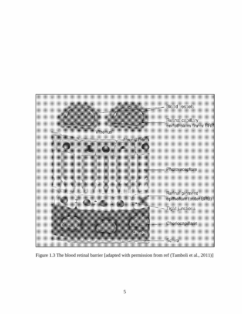
membrous barriers located in the cornea (Fig. 1.2), conjunctiva, iris-ciliary body and retina
(Fig. 1.3). Blood-Aqueous barrier (BAB), also known as anterior chamber barrier, primarily
prevents the entry of exogenous compound into the aqueous humor. It is formed from the
endothelial cells of uvea and restricts the movement of hydrophilic drug from plasma into the
anterior chamber. The drug molecules can cross the cornea either by transcellular
(intracellular) or paracellular (intercellular) pathway. The intracellular route dominates for
the entry hydrophilic compound or ions of small molecules, while the paracellular route
dominates for lipophilic molecules. The corneal epithelium is a lipophilic tissue that provides
resistance to hydrophilic compounds, whereas the stroma having an aqueous environment
controls the entry of hydrophobic compounds. The Bowman’s and descemet’s membrane do
not act as a barrier to drug absorption. The corneal endothelium is a monolayer of polygonal
cells with large intercellular junction that form cellular barrier between the stroma and
aqueous humor. The leaky hydrophilic gates of the endothelium allow the entry of
macromolecules from the stroma into the aqueous humor. It does not act as a rate limiting for
hydrophilic molecules but controls the entry of lipophilic molecules to some extent.

4
Figure 1.2 Corneal barriers [adapted with permission from ref (Tamboli et al., 2011)]
5
Figure 1.3 The blood retinal barrier [adapted with permission from ref (Tamboli et al., 2011)]
6
Role of non-corneal route in ocular drug absorption
Although the corneal route is considered to be a major route for penetration of topically
applied drugs, the conjunctival/sclera pathway is also a competitive and parallel route of
absorption. Conjunctival epithelium is leakier and has approximately 20 times more area than
cornea. It allows easy permeation of hydrophilic macromolecules. This non-corneal route has
a significant contribution for the absorption of large hydrophilic compounds such as insulin,
peptides and proteins. Moreover, small molecules such as timolol maleate, gentamycin and
prostraglandin PGF2 diffuse through conjunctiva and sclera because of their poor corneal
permeability (Koevary, 2002; Lee, 1990; Lehr et al., 1994). In general the non-corneal route
is comparatively less productive because the limbal area is full of blood vessels. The large
amount of the administered drug dissipates in the systemic circulation while crossing the
conjunctiva. The remaining drug diffuses through sclera and enters the posterior segment
while some penetrates across the conjunctiva, sclera and ciliary body and enter into the
anterior chamber. Blood-Retinal Barrier (BRB) primarily prevents the entry of molecules in
the posterior segment. It is formed by RPE and retinal endothelium. Due to limited blood
flow through choroid and presence of this barrier only a limited percentage of orally
administered drugs reach to the retina.
There are many influx and efflux transporters present in various tissues of the eye. Those
transporters also play an important role in absorption of drugs. Efflux pump are the
transporters which are responsible for extrusion of drug by transporting them out of the cell.
These pumps can act as barrier for both segments of eye. These pumps are the transport
protein, mainly responsible for the efflux of drugs from the cornea. The efflux pumps such as
P-glycoprotein (P-gp), Multiple drug resistance protein (MRP) and Breast cancer resistance

7
protein (BCRP) are found on the rabbit pigmented corneal epithelial cells (Mitra, 2009): a
brief overview of these transporters is given below:
1. P-glycoprotein (P-gp)
It is an ABC type transporter which can interact with various drug molecules. These drug
substrates are HIV protease, anti cancer, antibiotic, antihypertensive, cytotoxic agents. P-
gp is mainly localized on the apical side of the cell membrane although its mechanism of
extrusion is still not clear.
2. Multiple drug resistance protein family (MRP)
It is an ATP dependent efflux pump. Previously it is commonly known as canalicular
multispecific organic anion transporter as it mainly efflux lipophilic anions into the bile.
Its expression has been reported in various cancers such as colorectal, breast and ovarian.
Its substrate specificity is different from P-gp. Studies by Karla et al, has shown the
expression of MRP-2 on the rabbit pigmented corneal epithelial cells (Karla et al.,
2007a).
3. Breast Cancer Resistant Protein (BCRP)
It is a half transporter present in homodimer form in plasma membrane and extrudes
various structurally diverse compounds. BCRP is commonly expressed on the apical
membrane of small intestine and colon. Recent studies by Karla et al, showed the
presence of this transporter on cornea (Karla et al., 2009).
To overcome these barriers and to increase contact time of the drug on the eye
surface, absorption enhancers and/or viscosity enhancers are generally used in the ocular
formulations. So far, these approaches have limited success to address the problem of poor
bioavailability from topical route to treat anterior segment diseases. On the other hand, for

8
the treatment of posterior segment diseases either systemic or local route is preferred because
of the poor corneal drug permeation. Systemic administration requires higher dosage and
frequent administration that results in severe adverse effects. Local injections, particularly
intravitreal and subconjunctival injections are alternate strategies to achieve therapeutic
concentration in the vitreo-retinal disorders. However, to maintain the effective concentration
repeated injections are required, which causes clinical complications or patient discomfort
(Kimura and Ogura, 2001).
Biodegradable polymers for ocular drug delivery (Tamboli et al., 2012)
Many approaches have been evaluated to improve ophthalmic drug delivery.
Application of controlled drug delivery systems was anticipated as an effective approach to
circumvent all these limitations. Controlled drug delivery systems release the drug in a
sustained and controlled manner by which the therapeutic concentration is maintained for the
prolonged period of time. These systems provide many practical advantages: they avoid
frequent administration, which is a major non-compliance with many chronic eye disorders.
The delivery of emerging therapeutic macromolecules having very short biological half-lives
could be possible as these systems protect the protein drugs in situ and have an ability to
deliver them at desire rate by overcoming anatomical and biochemical barriers of drug
transport (Daugherty and Mrsny, 2003; Shell, 1984). These systems can be based on either
erodible or non-erodible matrices. In the early 1960s, first polymeric device was developed
for controlled drug delivery. Synthetic biodegradable polymers such as poly (glycolic acid)
(PGA) and poly (lactic acid) (PLA) had gained attention for biomedical applications. After
five-years, poly (lactide-co-glycolide) (PLGA) sutures emerged on the market. Since then, a
wide variety of biodegradable polymers were explored for the drug delivery (U. Adlund,

9
2002). In the past two decades the development and application of synthetic biodegradable
polymers for ocular drug delivery have gained significant momentum. Polymeric devices
such as micro and nanoparticles, microspheres, liposomes, hydrogels and ocular implants
have been designed to deliver the therapeutic agents in the controlled manner. The release
rate of the drug molecules from these polymeric devices depends on many factors such as,
molecular weight and degradation mechanisms of the polymer, physicochemical properties
of the drug, thermodynamic compatibility between the drug and polymer and the shape and
size of the devices (Park et al., 2005).
Biodegradation is an enzymatic or non-enzymatic hydrolysis of the polymeric
backbone into water soluble or insoluble products. Biodegradation involves two
complementary processes, degradation and erosion. In the degradation process cleavage of
the polymeric backbone into low molecular weight fractions takes place, whereas the erosion
mechanism refers to the physical phenomena such as dissolution and diffusion of low
molecular weight fractions from the polymer matrix. The degradation products are eventually
eliminated from the body via normal metabolic pathway (Katti et al., 2002).
Types of biodegradation
Heller has described three basic mechanisms of polymer degradation and classified the
polymers based on the degradation mechanisms (Heller, 1984). Schematic representation of
polymer degradation mechanisms is shown in Figure. 1.4.

10
Figure 1.4 Schematic representations of polymer degradation mechanisms [adapted with
permission from ref (Tamboli et al., 2012)]

11
Type I biodegradation
Cross linked water soluble polymers generally follow type-I erosion. Polymers such
as gelatin, collagen, polyacrylamides, poly (vinyl alcohol) (PVA) and poly (N-vinyl
pyrrolidone) (PVP) upon crosslinking form hydrogel, which is a water insoluble three
dimensional structure that undergoes type I hydrolysis. On the basis of hydrolysis product
generated, type I erosion mechanism can be further subdivided into type IA and IB. Type IA
erosion mechanism produces high molecular weight water soluble polymers, whereas type IB
generates low molecular weight polymers. Polymers having type IA erosion kinetics are best
suited for topical applications because of faster elimination of high molecular weight water
soluble polymers from the ocular surface. Polymers following type IB degradation kinetics
are generally utilized for designing implants. Polymeric systems that undergo type I erosion
are highly water permeable therefore; they are not suitable for the delivery of low molecular
weight compounds with appreciable water solubility. However, the crosslinked polymeric
matrix physically entangles the macromolecules and restricts them to diffuse out of the
matrix. Therefore, these polymers are well suited for the delivery of macromolecules such as
enzymes and antigens or sparingly water soluble molecules, which are released from
hydrogels initially via diffusion followed by degradation of the polymer (Heller, 1984).
Type II biodegradation
Conversion of water insoluble linear polymers into water soluble moiety through
hydrolysis, ionization, or protonation of pendant groups is defined as type II
erosion. However, since no backbone degradation is involved during erosion process overall
molecular weight of polymers does not change significantly. These polymers are generally
employed for the topical applications. Copolymers of alkyl vinyl ether and maleic anhydride

12
follow type II erosion mechanism where the degradation rate of the copolymers is affected by
the size of the alkyl substitute, pH of the degradation medium and pKa of the carboxylic
group (Woodruff et al., 1972).
Type III biodegradation
Type III erosion produces low molecular weight water soluble molecules by the
hydrolytic cleavage of water insoluble high molecular weight polymers. The polymers
demonstrating type III erosion kinetics produce non-toxic degradation products and thus
advantageous for topical and systemic administrations. These polymers are employed for
wound healing after surgery and also for chronic ocular diseases. These polymers are
available in a wide range of molecular weights having different physico-chemical properties,
which can be modulated to formulate drug delivery systems. PLA, PGA and their
copolymers PLGA, polyanhydrides, polyurathanes, polycaprolactone (PCL) and its
copolymers, poly (ortho esters) and poly (alkyl cynoacrylates) (PACA) exhibit type III
erosion mechanisms because of the characteristic hydrolytic instability in the polymer
backbone (Kimura and Ogura, 2001). The representative structures of these polymers are
shown in Figure. 1.5.

13
C
CN
COOR
* CH2 *
n
Poly(cyanoacrylates)
C O
O
C
O
*R*n
Poly(anhydrides)
C
OR'
R''
O O R* *
n
Poly(ortho esters)
* O
*
O
n
Polyester
Figure 1.5 Structures of different biodegradable polymers

14
Advantages of biodegradable polymers
Biodegradable polymers offer several advantages over non-biodegradable polymers
for controlled drug delivery. They do not require surgical removal after application, being the
most important advantage in ophthalmic drug delivery as it can circumvent surgical
complications associated with non-biodegradable implanted devices. The natural and
synthetic biodegradable polymers have many favorable properties such as biocompatibility
with ocular tissues, biodegradability and mechanical strength. They provide negligible
toxicity and also their degradation products are non-toxic in terms of both local and systemic
response. Due to the adequate mechanical properties, they can be tailored to wide range of
properties. Natural biodegradable polymers such as gelatin, albumin, chitosan, hyaluronic
acid and synthetic biodegradable polymers such as PVP, PACA, PCL, PEO, polyanhydrides
and thermoplastic aliphatic polyesters like PLA, PGA and PLGA have been thoroughly
explored for ocular delivery systems as summarized in Table 1.1. These polymers are
approved by FDA for human applications.

15
Table 1.1 Polymeric drug delivery systems used in ophthalmic research [adapted with
permission from ref (Tamboli et al., 2012)]
Polymer Example of
bioactivates Dosage form Model Ref.
Gelatin Timolol maleate Microsphere Rabbits (Bonferoni et al.,
2004)
Collagen Mitomycin c Implant In-vitro (Zimmerman, 2004)
Chitosan Indomethacin Nanoemulsions Rabbits (Badawi et al.,
2008)
PLA Ganciclovir Implant Rabbits (Kunou et al., 2000)
PLGA Vancomycine Microsphere Rabbits (Gavini et al., 2004)
PCL Dexamethasone Implants Rabbits (Silva-Cunha et al.,
2009)
PiBCA Pilocarpine Nanocapsules Rabbit (Desai and
Blanchard, 2000)
PECA Ganciclovir Nanoparticles Rabbit (EL- Samaligy,
1996)
Polyanhydride 5-fluorouridine Disks Monkeys (Jampel et al., 1990)
POE 5-chlorouracil and
fluorouracil
Injectable
solution Rabbits (Polak et al., 2008)

16
Synthetic biodegradable polymers used for ocular drug delivery
Poly N-vinylpyrrolidone (PVP)
PVP is a synthetic and biocompatible polymer widely utilized for vitreo-retinal drug
delivery. It is mainly employed for the preparation of hydrogels that exhibit viscoelastic
properties (Bruining et al., 1999). The decomposition products of PVP-based hydrogels are
easily eliminated from the vitreous through phagocytosis (Hong et al., 1998; Vijayasekaran et
al., 1996). Hydrogel prepared from cross linked PVP was used as a vitreous substitute
(Colthurst et al., 2000). Hong et al. evaluated biodegradation of poly (1-vinyl-2-
pyrrolidinone) cross-linked with 1% 14
C-methyl methacrylate. They observed that cross-
linked PVP hydrogel did not degrade in vitro in presence of proteolytic enzymes such as
trypsin or collagenase. However, in vivo half of the hydrogel disappeared from the rabbit
vitreous cavity within 4 weeks by phagocytosis (Hong et al., 1998). PVP based hydrogels
were transparent materials and remain at the site of injection for several weeks. However,
fragmentation of the hydrogels triggers an inflammatory response resulting in the vacuole
formation in the retinal pigment epithelium (Vijayasekaran et al., 1996). In addition, clinical
studies have shown that PVP-based hydrogels cause intravitreal opacity, hazy corneas and
inflammation and might not be suitable as vitreous substitutes (Colthurst et al., 2000).
Degradation kinetics of this biomaterial could be easily modulated by varying crosslinking
density. Niu et al. investigated injectable hydrogel of acrylamide/N-vinylpyrrolidone
copolymer crosslinked with reversible disulfide bond for ophthalmic applications. This
hydrogel showed characteristic in-situ sol-gel transition that facilitated the designing of
complex shapes, which was advantageous as artificial vitreous substance and scaffold for
lens regeneration (Niu et al., 2009). Hacker et al. explored the matrices composed of
17
photocrosslinked poly (propylenefumarate) (PPF)/ (PVP) for a long term delivery of
antiglaucoma drugs, such as acetazolamide (AZ), dichlorphenamide (DP) and timolol
maleate (TM). Authors suggested that the use of PVP based implants could be a valuable
strategy for controlled release of drugs over a period of 300 days in glaucoma therapy
(Hacker et al., 2009).
Poly (lacticide) (PLA), poly (glycolide), and their copolymers polylactide-co- glycolide
(PLGA)
PLA and PLGA are the most promising biodegradable polymers (Hyon, 2000). PGA
alone is highly prone to hydrolysis and remains insoluble in common organic solvents
therefore it is not widely acceptable for the fabrication of controlled drug delivery systems.
PLA alone and in combination with PGA with different ratios are mostly utilized in the
formulations. These polymers are synthesized by two methods. First involves direct
condensation reaction of monomers, which results in low molecular weight polymers and the
other method is based on ring opening polymerization of cyclic dimmers, which yields high
molecular weight polymers. These polymers upon non-enzymatic or enzymatic hydrolysis
produce water soluble metabolic products, which are not harmful to living tissues (Cam et
al., 1995; Hyon et al., 1998). These polymers belong to polyester class and degrade mainly
through bulk erosion. In vitro degradation of polyesters primarily occurs through hydrolytic
cleavage. However, in vivo, enzymes play an important role to initiate the degradation
process. The degradation products lactic acid and glycolic acid are nontoxic and eliminate in
the form of CO2 and water via Krebs cycle (Yasukawa et al., 2005). Chemical structures of
different polymers of polyester class are shown in Figure. 1.6.

18
Figure 1.6 Chemical structures of different polymers of polyester class
HO
O
O
O
H
O
y
x
PLGA
o
CH3
OH
O
H
n
PLA
oOH
O
H
n
PGA
C
O
H2C OHOH5 n
Polycaprolactone

19
Polymer degradation rate can be easily modulated by changing the molecular weight,
composition, conformation and crystallinity of the polymers (Thassu D., 2007). For example,
by varying the ratio of lactide and glycolide a wide range of diffusion and degradation
profiles can be obtained in PLGAs. PLGA with 50:50 ratio of lactic and glycolic acid
degrades faster than either PLA or PGA alone (Ogawa et al., 1988; Yasukawa et al., 2006).
The presence of methyl group provides more hydrophobicty to PLA and it degrades slowly in
comparison to PGA. These polymers have glass transition temperature ranging from 45 to 65
°C (Park P., 2006). Physico-chemical properties of different grades of PLGAs and other
polymers are summarized in Table 1.2.

20
Table 1.2 Physico-chemical properties of polymers [adapted with permission from ref
(Tamboli et al., 2012)]
Polymers Glass transition
temp, Tg (˚C)
Melting
temp,Tm(˚C)
Biodegradation
time(months)
Approximate
strength
( Modulus )
Poly (glycolic acid ) 35-40 225-230 6-12 7.0 Gpa
Poly ( l- lactic acid ) 60-65 173-178 >24 2.7 Gpa
Poly ( d, l- lactic
acid) 55-60 None 12-16 1.9 Gpa
Poly(ε-caprolactone) -65 to -60 58-63 >24 0.4 Gpa
Poly(d,l-lactic-co-
glycolic acid) [85/15] 50-55 None 5-6 2.0 Gpa
Poly (d,l-lactic-co-
glycolic acid) [50/50] 45-50 None 1-2 2.0 Gpa
Poly1,6[-bis
(carboxyphenoxy)
hexane]
-- -- 12 (in-vitro) 1.3 Mpa

21
Drug release from the PLGA system depends on the proportion of two monomer used,
porosity, and surface area of the carrier and physico-chemical properties of the incorporated
drug (Deshpande et al., 1998; Vega et al., 2008). These polymeric materials have been used
as surgical sutures due to their good biocompatibility and rapid clearance (Visscher et al.,
1985). PLA and PLGA are widely utilized in ocular drug delivery systems such as implants,
injectable microspheres and nanoparticles. PLA and PLGA microspheres have been
evaluated to reduce the intravitreal administration frequency for various chronic eye diseases
such as cytomegalovirus retinitis and endophthalmitis (Duvvuri et al., 2007; Moritera et al.,
1991). Dillen et al. developed cationic Eudragit®
coated PLGA nanoparticles loaded with
ciprofloxacin, a most commonly used fluoroquinolone for ocular infections. These authors
found that positively charged drug loaded nanoparticles can adhere to the negatively charged
bacterial surface. In addition, particulate systems enhanced the therapeutic drug
concentration at the target site by providing prolonged diffusion controlled release (Dillen et
al., 2006). Kunou et al. achieved pseudo zero-order release kinetics of GCV over a period of
one year by employing two monomers of PLA with different molecular weights and ratios
for the preparation of biodegradable scleral implant (Kunou et al., 2000). The PEG- coated
PLA nanospheres were more efficient for sustaining the drug release and improving the
ocular bioavailability of ACV in the treatment of viral infections (Giannavola et al., 2003).
PLGA was also utilized to encapsulate anti-VEGF RNA aptamer (EYE001) in microspheres.
In contrast to a characteristic triphasic release pattern of microsphere, the release of EYE001
from PLGA microspheres was a diffusion-controlled process that exhibited drug release in
continuous manner over a period of 20 days. Furthermore, the bioactivity of the aptamer was
retained in the formulation during the entire release period (Carrasquillo et al., 2003).

22
Duvvuri et al. discussed the conventional triphasic release pattern from PLGA microspheres
and optimized the drug release kinetics by employing various PLGA polymer blends
(Duvvuri et al., 2006). However, particulate systems such as microspheres and nanospheres
may cause vision obstruction or irritation to the retinal tissues after intravitreal injections. In
addition, most of the PLGA based drug delivery systems have initial burst release phase.
Authors investigated the composite approach to minimize the particulate system related
drawbacks. This dual approach involved the use of PLGA-PEG-PLGA triblock
thermogelling polymer to suspend the particulate system (Duvvuri et al., 2005; Zentner et al.,
2001). The thermosensitive polymer exists in the liquid state at room temperature and forms
gel upon contact with eye tissue i.e. 34 0C. Thermosensitive hydrogel holds particles at the
site of administration and avoids vision interference. In addition, gel matrix protects the
microspheres from enzymatic and cellular degradation. This dual system showed release of
drug in a more controlled manner for prolonged period of time (Duvvuri et al., 2007).
Poly- ε-caprolactone (PCL)
PCL is an aliphatic polyester synthesized form monomer ε- caprolactone through ring
opening polymerization catalyzed by stannous octoate at 140 0C (Sinha et al., 2004). It is a
tough semi crystalline polymer having the melting point in the range of 59 and 64 °C and a
glass transition temperature of -60 °C (Murthy R., 1997). Permeability and crystallinity of
the PCL can be modified by co-polymerization with PLA or PGA (Sinha et al., 2004).
Degradation of PCL occurs in two phases. First phase involves molecular weight (Mn) loss
up to 5000 due to cleavage of ester linkage in the polymer backbone (chain scission), that
produces ε- hydroxyl caproic acid and decreases the intrinsic viscosity of polymer. In the
second phase (commonly observed in vivo), chain scission of low molecular weight polymer

23
produces small fragments, which diffuse out of the polymer bulk and break the polymer in
small particles that undergo phagocytosis (Deshpande et al., 1998). PCL is utilized for
sustained drug delivery due to its higher permeability to various drug molecules and slower
degradation in comparison to other polymers (Murthy R., 1997). Degradation rate of PCL
can be improved by co-polymerizing with other fast degradating polymers. PCL implant
loaded with dexamethasone had released the drug within the therapeutic range over the
period of more than one year and was well tolerated in the rabbit eye (Fialho et al., 2008).
Rod shaped PCL implant loaded with triamcinolone was also well tolerated in sub-retinal
space of rabbit eye and had released the drug over the period of 4 weeks without any clinical
complications (Beeley et al., 2005). Yenice et al. evaluated hyaluronic acid coated PCL
nanospheres loaded with cyclosporine. Investigators found that bioavailability of
cyclosporine nanospheres was 10-15 fold higher than the drug solution in castor oil. PCL can
be utilized to prepare in situ gel-forming sustained drug delivery system. PCL based triblock
polymer was recently characterized for ophthalmic applications. Gong et al. evaluated the
toxicity of PEG-PCL-PEG triblock copolymer hydrogel after intracameral injections. This
hydrogel was biocompatible with ocular tissues and appeared to be a promising controlled
release systems for chronic ocular diseases (Yin et al., 2010).
Poly (alkyl cynoacrylates) (PACA)
PACA is synthesized from monomer alkyl cynoacrylate, which exhibits bioadhesive
properties. It can form a strong bond with polar surfaces including skin and living tissues.
Polymethylacrylate composed of smaller alkyl chain is not applicable to drug delivery due to
tissue toxicity and inflammation. Therefore, larger alkyl chains such as n-butyl, octyl
cyanoacrylates are used for clinical applications. In practice anionic or zwitterionic

24
polymerization are commonly used for synthesis of PACA due to rapid initiation at ambient
temperature. Polymer degradation occurs by enzymatic hydrolysis of alkyl side chain
producing an alkyl alcohol and poly (cyanoacrylic acid). The degradation products are
soluble in water and eliminate via kidney filtration (Vauthier et al., 2003). This polymer was
mostly explored for the preparation of biodegradable nanoparticles. Layre et al. reported the
encapsulation of highly crystalline alkylating drug, busulfan, utilizing five different PACA
polymers. The highest encapsulation of busulfan was found in poly (isobutyl cyanoacrylate)
(PIBCA) and poly (ethyl cyanoacrylate) due to the specific interaction between the drug and
the polymers. They suggested that this nanoparticulate formulation when given intravenously
have nigligible toxicity and also minimize the variability in bioavailability (Layre et al.,
2006). Peracchia et al. suggested the potential use of PEG-coated PIBCA nanoparticles as
drug delivery carriers, which are rapidly biodegradable. The covalently bound PEG avoids
interaction with blood components and prevents recognition by macrophages of the
mononuclear phagocyte system after intravenous injection (Peracchia et al., 1997). PEG-
coated polyethyl-2-cyanoacrylate nanospheres had increased the ocular bioavailability of
ACV by 25 fold when instilled in the conjunctival sac of rabbit eyes. The improved drug
bioavailability was attributed to the colloidal nature of nanospheres that can facilitate the
transport of drug paracellularly. In addition, presence of PEG provided better mucoadhesion
on the corneal surface and improved dug permeation (Fresta et al., 2001).
Polyanhydrides
In 1930s, Hill and Carothers proposed polyanhydrides as substitutes of polyesters for
textile applications. However, they were not useful for textile industry due to faster
hydrolytic cleavage of anhydride linkage in the polymer backbone. Instead, due to this

25
intrinsic property polyanhydrides are considered as an ideal candidate for formulation of
controlled drug delivery systems. Hydrolytically labile linkages of polyanhydrides provide
bio-degradability and regulate degradation rate. For example, poly [bis (p- carboxyphenoxy)
alkane anhydrides] degradation rate can be adjusted from 10-1
to 10-4
mg/hr/cm2 upon
changing the methyl group to the hexyl group. Polyanhydrides can be synthesized by three
methods: melt condensation, dehydrochlorination and dehydrative coupling. Melt
condensation method can produce high molecular weight polymer (up to 50,000) while other
two methods are useful for the synthesis of low molecular weight polymers (Leong, 1987).
Polyanhydrides show pH dependent degradation, which can be modulated by additives. Basic
additives primarily promote bulk erosion, whereas acidic additives favor surface erosion and
produces acetic acid upon degradation (Leong et al., 1985). Further, upon changing the
polymeric backbone drug release rate can be modulated over a thousand fold (Jain et al.,
2005). Mostly copolymer of bis(p-carboxyphenoxy propane) and sebacic acid is utilized for
drug delivery applications. Release of drug from this polymeric delivery system occurs
mainly by surface erosion rather than drug diffusion. It has a potential to provide almost
zero-order drug release rate and also undergoes relatively faster in-vivo biodegradation (Jain
et al., 2005). Rosen et al. demonstrated near zero order degradation and drug release kinetics
from poly [bis (p-carboxyphenoxy methane andride] for several months at two different
temperatures i.e. 37 and 60˚ C (Rosen et al., 1983). Polyanhydride microspheres have been
employed to avoid repeated intravitreal injections for the treatment of vitreoretinal diseases
(Jain et al., 2005). Microspheres prepared from poly (adipic anhydride) (PAA) exhibited
surface degradation. Release of timolol maleate from these microspheres was sustained for 7
hrs and mainly controlled by polymer degradation. Further, to improve ocular bioavailability
26
of timolol maleate PAA-microspheres were suspended in the Gelrite@ (an in situ
polysaccharide gel) (Albertsson, 1996). In another study by Lee et al., 5-fluorouracil (5-FU)
was incorporated in 3mm bio-erodible disc of bis (p-carboxyphenoxy) propane and sebacic
acid. They reported that 5-FU was delivered in a sustained manner and maintained intra-
ocular pressure for 3 weeks (Lee et al., 1988).
Poly (orthoester) (PEOs)
Since early 1970s, four families of poly (ortho esters) have been synthesized. POEs
are hydrophobic polymers having hydrolytically labile ortho ester bonds. The amount of
water available to react with these bonds is very less under physiological conditions that
make the polymer extremely stable. POEs undergo surface erosion and provide zero order
release rate for a longer period of time (Einmahl et al., 2003). POEs based formulations have
been proven promising in the treatment of ocular diseases such as glaucoma filtration surgery
and proliferative vitreoretinopathy (PVR) (Bernatchez et al., 1993). They can be injected
directly into the eye with a needle of appropriate size. Chemical structures of four types of
POEs are shown in Figure 1.7.

27
Figure 1.7 Chemical structures of different Poly (ortho esters) [adapted with permission from
ref (Tamboli et al., 2012)]

28
POE I is synthesized by transesterification reaction between a diol and diethoxy
tetrahydrofuran. It is a hydrophobic solid polymer having acid sensitive nature and easily
hydrolyzed in an aqueous environment. Basic ingredients such as sodium carbonate are
generally utilized to prevent autocatalytic hydrolysis. This polymer has been widely explored
for orthopedic applications, treatment of burns and for the delivery of narcotic antagonist and
contraceptive steroids. It is not explored much for ocular drug delivery (Bernatchez et al.,
1993; Heller, 2005; Heller et al., 2002).
POE II is synthesized by simple addition reaction between diol and di keteneacetal
3,9-di(ethylidene 2,4,8,10-tetraoxaspiro[5.5]undecane) (Heller et al., 2002). Monomers are
required to dissolve in tetrahydrofuran and trace of acidic catalyst is used to initiate polymer
synthesis instantaneously. Polymer hydrolysis occurs in two steps, unlike POE I there is an
absence of autocatalytic hydrolysis. This polymer is also synthesized by crosslinking a triol,
either alone or as a mixture with diols. It forms a dense polymer upon crosslinking, which
biodegrades to small water soluble fragments. The cross linked density can be adjusted by
varying the ratio of diol to triol. POE II can be fabricated as a hard glassy material to semi
solid material; mechanical and thermal properties can be controlled by using diols having
different degrees of chain flexibility (Heller, 2005; Heller et al., 2002). POE II have been
extensively explored for the release of 5-FU, which is mainly utilized as an adjunct to
glaucoma filtration surgery. The erosion rate of POE II can be controlled by incorporating
the acidic excipients such as suberic, adipic and itaconic acids in the polymer matrix. Nearly
zero–order release of 5-FU was obtained by incorporating different amount of suberic acid in
POE polymeric matrix (Einmahl et al., 2003). According to the United States of

29
Pharmacopoeia, this generation of polymer is nontoxic for cellular, subcutaneous,
intramuscular and systemic implant applications.
POE III is semi-solid at room temperature and synthesized via transesterification of 1,
2, 6- and trimethyl orthoacetate (Heller, 2005). This polymer has a very flexible backbone
and allows incorporation of therapeutic agents at room temperature without using organic
solvent. Therefore, it can be used for thermo labile and solvent sensitive drugs. The release
rate of incorporated drug can be controlled by modulating the molecular weight of the
polymer. Merkli et al. observed that the release of 5-FU occurred within 1 day from 3500 Da
and was sustained for 1 week from 33,300 Da POE III. (Merkli, 1994). Drug delivery
systems fabricated from this polymeric material do not show any burst release and the drug
release rate was governed by the polymer degradation rate (Einmahl et al., 2001). Sintzel et
al. reported that the drug release rate from POE III can be controlled by modulating the
hydrophobicity of the polymer by substituting triol from 1,2,6 hexanetriol to 1,2,10-
decanetriol (Sintzel, 1997). According to Einmahl et al., this new generation of POE has a
potential for application in glaucoma filtering surgery for the patients with higher risk of
surgery failure. This injectable polymer can provide sustained release of 5-FU for 2 weeks
after subconjunctival injection that can avoid frequent administrations and minimize the
adverse effects (Einmahl et al., 2001). These authors have also described that after
subconjunctival administration, polymer degradation products follow several pathways. One
major pathway involves direct entry into the anterior chamber through the fistula, to the
ciliary body, into the vitreous body, and then into the retina (Einmahl et al., 2003). Therefore
they evaluated the biocompatibility of this polymer in different parts of the eye including
anterior chamber and suprachoroidal space. They found that the anterior chamber of the

30
rabbit eye can tolerate up to 50 µl of polymer solution, which degrades within 1 week
(Einmahl et al., 2000). In addition, after suprachoroidal injection the retinal pigmented
epithelial (RPE) cells, retinal and choroidal vasculatures were not affected by the polymeric
formulation (Einmahl et al., 2000; Einmahl et al., 2002). POE III demonstrated excellent
biocompatibility to the different parts of the rabbit eye. Difficulties in synthesis and lack of
reproducibility have limited the use of POE III in biomedical applications (Heller, 2005).
POE IV is synthesized by reacting diols with the diketeneacetal 3, 9-diethylidene-2,
4, 8, 10-tetraoxaspiro [5.5]undecane. It is a modified form of POE II, which contains latent
acid in the polymer backbone that regulates the erosion rate. The latent acid is generally
composed of glycolic acid or lactic acid. POE IV does not require external acidic excipients
to control the erosion rate, unlike POE II. When the polymer is exposed to an aqueous
solution, the latent acid will hydrolyze to give lactic acid or glycolic acid that will further
assist in the hydrolysis of polymer. POE IV can be fabricated as solid or gel-like material by
changing the nature of diols. POE IV-based devices generally undergo surface erosion and
produce acidic degradation products which readily diffuse out from the device. Lactic acid
based fourth generation POEs are biocompatible and have long residence times following
intracameral, subconjunctival, intravitreal and suprachoroidal injections in the rabbit eyes
(Einmahl et al., 2003). Polak et al. evaluated the efficacy of 5-chlorouracil (5-CU) loaded
POE IV formulation in the glaucoma filtration surgery. They found that 5-CU suspended in
POE IV has maintained low IOP in the rabbit eye for 5 months (Polak et al., 2008).
Polymeric materials contribute a significant role in the controlled drug delivery. In particular,
biodegradable polymers have been extensively explored for ocular therapeutics in the recent
years. In this chapter we have summarized mainly the properties and applications of

31
biodegradable polymers having natural and synthetic origins. We have exemplified the
applications of biodegradable polymers for the delivery small molecules to the different parts
of the eye. Two major advantages of polymeric drug delivery devices, enhancing drug
bioavailability and minimizing side effects, are significant in ocular drug delivery. The
development of new biodegradable block polymers has gained significant momentum in the
recent years. These polymers would be advantageous in the delivery of newer therapeutic
agents including genes, therapeutic antibodies and bioactive proteins. It is challenging to
deliver these macromolecules to targeted tissues of the eye. Therefore, design of novel
biodegradable polymeric devices is currently under investigations for targeted delivery of
macromolecules.
Controlled release carriers for Ocular drug delivery
Various sight-threatening and chronic ocular diseases such as age-related macular
degeneration, proliferative vitreoretinopathy, chronic cytomegalovirus retinitis (CMV)
diabetic macular edema and other ocular inflammatory conditions require sustained levels of
therapeutic agents for longer duration (Mishra et al., 2011b). Treatment of these diseases
requires frequent drug administrations. Conventional routes fail to achieve required
therapeutic levels in the eye due to the presence of ocular barriers. In recent years,
nanotechnology has attained wide acceptance in ocular drug delivery. Novel nanocarriers
such as nanomicelles, microsphere, nanoparticles, liposomes and surface modified nano
formulations are very efficient in circumventing various ocular barriers (Sahoo et al., 2008;
Vandervoort and Ludwig, 2007). These systems can avoid frequent administrations and
release therapeutic molecules at the targeted site in a controlled manner for prolonged
periods. Nanotechnology based drug delivery systems may provide many advantages such as

32
enhanced cellular uptake, stimuli sensitive release and targeted delivery to specific ocular
tissues. Nanocarriers are particularly beneficial in many angiogenic ocular diseases such as
diabetic retinopathy, choroidal neovascularisation (CNV), central retinal vein occlusion and
intraocular solid tumors, where enhance permeation retention (EPR) effect can be achieved
by targeting drugs with nanocarriers (Mishra et al., 2010; Yasukawa et al., 2004). In addition,
polymeric nanocarriers are effective in gene delivery to specific ocular which can overcome
issues regarding short intravitreal half-life and transient gene expression. An ideal
nanocarrier for ophthalmic applications should possess appropriate size and narrow
distribution that ensure low irritation to ocular tissues and provide adequate ocular
bioavailability (Sahoo et al., 2008). Our research group has developed and evaluated
different nanocarriers for ophthalmic applications.
Microparticles and microspheres
PLGA polymer based controlled drug delivery systems such as microspheres and
microparticles have gained attention in ophthalmic applications. Availability of different
grades of PLGA polymers provides an excellent platform for formulation scientists to
develop a tailor made sustained release formulations according to drug of choice (Duvvuri et
al., 2006). An ideal sustained release microsphere formulation should provide continuous
release of entrapped drug for a desire period. Drug release from PLGA microsphere usually
follows three stages (Duvvuri et al., 2005). Initial burst release phase (Phase I) is attributed
to diffusion of surface absorbed and poorly encapsulated drug. Slow or no release phase
(Phase– II) can be attributed to possible drug-polymer interactions, which results in low drug
levels at the target site. Third phase of rapid release (Phase-III) is mainly attributed to faster
diffusion of drug molecules from polymer matrix due to degradation of polymer (Duvvuri et

33
al., 2006). PLGA polymers are available in wide range of molecular weights with different
lactide/glycolide ratios, the amount of drug release during each phase could vary
considerably depending upon type of PLGA utilized in microsphere formulation.
Particularly, phase-II release duration is dependent on the hydrophilicity of the matrix. In
addition, all these release phases are most likely evident for hydrophilic molecules compared
to lipophilic molecules (Duvvuri et al., 2006). The extent of drug release during different
release phases can be modulated by adding polymeric additives (Mishra et al., 2010), release
modifying agents such as tweens and polyethylene glycols or by utilizing polymeric blends
(Duvvuri et al., 2005). In an attempt to modify GCV release from PLGA microsphere,
different PLGA blends were used in the formulation. A small molecular weight hydrophilic
PLGA polymer, Resomer RG 502H (d,l-lactide : glycolide::50: 50, Mw=8000 Da) was
blended with PLGA 65/35 (d,l-lactide : glycolide::65: 35, Mw=45,000–75, 000 Da) to
modulate drug release kinetics from microspheres (Duvvuri et al., 2005). Drug entrapment
efficiency was also increased from 47.13% with Resomer RG 502H microspheres to 72.67 %
for polymer blend microspheres. GCV release from both Resomer RG 502H microspheres
and blend microsphere followed triphasic pattern. However, drug release rate constant of
each release phase was significantly lowered in the case of blend microsphere compared to
Resomer RG 502H microspheres. Although microsphere can provide controlled delivery of
entrapped molecules for longer time periods major obstacles anticipated with its application
are retinal irritation and vision obstruction due to movement of particulate system (floaters)
in the vitreous following intravitreal injection. Moreover, due to hydrophobic nature PLGA
microsphere can agglomerate in aqueous buffer and poses a challenge for formulation
development. One promising strategy is the development of composite formulation

34
comprising of nanoparticles in suspended thermosensitive hydrogel. Thermogelling polymer
solution remains in sol state at room temperature and forms gel at physiological temperature.
We observed that thermogelling polymer can form a depot upon administration in to vitreous
cavity and minimize the movement of GCV microspheres in the vitreous. PLGA-PEG-PLGA
copolymer was utilized as thermosensitive hydrogel (Duvvuri et al., 2005). Our research
group evaluated the intravitreal pharmacokinetics of GCV following single intravitreal
injection of GCV solution and equivalent GCV microsphere formulation (Resomer RG 502H
and a blend of Resomer RG 502H: PLGA 65/35::1:3, were physically mixed in a 1:1 ratio
and suspended in a 23% w/w aqueous solution of PLGA-PEG-PLGA) in male New Zealand
white rabbits (Duvvuri et al., 2007). Following administration of GCV solution, the vitreous
level of GCV was maintained above its minimum inhibitory concentration (MIC) for only 54
hrs with vitreous elimination half-life of 6.45 hrs. In contrast, with gel formulation the
vitreous level of GCV was maintained above its MIC for 14 days. These results suggest that
composite formulations can be used to deliver GCV to the vitreous and retina/choroid
following intravitreal administration continuously over 4–5 weeks. In addition, these
formulations is biodegradable and biocompatible in nature and do not require surgical
removal following completion of drug release. These studies also suggest that sustained
delivery of GCV can be tailor made by polymer blending strategy.
Nanoparticles
Nanoparticles are defined as sub-micron sized carriers (10 to 1000 nm) in which drug
molecules are dissolved, entrapped or adsorbed. Nanoparticles can be further divided in to
nanospheres and nanocapsules. Nanospheres have a solid matrix of polymers or lipids on
which drug molecules are simply adsorbed on the surface. Nanocapsules have polymeric

35
shell and aqueous core like structure in which molecules are mostly entrapped and dissolved
in the aqueous core. Nanoparticle surfaces can be functionalized with specific receptor
targeted moiety to achieve site specific delivery and to enhance transport of therapeutic
molecules across the various physiological ocular barriers such as corneal and blood retinal
barrier. Various natural and synthetic biodegradable polymers used in the development of
nanoparticles have demonstrated promising results in ophthalmic drug delivery. The most
common ones are polycaprolactone (PCL), polylactide (PLA) and poly(lactide-co-glycolide)
(PLGA). These are USFDA-approved polymers that degrade in vivo via the Kreb’s cycle,
resulting in biocompatible by-products (lactic and glycolic acids). These polymers can be
fabricated or formulated into devices such that they provide controlled drug release from a
few days to years (Janoria et al., 2007). PLGA nanoparticles loaded with different steroids
such as dexamethasone, hydrocortisone acetate, and prednisolone acetate were developed as
a vehicle to provide sustained levels of corticosteroids in the posterior ocular segment
(Janoria et al., 2007). PLGA nanoparticles prepared from single emulsion solvent
evaporation method resulted in uniform size particles with significant increase in entrapment
efficiency and drug loading relative to nanoparticles prepared by dialysis method. Different
grades of PLGA i.e. PLGA 50/50 and PLGA 65/35 were evaluated for nanoparticle
preparation. Nanoparticles prepared from PLGA 65/35 provided more prolonged release of
steroids in comparison to PLGA 50/50. This effect may be explained by the fact that PLGA
65/35 is comparatively more hydrophobic in nature that in turn retards the rate of water
penetration into polymer matrices. Initial burst release of steroids was also found to be less in
nanoparticles prepared from PLGA 65:35. Based on the in vitro release data, the researchers
concluded that PLGA 65/35 provides promising results in comparison to PLGA 50/50 for

36
sustained delivery of steroids from nanoparticles (Janoria et al., 2007). However,
nanoparticles prepared from PLGA polymer produce high amounts of lactic and glycolic
acids after degradation, which may cause local tissue irritation and degradation of therapeutic
molecules (Kang and Schwendeman, 2002; Meyer et al., 2012). In addition, PLGA
nanoparticles produce high initial burst release followed by no release phase (Boddu et al.,
2010b; Budhian et al., 2005). Considering this, we developed novel polymeric materials for
preparation of controlled release formulations that can address these limitations.
Micelles
Drug delivery to the posterior segments of the eye presents a greater challenge
because of the selective functionality of the biological barriers. Particularly, delivery of
hydrophobic molecules to the posterior segments via topical delivery is ineffective.
Moreover, the low aqueous solubility of many steroids limits the feasibility of formulating a
highly concentrated aqueous eye drop formulation, making the topical route inefficient in
delivering adequate drug levels to the retina (Forrest et al., 2006; Loftsson and Hreinsdottir,
2006). However, many investigators are now exploring the potential of topical delivery for
the back-of-the-eye diseases. Polymeric micelles are exploited as pharmaceutical
nanocarriers for the delivery of poorly water-soluble drugs, which can be solubilized in the
hydrophobic inner core of a nanomicelle. In this regard, LX214, a nanomicellar formulation,
10 to 15 nm in size containing up to 0.2% voclosporin, was developed by our research group
in collaboration with Lux Biosciences Inc (Mitra et al., 2010; Mitra et al., 2009, 2011).
Nanomicelles are colloidal particles in nanometer size ranges (10-100 nm), forming spherical
structures of amphiphilic molecules in water (Aliabadi and Lavasanifar, 2006; Torchilin,
2007). The nanomicellar corona is comprised of hydrophilic chains extended outwards (see

37
Figure 1). Micelles can therefore serve to improve solubility and bioavailability of various
hydrophobic (water-insoluble) drugs. LX214, mixed nanomicelles are composed of two non-
ionic surfactants, D-alphatocopheryl polyethylene glycol 1000 succinate (Vitamin E TPGS)
stabilized with octyl phenol ethoxylate (octoxynol-40) in a defined ratio. LX214, packaged in
single-use sterile low-density polyethylene, blow-fill -sealed vials, has demonstrated stability
for at least 1 year under refrigeration and for 2 months at room temperature. Hydrophobic
molecules, such as voclosporin, encapsulated in 15 nm nanomicelles, form spherical
structures in water. Due to their hydrophilic corona, these micellar nanocarriers can pass
through the aqueous channels/pores of the sclera, which range from about 30 nm to about
300 nm in size. Nanomicelles may then be absorbed onto the basolateral side of the retinal
pigment epithelium (RPE) through endocytosis. The contents of the micellar nanocarriers are
discharged inside the cell after fusion with the cell membrane. It is also believed that during
the transit, the hydrophilic nanomicellar corona helps drug molecules to evade wash-out into
the systemic circulation by the conjunctival/choroidal blood vessels and lymphatics. In recent
years, polymeric micelles composed of amphiphillic block copolymers such as PLGA-PEG,
PCL-PEG, polyethylene oxide (PEO)-b-poly (ester) and Pluronics have gained attention in
drug delivery. Chemical structure of block copolymers can be easily modified to achieve
high drug payload, improve stability, modulate drug release kinetics and achieve target
specific delivery (Croy and Kwon, 2006). In an attempt to evaluate the role of folate
receptor-targeted drug delivery system for the treatment of retinoblastoma doxorubicin
(DOX) loaded PLGA-PEG-folate micelles were developed by our research group (Boddu et
al., 2010a). The uptake of DOX was found to be 4 times higher in the presence of targeted
DOX micelles compared to DOX alone. Development of novel micellar formulation targeted

38
to specific receptor/transporter opens doors for non invasive delivery of hydrophobic drugs
for treatment of diseases affecting the anterior and/or posterior ocular segments.
Liposomes
In 1965 liposomes were first introduced as drug delivery carriers (Bangham et al.,
1965). Liposomes are usually within the size range of 10 nm to 1 µm or greater. These
vesicular systems are composed of an aqueous core enclosed by phospholipid bilayers of
natural or synthetic origin. Liposomes are structurally classified on the basis of lipid bilayers
such as small unilamellar vesicles (SUVs) or multilamellar vesicles (MLVs). Furthermore,
on the basis of size liposomes are classified into small unilamellar vesicles (SUVs), giant
unilamellar vesicles (GUVs) and large unilamellar vesicles (LUVs) (Fig.1.8). Unilamellar
vesicles are composed of single layer of lipid such as lecithin or phopshotidylglycerol
encapsulating aqueous interior core. Multilamellar vesicle is composed of various layers of
lipid bilayers (Elbayoumi and Torchilin, 2010; Kaur et al., 2004; Mainardes et al., 2005).
MLVs are metastable energy configuration having different facets depending upon the
polydispersity of the liposomal formulation. Various types of liposomes with size are
summarized in table 1.3.

39
Figure 1.8 Schematic representation of basic structures and different types of Liposomes
[adapted with permission from ref (Mishra et al., 2011a)]
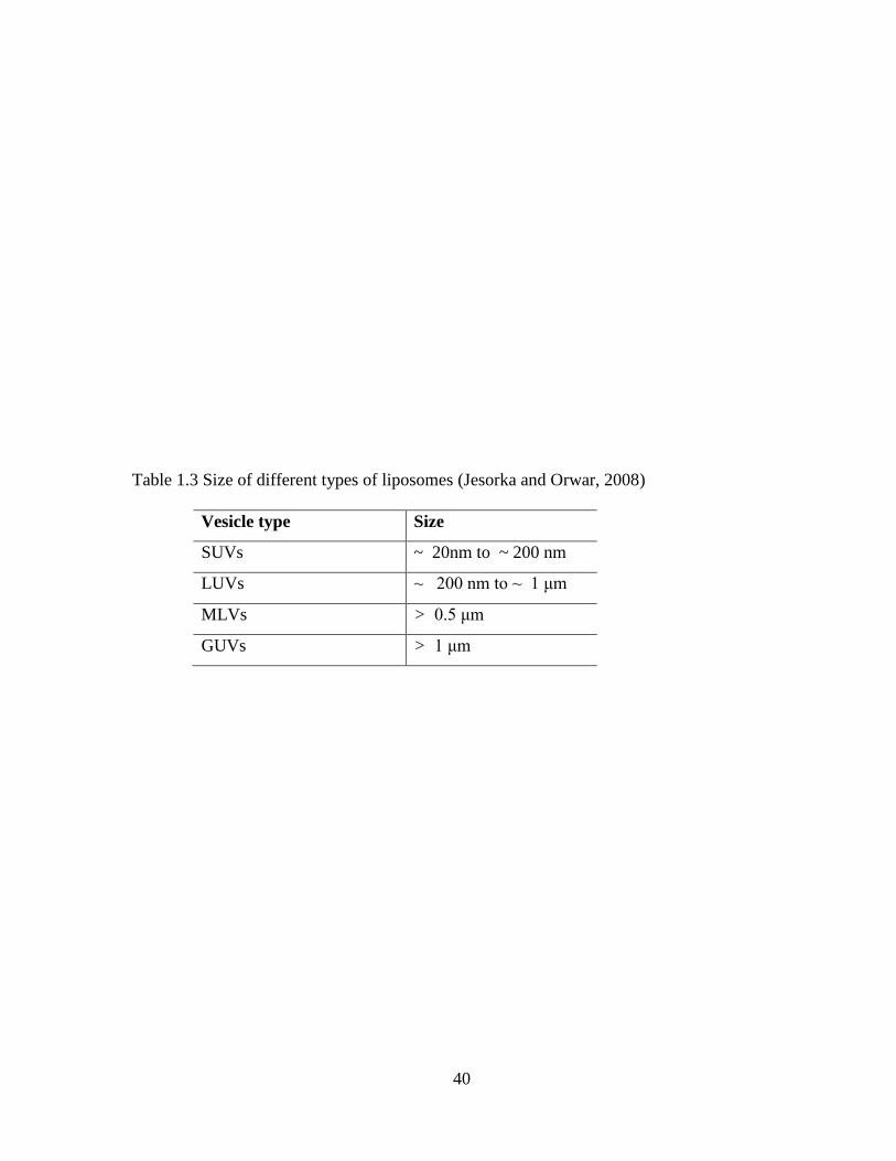
40
Table 1.3 Size of different types of liposomes (Jesorka and Orwar, 2008)
Vesicle type Size
SUVs ~ 20nm to ~ 200 nm
LUVs ~ 200 nm to ~ 1 μm
MLVs > 0.5 μm
GUVs > 1 μm

41
Drug loading capacity of liposomes depends on many factors such as size of
liposomes, types of lipid utilized for preparation and physicochemical properties of
therapeutic agent itself. For example, entrapping efficiency for SUVs are poor in comparison
to MLVs. However, LUVs provide a balance between size and drug loading capacity.
Liposomes are advantageous in encapsulating both lipophilic and hydrophilic molecules.
Hydrophilic drugs are entrapped in the aqueous layer while hydrophobic drugs are held in the
lipid bilayers. Loading capacity of ionic molecules can be further improved by using cationic
or anionic lipids for the preparation of liposomes (Ding X et al.).
Majority of liposomal formulations utilize phosphotidylcholine (PC) and other
constituents such as cholesterol and lipid conjugated hydrophilic polymers as the main
ingredients. Incorporation of cholesterol enhances the stability by improving the rigidity of
the membrane. Stability of liposomes depends upon the various properties such as surface
charge, size, surface hydration and fluidity of lipid bilayers. Surface charge determines
interaction of liposomes with ocular membrane. Positively charged liposomes display better
corneal permeation than the neutral and negatively charged liposomes. Neutral liposomes
upon systemic administration evade the elimination by reticuloendothelial system (RES).
However, these vesicles possess higher self aggregation tendency. In contrast, negative and
positively charged liposomes exhibit lower aggregation tendency but undergo rapid clearance
by RES cells due to higher interaction with serum proteins. In addition, size of the liposomes
can also regulate the clearance by RES. Liposomes of size less than 100 nm generally exhibit
significantly higher circulation time due to decrease in opsonization of liposomes with serum
protein (Lian and Ho, 2001).

42
Amphiphilic nature of phospholipids allows these molecules to form lipid bilayers.
This unique feature is utilized for the preparation of liposomes. In general, hydration of
phospholipids results into formation of MLVs, which can be processed into SUVs with
proper sonication. However, addition of aqueous solution of surfactant above the critical
micelle concentration results in the formation of phospholipids micelles. After the dialysis of
surfactant aggregation of micelles form LUVs Critical micelle concentrations of amphiphiles
which can form micelles are four to five orders of magnitude higher than the phospholipids
which form liposomes (Jesorka and Orwar, 2008). Numerous methods have been reported to
prepare liposomes. Most commonly solvent evaporation method, reverse phase evaporation
method and detergent dialysis method are employed. (Niesman, 1992). The encapsulated
drug from liposome can be released either through passive diffusion, vesicle erosion or
vesicle retention. In passive diffusion, drug molecules tend to penetrate through the lipid
layers of liposome to reach extra vesicular layer either by diffusion or convection
mechanism. The rate of diffusion depends on the size, lipid composition, and the properties
of the drug itself (Guy et al., 1983; Papahadjopoulos et al., 1973; Tsukada K et al., 1984).
Unilamellar liposomes exhibit faster release rate than multilamellar ones because in multi
layered liposome, drug diffusion occurs through a series of barriers hence the drug release is
delayed. Phospholipase and high density lipoprotein present in blood plasma can damage
phospholipid layers of liposome and thus results in vesicle erosion and releases the
encapsulated drug into the cell. The drug release rate depends on the extent of liposomal
membrane damage (Zeng L and X, 2010). Liposome-cell interactions depend on several
factors like size, surface charge, composition of liposomes, targeting ligand on the surface of
liposome and biological environment. Liposomes can interact with cells by four different

43
mechanisms: adsorption, fusion, lipid exchange and endocytosis (receptor mediated).
Liposomes can be specifically or nonspecifically adsorbed onto the cell surface or can be
fused with cell membranes, and release encapsulated drug inside the cell. During adsorption,
liposomes can release encapsulated drug in front of cell membrane and released drug can
enter cell via micropinocytosis. They can also be engulfed inside the cell by specific or
nonspecific endocytosis process. Negatively charged liposomes have been found to be more
efficient than neutral liposomes for internalization into the cells by endocytosis process.
Liposomes bind to the receptor present in the invaginations of cellular membrane and are
internalized into the cell by endocytotic pathway. After endocytosis, they can fuse with the
endosomal membrane to form endosome which can be delivered to lysosomes. In lysosomes,
the presence of peptidase and hydrolase degrade the liposomes and their content. To avoid
this degradation and thus to increase cytoplasmic bioavailability stimuli responsive
liposomes (such as pH or temperature) have been developed. pH sensitive liposomes can
undergo fusion with endosomal membrane and release their content directly into cytosol. In
some cases liposomes become destabilized inside the endosome and release their content or
they destabilize endosomal membrane resulting in leakage of encapsulated content into
cytosol (Bonacucina et al., 2009; Torchilin, 2005).
Application of liposomes in ophthalmic drug delivery
Liposomes have been investigated for ophthalmic drug delivery since it offers
advantages as a carrier system. It is a biodegradable and biocompatible nanocarrier. It can
enhance the permeation of poorly absorbed drug molecules by binding to the corneal surface
and improving residence time. It can encapsulate both hydrophilic and hydrophobic drug
molecules. In addition, liposomes can improve pharmacokinetic profile, enhance therapeutic

44
effect and reduce toxicity associated with higher dose. Owing to their versatile nature,
liposomes have been widely investigated for the treatment of both anterior and posterior
segment eye disorders. Current approaches for the anterior segment drug delivery are focused
on improving corneal adhesion and permeation by incorporating various bioadhesive and
penetration enhancing polymers. However, in the case of posterior segment disorders
improvement of intravitreal half life and targeted drug delivery to the retina are necessary.
Currently verteporfin is being used clinically in photodynamic therapy for the treatment of
subfoveal choroidal neovascularization (CNV), ocular histoplasmosis, or pathological
myopia effectively. Verteporfin is a light-activated drug which is administered by
intravenous infusion. In photodynamic therapy, after the drug is injected, a low energy laser
is applied to the retina through the contact lens in order to activate verteporfin that results in
closure of the abnormal blood vessels. Unfortunately, photodynamic therapy usually does not
permanently close the abnormal vessels and choroidal neovessels reappear after several
months. Another liposomal photosensitizing agent, rostaporfin, was evaluated for the
treatment of age related macular degeneration. It is now under Phase 3 clinical trial.
Rostaporfin requires less frequent administration compared to verteporfin. Liposome
technology has been explored for ophthalmic drug delivery. However, there are some issues
to be addressed such as formulation and storage of liposomes is very difficult and they are
known to cause long term side effects. Intravitreal administration of liposomes has resulted in
vitreal condensation, vitreal bodies in the lower part of eye, and retinal abnormalities.
Therefore all these factors should be taken into account while developing liposomal
formulation for ophthalmic application (2004; Chakravarthy et al., 2006; Lazzeri et al., 2011;

45
Ruiz-Moreno et al., 2006; Sachdeva et al., 2010).Recent applications of liposomal
formulations encapsulating various therapeutic molecules are summarized in table 1.4

46
Table 1.4 Application of liposomes for the delivery of various drug molecules [adapted with
permission from ref (Mishra et al., 2011a)]
Drug Formulation Result Ref
GCV Liposomes
In vitro transcorneal permeation and in vivo ocular
pharmacokinetics was improved
(Shen and
Tu, 2007)
Ciprofloxacin Liposomal
hydrogel
Fivefold higher transcorneal permeation than the
liposomes alone
(Hosny,
2010)
Levofloxacin
Liposomes
attached to
the contact
lense
Drug was released following first order kinetics for
more than 6 days and formulation had showed
activity against S. aureus
(Danion et
al., 2007)
Herpes simplex
virus antigens
Periocular
vaccine
Treated rabbits showed anti-gB immune response
and protected against reactivation of HSV infection
(Cortesi et
al., 2006)
Acetazolamide
Neutral and
surface
charged
liposomes
Positively charged liposomes reduced IOP and
ehhibited prolonged effect than negatively charged
liposomes
(Hathout et
al., 2007)
Tacrolimus Liposomes
more than 50 ng/ ml vitreous concentration was
maintained for 2 weeks and reduced drug related
toxicity
(Zhang et
al., 2010)
Vasoactive
intestinal
peptide
Rhodamine
conjugated
liposomes
Liposomes were internalized by retinal Müller glial
cells, resident macrophages
Majority of the liposomes reached the cervical
lymph nodes and resulted in slower release and
long term expression inside the eye
(Camelo et
al., 2007)
Clodronate Liposomes Effectively inhibit infiltration of ED2-positive
macrophages
(Fukushima
et al., 2005)
Plasmid DNA Cationic
liposomes
Significantly increased transfection efficiency of
pDNA
(Kawakami
et al., 2004)
Therapeutic
DNA
Cationic
lipoplexes
Achieved good vitreous mobility with moderately
pegylated cationic lipoplexes with size less than
500 nm
(Peeters et
al., 2005)
47
Niosomes
Niosomes are non-ionic surfactant vesicles physically similar to liposomes. This
vesicular system is formed by hydration of cholesterol and a single-alkyl chain, non-ionic
surfactant. Niosomes are preferred in topical ocular drug delivery because they are
chemically stable, biodegradable, biocompatible, non-immunogenic and have low toxicity
because of non-ionic nature. They can entrap both hydrophobic and hydrophilic drugs.
Niosomes do not require special handling techniques and show flexibility in structural
characterization such as size, composition and fluidity. Discomes are a modified form of
niosomes with higher amount of non-ionic surfactant. These carrier because of their shape
and larger size (12-60 µm) do not wash out quickly form the cul-de-sac. It has the capacity of
encapsulating a higher amount of hydrophilic drug than niosomes (Mainardes et al., 2005).
Many researchers reviewed significance of niosomes for glaucoma therapy. Niosomal
formulation of timolol meleate showed improved bioavailability and better efficacy for intra
ocular pressure lowering effect as a result of controlled release of drug from nisomes
(Aggarwal and Kaur, 2005; Kaur et al., 2010).
Hydrogel
Hydrogel is a hydrophilc homopolymer or copolymer network capable of imbibing
large amounts of water or biological fluids (Klouda and Mikos, 2008). It forms a three-
dimensional cross linked network by chemical (covalent bonds) and physical (secondary
forces, crystalline formation or chain entanglements) forces (Qiu and Park, 2001). Hydrogels
are mainly divided in to two groups, preformed gels and stimuli responsive hydrogels.
Preformed hydrogels are highly viscous in nature, which do not undergo any change after
administration. A stimuli responsive hydrogel which exists as an aqueous solution or

48
suspension before administration and undergo gelation in response to the physiological
environment is known as in-situ forming hydrogel (He et al., 2008). Stimuli-sensitive
hydrogels are called smart hydrogels. They undergo a reversible phase-transition or a sol-gel
transition in response to physical stimuli such as temperature, pressure, light, electromagnetic
radiation, sound or chemical stimuli such as pH and ionic strength (Nanjawade et al., 2007;
Qiu and Park, 2001). Solution nature allows them to incorporate various therapeutic agents
by simple physical mixing (Ruel-Gariepy and Leroux, 2004). These in-situ forming
hydrogels offer several advantages for application to ophthalmic drug delivery. For example
they can apply as eye drops, which undergo phase-transition in the ocular cul-de-sac to form
a viscoelastic gel and provide prolonged contact time of drugs on the eye surface. In addition,
they can be injected into the eye in a less invasive manner before they solidify (Nanjawade et
al., 2007). Hydrogel based formulations that do not create any problem with vision and
reduce the dosing frequency. Hydrogel can be useful for the delivery of different therapeutic
agents ranging from small hydrophilic and hydrophobic drugs to the gene and protein
molecules. It can be tailor-made for specific use (Klouda and Mikos, 2008). Many
investigators have evaluated the significance of hydrogel based ocular drug delivery systems.
For example, PEG hydrogel was evaluated for controlled delivery of pilocarpine. The in situ
gel forming solution was applied topically, which was able to resist the shear forces in cul-
de-sac. The hydrogel provided sustained drug release and shown prolonged pharmacological
response in comparison to eye drops (Anumolu et al., 2009). Rapid sol-gel transition,
transparent nature, biocompatibility and low toxicity have attracted pharmaceutical scientists
towards the utilization of biodegradable block polymers in ocular drug delivery. Recently,
PLGA-PEG-PLGA block polymer was utilized as thermosensitive in situ gel forming
49
solution for dexamethasone acetate delivery. Significantly enhanced therapeutic
concentration of DXA was achieved in the anterior chamber of rabbit cornea in comparison
the eye drops (Gao et al., 2010).

50
CHAPTER 2
INTRODUCTION
Statement of problem
Topical administration is the preferred and most convenient route when it comes to
treating eye disorders. However, the ocular bioavailability of most of the topically applied
drugs is less than 1% (Macha S et al., 2003). Poor bioavailability mainly results from
precorneal factors such as blinking, transient residence time in cul-de-sac, and nasolacrimal
drainage. In addition, the lipoidal nature of the corneal epithelium restricts the entry of
hydrophilic drug molecules and the water- laden stroma acts as a rate limiting membrane for
lipophilic molecules. Moreover, physicochemical properties of a drug entity itself determine
the diffusion resistance and relative impermeability offered by various ocular tissues (Ghate
and Edelhauser, 2008; Mitra, 2009; Watsky et al., 1988). Also, Multidrug efflux pumps such
as the P-glycoprotein (P-gp) and multi-drug resistance protein (MRP) have been reported to
limit the in vivo ocular bioavailability of topically applied drugs (Dey et al., 2003; Karla et
al., 2007a; Karla et al., 2007b).
Glaucoma is a progressive optic nerve disease often associated with elevated
intraocular pressure and characterized by optic disc cupping and visual field loss. Glaucoma
is the second most common cause of blindness in the United States affecting approximately
2.5 million people (Quigley and Broman, 2006). Almost 50% of the patients affected by this
disease are unaware because they do not experience symptoms and therefore do not seek
treatment (Hoyng and Van beek, 2000). Early diagnosis and treatment play a vital role in
halting the progression of this vision threatening condition. A major cause for glaucoma is
the relative obstruction to the outflow of aqueous humor from the anterior segment, which

51
leads to increased intraocular pressure. The goal of glaucoma therapy is to preserve vision by
reducing intraocular pressure to a level thought to be safe for the optic nerve, while
preserving the patient’s quality of life. This can involve any or all of the following: (i) topical
ocular therapy (ii) laser therapy (iii) surgical therapy. Currently, the only non invasive mode
of treating glaucoma is with eye drops taken on a daily basis. Amongst the various β- blocker
drugs used for glaucoma, direct clinical comparisons have demonstrated that timolol is the
most efficacious. Timolol acts by decreasing aqueous humor production from the cilliary
body. Timolol is available as marketed eye-drops formulations (example- TIMOPTIC).
However, the effectiveness of eye-drops is less than 1% and frequent administration is
required to maintain the therapeutic levels of drug. Glaucoma generally affects older people
and taking eye drops twice a day is difficult for these patients. Administering frequent eye
drops plays a major role in non-compliance with patients (Guttman, 2005). Poor compliance
with medications is a important reason for vision loss in glaucoma patients. There is a need
for a better delivery strategy which can provide constant levels of drug over a long period of
time and thus helps in reducing the frequent application of eye drops.
Our hypothesis is to develop novel pentablock polymers, based on optimization of
both diffusion and degradation mediated drug release from polymer matrix. These polymers
will be comprised of multi-polymer blocks such as polyethylene glycol (PEG),
polycaprolactone (PCL) and polylactide (PLA) which are FDA approved for human use. In
comparison to PLGA, these novel polymers will release lesser amount of lactic acid upon in
vivo degradation due to low molecular weight of PLA block. Existing PCL or PLA based
block polymers primarily cause diffusion mediated drug release due to extremely slow
degradation (Gou et al., 2009a). So our approach may lead to development of novel polymers

52
with optimum degradation rate to achieve constant (zero order) drug release via both
diffusion and degradation pathway. These polymers will be utilized for the development of
drug delivery systems such as of nanoparticles embedded in thermosensitive gel in order to
attain long term constant release over a period of one week. Pentablock polymers with
different block ratios of PEG and PLA will be prepared and utilized for the nanoparticle
preparation. These polymers have similar composite molecular structures with different
molecular weights and block ratios. Pentablock polymers based thermosensitive gel
formulation will remain in liquid aqueous state at room temperature (25 °C) and will form a
thin transparent film on the surface of cornea upon contact with eye temperature i.e. 34 °C.
The rationale behind developing novel biodegradable and biocompatible pentablock based
biomaterial is to achieve controlled drug delivery over a period of several days from a single
drop. Our dual approach of nanoparticles suspended in a thermogelling system could
minimize burst release due to longer diffusion pathway of entrapped molecules from the
system. Such a novel technology upon instillation as an eye drop will result in a prolonged
duration of action of the drug and thereby eliminates the need for repeated administrations.
Objectives
Therefore objective of this research are as follows:
1. To prepare nanoparticle formulation of model hydrophobic drug using PEG-PCL-
PEG based triblock copolymers with different molecular weight of PEG
This objective will be studied by synthesizing triblock copolymers with PEG of molecular
weight 2000-8000. In the next step we will characterize the copolymer compositions for
molecular weight, and crystallinity using 1HNMR, GPC and XRD. We will evaluate the
effect of copolymer compositions on release profile of triamcinolone acetonide from
53
nanoparticles. Nanoparticles will be characterized for size, surface morphology, drug loading
and in vitro release kinetics. Nanoparticles size, surface charge and morphology will be
characterized by DLS, Zetasizer and SEM. In-vitro release studies will be performed by
dialysis method and amount of drug release at different time intervals will be measured by
HPLC analysis.
2. To design and evaluate Pentablock polymers for controlled drug delivery and
evaluate the nanoparticle formulation of various steroids.
This objective will be studied by synthesizing novel PLA based pentablock polymers of
different molecular weight and block ratios. The polymers will be optimized by changing the
block ratio of PEG, PCL and PLA for the preparation of nanoparticles. The objective behind
changing the block length is to achieve near zero-order drug release kinetics from the
nanoparticles. Pentablock copolymers will be characterized for molecular weight, molecular
weight distribution, crystallinity and biocompatibility of the polymers. Triamcinolone
acetonide loaded nanoparticles utilizing different pentablck polymers will be prepared and
characterized for degradation, drug release profile, size, surface morphology and charge.
Nanoparticles encapsulating different steroids will be prepared from optimized pentablock
polymer. Drug release from nanoparticles alone and nanoparticles suspended in
thermosensitive gel are performed by standard dialysis method in phosphate buffer saline
(pH 7.4) at 37 °C.
3. To develop novel composite strategy for the treatment of glaucoma
Timolol loaded nanoparticles will be prepared using different polymer compositions and
characterized for surface morphology, charge, particle size and drug entrapment efficiency.
Different pentablock copolymers synthesized in objective 2 will be utilized for preparation of
54
timolol loaded nanoparticles. Timolol nanoparticles will be characterized for drug release
profile, size and drug entrapment efficiency. In vitro release kinetics from nanoparticle alone
and suspended in thermosensitive gel will be evaluated. Two different compositions of
thermosensitive gel will be utilized to optimize drug release profile from composite
formulations. In vitro cell cytotoxicity and cell viability will be evaluated in various ocular
cell-lines.
4. To develop controlled release formulations of macromolecules using thermosensitive
hydrogel.
Drug release kinetics of various large molecules of different molecular weight from
thermosensitive hydrogel will be evaluated. FITC-Dextran of different molecular weight will
be screened to evaluate effect of drug molecular size on release profile from thermosensitive
hydrogel. Release profile of lysozyme (model protein molecule) will be evaluated from
different compositions of thermosensitive hydrogels. Physical and chemical stability of
lysozyme in released samples will be determined by CD. Biological activity of the protein
molecule will be determined by enzyme activity assay.
55
CHAPTER 3
NOVEL PENTABLOCK COPOLYMER (PLA-PCL-PEG-PCL-PLA)
BASED NANOPARTICLES FOR CONTROLLED DRUG DELIVERY: EFFECT OF
COPOLYMER COMPOSITIONS ON POLYMER CRYSTALLINITY AND DRUG
RELEASE PROFILE FROM NANOPARTICLES
Rationale
Chronic posterior segment diseases such as diabetic retinopathy, age-related macular
degeneration (AMD), macular edema and retinovascular diseases require sustained levels of
corticosteroids (Jonas et al., 2002). Direct intravitreal injections provide therapeutic levels in
ocular tissues and avoid systemic toxicity (Tamura et al., 2005; Young et al., 2001).
However, high bolus dose and repeated intravitreal injections are required to maintain
therapeutic concentrations. The common side effects associated with frequent high
dose intravitreal injections are development of endophthalmitis, blurred vision, increase in
intraocular pressure, cataract formation and increased risk of retinal detachment (van Kooij et
al., 2006). Moreover, higher doses of steroids may cause retinal toxicity (Kwak and
D'Amico, 1992). To overcome these problems and to avoid direct tissue exposure of high
concentration of steroids, encapsulation of the drug molecules in polymeric nanoparticles
may be an ideal strategy. In this regard, biodegradable polymeric nanoparticles hold
significant promise in the treatment of chronic ocular diseases. Unlike implants
biodegradable polymeric nanoparticles do not require surgical removal and thus may avoid
autoimmune responses and disorganization of ocular tissues. In the last decade, numerous
investigators evaluated the significance of nanoparticles for ocular drug delivery (Araujo et
al., 2009; Cai et al., 2008; Diebold and Calonge, 2010). Biodegradable nanoparticles with

56
appropriate size and narrow size distribution can provide adequate ocular bioavailability
(Sahoo et al., 2008). Biodegradable polymers such as poly (DL-glycolide-co-lactide)
(PLGA) (Gupta et al., 2010), poly (lactide) (PLA) (Liang et al., 2005) and poly
(caprolactone) (PCL) (Marchal-Heussler et al., 1993) are widely studied for the preparation
of nanoparticles. Particularly, micelle- like nanoparticles having amphiphillic block with poly
ethylene glycol (PEG) such as PLA-PEG (Venkatraman et al., 2005), PCL-PEG (Li et al.,
2009), PCL-PEG-PLA (Ahmed and Discher, 2004), PLGA-PEG (Dalwadi and Sunderland,
2009), and PCL-PEG-PCL have gained attention in ocular drug delivery. In these block
copolymers PEG block forms the outer shell of nanoparticles whereas PCL or PLA due their
hydrophobic nature forms nanoparticle core. PEG is well known for its non-antigenicity and
non immunogenicity (Hu et al., 2003; Hu et al., 2007). Because of its hydrophilic nature it
facilitates the diffusion of water into nanoparticles matrix and provides diffusion mediated
drug release from nanoparticles. PCL is hydrophobic biodegradable polyester which enables
high permeability for small molecules (Ghoroghchian et al., 2006). Being hydrophobic in
nature it provides good encapsulation efficiency to lipophilic drugs via hydrophobic
interactions. However, it exhibits very slow degradation because of its hydrophobic and
crystalline nature. Existing PCL and PEG based di or triblock copolymers such as PEG-PCL
or PCL-PEG-PCL have limitations of burst release from nanoparticles due to crystallinity of
PCL block. Nanoparticles prepared from PCL based block polymers primarily exhibit
diffusion mediated drug release due to extremely slow degradation of PCL (Frank et al.,
2005; Mishra et al., 2010). In addition, hydrophobic drugs often are trapped in the
hydrophobic core of the nanoparticles and achieve no or limited release from nanoparticles.
Previous reports suggest that high molecular weight PCL needs more than 1 year for
57
complete degradation in vitro (Lam et al., 2008). Therefore, there is a need of controlled
delivery system that releases drugs via both diffusion and degradation mechanism.
Crystallinity and slow degradation of PCL can be improved by conjugating with relatively
faster degrading PLA. Block ratio of PCL to PLA can be adjusted to optimize release of
hydrophobic drugs via both diffusion and degradation mediated pathway. In vitro drug
release may be further optimized by adjusting the molecular weight of each polymeric block.
Considering these facts we have developed novel pentablock copolymers (PLA-PCL-PEG-
PCL-PLA) by conjugating faster degrading hydrophobic segment (PLA) to the triblock
copolymer (PCL-PLA-PCL) to achieve near zero order Triamcinolone acetonide release from
nanoparticles.
Triamcinolone acetonide was selected as a model hydrophobic drug to evaluate the
potential of PLA-PCL-PEG-PCL-PLA based nanoparticles as carrier of hydrophobic
molecules. Triamcinolone acetonide is a water insoluble synthetic corticosteroid with
longer intravitreal half-life compared to other steroids (Chin et al., 2005). It is very effective
and being used off-label for the treatment of many sight threatening ocular disorders such as
AMD and vitreoratinopathy (Jermak et al., 2007; Jonas et al., 2002). Marketed formulations
of triamcinolone acetonide are available in the form of injectable suspension and require
repeated administrations to maintain therapeutic levels at the target sites. (Jermak et al.,
2007; Jonas et al., 2002). High dose intravitreal injections of triamcinolone acetonide have
many injection-related complications as described above. In addition, drug release rate
cannot be controlled adequately from the suspension. Therefore, there is a need for sustained-
release drug delivery system for lipophilic drugs such as triamcinolone acetonide, which can

58
provide therapeutic levels in ocular tissues over longer periods and avoid repeated
administrations (Okabe et al., 2003).
Hence, the aim of this study was to develop triamcinolone acetonide loaded
nanoparticles utilizing novel pentablock copolymers (PLA-PEG-PCL-PCL-PLA) and to
investigate the effect of polymeric compositions on drug release profile from nanoparticles.
Pentablock copolymers were characterized by 1HMNR, GPC, FT-IR, XRD and DSC.
Nanoparticles were characterized for size, surface morphology and surface charge. Change in
crystallinity of copolymers with change in ratio of PCL/PLA was discussed. Moreover, effect
of copolymer compositions on triamcinolone release profile was discussed.
Materials and Methods
Material
Triamcinolone acetonide, poly (ethylene glycol) (PEG, Mw: 2 kDa), ε- caprolactone,
stannous octoate, poly (vinyl alcohol) (PVA) were purchased from sigma aldarich company
(St. Louis, MO; USA). D, L lactide and L-lactide were purchased from Acros organics
(Morris Plains, NJ; USA). All other chemicals purchased were of analytical grade and used
as received.
Methods
Synthesis of Triblock copolymers
Triblock copolymers (PCL-PEG-PCL) were synthesized by ring opening polymerization
of ε-caprolactone as reported in the literature. PEG of Mw 2000-8000 was selected as
initiator and stannous octoate was added as catalyst (Gou et al., 2009a). After purification
predetermined amount of PCL-PEG-PCL was used as initiator for the synthesis of D, L-
lactide or L-lactide containing pentablock copolymers.

59
Synthesis of pentablock copolymers
Briefly, for synthesis of triblock copolymer (P-1) PEG (Mw 2000) was dried under
vacuum for 3 h before copolymerization. Calculated amount of PEG (0.001 mol), ε-
Caprolactone (0.001 mol) and stannous octoate (0.5 wt %) were added in the round bottom
flask and degassed for 30 minutes. Then the flask was purged with nitrogen and the reaction
was performed for 24 h at 130 °C. The resulting crude product was dissolved in methylene
chloride and precipitated with cold petroleum ether to remove un-reacted monomers. The
precipitated polymer was filtered and vacuum dried for 24 h.
For the synthesis of pentablock copolymer (P-4), purified PCL-PEG-PCL (P-1) was
utilized as initiator for copolymerization with L-lactide. Triblock copolymer and L-lactide
(0.001 mol) monomer were added in round bottom flask and stannous octoate (0.5 wt %) was
added as a catalyst. Then the flask was purged with nitrogen and reaction was performed for
24 h at 130 °C. The final product was again purified by dissolving in methylene chloride
followed by precipitation with cold petroleum ether and the precipitate was vacuum dried for
24 h. Same procedure was followed for the synthesis of different compositions of
pentablockcopolymer.
Characterization of copolymers
NMR
1H NMR spectroscopy was performed to characterize copolymer compositions. Spectra
were recorded by dissolving polymeric material in deuterated chloroform (CDCl3) and then
analyzed the proton NMR spectra recorded with a Varian-400 NMR instrument.

60
Gel Permeation Chromatography (GPC) analysis
GPC analysis was performed with the Shimadzu refractive index detector to
determine molecular weight and its distribution. 1.5 mg polymeric material was dissolved in
1.5 ml Tetrahydrofuran (THF). Polystyrenes standards of different molecular weights were
utilized as reference. THF was used as eluting solvent at a flow rate of 1 ml/min and Styragel
HR-3 column maintained at 35 °C was utilized for separation.
Fourier transform infrared spectroscopy (FTIR)
Fourier transform infrared spectroscopy (FT-IR) spectra were recorded with a
Nicolet-100 infrared spectrophotometer at a resolution of 4 sec-1
. Polymer was dissolved in
methylene chloride and casted on KBr plates.
X-ray diffraction analysis of copolymers
X-ray diffraction (XRD) analysis was performed for tri block and pentablock
copolymers, by MiniFlex automated X-ray diffractometer (Rigaku, The Woodlands, Texas)
with Ni-filtered Cu-kα radiation (30 kV and 15 mA) at room temperature.
DSC analysis of copolymers
Differential scanning calorimetry (DSC) analysis was performed for tri and
pentablock copolymers by Perkin Elmer, Diamond DSC thermal analyzer. Approximately 5
mg sample mass of each copolymer was taken. DSC heating and cooling run were performed
at the rate of 5 °C/min. Samples were heated till 100 °C and kept for 2 min at that
temperature. Then samples were cooled down to 10 °C and followed by a second heating till
100 °C.
61
Preparation of blank and drug loaded nanoparticles
Triamcinolone acetonide loaded as well as blank nanoparticles were prepared as per a
previous reported method with minor modifications (Li et al., 2009). Concisely, single o/w
emulsion solvent evaporation method was used to prepare triblock and pentablock
nanoparticles. Copolymer and drug in the ratio of 1:10 were dissolved in 1 ml methylene
chloride at room temperature. The organic phase containing drug and copolymer was
emulsified in an aqueous solution of 2 wt. % PVA by sonication for 5 minutes at 65W to
obtain o/w emulsion. The organic solvent was evaporated by continuous stirring for 2 h to
generate nanoparticles. The resulting nanoparticle suspension was then centrifuged at 21,000
rpm, 4 °C for 1h. The prepared nanoparticles were washed three times with distilled water to
remove un-entrapped drug and excess PVA. Blank nanoparticles were prepared by the same
method without drug incorporation in organic phase. Prepared nanoparticle suspensions were
lyophilized with 5 wt % mannitol (act as lyoprotectant) for 24h and stored at 4 °C for further
studies.
Characterization of nanoparticles
Size and zeta potential determination of nanoparticles
The mean diameter and size distribution of nanoparticles were determined by
dynamic light scattering (DLS) with a 90 Plus Particle Size Analyzer (Brookhaven
Instruments Corporation). Nanoparticle suspension was diluted with distilled water and
analysis was performed over 3 minutes at 25 °C and 90° scattering angle. All measurements
were made in triplicate and average particle size and size distribution data were reported ±
SD.

62
Zeta potential of nanoparticles was determined by Malvern Zeta sizer (Nano-ZS, Malvern
Instrument, UK). All analyses were performed at 25 °C. Values were reported as mean value
± SD of 3 test runs.
Drug loading and entrapment efficiency
Drug loaded freeze dried nanoparticles were employed for the determination of drug
loading and entrapment efficiency. Briefly, 5 mg of nanoparticles were dissolved in 0.5 ml of
dichloromethane and diluted with deionized water. Amount of drug extracted in water was
analyzed by high performance liquid chromatography (HPLC) instrument (Shimadzu, Japan)
at a wavelength of 240 nm. The mobile phase containing acetonitrile / water (40/60) mixture
was used as eluent solvent and C18 reverse phase column was utilized for separation
(Havlikova et al., 2008). The total amount of drug in nanoparticles was calculated and drug
loading and entrapment efficiency were determined by the following equations 1 and 2:
Drug loading =
Entrapment Efficiency =
Surface morphology study
The surface morphology of lyophilized nanoparticles was observed by scanning
electron microscopy (SEM). Lyophilized nanoparticles were coated with gold/palladium at
0.6 kV. Samples were examined by FEG ESEM XL 30 electron microscope.
In vitro release studies of TA-loaded nanoparticles
Drug loaded nanoparticles were suspended in 500 µl 10mM phosphate buffer saline
(PBS) of pH 7.4 and this nanosuspension was placed in dialysis bag with molecular weight
cutoff of 6500 Da. (Sigma Aldrich, MO; USA). The dialysis bag was immersed in tube
containing 10 ml of 10 mM PBS (pH 7.4) at 37 °C with continuous shaking (60 rpm). At pre-

63
determined time intervals samples were withdrawn and the entire release medium was
replaced with fresh buffer to maintain sink conditions. The amount of triamcinolone
acetonide released at each time point was determined by HPLC analysis. The experiments
were repeated 3 times and mean value ± SD was expressed as cumulative % drug released
with time.
Drug release kinetics
Drug release parameters were computed by two different methods utilizing Higuchi and
Korsmeyer equations. Drug release mechanism was evaluated by model equations as
described below (Mishra et al., 2010).
Higuchi equation:
Mt = KHt1/2
KH indicates the Higuchi release rate constant obtained by plotting cumulative percent drug
released against the square root of time.
Korsmeyer-peppas equation:
Mt/ M∞ = ktn
Mt and M∞ denote the cumulative amount of drug released at time t and at the equilibrium,
respectively. The constant k represents the kinetic constant and n is the release exponent that
indicates the drug release mechanism. Values of n< 0.5 indicates Fickian (ideal) diffusion
mechanism and values of 0.5<n<1.0 suggest non-Fickian diffusion. When value of n is
greater than 1.0, it represents case II transport or zero order release kinetics. Drug release
data is employed to obtain the release exponent for Mt/ M∞ ≤ 0.6 (Mishra et al., 2010).
64
Statistical analysis
All release studies were performed in triplicate. The results were reported as mean ±
standard deviation. Statistical analysis of the effect of copolymer compositions on
triamciolone acetonide release profile from nanoparticles were compared by one-way
ANOVA. Statistical package for social science (SPSS) version 11 was applied to compare
mean of each group. A level of P < 0.05 was considered statistically significant in all cases.
Results and discussion
Polymer synthesis and characterization
Pentablock copolymers were synthesized by sequential ring opening polymerization of ε-
caprolactone and L-lactide or D, L-lactide. In the first step triblock copolymers with different
ratios of PEG/PCL were synthesized by ring opening polymerization of ε-caprolactone in the
presence of PEG and a small amount of stannous octoate. In the second step these tribolck
copolymers served as initiators for the synthesis of pentablock (PLA-PCL-PEG-PCL-PLA)
copolymers by ring opening polymerization of D, L-lactide or L-lactide (Fig. 3.1). Different
compositions of pentablock copolymers were synthesized by changing the molar ratios of
PEG/PCL/PLA, as listed in Table 3.1. We attempted to modify the properties of copolymers
such as molecular weight and crystallinity by changing the hydrophobic segment of
amphiphillic block copolymers. In this study, we want to evaluate the effect of hydrophobic
segment of copolymer on the release profile of a hydrophobic drug. Pentablock copolymers
are composed of relatively low molecular weight PLA in comparison to PLGA polymers.
PLGA polymers are conventionally utilized for the preparation of nanoparticles and known
for producing high concentration of lactic and glycolic acid after degradation that may cause
irritation to the local tissue or degradation of entrapped molecules (Meyer et al., 2012). In
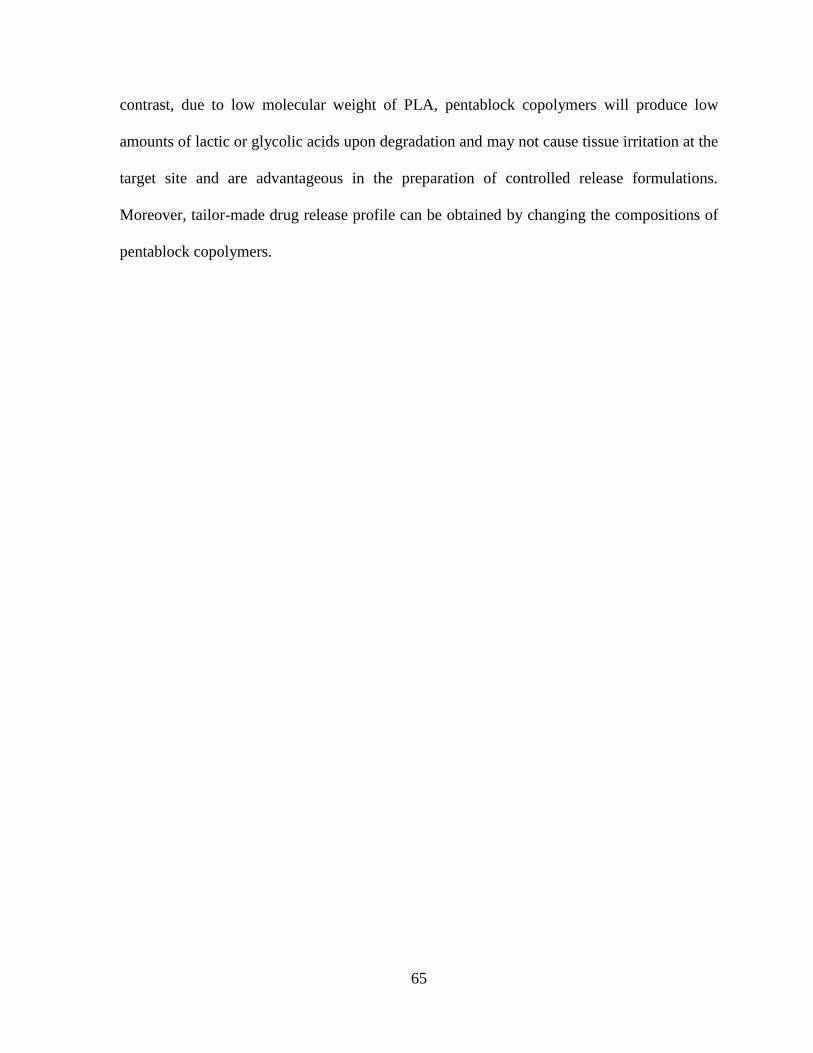
65
contrast, due to low molecular weight of PLA, pentablock copolymers will produce low
amounts of lactic or glycolic acids upon degradation and may not cause tissue irritation at the
target site and are advantageous in the preparation of controlled release formulations.
Moreover, tailor-made drug release profile can be obtained by changing the compositions of
pentablock copolymers.
66
Sn(Oct)2
130 oC
O
O
O
O
C OH2C C
O
O CH2CH2O C
O
CH2 O C5 5Y XX
HC
O
O HZ
HCOH
O
Z
Lactide
CH3 CH3
PLA-PCL-PEG-PCL-PLA
Y
O
O
Sn(Oct)2
130 oC
H OH2C C
O
O CH2CH2O C
O
CH2 O H5
5Y XX
PEG Caprolactone PCL-PEG-PCL
H OCH2CH2 OH
H OH2C C
O
O CH2CH2O C
O
CH2 O H5
5Y XX
PCL-PEG-PCL
Figure 3.1 Synthetic scheme of pentablock copolymer (PLA-PCL-PEG-PCL-PLA)
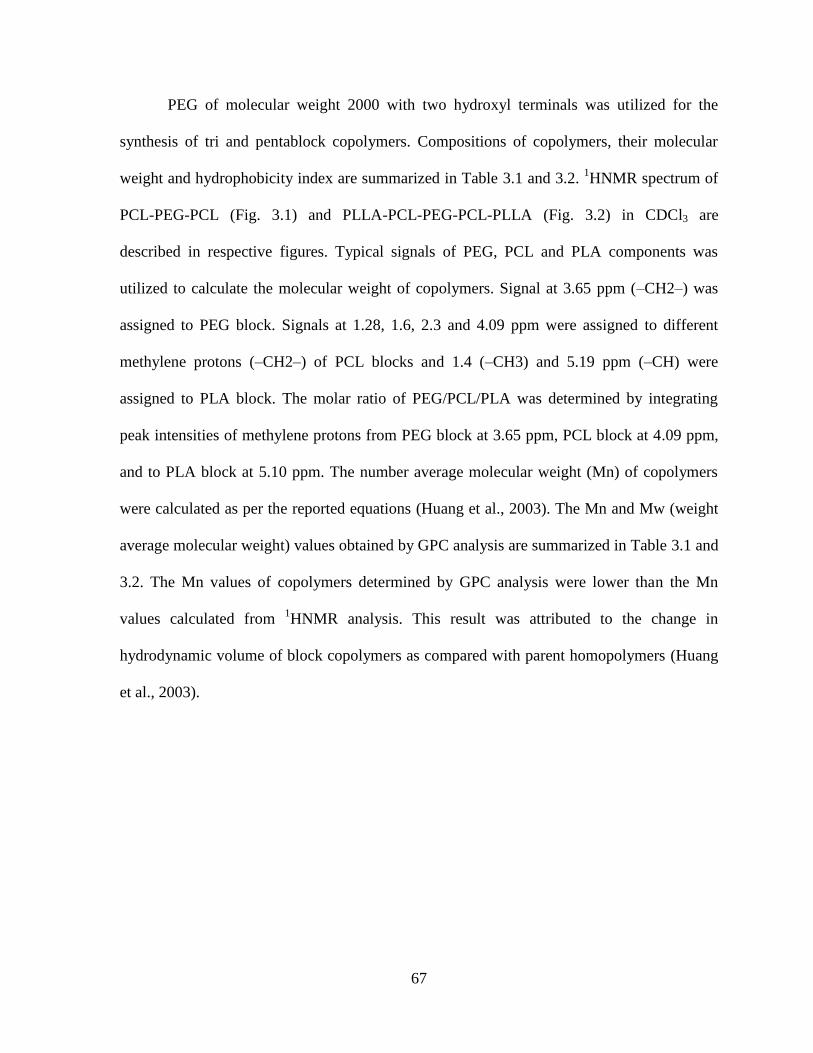
67
PEG of molecular weight 2000 with two hydroxyl terminals was utilized for the
synthesis of tri and pentablock copolymers. Compositions of copolymers, their molecular
weight and hydrophobicity index are summarized in Table 3.1 and 3.2. 1HNMR spectrum of
PCL-PEG-PCL (Fig. 3.1) and PLLA-PCL-PEG-PCL-PLLA (Fig. 3.2) in CDCl3 are
described in respective figures. Typical signals of PEG, PCL and PLA components was
utilized to calculate the molecular weight of copolymers. Signal at 3.65 ppm (–CH2–) was
assigned to PEG block. Signals at 1.28, 1.6, 2.3 and 4.09 ppm were assigned to different
methylene protons (–CH2–) of PCL blocks and 1.4 (–CH3) and 5.19 ppm (–CH) were
assigned to PLA block. The molar ratio of PEG/PCL/PLA was determined by integrating
peak intensities of methylene protons from PEG block at 3.65 ppm, PCL block at 4.09 ppm,
and to PLA block at 5.10 ppm. The number average molecular weight (Mn) of copolymers
were calculated as per the reported equations (Huang et al., 2003). The Mn and Mw (weight
average molecular weight) values obtained by GPC analysis are summarized in Table 3.1 and
3.2. The Mn values of copolymers determined by GPC analysis were lower than the Mn
values calculated from 1HNMR analysis. This result was attributed to the change in
hydrodynamic volume of block copolymers as compared with parent homopolymers (Huang
et al., 2003).

68
Table 3.1 Characterization of triblock polymers
Code Copolymers Mna Mn
b Mw
b PDI
b
A PCL5000-PEG2000-
PCL5000 12450 8975 11630 1.50
B PCL5000-PEG4000-
PCL5000 14099 9795 13115 1.33
C PCL5000-PEG8000-
PCL5000 18027 14652 18068 1.23
a. Values calculated from 1HNMR spectra
b. Values obtained by GPC analysis

69
Table 3.2 Characterization of pentablock copolymers
Code Copolymers PEG/PCL/PLA
ratio Mn
a Mn
b Mw
b PDI
b
P-1 PCL-PEG-PCL 1/2.5 7105 4083 6080 1.63
P-2 PCL-PEG-PCL 1/5 12450 8975 11630 1.50
P-3 PLLA-PCL-PEG-
PCL-PLLA 1/5/2.5 19072 10964 16260 1.48
P-4 PLLA-PCL-PEG-
PCL-PLLA 1/2.5/2.5 11062 6186 9526 1.54
P-5 PDLLA-PCL-
PEG-PCL-PDLLA 1/5/2.5 18803 11739 16246 1.38
P-6 PDLLA-PCL-
PEG-PCL-PDLLA 1/2.5/2.5 11665 6165 10160 1.64
a. Values calculated from 1HNMR spectra
b. Values obtained by GPC analysis

70
Figure 3.2 1H-NMR spectra of PCL-PEG-PCL copolymer in CDCl3

71
Figure 3.3 1H-NMR spectra of PLA-PCL-PEG-PCL-PLA copolymer in CDCl3

72
Figure 3.4 depicts FT-IR spectra of P-2, P-3 and P-5 block copolymers. On the spectrum of
PCL-PEG-PCL (P-2) (Fig. 3.4A), band for C=O stretching appeared at 1732 cm-1
and
bands for C-H stretching appeared at 2941 cm-1
and 2860 cm-1
for PCL block. Absorption
band at 1140 cm-1
appeared because of C-O-C stretching vibrations of the repeated
OCH2CH2 units of PEG and band at 1279 cm-1
was attributed to the -COO- stretching
vibrations (Li et al., 2009). On the P-3 (Fig. 3.4B) and P-5 (Fig. 3.4C) spectra another C=O
stretching band was observed at 1757 cm-1
, which can be attributed to PLA block.

73
Figure 3.4 FTIR spectra of copolymers (A) P-2 (B) P-3 (C) P-5
C
B
A
74
To improve the crystallinity and degradation of PCL based triblock copolymers, PLA block
was introduced in the copolymers. Earlier reports suggest that by changing the
hydrophobic/hydrophilic constituents, crystallinity and degradation of the copolymers can be
modulated (Frank et al., 2005; Gou et al., 2010; Huang et al., 2004; Li et al., 2005; Li et al.,
2002). We synthesized four compositions of pentablock copolymers with different ratios of
PCL/PLA in the hydrophobic segments. We also evaluated the effect of different isomeric
forms of PLA block on the crystallinity of copolymers. L-lactide is semicrystalline in nature
where as D, L-lactide has amorphous structure. Both polymers may act differently in
reducing the crystallinity of PCL block, which eventually may affect drug release profile.
The X-ray diffractograms obtained for different triblock and pentablock copolymer
compositions are shown in figure 4. For triblock copolymer (P-1) (Fig. 3.5A), diffraction
pattern exhibited two characteristic crystalline peaks of PCL block at 2θ = 21.5° and 23.8°.
L-lactide containing pentablock copolymer (P-4) Fig. 4a, showed less intense peaks for PCL
and two another crystalline peaks at 2θ = 16.5° and 19.05° for PLA block. These peaks
suggest that incorporation of L-lactide in the copolymers has reduced the crystallinity of PCL
and because of its semicrystalline nature PLA exhibited its own crystalline peaks. However,
P-6 represented a wide amorphous peak suggesting that D, L-lactide containing pentablock
copolymer may exist in amorphous state. This result suggested that D, L-lactide exhibited
significant effect in reducing crystallinity of PCL in comparison to L-lactide. However, there
is no significant effect on crystalline peak of PCL in P-3 and P-5 (Fig. 3.5B). The PCL/PLA
block ratio in the case of P-3 and P-5 was higher as compared to P-4 and P-6, respectively.
Therefore, effect of PLA in reducing the PCL crystallinity was not as intense as observed in

75
the case of P-4 and P-6. From the XRD results it can be concluded that a proper ratio of
PCL/PLA in the copolymer compositions can alter the crystallinity of PCL.

76
Figure 3.5A X-ray diffraction diagrams of polymers P-1, P-4, P-6
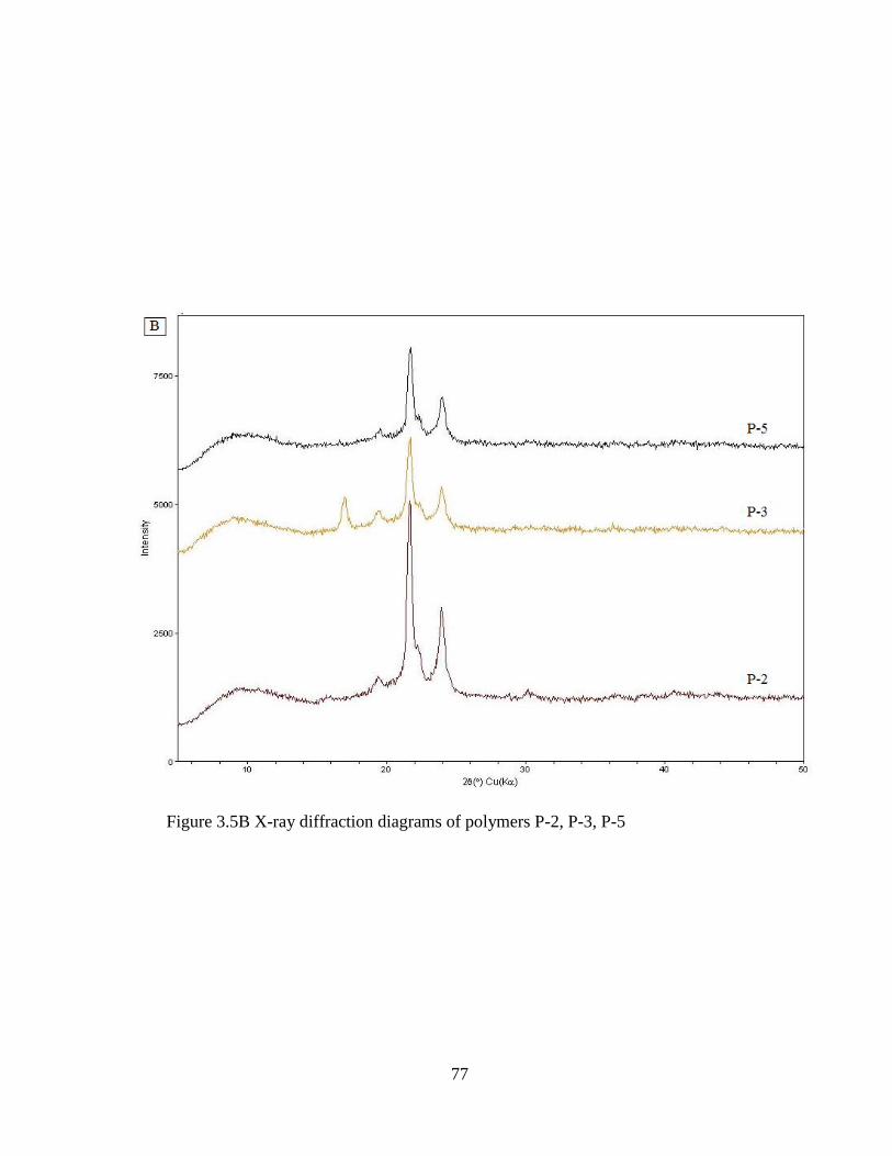
77
Figure 3.5B X-ray diffraction diagrams of polymers P-2, P-3, P-5

78
B
A
Figure 3.6 DSC thermograms of polymers (A) First heating (B) Second heating

79
Figure 3.6A shows DSC results of first heating scan and 4.5B shows the DSC scan of second
heating cycle of all prepared copolymers. The endotherms located in between 30 °C to 60 °C
correspond to the melting of PCL component in the copolymers. The melting points of PCL
in copolymers were clearly decreased with incorporation of PLLA or PDLLA in the polymer.
This finding suggests that PLA block has reduced the crystallization ability of PCL block.
Further, in first heating scan melting point of PDLLA containing copolymer is slightly lower
than PLLA containing copolymer. These were expected results since it is widely reported
that PDLLA contains random mixture of L-lactide and D-lactide and interrupts the
crystallization of PCL more significantly than PLLA. Also, second heating scan shows
similar decrease in melting points of pentablock copolymer in comparison to triblock
copolymers. It is interesting to observe that second heating cycle shows two melting peaks
for PCL. The second melting peaks were attributed to the imperfect crystallization of PCL
chains during cooling and second heating process. Our DSC results further confirmed that
crystallization ability of PCL in copolymer compositions was altered by incorporation of
PLLA or PDLLA. Hydrolytic degradation of polymers depends on the crystallinity.
Amorphous polymers degrade at a faster rate than the crystalline polymers. Earlier reports
suggest that higher the PLA content in PCL-PLA block copolymers or PCL/PLA blends,
lower the crystallinity of PCL (Newman et al., 2009). Also, degradation rate of block
copolymer was faster in comparison to PCL and PLA homopolymers (Haung et al., 2006).
However, no literature available that suggests the effect of block copolymer crystallinity on
drug release profile from nanoparticles. In this study, two feed ratios of PCL/PLA i.e. 1: 0.5
and 1:1 were selected for the synthesis of D, L-lactide and L-lactide containing pentablock
copolymers in which PLA content was more than 50 % in each prepared copolymer. We

80
attempted to improve the crystallinity of triblock copolymers by incorporating PLA segment.
In addition, we observed that effect of D, L-lactide on reducing the crystallinity of PCL was
more pronounced in comparison the L-lactide. A change in crystallinity of PCL has also
affected release rate of triamcinolone acetonide from nanoparticles which was discussed in
another section. The hydrophobicity index of copolymers also changed with hydrophobic
segment of copolymer which was determined by the ratio of molecular weight of
hydrophobic segment of copolymer over the total molecular weight of copolymer.
Preparation and characterization of nanoparticles
Particle size and zeta potential
Triblock and pentablock copolymers based nanoparticles were prepared by single o/w
emulsion solvent evaporation method. Table 2 summarizes the properties of each
nanoparticle sample. There was not much difference on size of nanoparticles prepared from
different copolymer compositions. Nanoparticles were in the range of 160- 280 nm and were
monodispersed. Figure 3.7 shows the particle size distribution. It appears entrapped drug may
not significantly alter the size of nanoparticles. Zeta potential of each prepared nanoparticles
sample was also listed in Table 2. It appears that nanoparticles have very high negative zeta
potential, indicating stable nature of the particles in water due to repulsion. However,
hydrophobic nanoparticles tend to aggregate upon storage due to hydrophobic attraction
(Soppimath et al., 2001).

81
Figure 3.7 Particle size distributions
82
Drug loading content and entrapment efficiency
Nanoparticles drug loading and entrapment efficiency depend mainly on copolymer
composition. It can be modulated by many factors, such as molecular weight, ratio of
hydrophobic to hydrophilic segment of copolymers, crystallinity, drug solubility and drug-
polymer interactions. Since triamcinolone acetonide has very low aqueous solubility, it can
precipitate in water during fabrication of nanoparticles that resulted in higher drug loading in
all nanoparticle batches. As shown in Table 3.4, molecular weight or hydrophobic segment of
the copolymer affected drug loading and percentage entrapment efficiency of nanoparticles.
Higher drug loading in P-3 and P-5 in comparison to P-4 and P-6, respectively could be
attributed to longer chain length of PCL segment that provides more hydrophobicity to the
polymers.
Surface morphology of nanoparticles
To investigate the morphology of prepared nanoparticles SEM analysis was
conducted. Fig. 6B shows SEM graph of drug loaded nanoparticles. It can be seen that
nanoparticles are spherical in shape and possess smooth surface morphology.

83
Table 3.3 Characterization of triblock nanoparticles
Polymer Size
(nm) PDI % EE Drug loading
Zeta
Potential
A 229 ± 2 0.294 74.4 ± 2.4 % 6.7 ± 1.2 % -26.4 ± 4.5
B 219.4 ± 8 0.297 66.9 ± 3.9 % 6.1 ± 0.8 % -24.5 ± 4.8
C 199.4 ± 5 0.284 60.6 ± 1.2 % 5.5 ± 0.3 % -22.9 ± 4.9
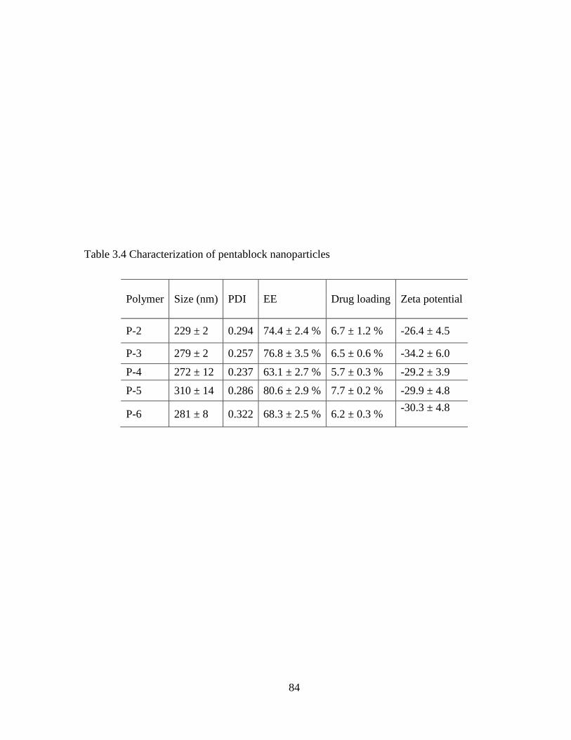
84
Table 3.4 Characterization of pentablock nanoparticles
Polymer Size (nm) PDI EE Drug loading
Zeta potential
P-2 229 ± 2 0.294 74.4 ± 2.4 % 6.7 ± 1.2 % -26.4 ± 4.5
P-3 279 ± 2 0.257 76.8 ± 3.5 % 6.5 ± 0.6 % -34.2 ± 6.0
P-4 272 ± 12 0.237 63.1 ± 2.7 % 5.7 ± 0.3 % -29.2 ± 3.9
P-5 310 ± 14 0.286 80.6 ± 2.9 % 7.7 ± 0.2 % -29.9 ± 4.8
P-6 281 ± 8 0.322 68.3 ± 2.5 % 6.2 ± 0.3 % -30.3 ± 4.8

85
Figure 3.8 SEM image of nanoparticles prepared from pentablock polymers
86
In vitro drug release study
Figures 3.10 and 3.11 show the comparison of triamcinolone acetonide release profile from
triblock (P-2) and pentablock based nanoparticles. Different pentablock copolymer
compositions with various block ratios of PCL/PLA were used to prepare nanoparticles. In
vitro studies revealed that drug release from nanoparticles depended on length of
hydrophobic segment, molecular weight and crystallinity of the copolymers. Also,
pentablock copolymer based nanoparticles exhibited very low initial burst release phase in
comparison to triblock nanoparticles. These results were in agreement from other reported
results and explained by the fact that pentablock copolymer based nanoparticles have very
low surface absorbed drug in comparison to triblock nanoparticles (Hu et al., 2003). Figure
3.10 suggests that in comparison to triblock copolymer (P-2), drug release rate from both
pentablock copolymers (P-3 and P-5) was slower. This observation may be explained by the
fact that pentablock copolymers (P-3 and P-5) were of higher molecular weight. Although all
three copolymers have same PCL block length P-3 and P-5 have additional PLA segment in
their composition, which made their structure more hydrophobic in comparison to triblock
copolymers. However, there is no significant difference in triamcinolone acetonide release
profile from nanoparticles prepared from P-3 and P-5 copolymers. P-3 contain L-lactide in its
structure whereas, P-5 contain D, L- lactide. In both P-3 and P-5 copolymer block length of
PCL polymer is higher in comparison to PLA block. Also, in copolymers P-3 and P-5
crystallinity of PCL block was not much affected with incorporation of PLA (as per our XRD
results). Moreover, triamcinolone acetonide is hydrophobic in nature which has high
tendency to remain with hydrophobic segment of polymer. Therefore, we can conclude that
PCL segment has predominant effect on drug release profile of nanoparticles prepared from

87
P-3 and P-4 copolymers. In addition, because of high hydrophobicity of PCL block release of
lipophilic drug such as triamcinolone acetonide was much slower in later phase. Lipophilic
drugs get trapped in the hydrophobic core of the nanoparticles and achieve no or limited
release from nanoparticles as time passes. We also observed no release phase from P-3 and
P-5 nanoparticles after 9 days. Moreover, due to crystalline nature of polymers nanoparticles
exhibited initial burst release phase with almost 37 cumulative % of drug released in 2 days.
However, our aim was to modulate the drug release kinetics from nanoparticles to achieve
near zero-order release and reduce the initial burst release phase. Drug release rate from
crystalline copolymers is higher than the amorphous matrix of copolymers (Miyajima et al.,
1997). Therefore, we tried to optimize the drug release rate by changing the molecular state
of the copolymers. In copolymers P-4 and P-6, we decreased the block length of PCL in
comparison to P-3 and P-5. Pentablock copolymers P-4, P-6 and triblock copolymer P-2 have
same hydrophobic/hydrophilic block ratio. However, P-4 and P-6 contains both PLA and
PCL segment in their structure in comparison to P-2 that contains only PCL. The overall
block ratio of PCL/PLA was changed in P-4 and P-6 as compared to P-3 and P-5,
respectively to reduce the crystallinity of copolymers. Our XRD results also suggested that
incorporation of L-lactide has reduced the crystallinity of PCL block in P-4, whereas in the
case of P-6, PCL was present in amorphous state due to conjugation of D, L-lactide.
Diffusion of drug from the pores of crystalline copolymers is much faster than the amorphous
structure of the polymers. Therefore, we observed slow release of triamcinolone acetonide
from P-4 and P-6 (fig. 3.11) in comparison to other copolymers. P-4 and P-6 copolymer
based nanoparticles exhibited continuous release of triamcinolone acetonide for 14 days
without any slow release phase because of low crystallinity and hydrophobicity of
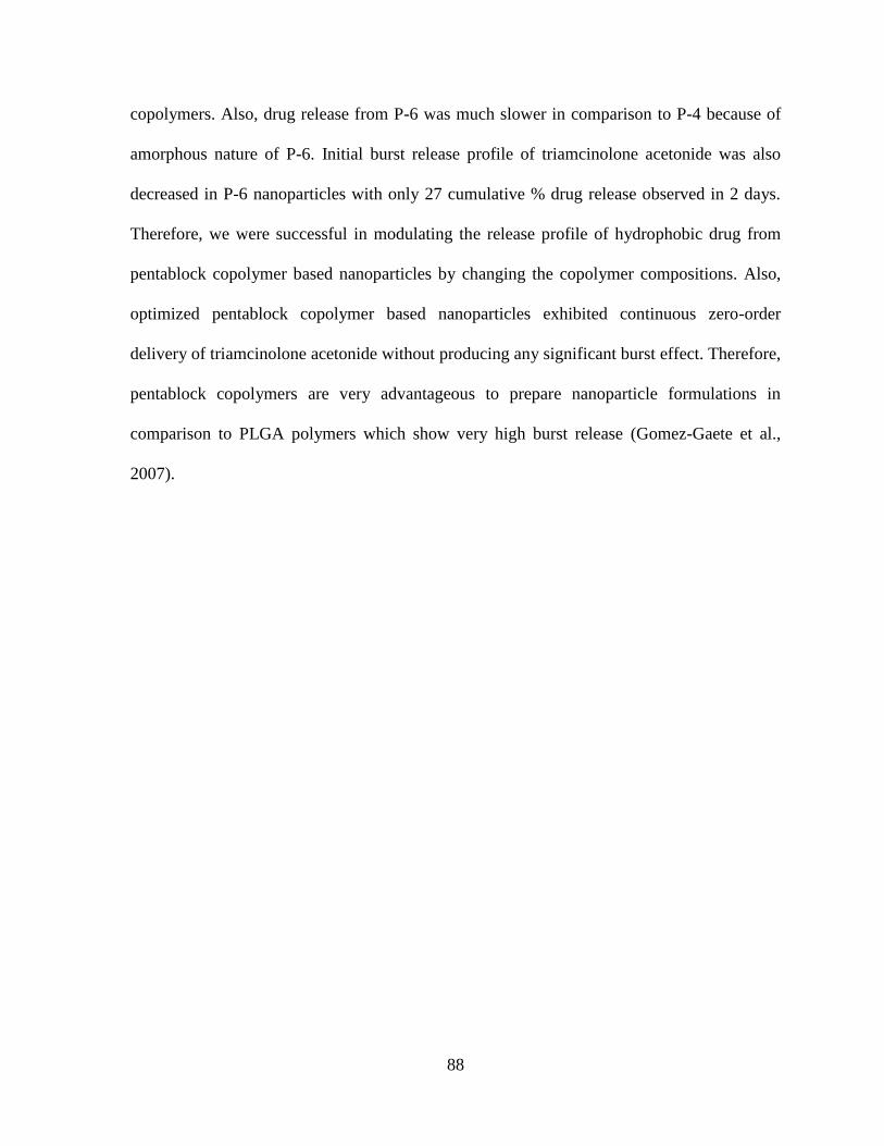
88
copolymers. Also, drug release from P-6 was much slower in comparison to P-4 because of
amorphous nature of P-6. Initial burst release profile of triamcinolone acetonide was also
decreased in P-6 nanoparticles with only 27 cumulative % drug release observed in 2 days.
Therefore, we were successful in modulating the release profile of hydrophobic drug from
pentablock copolymer based nanoparticles by changing the copolymer compositions. Also,
optimized pentablock copolymer based nanoparticles exhibited continuous zero-order
delivery of triamcinolone acetonide without producing any significant burst effect. Therefore,
pentablock copolymers are very advantageous to prepare nanoparticle formulations in
comparison to PLGA polymers which show very high burst release (Gomez-Gaete et al.,
2007).

89
Figure 3.9 Release of triamcinolone acetonide from A , B , and C
triblock copolymers nanoparticles in PBS buffer (pH 7.4) at 37 °C. The values are
represented as mean ± standard deviation of n=3

90
Figure 3.10 Release of triamcinolone acetonide from P-2 , P-3 and P-5
copolymers nanoparticles in PBS buffer (pH 7.4) at 37 °C. The values are represented as
mean ± standard deviation of n=3
91
Figure 3.11 Release of triamcinolone acetonide from P-2 , P-4 and P-6
copolymers nanoparticles in PBS buffer (pH 7.4) at 37 °C. The values are represented as
mean ± standard deviation of n=3

92
Figure 3.12 Release of triamcinolone acetonide from P-6 nanoparticles alone and P-6
nanoparticles suspended in gel copolymers nanoparticles in PBS buffer (pH 7.4) at 37
°C. The values are represented as mean ± standard deviation of n=3

93
Drug release kinetics
Drug release from nanoparticles generally follows diffusion/degradation or a combination of
diffusion and degradation mediated release phenomena (Wu and Chu, 2008). Copolymer
compositions have significant effect on the drug release profile from nanoparticles. Analysis
of drug release kinetics correlated well with Korsmeyer model. This observation suggested
that drug release depended primarily on diffusion from the nanoparticles matrix rather than
erosion process of the copolymer. In addition, analysis of first 60% release data according to
Korsmeyer model suggested that release rate of triamcinolone acetonide was slowest and
followed near zero-order kinetics from the nanoparticles prepared from P-6 pentablock
copolymer. A diffusion exponent 0.685<n<0.869 suggest anomalous diffusion mechanism
from all nanoparticles matrixes.
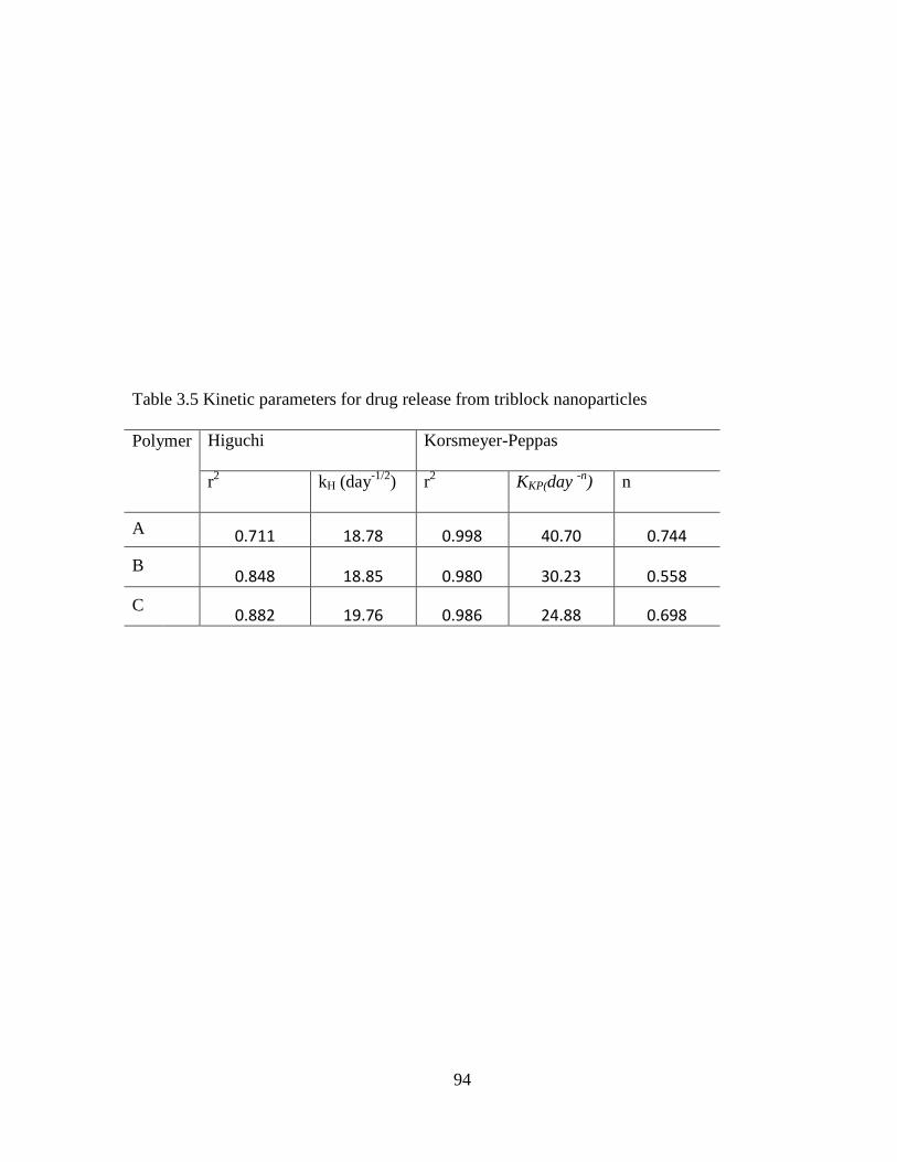
94
Table 3.5 Kinetic parameters for drug release from triblock nanoparticles
Polymer
Higuchi Korsmeyer-Peppas
r2 kH (day
-1/2)
r2 KKP(day
-n) n
A 0.711 18.78 0.998 40.70 0.744
B 0.848 18.85 0.980 30.23 0.558
C 0.882 19.76 0.986 24.88 0.698

95
Table 3.6 Kinetic parameters for drug release from pentablock nanoparticles
Polymer
Higuchi Korsmeyer-Peppas
r2 kH (day
-1/2)
r2 KKP(day
-n) n
P-2 0.711 18.79 0.998 40.70 0.744
P-3 0.891 17.43 0.983 23.57 0.583
P-4 0.974 15.71 0.920 15.97 0.706
P-5 0.913 17.05 0.989 19.27 0.634
P-6 0.996 14.86 0.952 8.22 0.847

96
Conclusions
PLA-PCL-PEG-PCL-PLA pentablock copolymer with different block ratios of
PEG/PCL/PLA was synthesized. Pentablock copolymer crystallinity can be modulated by
changing the PCL/PLA ratio. Triamcinolone acetonide nanoparticles were successfully
prepared from pentablock copolymers for ocular delivery. The copolymer compositions,
molecular weight and crystallinity can influence the drug release kinetics from nanoparticles.
Drug release rate from nanoparticles prepared from amorphous pentablock copolymers is
slower than crystalline copolymers. Novel pentablock copolymers are excellent biomaterials
that could serve as a vehicle for ophthalmic drug delivery as well as for other disorders where
sustained levels of corticosteroids are required.

97
CHAPTER 4
A NOVEL PENTABLOCK COPOLYMER BASED COMPOSITE DRUG DELIVERY
SYSTEM FOR TIMOLOL MALEATE DELIVERY
Rationale
Glaucoma is the second leading cause of blindness in the world, affecting
approximately 70 million patients aged 60 and over. It is an ocular disorder that
progressively causes degeneration of retinal ganglion cells causing damage to the optic nerve
head. Although there are many risk factors associated with the development of glaucoma
(age, race, myopia, family history, and injury), development of the disease is mainly
associated with elevated intra ocular pressure (IOP) caused by blockage of ocular fluid
outflow via trabecular meshwork pathway and/or uveoscleral pathway (Ghate and
Edelhauser, 2008). Progression of the disease leads to partial vision loss ultimately
progressing to complete blindness if remained untreated. Early diagnosis and effective
treatment play a vital role in halting the progression of this vision threatening condition.
Timolol maleate, a non-selective β-adrenergic blocker, is considered a gold standard
drug for the treatment of glaucoma based on its excellent pressure lowering efficacy (Kaur et
al.). It reduces the IOP by blocking the production of aqueous humor from ciliary bodies.
Timolol maleate eye drop is the primary mode of drug administration. Topical administration
is the most preferred and convenient route to treat anterior segment diseases. However,
ocular bioavailability of eye-drops is less than 1% and frequent administration is required to
maintain therapeutic levels. Poor bioavailability mainly results from precorneal factors such
as blinking, transient residence time in cul-de-sac and nasolacrimal drainage. Corneal
epithelium is composed of 6-7 layers of epithelial cells with tight junctions, which is highly
98
lipophilic in nature. It restricts the entry of hydrophilic molecules such as timolol maleate
(Ghate and Edelhauser, 2008). Frequent eye drop administrations play a major role in patient
non-compliance. Poor compliance with medications is an important reason for vision loss in
glaucoma patients. In addition, a majority of the topically applied timolol maleate eye drops
exhibit systemic absorption that leads to severe side effects such as cardiac arrhythmia,
congestive heart failure, bronchospasm and status asthmaticus (Kaur et al.). Therefore, there
is a need for a better delivery system which can avoid the frequent administration of eye
drops and reduce the systemic side effects of timolol maleate while maintaining sustained
drug levels over a longer time periods.
In recent years, nanoparticles have gained significant attention in ocular drug delivery
(Araujo et al., 2009; Cai et al., 2008; Diebold and Calonge). Biodegradable nanoparticles
with appropriate size and distribution that can provide adequate bioavailability are always
desirable for ocular drug delivery. However, previous reports suggest that subconjunctival
administration of nanoparticles do not maintain prolonged therapeutic concentration in ocular
tissues (Kompella et al., 2003). The major concern is that nanoparticles observe high burst
release in initial time points followed by slow release phase. Because of the smaller diameter
and large surface area, these exhibit rapid release of surface adsorbed drug followed by slow
release from the nanoparticle core (Kompella et al., 2003). Moreover, blood and lymphatic
circulations in the periocular region can washout a large fraction of drug loaded nanoparticles
from the subconjunctival space (Amrite et al., 2008). Drug loaded nanoparticles can be
suspended in themosensitive hydrogel to modify overall drug release rate. Thermosensitive
hydrogel can be utilized in the development of in situ depot forming injectable controlled
release formulations (Lee et al.; Packhaeuser and Kissel, 2007). Thermosensitive hydrogel

99
exhibits sol-gel transition at physiological temperature. Drug loaded nanoparticles can be
incorporated in aqueous polymer solution at room temperature, that gels to form depot
system in subconjunctival space. Thermosensitive gel can hold the drug loaded nanoparticles
at injection site that minimizes nanoparticle loss due to periocular circulation. Composite
drug delivery system (biodegradable nanoparticles suspended in injectable thermosensitive
hydrogel) will provide more sustained release over an extended period of time compared to
nanoparticles without thermosensitive gel. Drug release from the polymeric depot system
takes place in sustained manner by diffusion and degradation mechanism. Nanoparticles in
gel will also significantly reduce the initial burst release by proving additional layer for drug
diffusion. Therefore, it is very attractive to evaluate the novel pentablock polymer based
composite polymeric system for sustained delivery of timolol maleate in glaucoma therapy.
In the present work, we evaluated release of timolol from nanoparticles alone and
nanoparticles suspended in gel, which were prepared from novel biodegradable pentablock
copolymers. PLA-PCL-PEG-PCL-PLA (polylatide- polycaprolactone- polyethylene glycol-
polycaprolactone- polylatide) was utilized for preparation of nanoparticles and polyethylene
glycol- polycaprolactone- polylatide- polycaprolactone- polyethylene glycol (PEG-PCL-
PLA-PCL-PEG) was utilized for the preparation of thermosensitive hydrogel. It is interesting
to observe that both types of copolymers are composed of PEG, PCL and PLA; however they
are different in molecular weight and block arrangements. PLA block in the outer segment
makes the polymer more hydrophobic that can be utilized as nanoparticle biomaterial. Whereas
PEG block in the outer segment makes the polymer hydrophilic that can be solubilized in water
and can be utilized to prepare thermosensitive hydrogel. Drug release from nanoparticles
depended on the molecular weight of the copolymers. Burst release of timolol was significantly

100
reduced by suspending nanoparticles in thermosensitive gel. Polymeric blocks utilized for the
synthesis of pentablock copolymers are FDA approved. However, earlier investigations
suggest that biocompatibility also depends on the processing method and properties of final
polymeric biomaterial. Therefore, cytotoxicity of nanoparticles and thermosensitive hydrogel
was investigated utilizing rabbit primary corneal epithelial culture cells (rPCEC) and human
retinal pigmented epithelial cells (ARPE-19) to evaluate the suitability of these polymeric
systems for ocular drug delivery. These delivery systems were biocompatible in nature. The
composite formulation i.e. timolol loaded nanoparticles suspended in thermosensitive gel can
be injected in to the subconjunctival space to achieve long term delivery of timolol for more
than 1 month.
Method and materials
Materials
Poly (ethylene glycol) (PEG, Mw = 2000), methoxy poly(ethylene glycol) (mPEG,
Mw = 550), ε- caprolactone, stannous octoate, poly (vinyl alcohol) (PVA), were obtained
from sigma aldrich company (St. Louis, MO; USA). Hexamethylene diisocyanate and L-
lactide were purchased from Acros organics (Morris Plains, NJ; USA). Analytical grade
petroleum ether, dichloromethane (DCM) and Acetonitrile (ACN) were procured from Fisher
Scientific (Morris Plains, NJ; USA). Timolol maleate was purchased from sigma aldrich
company (St. Louis, MO; USA).
Methods
Synthesis of pentablock copolymers
PLA-PCL-PEG-PCL-PLA copolymer was synthesized for the preparation of
nanoparticles. Briefly, in the first step, PCL-PEG-PCL triblock copolymer was synthesized
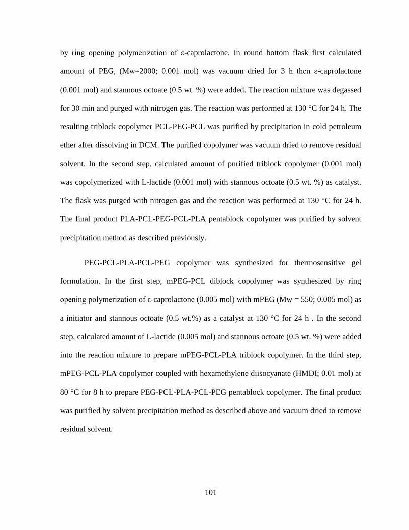
101
by ring opening polymerization of ε-caprolactone. In round bottom flask first calculated
amount of PEG, (Mw=2000; 0.001 mol) was vacuum dried for 3 h then ε-caprolactone
(0.001 mol) and stannous octoate (0.5 wt. %) were added. The reaction mixture was degassed
for 30 min and purged with nitrogen gas. The reaction was performed at 130 °C for 24 h. The
resulting triblock copolymer PCL-PEG-PCL was purified by precipitation in cold petroleum
ether after dissolving in DCM. The purified copolymer was vacuum dried to remove residual
solvent. In the second step, calculated amount of purified triblock copolymer (0.001 mol)
was copolymerized with L-lactide (0.001 mol) with stannous octoate (0.5 wt. %) as catalyst.
The flask was purged with nitrogen gas and the reaction was performed at 130 °C for 24 h.
The final product PLA-PCL-PEG-PCL-PLA pentablock copolymer was purified by solvent
precipitation method as described previously.
PEG-PCL-PLA-PCL-PEG copolymer was synthesized for thermosensitive gel
formulation. In the first step, mPEG-PCL diblock copolymer was synthesized by ring
opening polymerization of ε-caprolactone (0.005 mol) with mPEG (Mw = 550; 0.005 mol) as
a initiator and stannous octoate (0.5 wt.%) as a catalyst at 130 °C for 24 h . In the second
step, calculated amount of L-lactide (0.005 mol) and stannous octoate (0.5 wt. %) were added
into the reaction mixture to prepare mPEG-PCL-PLA triblock copolymer. In the third step,
mPEG-PCL-PLA copolymer coupled with hexamethylene diisocyanate (HMDI; 0.01 mol) at
80 °C for 8 h to prepare PEG-PCL-PLA-PCL-PEG pentablock copolymer. The final product
was purified by solvent precipitation method as described above and vacuum dried to remove
residual solvent.

102
Sn(Oct)2
130 oC
O
O
O
O
C OH2C C
O
O CH2CH2O C
O
CH2 O C5 5Y XX
HC
O
O HZ
HCOH
O
Z
Lactide
CH3 CH3
PLA-PCL-PEG-PCL-PLA
Y
O
O
Sn(Oct)2
130 oC
H OH2C C
O
O CH2CH2O C
O
CH2 O H5
5Y XX
PEG Caprolactone PCL-PEG-PCL
H OCH2CH2 OH
H OH2C C
O
O CH2CH2O C
O
CH2 O H5
5Y XX
PCL-PEG-PCL
Figure 4.1 Synthetic scheme of PLA-PCL- PEG -PCL-PLA

103
Figure 4.2 Synthetic scheme of PEG-PCL-PLA-PCL-PEG

104
Table 4.1 Characterization of copolymers
Code Copolymers Mna Mn
b Mw
b PDI
b
P-1
PLA2500-PCL5000-PEG2000-PCL5000-
PLA2500
19072 10964 16260 1.48
P-2
PLA2500-PCL2500-PEG2000-PCL2500-
PLA2500
11062 6186 9526 1.54
P-3
PEG550-PCL825-PLA550-PCL825-
PEG550
3300 3872 5260 1.35
a. Values calculated from 1HNMR spectra
b. Values obtained by GPC analysis

105
Characterization of pentablock copolymers
NMR
Both pentablock copolymers were characterized by 1H NMR spectroscopy. Each
copolymer (10 mg/mL) was dissolved in deuterated chloroform (CDCl3) and proton NMR
spectra were recorded with a Varian-400 NMR spectrophotometer.
Gel permeation chromatography (GPC)
GPC analysis was performed with the Shimadzu refractive index detector to
determine molecular weight and its distribution. Polymeric material (1.5 g) was dissolved in
1.5 ml tetrahydrofuran (THF). Polystyrenes standards of different molecular weight were
utilized for GPC analysis. THF was used as eluting solvent at a flow rate of 1 ml/min and
Styragel HR-3 column maintained at 35 °C was utilized for separation.
Fourier transforms infrared spectroscopy (FTIR)
Fourier transform infrared spectroscopy (FT-IR) spectra were recorded with a
Nicolet-100 infrared spectrophotometer at a resolution of 8 sec-1
. Each copolymer was
dissolved in DCM and polymer films were casted on KBr plates.
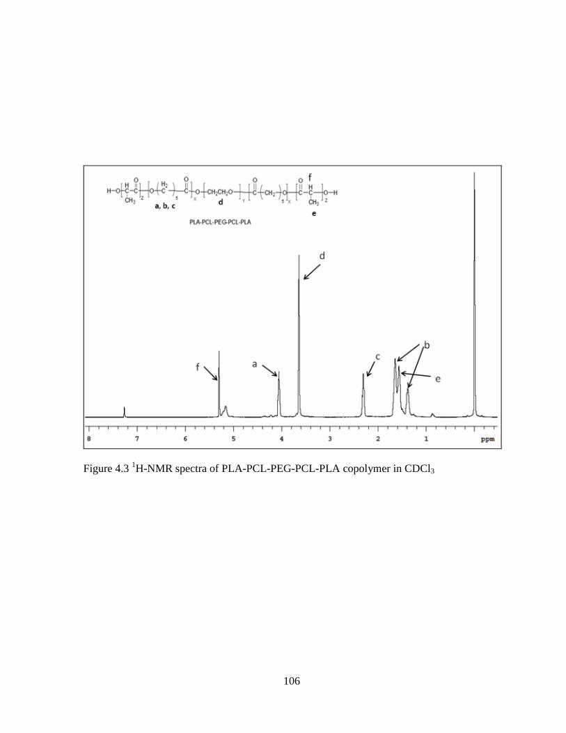
106
Figure 4.3 1H-NMR spectra of PLA-PCL-PEG-PCL-PLA copolymer in CDCl3
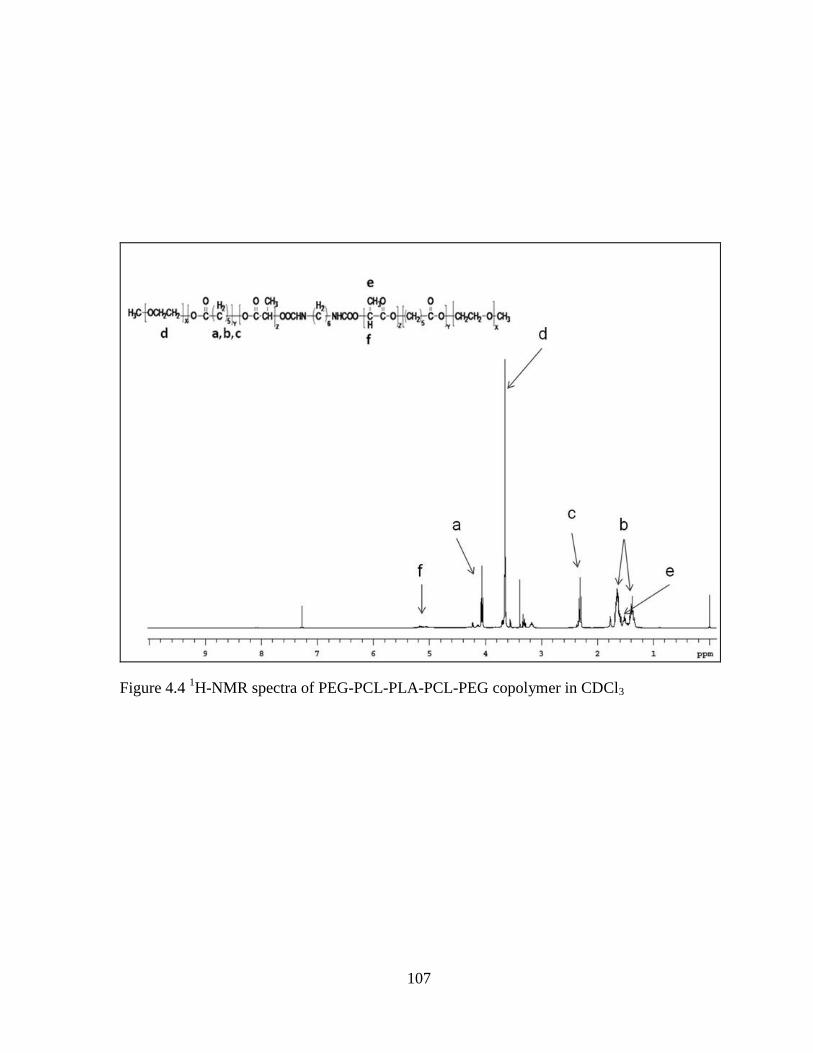
107
Figure 4.4 1H-NMR spectra of PEG-PCL-PLA-PCL-PEG copolymer in CDCl3
108
Figure 4.5 FT-IR spectra (A) PLA-PCL-PEG-PCL-PLA (B) PEG-PCL-PLA-PCL-PEG
A
B

109
Phase transition study of thermosensitive gel
The sol-gel transition behavior of aqueous co-polymeric solution was investigated by
test tube inverting method. The pentablock copolymer was dissolved in 10 mM PBS (pH 7.4)
at different concentrations (15- 40 wt. %) and then incubated for 12 h at 4 °C. Then 1 ml of
polymer solution was taken in 4 ml glass vial with an inner diameter of 12 mm and placed in
water bath. The water bath temperature was raised slowly from 20 to 60 °C at the rate of 5
°C increment per 5 min. The gel formation was characterized visually by inverting the tube.
A physical state of flow was characterized as sol phase, whereas a state of no flow was
characterized as gel phase (Mishra et al.).
Preparation of timolol loaded PLA-PCL-PEG-PCL-PLA nanoparticles
Timolol maleate is a highly water soluble salt form of the drug and incorporation of
salt in the oil phase is generally difficult (Li et al., 2007), which results in less entrapment
efficiency. Since salt form is not suitable for nanoparticle preparation, we converted timolol
maleate salt into the base form of timolol as per the earlier reported method (Li et al., 2007).
Timolol base is comparatively lipophilic in nature and mostly preferred for the preparation of
controlled release formulations (Bertram et al., 2009). Briefly, 83 mg timolol maleate was
dissolved in 6 ml of 0.77 M NaOH solution. A majority of salt was converted to the base
form when pH of the solution was adjusted above the pKa of timolol i.e. (pKa ~ 9.2).
Timolol base was an oily liquid phase which was allowed to be separated from aqueous
phase at room temperature. After phase separation, 5 ml of aqueous phase was removed and
500 μl of ethyl butyrate was added into the mixture. It was again allowed to phase separate
into timolol base rich ethyl butyrate phase and aqueous phase. The timolol base containing
ethyl butyrate phase was pipette out and

110
Figure 4.6 Sol-gel transition study of P-3

111
vacuum dried for 30 min. The timolol base was an oily liquid at room temperature, which
was utilized for the nanoparticle preparation (Li et al., 2007).
Timolol nanoparticles were prepared by o/w single emulsion solvent evaporation
method. One hundred mg polymer and 10 mg drug were dissolved in 2 ml DCM. The organic
phase was emulsified in aqueous phase containing 1 % PVA by tip sonication for 5 minutes
at 65W to obtain o/w emulsion. The organic phase was evaporated by continuous stirring at
room temperature to form nanoparticles. The resulting nanoparticle suspension was then
centrifuged at 21,000 rpm, 4 °C for 1 h. The prepared nanoparticles were washed three times
with distilled water to remove un-entrapped drug and excess PVA. Nanoparticle were
lyophilized with 5 wt.% mannitol (act as lyoprotectant) for 24 h and stored at 4 °C for further
studies.
Characterization of nanoparticles
Size and zeta potential determination of nanoparticles
Nanoparticles were analyzed for mean size and distribution by dynamic light
scattering (DLS) with a 90 Plus Particle Size Analyzer (Brookhaven Instruments
Corporation). The lyophilized nanoparticles were suspended in distilled water and analysis
was performed for 3 minutes at 25 °C and 90° scattering angle. All measurements were
made in triplicate and average particle size and size distribution data were reported as mean ±
SD.
Zeta potential of nanoparticles was determined by Nano-ZS Malvern Zeta sizer. All
analyses were performed at 25 °C. Values were reported as mean value ± SD of 3 test runs.

112
Figure 4.7 Particle size distribution for P-1

113
Drug loading and entrapment efficiency
Five mg of timolol loaded freeze dried nanoparticles were dissolved in 0.5 ml of
dimethyl sulfoxide (DMSO) and diluted approximately with deionized water. Total amount
of drug in solution was analyzed by high performance liquid chromatography (HPLC) at a
wavelength of 295 nm. The mobile phase containing acetonitrile / water/ triethylamine
(18:81:1, v/v/v) mixture was used as eluent solvent at the rate of 1 mL/min and C18 reverse
phase column was utilized for separation (El-Laithy, 2009). Drug loading and entrapment
efficiency of nanoparticles were determined by the equations:
Drug loading =
Entrapment Efficiency =

114
Table 4.2 Characterization of nanoparticles
Polymer Size PDI EE Drug loading Zeta potential
P-1 250.2 ± 2.6 0.211 48.62 ± 1.6 % 5.3 ± 1.6 % -32.4 ± 5.2
P-2 220.8 ± 3.7 0.218 37.77 ± 1.8 % 2.4 ± 0.8 % -30.5 ± 4.6

115
Figure 4.8 XRD analysis of nanoparticles loaded with timolol

116
In vitro release studies of timolol from nanoparticles alone, gel alone and nanoparticles
suspended in gel
Drug loaded nanoparticles (corresponding to 0.5 mg timolol) were suspended in 500
µl 10mM phosphate buffer saline (PBS) of pH 7.4 and this nanosuspension was placed in
dialysis bag with molecular weight cutoff of 6500 Da. (Sigma Aldrich, MO; USA). The
dialysis bag was immersed in a tube containing 10 ml of 10 mM PBS (pH 7.4) at 37 °C with
continuous shaking (60 rpm). At pre-determined time intervals samples were withdrawn and
the entire release medium was replaced with fresh buffer to maintain sink conditions. To
perform the release study of nanoparticles suspended in thermosensitive gel, 0.5 mg drug
equivalent nanoparticles were first suspended in 0.5 ml hydrogel solution by simple physical
mixing. Nanoparticle suspended gel was then placed in dialysis bag with mol. wt cutoff of
6500 Da. The rest of the procedure was followed as described above. The amount of timolol
released at each time point was determined by HPLC analysis as mentioned above. The
experiments were repeated three times and mean value ± SD was expressed as a cumulative
% drug released with time.
Drug release kinetics
Drug release parameters were computed by two different methods utilizing Higuchi
and Korsmeyer equations. Drug release mechanism was evaluated by model equations as
described below.
Higuchi equation:
Mt = KHt1/2
KH indicates the Higuchi release rate constant obtained by plotting cumulative percent drug
released against the square root of time.
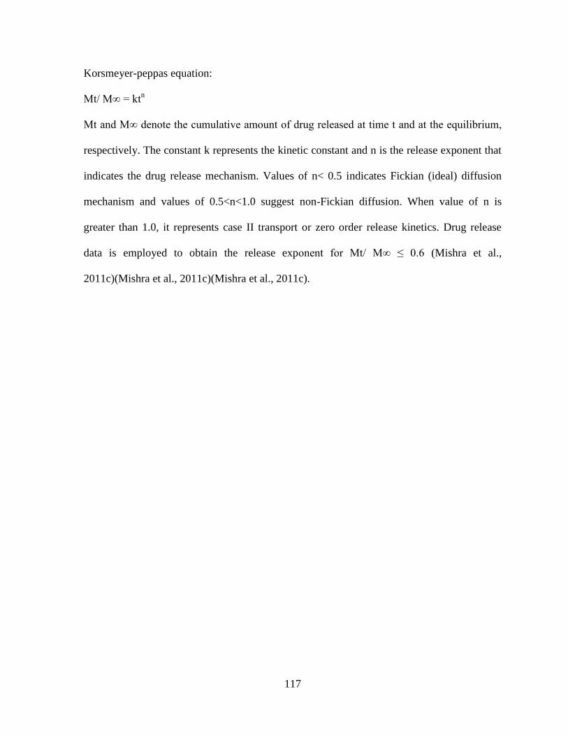
117
Korsmeyer-peppas equation:
Mt/ M∞ = ktn
Mt and M∞ denote the cumulative amount of drug released at time t and at the equilibrium,
respectively. The constant k represents the kinetic constant and n is the release exponent that
indicates the drug release mechanism. Values of n< 0.5 indicates Fickian (ideal) diffusion
mechanism and values of 0.5<n<1.0 suggest non-Fickian diffusion. When value of n is
greater than 1.0, it represents case II transport or zero order release kinetics. Drug release
data is employed to obtain the release exponent for Mt/ M∞ ≤ 0.6 (Mishra et al.,
2011c)(Mishra et al., 2011c)(Mishra et al., 2011c).
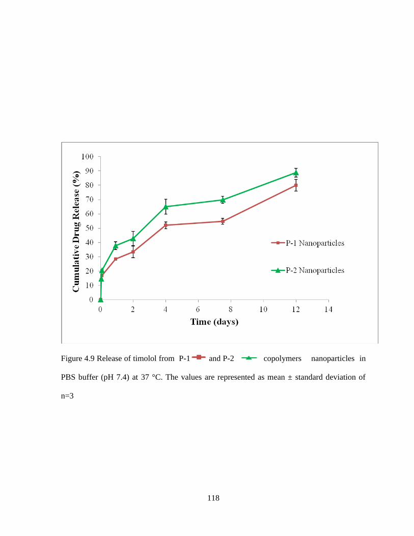
118
Figure 4.9 Release of timolol from P-1 and P-2 copolymers nanoparticles in
PBS buffer (pH 7.4) at 37 °C. The values are represented as mean ± standard deviation of
n=3

119
Figure 4.10 Release of timolol from P-1 Nanoparticles alone and P-1 nanoparticles in
gel copolymers nanoparticles in PBS buffer (pH 7.4) at 37 °C. The values are
represented as mean ± standard deviation of n=3

120
Table 4.3 Kinetic parameters for drug release
Polymer
Higuchi Korsmeyer-Peppas
r2 kH (day
-1/2)
r2 KKP(day
-n) n
P-1 0.964 20.18 0.973 31.54 0.263
P-2 0.964 23.60 0.983 39.03 0.294
P-3 0.961 14.17 0.983 1.18 1.3061

121
Cell line experiments
Rabbit primary corneal epithelial culture cells (rPCEC) and human retinal pigment
epithelial cells (ARPE-19) were cultured in Dulbecco's modified essential medium (DMEM,
Sigma Aldarich, USA) containing 10% fetal bovine serum, HEPES (4-(2-hydroxyethyl)-1-
piperazineethanesulfonic acid), 100 U/ml streptomycin-penicillin. The cells were cultivated
in humidified environment of 5 % CO2 at 37 °C and sub cultured after reaching 80 %
confluences.
Cytotoxicity experiments
MTS Assay
Cells were seeded in 96-well culture plates at a density of 10,000 cells per well. At 70
% confluency, 200 µl fresh medium containing nanoparticles or fresh medium containing
hydrogel of different concentrations ranging from 1 mg/mL to 20 mg/mL were added. Cells
were incubated for 48 h at the humidified atmosphere at 37° C and 5% CO2. Following
incubation, nanoparticles suspension was removed and wells were washed three times with
PBS then 20 μL of MTS (3-(4, 5-dimethylthiazol-2-yl)-5-(3-carboxymethoxyphenyl)-2-(4-
sulfophenyl)-2H-tetrazolium) solution and 80 μL of serum free medium were added into each
well. After 4 h incubation, absorbance of each well was measured at 450nm by ELISA plate
reader. Absorbance is directly proportional to viability of cells. In MTS assay, Trioton X
treated cells were served as negative control and untreated cells served as positive control.
Cell viability (%) was calculated by following equation.
Cell viability % =
Lactate Dehydrogenase (LDH) assay

122
To estimate the cytotoxicity of polymeric nanoparticles to the exposed cells lactate
Dehydrogenase (LDH) assay was performed. Cell growth conditions were kept same as the
MTS assay. Cells were seeded in 96-well culture plates at a density of 10,000 cells per well.
At 70 % confluency, 200 µl fresh medium containing nanoparticles or fresh medium
containing hydrogel of different concentrations ranging from 1 mg/mL to 20 mg/mL were
added. Cells were incubated for 48 h at the humidified atmosphere at 37° C and 5% CO2.
Damage of the cell membrane or cell death releases the lactate dehydrogenase (LDH) in the
supernatant. Concentration of LDH released in the supernatant of the rPCEC and ARPE-19
cells was measured by Cytotoxicity Detection Kit (Takara Bio Inc., Japan). Amount of the
LDH release is directly proportional to the number of damaged cells. Less than 10% LDH
release was considered as non toxic in our experiment and spectrophotometric determination
was carried out at 450 nm. LDH (%) release was calculated by following equation.
Cell viability % =

123
Figure 4.11 Cell viability studies (MTS assay) on P-1 and P-2 nanoparticles. The values are
represented as mean ± standard deviation of n=6.

124
Figure 4.12 Cell cytotoxicity studies (LDH assay) on P-1 and P-2 nanoparticles. The values
are represented as mean ± standard deviation of n=6.
125
Figure 4.13 Cell viability studies (MTS assay) on P-3 hydrogel. The values are represented as
mean ± standard deviation of n=6.

126
Results and discussion
Currently, eye drops are the primary method for reducing intraocular pressure in
glaucoma therapy. However, 80% of the topically applied drug passes through the
nasolacrimal canal and absorbs systemically. Only less than 1% of administered drug reaches
the aqueous humor. To control the intraocular pressure frequent high dose eye drops are
required which is the major reason for non-compliance in the glaucoma patients. It is
reported that to 80% of glaucoma patients do not take their medication as prescribed which
leads to the treatment failure (Olthoff et al., 2005). Poor compliance with medications is an
important reason for vision loss in glaucoma patients. Moreover, topical administration of
timolol maleate in the form of eye drops is reported to be extensively absorbed into the
systemic circulation, which can result in severe cardiopulmonary side effects (Schuman,
2000). Thus, a formulation that provides sustain levels of timolol maleate while reducing the
systemic side effects could increase the patient compliance in glaucoma therapy. Controlled
release formulation(s) of timolol maleate are reported to be a better alternative. In the current
study, we studied novel pentablock copolymers based controlled release formulations of
timolol maleate. PLA-PCL-PEG-PCL-PLA copolymer was utilized for nanoparticle
preparation and PEG-PCL-PLA-PCL-PEG copolymer was utilized for thermosensitive
hydrogel formulation. PCL and PEG based triblock copolymers (i.e. PCL-PEG-PCL or PEG-
PCL-PEG) are widely explored for drug delivery. In attempt to sustain the drug release
profile investigators generally increase the molecular weight of PCL block. However, high
molecular weight PCL block increases the overall crystallinity and hydrophobicity of the
polymer. Nanoparticles prepared from these copolymers generally exhibit high burst release,
exhibiting almost 60 % drug release during initial time points (Gou et al., 2009b; Jia et al.,
127
2008; Li et al., 2009). Moreover, drugs often get trapped in the hydrophobic core of the
nanoparticles and achieve no or limited release in the later time intervals (Gou et al., 2009a;
Jia et al., 2008). PCL exhibits very slow degradation because of its hydrophobic and
crystalline nature. Crystallinity and degradation profile of the PCL can be modulated by
conjugating with PLA (Chen et al., 2003). To modulate the crystallinity of PCL based
triblock copolymers PLA block was introduced in the pentablock copolymers.
Pentablock copolymers were synthesized by sequential ring opening polymerization
of ε-caprolactone and DL-lactide. In the first step triblock copolymers with different ratios of
PEG/PCL were synthesized by ring opening polymerization of ε-caprolactone in the presence
of PEG (Mw=2000) and a small amount of stannous octoate (Gou et al., 2009a). In the
second step these tribolck copolymers served as initiators for the synthesis of pentablock
(PLA-PCL-PEG-PCL-PLA) copolymers by ring opening polymerization of D, L-lactide (Fig.
1). For the synthesis of thermosensitive polymer mPEG of Mw= 550 was used as initiator.
Figure 4.1 and 4.2 represents the synthesis schemes for PLA-PCL-PEG-PCL-PLA (P-1) and
PEG-PCL-PLA-PCL-PEG (P-3) copolymers. Figure 4.3 and 4.4 shows the 1H NMR spectra
of both polymers. Typical signals of PEG, PCL and PLA components were utilized to
calculate the molecular weight of copolymers. Signal at 3.65 ppm (–CH2–) was assigned to
PEG block. Signals at 1.28, 1.6, 2.3 and 4.09 ppm were assigned to different methylene
protons (–CH2–) of PCL blocks and 1.4 (–CH3) and 5.19 ppm (–CH) were assigned to PLA
block. The molar ratio of PEG/PCL/PLA was determined by integrating peak intensities of
methylene protons from PEG block at 3.65 ppm, PCL block at 4.09 ppm, and to PLA block
at 5.10 ppm. The number average molecular weight (Mn) of copolymers were calculated as
per the previously reported equations (Huang et al., 2003). The Mn and Mw (weight average
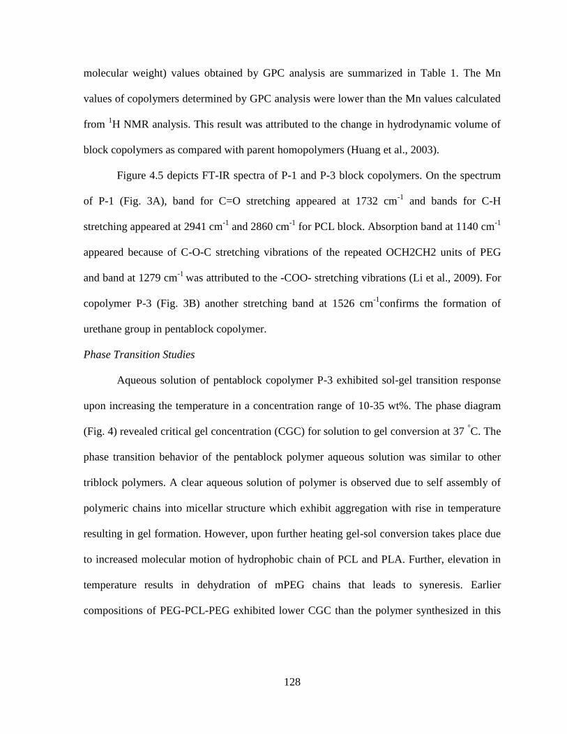
128
molecular weight) values obtained by GPC analysis are summarized in Table 1. The Mn
values of copolymers determined by GPC analysis were lower than the Mn values calculated
from 1H NMR analysis. This result was attributed to the change in hydrodynamic volume of
block copolymers as compared with parent homopolymers (Huang et al., 2003).
Figure 4.5 depicts FT-IR spectra of P-1 and P-3 block copolymers. On the spectrum
of P-1 (Fig. 3A), band for C=O stretching appeared at 1732 cm-1
and bands for C-H
stretching appeared at 2941 cm-1
and 2860 cm-1
for PCL block. Absorption band at 1140 cm-1
appeared because of C-O-C stretching vibrations of the repeated OCH2CH2 units of PEG
and band at 1279 cm-1
was attributed to the -COO- stretching vibrations (Li et al., 2009). For
copolymer P-3 (Fig. 3B) another stretching band at 1526 cm-1
confirms the formation of
urethane group in pentablock copolymer.
Phase Transition Studies
Aqueous solution of pentablock copolymer P-3 exhibited sol-gel transition response
upon increasing the temperature in a concentration range of 10-35 wt%. The phase diagram
(Fig. 4) revealed critical gel concentration (CGC) for solution to gel conversion at 37 °C. The
phase transition behavior of the pentablock polymer aqueous solution was similar to other
triblock polymers. A clear aqueous solution of polymer is observed due to self assembly of
polymeric chains into micellar structure which exhibit aggregation with rise in temperature
resulting in gel formation. However, upon further heating gel-sol conversion takes place due
to increased molecular motion of hydrophobic chain of PCL and PLA. Further, elevation in
temperature results in dehydration of mPEG chains that leads to syneresis. Earlier
compositions of PEG-PCL-PEG exhibited lower CGC than the polymer synthesized in this

129
study due to the fact that incorporation of PLA lowers hydrophobicity of central block
(Ghoroghchian et al., 2006; Hariharan et al., 2009).
Preparation and characterization of timolol loaded PLA-PCL-PEG-PCL-PLA copolymer
based nanoparticles
Table 2 summarizes the properties of nanoparticles prepared by utilizing two different
polymer compositions. Our study suggests that nanoparticles prepared from P-1 have larger
particle size in comparison to P-2. Nanoparticles size was increased with increase in the PCL
chain length in the polymer. All prepared nanoparticles have polydispersity in the range of
0.218-0.231. Figure 4.7 shows the particle size distribution. Entrapment efficiency of timolol
was also depended on the pentablock copolymer composition. In the case of P-1 entrapment
efficiency was around 48.62 ± 1.6 % whereas copolymer P-2 based nanoparticles have
entrapment efficiency around 37.77 ± 1.8 %. This difference in entrapment efficiency can be
attributed to the difference in molecular weight polymers. Therefore, polymer molecular
weight is a dominant factor that regulates the size and encapsulation efficiency of
nanoparticles. Zeta potential of prepared nanoparticles sample was also listed in Table 2. It
appears that nanoparticles have very high negative zeta potential, indicating stable nature of
the particles in water due to repulsion (Soppimath et al., 2001).
In vitro drug release study
Figure 4.9 represents the drug release data of timolol from two different
nanoparticles. It is interesting to notice that molecular weight of polymer does not have
significant effect on drug release rate from nanoparticles. Timolol is a small hydrophilic
molecule, which can easily diffuse out from the nanoparticles pores upon diffusion of water
molecules inside the nanoparticles matrix. However, suspension of nanoparticles in to the
130
thermosensitive hydrogel significantly restricts the diffusion of water molecules in to
polymeric matrix. In the case of P-1 nanoparticles alone, 88.0 ± 9.4% drug release was
observed in 12 days whereas in composite formulation 90.9 ± 1.9 % of drug release was
observed in 25 days (Fig. 4.10). Moreover, nanoparticles alone exhibited very high burst
release from nanoparticles due to surface adsorbed drug, whereas in the case of nanoparticles
suspended in thermosensitive gel burst effect was significantly minimized due the additional
diffusion layer of thermosensitive polymer. These results suggest that our strategy has
minimized the burst release phase and significantly prolonged the drug release duration.
Drug Release Kinetics
We have evaluated the drug release profile of composite formulations through
Higuchi, and Korsmeyer model. In the case of nanoparticles alone release was primarily
depended (Table 3) on diffusion mechanisms whereas in the case of composite formulations
diffusion exponent value is greater than 0.5 which suggest that release process was mediated
by diffusion and degradation mechanisms. We also observed that release rate constant was
significantly decreased in the case of composite formulations in comparison to nanoparticles
alone.
Cytotoxicity studies
Cell viability responses suggested that nanoparticles prepared from both P-1 and P-2
copolymers as well as thermogelling polymer were not cytotoxic to ARPE-19 and rPCEC
cells. Figs. 8 and 9 suggest that of even at higher polymer concentration (i.e., 10 mg/ml) cell
viability was maintained almost 95 % for both ocular cell-lines However, other investigators
reported that PCL-PEG-PCL triblock copolymers are cytotoxic to cancer cell-line at
concentration of 5 mg/ml. Also, as shown in Figure 4.11 and 4.13, MTS assay results
131
suggested that nanoparticles and thermosensitive gel were not cytotoxic or negligible
cytotoxic to both cell-lines at all Therefore, MTS and LDH assay suggested that PLA
containing copolymers based nanoparticles might be a safe vector for ocular drug delivery.
Conclusions
We have successfully prepared composite formulation of nanoparticles suspended in
the hydrogel matrix for timolol delivery. This formulation had successfully minimized the
burst release phase and provided sustained zero-order release kinetics of timolol. Such novel
technology will result in a prolonged duration of action of the drug and thereby eliminates
the need for repeated administrations. Composite formulation has potential for clinical
applications based on their excellent biocompatible nature.

132
CHAPTER 5
HYDROLYTIC AND ENZYMATIC DEGRADATION OF PENTABLOCK POLYMERS
Rationale
Biodegradable polyesters such as PGA, PLA and PCL and their copolymers have been
widely explored for the various therapeutic applications such as tissue engineering scaffolds,
sutures, osteosythetic devices and controlled release drug delivery systems. These polymers
are FDA approved for human use and are biocompatible and biodegradable in nature. The
degradation products are pharmacologically inactive and can be excreted by human body.
PLA upon degradation produces lactic acid and PCL produces 6-hydroxycaproic acid, which
are re-absorbable by the human body or removable by metabolism (Zhao et al., 2004).
Hydrolytic degradation of these homopolymers and copolymers is widely explored by
various researchers. However, most of the studies dealt with either bulk materials or polymer
films. We synthesized pentablock polymers for the preparation of nanoparticles and
thermosensitive hydrogel formulations. In-vitro degradation data generally do not match with
in-vivo degradation process. We selected pseudomonas lipase enzyme to evaluate the
enzymatic degradation study. Pseudomonas lipase is an esterase capable of cleaving ester
bonds on hydrophobic substrates.
Materials and methods
Materials
Poly (ethylene glycol) (PEG, Mw = 2000), methoxy poly (ethylene glycol) (mPEG, Mw =
550), ε- caprolactone, stannous octoate, poly (vinyl alcohol) (PVA), Pseudomonas lipase (40
IU/mg) were purchased from sigma aldrich company (St. Louis, MO; USA). Hexamethylene
diisocyanate and L-lactide were purchased from Acros organics (Morris Plains, NJ; USA).

133
Analytical grade petroleum ether and dichloromethane (DCM) were purchased from Fisher
Scientific (Morris Plains, NJ; USA).
Methods
Synthesis of pentablock copolymers
Synthesis methods for PLA-PCL-PEG-PCL-PLA and PEG-PCL-PLA-PCL-PEG were
described in the previous chapter. PLA-PCL-PEG-PCL-PLA of different molecular weight
was synthesized for nanoparticle preparation and PEG-PCL-PLA-PCL-PEG was synthesized
for thermosensitive gel preparation.
Preparation of nanoparticles and thermosensitive gel
We followed the similar methods for the preparation of nanoparticles and hydrogel as
described in the chapter 4.
Hydrolytic and enzymatic degradation studies of nanoparticles
Degradation studies were performed at 37 °C in 0.05 M phosphate buffer pH 7.4 with and
without 0.2 mg/mL pseudomonas lipase enzyme. Sodium azide (0.02 wt. %) was added in
the solution to prevent the growth of microorganisms. Nanoparticles prepared from different
polymers were suspended in 5 mL of PBS. The buffer solution was replaced at 24 h to
maintain the enzyme activity. At definite time intervals, the nanoparticles were collected and
washed thoroughly with distilled deionied water by ultracentrifugation. The washed
nanoparticles were freeze dried over night for further analysis (Kulkarni et al., 2008; Zeng et
al., 2004).

134
Characterization of nanoparticles
NMR
1H NMR spectroscopy was performed to characterize copolymer compositions.
Spectra were recorded with a Varian-400 NMR instrument by dissolving polymeric material
in deuterated chloroform (CDCl3).
Gel Permeation Chromatography (GPC) analysis
GPC analysis was performed with the Shimadzu refractive index detector to
determine molecular weight and its distribution. 1.5 mg polymeric material was dissolved in
1.5 ml Tetrahydrofuran (THF). Polystyrenes standards of different molecular weights were
utilized as reference. THF was used as eluting solvent at a flow rate of 1 ml/min and Styragel
HR-3 column maintained at 35 °C was utilized for separation.
XRD analysis of nanoparticles
X-ray diffraction (XRD) analysis was performed for tri block and pentablock
copolymers, by MiniFlex automated X-ray diffractometer (Rigaku, The Woodlands, Texas)
with Ni-filtered Cu-kα radiation (30 kV and 15 mA) at room temperature.
Results and discussion
Degradation of copolymers depends on many factors such as molecular weight,
composition and hydrophobicity. We found that both hydrolytic and enzymatic degradation
of pentablock copolymer depended on the composition of polymers rather than molecular
weight or hydrophobicity of the polymers. Reports suggest that surface area of the polymer is
also a main factor responsible for degradation of copolymers. For example, rate of
degradation for nanoparticles is much higher compare to polymeric films or intact polymers
(Singh et al., 2010). Therefore, we prepared nanoparticles of different pentablock polymers

135
Figure 5.1 XRD analysis of PCL2500-PEG2000-PCL2500 after hydrolytic degradation
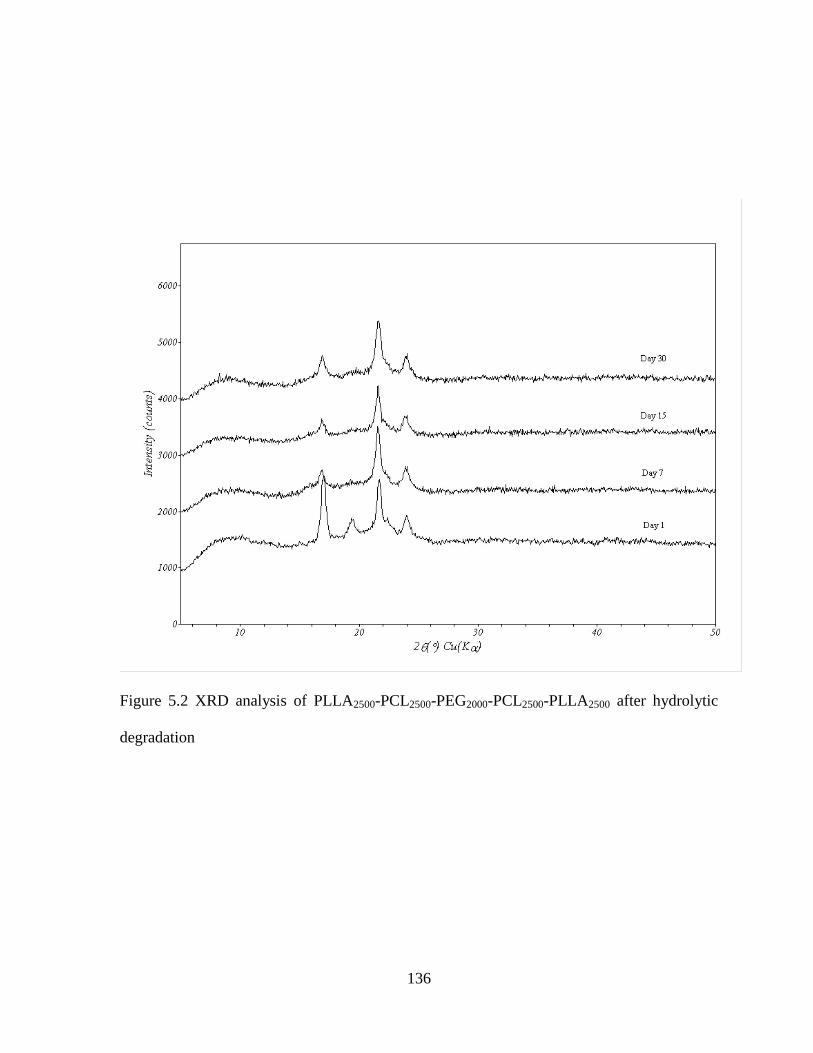
136
Figure 5.2 XRD analysis of PLLA2500-PCL2500-PEG2000-PCL2500-PLLA2500 after hydrolytic
degradation

137
Figure 5.3 XRD analysis of PDLLA2500-PCL2500-PEG2000-PCL2500-PDLLA2500 after
hydrolytic degradation
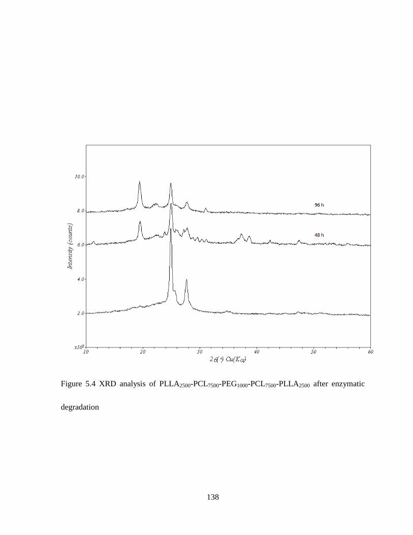
138
Figure 5.4 XRD analysis of PLLA2500-PCL7500-PEG1000-PCL7500-PLLA2500 after enzymatic
degradation

139
Figure 5.5 XRD analysis of PLLA2500-PCL2500-PEG2000-PCL2500-PLLA2500 after enzymatic
degradation
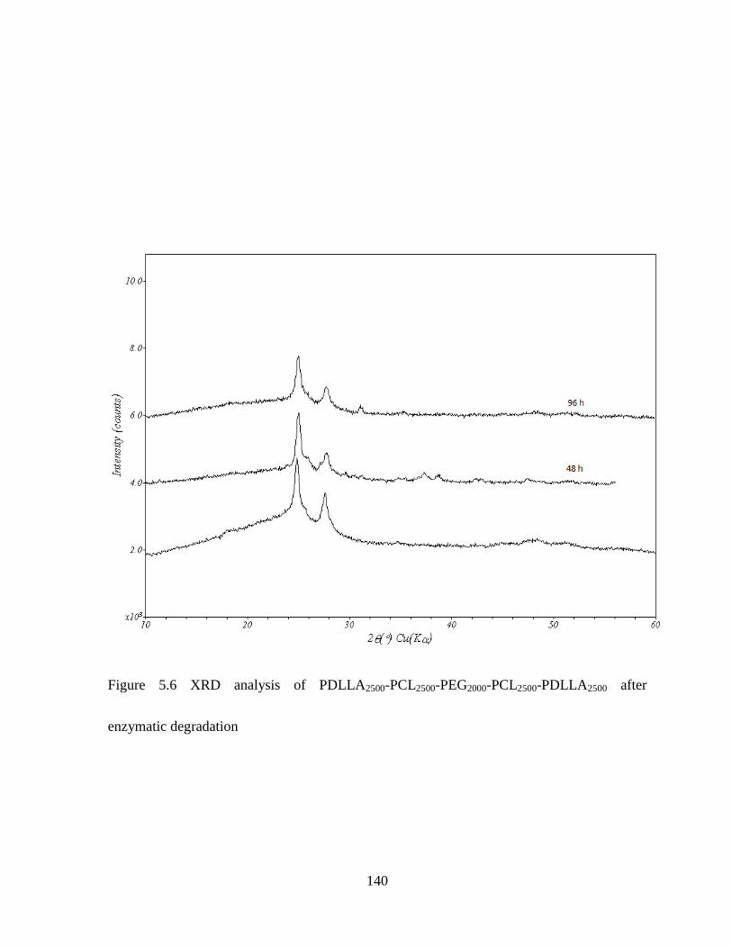
140
Figure 5.6 XRD analysis of PDLLA2500-PCL2500-PEG2000-PCL2500-PDLLA2500 after
enzymatic degradation

141
Figure 5.7 XRD analysis of PEG550-PCL825-PLA550-PCL825-PLA550 after enzymatic
degradation

142
Table 5.1 Degradation of PLA2500-PCL7500-PEG1000-PCL7500-PLA2500
Table 5.2 Degradation of PLLA2500-PCL2500-PEG2000-PCL2500-PLLA2500
Table 5.3 Degradation of PDLLA2500-PCL2500-PEG2000-PCL2500-PDLLA2500
Table 5.4 Degradation of PEG550-PCL825-PLA550-PCL825-PLA550
Time (h) Mnb Mw
b PDI
b
0 12932 19195 1.48
96 5385 11685 2.17
Time
(h) Mn
b Mw
b PDI
b
0 6186 9526 1.54
96 4112 5289 1.26
Time (h) Mnb Mw
b PDI
b
0 6165 10160 1.64
96 6177 9348 1.51
Time (h) Mnb Mw
b PDI
b
0 3872 5260 1.35
96 2763 3932 1.42

143
that are subjected to hydrolytic and enzymatic degradation. Moreover, we evaluated the
degradation of thermosensitive gel also. Our XRD results suggested that degradation of
pentablock copolymers first occurs in the amorphous region of the polymer followed by
crystalline region. Therefore, we observed the increase in polymer crystallinity with
degradation. Earlier reports by other investigators also suggest the similar pattern of
degradation (Zhao et al., 2007). Moreover, we observed that hydrolytic degradation of
polymers is much slower in comparison to enzymatic degradation. Due to high
hydrophobicity and crystallinity hydrolytic degradation of PCL is very slow (Liu et al.,
2000). That has decrease the overall degradation rate of copolymer. Change in crystallinity
was very prominent in the case of enzymatic degradation in comparison to hydrolytic
degradation. We evaluated the hydrolytic degradation of triblock and pentablock copolymers
having same molecular weight of PCL. We found that degradation of pentablock copolymer
was faster compared to triblock copolymers. These results support our hypothesis that in
pentablock copolymer incorporation of PLA block reduces the crystallinity of PCL block
thereby increase the degradation rate. Our previous XRD results suggest that the crystallinity
of pentablock copolymers is lower than the triblock copolymers. Enzyme is a macromolecule
and cannot enter the polymer matrix very easily. Therefore, enzymatic degradation of
nanoparticles or thermosensitive gel depends on how fast enzyme can enter the polymer
matrix (Zhao et al., 2007). Water uptake by the nanoparticles and hydrogel structure is
considered a major factor for enzymatic degradation process since water uptake could cause
swelling of the polymer and facilitate the enzymatic attack. As per release studies results
water uptake in PDLLA containing pentablock polymer is much slower in compared to
PLLA containing pentablock copolymers. Therefore, we observed the negligible change in

144
molecular weight and crystallinity of copolymers containing PDLLA. Moreover, we
observed more decrease in the molecular weight for our most hydrophobic pentablock
copolymer with PCL mw= 15000 compared to hydrophilic pentablock polymer with PCL
mw=5000. These results also can be explained by the fact that high molecular weight of PCL
provides higher crystallinity to copolymer which can lead to faster enzyme uptake through
the pore of the nanoparticles compared to less crystalline polymer. Therefore, we conclude
that crystallinity of polymer could affect the degradation of polymers. However, degradation
of copolymers is very complex process and many factors can affect the rate of degradation of
polymers. Reports suggest that although lipase is the PCL specific enzyme it can also
degrade the amorphous PLLA block. Therefore we observed the higher change in the
molecular weight of hydrophobic pentablock copolymer (Liu et al., 2000; Zhao et al., 2007).
In this scenario degradation of pentablock polymers with similar PCL content should be
similar. However, as described in table 5.2 and 5.3 there is a lot of difference in the
molecular weight change of two pentablock with similar molecular weight of PCL. These
results further support our hypothesis that the water uptake in the polymer uptake is the
possible factors that affect the degradation of copolymers. Pentablock copolymer having
PDLLA in the structure is amorphous in nature while PLLA containing copolymer has
crystalline structure. Diffusion of water across the crystalline matrix is much faster the
amorphous structure. This could be the possible reason for less change in the molecular
weight of polymer containing PDLLA.
Conclusions
Our results suggest that incorporation of PLA block not only improves the drug release rate
from nanoparticles but also increased the degradation rate of polymers. Hydrolytic

145
degradation of copolymer occurs at slower rate compared to enzymatic degradation.
Copolymer degradation depends on the composition of polymer. However, degradation of
copolymers depends on the many factors and advance polymer characterization techniques
are required to delineate the effect of each factor responsible for degradation of copolymers.

146
CHAPTER 6
PENTABLOCK COPOLYMER BASED THERMOSENSITIVE HYDROGEL FOR
MACRO MOLECULE DELIVERY
Rationale
Age-related macular degeneration (AMD) is a vision threatening ocular disease. The
most common cause of central vision loss in AMD is development of choroidal
neovascularization (CNV) characterized by fluid and blood leakage into the subretinal space
and fibrovascular scar formation (wet AMD) (Kulkarni and Kuppermann, 2005). The disease
affects the macular region of the retina, RPE, Bruch’s membrane and choriocapillaris. It may
cause the irreversible blindness in older age patients (Munoz et al., 2000). Vascular
endothelial growth factor (VEGF), a naturally occurring large lipoprotein molecule is
responsible for the growth of blood vessels (Leung et al., 1989). Elevated levels of VEGF
have been reported in many sight threatening eye diseases such as AMD and diabetic
retinopathy. It is a potent mitogen for endothelial cells. This growth factor increases vascular
permeability by leukocyte-mediated endothelial cell injury, formation of fenestration, and
dissolution of tight junctions. These manifestations cause intra-retinal fluid accumulation
which has a negative effect on visual acuity. Moreover, VEGF can also cause release of
inflammatory cytokines which can further reinforce the cycle of inflammation and
angiogenesis (Daughaday, 1992). Thus, anti-VEGF agents seem to be attractive therapeutic
molecules for the treatment of wet AMD. Anti-VEGF molecules of different sizes such as
Avastin (a 149 kDa antibody), Lucentis (a 48 kDa antibody, fab fragment) and Macugen (a
28-base ribonucleic acid aptamer covalently linked to two branched 50 kDa PEG moieties)
are being used for the treatment of different ocular conditions. Direct intra-vitreal injection of

147
antibodies appears found to be a more effective treatment of wet-AMD (McVey et al., 2008).
Monthly injection of Lucentis has become the standard care for wet AMD (El Sanharawi et
al., 2010). Since wet AMD is the chronic ocular disease therapeutic concentration of anti
VEGF proteins must be maintained for longer durations, which requires repeated intravitreal
injections. This thereby can be costly and inconvenient to patients. Moreover, various side
effects are associated with repeated intravitreal injections as summarized earlier. Delivery of
large molecules to the posterior segment of the eye has always been a challenge. Various
reports suggest that trasscleral route may be a viable option for delivery of large molecules to
the posterior segment (Ambati et al., 2000; Pescina et al., 2011). Sclera has large accessible
surface area and relatively high permeability that does not decrease with age. Different ex-
vivo studies reported the permeability of various macromolecules across the sclera (Ambati
et al., 2000). Human sclera is permeable to 70 kDa dextran (Olsen et al., 1995) whereas
rabbit sclera is permeable up to 150 kDa dextran and IgG (Ambati and Adamis, 2002;
Ambati et al., 2000). Literature suggests that molecular radius of the molecule is more
important factor than the molecular weight because reports mention that the globular proteins
has higher permeability through sclera than the liner protein (Ambati et al., 2000; Boubriak et
al., 2000; Olsen et al., 1995). Recently, in-situ depot forming drug delivery systems have
gained significant attention for sustained delivery of various therapeutic macromolecules.
This novel delivery system provides the ease of administration, delivery of accurate dose and
enhanced contact time with the target tissue. We have synthesized novel block copolymers
for the preparation of macromolecules loaded thermosensitive hydrogel formulation.
Thermosensitive hydrogels are environment-sensitive polymeric systems, in which sol-gel
transition is triggered by change in temperature. The ideal critical temperature for this

148
delivery system is in the range of physiological temperature where the source of heat to form
a gel is supplied by the body, which makes this system more flexible (Rathore, 2010). The
thermosensitive gels are liquid polymeric clear aqueous solution at room temperature. Large
molecules can be easily dispersed in liquid polymeric solution. Drug loaded polymeric
solution can be injected at subconjunctival space to form a depot system. A subconjunctival
injection can deliver up to 500μl of drug volume underneath the conjunctiva. Sustained
delivery via the subconjunctival route offers easier access and a safer administration route.
Though there are minor risks associated with subconjunctival injections, proper selection and
utilization of advanced techniques can minimize such risk (Ghate et al., 2007; Raghava et al.,
2004).
Pentablock copolymers (PEG-PCL-PLA-PCL-PEG) were synthesized as ingredients
of thermosensitive gel formulation. Each block of these pentablock copolymers is FDA
approved for human use. PLGA polymer is most commonly incorporated in the preparation
of controlled release formulation. Drug delivery systems prepared from this polymer
following degradation produce high molar mass of lactic and glycolic acids, which may
cause tissue irritation and toxicity by lowering pH (Kang and Schwendeman, 2002). PLGA
particles can cause rapid release (40-50% of payload) of the cargo within one to two days
(Budhian et al., 2005; Jwala et al., 2011). Pentablock copolymers are composed of relatively
low molecular weight of PLA block. Upon degradation of copolymer, due to low molecular
weight of PLA, the polymer will produce negligible amount of lactic acid. Therefore,
pentablock polymers are more suitable than the existing PLGA based gel for delivery of
macromolecules, particularly proteins and antibodies. We have synthesized two different
compositions of copolymers with different ratios of PCL/PLA blocks. The copolymers were

149
characterized for molecular weight and distribution with GPC and 1HNMR. The functional
group modification was confirmed by FT-IR. Crystallinity was characterized by XRD
analysis. Effect of polymer composition and concentration on drug release profile of
Lysozyme were also evaluated. Effect of size of the large molecule on release profile from
hydrogel also evaluated utilizing FITC-dextran of different molecular weight.
Materials and methods
Methoxy poly(ethylene glycol) (mPEG, Mw = 550), ε- caprolactone, stannous octoate,
Lysozyme from chicken egg white and Micrococcus luteus were purchased from Sigma
Aldrich (St. Louis, MO; USA). Hexamethylene diisocyanate and L-lactide were obtained
from Acros organics (Morris Plains, NJ; USA). Analytical grade petroleum ether,
dichloromethane (DCM) and Micro BCA protein assay kit were procured from Fisher
Scientific (Morris Plains, NJ; USA).
Synthesis of pentablock copolymers
Two compositions of PEG-PCL-PLA-PCL-PEG copolymer were synthesized for
thermosensitive gel formulation. In the first step, mPEG-PCL diblock copolymer was
synthesized by ring opening polymerization of ε-caprolactone (0.01 mol) using mPEG (Mw
= 550; 0.01mol) as a initiator and stannous octoate (0.5 wt.%) as a catalyst at 130 °C for 24 h
. In the second step, calculated amount of L-lactide (0.01 mol) and stannous octoate (0.5
wt.%) was added into the reaction mixture to prepare mPEG-PCL-PLA triblock copolymer.
In the third step, mPEG-PCL-PLA copolymer coupled with hexamethylene diisocyanate
(HMDI; 0.01 mol) at 80 °C for 8 h to prepare PEG-PCL-PLA-PCL-PEG pentablock
copolymer. The final product was purified by precipitation in cold petroleum ether after
dissolving in DCM. The purified copolymer was vacuum dried to remove residual solvent

150
Characterization of pentablock copolymers
NMR
Pentablock copolymers were characterized by 1H NMR spectroscopy. Each
copolymer (10 mg/mL) was dissolved in deuterated chloroform (CDCl3) and proton NMR
spectra were recorded with a Varian-400 NMR spectrophotometer.
Gel permeation chromatography (GPC)
GPC analysis was performed with the Shimadzu refractive index detector to
determine molecular weight and its distribution. The polymeric material (1.5 mg) was
dissolved in 1.5 ml tetrahydrofuran (THF). Polystyrenes standards of different molecular
weight were employed for GPC analysis. THF as eluting solvent was pumped at a flow rate
of 1 ml/min through Styragel HR-3 column maintained at 35 °C for separation.
Fourier transforms infrared spectroscopy (FTIR)
Fourier transform infrared spectroscopy (FT-IR) spectra were recorded with a
Nicolet-100 infrared spectrophotometer at a resolution of 8 sec-1
. Each copolymer was
approximately dissolved in DCM and polymer films were casted on KBr plates.
Phase transition study of thermosensitive gel
The sol-gel transition behavior of aqueous co-polymeric solution was investigated by
test tube inversion method. The pentablock copolymer was dissolved in 10 mM PBS (pH 7.4)
at different concentrations (15- 40 wt. %) and then incubated for 12 h at 4 °C. One ml of
polymer solution was taken in 4 ml glass vial with an inner diameter of 12 mm and placed in
water bath. The water bath temperature was raised from 20 to 60 °C at the rate of 5 °C
increment over 5 min. The gel formation was observed visually by inverting the tube. A

151
physical state of flow was characterized as sol phase, whereas a state of no flow was
considered as gel phase (Mishra et al., 2010).
Rheological Measurement
Rheology of polymer solution was conducted in Oscillatory HR-3 rheometer, TA
instruments with cone and plate geometry. The angle of the cone was 2˚ with a diameter of
60mm. Two mL sample was placed on the temperature controlled peltier plate of rheometer
at 5˚C and further analysis was conducted at a angular frequency of 1 rad. All the rheological
studies were conducted within the linear viscoelastic transition range of the polymer. Sol- gel
phase transition was performed at temperatures ranging from 10-42 °C with 1 °C intervals.
The gelation time was studied by time sweep measurements at 37 °C and the angular
frequency was kept at 1 rad/s.
Preparation of in situ gel forming formulations for lysozyme
Pentablock copolymers were dissolved in PBS solution at a concentration of 20 wt%.
Calculated amount of lysozyme was suspended into the polymeric solution and the vial was
vortexed until a homogeneous solution was formed. A 500 µL volume of lysozyme loaded
polymeric solution was injected into the glass tube maintained at 37 °C water bath. After 10
min the polymeric solution turned into a gel.
In vitro release of lysozyme
Five mL PBS solution (0.01 M, pH 7.4, 0.02% Sodium azide) was added onto the top
of the gel inside a tube. The tube was incubated in the shaker bath at 30 rpm and 37 °C. To
maintain the sink condition samples were withdrawn at predetermined time interval and
replaced with fresh PBS solution. The amount of lysozyme released at each time points was
calculated by Micro-BCA assay. Briefly, 150 µL of diluted sample was mixed with 150 µL

152
of Micro-BCA working reagent (Micro-BCA reagent A, B, C at the ratio of 50:48:2) and
poured into the 96-well microplate reader. The solution mixture was incubated at 37 °C for 2
h and allowed to cooled down to 25 °C. The micro plate reader analysis was performed at
595 nm. To prepare the standard curve freshly diluted lysozyme solution was prepared at
different concentration range of 25-200 µg/mL. The effect of polymer composition on the
release profile of lysozyme was evaluated by plotting the cumulative % lysozyme released
with time. To determine the effect of polymer concentration on the release profile of
lysozyme, two different concentrations of polymer (i.e. 20 wt. % and 25 wt. %) were
selected. The rest of the conditions were kept similar to previous condition.
In vitro release of dextran
FITC-dextran of different molecular weights (i.e. 9200, 38000 and 72000) were selected to
determine the effect of molecule size on the release profile from thermosensitive hydrogel.
PB-2 polymer was selected for this study. Pentablock copolymer was dissolved in PBS
solution at the concentration of 20 wt%. Calculated amount of FITC-dextran of different size
were suspended into the polymeric solutions and vial were vertexed until the homogeneous
solution was formed. A 500 µL of polymeric solution was injected into the glass tube. The
tube was put into the 37 °C water bath. After 10 min the polymeric solution became the gel.
A 5 mL of PBS solution (0.01 M, pH 7.4, 0.02% Sodium azide) was added on the top of the
gel in the tube. The tube was incubated in the shaker bath at 30 rpm and 37 °C. To maintain
the sink condition the samples were withdrawn at predetermined time interval and replaced
with fresh PBS solution. The amount of FITC-dextran released at each time points was
calculated using plate reader with excitation wavelength of 485 nm and emission wavelength
of 535 nm.
153
Biological activity of lysozyme by enzyme activity assay
To determine enzymatic activity stock solution of Micrococcus luteus (0.01%, w/v)
was prepared in phosphate buffer (66 mM, pH 6.15) and diluted to obtain absorbance in
between 0.2 to 0.6 at 450 nm. A 2.5 ml of M. luteus solution was added to a 0.1 ml of
approximately diluted sample of lysozyme. The reaction mixture was then immediately
transferred to a spectrophotometer cell having path length of 1 cm and absorbance was
determined at 450 nm with UV spectrophotometer. A rate of decrease of absorbance at 450
nm was monitored over a period of 5 min at 25 °C. The slope of the linear portion of the plot
(obtained by plotting absorbance vs time) determined the amount of lysozyme in one enzyme
unit (EU). The units of active lysozyme were calculated as per the previously reported
formula (Tang and Singh, 2009).
Where, df is the dilution factor, 0.001 was obtained from the definition of lysozyme unit, and
0.1 is the volume (in ml) of sample or standard. The biological activity of lysozyme from
released sample was compared with the control lysozyme sample in phosphate buffer to
determine the effect of pentablock polymer based hydrogel formulation on lysozyme activity.
Secondary structure stability study
Structural stability of lysozyme was determined by Circular dichroism (CD)
measurement. CD analysis was performed using Jasco 720 spectropolarimeter at 25 °C. CD
spectra were recorded over a range of 260 to 200 nm using 1 cm cell, band width of 1 nm and
a scanning speed of 100 nm/min. A spectrum for the blank PBS solution was obtained at the

154
same wavelength range as the blank subtraction. CD measurements were reported as molar
ellipticity [θ]
Statistical analysis
All release studies were performed in triplicate. The results were reported as mean ±
standard deviation. Effects of copolymer compositions on lysozyme release profile from
thermosensitive hydrogel were compared by student’s t-test.
Results and discussion
Pentablock copolymers were synthesized by sequential ring opening polymerization
of ε-caprolactone and L-lactide. In the first step mPEG of Mw= 550 was copolymerized with
caprolactone. Figure 6.1 represents the synthetic scheme for PEG-PCL-PLA-PCL-PEG
copolymer. Figure 6.2 illustrates the 1H NMR spectra of both polymers. Typical signals of
PEG, PCL and PLA components were utilized to calculate the molecular weight of
copolymers. Signal at 3.65 ppm (–CH2–) was assigned to PEG block. Signals at 1.28, 1.6,
2.3 and 4.09 ppm were assigned to different methylene protons (–CH2–) of PCL blocks and
1.4 (–CH3) and 5.19 ppm (–CH) were assigned to PLA block. The molar ratio of
PEG/PCL/PLA was determined by integrating peak intensities of methylene protons from
PEG block at 3.65 ppm, PCL block at 4.09 ppm, and to PLA block at 5.10 ppm. The number
average molecular weight (Mn) of copolymers were calculated according to a previous report
(Huang et al., 2003). The Mn and Mw (weight average molecular weight) values obtained by
GPC analysis are summarized in Table 6.1. Mn values of copolymers determined by GPC
analysis are lower than Mn values calculated from 1H NMR analysis. This result was
attributed to the change in hydrodynamic volume of block copolymers relative to parent
homopolymers (Huang et al., 2003).

155
Figure 6.3 depicts FT-IR spectra of pentablock copolymer. On the spectrum of P-1,
band for C=O stretching at 1732 cm-1
and bands for C-H stretching at 2941 cm-1
and 2860
cm-1
for PCL block appeared. Absorption band at 1140 cm-1
signifies C-O-C stretching
vibrations of OCH2CH2 repeat units of PEG and band at 1279 cm-1
can be attributed to -
COO- stretching vibrations (Li et al., 2009). Another band for N-H stretching at 1526 cm-1
confirms the formation of urethane group in pentablock copolymer.
P-1 is more viscous than the other polymer P-2. The storage modulus (G’) values for
P-1 and P-2 are higher than the loss modulus (G’’) values, indicating the formation of more
elastic (solid) gels.

156
Figure 6.1 Synthetic scheme of PEG-PCL-PLA-PCL-PEG

157
Figure 6.2 1HNMR spectra of P-2

158
Figure 6.3 FTIR spectrum of P-1

159
Table 6.1 Molecular weight characterization of polymers
Code Copolymers Mna Mn
b Mw
b PDI
b
P-1 PEG550-PCL825-PLA550-PCL825-PEG550 2893 2810 4211 1.49
P-2 PEG550-PCL550-PLA1100-PCL550-PEG550 2705 2625 3763 1.43
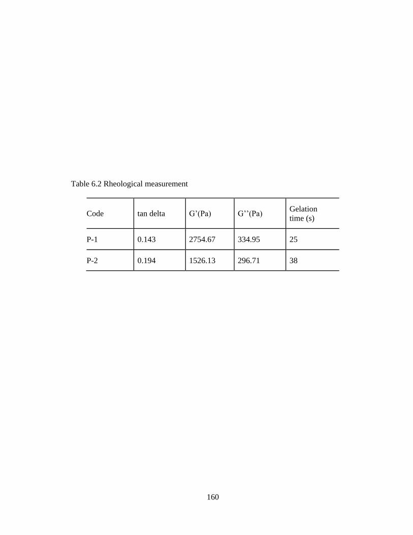
160
Table 6.2 Rheological measurement
Code tan delta G’(Pa) G’’(Pa) Gelation
time (s)
P-1 0.143 2754.67 334.95 25
P-2 0.194 1526.13 296.71 38
161
Phase Transition Studies
Aqueous solution of pentablock copolymer P-1 and P-2 exhibited sol-gel transition
with rising temperature within a concentration range of 10-35 wt %. The phase diagram (Fig.
6.4) revealed that critical gel concentration (CGC) for P-1 is 18 wt %, whereas for P-2 it is 20
wt %. This change in the CGC may be attributed to the higher hydrophobicity of the
polymer. Below the gelling temperature a clear aqueous solution of polymer is observed due
to self assembly of polymeric chains into micellar structure. Polymeric micelles exhibit
aggregation with rise in temperature resulting in gel formation. However, upon further
heating at elevated temperature causes dehydration of mPEG chains leading to syneresis.
Results of sol-gel transition process is consistent with our previously reported data for
triblock polymers (Mishra et al., 2011c).

162
Figure 6.4 Phase diagram of thermosensitive hydrogel formulations of P-1 and P-2
163
In vitro release of lysozyme from thermosensitive hydrogel
Release of lysozyme from the hydrogel formulation appears to depend on the
copolymer composition as shown in Figure 6.5. Rate of lysozyme release is slower from P-1
copolymer formulation than P-2. In the first 24 h cumulative percentage release from P-1 was
20.5 ± 2.6 % whereas in P-2 it was 25.5 ± 1.5 %. This result can be attributed to the fact that
P-1 is more hydrophobic in nature compared to P-2 since it possess relatively longer PCL
chains that restrict entry of water molecule in the hydrogel matrix. Therefore, diffusion of
lysozyme molecules across the hydrogel matrix is slower in P-1 than P-2. These results
suggest that release rate of lysozyme from the hydrogel formulation may be affected by
hydrophilic/hydrophobic balance of the copolymer. Figure 6.6 suggests that copolymer
concentration has significant effect on the release profile of lysozyme from P-2. Release of
lysozyme from 20 wt. % is more rapid than 25 wt. % formulation due to volume effect of the
gel matrix. Since, 25 wt. % gel forms dense matrix compared to 20 wt. % it restricts the
diffusion of lysozyme molecules.
As shown in figure 6.7 release of FITC-dextran from thermosensitive gel P-2 was depended
on the size of the molecule. Molecular chains of large molecules get trapped in the hydrogel
matrix that provide hinder for the diffusion of molecules from the hydrogel and result delay
in release. Therefore, we observed the release of FITC-dextran the following order
(Mw=70000, size=58 °A) < (Mw=40000, size=44.5 °A) < (Mw=10000, size=23.6 °A). From
our studies we can conclude that release of large molecules from the thermosensitive gel is
mainly depended on the size of the molecules. These data are in consistent with literature and
with our previous study with the small molecules, where we did not found any significant

164
Figure 6.5 Lysozyme release from two different thermosensitive gels PB-1 and PB-2 (20 wt
%) at 37 °C. The values are represented as mean ± standard deviation of n=3 (p< 0.05)
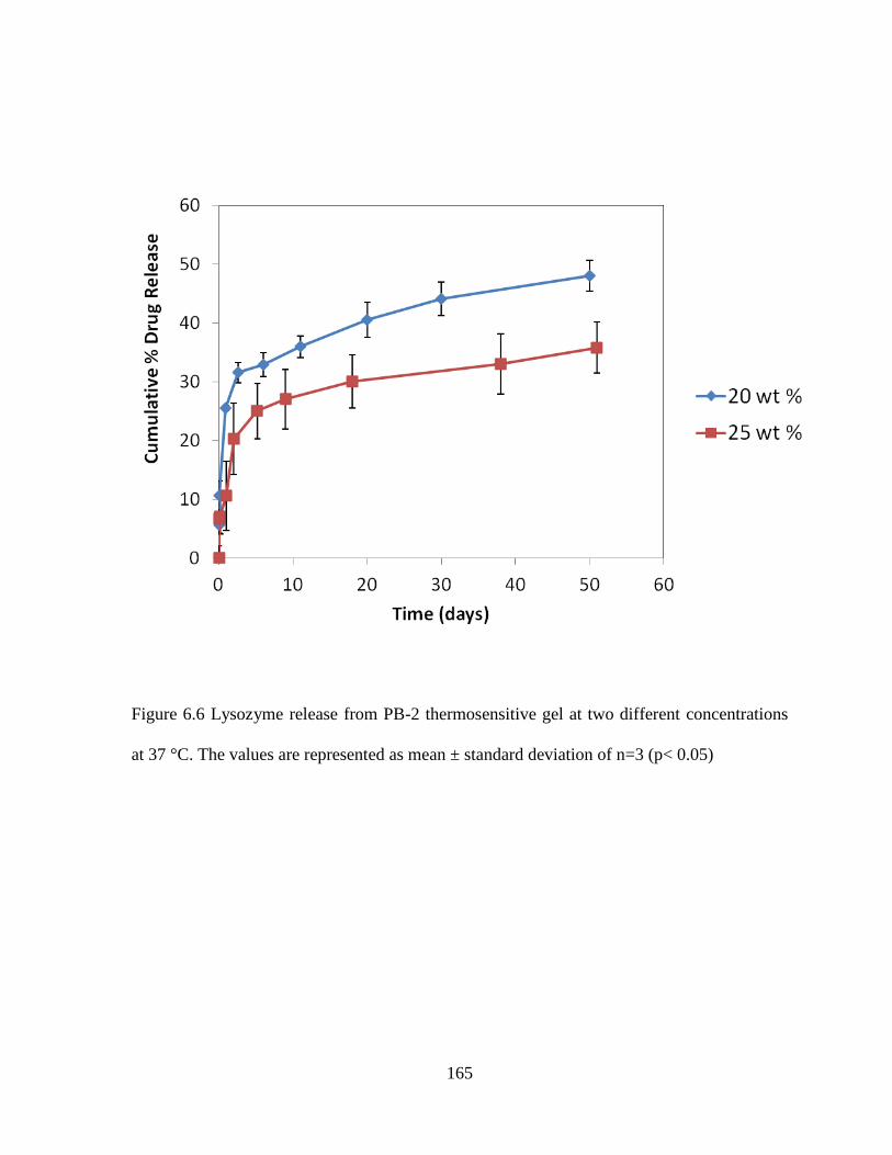
165
Figure 6.6 Lysozyme release from PB-2 thermosensitive gel at two different concentrations
at 37 °C. The values are represented as mean ± standard deviation of n=3 (p< 0.05)

166
Figure 6.7 Release profile of FITC-Dextran of different Mw was suspended in
thermosensitive gel PB-2 (20 wt %) at 37 °C. The values are represented as mean ± standard
deviation of n=3 (p< 0.05)

167
difference in the release profile with small molecules having same size but different
lipophilicity (Lu and Anseth, 2000).
Biological Activity
Table 6.3 displays biological activity of lysozyme in released samples. Protein
activity of freshly prepared lysozyme sample is 22.2 ± 2.0 (EU/mg) ×103. Enzymatic activity
in released samples taken at different time points is higher than the respective controls.
Thermosensitive hydrogel polymers are water soluble and hydrogel formulations are
prepared by vertexing lysozyme solution with the polymer in a phosphate buffer solution.
There is no harsh treatment or organic solvent required to prepare the thermosensitive gel
formulations. Therefore, we conclude that protein activity of the entrapped lysozyme
remained unaltered for a long duration in comparison to control samples which are in
phosphate buffer for the entire duration. However, we observed that enzymatic activity of
lysozyme at each time points is higher in P-2 formulation than P-1. This result can be
explained by the fact that P-1 is more hydrophobic than the P-2. Hydrophobic residues can
cause protein adsorption on the polymer surface that will result in loss of biological activity
of the entrapped protein. However, pentablock polymers are amphiphilic in nature which are
more suitable for protein delivery. As shown in table 3 protein activity in the released sample
diminished with time. It may be explained by the fact that protein molecules remained in the
release media for a specific time interval. Control samples have been collected at respective
time intervals by suspending lysozyme in phosphate buffer at the same concentration as in
formulations. A decrease in enzymatic activity can be attributed to storage conditions which
will not be the case under in vivo conditions. Under in vivo conditions proteins would be
absorbed immediately after release from the gel. The thermosensitive gel slows diffusion of
168
water molecules to the core of the hydrogel and protects the protein (Crotts and Park, 1997;
Singh et al., 2007).
Secondary structure stability by circular dichroism (CD)
Figure 6.8 represents the CD spectra of released sample from P-1 formulation and
native lysozyme. CD spectra from the sample at day 20 and day 50 displayed a strong
negative band at 208 nm and 222 nm, which were similar to the native sample. CD is a very
sensitive method and can be utilized to investigate the secondary structure of proteins.
Previous work suggests that copolymer degradation products such as lactic acid can degrade
the protein (Determan et al., 2006). Following degradation pentablock copolymer will release
lactic acid but as per the CD spectra the released sample did not exhibit any significant
difference from native lysozyme indicating stable nature of the protein inside the hydrogel.

169
Table 6.3 Biological activity of lysozyme in the released samples
Days Specific enzyme activity (EU/mg)×103
Control PB-1 PB-2
1 19.0 ± 2.0 19.3 ± 0.5 21.8 ± 1.1
20 12.5 ± 1.2 16.8 ± 1.2 18.8 ± 1.1
30 9.2 ± 0.6 13.6 ± 1.4 15.3 ± 1.8
50 5.5 ± 0.3 11.1 ± 2.4 12.1 ± 1.2

170
Figure 6.8 CD spectra of released lysozyme sample from P-1 formulation
171
Conclusions
Novel pentablock polymers for the preparation of hydrogel formulations have been
synthesized. Release kinetics of lysozyme from the hydrogel depends on the pentablock
copolymer compositions and concentration. In addition, release profile of FITC-dextran
depends on the size of the molecules. Results from our laboratory also suggest that
pentablock copolymers are biocompatible with the different ocular cell-lines (results are not
shown). Therefore, pentablock copolymers have the potential for providing sustained release
profile for protein molecules without affecting its structure and activity. Pentablock
copolymers appear to be excellent biomaterials for ocular protein delivery.

172
CHAPTER 7
SUMMARY AND RECOMMENDATIONS
Summary
Different compositions of pentablock copolymers were synthesized and characterized to
modulate the drug release profile from nanoparticles. We evaluated the effect of PEG on the
release profile of triamcinolone acetonide by synthesizing triblock copolymers PCL-PEG-
PCL with different molecular weights of PEG in the range from 2000- 8000 and found that
the hydrophilic segment does not have significant effect on the release profile of a
hydrophobic drug. However, a relatively slower release rate was observed from PEG of
mw=2000 in comparison to PEG of mw=8000. Therefore, we seleacted PEG=2000
molecular weight for the synthesis of pentablock polymers.
We synthesized pentablock copolymers containing PEG, PCL and PLA blocks. Each
block has its unique properties. Literature suggests that the block copolymers can retain the
properties of individual blocks. PEG is well known for its non-antigenic and non
immunogenic nature. In addition, because of its hydrophilic nature it facilitates the diffusion
of water into nanoparticles matrix and provides diffusion mediated drug release from
nanoparticles. PCL is hydrophobic biodegradable polyester which enables high permeability
for small molecules. Being hydrophobic in nature it also provides good encapsulation
efficiency to lipophilic drugs via hydrophobic interactions. However, it exhibits very high
crystallinity that is responsible for high burst release. PLA can influence the crystallinity of
PCL. We wanted to have all these properties in a single polymer to prepare an ideal drug
delivery system for delivery of steroids. Therefore, we optimize block ratio of PCL/PLA by
synthesizing a series of pentablock polymers. We attempted to improve the limitations of
triblock polymers i.e. PCL-PEG-PCL by incorporating the additional block PLA at the end

173
by covalent conjugation, i.e. PLA-PCL-PEG-PCL-PLA. Triblock copolymers have limitation
of burst release of drug from the nanoparticles. Burst release mainly responsible for the high
crystallinity of PCL and surface adsorbed drug. For the first time we described a strategy,
using covalently conjugated PLA blocks to reduce the crystallinity of PCL-PEG-PCL and
affect a drug release profile from nanoparticles. For reference, we synthesized the triblock
copolymers as well with same molecular weight of PCL, and characterized the triblock
copolymers for crystallinity and drug release profile. We evaluated and compared the effect
on crystallinity and drug release profile of pentablock polymers having the same molecular
weight of PCL. We were successful in changing the crystalline properties of triblock
polymers by synthesizing pentablock polymers, which has altered the release profile of
triamcinolone acetonide from nanoparticles. Moreover, pentablock copolymer based
nanoparticles exhibited minimal burst release profile. Therefore, we concluded that different
compositions of pentablock polymer (PLA-PCL-PEG-PCL-PLA) have better properties
compared to respective triblock polymers and can; provide sustained release profile for
longer duration without producing significant burst release.
Further we evaluated the pentablock copolymers for long term delivery of timolol.
Our earlier attempt of modulating the sustained release profile of timolol from PEG-PCL-
PEG hydrogel by adding the polymeric additives did not show significant improvement
(Mishra et al., 2011c). Therefore, we synthesized a pentablock copolymer based
thermosensitive hydrogel i.e. PEG-PCL-PLA-PCL-PLA. We found that pentablock
copolymer based hydrogel had better reproducibility in terms of sol-gel transitive reversion
compared to triblock polymer based hydrogels. However, to further improve the long term
release profile we evaluated the composite approach. We suspended the nanoparticles in to
174
thermosensitive hydrogel solution. We found that release profile of timolol from both
nanoparticles and thermosensitive gel alone was faster compared to the combination of both
formulations. Since, timolol is a hydrophilic molecule diffusion of molecules from the gel
was faster. In addition, nanoparticle performance suffers from the surface adsorbed drug
causing initial burst release. Our combination approach has significantly reduced the drug
release rate because of the fact that drug has to travel longer through the polymeric matrix in
the case of composite formulation.
Further, we evaluated the degradation kinetics of different pentablock copolymers.
We performed the hydrolytic and enzymatic degradation of pentablock copolymer based
nanoparticles and thermosensitive hydrogel. We found that hydrolytic degradation of these
formulations was much slower compared to enzymatic degradation. These results could be
explained by the fact that due to high hydrophobicity, hydrolytic degradation of PCL is very
slow. However, lipase enzyme can specifically degrade the PCL chains and low molecular
weight PLLA chains, but not the PDLLA. Therefore, we found faster degradation of PLLLA
containing pentablock polymers compared to PDLLA containing structure.
Finally, we evaluated the potential of thermosensitive hydrogel copolymers for long
term delivery of sensitive protein molecules. Thermosensitive hydrogels and nanoparticles
are porous structures and size of the molecules has highest impact on the drug release rate
compared to other parameters. We evaluated the effect of size of the macromolecules on the
release profile by utilizing FITC-dextran of molecular weights. We found that size of the
molecules have significant effect on the release profile, with smaller sized molecules
diffusing more rapidly than larger sized. These results were in agreement with our previous
results with small molecules. We found that various small molecules having different
175
hydrophobicity and log P values did not show significant difference in the release pattern
because of the similar smaller size. In contrast, large molecules get trapped in the polymer
matrix that provides hindrance to the diffusion of molecules. Moreover, we evaluated the
biological activity and structural stability of lysozyme (selected as model protein molecule)
released from the thermosensitive hydrogel formulation. Since, hydrogel preparation does not
involve any harsh treatment such as use of organic solvent or sonication, protein molecules
can retain structural integrity and activity during preparation of the formulation. In addition,
hydrogel can minimize the direct exposure of protein molecules with water molecules,
further retarded the degradation of protein molecules inside the formulation. We also found
that structural integrity and biological activity of lysozyme was maintained in the release
medium during the entire release period. Therefore, we conclude that our novel pentablock
copolymers based thermosensitive hydrogel and nanoparticles have potential for long term
delivery of small as well as macromolecules in the treatment of chronic ocular diseases
where sustained levels of therapeutic molecules are required.
176
Recommendations
Pentablock copolymers can be further evaluated for the delivery of sensitive molecules such
as proteins and antibodies. Preliminary results from our laboratory suggest that PEG is more
compatible to hydrophilic molecules such as antibodies and proteins. Determination of the
effect of PEG on the release profile of large hydrophilic molecules would be helpful to
optimize the controlled release formulations for proteins. Further, our composite approach of
nanoparticles suspended in a thermosensitive gel can be applicable to large molecules which
will further prolong the release of therapeutic molecules. Since, our results suggest size of the
large molecules has impact on the release profile from gel, different compositions of
copolymers should tried. Pentablock copolymer based-drug delivery systems can be
introduced in to subconjunctival space or in the intravitreal cavity. Such a delivery system
may result in prolonged duration of action, thereby completely eliminating the need for
repeated intravitreal injections. Moreover, target specific drug delivery can be obtained by
using the targeted polymers along with pentablock copolymers in the formulation of
controlled release delivery systems. For example, peptide transporter present on the
basolateral side of the RPE and Bruch’s membrane. These novel polymers could be utilize
for delivery of siRNA, protein, antibody and fragments. Moreover, determination of
intravitreal pharmacokinetics of various small and macromolecules after administration in to
rabbit eye could provide new mechanistic approaches to biomaterial design.

177
Confirmation Number: 110:57:59:5 Order ~te: 01/ 02/2013
Customer Jnfonnation
Customer. wI! tfmboli "<COIInl Number. 3000578120 Or'9aniZitlon: viral tamboli Email: vtamboliOma~.um~c.edu I'tlOIll:: +1 (551)5805580
~ Pnnlthos~ Pnnl terms &. concICJons Pnnl atabon Iflfonnatioll (Wbtt's thos?)
Search Ofde r dehils by: I C/Ioo$e One v II
Order Details
Th.~ptutiC delivery
Order ddaillO: 63300113
ISSN: 2()4I·sm Publication Typt! : )oIIrnal Volume: ISlIIt:: Start INlge: Publil her:
~rmission Stalul: ti Grlnled
Permillion type: Republish or disolay content
Bill irlg Status: .,. Typt! of ust: Republish in . thestsldissertltion
Order lic~nst Id: 3061091178202
Nole: This ~em wts wwoictd $tPlfatr/y through our Rightslink 'leNice. MoI'e Info So.OO

178
I Copy order > I
Confi rm~ lion Number: 11052144 Order O~te: 12/07/20 12
Cuslome r Informlltion
( u~tomer: virlltlmboli Account Number: 3000576120 01'1l"nIZlll lon: viral tlmboli Em"II: vtlmbcH iOm.il ,um kc.edu Phorn:: +1 (551)5805580
Search ord" d.tl il. by : I Choose One
Order Deill ii s
Colloid & polymer $eitnee
Order detai l 10:
Art icle Tille:
AulhlH"('):
001: Dale: ISS N:
63219057
I-Iovel pentltH<xk wPQiym er (M- PCl -PEG-PCL-PL.A.)-bl..,d nl nopartide. for controlled drug delivery: effed of wpoiymer oompo<ilion. on the (t)':St/lllinity 01 (oPQiymers and in vitro drug reluse pro~le from nanopartid"" Tlmboli, Virll: Hishrl, GYln p, M ~ ... Ad, im K. 10. 1007 f S00396-0 12 -2854-0 I-Iov 21, 2012 1435-153£
Publication Type: e-)ournal Vo lume : In ue: Sla rt 1'"11": Publioher: SPRINGER-VERlAG
BERLIWHEIDELBERG
ta Print tIli. paQ" Print terms'" conditi""s Print c~~on inform.tion (What'. tIli'?)
Perminlon Slalu., ~ Grl nted
Pe rminion type: ReputHish or display oontent Type of u..,: u.., in. the~diner1.tion
Order Liu n ... Id: 3043770565542
I%l View detlils
Nn ' ,,· ThO. ".m .... ino"" ..... "" •••• .." . h ..... 'nh .... ., ....... . 'n • ....... r . Mn ... in'n
179
Re: Permission for reuse the figure and text of my article in my dissertation
Noran EI-Zoheary [noran.el [email protected][
To: 1II 1~ m ~ol\ Vir~1 M, {UMKC->ludenll
• F~ lorlollow up. StaJt by Monday. ~mberOS. 2012. Due by Monday. ~mberOS. 2012.
Dear Dr . Tamboli ,
Thank you for your email and for your i nterest i n Journal of Drug Delivery, which is an open access journal . Open Access authors retain the copyrights of their papers , and all open access articles are dist ribut ed under t he terms of the creat ive Commons Attribution license, which permit s unrest rict ed use, dist ribut ion and reproduct ion i n any medi um, provided that the original work is properly cited . Accordingly, any reader/visitor is allowed to reuse any of our open access publications without getting a permission, provided that the original work is properly cited .
Please fee l free to contact me i f I can be of any further help .
Best regards,
Noran
******************************* Noran El-Zoheary Editorial Office Hi ndawi Publishi ng Corporation http : //www.hindawi.com
180
Re: Policy on reuse the material
Shankar Pandalai [email protected]
To: T~l!Ibcl\ Vir.1 M. (IIMlC.Studml)
October 6, 2012
il<!ar Dr. Viral Tamboli,
RESEARCH SIGNPOST TRANSWORLD RESEARCH NETWORK
Thank you for you e-mail dated October 4, 2012. I have talked to our management. They have given permission to use your own chapter for free. Kindly quote source,
Thanking you
Sincerely yours,
A. Gayathri Publication Manager.

181
References
Aggarwal, D., Kaur, I.P., 2005. Improved pharmacodynamics of timolol maleate from a
mucoadhesive niosomal ophthalmic drug delivery system. Int J Pharm 290, 155-159.
Ahmed, F., Discher, D.E., 2004. Self-porating polymersomes of PEG-PLA and PEG-PCL:
hydrolysis-triggered controlled release vesicles. J Control Release 96, 37-53.
Albertsson, A., C., Carlfors, J., and sturesson, C., 1996. J Applied polymer sci. 62, 695.
Aliabadi, H.M., Lavasanifar, A., 2006. Polymeric micelles for drug delivery. Expert Opin
Drug Deliv 3, 139-162.
Ambati, J., Adamis, A.P., 2002. Transscleral drug delivery to the retina and choroid. Progress
in retinal and eye research 21, 145-151.
Ambati, J., Canakis, C.S., Miller, J.W., Gragoudas, E.S., Edwards, A., Weissgold, D.J., Kim,
I., Delori, F.C., Adamis, A.P., 2000. Diffusion of high molecular weight compounds through
sclera. Investigative ophthalmology & visual science 41, 1181-1185.
Amrite, A.C., Edelhauser, H.F., Singh, S.R., Kompella, U.B., 2008. Effect of circulation on
the disposition and ocular tissue distribution of 20 nm nanoparticles after periocular
administration. Mol Vis 14, 150-160.
Anumolu, S.S., Singh, Y., Gao, D., Stein, S., Sinko, P.J., 2009. Design and evaluation of
novel fast forming pilocarpine-loaded ocular hydrogels for sustained pharmacological
response. J Control Release 137, 152-159.
Araujo, J., Gonzalez, E., Egea, M.A., Garcia, M.L., Souto, E.B., 2009. Nanomedicines for
ocular NSAIDs: safety on drug delivery. Nanomedicine 5, 394-401.
Badawi, A.A., El-Laithy, H.M., El Qidra, R.K., El Mofty, H., El dally, M., 2008. Chitosan
based nanocarriers for indomethacin ocular delivery. Arch Pharm Res 31, 1040-1049.

182
Bangham, A.D., Standish, M.M., Watkins, J.C., 1965. Diffusion of univalent ions across the
lamellae of swollen phospholipids. J Mol Biol 13, 238-252.
Beeley, N.R., Rossi, J.V., Mello-Filho, P.A., Mahmoud, M.I., Fujii, G.Y., de Juan, E., Jr.,
Varner, S.E., 2005. Fabrication, implantation, elution, and retrieval of a steroid-loaded
polycaprolactone subretinal implant. J Biomed Mater Res A 73, 437-444.
Bernatchez, S.F., Merkli, A., Tabatabay, C., Gurny, R., Zhao, Q.H., Anderson, J.M., Heller,
J., 1993. Biotolerance of a semisolid hydrophobic biodegradable poly(ortho ester) for
controlled drug delivery. J Biomed Mater Res 27, 677-681.
Bertram, J.P., Saluja, S.S., McKain, J., Lavik, E.B., 2009. Sustained delivery of timolol
maleate from poly(lactic-co-glycolic acid)/poly(lactic acid) microspheres for over 3 months.
J Microencapsul 26, 18-26.
Boddu, S.H., Jwala, J., Chowdhury, M.R., Mitra, A.K., 2010a. In vitro evaluation of a
targeted and sustained release system for retinoblastoma cells using Doxorubicin as a model
drug. J Ocul Pharmacol Ther 26, 459-468.
Boddu, S.H., Jwala, J., Vaishya, R., Earla, R., Karla, P.K., Pal, D., Mitra, A.K., 2010b. Novel
nanoparticulate gel formulations of steroids for the treatment of macular edema. J Ocul
Pharmacol Ther 26, 37-48.
Bonacucina, G., Cespi, M., Misici-Falzi, M., Palmieri, G.F., 2009. Colloidal soft matter as
drug delivery system. J Pharm Sci 98, 1-42.
Bonferoni, M.C., Chetoni, P., Giunchedi, P., Rossi, S., Ferrari, F., Burgalassi, S., Caramella,
C., 2004. Carrageenan-gelatin mucoadhesive systems for ion-exchange based ophthalmic
delivery: in vitro and preliminary in vivo studies. Eur J Pharm Biopharm 57, 465-472.
183
Boubriak, O.A., Urban, J.P., Akhtar, S., Meek, K.M., Bron, A.J., 2000. The effect of
hydration and matrix composition on solute diffusion in rabbit sclera. Experimental eye
research 71, 503-514.
Bruining, M.J., Edelbroek-Hoogendoorn, P.S., Blaauwgeers, H.G., Mooy, C.M., Hendrikse,
F.H., Koole, L.H., 1999. New biodegradable networks of poly(N-vinylpyrrolidinone)
designed for controlled nonburst degradation in the vitreous body. J Biomed Mater Res 47,
189-197.
Budhian, A., Siegel, S.J., Winey, K.I., 2005. Production of haloperidol-loaded PLGA
nanoparticles for extended controlled drug release of haloperidol. J Microencapsul 22, 773-
785.
Cai, X., Conley, S., Naash, M., 2008. Nanoparticle applications in ocular gene therapy.
Vision Res 48, 319-324.
Cam, D., Hyon, S.H., Ikada, Y., 1995. Degradation of high molecular weight poly(L-lactide)
in alkaline medium. Biomaterials 16, 833-843.
Camelo, S., Lajavardi, L., Bochot, A., Goldenberg, B., Naud, M.C., Fattal, E., Behar-Cohen,
F., de Kozak, Y., 2007. Ocular and systemic bio-distribution of rhodamine-conjugated
liposomes loaded with VIP injected into the vitreous of Lewis rats. Mol Vis 13, 2263-2274.
Carrasquillo, K.G., Ricker, J.A., Rigas, I.K., Miller, J.W., Gragoudas, E.S., Adamis, A.P.,
2003. Controlled delivery of the anti-VEGF aptamer EYE001 with poly(lactic-co-
glycolic)acid microspheres. Investigative ophthalmology & visual science 44, 290-299.
Chakravarthy, U., Soubrane, G., Bandello, F., Chong, V., Creuzot-Garcher, C., Dimitrakos,
S.A., 2nd, Korobelnik, J.F., Larsen, M., Mones, J., Pauleikhoff, D., Pournaras, C.J.,
Staurenghi, G., Virgili, G., Wolf, S., 2006. Evolving European guidance on the medical

184
management of neovascular age related macular degeneration. Br J Ophthalmol 90, 1188-
1196.
Chen, C.C., Chueh, J.Y., Tseng, H., Huang, H.M., Lee, S.Y., 2003. Preparation and
characterization of biodegradable PLA polymeric blends. Biomaterials 24, 1167-1173.
Chin, H.S., Park, T.S., Moon, Y.S., Oh, J.H., 2005. Difference in clearance of intravitreal
triamcinolone acetonide between vitrectomized and nonvitrectomized eyes. Retina 25, 556-
560.
Colthurst, M.J., Williams, R.L., Hiscott, P.S., Grierson, I., 2000. Biomaterials used in the
posterior segment of the eye. Biomaterials 21, 649-665.
Cortesi, R., Argnani, R., Esposito, E., Dalpiaz, A., Scatturin, A., Bortolotti, F., Lufino, M.,
Guerrini, R., Cavicchioni, G., Incorvaia, C., Menegatti, E., Manservigi, R., 2006. Cationic
liposomes as potential carriers for ocular administration of peptides with anti-herpetic
activity. Int J Pharm 317, 90-100.
Crotts, G., Park, T.G., 1997. Stability and release of bovine serum albumin encapsulated
within poly(lactide-co-glycolide) microparticles. Journal of controlled release 44, 123-134.
Croy, S.R., Kwon, G.S., 2006. Polymeric micelles for drug delivery. Curr Pharm Des 12,
4669-4684.
Dalwadi, G., Sunderland, B., 2009. An ion pairing approach to increase the loading of
hydrophilic and lipophilic drugs into PEGylated PLGA nanoparticles. Eur J Pharm Biopharm
71, 231-242.
Danion, A., Arsenault, I., Vermette, P., 2007. Antibacterial activity of contact lenses bearing
surface-immobilized layers of intact liposomes loaded with levofloxacin. J Pharm Sci 96,
2350-2363.

185
Daughaday, W.H., 1992. Pituitary gigantism. Endocrinology and metabolism clinics of North
America 21, 633-647.
Daugherty, A.L., Mrsny, R.J., 2003. Emerging technologies that overcome biological barriers
for therapeutic protein delivery. Expert Opin Biol Ther 3, 1071-1081.
Desai, S.D., Blanchard, J., 2000. Pluronic F127-based ocular delivery system containing
biodegradable polyisobutylcyanoacrylate nanocapsules of pilocarpine. Drug Deliv 7, 201-
207.
Deshpande, A.A., Heller, J., Gurny, R., 1998. Bioerodible polymers for ocular drug delivery.
Crit Rev Ther Drug Carrier Syst 15, 381-420.
Determan, A.S., Wilson, J.H., Kipper, M.J., Wannemuehler, M.J., Narasimhan, B., 2006.
Protein stability in the presence of polymer degradation products: consequences for
controlled release formulations. Biomaterials 27, 3312-3320.
Dey, S., Patel, J., Anand, B.S., Jain-Vakkalagadda, B., Kaliki, P., Pal, D., Ganapathy, V.,
Mitra, A.K., 2003. Molecular evidence and functional expression of P-glycoprotein (MDR1)
in human and rabbit cornea and corneal epithelial cell lines. Investigative ophthalmology &
visual science 44, 2909-2918.
Diebold, Y., Calonge, M., Applications of nanoparticles in ophthalmology. Prog Retin Eye
Res 29, 596-609.
Diebold, Y., Calonge, M., 2010. Applications of nanoparticles in ophthalmology. Prog Retin
Eye Res 29, 596-609.
Dillen, K., Vandervoort, J., Van den Mooter, G., Ludwig, A., 2006. Evaluation of
ciprofloxacin-loaded Eudragit RS100 or RL100/PLGA nanoparticles. Int J Pharm 314, 72-
82.

186
Ding X, Alani WG, JR, R., Extended-release and targeted drug delivery systems, in: Troy,
D.B. (Ed.), Remington: the science and practice of pharmacy. Lippincott Williams and
wilkins.
Duvvuri, S., Gaurav Janoria, K., Mitra, A.K., 2006. Effect of polymer blending on the release
of ganciclovir from PLGA microspheres. Pharm Res 23, 215-223.
Duvvuri, S., Janoria, K.G., Mitra, A.K., 2005. Development of a novel formulation
containing poly(d,l-lactide-co-glycolide) microspheres dispersed in PLGA-PEG-PLGA gel
for sustained delivery of ganciclovir. J Control Release 108, 282-293.
Duvvuri, S., Janoria, K.G., Pal, D., Mitra, A.K., 2007. Controlled delivery of ganciclovir to
the retina with drug-loaded Poly(d,L-lactide-co-glycolide) (PLGA) microspheres dispersed in
PLGA-PEG-PLGA Gel: a novel intravitreal delivery system for the treatment of
cytomegalovirus retinitis. J Ocul Pharmacol Ther 23, 264-274.
Einmahl, S., Behar-Cohen, F., D'Hermies, F., Rudaz, S., Tabatabay, C., Renard, G., Gurny,
R., 2001. A new poly(ortho ester)-based drug delivery system as an adjunct treatment in
filtering surgery. Investigative ophthalmology & visual science 42, 695-700.
Einmahl, S., Behar-Cohen, F., Tabatabay, C., Savoldelli, M., D'Hermies, F., Chauvaud, D.,
Heller, J., Gurny, R., 2000. A viscous bioerodible poly(ortho ester) as a new biomaterial for
intraocular application. J Biomed Mater Res 50, 566-573.
Einmahl, S., Ponsart, S., Bejjani, R.A., D'Hermies, F., Savoldelli, M., Heller, J., Tabatabay,
C., Gurny, R., Behar-Cohen, F., 2003. Ocular biocompatibility of a poly(ortho ester)
characterized by autocatalyzed degradation. J Biomed Mater Res A 67, 44-53.
187
Einmahl, S., Savoldelli, M., D'Hermies, F., Tabatabay, C., Gurny, R., Behar-Cohen, F., 2002.
Evaluation of a novel biomaterial in the suprachoroidal space of the rabbit eye. Investigative
ophthalmology & visual science 43, 1533-1539.
EL- Samaligy, M., S., Rojanasakul, Y., Charlton, J., F., Weinstein, G. W., and Lim J., K.,,
1996. Ocular disposition of nanoencapsulated acyclovir and ganciclovir via intravitreal
injection in rabbit’s eye. Drug Delivery 3, 93-97.
El-Laithy, H.M., 2009. Novel transdermal delivery of Timolol maleate using sugar esters:
preclinical and clinical studies. Eur J Pharm Biopharm 72, 239-245.
El Sanharawi, M., Kowalczuk, L., Touchard, E., Omri, S., de Kozak, Y., Behar-Cohen, F.,
2010. Protein delivery for retinal diseases: from basic considerations to clinical applications.
Progress in retinal and eye research 29, 443-465.
Elbayoumi, T.A., Torchilin, V.P., 2010. Current trends in liposome research. Methods Mol
Biol 605, 1-27.
Fialho, S.L., Behar-Cohen, F., Silva-Cunha, A., 2008. Dexamethasone-loaded poly(epsilon-
caprolactone) intravitreal implants: a pilot study. Eur J Pharm Biopharm 68, 637-646.
Forrest, M.L., Won, C.Y., Malick, A.W., Kwon, G.S., 2006. In vitro release of the mTOR
inhibitor rapamycin from poly(ethylene glycol)-b-poly(epsilon-caprolactone) micelles. J
Control Release 110, 370-377.
Frank, A., Rath, S.K., Venkatraman, S.S., 2005. Controlled release from bioerodible
polymers: effect of drug type and polymer composition. J Control Release 102, 333-344.
Fresta, M., Fontana, G., Bucolo, C., Cavallaro, G., Giammona, G., Puglisi, G., 2001. Ocular
tolerability and in vivo bioavailability of poly(ethylene glycol) (PEG)-coated polyethyl-2-
cyanoacrylate nanosphere-encapsulated acyclovir. J Pharm Sci 90, 288-297.
188
Fukushima, A., Ozaki, A., Ishida, W., van Rooijen, N., Fukata, K., Ueno, H., 2005.
Suppression of macrophage infiltration into the conjunctiva by clodronate liposomes in
experimental immune-mediated blepharoconjunctivitis. Cell Biol Int 29, 277-286.
Gao, Y., Sun, Y., Ren, F., Gao, S., 2010. PLGA-PEG-PLGA hydrogel for ocular drug
delivery of dexamethasone acetate. Drug development and industrial pharmacy 36, 1131-
1138.
Gavini, E., Chetoni, P., Cossu, M., Alvarez, M.G., Saettone, M.F., Giunchedi, P., 2004.
PLGA microspheres for the ocular delivery of a peptide drug, vancomycin using
emulsification/spray-drying as the preparation method: in vitro/in vivo studies. Eur J Pharm
Biopharm 57, 207-212.
Ghate, D., Brooks, W., McCarey, B.E., Edelhauser, H.F., 2007. Pharmacokinetics of
intraocular drug delivery by periocular injections using ocular fluorophotometry.
Investigative ophthalmology & visual science 48, 2230-2237.
Ghate, D., Edelhauser, H.F., 2006. Ocular drug delivery. Expert Opin Drug Deliv 3, 275-287.
Ghate, D., Edelhauser, H.F., 2008. Barriers to glaucoma drug delivery. J Glaucoma 17, 147-
156.
Ghoroghchian, P.P., Li, G., Levine, D.H., Davis, K.P., Bates, F.S., Hammer, D.A., Therien,
M.J., 2006. Bioresorbable Vesicles Formed through Spontaneous Self-Assembly of
Amphiphilic Poly(ethylene oxide)-block-polycaprolactone. Macromolecules 39, 1673-1675.
Giannavola, C., Bucolo, C., Maltese, A., Paolino, D., Vandelli, M.A., Puglisi, G., Lee, V.H.,
Fresta, M., 2003. Influence of preparation conditions on acyclovir-loaded poly-d,l-lactic acid
nanospheres and effect of PEG coating on ocular drug bioavailability. Pharm Res 20, 584-
590.
189
Gomez-Gaete, C., Tsapis, N., Besnard, M., Bochot, A., Fattal, E., 2007. Encapsulation of
dexamethasone into biodegradable polymeric nanoparticles. Int J Pharm 331, 153-159.
Gou, M., Gong, C., Zhang, J., Wang, X., Gu, Y., Guo, G., Chen, L., Luo, F., Zhao, X., Wei,
Y., Qian, Z., 2010. Polymeric matrix for drug delivery: honokiol-loaded PCL-PEG-PCL
nanoparticles in PEG-PCL-PEG thermosensitive hydrogel. J Biomed Mater Res A 93, 219-
226.
Gou, M., Zheng, L., Peng, X., Men, K., Zheng, X., Zeng, S., Guo, G., Luo, F., Zhao, X.,
Chen, L., Wei, Y., Qian, Z., 2009a. Poly(epsilon-caprolactone)-poly(ethylene glycol)-
poly(epsilon-caprolactone) (PCL-PEG-PCL) nanoparticles for honokiol delivery in vitro. Int
J Pharm 375, 170-176.
Gou, M., Zheng, X., Men, K., Zhang, J., Zheng, L., Wang, X., Luo, F., Zhao, Y., Zhao, X.,
Wei, Y., Qian, Z., 2009b. Poly(epsilon-caprolactone)/poly(ethylene glycol)/poly(epsilon-
caprolactone) nanoparticles: preparation, characterization, and application in doxorubicin
delivery. J Phys Chem B 113, 12928-12933.
Gupta, H., Aqil, M., Khar, R.K., Ali, A., Bhatnagar, A., Mittal, G., 2010. Sparfloxacin-
loaded PLGA nanoparticles for sustained ocular drug delivery. Nanomedicine 6, 324-333.
Guttman, C., 2005. Multiple factors underlie non-compliance with treatment.,
Ophthalmology Times.
Guy, R.H., Hadgraft, J., Taylor, M.J., Kellaway, I.W., 1983. Release of non-electrolytes from
liposomes. J Pharm Pharmacol 35, 12-14.
Hacker, M.C., Haesslein, A., Ueda, H., Foster, W.J., Garcia, C.A., Ammon, D.M., Borazjani,
R.N., Kunzler, J.F., Salamone, J.C., Mikos, A.G., 2009. Biodegradable fumarate-based drug-
delivery systems for ophthalmic applications. J Biomed Mater Res A 88, 976-989.
190
Hariharan, S., Minocha, M., Mishra, G.P., Pal, D., Krishna, R., Mitra, A.K., 2009. Interaction
of ocular hypotensive agents (PGF2 alpha analogs-bimatoprost, latanoprost, and travoprost)
with MDR efflux pumps on the rabbit cornea. J Ocul Pharmacol Ther 25, 487-498.
Hathout, R.M., Mansour, S., Mortada, N.D., Guinedi, A.S., 2007. Liposomes as an ocular
delivery system for acetazolamide: in vitro and in vivo studies. AAPS PharmSciTech 8, 1.
Haung, M., Li, S., Hutmacher, D.W., Coudane, J., Vert, M., 2006. Degradation
Characteristics of Poly(ε-caprolactone)-Based Copolymers and Blends. J Applied Poly Sci
102, 1681-1687.
Havlikova, L., Matysova, L., Hajkova, R., Satinsky, D., Solich, P., 2008. Advantages of
pentafluorophenylpropyl stationary phase over conventional C18 stationary phase--
application to analysis of triamcinolone acetonide. Talanta 76, 597-601.
He, C., Kim, S.W., Lee, D.S., 2008. In situ gelling stimuli-sensitive block copolymer
hydrogels for drug delivery. J Control Release 127, 189-207.
Heller, J., 1984. Biodegradable polymers in controlled drug delivery. Crit Rev Ther Drug
Carrier Syst 1, 39-90.
Heller, J., 2005. Ocular delivery using poly(ortho esters). Adv Drug Deliv Rev 57, 2053-
2062.
Heller, J., Barr, J., Ng, S.Y., Abdellauoi, K.S., Gurny, R., 2002. Poly(ortho esters): synthesis,
characterization, properties and uses. Adv Drug Deliv Rev 54, 1015-1039.
Hong, Y., Chirila, T.V., Vijayasekaran, S., Shen, W., Lou, X., Dalton, P.D., 1998.
Biodegradation in vitro and retention in the rabbit eye of crosslinked poly(1-vinyl-2-
pyrrolidinone) hydrogel as a vitreous substitute. J Biomed Mater Res 39, 650-659.

191
Hosny, K.M., 2010. Ciprofloxacin as ocular liposomal hydrogel. AAPS PharmSciTech 11,
241-246.
Hoyng, P.J., Van beek, L.M., 2000. Pharmacological therapy for glaucoma: a review. Drugs
59, 411-434.
Hu, Y., Jiang, X., Ding, Y., Zhang, L., Yang, C., Zhang, J., Chen, J., Yang, Y., 2003.
Preparation and drug release behaviors of nimodipine-loaded poly(caprolactone)-
poly(ethylene oxide)-polylactide amphiphilic copolymer nanoparticles. Biomaterials 24,
2395-2404.
Hu, Y., Xie, J., Tong, Y.W., Wang, C.H., 2007. Effect of PEG conformation and particle size
on the cellular uptake efficiency of nanoparticles with the HepG2 cells. J Control Release
118, 7-17.
Huang, M.H., Li, S., Coudane, J., Vert, M., 2003. Synthesis and Characterization of Block
Copolymers of e-Caprolactone and DL-Lactide Initiated by Ethylene Glycol or Poly(ethylene
glycol). Macromol Chem and Phys 204, 1994-2001.
Huang, M.H., Li, S., Hutmacher, D.W., Schantz, J.T., Vacanti, C.A., Braud, C., Vert, M.,
2004. Degradation and cell culture studies on block copolymers prepared by ring opening
polymerization of epsilon-caprolactone in the presence of poly(ethylene glycol). J Biomed
Mater Res A 69, 417-427.
Hyon, S.H., 2000. Biodegradable poly (lactic acid) microspheres for drug delivery systems.
Yonsei Med J 41, 720-734.
Hyon, S.H., K., J., Ikada, Y., 1998. Polymer Int 46, 196.
Jain, J.P., Modi, S., Domb, A.J., Kumar, N., 2005. Role of polyanhydrides as localized drug
carriers. J Control Release 103, 541-563.

192
Jampel, H.D., Leong, K.W., Dunkelburger, G.R., Quigley, H.A., 1990. Glaucoma filtration
surgery in monkeys using 5-fluorouridine in polyanhydride disks. Arch Ophthalmol 108,
430-435.
Janoria, K.G., Gunda, S., Boddu, S.H., Mitra, A.K., 2007. Novel approaches to retinal drug
delivery. Expert Opin Drug Deliv 4, 371-388.
Jermak, C.M., Dellacroce, J.T., Heffez, J., Peyman, G.A., 2007. Triamcinolone acetonide in
ocular therapeutics. Surv Ophthalmol 52, 503-522.
Jesorka, A., Orwar, O., 2008. Liposomes: technologies and analytical applications. Annu Rev
Anal Chem (Palo Alto Calif) 1, 801-832.
Jia, W., Gu, Y., Gou, M., Dai, M., Li, X., Kan, B., Yang, J., Song, Q., Wei, Y., Qian, Z.,
2008. Preparation of biodegradable polycaprolactone/poly (ethylene
glycol)/polycaprolactone (PCEC) nanoparticles. Drug Deliv 15, 409-416.
Jonas, J.B., Kreissig, I., Degenring, R., 2002. Repeated intravitreal injections of
triamcinolone acetonide as treatment of progressive exudative age-related macular
degeneration. Graefe's archive for clinical and experimental ophthalmology = Albrecht von
Graefes Archiv fur klinische und experimentelle Ophthalmologie 240, 873-874.
Jwala, J., Boddu, S.H., Shah, S., Sirimulla, S., Pal, D., Mitra, A.K., 2011. Ocular sustained
release nanoparticles containing stereoisomeric dipeptide prodrugs of acyclovir. Journal of
ocular pharmacology and therapeutics : the official journal of the Association for Ocular
Pharmacology and Therapeutics 27, 163-172.
Kang, J., Schwendeman, S.P., 2002. Comparison of the effects of Mg(OH)2 and sucrose on
the stability of bovine serum albumin encapsulated in injectable poly(D,L-lactide-co-
glycolide) implants. Biomaterials 23, 239-245.
193
Karla, P.K., Earla, R., Boddu, S.H., Johnston, T.P., Pal, D., Mitra, A., 2009. Molecular
expression and functional evidence of a drug efflux pump (BCRP) in human corneal
epithelial cells. Current eye research 34, 1-9.
Karla, P.K., Pal, D., Mitra, A.K., 2007a. Molecular evidence and functional expression of
multidrug resistance associated protein (MRP) in rabbit corneal epithelial cells. Experimental
eye research 84, 53-60.
Karla, P.K., Pal, D., Quinn, T., Mitra, A.K., 2007b. Molecular evidence and functional
expression of a novel drug efflux pump (ABCC2) in human corneal epithelium and rabbit
cornea and its role in ocular drug efflux. Int J Pharm 336, 12-21.
Katti, D.S., Lakshmi, S., Langer, R., Laurencin, C.T., 2002. Toxicity, biodegradation and
elimination of polyanhydrides. Adv Drug Deliv Rev 54, 933-961.
Kaur, I.P., Aggarwal, D., Singh, H., Kakkar, S., Improved ocular absorption kinetics of
timolol maleate loaded into a bioadhesive niosomal delivery system. Graefe's archive for
clinical and experimental ophthalmology = Albrecht von Graefes Archiv fur klinische und
experimentelle Ophthalmologie 248, 1467-1472.
Kaur, I.P., Aggarwal, D., Singh, H., Kakkar, S., 2010. Improved ocular absorption kinetics of
timolol maleate loaded into a bioadhesive niosomal delivery system. Graefe's archive for
clinical and experimental ophthalmology = Albrecht von Graefes Archiv fur klinische und
experimentelle Ophthalmologie 248, 1467-1472.
Kaur, I.P., Garg, A., Singla, A.K., Aggarwal, D., 2004. Vesicular systems in ocular drug
delivery: an overview. Int J Pharm 269, 1-14.
194
Kawakami, S., Harada, A., Sakanaka, K., Nishida, K., Nakamura, J., Sakaeda, T., Ichikawa,
N., Nakashima, M., Sasaki, H., 2004. In vivo gene transfection via intravitreal injection of
cationic liposome/plasmid DNA complexes in rabbits. Int J Pharm 278, 255-262.
Kimura, H., Ogura, Y., 2001. Biodegradable polymers for ocular drug delivery.
Ophthalmologica 215, 143-155.
Klouda, L., Mikos, A.G., 2008. Thermoresponsive hydrogels in biomedical applications. Eur
J Pharm Biopharm 68, 34-45.
Koevary, S.B., 2002. Trends in the noncorneal delivery of drugs into the eye. Archivos de la
Sociedad Espanola de Oftalmologia 77, 347-349.
Kompella, U.B., Bandi, N., Ayalasomayajula, S.P., 2003. Subconjunctival nano- and
microparticles sustain retinal delivery of budesonide, a corticosteroid capable of inhibiting
VEGF expression. Investigative ophthalmology & visual science 44, 1192-1201.
Kulkarni, A., Reiche, J., Hartmann, J., Kratz, K., Lendlein, A., 2008. Selective enzymatic
degradation of poly(epsilon-caprolactone) containing multiblock copolymers. Eur J Pharm
Biopharm 68, 46-56.
Kulkarni, A.D., Kuppermann, B.D., 2005. Wet age-related macular degeneration. Advanced
drug delivery reviews 57, 1994-2009.
Kunou, N., Ogura, Y., Yasukawa, T., Kimura, H., Miyamoto, H., Honda, Y., Ikada, Y., 2000.
Long-term sustained release of ganciclovir from biodegradable scleral implant for the
treatment of cytomegalovirus retinitis. J Control Release 68, 263-271.
Kwak, H.W., D'Amico, D.J., 1992. Evaluation of the retinal toxicity and pharmacokinetics of
dexamethasone after intravitreal injection. Arch Ophthalmol 110, 259-266.
195
Lam, C.X., Savalani, M.M., Teoh, S.H., Hutmacher, D.W., 2008. Dynamics of in vitro
polymer degradation of polycaprolactone-based scaffolds: accelerated versus simulated
physiological conditions. Biomed Mater 3, 034108.
Layre, A.M., Couvreur, P., Chacun, H., Aymes-Chodur, C., Ghermani, N.E., Poupaert, J.,
Richard, J., Requier, D., Gref, R., 2006. Busulfan loading into poly(alkyl cyanoacrylate)
nanoparticles: physico-chemistry and molecular modeling. J Biomed Mater Res B Appl
Biomater 79, 254-262.
Lazzeri, S., Figus, M., Di Bartolo, E., Rizzo, S., Nardi, M., 2011. Verteporfin photodynamic
therapy for retinal hemangioblastoma associated with Von Hippel-Lindau Disease in a 9-
year-old child. Clin Experiment Ophthalmol.
Lee, D.A., Leong, K.W., Panek, W.C., Eng, C.T., Glasgow, B.J., 1988. The use of
bioerodible polymers and 5-fluorouracil in glaucoma filtration surgery. Investigative
ophthalmology & visual science 29, 1692-1697.
Lee, J.Y., Kim, K.S., Kang, Y.M., Kim, E.S., Hwang, S.J., Lee, H.B., Min, B.H., Kim, J.H.,
Kim, M.S., In vivo efficacy of paclitaxel-loaded injectable in situ-forming gel against
subcutaneous tumor growth. Int J Pharm 392, 51-56.
Lee, V.H., 1990. New directions in the optimization of ocular drug delivery. Journal of
ocular pharmacology 6, 157-164.
Lehr, C.M., Lee, Y.H., Lee, V.H., 1994. Improved ocular penetration of gentamicin by
mucoadhesive polymer polycarbophil in the pigmented rabbit. Investigative ophthalmology
& visual science 35, 2809-2814.

196
Leong, K.W., Brott, B.C., Langer, R., 1985. Bioerodible polyanhydrides as drug-carrier
matrices. I: Characterization, degradation, and release characteristics. J Biomed Mater Res
19, 941-955.
Leong, K.W., Simonte, V., and langer R., 1987. Macromolecules 20, 705.
Leung, D.W., Cachianes, G., Kuang, W.J., Goeddel, D.V., Ferrara, N., 1989. Vascular
endothelial growth factor is a secreted angiogenic mitogen. Science 246, 1306-1309.
Li, C.C., Abrahamson, M., Kapoor, Y., Chauhan, A., 2007. Timolol transport from
microemulsions trapped in HEMA gels. J Colloid Interface Sci 315, 297-306.
Li, R., Li, X., Xie, L., Ding, D., Hu, Y., Qian, X., Yu, L., Ding, Y., Jiang, X., Liu, B., 2009.
Preparation and evaluation of PEG-PCL nanoparticles for local tetradrine delivery.
International journal of pharmaceutics 379, 158-166.
Li, S., Dobrzynski, P., Kasperczyk, J., Bero, M., Braud, C., Vert, M., 2005. Structure-
property relationships of copolymers obtained by ring-opening polymerization of glycolide
and epsilon-caprolactone. Part 2. Influence of composition and chain microstructure on the
hydrolytic degradation. Biomacromolecules 6, 489-497.
Li, S., Molina, I., Martinez, M.B., Vert, M., 2002. Hydrolytic and enzymatic degradations of
physically crosslinked hydrogels prepared from PLA/PEO/PLA triblock copolymers. J Mater
Sci Mater Med 13, 81-86.
Lian, T., Ho, R.J., 2001. Trends and developments in liposome drug delivery systems. J
Pharm Sci 90, 667-680.
Liang, H.F., Yang, T.F., Huang, C.T., Chen, M.C., Sung, H.W., 2005. Preparation of
nanoparticles composed of poly(gamma-glutamic acid)-poly(lactide) block copolymers and
evaluation of their uptake by HepG2 cells. J Control Release 105, 213-225.

197
Liu, L., Li, S., Garreau, H., Vert, M., 2000. Selective enzymatic degradations of poly(L-
lactide) and poly(epsilon-caprolactone) blend films. Biomacromolecules 1, 350-359.
Loftsson, T., Hreinsdottir, D., 2006. Determination of aqueous solubility by heating and
equilibration: a technical note. AAPS PharmSciTech 7, E4.
Lu, S., Anseth, K., 2000. Release Behavior of High Molecular Weight Solutes from
Poly(ethylene glycol)-Based Degradable Networks. Macromolecules 33, 2509-2515.
Macha S, Hughes PM, Mitra AK, 2003. Overview of Ocular Drug Delivery, in: Mitra, A.K.
(Ed.), Ophthalmic Drug delivery systems. Marcell Dekker, New york, USA.
Mainardes, R.M., Urban, M.C., Cinto, P.O., Khalil, N.M., Chaud, M.V., Evangelista, R.C.,
Gremiao, M.P., 2005. Colloidal carriers for ophthalmic drug delivery. Curr Drug Targets 6,
363-371.
Marchal-Heussler, L., Sirbat, D., Hoffman, M., Maincent, P., 1993. Poly(epsilon-
caprolactone) nanocapsules in carteolol ophthalmic delivery. Pharm Res 10, 386-390.
McVey, D., Hamilton, M.M., Hsu, C., King, C.R., Brough, D.E., Wei, L.L., 2008. Repeat
administration of proteins to the eye with a single intraocular injection of an adenovirus
vector. Molecular therapy : the journal of the American Society of Gene Therapy 16, 1444-
1449.
Merkli, A., Heller, J., Tabatabay, C., and Gurny, R., 1994. Semi-solid hydrophobic
bioerodible poly(ortho ester) for potential application in glaucoma filtration surgery. J
Control Release 29, 105-112.
Meyer, F., Wardale, J., Best, S., Cameron, R., Rushton, N., Brooks, R., 2012. Effects of
lactic acid and glycolic acid on human osteoblasts: A way to understand PLGA involvement
in PLGA/calcium phosphate composite failure. J Orthop Res.

198
Mishra, G., Gaudana, R., Tamboli, V., Mitra, A., 2010. Recent Advances in Ocular Drug
Delivery: Role of Transporters, Receptors, and Nanocarriers. , in: RI, M., AS, N. (Eds.),
Targeted Delivery of Small and Macromolecular Drugs. Taylor and Francis, New york, pp.
421-456.
Mishra, G.P., Bagui, M., Tamboli, V., Mitra, A.K., 2011a. Recent applications of liposomes
in ophthalmic drug delivery. Journal of drug delivery 2011, 863734.
Mishra, G.P., Tamboli, V., Jwala, J., Mitra, A.K., 2011b. Recent patents and emerging
therapeutics in the treatment of allergic conjunctivitis. Recent Pat Inflamm Allergy Drug
Discov 5, 26-36.
Mishra, G.P., Tamboli, V., Mitra, A.K., Effect of hydrophobic and hydrophilic additives on
sol-gel transition and release behavior of timolol maleate from polycaprolactone-based
hydrogel. Colloid Polym Sci 289, 1553-1562.
Mishra, G.P., Tamboli, V., Mitra, A.K., 2011c. Effect of hydrophobic and hydrophilic
additives on sol-gel transition and release behavior of timolol maleate from
polycaprolactone-based hydrogel. Colloid Polym Sci 289, 1553-1562.
Mitra, A.K., 2009. Role of transporters in ocular drug delivery system. Pharm Res 26, 1192-
1196.
Mitra, A.K., Velagaleti, P.R., Grau, U.M., 2010. Topical drug delivery systems for
ophthalmic use. Lux Biosciences, Inc.
Mitra, A.K., Velagaleti, P.R., Natesan, S., 2009. Ophthalmic compostions comprising
calcineurin inhibitors or mTOR inhibitors. Lux Biosciences, Inc.
Mitra, A.K., velagaleti, P.R., Natesan, S., 2011. Ophthalmic compositions comprising
calcineurin inhibitors or mTOR inhibitors. Lux Biosciences, Inc.

199
Miyajima, M., Koshika, A., Okada, J., Ikeda, M., Nishimura, K., 1997. Effect of polymer
crystallinity on papaverine release from poly(L-lactic acid) matrix. J Control Release 49,
207-215.
Moritera, T., Ogura, Y., Honda, Y., Wada, R., Hyon, S.H., Ikada, Y., 1991. Microspheres of
biodegradable polymers as a drug-delivery system in the vitreous. Investigative
ophthalmology & visual science 32, 1785-1790.
Munoz, B., West, S.K., Rubin, G.S., Schein, O.D., Quigley, H.A., Bressler, S.B., Bandeen-
Roche, K., 2000. Causes of blindness and visual impairment in a population of older
Americans: The Salisbury Eye Evaluation Study. Archives of ophthalmology 118, 819-825.
Murthy R., S.R., 1997. Controlled and Novel Drug Delivery. CBS Publisher, New Delhi.
Nanjawade, B.K., Manvi, F.V., Manjappa, A.S., 2007. In situ-forming hydrogels for
sustained ophthalmic drug delivery. J Control Release 122, 119-134.
Newman, D., Loredo, E., Bello, A., Grillo, A., eijoo, A., M ller, A., 2009. Molecular
Mobilities in Biodegradable Poly(dl-lactide)/Poly(ε-caprolactone) Blends. Macromol 42,
5219–5225.
Niesman, M.R., 1992. The use of liposomes as drug carriers in ophthalmology. Crit Rev Ther
Drug Carrier Syst 9, 1-38.
Niu, G., Yang, Y., Zhang, H., Yang, J., Song, L., Kashima, M., Yang, Z., Cao, H., Zheng, Y.,
Zhu, S., Yang, H., 2009. Synthesis and characterization of acrylamide/N-vinylpyrrolidone
copolymer with pendent thiol groups for ophthalmic applications. Acta Biomater 5, 1056-
1063.
Ogawa, Y., Okada, H., Yamamoto, M., Shimamoto, T., 1988. In vivo release profiles of
leuprolide acetate from microcapsules prepared with polylactic acids or

200
copoly(lactic/glycolic) acids and in vivo degradation of these polymers. Chem Pharm Bull
(Tokyo) 36, 2576-2581.
Okabe, K., Kimura, H., Okabe, J., Kato, A., Kunou, N., Ogura, Y., 2003. Intraocular tissue
distribution of betamethasone after intrascleral administration using a non-biodegradable
sustained drug delivery device. Investigative ophthalmology & visual science 44, 2702-2707.
Olsen, T.W., Edelhauser, H.F., Lim, J.I., Geroski, D.H., 1995. Human scleral permeability.
Effects of age, cryotherapy, transscleral diode laser, and surgical thinning. Investigative
ophthalmology & visual science 36, 1893-1903.
Olthoff, C.M., Schouten, J.S., van de Borne, B.W., Webers, C.A., 2005. Noncompliance with
ocular hypotensive treatment in patients with glaucoma or ocular hypertension an evidence-
based review. Ophthalmology 112, 953-961.
Packhaeuser, C.B., Kissel, T., 2007. On the design of in situ forming biodegradable
parenteral depot systems based on insulin loaded dialkylaminoalkyl-amine-poly(vinyl
alcohol)-g-poly(lactide-co-glycolide) nanoparticles. J Control Release 123, 131-140.
Papahadjopoulos, D., Jacobson, K., Nir, S., Isac, T., 1973. Phase transitions in phospholipid
vesicles. Fluorescence polarization and permeability measurements concerning the effect of
temperature and cholesterol. Biochim Biophys Acta 311, 330-348.
Park, J.H., Ye, M., Park, K., 2005. Biodegradable polymers for microencapsulation of drugs.
Molecules 10, 146-161.
Park P., J.S., 2006. J. Applied Poly. Sci. 100, 1983.
Peeters, L., Sanders, N.N., Braeckmans, K., Boussery, K., Van de Voorde, J., De Smedt,
S.C., Demeester, J., 2005. Vitreous: a barrier to nonviral ocular gene therapy. Investigative
ophthalmology & visual science 46, 3553-3561.

201
Peracchia, M.T., Vauthier, C., Puisieux, F., Couvreur, P., 1997. Development of sterically
stabilized poly(isobutyl 2-cyanoacrylate) nanoparticles by chemical coupling of
poly(ethylene glycol). J Biomed Mater Res 34, 317-326.
Pescina, S., Santi, P., Ferrari, G., Nicoli, S., 2011. Trans-scleral delivery of macromolecules.
Therapeutic delivery 2, 1331-1349.
Polak, M.B., Valamanesh, F., Felt, O., Torriglia, A., Jeanny, J.C., Bourges, J.L., Rat, P.,
Thomas-Doyle, A., BenEzra, D., Gurny, R., Behar-Cohen, F., 2008. Controlled delivery of 5-
chlorouracil using poly(ortho esters) in filtering surgery for glaucoma. Investigative
ophthalmology & visual science 49, 2993-3003.
Qiu, Y., Park, K., 2001. Environment-sensitive hydrogels for drug delivery. Adv Drug Deliv
Rev 53, 321-339.
Quigley, H.A., Broman, A.T., 2006. The number of people with glaucoma worldwide in
2010 and 2020. Br J Ophthalmol 90, 262-267.
Raghava, S., Hammond, M., Kompella, U.B., 2004. Periocular routes for retinal drug
delivery. Expert opinion on drug delivery 1, 99-114.
Rathore, K.S., 2010. Insitu gelling ophthalmic drug delivery system: An overview.
International Journal of Pharmacy and Pharmaceutical Sciences 2, 30-34.
Rosen, H.B., Chang, J., Wnek, G.E., Linhardt, R.J., Langer, R., 1983. Bioerodible
polyanhydrides for controlled drug delivery. Biomaterials 4, 131-133.
Ruel-Gariepy, E., Leroux, J.C., 2004. In situ-forming hydrogels--review of temperature-
sensitive systems. Eur J Pharm Biopharm 58, 409-426.
Ruiz-Moreno, J.M., Montero, J.A., Arias, L., Sanabria, M.R., Coco, R., Silva, R., Araiz, J.,
Gomez-Ulla, F., Garcia-Layana, A., 2006. Photodynamic therapy in subfoveal and

202
juxtafoveal idiopathic and postinflammatory choroidal neovascularization. Acta Ophthalmol
Scand 84, 743-748.
Sachdeva, R., Dadgostar, H., Kaiser, P.K., Sears, J.E., Singh, A.D., 2010. Verteporfin
photodynamic therapy of six eyes with retinal capillary haemangioma. Acta Ophthalmol.
Sahoo, S.K., Dilnawaz, F., Krishnakumar, S., 2008. Nanotechnology in ocular drug delivery.
Drug Discov Today 13, 144-151.
Schuman, J.S., 2000. Antiglaucoma medications: a review of safety and tolerability issues
related to their use. Clin Ther 22, 167-208.
Shell, J.W., 1984. Ophthalmic drug delivery systems. Surv Ophthalmol 29, 117-128.
Shen, Y., Tu, J., 2007. Preparation and ocular pharmacokinetics of ganciclovir liposomes.
AAPS J 9, E371-377.
Silva-Cunha, A., Fialho, S.L., Naud, M.C., Behar-Cohen, F., 2009. Poly-epsilon-
caprolactone intravitreous devices: an in vivo study. Investigative ophthalmology & visual
science 50, 2312-2318.
Singh, N.K., Das Purkayastha, B., Roy, J.K., Banik, R.M., Yashpal, M., Singh, G., Malik, S.,
Maiti, P., 2010. Nanoparticle-induced controlled biodegradation and its mechanism in
poly(epsilon-caprolactone). ACS applied materials & interfaces 2, 69-81.
Singh, S., Webster, D.C., Singh, J., 2007. Thermosensitive polymers: synthesis,
characterization, and delivery of proteins. International journal of pharmaceutics 341, 68-77.
Sinha, V.R., Bansal, K., Kaushik, R., Kumria, R., Trehan, A., 2004. Poly-epsilon-
caprolactone microspheres and nanospheres: an overview. Int J Pharm 278, 1-23.
Sintzel, M., B., Merkli, A., Heller, J., Tabatabay, C., and Gurny, R., 1997. Int J Pharm 155,
263.
203
Soppimath, K.S., Aminabhavi, T.M., Kulkarni, A.R., Rudzinski, W.E., 2001. Biodegradable
polymeric nanoparticles as drug delivery devices. J Control Release 70, 1-20.
Tamboli, V., Mishra, G.P., Mitra, A.K., 2011. Polymeric vectors for ocular gene delivery.
Ther Deliv 2, 523-536.
Tamboli, V., Mishra, G.P., Mitra, A.K., 2012. Biodegradable polymers for ocular drug
delivery, in: Mitra, A.K. (Ed.), Advances in Ocular Drug Delivery. Research Signpost, India.
Tamura, H., Miyamoto, K., Kiryu, J., Miyahara, S., Katsuta, H., Hirose, F., Musashi, K.,
Yoshimura, N., 2005. Intravitreal injection of corticosteroid attenuates leukostasis and
vascular leakage in experimental diabetic retina. Investigative ophthalmology & visual
science 46, 1440-1444.
Tang, Y., Singh, J., 2009. Biodegradable and biocompatible thermosensitive polymer based
injectable implant for controlled release of protein. International journal of pharmaceutics
365, 34-43.
Thassu D., D.M., and Pathak Y., 2007. Nanoparticulate Drug Delivery Systems
Informa Healthcare USA, Inc, New York.
Torchilin, V.P., 2005. Recent advances with liposomes as pharmaceutical carriers. Nat Rev
Drug Discov 4, 145-160.
Torchilin, V.P., 2007. Micellar nanocarriers: pharmaceutical perspectives. Pharm Res 24, 1-
16.
Tsukada K, Ueda S, R, O., 1984. Preparation of liposome-encapsulated antitumordrugs;
relation-ship between lipophilicity of drugs and in vitro drug release. Chem Pharm Bull 32,
1929-1935.
204
U. Adlund, A.C.A., 2002. Degradable polymer microspheres for controlled drug delivery.
Spinger-verlag berlin Heidelberg New york Germany.
van Kooij, B., Rothova, A., de Vries, P., 2006. The pros and cons of intravitreal
triamcinolone injections for uveitis and inflammatory cystoid macular edema. Ocul Immunol
Inflamm 14, 73-85.
Vandervoort, J., Ludwig, A., 2007. Ocular drug delivery: nanomedicine applications.
Nanomedicine (Lond) 2, 11-21.
Vauthier, C., Dubernet, C., Fattal, E., Pinto-Alphandary, H., Couvreur, P., 2003.
Poly(alkylcyanoacrylates) as biodegradable materials for biomedical applications. Adv Drug
Deliv Rev 55, 519-548.
Vega, E., Gamisans, F., Garcia, M.L., Chauvet, A., Lacoulonche, F., Egea, M.A., 2008.
PLGA nanospheres for the ocular delivery of flurbiprofen: drug release and interactions. J
Pharm Sci 97, 5306-5317.
Venkatraman, S.S., Jie, P., Min, F., Freddy, B.Y., Leong-Huat, G., 2005. Micelle-like
nanoparticles of PLA-PEG-PLA triblock copolymer as chemotherapeutic carrier. Int J Pharm
298, 219-232.
Vijayasekaran, S., Chirila, T.V., Hong, Y., Tahija, S.G., Dalton, P.D., Constable, I.J.,
McAllister, I.L., 1996. Poly(1-vinyl-2-pyrrolidinone) hydrogels as vitreous substitutes:
histopathological evaluation in the animal eye. J Biomater Sci Polym Ed 7, 685-696.
Visscher, G.E., Robison, R.L., Maulding, H.V., Fong, J.W., Pearson, J.E., Argentieri, G.J.,
1985. Biodegradation of and tissue reaction to 50:50 poly(DL-lactide-co-glycolide)
microcapsules. J Biomed Mater Res 19, 349-365.

205
Watsky, M.A., Jablonski, M.M., Edelhauser, H.F., 1988. Comparison of conjunctival and
corneal surface areas in rabbit and human. Current eye research 7, 483-486.
Woodruff, C.W., Peck, G.E., Banker, G.S., 1972. Dissolution of alkyl vinyl ether-maleic
anhydride copolymers and ester derivatives. J Pharm Sci 61, 1912-1916.
Wu, D.Q., Chu, C.C., 2008. Biodegradable hydrophobic-hydrophilic hybrid hydrogels:
swelling behavior and controlled drug release. J Biomater Sci Polym Ed 19, 411-429.
Yasukawa, T., Ogura, Y., Kimura, H., Sakurai, E., Tabata, Y., 2006. Drug delivery from
ocular implants. Expert Opin Drug Deliv 3, 261-273.
Yasukawa, T., Ogura, Y., Sakurai, E., Tabata, Y., Kimura, H., 2005. Intraocular sustained
drug delivery using implantable polymeric devices. Adv Drug Deliv Rev 57, 2033-2046.
Yasukawa, T., Ogura, Y., Tabata, Y., Kimura, H., Wiedemann, P., Honda, Y., 2004. Drug
delivery systems for vitreoretinal diseases. Prog Retin Eye Res 23, 253-281.
Yin, H., Gong, C., Shi, S., Liu, X., Wei, Y., Qian, Z., 2010. Toxicity evaluation of
biodegradable and thermosensitive PEG-PCL-PEG hydrogel as a potential in situ sustained
ophthalmic drug delivery system. J Biomed Mater Res B Appl Biomater 92, 129-137.
Young, S., Larkin, G., Branley, M., Lightman, S., 2001. Safety and efficacy of intravitreal
triamcinolone for cystoid macular oedema in uveitis. Clin Experiment Ophthalmol 29, 2-6.
Zeng, J., Chen, X., Liang, Q., Xu, X., Jing, X., 2004. Enzymatic degradation of poly(L-
lactide) and poly(epsilon-caprolactone) electrospun fibers. Macromolecular bioscience 4,
1118-1125.
Zeng L, X, W., 2010. Modeling the sustained release of lipophilic drugs from liposomes. .
Applied Physics Letters 97, 073701-073703.
206
Zentner, G.M., Rathi, R., Shih, C., McRea, J.C., Seo, M.H., Oh, H., Rhee, B.G., Mestecky,
J., Moldoveanu, Z., Morgan, M., Weitman, S., 2001. Biodegradable block copolymers for
delivery of proteins and water-insoluble drugs. J Control Release 72, 203-215.
Zhang, R., He, R., Qian, J., Guo, J., Xue, K., Yuan, Y.F., 2010. Treatment of experimental
autoimmune uveoretinitis with intravitreal injection of tacrolimus (FK506) encapsulated in
liposomes. Investigative ophthalmology & visual science 51, 3575-3582.
Zhao, Y., Fu, J., Ng, D.K., Wu, C., 2004. Formation and degradation of poly(D,L-lactide)
nanoparticles and their potential application as controllable releasing devices.
Macromolecular bioscience 4, 901-906.
Zhao, Z., Yang, L., Hu, Y., He, Y., Wei, J., Li, S., 2007. Enzymatic degradation of block
copolymers obtained by sequential ring opening polymerization of L-lactide and e-
caprolactone. Polymer Degradation and Stability 92, 1769-1777.
Zimmerman, C., Drewe, J., Flammer, J., and Shaarawy, T., 2004. Current eye research 21, 1.

207
VITA
Viral M. Tamboli was born on April 24, 1985, in Ahmedabad, Gujarat, India. She
completed her Bachelor of Pharmacy degree from Hemchandracharya North Gujarat
University in 2006. Following completion of her degree she joined Zydus Cadila as research
associate. She also worked at Sterling hospital in Ahmedabad as hospital pharmacist.
Viral Tamboli initiated her Doctoral studies at University of Missouri Kansas City in
the January, 2008. She is the active member of American Association of Pharmaceutical
Scientists (AAPS), Control Release Society (CRS), Association of Research in Vision and
Ophthalmology (ARVO) and Pharmaceutical Sciences Graduate Student Association
(PSGSA). She was awarded Chancellor’s doctoral fellowship from school of graduate studies
in the year 2011. She received travelship award from Astrazeneca to present her work at
AAPS 2010 annual meeting. She received travelship award from AAPS biotechnology
conference to present her work at AAPS 2011 annual meeting. She also received Amgen
travelship award for AAPS national biotechnology conference in 2012. She was awarded
Women’s council graduate assistance fund fellowship for her dissertation project. Viral
Tamboli has completed her doctoral studies in 2012 under the guidance of Dr. Ashim K.
Mitra and authored/co-authored several peer reviewed publications published in prestigious
books and journals


![Controlled polymers for pigment dispersants€¦ · pigment dispersants [6]. In this contribution acrylic block copolymer type "controlled pigment dispersants" are presented based](https://img.pdfslide.us/doc/110x75/5ea9e9ef0447ea48144fa6b6/controlled-polymers-for-pigment-pigment-dispersants-6-in-this-contribution-acrylic.jpg)