Embed Size (px)
Citation preview
Pelvic and Perineal Anatomy of the Male Gorilla: Selected Observations
THOMAS M. OELRICH Department of Anatomy, 3734 Medical Science Building 11, The University of Michigan, Ann Arbor, Michigan 48109
ABSTRACT The anatomy of parts of the pelvic outlet and perineum is de- scribed in an adult male gorilla. Two previously undescribed muscles are pre- sented: (1) The puborectalis muscle, completely separated from the levator ani, arises from the region of the symphysis and forms a sling for the rectum while i t also substitutes for the perineal membrane. (2) The puboampullaris muscle, a paired smooth muscle, arises from the pubis and inserts into the rectum to ele- vate the rectum w.hile additionally providing support for the urogenital viscera. The levator ani muscle is recounted to point out its lack of attachment to the pelvic viscera while allowing a hiatus in which the rectum is exposed within the perineum. The sphincter urethrae muscle is presented emphasizing its true sphincteric characteristics, its absence of lateral attachments and its similarity to man. Other muscles of the pelvis and perineum as well as urogenital viscera are described or modified where necessary. The manner in which these struc- tures enter into the support of the pelvic viscera is considered.
Death of an adult male gorilla in the Detroit Zoological Garden has provided anatomists and physical anthropologists with an oppor- tunity to add to the meager literature on the anatomy of this great ape.
Because of a long standing personal interest in the development and adult structure of the pelvic diaphragm and perineum in man, this specimen was looked upon as an opportunity to correlate these structures in the gorilla. Not only did this specimen provide illumina- tion for problems of the human pelvis and perineum but presented some anatomy of the pelvic diaphragm and perineum not previous- ly recorded for the gorilla. The dissection was made from the pelvic outlet in order to pre- serve the skeletal elements. The present ob- servations deal primarily with musculature and are new or in part clarifications of struc- tures previously describedl. This is not meant to be a comparative or an exhaustive study of the pelvis and perineum since the descriptions are derived from a single specimen.
The specimen examined was MAXIMO, a male lowland gorilla (Gorilla gorilla gorilla) that died in the Detroit Zoological Garden in 1975. MAXIMO was received by the Zoo on May 6,1955 and was estimated to be 1 + years old. Upon arrival at the zoo he was considered
ANAT. REC. (1978) 191: 433-446.
to be overweight and seemed to maintain that condition throughout life. At death he weighed 538 pounds.
The literature on primate anatomy is vo- luminous. Studies on the anatomy of the goril- la are limited, especially so in the region of the pelvis and the perineum. The literature cited is limited to those studies in which the anat- omy of the gorilla is specifically considered.
Eggeling (18961, Elftman ('321, Raven ('50) and Hill ('50) described much of the pelvic structure, musculature and organ systems of the gorilla. Eggeling described his specimen as Troglodytes gorilla, Elftman did not indicate his species, while Raven and Hill indicated their specimen was Gorilla gorilla.
The pelvic floor M. levator ani. The levator ani muscle
(Hill, '50) consists of two muscle masses, pubo- coccygeus and iliococcygeus (Elftman, '32; Raven, '50).
In the gorilla the term levator ani seems ap- propriate since only the origin of these mus- cles can be used to differentiate them. No separation between the muscles nor overlap- ping of component parts exists as in man. The
Received Oct. 12, '17. Accepted Mar. I , '18.
433

434 THOMAS M. OELRICH
descriptive character of the term levator ani might also be questioned as i t applies func- tionally in the gorilla. Regardless of these facts, and t o maintain a logical phylogenetic relationship, the terms pubococcygeus and iliococcygeus are used in the following de- scriptions, although pubocaudalis and iliocau- dalis respectively have been used inter- changeably with the pubococcygeus and ilio- coccygeus in primates (Eggeling, 1896).
M . pubococcygeus. The pubococcygeus muscle (figs. lA,B, 2A) arises from the pubic bone, near the symphysis, and the fascia of the obturator internus muscle. The origin is fleshy, providing a muscle approximately 5 mm thick. The parallel muscle fascicles pass dorsally behind the rectum and are con- tinuous with the corresponding muscle of the opposite side. Fibers of the pubococcygeus do not reach to the coccyx. There is no inter- digitation of muscle fibers or an anococcygeal raphe; however, on the perineal surface of this portion of the levator ani the thickened inferior fascia between the external anal sphincter and the coccyx appears to form an anococcygeal raphe. I t may function as a liga- ment (anococcygeal ligament) from the coc- cyx t o the external anal sphincter but in no way interrupts the fibers of the pubococ- cygeus (or the iliococcygeus muscle) or be- comes an insertion for the muscles of the two sides.
Along the lower edge of the pubococcygeus muscle, immediately behind the rectum, an intermingling of fibers from the deep part of the external anal sphincter (figs. lB, 2A) may be mistaken for an interdigitation of fibers of the two sides of the pubococcygeus.
M. iliococcygeus. The iliococcygeus muscle (figs. 1A,B, 2A) arises from the fascia of the obturator internus by an aponeurosis and dis- tinctive tendons, each giving rise to from three to five muscle fascicles. Although fused to the fascia of the obturator internus muscle, these tendons may extend as far as the ilium with no separation between this muscle and the pubococcygeus. The muscle fascicles par- allel those of the pubococcygeus and continue from one side to the other behind the rectum. Near the coccyx a small aponeurotic area in the iliococcygeus part of the levator ani (fig. la) is an insertion of its upper fibers into the coccyx.
As the muscles of the levator ani sweep dor- sally they pass lateral to the bladder-prostate-
urethra-sphincter urethrae complex and, fur- ther dorsally, the rectum. These fibers do not attach to any of the pelvic viscera despite Elftman, ('32) and Ravens ('50) remark indi- cating these fibers do insert into the rectum. The confusion is due to the presence of a dis- tinctive smooth muscle mass, the puboam- pullaris, which they apparently did not sepa- rate. The absence of an attachment of the pelvic diaphragm to the rectal wall makes it impossible to use this as a guide to the end of the rectum and the beginning of the anal canal as is sometimes used in man. Indirectly, through the continuity of the rectal wall with the superficial and the deep external anal sphincters, there is a point of fixation behind the rectum.
The inferior margin of the pubococcygeus lies adjacent t o the sphincter urethrae and further dorsally lies lateral to that portion of the rectum corresponding t o the ampulla. The muscles of the levator ani, passing dorsally and around the rectum, provide a large open- ing, the recto-urogenital hiatus (fig. lB), through which the urogenital and alimentary structures pass from the pelvic cavity t o the perineum. This opening leaves a great dis- parity between that portion of the rectum covered by the levator ani dorsally and ven- trally. Dorsally the pubococcygeus and the iliococcygeus completely cover the rectum. Anteriorly the ampullary part of the rectum and the anal canal lie caudal to the levator ani, exposed in the perineum. Approximately 8 cm of the anterior wall of the rectum is ex- posed below the pubococcygeus.
The striated parts of the levator ani just de- scribed appear to have no direct function in support of the alimentary canal and the blad- der-prostate-urethra complex. As the two sides of the muscle contract and advance the rectum toward the pubis they may act as a sphincter to close off the recto-urogenital hiatus by approximating the mid-line. With the appearance of the following muscle a more logical arrangment for support of the pelvic viscera becomes apparent. M. puborectalis. The puborectalis muscle
(figs. lA, 2D) is described until the present as characteristic of man. Eggeling, ('29) used the term to describe some of the caudal fibers of the pubococcygeus muscle encircling the dor- sal side of the rectum in the orangutan. In man it has been considered both as a part of the pubococcygeus and as a separate muscle.

PELVIC AND PERINEAL ANATOMY OF THE GORILLA 435
In the gorilla it is a completely separate mus- cle not entirely comparable to the puborec- talis of man.
The puborectalis in the gorilla arises by means of an aponeurosis (fig. 2D) from the connective tissue in the region of the sub-sym- physial angle (arcuate pubic ligament), the fibrous tissue associated ‘with the attachment of the tunica albuginea of the corpus caverno- sum penis and the symphgsis. I t is slightly ex- panded at its immediate ,attachment and per- forated by branches of the internal pudental artery and vein and pudendal nerve as these pass to the dorsum of the penis and to the cav- ernous bodies. Being biliateral, the two apo- neurotic origins surround the urethra immedi- ately deep to the corpue, spongiosum. These aponeurotic origins seem to function as a perineal membrane (inferior fascia of the so- called urogenital diaphragm) in as much as they seem to support the urethra, the sphinc- ter urethrae and the corpus spongiosum; how- ever, they lie parallel to the mid-line and do not fill in the entire sub-symphysial angle as the perineal membrane does in man. On a plane cephalic to this aponeurotic origin lie the nerves, arteries and veins of the pudendal canal, the sphincter urethrae muscle and the deep transverse perineus muscle, the latter extending lateral to the puborectalis muscles (fig. 2D). The origins of the puborectalis mus- cles become fleshy near the posterior border of the corpus spongiosum iwhere they parallel one another and lie inferior to the anterior rectal wall, exposed within the recto-urogeni- tal hiatus. Continuing dorsally they parallel the lower border of the pubococcygeus muscle (fig. 1A) and lie lateral t o the rectum as a cylindrical mass 2 cm X 1 cm. The muscle con- tinues overlying the inferior border of the pubococcygeus and part of the deep external anal sphincter. Behind t h e rectum i t becomes continuous with the corriesponding muscle of the opposite side (fig. 1A) to form a character- istic “sling muscle.” Here a few fascicles of muscle intermingle with the deep external anal sphincter but do not attach to the levator ani.
This muscle undoubtedly functions to bring the rectum toward the pubis. The presence of these puborectalis masses near the mid-line just behind the corpus spongiosum tends to close off the large recto-urogenital hiatus and prevent rectal prolapse.
Though the puborectalis of the gorilla and
of man are not entirely comparable, the pubo- coccygeus part of the levator ani also has char- acteristics similar to the puborectalis in man since i t actually has no attachment to the coc- cyx and sweeps behind the rectum in con- tinuity with the external anal sphincter.
Previous investigators may have seen the muscle but did not thoroughly appreciate its unique structural characteristics. Elftman (’32) indicates in his discussion of the external anal sphincter that: “-an especially strong band crosses to either side of the urethra, in addition sending some bundles to insert in the ischium near the symphysis.” Raven (’50, plate 93) shows a similar, unlabeled mass of muscle lying between the ischiocavernosus and the bulbospongiosus muscles.
Within the pelvic cav- ity, lying on the superior surface of each half of the pubococcygeus muscle, a large (30 mm high x 5 mm thick), well defined paired smooth muscle (fig. 2A) arises from the pubic bone (right and left) just medial to the origin of the pubococcygeus muscle and passes dor- sally paralleling the inner surface and the lower border of the pubococcygeus. Though lying adjacent to the pubococcygeus i t by no means arises from or attaches to that muscle. I t is separated from the pubococcygeus by a thin fascia1 plane, the superior fascia of the pelvic diaphragm. Each muscle passes dorsal- ly, lateral to the prostate and the sphincter urethrae muscle and attaches to the wall of the rectum, becoming distinctly fasciculated. The fascicles pass between the longitudinally arranged fascicles of smooth muscle of the rec- tal wall (fig. 2B), t u m inferiorly and blend to form a more homogeneous sheet becoming in- distinguishable from the longitudinal smooth muscle of the rectum (fig. 2A). The most cau- dal fibers sweep toward the mid-line where they unite in front of the rectum with the fibers of the opposite side to blend into that re- gion of the rectum and anal canal exposed be- tween the portions of the pelvic diaphragm or the area exposed in the recto-urogenital hia- tus (fig. 1B). As these two muscles sweep dor- sally and join together on the rectum they in effect provide a smooth muscle urogenital hiatus (fig. 2A) similar to the recto-urogenital hiatus of the pubococcygeus part of the leva- tor ani. Within this hiatus and passing from the pelvis to the perineum lie the urethra and its investing sphincter urethrae muscle.
Just above the anal columns the anal canal
M. puboampullaris.

436 THOMAS M. OELRICH
is angulated dorsally (presumably the effect of the puborectalis) so that the rectum and anal canal lie a t approximately a 45" angle. The point of angulation is the point of attach- ment of the smooth muscles just described. Al- though the rectum is not dilated in this re- gion, the term ampulla seems appropriate.
The term pubovesical (M. pubovesicales) has been used to describe what appears to cor- respond with this muscle by Raven ('50). He also included i t in illustrations (plates 59, 96) as having a common origin with the pubo- coccygeus and that i t had an insertion into the: "-lateral aspect of the bladder near the margin of the prostate area." Elftman ('32) does not mention this muscle specifically but indicates that the pubococcygeus inserts into the rectal wall. The difficulties here center, as in man, with the confusion created by the den- sities of the endopelvic fascia. Elftman saw this muscle inserting into the rectum but could not separate it, as smooth muscle, from the pubococcygeus and thus indicated that the pubococcygeus inserts into the rectal wall. There is no pubovesical muscle in the gorilla. I therefore suggest the term puboampullaris for this muscle so that confusion does not exist be- tween this smooth muscle and the striated muscle, puborectalis.
Functionally these smooth muscle bundles elevate and pull the rectum forward. Addi- tionally the two muscle masses provide a sling for the urogenital structures as well as sup- port since in effect contraction would narrow the urogenital hiatus.
The external anal sphincter consists of two parts, superficial and deep. Both surround the anal canal and are thick, heavily fasciculated muscles. The anal canal is characterized by a distinctive mucosa, anal valves, anal sinuses and anal columns. Since the pelvic diaphragm does not attach or adhere t o the rectal wall this attachment can- not be used to distinguish the rectum from the anal canal. The anal canal therefore extends to the upper border of the anal columns.
The superficial external anal sphincter (figs. lB, 2A) can be distinguished by two characteristics. I t is fusiform in its anterior (ventral) portion and the longitudinal smooth muscle of the rectum passes between adjacent fascicles to attach to the skin surrounding the anal verge, as in man. The superficial external anal sphincter surrounds the anal verge a t the same plane as the internal anal sphincter, just inferior and adjacent to the deep external anal
M. sphincter ani externi.
sphincter. Through a cylindrical muscle, i t is somewhat flattened and horizontal in disposi- tion. On the dorsal side of the anus, the fasci- cles encircle the anal canal. A few peripheral fascicles may radiate toward the coccyx but by no means reach i t as they disperse within the superficial fascia. The anterior (ventral) part is fusiform. Though a few deep fascicles encir- cle the anterior portion of the anal canal the major fibers extend forward, superficial to the bulbospongiosus muscle, paralleling the mid- line. These fibers radiate upward toward the scrota1 septum and the raphe of the bulbo- spongiosus muscle where they attach. This arrangement is necessitated since the plane of the fusiform part of the external anal sphincter is approximately 25 mm superficial to the more anterior perineal structures. A few fibers radiating toward the ischial tuberosities resemble a superficial transverse perineus muscle, but they are thin and sparse and do not reach the ischial tuberosities. At the periphery of the external anal sphincter a few muscle fascicles radiate into the superfi- cial fascia of the ischiorectal fossa. These fibers are the exception and by no means form the massive radiating superficial external anal sphincter seen in the chimpanzee (per- sonal observation).
The deep external anal sphincter (figs. lB , 2A) is a dense circular mass, somewhat flat- tened vertically, surrounding the anal canal deep to the superficial external anal sphinc- ter. No longitudinal fibers of the rectum pass between its fascicles as i t lies completely ex- ternal to the longitudinal smooth muscle of the anal canal. Ventrally its vertical face lies against the posterior portion of the bulbospon- giosus muscle. Along its dorsal side i t is con- tinuous with and blends inseparably from the caudal fibers of the pubococcygeus part of the levator ani muscle (figs. lB, 2A). There is no attachment t o the wall of the anal canal. As the puborectalis muscle passes dorsally, it lies along the upper (cephalic) border of the deep external anal sphincter; however, as it con- tinues to the mid-line behind the anal canal it comes to overlie the deep external anal sphincter (fig. 1A).
Perineal and urogenital muscles
The sphincter ure- thrae muscle (figs. 2B,C) embraces the ure- thra from the base of the bladder to the ce- phalic border of the corpus spongiosum. It is separated from the corpus spongiosum by the
M. sphincter urethrae.

PELVIC AND PERINEAL ANATOMY OF THE GORILLA 437
aponeurotic origin of the puborectalis muscle on which the sphincter rests. The muscle fibers are individual, not well fasciculated, and frequently separated from one another by fat. In addition the muscle contains a vascular plexus continuous with and part of the vesical plexus, typical of mature adult and old human sphincter urethrae muscles.
The sphincter urethrae muscle does not ex- tend throughout the entire length of the urethra on the dorsal side due to the presence of the paired prostate, thle ductuli deferentes and the seminal vesicles extending as far caudally as the seminal colliculus. The extent of the sphincter along the anterior (ventral) wall of the urethra is 45 mm while i t is only 25 mm posteriorly (dorsally). The upper border of the muscle therefore slopes caudally (fig. 2B). Though the prostates, seminal vesicles and ductuli deferentes cause the muscle to lie more caudally on the dorsal side, the muscle somewhat ensheathes the lateral border of these structures and forrns a cone like cavity or compartment for them.
The major portion of the sphincter urethrae lies within the pelvic cavity. At the inferior border of the puboampullaris muscles the sphincter urethrae passes through its urogeni- tal hiatus. Below this narrowed hiatus the sphincter expands to fill the area between the ischial rami or the region of the sub-symphy- sial angle. The muscle rests on the aponeurot- ic origins of the puborectalis muscles which parallel the mid-line (fig. 2D). The fibers lying immediately adjacent to the urethra are sphincteric while those fibers situated more peripherally radiate outward extending to- ward the obturator fascia and the fascia lunata, investing the pudendal vessels, but do not attach. The posterior part of the sphincter urethrae is enlarged or bulging as i t contains the bulbourethral glands. Dorsally i t is in con- tact and inseparable from the deep transverse perineus muscle. The sphincter urethrae mus- cle therefore lies within the pelvic cavity, in the urogenital hiatus and within the urogeni- tal triangle of the perineum. Being muscular i t is surrounded by a muscular fascia. In the pelvis, this fascia is inseparable from the endopelvic condensations which also invest the paired prostate, seminal vesicles, ductuli deferentes and the base of the bladder. The fascia fuses with the superior fascia of the pelvic diaphragm to close off the urogenital hiatus.
The sphincter urethrae of the gorilla, be-
cause of its size, simplicity and absence of an invasive prostate presents us with a true sphincter mechanism. A similar arrangement can be seen in man a t term and in early de- velopmental stages prior to puberty. The sphincter urethrae in the gorilla is a cylindri- cal sphincter surrounding the urethra without lateral attachments, and not a horizontal plane of muscle as often incorrectly depicted in man. In man the continued growth of the prostate (encircling the urethra) has invaded the sphincter urethrae muscle and resulted in a major loss of fibers. Though striated muscle sphincter fibers still exist external to the cap- sule of the prostate and overlain by the invest- ing prostate anteriorly, the appearance of the grossly visible sphincter muscle, surrounding the membranous urethra, has resulted in a false concept of a horizontal muscular dia- phragm (urogenital diaphragm) with a fascia above and below and surmounted by the pros- tate. It is not the purpose of this paper to delve into the validity of the urogenital diaphragm in man, however, i t is important to point out that with only a minor modification (the in- crease in growth of the prostate) we see in the gorilla the same sphincter urethrae muscle in all aspects and relationships that we see in man. In addition in man the sphincter ure- thrae (bladder and prostate) rest on a perineal membrane. In the gorilla the anterior parts of the puborectalis seem to support these struc- tures and form a support and attachment for the corpus spongiosum. Elftman ('32) denied the existence of the urogenital diaphragm in primates (excluding man) with the exception of the chimpanzee. Having dissected both male and female chimpanzees I find no uro- genital diaphragm there either.
M . transversus perinei profundus. The deep transverse perineus muscle (figs. 2B,D) consists of well fasciculated, transversely ori- ented fibers lying adjacent to the posterior border of the sphincter urethrae (that portion which lies in the perineum); and arising from the fascia of the obturator internus muscle about 2 mm cephalic to the ischial ramus. The origin is crossed inferiorly (superficially) by the pudendal canal and its contained vascu- lature. Some thin anterior fibers, possibly apo- neurotic, lie inferior to the sphincter urethrae muscle and radiate forward to attach to the obturator fascia. A t the mid-line dorsal to the corpus spongiosum continuity with the sphincter urethrae prevents a clear line of separation. At the mid-line some of the fibers

438 THOMAS M. OELRICH
interdigitate with the superficial external anal sphincter, the bulbospongiosus and some of the smooth muscle on the anterior wall of the rectum, certainly a point of muscular fixa- tion similar to a perineal body.
The deep transverse perineus lies just ce- phalic to and rests upon the tendon of origin of the puborectalis muscle (fig. 2D).
M. rectourethralis. Additional smooth muscles can be found passing from the rectal wall to the sphincter urethrae. This mass arises from that part of the rectal wall angu- lated forward and in contact with the pros- tates, seminal vesicles, sphincter urethrae, 8- 10 cm above the anal verge. The rectourethra- lis muscle (figs. 2B,C) consists of thin sheets (approximately 8 near the mid-line) which arise between the longitudinal fascicles of smooth muscle of the rectum and pass forward to the sphincter urethrae muscle. These fibers extend as high as the upper border of the sphincter urethrae and as far caudally as the bulb of the corpus spongiosum. The fibers for the most part, interdigitate, intermingle and diffuse into the sphincter urethrae; however, those near the mid-line pass through the sphincter and attach to the dorsal side of the urethra and to the bulb of the corpus spongi- osum (fig. 2C). Though the fibers attach to the urethral wall, they do not encircle i t or provide any form of a smooth muscle sphincter in addi- tion to the smooth muscle wall of the urethrae itself. These smooth muscle fibers attach t o the urethrae below the level of colliculus semi- nalis and appear to be only anchoring points between the rectum and the urethrae with its sphincter.
M. bul bospongiosus. The bulbospongiosus muscle arises from the ischial rami and the septum of the penis. The posterior fibers arise by an aponeurosis from the ischial ramus just above and adjacent to the attachment of the crus of the corpus cavernosum penis. The paired origins, along the ischial rami, con- tinue forward converging on one another a t the mid-line beneath the symphysis to unite just anterior to the urethra as it enters the corpus spongiosum. The two united origins continue forward attaching to the septum of the body of the penis (corpus cavernosum). As the septum disappears within the shaft of the penis, the muscle origins again become paired and arise from the tunica albuginea of the inferior surface of the shaft of the penis. This origin continues forward to a point 25 mm from the tip of the penis. The entire length of
the origin of each side is approximately 110 mm long. The muscle fibers extend from their origins to pass around the corpus spongiosum and its contained urethra and ultimately in- sert by interdigitating with the corresponding muscle of the opposite side in a mid-line raphe along the inferior surface of the corpus spongi- osum. Fusiform fibers of the superficial exter- nal anal sphincter extend through the superfi- cial fascia to attach to the mid-line raphe of the bulbospongiosus muscle as in man. The muscle is obviously a compressor of the bulb, the corpus spongiosum and the contained urethra.
In man the bulbospongiosus muscle arises from the inferior surface of the perineal mem- brane and encircles the bulb. With the ab- sence of a perineal membrane, in the gorilla, the posterior origin arises from the ischial ramus. This origin is a t first aponeurotic and then becomes muscular passing posteriorly and inferiorly around the hemispheric bulb of the corpus spongiosum. As this aponeurotic sheet extends dorsally toward the bulb it pro- vides a distinct plane of connective tissue which appears as a “perineal membrane”. Moreover, by this attachment and disposition i t does provide fixation for the corpus spongio- sum and the contained urethra, thereby sup- porting the urogenital viscera cephalic to it.
M. ischiocavernosus. The ischiocaverno- sus muscle is relatively simple and compara- ble to the ischiocavernosus found in most other primates. I t arises from the ischium, just anterior t o the ischial tuberosity, and passes forward along the ischial ramus for 25 mm where i t reaches the tip of the crus of the corpus cavernosum penis. Fibers of the muscle then fan medially, laterally and inferiorly, t o the crus of the corpus cavernosum penis and insert into its tunica albuginea. The major muscle mass continues forward on the inferior and lateral wall of the crus to the shaft of the penis where fibers insert, in a vertical line, into the tunica albuginea of the proximal end of the shaft of the penis just prior to its perineal flexure. A few of the fibers near the dorsum of the penis become aponeurotic and cross the mid-dorsal-line (covering the deep dorsal vein, the dorsal arteries and the dorsal nerves) to interdigitate with the corre- sponding fibers of the opposite side.
Urogenital viscera The bladder (figs. 2B,C) is distinctly tapered
(funnel shaped) toward the urethra. I t is con-

PELVIC AND PERINEAL ANATOMY OF THE GORILLA 439
cave on its dorsal side forming a cavity for the ductuli deferentes and seminal vesicles. In the lower 25 mm the ventral wall becomes thickened and gives the appearance of a smooth muscle sphincter. This disappears sud- denly and only the urethral wall surrounded by the straited sphincter urethrae continues.
The urethra (fig. 2C) from the base of the bladder to the corpus spongiosum is 45 mm long and 7.5 mm in diameter. Its course is slightly concave (ventrally). Since the pros- tate does not surround the urethra it cannot be divided into prostatic and membranous parts, however, i t would be appropriate to call i t the membranous urethra due to its invest- ment by the sphincter urethrae muscle. The mucosa in the collapsed state is longitudinally folded throughout. On the dorsal side 15 mm from the base of the bladder the seminal col- liculus surmounts a typical urethral crest. The paired ejaculatory ducts open onto the apex of the colliculus seininalis separated by the shallow prostatic utricle. At the sides of the colliculus and the urethral crest the prostatic sinuses receive the ducts of the pros- tate glands. Just as the urethra enters the cor- pus spongiosum it receives the ducts of the bulbourethral glands (fig. 2C) on its dorsal side. The urethra enters the corpus spongio- sum and lies in its cephalic part surrounded by erectile tissue on three stdes. Only as the ure- thra reaches the glans ]penis does i t become centrally located within the corpus spongi- osum.
The ductuli deferentes (figs. 2B,C) pass to the dorsal side of the bladder to converge toward the mid-line. Approximately 60 mm from their termination they become tortuous, somewhat visible externally. They continue caudally lying between the bladder and the corresponding seminal vesicle. The wall throughout is very thick exceeding in thick- ness the diameter of the lumen. Just before perforating the urethral wall each unites with the duct of a seminal vesicle. The thinwalled ejaculatory duct thus formed, approximately 10 mm long, continues within the connective tissue surrounding the base of the seminal vesicle and the ductus deferens. Each enters the posterior wall of the urethra but does not pass through the prostate.
The seminal vesicles (figs. 2B,C) are 50-60 mm long, 5-7 mm broad; their distinct tor- tuosity becomes less pronounced as the semi- nal vesicles lie between the prostate glands. The duct of the seminal vesicle joins the duc-
tus deferens 10 mm from the urethral wall. For the distal 20 mm of their course the duc- tuli deferentes and the seminal vesicles are embraced by the pair of prostate glands and the posterior wall of the bladder. The fascia of the pelvis provides an additional investment of great density.
Each of the paired prostate glands (figs. 2B,C) is 25 mm long, 15 mm wide and 7 mm thick. The cephalic end is pointed and lies against the base of the bladder; the caudal end is blunt and rounded with a medial concave surface. In its caudal one-third each gland lies against the posterolateral wall of the urethra in the region of the corresponding prostatic sinus (fig. 2 0 , where the multiple ducts open into the urethral lumen. The glands contact only the posterior one-third of the urethra. They do not lie lateral to or encircle the urethra. The prostate gland does not resemble what Raven ('50) described (also his plate 94). Steiner ('54) indicates only measurements for the prostate but assumes the gland to be un- paired.
The bulbourethral glands (fig. 2C) are paired cylindrical glands without distinct cap- sules 10 mm in dimension situated within the most caudal part of the sphincter urethrae muscle posterolateral to the urethra. The duct seems to arise in a hilar region of the gland, is 0.5 mm in diameter and courses 10 mm for- ward through the sphincter urethrae muscle to reach the proximal portion of the penile urethra. The bulbourethral glands were readi- ly identified in our specimen.
DISCUSSION
Support of pelvic viscera is an important consideration in the structure of the pelvic outlet. Although fascias do provide support, muscles are the most useful on a comparative basis. Muscular closure of the pelvic floor of the gorilla is incomplete when compared with man but all of the muscle elements are well developed, either smooth or striated, provid- ing active dynamic support.
The gorilla lacks smooth muscle support of the alimentary canal from the vertebral col- umn, though atrophic remnants of the caudo- analis and caudorectalis do persist. The smooth muscle support for the rectum has ap- parently shifted to the pubis in the form of a well developed muscle, the puboampullaris.
The striated muscle levator ani, represented by the pubococcygeus and iliococcygeus, though in part closing off the pelvic outlet,
440 THOMAS M. OELRICH
provide no direct support for the alimentary or urogenital viscera since these structures lie in the large rectourogenital hiatus between the two halves of the levator ani. The rectum is supported indirectly by the attachment of the deep part of the external anal sphincter to the caudal fibers of pubococcygeus. Since the pubococcygeus has no direct attachment to the coccyx and merely “slings” the rectum its probable action would be to move the rectum toward the symphysis stabilizing i t against the more anterior urogenital viscera as well as slightly elevating it. It may assist the exter- nal anal sphincter in its function of closing the alimentary canal. The puborectalis mus- cle, by its position, supports the exposed ante- rior rectal wall and assist in closing off the rectourogenital hiatus. Since it parallels the lower border of the pubococcygeus its probable function in contraction would be similar t o that of the pubococcygeus.
Elftman (’32) emphasized the function of the external anal sphincter in supporting the pelvic floor as well as its strength and dis- tribution. The extent and strength seen here is only comparable to the size of the anal canal and the animal and certainly not as propor- tionately large and elaborately distributed as the external anal sphincter of the chim- panzee. The sphincters in many respects re- semble man but cannot be considered to sup- port the pelvic floor since they lack true sup- portive attachments. Elftman described some heavy bands of muscle, of the external anal sphincter, crossing to either side of the urethra and inserting into the ischium near the symphysis. He may have seen the puborec- talis incompletely and considered it a part of the external anal sphincter, thereby giving the external anal sphincter undue credit for support of the pelvic outlet.
The absence of a distinct perineal mem- brane in the urogenital region seems t o necessitate the specialization of other struc- tures to provide support. Three distinct ele- ments appear to support the urogenital vis- cera as well as close off the urogenital portion of the pelvic outlet. First: the puboampullaris muscle with its attachment to the rectal wall forms a narrow sling surrounding the urethra and its sphincter. Upon contraction, not only would it narrow the urogenital hiatus, but move the rectum forward, reducing the ante- rior-posterior dimension of the hiatus. Sec-
ondly: that portion of the sphincter urethrae below the puboampullaris and the pelvic diaphragm rests directly on the anterior aponeurotic origin of the puborectalis. This shelf not only provides direct support for the sphincter and the viscera above but provides a point of fixation for the urethra and the cor- pus spongiosum below. Lastly: the bulbospon- giosus muscle, by arising from the ischial ramus in part, and extending dorsally to invest the bulb of the corpus spongiosum, stabilizes, fixes and supports the corpus spon- giosum in the subsymphysial angle.
A perineal body does not appear t o be a dis- tinctive feature of the perineum; however, some fibers of the deep transverse perineus, the superficial and deep external anal sphinc- ter, the bulbospongiosus muscles and the rec- tal wall do intermingle. Additionally the rec- toure thra l i s (smooth) muscle provides anchorage between the rectal wall and the sphincter urethrae muscle.
ACKNOWLEDGMENTS
Use and permission to dissect the pelvic out- let was granted through the courtesy of James Savoy, director of the Detroit Zoological Garden, David S. Carlson, Assistant Professor of Anthropology, and Nicholas Mizeres, Pro- fessor of Anatomy, Wayne State University, Detroit, Michigan. Many helpful editorial sug- gestions were provided by William S. Pollitzer, Professor of Anatomy, University of North Carolina.
LITERATURE CITED
Eggeling, H. 1896 Zur Morphologie der Dammus- kulatur. Morph. Jahrb., 24: 511-631.
1929 Zur Morphologie des Beckensbodens. Morph. Jahrb., 63; 243-259.
Elftman, H. 0. The evolution of the pelvic floor of primates. Am. J. Anat., 51; 307-346.
Hill, J. E. 1950 In: The anatomy of the Gorilla. W. K. Gregory, ed. Columbia University Press, New York, pp. 15-188.
Raven, H. C. 1950 Regional anatomy of the gorilla. In: The Anatomy of the Gorilla. W. K. Gregory, ed. Columbia Univ. Press, New York, pp. 15-188.
Steiner, P. E. 1954 Anatomical observations in a Gorilla gorilla. Am. J. Phys. Anthrop., 12: 145-179.
1932
SELECTED BIBLIOGRAPHIES OF PRIMATE ANATOMY
Ruch, T. C. 1941 Bibliographia Primatologica. Charles
Voss, H. 1955 Bibliographie der Menschenaffen. Gustaf Thomas, Springfield, Illinois.
Fischer, Jena.

PLATES

PLATE 1
EXPLANATION OF FIGURES
1 Lateral views of the pelvic musculature of the male gorilla; skeletal elements removed.
Muscles of the anal region intact (1/2 natural size). A
B Puborectalis muscle removed demonstrating the recto-urogenital hiatus (1/2 natural size).
442

PELVIC AND PEKINEAL ANATOMY OF THE GORILLA Thomas M. Oelrich
PLATE 1
J C0ccyx-A
t
Levator ani :
Iliococcygeus rn
Pu bococcygeus rn
Puborectalis m
443
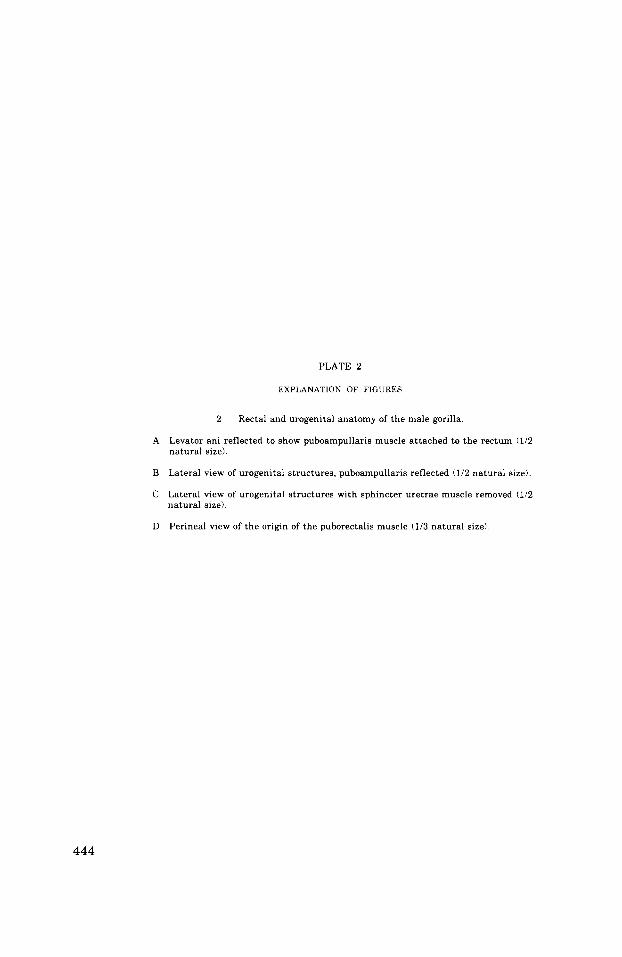
PLATE 2
EXPLANATION OF FIGURES
2 Rectal and urogenital anatomy of the male gorilla.
A Levator ani reflected to show puboampullaris muscle attached to the rectum (112 natural size).
B Lateral view of urogenital structures, puboampullaris reflected (1/2 natural size).
C Lateral view of urogenital structures with sphincter uretrae muscle removed (112 natural size).
Perineal view of the origin of the puborectalis muscle (113 natural size) D
444

PELVIC AND PERINEAL ANATOMY OF THE GORILLA Thomas M. Oelrich
PLATE 2
Levator ani (reflected) :
I~iococcygeuG m.
Pubococcygei m.
External anal sphincter
Rectourethralis (smooth
Bulbourethrol gland & duct