Embed Size (px)
Citation preview

PEER-REVIEWED ARTICLE bioresources.com
Songsamoe et al. (2020). “Essential oil & packaging,” BioResources 15(2), 2147-2162. 2147
Treatment of Fresh Water Hyacinth with Essential Oil Emulsion and Subsequent Use of the Plant Material for Control of Aspergillus flavus on Rice Dessert Products
Sumethee Songsamoe and Narumol Matan *
Essential oil emulsion, which is generally functionalized to have increased antifungal activity, has high solubility in water. The objective of this research was to develop an essential oil emulsion for absorption by water hyacinth plants in order to produce an essential oil absorbent material as a carrier of essential oil vapor that can be released to control mold in an enclosed packaging system. The fresh water hyacinth adsorbed the Michelia alba oil emulsion (100 µL/mL to 500 µL/mL) through its root system, transporting it to the stems and leaves after submerging the plant into the M .alba oil emulsion for 48 h. Dried sections (root, stem, and leaf) of the water hyacinth plant with absorbed M. alba essential oil emulsion released the M. alba vapor, inhibited Aspergillus flavus growth on malt extract agar (MEA), and also Thai dessert (Ja Mongkut) with the highest antifungal activity achieved with the concentration 500 µL/mL. The maximum essential oil absorption (345 µL/g) in the fresh plant was achieved after 48 h. The dried water hyacinth (≥ 0.4 g/L air) completely inhibited the growth of A. flavus for at least 7 days. In addition, the absorbent material prevented the growth of A. flavus on the Thai dessert (Ja Mongkut) with approximately 100% effectiveness (versus the control) for 10 days, thereby extending the shelf-life 2.5-fold in when compared with the control without essential oil.
Keywords: Absorbent material; Aspergillus flavus; Essential oil emulsion; Water hyacinth
Contact information: Food Science and Technology, School of Agricultural Technology, Walailak
University, Nakhon Si Thammarat 80160, Thailand;
*Corresponding author: [email protected]
INTRODUCTION
The growth of spoilage fungi and pathogenic molds on rice grains is undesirable,
as it affects the quality of grain by causing discoloration, loss in dry matter, degradation of
lipids and proteins, texture changes, and off odors (Christensen 1978). Moreover, some
molds produce mycotoxins, namely aflatoxin, ochratoxin, and deoxynivalenol, which are
associated with a high risk of human health problems if ingested (Magan and Aldred 2007;
Reddy et al. 2011). There are many reports of contaminated mold or mycotoxin from rice
grains affecting rice products (Iqbal et al. 2016), such as brown rice snack bars deteriorated
by Aspergillus flavus and natural mold within 3 d of storage (Suhem et al. 2015). Many
Thai desserts also have a short shelf-life caused by mold spoilage, which is a major issue
for the Thai dessert business (Nopwinyuwong et al. 2010). Thus, finding alternative
methods for controlling mold is necessary to improve the safety and quality of grains and
grain products.
PEER-REVIEWED ARTICLE bioresources.com
Songsamoe et al. (2020). “Essential oil & packaging,” BioResources 15(2), 2147-2162. 2148
Currently, many types of natural antimicrobial agents are applied in food products
to avoid the use of chemical preservatives. Essential oil is one of the most interesting
natural antimicrobial agents because it contains numerous natural compounds that can
inhibit microbial growth (Chen et al. 2014; Tao et al. 2019). Application of essential oil in
the vapor phase in antimicrobial packaging as a means to inhibiting mold on food has been
demonstrated to be effective and harmless for humans (Tyagi et al. 2012; Suhem et al.
2019). However, the essential oil vapor can easily vaporize over time, so high
concentrations are required for long term storage. Therefore, many researchers are trying
to create food packaging with a vapor-based essential oil absorbent material, such as edible
films, coatings, and sachets or pads (Oral et al. 2009; Seo et al. 2012; Balaguer et al. 2013).
The water hyacinth (Eichhornia crassipes (Mart.) Solms) is normally found in
garden ponds around tropical and subtropical regions (Zhang et al. 2010). This plant is
known to grow quickly, forming a large plant on the surface of the water (Villamagna and
Murphy 2010) that rapidly covers the pond surface in a short time (Malik 2007). In fact, it
can double in size between 6 d to 28 d and weigh in the range of 270 tons per hectare to
400 tons per hectare (Epstein 1998; Malik 2007). According to its extensive root system,
the water hyacinth root can consume large amounts of chemical pollutants in the water
(Gong et al. 2018), and its high cellulose polymer content can be used to produce low-cost
cellulose membranes and cellulose nanofibers (Sundari and Ramesh 2012; Istirokhatun et
al. 2015).
The objective of this research was to develop an essential oil emulsion for absorp-
tion by freshly harvested water hyacinth plants and thereafter to produce an absorbent ma-
terial serving as a carrier of the essential oil. The concept is that the vapor of the essential
oil can be released to control mold in an enclosed packaging system.
EXPERIMENTAL
Materials Essential oil
Michelia alba oil, derived by steam distillation, was provided by the Thai China
Flavors & Fragrances Industry Co., Ltd. (Bangkok, Thailand). Linalool (73.74%) and
caryophyllene (7.35%) are the two main compounds of Michelia oil.
Water hyacinth plants
The water hyacinth plant )60 ± 5 g(, including all the leaves and stem with a
diameter of 25 cm, 30 cm in height, and roots 6-cm-width× 0.2-cm-thick × 20-cm-length,
was harvested from a pond at Walailak University, Nakhon Si Thammarat, Thailand.
Before use, the whole plant was washed eight times by dipping into deionized water for 48
h.
Culture
Aspergillus flavus (106 CFU/mL) was isolated from brown rice. The A. flavus was
cultured on malt extract agar (MEA) before incubation at 25 °C for 7 days. Spore suspen-
sions of A. flavus (10-1 - 10-8 spore ml-1) were collected by flooding the surface of the plates

PEER-REVIEWED ARTICLE bioresources.com
Songsamoe et al. (2020). “Essential oil & packaging,” BioResources 15(2), 2147-2162. 2149
with ~9 mL of sterile water. The number of viable spores was checked by the plate count
method (AOAC 1992).
Methods Investigation of water hyacinth for essential oil emulsion absorption
First, the essential oil emulsion was prepared at concentrations of 100 µL/mL to
500 µL/mL by mixing M. alba oil, polyethylene glycol sorbitan monooleate (Tween 80;
3%, w/w; Tariko Co., Ltd., Bangkok, Thailand), and distilled water. The solution was
mixed using a magnetic stirrer for 1 min and then homogenized (T25 Digital Ultra-Turrax;
IKA, Staufen, Germany) at 24,000 rpm for 20 min. The droplet size of the emulsions was
observed by microscope (Carl Zeiss Jena Gmbh, Oberkochen, Germany) for 60 h, and the
stability of emulsion (d) was also assessed.
Second, to investigate the ability of the water hyacinth to absorb an essential oil
emulsion, the fresh plant was first submerged in each concentration of the essential oil
emulsion (100 µL/mL to 500 µL/mL) at room temperature. The flow of essential oil into
the plant and capacity of the essential oil emulsion in each plant was assessed every 6 h for
60 h. Then, the plant was dried at 50 °C in a hot air oven for 12 h. Next, the dried plant was
ground and kept at 20 °C and 65% relative humidity before it was weighed using a digital
balance (PA214; Ohaus Co., Ltd., Parsippany, NJ, USA) or 14% moisture content was
achieved. A control treatment was performed in the same way but without essential oil.
The capacity of essential oil uptake in water hyacinth plant during and after the absorption
process was calculated according to Eqs. 1 and 2, respectively,
Essential oil emulsion uptake capacity (µL/g) = Vt/W (1)
where Vt is the absorbed volume of essential oil emulsion (µL), at time and W is the initial
wet weight of water hyacinth plant (g),
Pure essential oil uptake capacity (µL/g) = (Vt × C)/W (2)
where Vt is the absorbed volume of essential oil emulsion (µL), at time t, C is the
concentration of essential oil emulsion (µL/mL), and W is the initial wet weight of water
hyacinth plant (g).
The morphology of the water hyacinth plant before and after uptake of the essential
oil emulsion was observed using an epifluorescent microscope and Nile red (Sigma-
Aldrich, St. Louis, MO, USA) fluorescent lipophilic stain. The Nile red solution was
prepared by dissolving it in methanol at a concentration of 1 g/L. The plant sections were
stained by Nile red solution, placed on a glass slide, enclosed with a cover slip, and
observed under an epifluorescent microscope (Nikon Eclipse E600 Microscope Y-FL EPI
Fluorescence Attachment; Nikon, Tokyo, Japan) using excitation wavelengths from 380
nm to 420 nm.
Antifungal effect of the absorbent material made from water plants containing essential
oil against A. flavus
To develop an absorbent material from the water plant, each root, stem, and leaf of
water plant was cut and dried in a tray for 12 h at 50 ºC. Then, 0.2 g to 2.4 g of the dried
plant parts were packed into a cellulose bag before they were placed into a 1-L plastic box.
Mycelia of A. flavus (diameter approximately 5 mm) grown for 7 days on MEA were added

PEER-REVIEWED ARTICLE bioresources.com
Songsamoe et al. (2020). “Essential oil & packaging,” BioResources 15(2), 2147-2162. 2150
to the center of a MEA plate and added into the plastic box. In addition, a spore suspension
of A. flavus (0.1 mL) was spread on a MEA plate and placed into the plastic box. All plates
(without lid) inside the box were incubated at 25 °C for 7 days. The colony diameter (mm)
of A. flavus on each plate was checked every day for 7 days. The MIC was defined as the
lowest concentration tested for which no mold growth was detected. Every test was per-
formed in triplicate and palm oil (Morakot Industries PCL, Bangkok, Thailand) was used
as a control.
Antifungal effect of the developed absorbent material on the growth of A. flavus on rice
dessert products
Rice desserts (Ja Mongkuts) were produced using a Thai traditional recipe, rice
flour (made from rice grain), sugar, egg yolk, coconut milk, and watermelon seeds as the
main ingredients. After stirring all ingredients at 60 °C for 30 min, the paste was added
into the flour base before adding the melon seeds. Before testing, the desserts were exposed
to UV-C radiation ( = 253.7 nm, Phillips UV-C 30 W lamp; Phillips, Amsterdam,
Netherlands) for 30 min in a biological safety cabinet (Cellgard, Charlotte, NC, USA). The
desserts were then incubated with the A. flavus spore suspension at 25 °C and 100% relative
humidity for 10 d in the 1-L plastic box with 1 g of dried water plant containing M. alba
emulsion (100 µL/mL to 500 µL/mL). Palm oil (Morakot Industries PCL, Bangkok,
Thailand) at the same concentrations was used as a control. The inhibitory period (d) was
recorded as the time when no growth of mold on the surface was observed. The number of
A. flavus was also counted on MEA plates. Then, the percent inhibition of mold growth
was calculated by the following Eq. 3,
Inhibition (%) = ((Nc - Ns)/Nc) × 100 (3)
where Nc is the colony count number (log10 cfu/mL) in the control and Ns is the number of
the colony count (log10 cfu/mL) in the sample.
In addition, the dessert specimens with 1 g of dried water plant containing M. alba
emulsion (100 µL/mL to 500 µL/mL) and the control stored at 25 °C for 48 h were
subjected to sensory analysis by an untrained panel (43 panelists: 25 females and 18 males)
that ranged from 18 to 45 years of age. Panelists were selected from students and staffs at
Walailak University, Nakon Si Tammarat, Thailand. The dessert was randomly coded and
presented to each panelist seated separately in a control booth. A 9-point hedonic scale
ranging from “like extremely” to “dislike extremely” was used to determine the degree of
acceptance of the specimens (Meilgaard et al. 1999).
Fourier transform infrared spectroscopy (FTIR)
Fourier transform infrared spectroscopy was used to investigate the changes in
chemical bonds on the surface of the dried absorbent (without M. alba oil emulsion and
500 µL/mL). The internal reflection crystal, which was made of zinc selenide, had a 45º
angle of incidence to the IR beam. Spectra were acquired at a resolution of 4 cm-1 and the
measurement range was from 4000 cm-1 to 600 cm-1 (mid-IR region) at room temperature.
Automatic signals were collected in 32 scans at a resolution of 4 cm-1 and normalized
against a background spectrum recorded from the clean, empty cell at 25 °C. Analysis of
spectral data was completed using the Spectrum One software program (Perkin Elmer,
PEER-REVIEWED ARTICLE bioresources.com
Songsamoe et al. (2020). “Essential oil & packaging,” BioResources 15(2), 2147-2162. 2151
version 6.3.5, Waltham, MA, USA). A change in the chemical composition of each sample
was observed (Suhem et al. 2017).
Statistical analysis
All results were expressed as the mean ± standard deviation. One-way analysis of
variance (ANOVA) and Duncan's post hoc test, with p < 0.05 being considered statistically
significant, were employed in the statistical analysis using Statistica software (StatSoft
Inc., version 11.0, Tulsa, OK, USA).
RESULTS AND DISCUSSION
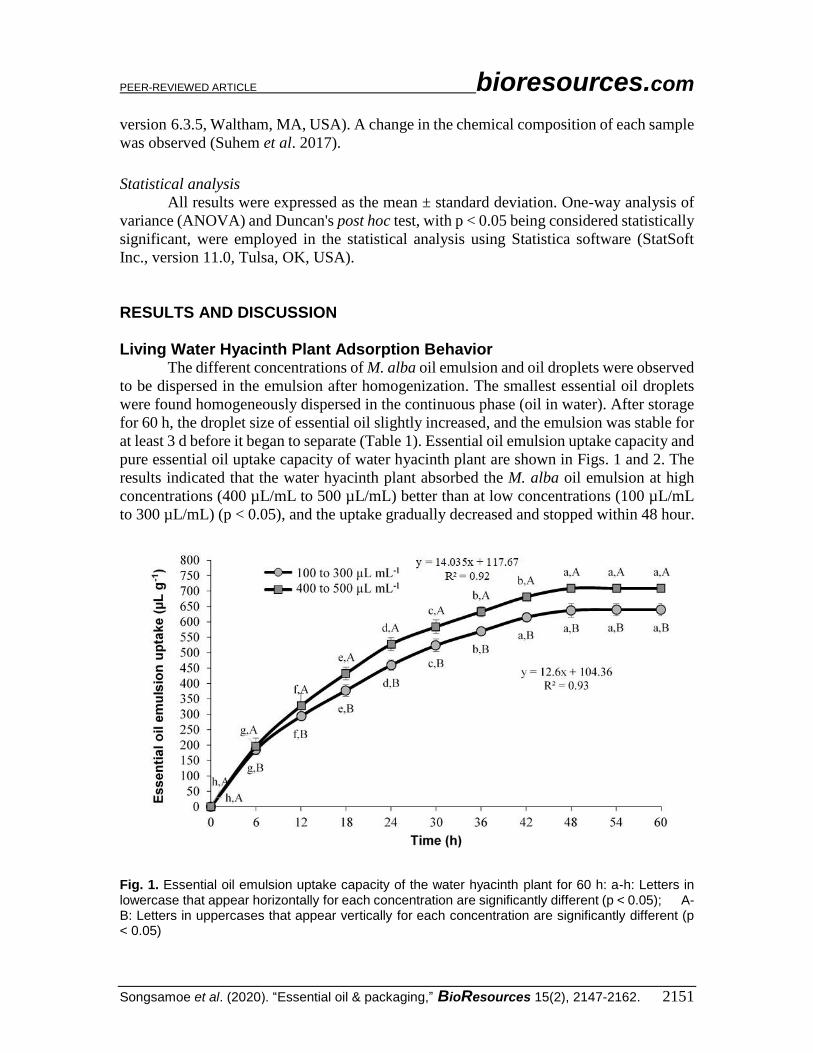
Living Water Hyacinth Plant Adsorption Behavior The different concentrations of M. alba oil emulsion and oil droplets were observed
to be dispersed in the emulsion after homogenization. The smallest essential oil droplets
were found homogeneously dispersed in the continuous phase (oil in water). After storage
for 60 h, the droplet size of essential oil slightly increased, and the emulsion was stable for
at least 3 d before it began to separate (Table 1). Essential oil emulsion uptake capacity and
pure essential oil uptake capacity of water hyacinth plant are shown in Figs. 1 and 2. The
results indicated that the water hyacinth plant absorbed the M. alba oil emulsion at high
concentrations (400 µL/mL to 500 µL/mL) better than at low concentrations (100 µL/mL
to 300 µL/mL) (p < 0.05), and the uptake gradually decreased and stopped within 48 hour.
Fig. 1. Essential oil emulsion uptake capacity of the water hyacinth plant for 60 h: a-h: Letters in lowercase that appear horizontally for each concentration are significantly different (p < 0.05); A-B: Letters in uppercases that appear vertically for each concentration are significantly different (p < 0.05)

PEER-REVIEWED ARTICLE bioresources.com
Songsamoe et al. (2020). “Essential oil & packaging,” BioResources 15(2), 2147-2162. 2152
Fig. 2. Pure essential oil uptake capacity of water hyacinth plant for 60 h: a-h: Letters in lowercase that appear horizontally for each concentration are significantly different (p < 0.05); A-D: Letters in uppercases that appear vertically for each concentration are significantly different (p < 0.05)
Table 1. Stability of M. alba Oil Emulsion
Concentration (µL/mL) Stability Time (d) Oil Droplet Mean Diameter (µm)
After Homogenization
After 60 h
100 4 1.23 1.49
200 4 1.57 1.86
300 4 1.61 2.08
400 3 1.71 2.40
500 3 2.09 2.73
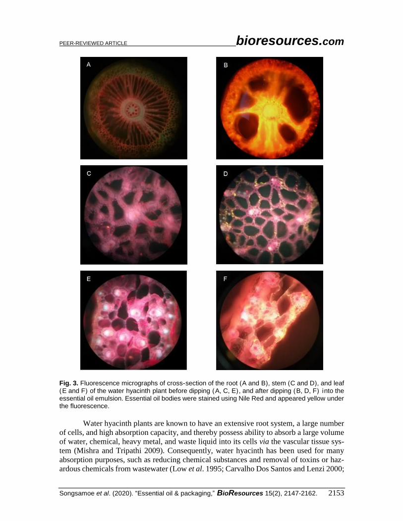
The fluorescence (380 nm to 420 nm wavelengths) micrographs of essential oil in
the plant are shown in Fig. 3. The M. alba emulsion was observed in the lipophilic parts of
the plant tissue, as indicated by the fluorescence in the root, stem, and leaves, with the
highest concentration found in the root.
The results from this study confirmed that water hyacinth could also absorb
essential oil emulsions. The essential oil emulsion passed through the vascular tissue
system of the aquatic vascular plants before accumulation in the root, stem, and leaf
sections. The water hyacinth reached its capacity of essential oil emulsion within 48 h. In
comparison with other substances, the whole plant of water hyacinth after submersion or
floating on hazardous wastewater was found to be fresh (Mukherjee and Mondal 1995; Al
Rmalli et al. 2005; Malik 2007) and difficult to harvest.
PEER-REVIEWED ARTICLE bioresources.com
Songsamoe et al. (2020). “Essential oil & packaging,” BioResources 15(2), 2147-2162. 2153
Fig. 3. Fluorescence micrographs of cross-section of the root )A and B(, stem )C and D(, and leaf )E and F( of the water hyacinth plant before dipping )A, C, E( , and after dipping )B, D, F i ( nto the essential oil emulsion. Essential oil bodies were stained using Nile Red and appeared yellow under the fluorescence.
Water hyacinth plants are known to have an extensive root system, a large number
of cells, and high absorption capacity, and thereby possess ability to absorb a large volume
of water, chemical, heavy metal, and waste liquid into its cells via the vascular tissue sys-
tem (Mishra and Tripathi 2009). Consequently, water hyacinth has been used for many
absorption purposes, such as reducing chemical substances and removal of toxins or haz-
ardous chemicals from wastewater (Low et al. 1995; Carvalho Dos Santos and Lenzi 2000;

PEER-REVIEWED ARTICLE bioresources.com
Songsamoe et al. (2020). “Essential oil & packaging,” BioResources 15(2), 2147-2162. 2154
Hasan et al. 2010; Murithi et al. 2014; Gong et al. 2018). For example, Mukherjee and
Mondal (1995) showed that the living water hyacinth reduced the concentration of Pb (0.5
mg/L to 10 mg/L) by 85% to 92% within 10 days. Delgado et al. (1993) reported that living
water hyacinth completely removed Zn, Cr, and Cd from water within 24 hour. Further-
more, Al Rmalli et al. (2005) showed that the powdered dried root of water hyacinth ab-
sorbed 93% of As (III) and 95% of As (V) from 200 μg As/L within 60 min. Additionally,
Low et al. (1995) revealed that the dried roots of water hyacinth have a similarly high
ability to absorb and remove two basic dyes (methylene blue and Victoria blue) from aque-
ous solutions as commercial activated carbon.
The morphology of the water hyacinth plant after uptake of the essential oil emul-
sion (Fig. 3) and the antifungal activity of the absorbent from each section of plant (Fig. 4)
demonstrated that water hyacinth plant absorbed essential oil emulsion and accumulated it
in the root section with the highest level of essential oil, followed by stem and leaf sections.
This phenomenon has also previously been observed and reported (Mishra and Tripathi
2009).
Fig. 4. Effects of M. alba vapor released from each section of the dried water hyacinth ) 0. 4 g/ L) air aborbed M .alba oil emulsion at concentration of 500 µL/mL on the mycelium growth of A .flavus
This research also confirmed that essential oil in the form of an emulsion could be
taken up by the water plants. The essential oil in an emulsion was stable, and the constant
small droplet size helped disperse the essential oil in the plant after absorption, and was
less costly for large-scale essential oil preparation (Donsì and Ferrari 2016). Moreover, the
small droplets enhanced the transportation of active molecules throughout the cell
membranes, thereby achieving higher antifungal activity. Furthermore, the water hyacinth
absorbed the essential oil emulsion by its natural vascular system without the need for any

PEER-REVIEWED ARTICLE bioresources.com
Songsamoe et al. (2020). “Essential oil & packaging,” BioResources 15(2), 2147-2162. 2155
additional devices, such as a high-pressure pump, and therefore the absorption process does
not require additional energy costs.
The essential oil (M. alba) emulsion was created to improve the dispersion of oil in
the water phase for absorption by the plant. The results of this experiment confirmed that
the polysorbate 80 (Tween 80; 3%, w/w) and colloid milling homogenization could be used
to prepare a high-quality essential oil emulsion with small-sized droplets, good oil
dispersion, and satisfactory stability. These results agreed with many reports that high shear
homogenization could be used to create essential oil emulsions at a low cost (Donsì et al.
2012; Ribes et al. 2016). Polysorbate 80 is a non-ionic surfactant made from sorbitol, oleic
acid, and epoxy ethane, which is widely used as an emulsifier, solubilizer, and stabilizer in
food, cosmetics, drugs, and biodegradation media due to its high surface activity and low
toxicity (Pan et al. 2016). Hence, it is considered safe for the production of essential oil
emulsion.
MICs of Plant Absorbent Material Containing M. alba Oil The effects of M. alba vapor released from sections of the root, stem, and leaf )of
the dried water hyacinth plants) absorbent material on the mycelium growth of A. flavus in
MEA are presented in Fig. 4 and Table 2. The results showed that the absorbents from all
parts of the plant reduced the growth of A. flavus for 7 days at 25 °C. The root section of
the dried plant had the highest antifungal activity, followed by stem and leaf section,
respectively (Fig. 4). The highest concentration of 500 µL/mL was more effective at
preventing A. flavus growth than the lowest concentration of 100 µL/mL .However, it was
observed that the M. alba oil emulsion at 200 µL/mL had similar antifungal activity with
the 500 µL/mL concentration
Additionally, the MIC values of the dried water hyacinth plants on the spore
germination of A. flavus are presented in Table 2. The results indicated that the root section
of the dried plant had the highest antifungal activity, and ≥ 0.4 g/L air completely inhibited
the growth of A. flavus for at least 7 days.
While the stem of the dried plant had lower antifungal activity than the root section,
the MIC of all treatments was not less than 0.8 g/L air. The leaf section of the dried plant
showed the lowest antifungal activity, and the MIC of all treatments was not less than 2.0
g/L air.
The antifungal activity depended on the volume of pure essential oil in plant, and
antifungal activity increased when the volume of pure essential oil in the plant increased.
Additionally, the micrographs confirmed that the highest volume of essential oil was detected
in the root section, followed by the stem and leaf.

PEER-REVIEWED ARTICLE bioresources.com
Songsamoe et al. (2020). “Essential oil & packaging,” BioResources 15(2), 2147-2162. 2156
Table 2. MICs of Plant Absorbent Material Containing M. alba Oil on the Growth of A. flavus
Treatments Part of Dried Plant MIC (g/L air)
Dried plant absorbed EO 100 µL/mL
Root 1.6
Stem 2.0
Leaf > 2.4
Dried plant absorbed EO 200 µL/mL
Root 0.8
Stem 1.0
Leaf 2.0
Dried plant absorbed EO 300 µL/mL
Root 1.0
Stem 1.2
Leaf 2.4
Dried plant absorbed EO 400 µL/mL
Root 1.0
Stem 1.2
Leaf 2.0
Dried plant absorbed EO 500 µL/mL
Root 0.4
Stem 0.8
Leaf 2.0
Shelf-life Extension of Thai Dessert Mold inhibition (%) and period inhibition (day) of A. flavus on rice dessert (Ja
Mongkut) are presented in Table 3. The results indicated that the dried plant absorption
material that contained the emulsion of M. alba oil (500 µL/mL) prevented the growth of
A. flavus on Ja Mongkut with approximately 100% effectiveness (based control) for 10
days, thereby extending the shelf-life of the Ja Mongkut 2.5-fold when compared with the
control without essential oil (4 days). Furthermore, at highest concentration (500 µL/mL),
the sensory test results showed that the consumer gave higher scores (9 point hedonic of
“like slightly” to “like moderately”) to Ja Mongkut with M. alba vapor than the control
(neither like nor dislike).
Table 3. Inhibition and Inhibition Period of A. flavus on Thai Dessert after Being Packed with 1 g of Absorbent Material Containing M. alba oil
Samples Inhibition (%) Period Inhibition (d)
Absorbed EO 100 µL/mL 10 4
Absorbed EO 200 µL/mL 93 8
Absorbed EO 300 µL/mL 70 6
Absorbed EO 400 µL/mL 87 7
Absorbed EO 500 µL/mL 100 10
Control 0 4
Possible Modes of Action The FTIR spectra of the dried water hyacinth with and without M. alba oil emulsion
and linalool emulsion are shown in Fig. 5. The FTIR spectra of all sections of water
hyacinth )root, stem, and leaf( containing M. alba oil emulsion displayed some
characteristic changes )compared with control(, especially in the peak range of 3000 cm-1
to 2800 cm-1 (representative of C-H groups).

PEER-REVIEWED ARTICLE bioresources.com
Songsamoe et al. (2020). “Essential oil & packaging,” BioResources 15(2), 2147-2162. 2157
Fig. 5. FTIR spectra of absorbent from water hyacinth (root, stem, and leaf) with and without M .alba oil emulsion and linalool emulsion

PEER-REVIEWED ARTICLE bioresources.com
Songsamoe et al. (2020). “Essential oil & packaging,” BioResources 15(2), 2147-2162. 2158
In addition, the amplitude of the peak range of 900 to 1200 cm-1 (representative of
C-O-C and C-O stretching the ester bond), 1200 to 1700 cm-1 (representative of C-OH, C-
C and C-O-C stretching), and 1625 to 1710 cm-1 (representative of C=O stretching and
hydrogen bonding coupled with COO-1, respectively) decreased, whereas the region of
3000 to 3700 (representative of hydrogen stretching the H-bonded OH group) clearly
increased. Additionally, the peak pattern of water hyacinth containing M. alba oil emulsion
was similar to that of the water hyacinth containing the linalool emulsion. In the
comparison of the FTIR results of each section of the dried plant, the peak pattern of root
and stem sections clearly changed, whereas the peak pattern of the leaf section showed
little change when compared with the control.
The FTIR spectra of all sections of water hyacinth )root, stem, and leaf( containing
M. alba oil emulsion suggested that linalool was the main active component of the essential
oil emulsion, as peak pattern changes in the peak range of 3000 cm-1 to 2800 cm-1 might
have indicated changes in the C-H group of linalool. Previous studies by Kinninmonth et
al. )2013( and Rani et al. )2014( reported that the peak at 2923 cm-1 to 3000 cm-1 indicates
the presence of C-H stretching in linalool. In addition, changes of the amplitude of the other
peak ranges suggested that M. alba oil emulsion could be incorporated into the water hya-
cinth and active on the surface, and this binding might cause a change in the water hyacinth
structure, which may affect its physical properties.
This study was aimed at developing an essential oil absorbent material from water
hyacinth plant for use as an antifungal agent in rice desserts. The findings from this study
confirmed that the developed absorbent from the water hyacinth plant could be applied to
prevent mold growth on the surface of rice dessert. The M. alba oil contains aroma constit-
uents and bioactive components that have been used as anti-inflammatory agents to treat
cramps, abdominal pain (Lee et al. 2005), fever, syphilis, and malaria (Asaruddin et al.
2003). Additionally, it has been shown to inhibit the growth of Aspergillus niger, Asper-
gillus flavus, Penicillium sp., Rhizopus sp., Fusarium sp., and Cladosporium sp. (Song-
samoe et al. 2017; Suhem et al. 2017). Moreover, its major component, linalool, has been
shown to inhibit bacteria and molds, such as Escherichia coli, Staphylococcus aureus, Ba-
cillus subtilis, Pseudomonas aeruginosa, Botrytis cinerea, Candida albicans, Penicillium
italicum, and Fusarium oxysporum (Bakkali et al. 2008; Shimada et al. 2014). The findings
from this study confirmed that the developed absorbent from the water hyacinth plant could
be applied to prevent mold growth on the surface of rice dessert. The vapor phase of the
active main component of M. alba oil, linalool, was released from the plant absorbent ma-
terial (≥ 0.4 g/L air) and inhibited mold growth on the rice-based dessert while maintaining
the flavor of the dessert.
CONCLUSIONS
1. The M. alba oil emulsion at concentrations from 100 µL/mL to 500 µL/mL was
absorbed into the plant via the plant vascular system.
2. The M. alba oil emulsion at a concentration of 500 µL/mL in the root exhibited the
highest inhibition for mold growth

PEER-REVIEWED ARTICLE bioresources.com
Songsamoe et al. (2020). “Essential oil & packaging,” BioResources 15(2), 2147-2162. 2159
3. The vapor of M. alba oil released from the absorbent (500 µL/mL) from the root (1 g)
inhibited fungal growth, which extended the shelf-life of the Ja Mongkut up to 10 days,
with a high score of 9 points on the hedonic scale.
4. The novel absorbent material from water hyacinth plants containing essential oil
emulsion has great potential as a food preservative in packaging for rice desserts to
prevent against fungal spoilage.
ACKNOWLEDGMENTS
This study was supported by the Thailand Research Fund (TRF) through the Royal
Golden Jubilee Ph.D. Program (Grant No. PHD/0090/2014), the Walailak University Fund.
REFERENCES CITED
Al Rmalli, S. W., Harrington, C. F., Ayub, M., and Haris, P. I. (2005). “A biomaterial
based approach for arsenic removal from water,” J. Environ. Monitor. 7(4), 279-282.
DOI: 10.1039/B500932D
AOAC International. (1992). FDA Bacteriology Analytical Manual, 7th Edition,
Arlington, VA 22201-3301 USA, 227-234.
Asaruddin, M. R., Honda, G., Tsubouchi, A., Nakajima-Shimada, J., Aoki, T., and
Kiuchi, F. (2003). “Trypanocidal constituents from Michelia alba,” Nat. Med. 57(2),
61-63.
Bakkali, F., Averbeck, S., Averbeck, D., and Idaomar, M. (2008). “Biological effects of
essential oils – A review,” Food Chem. Toxicol. 46(2), 446-475. DOI:
10.1016/j.fct.2007.09.106
Balaguer, M. P., Lopez-Carballo, G., Catala, R., Gavara, R., and Hernandez-Munoz, P.
(2013). “Antifungal properties of gliadin films incorporating cinnamaldehyde and
application in active food packaging of bread and cheese spread foodstuffs,” Int. J.
Food Microbiol. 166(3), 369-377. DOI: 10.1016/j.ijfoodmicro.2013.08.012
Carvalho Dos Santos, M., and Lenzi, E. (2000). “The use of aquatic macrophytes
(Eichhornia crassipes) as a biological filter in the treatment of lead contaminated
effluents,” Environ. Technol. 21(6), 615-622. DOI: 10.1080/09593330.2000.9618946
Chen, C., Tong, Z., Liao, D., Li, Y., Yang, G., and Li, M. (2014). “Chemical composition
and antimicrobial and DPPH scavenging activity of essential oil of Toona sinensis (A.
Juss.) Roem from China,” BioResources 9(3), 5262-5278. DOI:
10.15376/biores.9.3.5262-5278
Christensen, C. M. (1978). “Fungi and seed quality,” Outlook Agr. 9(5), 209-213.
DOI: 10.1177/003072707800900503
Delgado, M., Bigeriego, M., and Guardiola, E. (1993). “Uptake of Zn, Cr and Cd by
water hyacinths,” Water Res. 27(2), 269-272. DOI: 10.1016/0043-1354(93)90085-V
Donsì, F., and Ferrari, G. (2016). “Essential oil nanoemulsions as antimicrobial agents in
food,” J. Biotechnol. 233, 106-120. DOI: 10.1016/j.jbiotec.2016.07.005

PEER-REVIEWED ARTICLE bioresources.com
Songsamoe et al. (2020). “Essential oil & packaging,” BioResources 15(2), 2147-2162. 2160
Donsì, F., Annunziata, M., Vincensi, M., and Ferrari, G. (2012). “Design of
nanoemulsion-based delivery systems of natural antimicrobials: Effect of the
emulsifier,” J. Biotechnol. 159(4), 342-350. DOI: 10.1016/j.jbiotec.2011.07.001
Epstein, P. (1998). “Weeds bring disease to the east African waterways,” Lancet
351(9102), 577. DOI: 10.1016/s0140-6736(05)78570-6
Gong, Y., Zhou, X., Ma, X., and Chen, J. (2018). “Sustainable removal of formaldehyde
using controllable water hyacinth”. J. Clean. Prod. 181, 1-7. DOI:
10.1016/j.jclepro.2018.01.220
Hasan, S. H., Ranjan, D., and Talat, M. (2010). “Water hyacinth biomass (WHB) for the
biosorption of hexavalent chromium: Optimization of process parameters,”
BioResources 5(2), 563-575. DOI: 10.15376/biores.5.2.563-575
Iqbal, S. Z., Asi, M. R., Hanif, U., Zuber, M., and Jinap, S. (2016). “The presence of
aflatoxins and ochratoxin A in rice and rice products; and evaluation of dietary
intake,” Food Chem. 210, 135-140. DOI: 10.1016/j.foodchem.2016.04.104
Istirokhatun, T., Rokhati, N., Rachmawaty, R., Meriyani, M., Priyanto, S., and Susanto,
H. (2015). “Cellulose isolation from tropical water hyacinth for membrane
preparation,” Procedia Environ. Sci. 23, 274-281. DOI:
10.1016/j.proenv.2015.01.041
Kinninmonth, M. A., Liauw, C. M., Verran, J., Taylor, R., Edwards-Jones, V., Shaw, D.,
and Webb, M. (2013). “Investigation into the suitability of layered silicates as
adsorption media for essential oils using FTIR and GC–MS,” Appl. Clay Sci. 83-84,
415-425. DOI: 10.1016/j.clay.2013.07.009
Lee, J., Jung, E., Park, J., Jung, K., Lee, S., Hong, S., Park, J., Park, E., Kim, J., Park, S.,
et al. (2005). “Anti-inflammatory effects of magnolol and honokiol are mediated
through inhibition of the downstream pathway of MEKK-1 in NF-κB activation
signaling,” Planta. Med. 71(4), 338-343. DOI: 10.1055/s-2005-864100
Low, K. S., Lee, C. K., and Tan, K. K. (1995). “Biosorption of basic dyes by water
hyacinth roots,” Bioresource Technol. 52(1), 79-83. DOI: 10.1016/0960-
8524(95)00007-2
Magan, N., and Aldred, D. (2007). “Post-harvest control strategies: Minimizing
mycotoxins in the food chain,” Int. J. Food Microbiol. 119(1-2), 131-139. DOI:
10.1016/j.ijfoodmicro.2007.07.034
Malik, A. (2007). “Environmental challenge vis a vis opportunity: The case of water
hyacinth,” Environ. Int. 33(1), 122-138. DOI: 10.1016/j.envint.2006.08.004
Mishra, V. K., and Tripathi, B. D. (2009). “Accumulation of chromium and zinc from
aqueous solutions using water hyacinth (Eichhornia crassipes),” J. Hazard. Mater.
164(2-3), 1059-1063. DOI: 10.1016/j.jhazmat.2008.09.020
Mukherjee, S., and Mondal, G. C. (1995). “Removal of lead by water hyacinth,” Indian J.
Chem. Technol. 2, 59-62.
Murithi, G., Onindo, C. O., Wambu, E. W., and Muthakia, G. K. (2014). “Removal of
cadmium (II) ions from water by adsorption using water hyacinth (Eichhornia
crassipes) biomass,” BioResources 9(2), 3613-3631. DOI: 10.15376/biores.9.2.3613-
3631
Nopwinyuwong, A., Trevanich, S., and Suppakul, P. (2010). “Development of a novel
colorimetric indicator label for monitoring freshness of intermediate-moisture dessert
spoilage,” Talanta. 81(3), 1126-1132. DOI: 10.1016/j.talanta.2010.02.008.

PEER-REVIEWED ARTICLE bioresources.com
Songsamoe et al. (2020). “Essential oil & packaging,” BioResources 15(2), 2147-2162. 2161
Oral, N., Vatansever, L., Sezer, Ç., Aydın, B., Güven, A., Gülmez, M., Baser, K. H. C.,
and Kürkçüoğlu, M. (2009). “Effect of absorbent pads containing oregano essential
oil on the shelf life extension of overwrap packed chicken drumsticks stored at four
degrees Celsius,” Poultry Sci. 88(7), 1459-1465. DOI: 10.3382/ps.2008-00375
Pan, J., Ji, Y., Du, Z., and Zhang, J. (2016). “Rapid characterization of commercial
polysorbate 80 by ultra-high performance supercritical fluid chromatography
combined with quadrupole time-of-flight mass spectrometry,” J. Chromatogr. A
1465, 190-196. DOI: 10.1016/j.chroma.2016.08.051
Rani, P. U., Madhusudhanamurthy, J., and Sreedhar, B. (2014). “Dynamic adsorption of
α-pinene and linalool on silica nanoparticles for enhanced antifeedant activity against
agricultural pests,” J. Pest Sci. 87(1), 191-200. DOI: 10.1007/s10340-013-0538-2
Reddy, K. R. N., Raghavender, C. R., Salleh, B., Reddy, C. S., and Reddy, B. N. (2011).
“Potential of aflatoxin B1 production by Aspergillus flavus strains on commercially
important food grains,” Int. J. Food Sci. Technol. 46(1), 161-165. DOI:
10.1111/j.1365-2621.2010.02468.x
Ribes, S., Fuentes, A., Talens, P., and Barat, J. M. (2016). “Use of oil-in-water emulsions
to control fungal deterioration of strawberry jams,” Food Chem. 211, 92-99. DOI:
10.1016/j.foodchem.2016.05.040
Seo, H.-S., Bang, J., Kim, H., Beuchat, L. R., Cho, S. Y., and Ryu, J.-H. (2012).
“Development of an antimicrobial sachet containing encapsulated allyl isothiocyanate
to inactivate Escherichia coli O157: H7 on spinach leaves,” Int. J. Food Microbiol.
159(2), 136-143. DOI: 10.1016/j.ijfoodmicro.2012.08.009
Shimada, T., Endo, T., Fujii, H., Rodríguez, A., Peña, L., and Omura, M. (2014).
“Characterization of three linalool synthase genes from Citrus unshiu Marc. and
analysis of linalool-mediated resistance against Xanthomonas citri subsp. citri and
Penicillium italicum in citrus leaves and fruits,” Plant Sci. 229, 154-166. DOI:
10.1016/j.plantsci.2014.09.008
Songsamoe, S., Matan, N., and Matan, N. (2017). “Antifungal activity of Michelia alba
oil in the vapor phase and the synergistic effect of major essential oil components
against Aspergillus flavus on brown rice,” Food Control 77, 150-157. DOI:
10.1016/j.foodcont.2017.02.010
Suhem, K., Matan, N., and Matan, N. (2019). “Effect of high temperature with Litsea
cubeba Pers. to control mold growth on bamboo food packaging and its possible
modes of action,” BioResources 14(1), 1289-1302. DOI: 10.15376/biores.14.1.1289-
1302
Suhem, K., Matan, N., Matan, N., Danworaphong, S., and Aewsiri T. (2015).
“Improvement of the antifungal activity of Litsea cubeba vapor by using a helium–
neon (He–Ne) laser against Aspergillus flavus on brown rice snack bars,” Int. J. Food
Microbiol. 215, 157-160. DOI: 10.1016/j.ijfoodmicro.2015.09.008
Suhem, K., Matan, N., Matan, N., Danworaphong, S., and Aewsiri, T. (2017). “Enhanced
antifungal activity of michelia oil on the surface of bamboo paper packaging boxes
using helium-neon (HeNe) laser and its application to brown rice snack bar,” Food
Control 73, 939-945. DOI: 10.1016/j.foodcont.2016.10.006
Sundari, M. T., and Ramesh, A. (2012). “Isolation and characterization of cellulose
nanofibers from the aquatic weed water hyacinth – Eichhornia crassipes,” Carbohyd.
Polym. 87(2), 1701-1705. DOI: 10.1016/j.carbpol.2011.09.076

PEER-REVIEWED ARTICLE bioresources.com
Songsamoe et al. (2020). “Essential oil & packaging,” BioResources 15(2), 2147-2162. 2162
Tao, C., Wang, Y., Zhang, X., Li, L., Wu, Y., Han, X., Jiang, X., and Lv, Z. (2019).
“Mechanism of action of essential oils extracted from bamboo (Phyllostachys
heterocycla cv. pubescens) leaves: Chemical composition and antimicrobial activity
against four food-related microorganisms,” BioResources 14(1), 1419-1434. DOI:
10.15376/biores.14.1.1419-1434
Tyagi, A. K., Malik, A., Gottardi, D., and Guerzoni, M. E. (2012). “Essential oil vapour
and negative air ions: A novel tool for food preservation,” Trends Food Sci. Tech.
26(2), 99-113. DOI: 10.1016/j.tifs.2012.02.004
Villamagna, A. M., and Murphy, B. R. (2010). “Ecological and socio-economic impacts
of invasive water hyacinth (Eichhornia crassipes): A review,” Freshwater Biol.
55(2), 282-298. DOI: 10.1111/j.1365-2427.2009.02294.x
Zhang, Y.-Y., Zhang, D.-Y., and Barrett, S. H. (2010). “Genetic uniformity characterizes
the invasive spread of water hyacinth (Eichhornia crassipes), a clonal aquatic plant,”
Mol. Ecol. 19(9), 1774-1786. DOI: 10.1111/j.1365-294X.2010.04609.x
Article submitted: August 29, 2019; Peer review completed: November 23, 2019;
Revised version received: January 6, 2020; Accepted: January 7, 2020; Published:
February 3, 2020.
DOI: 10.15376/biores.15.2.2147-2162