Embed Size (px)
Citation preview
ABSTRACT
Sao Paulo Med J. 2008;126(3):190-3.
Case R
eport Pedro Popoutchi
Carlos Renato dos Reis Lemos
Julio César Rosa e Silva
Antônio Alberto Nogueira
Omar Feres
José Joaquim Ribeiro da Rocha
Postmenopausal intestinal obstructive endometriosis: case report and review of the literatureDiscipline of Coloproctology, Department of Surgery and Anatomy, Faculdade de Medicina de Ribeirão Preto (FMRP), Universidade de São Paulo (USP), Ribeirão Preto, São Paulo, Brazil
CONTEXT: Endometriosis is characterized by the presence of endometrial tissue outside the uterine cavity, which is commonly detected in gynecological practice but rarely reported as a coloproctological disorder. The objective of the present report was to discuss a rare case of postmenopausal intestinal endometriosis simulating a malignant lesion, following a review of the literature.
CASE REPORT: A 74-year-old woman with com-plaints of hematochezia and tenesmus of two months’ duration accompanied by liquid feces and pelvic pain, but with no other gastrointestinal or gynecological complaints, was referred to our service. She had been menopausal for 22 years, with no hormone replacement treatment, and had undergone panhysterectomy three years before the referral to us, due to endometrial thickening and a right adnexal cyst. Five months before this referral, she had undergone laparotomy due to acute obstructive abdomen, which revealed a tumor mass involving the small bowel. Anato-mopathological examination of the enterectomy suggested a hypothesis of intestinal endometrio-sis. A proctological examination was normal. Computed tomography of the pelvis revealed thickening of the rectosigmoid transition and colonoscopy revealed friable tumor formation in the rectum. A biopsy of the lesion revealed muco-sal fragments of endometrial type, which led to a review of the previous anatomopathological examination. The patient underwent rectosig-moidectomy with protective transversotomy, with a good postoperative course, and anatomical examination confi rmed the intestinal endometrio-sis. The patient subsequently suffered a stenosing recurrence of the lesion and has undergone colostomy since then.
KEY WORDS: Intestines. Endometriosis. Surgery. Diagnosis. Postmenopause.
INTRODUCTIONEndometriosisisanestrogen-dependent
diseasethatusuallyoccursinwomenduringthe menacme. Its etiopathogenesis is still amatterofcontroversy.1-3However,occurrencesofendometriosisinpatientswithnomenstrualflow,4,5orthepresenceoflesionsatsiteswherethereisnodirectcontactwithmenstrualflow,suchasthelungsandintestine,6,7raisethehy-pothesisthatmetaplasiamaydevelopintheseareas and/or vascular transport may occur,especiallyifestrogenisnotpresent.
Theincidenceofintestinalendometriosisranges from 3 to 34%,8-10 and the sigmoidand rectum are more commonly involved.Thisdisorderismorefrequentamongwomenduringthemenacme6anditsimportanceliesmainlyintheneedforadifferentialdiagnosiswith colon adenocarcinoma, which is thethirdmostcommontypeofcancerdiagnosedinwomen.11
Theobjectiveofthepresentreportwastodescribeanddiscussararecaseofpostmeno-pausal intestinal endometriosis simulatingamalignantlesion,withemphasisondiagnosticandtherapeuticmethods,followingareviewoftheliterature.
CASE REPORTA74-year-oldwhitewomanfromItabuna,
StateofBahia,Brazil,soughttheColoproctol-ogyServiceofourinstitutionwithcomplaintsof hematochezia and liquid feces of twomonths’ duration, as well as tenesmus andpelvicpain.
Herantecedentsofinterestincludedeightpregnancies,twoabortions,sixvaginaldeliver-ies,menopauseof22years’duration,presenceofdiabetesandhypertension,andnohormonereplacementtreatment.Shereachedthemen-archeat13yearsofage,anddidnothaveanyhistoryofdysmenorrhea.Shehadundergonehysterectomyplus bilateral salpingo-oopho-rectomythreeyearsbeforeherreferraltoour
service,duetoendometrialthickening,withapostoperativeanatomopathologicaldiagnosisofendometrialglandularhyperplasiaanddis-cretecellularatypia.Onthesameoccasion,amassofchocolate-coloredcontentthatocclud-edtheupperthirdofthevaginaandaparatubalcystalsocontainingchocolate-coloredmatterhadbeendetectedintraoperatively.Histologi-calanalysisledtoadiagnosisofhematosalpinxwithhemosiderinontheleftside.
Fivemonthsbeforebeforeherreferraltoourservice,shewasattendedattheemergencyservice of Hospital das Clínicas, FaculdadedeMedicina de Ribeirão Preto, Universi-dadede São Paulo (HC/FMRP/USP) withsignsandsymptomsofintestinalobstruction.Sheunderwentexploratorylaparotomy,whichrevealedatumormasssurroundingsomeloopsofthesmall intestine.Anatomopathologicalexaminationconfirmedthepresenceofintes-tinalendometriosis.
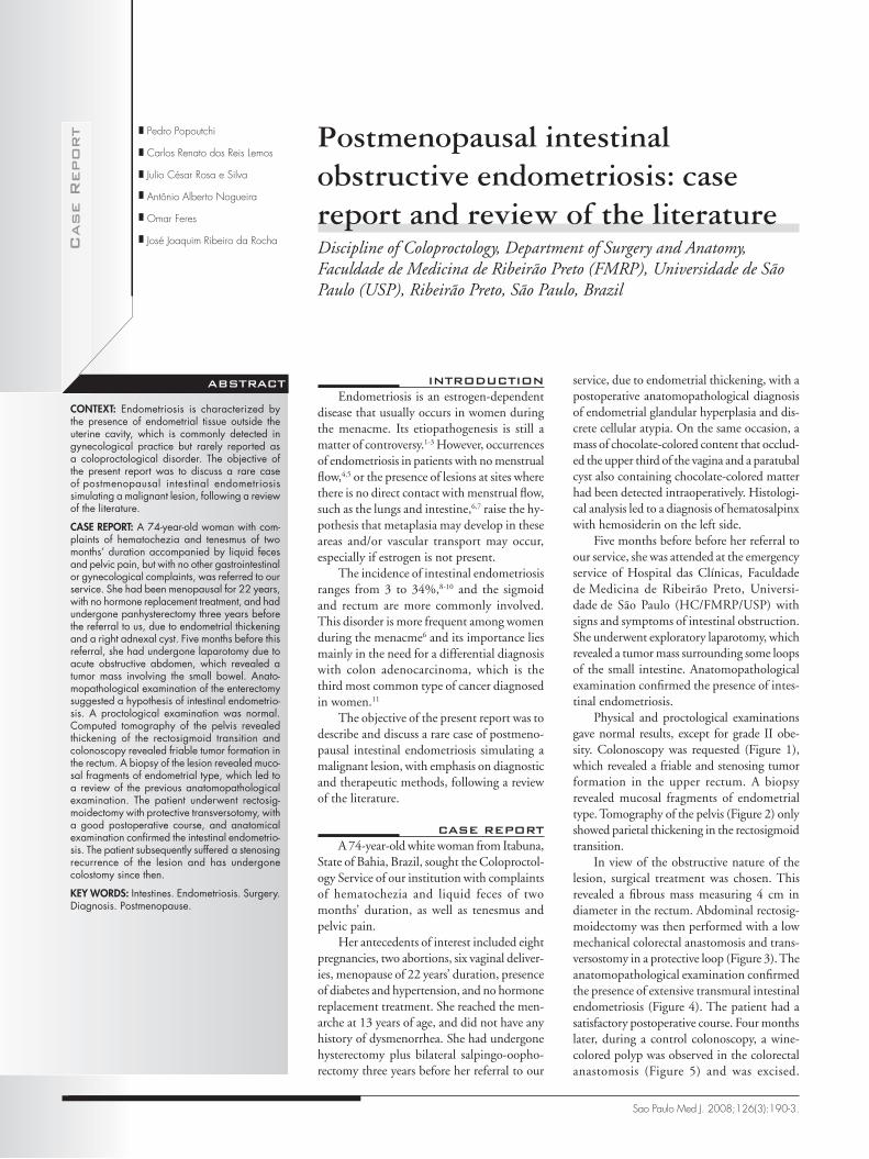
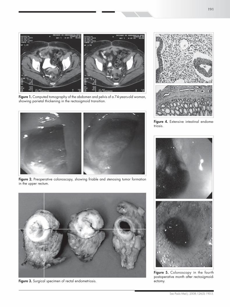
Physical andproctological examinationsgavenormal results, except forgrade IIobe-sity.Colonoscopywas requested (Figure1),whichrevealedafriableandstenosingtumorformation in the upper rectum. A biopsyrevealed mucosal fragments of endometrialtype.Tomographyofthepelvis(Figure2)onlyshowedparietalthickeningintherectosigmoidtransition.
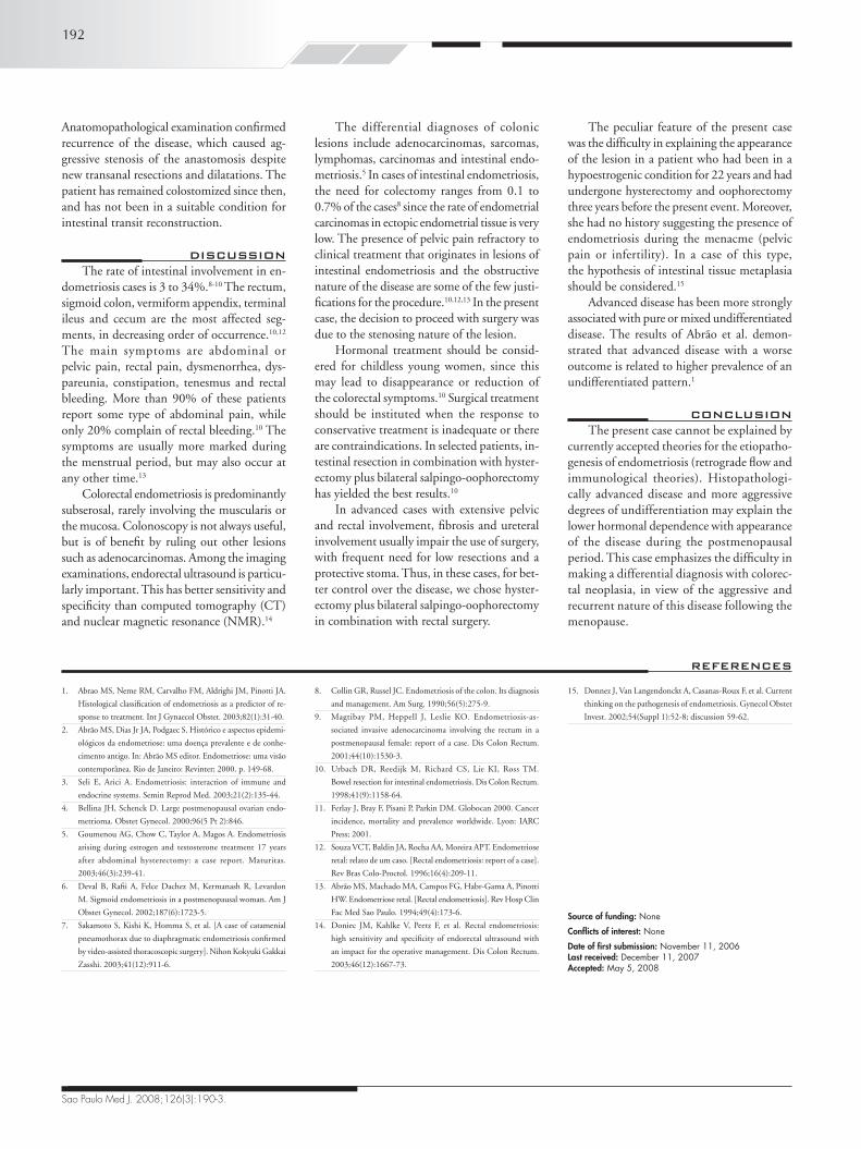
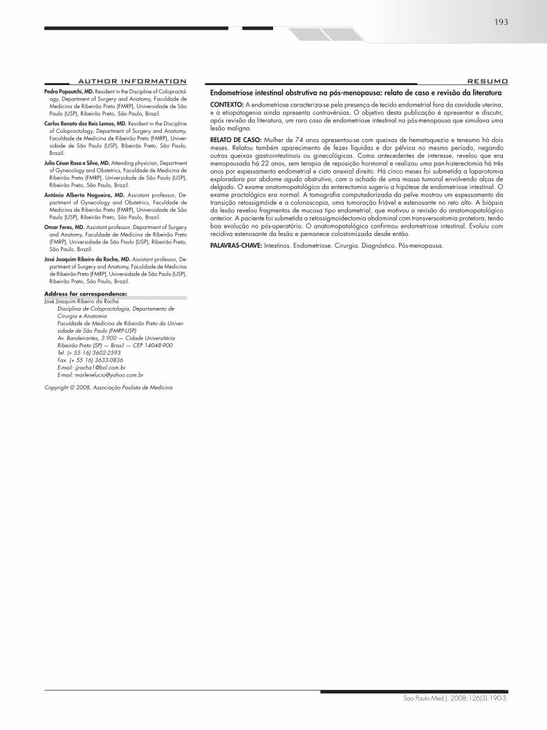
Inviewoftheobstructivenatureofthelesion, surgical treatment was chosen.Thisrevealed a fibrous mass measuring 4 cm indiameterintherectum.Abdominalrectosig-moidectomywasthenperformedwithalowmechanicalcolorectalanastomosisandtrans-versostomyinaprotectiveloop(Figure3).Theanatomopathologicalexaminationconfirmedthepresenceofextensivetransmuralintestinalendometriosis(Figure4).Thepatienthadasatisfactorypostoperativecourse.Fourmonthslater,duringacontrolcolonoscopy,awine-coloredpolypwasobservedinthecolorectalanastomosis (Figure 5) and was excised.
191
Sao Paulo Med J. 2008;126(3):190-3.
Figure 1. Computed tomography of the abdomen and pelvis of a 74-years-old woman, showing parietal thickening in the rectosigmoid transition.
Figure 2. Preoperative colonoscopy, showing friable and stenosing tumor formation in the upper rectum.
Figure 3. Surgical specimen of rectal endometriosis.
Figure 4. Extensive intestinal endome-triosis.
Figure 5. Colonoscopy in the fourth postoperative month after rectosigmoid-ectomy.
192
Sao Paulo Med J. 2008;126(3):190-3.
1. AbraoMS,NemeRM,CarvalhoFM,AldrighiJM,PinottiJA.
Histologicalclassificationofendometriosisasapredictorofre-
sponsetotreatment.IntJGynaecolObstet.2003;82(1):31-40.
2. AbrãoMS,DiasJrJA,PodgaecS.Históricoeaspectosepidemi-
ológicosdaendometriose:umadoençaprevalenteedeconhe-
cimentoantigo.In:AbrãoMSeditor.Endometriose:umavisão
contemporânea.RiodeJaneiro:Revinter;2000.p.149-68.
3. Seli E, Arici A. Endometriosis: interaction of immune and
endocrinesystems.SeminReprodMed.2003;21(2):135-44.
4. BellinaJH,SchenckD.Largepostmenopausalovarianendo-
metrioma.ObstetGynecol.2000;96(5Pt2):846.
5. GoumenouAG,ChowC,TaylorA,MagosA.Endometriosis
arising during estrogen and testosterone treatment 17 years
after abdominal hysterectomy: a case report. Maturitas.
2003;46(3):239-41.
6. DevalB,RafiiA,FelceDachezM,KermanashR,Levardon
M.Sigmoidendometriosisinapostmenopausalwoman.AmJ
ObstetGynecol.2002;187(6):1723-5.
7. SakamotoS,KishiK,HommaS,etal.[Acaseofcatamenial
pneumothoraxduetodiaphragmaticendometriosisconfirmed
byvideo-assistedthoracoscopicsurgery].NihonKokyukiGakkai
Zasshi.2003;41(12):911-6.
8. CollinGR,RusselJC.Endometriosisofthecolon.Itsdiagnosis
andmanagement.AmSurg.1990;56(5):275-9.
9. Magtibay PM, Heppell J, Leslie KO. Endometriosis-as-
sociated invasive adenocarcinoma involving the rectum in a
postmenopausalfemale:reportofacase.DisColonRectum.
2001;44(10):1530-3.
10. Urbach DR, Reedijk M, Richard CS, Lie KI, RossTM.
Bowelresectionforintestinalendometriosis.DisColonRectum.
1998;41(9):1158-64.
11. FerlayJ,BrayF,PisaniP,ParkinDM.Globocan2000.Cancer
incidence,mortality andprevalenceworldwide.Lyon: IARC
Press;2001.
12. SouzaVCT,BaldinJA,RochaAA,MoreiraAPT.Endometriose
retal:relatodeumcaso.[Rectalendometriosis:reportofacase].
RevBrasColo-Proctol.1996;16(4):209-11.
13. AbrãoMS,MachadoMA,CamposFG,Habr-GamaA,Pinotti
HW.Endometrioseretal.[Rectalendometriosis].RevHospClin
FacMedSaoPaulo.1994;49(4):173-6.
14. Doniec JM, KahlkeV, Peetz F, et al. Rectal endometriosis:
high sensitivityand specificityofendorectalultrasoundwith
animpactfortheoperativemanagement.DisColonRectum.
2003;46(12):1667-73.
15. DonnezJ,VanLangendoncktA,Casanas-RouxF,etal.Current
thinkingonthepathogenesisofendometriosis.GynecolObstet
Invest.2002;54(Suppl1):52-8;discussion59-62.
Source of funding: None
Conflicts of interest: None
Date of first submission: November 11, 2006Last received: December 11, 2007Accepted: May 5, 2008
REFERENCES
Anatomopathologicalexaminationconfirmedrecurrence of the disease, which caused ag-gressive stenosis of the anastomosis despitenewtransanalresectionsanddilatations.Thepatienthasremainedcolostomizedsincethen,andhasnotbeeninasuitableconditionforintestinaltransitreconstruction.
DISCUSSIONTherateofintestinalinvolvementinen-
dometriosiscasesis3to34%.8-10Therectum,sigmoidcolon,vermiformappendix,terminalileus and cecum are the most affected seg-ments,indecreasingorderofoccurrence.10,12The main symptoms are abdominal orpelvicpain, rectalpain,dysmenorrhea,dys-pareunia, constipation, tenesmus and rectalbleeding. More than 90% of these patientsreport some type of abdominal pain, whileonly20%complainofrectalbleeding.10Thesymptoms are usually more marked duringthemenstrualperiod,butmayalsooccuratanyothertime.13
Colorectalendometriosisispredominantlysubserosal,rarelyinvolvingthemuscularisorthemucosa.Colonoscopyisnotalwaysuseful,but isofbenefitby rulingoutother lesionssuchasadenocarcinomas.Amongtheimagingexaminations,endorectalultrasoundisparticu-larlyimportant.Thishasbettersensitivityandspecificitythancomputedtomography(CT)andnuclearmagneticresonance(NMR).14
The differential diagnoses of coloniclesions include adenocarcinomas, sarcomas,lymphomas,carcinomasandintestinalendo-metriosis.5Incasesofintestinalendometriosis,the need for colectomy ranges from 0.1 to0.7%ofthecases8sincetherateofendometrialcarcinomasinectopicendometrialtissueisverylow.Thepresenceofpelvicpainrefractorytoclinicaltreatmentthatoriginatesinlesionsofintestinal endometriosis and theobstructivenatureofthediseasearesomeofthefewjusti-ficationsfortheprocedure.10,12,13Inthepresentcase,thedecisiontoproceedwithsurgerywasduetothestenosingnatureofthelesion.
Hormonal treatment should be consid-ered for childless young women, since thismay lead to disappearance or reduction ofthecolorectalsymptoms.10Surgicaltreatmentshould be instituted when the response toconservativetreatmentisinadequateortherearecontraindications.Inselectedpatients,in-testinalresectionincombinationwithhyster-ectomyplusbilateralsalpingo-oophorectomyhasyieldedthebestresults.10
In advanced cases with extensive pelvicandrectalinvolvement,fibrosisandureteralinvolvementusuallyimpairtheuseofsurgery,withfrequentneedfor lowresectionsandaprotectivestoma.Thus,inthesecases,forbet-tercontroloverthedisease,wechosehyster-ectomyplusbilateralsalpingo-oophorectomyincombinationwithrectalsurgery.
Thepeculiar featureof thepresentcasewasthedifficultyinexplainingtheappearanceofthelesioninapatientwhohadbeeninahypoestrogenicconditionfor22yearsandhadundergonehysterectomyandoophorectomythreeyearsbeforethepresentevent.Moreover,shehadnohistorysuggestingthepresenceofendometriosis during the menacme (pelvicpain or infertility). In a case of this type,thehypothesisofintestinaltissuemetaplasiashouldbeconsidered.15
Advanceddiseasehasbeenmorestronglyassociatedwithpureormixedundifferentiateddisease.The results of Abrão et al. demon-strated that advanced disease with a worseoutcomeisrelatedtohigherprevalenceofanundifferentiatedpattern.1
CONCLUSIONThepresentcasecannotbeexplainedby
currentlyacceptedtheoriesfortheetiopatho-genesisofendometriosis(retrogradeflowandimmunological theories). Histopathologi-callyadvanceddiseaseandmoreaggressivedegreesofundifferentiationmayexplainthelowerhormonaldependencewithappearanceof the disease during the postmenopausalperiod.Thiscaseemphasizesthedifficultyinmakingadifferentialdiagnosiswithcolorec-talneoplasia, inviewof theaggressiveandrecurrentnatureofthisdiseasefollowingthemenopause.
193
Sao Paulo Med J. 2008;126(3):190-3.
AUTHOR INFORMATION Pedro Popoutchi, MD. Resident in the Discipline of Coloproctol-
ogy, Department of Surgery and Anatomy, Faculdade de Medicina de Ribeirão Preto (FMRP), Universidade de São Paulo (USP), Ribeirão Preto, São Paulo, Brazil.
Carlos Renato dos Reis Lemos, MD. Resident in the Discipline of Coloproctology, Department of Surgery and Anatomy, Faculdade de Medicina de Ribeirão Preto (FMRP), Univer-sidade de São Paulo (USP), Ribeirão Preto, São Paulo, Brazil.
Julio César Rosa e Silva, MD. Attending physician, Department of Gynecology and Obstetrics, Faculdade de Medicina de Ribeirão Preto (FMRP), Universidade de São Paulo (USP), Ribeirão Preto, São Paulo, Brazil.
Antônio Alberto Nogueira, MD. Assistant professor, De-partment of Gynecology and Obstetrics, Faculdade de Medicina de Ribeirão Preto (FMRP), Universidade de São Paulo (USP), Ribeirão Preto, São Paulo, Brazil.
Omar Feres, MD. Assistant professor, Department of Surgery and Anatomy, Faculdade de Medicina de Ribeirão Preto (FMRP), Universidade de São Paulo (USP), Ribeirão Preto, São Paulo, Brazil.
José Joaquim Ribeiro da Rocha, MD. Assistant professor, De-partment of Surgery and Anatomy, Faculdade de Medicina de Ribeirão Preto (FMRP), Universidade de São Paulo (USP), Ribeirão Preto, São Paulo, Brazil.
Address for correspondence:José Joaquim Ribeiro da Rocha
Disciplina de Coloproctologia, Departamento de Cirurgia e AnatomiaFaculdade de Medicina de Ribeirão Preto da Univer-sidade de São Paulo (FMRP-USP)Av. Bandeirantes, 3.900 — Cidade UniversitáriaRibeirão Preto (SP) — Brasil — CEP 14048-900Tel. (+ 55 16) 3602-2593 Fax. (+ 55 16) 3633-0836E-mail: [email protected]: [email protected]
Copyright © 2008, Associação Paulista de Medicina
RESUMO
Endometriose intestinal obstrutiva na pós-menopausa: relato de caso e revisão da literatura
CONTEXTO: A endometriose caracteriza-se pela presença de tecido endometrial fora da cavidade uterina, e a etiopatogenia ainda apresenta controvérsias. O objetivo desta publicação é apresentar e discutir, após revisão da literatura, um raro caso de endometriose intestinal na pós-menopausa que simulava uma lesão maligna.
RELATO DE CASO: Mulher de 74 anos apresentou-se com queixas de hematoquezia e tenesmo há dois meses. Relatou também aparecimento de fezes líquidas e dor pélvica no mesmo período, negando outras queixas gastrointestinais ou ginecológicas. Como antecedentes de interesse, revelou que era menopausada há 22 anos, sem terapia de reposição hormonal e realizou uma pan-histerectomia há três anos por espessamento endometrial e cisto anexial direito. Há cinco meses foi submetida a laparotomia exploradora por abdome agudo obstrutivo, com o achado de uma massa tumoral envolvendo alças de delgado. O exame anatomopatológico da enterectomia sugeriu a hipótese de endometriose intestinal. O exame proctológico era normal. A tomografia computadorizada da pelve mostrou um espessamento da transição retossigmóide e a colonoscopia, uma tumoração friável e estenosante no reto alto. A biópsia da lesão revelou fragmentos de mucosa tipo endometrial, que motivou a revisão do anatomopatológico anterior. A paciente foi submetida a retossigmoidectomia abdominal com transversostomia protetora, tendo boa evolução no pós-operatório. O anatomopatológico confirmou endometriose intestinal. Evoluiu com recidiva estenosante da lesão e pemanece colostomizada desde então.
PALAVRAS-CHAVE: Intestinos. Endometriose. Cirurgia. Diagnóstico. Pós-menopausa.