Embed Size (px)
DESCRIPTION
bahan
Citation preview
Pediatric Lower Respiratory Infections
1 april 2015
Alessandra Scaparrotta1*, Marina Attanasi1, Sabrina Di Pillo1 and Francesco Chiarelli1 1 Department of Pediatrics, University of Chieti, Italy *Corresponding author: Alessandra Scaparrotta, Department of Pediatrics, University of Chieti, Italy
1 Acute and Chronic Bronchitis 3.4 Diagnosis1.1 Acute Bronchitis 3.5 Therapy
1.1.1 Pathophysiology and Etiology 3.6 Prognosis1.1.2 Sign and symptoms 4 Pneumonia1.1.3 Diagnosis 4.1 Definition1.1.4 Differential Diagnosis 4.2 Epidemiology1.1.5 Therapy 4.3 Etiology1.2 Chronic Bronchitis 4.4 Bacterial Pneumonia
1.2.1 Differential Diagnosis 4.4.1 Pathophysiology1.2.2 Diagnosis 4.4.2 Symptoms and Signs1.2.3 Therapy 4.4.3 Diagnostic Tests
2 Infectious Asthmatic Bronchitis (Wheezing/Wheeze)
4.4.4 Treatment
2.1 Etiology 4.4.5 Complications2.2 Diagnosis 4.5 Non-Bacterial Pneumonia2.3 Therapy 4.5.1 Pathophysiology2.4 Prognosis 4.5.2 Symptoms and Signs3 Bronchitis 4.5.3 Diagnostic Tests
3.1 Etiology and Pathophysiology 4.5.4 Treatment3.2 Clinical Symptoms 4.5.5 Complications3.3 Differential diagnosis 4.6 Recurrent or Chronic Pneumonia
Lower respiratory tract infections include conditions, which may or may not involve the parenchyma [1]:
Infections not involving the parenchyma as acute bronchitis, exacerbation of chronic bronchitis, asthmatic bronchitis and bronchiolitis
Infections involving the parenchyma as pneumonia.
1. Acute and Chronic Bronchitis 1.1 Acute bronchitis
Many terms are used to describe diseases characterized by cough: bronchitis, wheezy bronchitis, asthmatic bronchitis, and tracheobronchitis. There is a lack of consensus regarding clinical definition of cough illnesses and nomenclature, caused by difficulty in comparing results from cough illness or bronchitis studies, with a lack of a firm consensus on diagnosis and treatment [2]. Acute bronchitis is an acute or subacute cough illness lasting less than 2-3 weeks, with or without phlegm production, frequently associated to other upper respiratory tract and constitutional symptoms [3-5].
The cough is the most frequently mentioned symptom necessitating office evaluation; so, acute bronchitis is one of the top 10 diagnoses in ambulatory care medicine [6,7]. Physicians exhibit extensive variability in diagnostic requirements and treatment, because the diagnosis is clinical, without standardized diagnostic signs and sensitive or specific confirmatory laboratory tests [
Diagnosis of bronchitis often results in a prescription for an antimicrobial agent, reflecting the physicians’ belief of bacterial infection, although the term bronchitis does not imply specific etiology and it is most commonly caused by viral pathogens [2]. 1.1.1 Pathophysiology and etiology: Acute bronchitis is defined as inflammation of the bronchial respiratory mucosa, resulting in productive cough. For most clinicians, bronchitis is a disease clinically characterized by cough, with or without fever or sputum production [2].
Bronchial epithelial injury is induced by infectious or noninfectious triggers, which cause an inflammatory response with consequent airway hyperresponsiveness and mucus production [7,9]. International literature suggests that clinical features of uncomplicated acute bronchitis develop in sequential phases: an acute infection phase, resulting from direct inoculation by the infectious virus of the tracheobronchial epithelium, leading to cytokine release and inflammatory cell activation. In this phase there are variable constitutional symptoms, such as fever, myalgia, and malaise, that last 1 to 5 days depending on the infectious agent. The protracted phase results from hypersensitivity of the tracheobronchial epithelium and airway receptors (bronchial hyperresponsiveness), characterized primarily by cough, often accompanied by phlegm production and wheezing, and usually lasts 1 to 3 weeks. Respiratory epithelial cell function plays an important role in airway inflammation, and vagal-mediated airway hyperresponsiveness has been shown to coincide with repair of the bronchial epithelial surface. Other mechanisms of bronchial hyperresponsiveness may also be present, as adrenergic-cholinergic tone imbalance and IgE-mediated histamine release [5]. Selected triggers that can begin the cascade leading to acute bronchitis are [7]:
Viruses: adenovirus, coronavirus, coxsackievirus, enterovirus, influenza virus, parainfluenza virus, respiratory syncytial virus, rhinovirus, human metapneumovirus.
Bacteria: Bordetella pertussis, Bordetella parapertussis, Branhamella catarrhalis, Haemophilus influenzae, Streptococcus pneumoniae, atypical bacteria (e.g., Mycoplasma pneumoniae, Chlamydia pneumoniae, Legionella species).
Yeast and fungi: Blastomyces dermatitidis, Candida albicans, Candida tropicalis, Coccidioides immitis, Cryptococcus neoformans, Histoplasma capsulatum.
Noninfectious triggers: asthma, air pollutants, ammonia, cannabis, tobacco, trace metals.
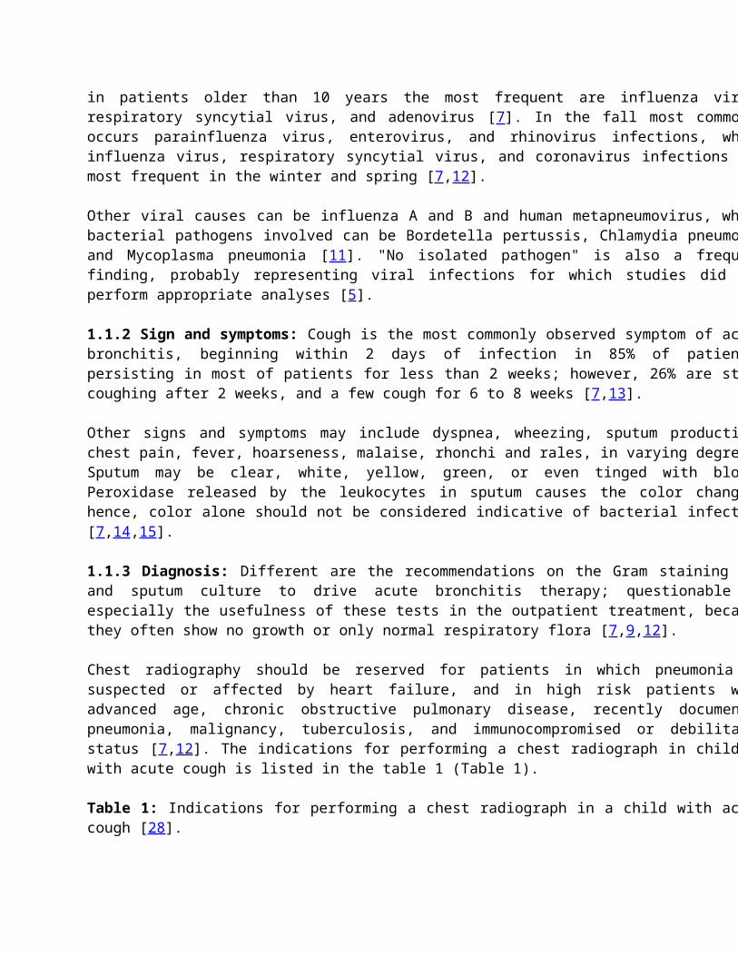
Acute bronchitis is usually caused by a viral infection [10,11]: in patients younger than one year, commonly by respiratory syncytial virus, parainfluenza virus, and coronavirus; in patients one to 10 years of age predominate parainfluenza virus, enterovirus, respiratory syncytial virus, and rhinovirus; in patients older than 10 years the most frequent are influenza virus, respiratory syncytial virus, and adenovirus [7]. In the fall most commonly occurs parainfluenza virus, enterovirus, and rhinovirus infections, while influenza virus, respiratory syncytial virus, and coronavirus infections are most frequent in the winter and spring [7,12]. Other viral causes can be influenza A and B and human metapneumovirus, while bacterial pathogens involved can be Bordetella pertussis, Chlamydia pneumonia and Mycoplasma pneumonia [11]. "No isolated pathogen" is also a frequent finding, probably representing viral infections for which studies did not perform appropriate analyses [5 1.1.2 Sign and symptoms: Cough is the most commonly observed symptom of acute bronchitis, beginning within 2 days of infection in 85% of patients, persisting in most of patients for less than 2 weeks; however, 26% are still coughing after 2 weeks, and a few cough for 6 to 8 weeks [7,13]. Other signs and symptoms may include dyspnea, wheezing, sputum production, chest pain, fever, hoarseness, malaise, rhonchi and rales, in varying degrees. Sputum may be clear, white, yellow, green, or even tinged with blood. Peroxidase released by the leukocytes in sputum causes the color changes; hence, color alone should not be considered indicative of bacterial infection [7,14,15]. 1.1.3 Diagnosis: Different are the recommendations on the Gram staining use and sputum culture to drive acute bronchitis therapy; questionable is especially the usefulness of these tests in the outpatient treatment, because they often show no growth or only normal respiratory flora [7,9,12]. Chest radiography should be reserved for patients in which pneumonia is suspected or affected by heart failure, and in high risk patients with advanced age, chronic obstructive pulmonary disease, recently documented pneumonia, malignancy, tuberculosis, and immunocompromised or debilitated status [7,12]. The indications for performing a chest radiograph in children with acute cough is listed in the table 1 (Table 1). Table 1: Indications for performing a chest radiograph in a child with acute cough [28].
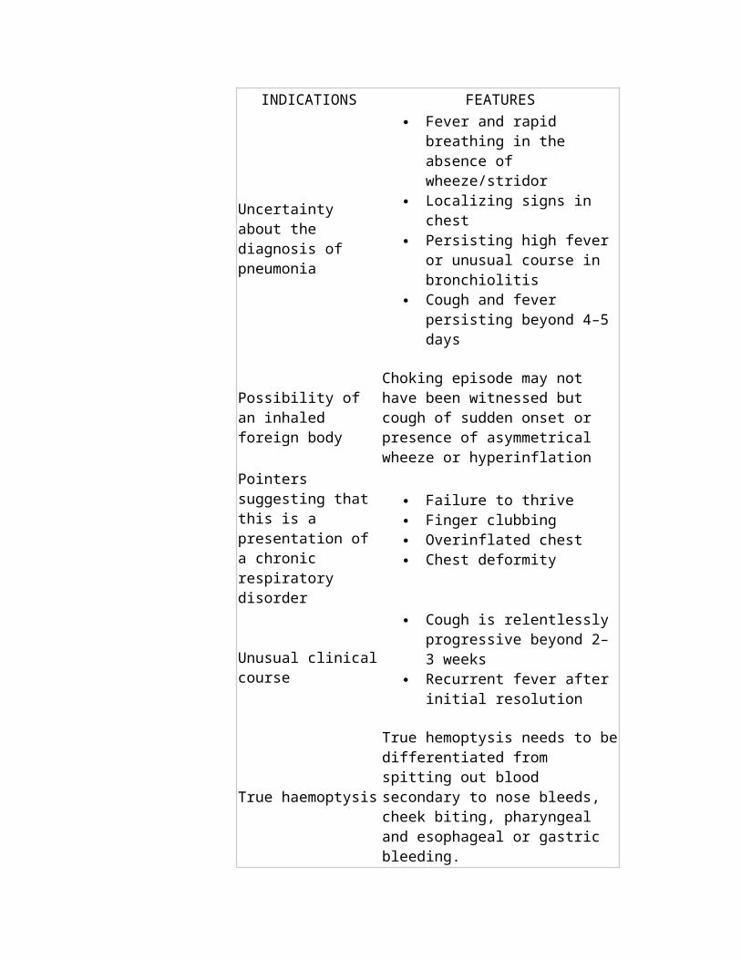
INDICATIONS FEATURES
Uncertainty about the diagnosis of pneumonia
Fever and rapid breathing in the absence of wheeze/stridor
Localizing signs in chest Persisting high fever or unusual
course in bronchiolitis Cough and fever persisting
beyond 4–5 days
Possibility of an inhaled foreign body
Choking episode may not have been witnessed but cough of sudden onset or presence of asymmetrical wheeze or hyperinflation
Pointers suggesting that this is a presentation of
Failure to thrive Finger clubbing
a chronic respiratory disorder
Overinflated chest Chest deformity
Unusual clinical course
Cough is relentlessly progressive beyond 2– 3 weeks
Recurrent fever after initial resolution
True haemoptysis
True hemoptysis needs to be differentiated from spitting out blood secondary to nose bleeds, cheek biting, pharyngeal and esophageal or gastric bleeding.
Pulmonary function testing as spirometry are not routinely used in the acute bronchitis diagnosis, but performed only when underlying obstructive pathology is suspected or if there are repeated bronchitis episodes. Pulse oximetry may determine the severity of the illness, but results do not confirm or rule out bronchitis, asthma, pneumonia, or other specific diagnoses [7]. 1.1.4 Differential diagnosis: The differential diagnosis includes the most common causes of acute cough [11]: ✓ Acute bronchitis✓ Allergic rhinitis✓Asthma✓ Chronic obstructive pulmonary disease exacerbation✓ Congestive heart failure exacerbation ✓ Gastroesophageal reflux disease ✓ Malignancy✓ Pneumonia✓ Post-infectious cough✓ Postnasal drip✓ Sinusitis✓ Viral syndrome. 1.1.5 Therapy: Antiviral medications against influenza may be considered during influenza season for high-risk patients who present within 36 hours of symptom onset [11]. Antibiotics should not be routinely used in the treatment of acute bronchitis, especially in younger patients. Although viruses cause 90% of bronchitis infections, approximately two thirds of patients in the United States are treated with antibiotics. Antibiotics do not significantly change the course of acute bronchitis, providing only minimal benefit compared with the risk of antibiotic use itself. Routine use of antibiotics is not recommend by the American College of Chest Physicians (ACCP) for patients with acute bronchitis [16], but they may be considered in certain situations: when pertussis is suspected as cough etiology, initiation of a macrolide is recommended as soon as possible to reduce transmission; however, antibiotics do not reduce duration of symptoms [11]. A very recent Cochrane concludes that there is limited evidence to support the use of antibiotics in acute bronchitis; they may have a modest beneficial effect in some patients with acute bronchitis, although data on subsets of patients
who may benefit more from treatment are lacking. However, the magnitude of this benefit needs to be considered in the broader context of potential side effects, medicalization for a self limiting condition, increased respiratory pathogens resistance and antibiotic treatment cost [17]. The use of antibiotics in acute bronchitis may decrease the risk of subsequent pneumonia. The use of serologic markers may help to guide antibiotic use, because of the clinical uncertainty in distinguishing acute bronchitis from pneumonia [11]. Over-the-counter (OTC) cough preparations are often as self prescribed as recommended by health practitioners for the initial treatment of cough [18]. Dextromethorphan is ineffective for cough suppression in children with bronchitis [19]. Other common therapies include antitussives, expectorants, inhaler medications, and alternative therapies. Expectorants, which have been shown to be ineffective in the treatment of acute bronchitis, and inhaler medications are not recommended for routine use in patients with bronchitis, although they are commonly used and suggested by physicians [18,20]. A Cochrane review does not suggest the routine use of beta-agonist inhalers in adults and children with acute bronchitis without airflow obstruction evidence; however, the subset of patients with wheezing during the illness responded to this therapy [20,21]. There may be some benefit only to high-dose, episodic inhaled corticosteroids, but no benefit occurred with preventive therapy [22]. No data support the use of oral corticosteroids in patients with acute bronchitis without asthma [11]. Antitussives, antihistamines, antihistamine decongestants and antitussive/bronchodilator combinations were no more effective than placebo in children with acute cough; however, many studies were of low quality and very different from each other, making very difficult the evaluation of overall efficacy [18]. 1.2 Chronic bronchitis There is a lack of clarity regarding the definition of chronic bronchitis. The definition of chronic bronchitis in adults is clear: “the presence of chronic productive cough for 3 months in each of 2 successive years, in a patient under whom other causes of chronic cough have been excluded” [23]. Whether this definition can be applied to childhood chronic bronchitis remains unclear [24,25]. The diagnosis of chronic bronchitis should occur in two phases. The first is consideration and identification of several well-defined respiratory disorders according to a staged management protocol. The second but simultaneous phase is elimination or modification of exogenous factors that produce or maintain the child’s illness [24]. However, this diagnosis has the potential to divert the pediatrician from detecting a more specific respiratory condition [24Despite coughing in childhood is common, there is remarkably little in the literature regarding etiology, investigation and management of chronic cough in childhood. Recent reports have emphasized the importance of making a specific diagnosis in children with a chronic cough (>3 weeks) [26,27].
Juvenile chronic bronchitis with persistent endobronchial infection (recently labeled persistent bacterial bronchitis) has been described for many decades. Children have chronic or recurrent cough with sputum production [28]. The persistent bacterial bronchitis (PBB) is, for some authors, the most common cause of a chronic cough [26,29variety of diagnostic labels have been used to describe this condition: terms such as chronic suppurative lung disease [30-32], persistent endobronchial infection [33] and PBB [29] describe the pathological process and site of infection, while terms such as ‘‘chronic bronchitis’’ [34-36] or ‘‘protracted bronchitis’’ [37,38] describe the clinical phenotype. Others have suggested using the term ‘‘pre-bronchiectasis’’ [39,40] to highlight the condition’s probable role in leading to damaged airways, as evident on high-resolution computed tomography or at bronchography [
PBB is a pediatric condition characterized by the presence of an isolated moist or wet cough, lasting more than 4 weeks in the absence of other specific causes. It usually affects children younger than 5 years and it has been recognized more by pediatric pulmonologists that resolves it with antibiotic treatment. The most common organisms involved in infants and children are Streptococcus pneumoniae, Haemophilus influenzae, and Moraxella catarrhalis. PBB is underdiagnosed and often misdiagnosed as asthma [27,29,41,42]. 1.2.1 Differential diagnosis: The term of “chronic bronchitis” should only be used after underlying causes have been excluded [28,42,43]:
Asthma Tracheobronchomalacia Foreign body aspiration Mechanical airway obstruction Gastroesophageal reflux/aspiration syndromes Cystic fibrosis Primary ciliary disorders Congenital malformation Passive smoking Environmental pollution Pulmonary tuberculosis Bronchiectasis Immune deficiencies.
1.2.2 Diagnosis: Careful history - taking and physical examination, together with appropriate investigations, enable the correct diagnoses to be made for most cases of chronic cough within a reasonable time frame [43]. The diagnostic approach includes [28,42]:
1. Complete blood cell count2. Chest x-ray examination3. Pulse oximetry4. Sputum for cultures5. Mantoux6. Sweat chloride measurement 7. Allergological evaluation 8. B-cell function (if immunological deficit is suspected)9. High-resolution computed tomographic chest scan10. Fiberoptic bronchoscopy with bronchoalveolar lavage and endobronchial biopsies 11. Placement of esophageal pH probe.
Therapy: The management of chronic coughing relates to first making an accurate underlying diagnosis and then applying specific treatment for that condition. Macrolide antibiotics should be early used if diagnosis of pertussis exists [28].
PBB often resolves after a course of antibiotic such as amoxicillin - clavulanate for 2 weeks; however, some require a longer 4–6 weeks antibiotic. Children with PBB should first have other underlying conditions excluded, especially if PBB becomes recurrent or fails to respond to antibiotics and further investigations as sputum cultured are required, to rule out the other conditions such as immunodeficiencies or other causes of chronic suppurative lung disease. A trial treatment of physiotherapy and a prolonged course (4–6 weeks) of appropriate antibiotics may be tried [28,44
1. Bru JP, Floret D, Pariset C, Ploy MC Diagnosis of acute Lower Respiratory Tract Infections. BioMe?rieux SA.
2. O'Brien KL, Dowell SF, Schwartz B, et al. (1998) Cough Illness/Bronchitis-Principles of Judicious Use of Antimicrobial Agents. Pediatrics 101: 178-181.
3. Evans AS (1967) Clinical syndromes in adults caused by respiratory infection. Med Clin North Am 51: 803-818.
4. Gonzales R, Barrett PH Jr, Crane LA, Steiner JF (1998) Factors associated with antibiotic use for acute bronchitis. J Gen Intern Med 13: 541-548.
5. Gonzales R, Sande MA (2000) Uncomplicated acute bronchitis. Ann Intern Med 133: 981-991.
6. Slusarcick AL, McCaig LF (2000) National hospital ambulatory medical care survey: 1998 outpatient department summary. Adv Data 317: 1-23.
7. Knutson D, Braun C (2002) Diagnosis and management of acute bronchitis. Am Fam Physician 65: 2039-2044.
8. Hueston WJ, Mainous AG 3rd (1998) Acute bronchitis. Am Fam Physician 57: 1270-1276, 1281-2.
9. Treanor JJ, Hayden FG (2000) Viral infections. In: Murray JF (eds.). Textbook of respiratory medicine. (3rdedn), Saunders, Philadelphia.
10. Marrie TJ (1998) Acute bronchitis and community acquired pneumonia. In: Fishman AP, Elias JA, (eds.). Fishman’s Pulmonary diseases and disorders. (3dedn), McGraw-Hill, New York.
11. Albert RH (2010) Diagnosis and treatment of acute bronchitis. Am Fam Physician 82: 1345-1350.
12. Blinkhorn RJ Jr. (1998) Upper respiratory tract infections. In: Baum GL (eds.). Textbook of pulmonary diseases. (6thedn), Lippincott-Raven, Philadelphia.
13. Chesnutt MS, Prendergast TJ Lung (2002) In: Tierney LM, Stephen J McPhee, Maxine A Papadakis, (eds.). Current medical diagnosis & treatment, 2002. (41stedn), McGraw-Hill, New York.
14. Mufson MA (2000) Viral pharyngitis, laryngitis, croup and bronchitis. In: Goldman L, Bennett JC (eds.). Cecil Textbook of medicine. (21stedn), Elsiever Health Sciences, Philadelphia.
15. Chodosh S (1987) Acute bacterial exacerbations in bronchitis and asthma. Am J Med 82: 154-163.
16. Braman SS (2006) Chronic cough due to acute bronchitis: ACCP evidence-based clinical practice guidelines. Chest 129: 95S-103S.
17. Smucny J, Fahey T, Becker LA, Glazier R (2004) Antibiotics for acute bronchitis. Cochrane Database Syst Rev (4): CD000245.
18. Smith SM, Schroeder K, Fahey T (2008) Over-the-counter (OTC) medications for acute cough in children and adults in ambulatory settings. Cochrane Database Syst Rev (1):CD001831..
19. Paul IM, Yoder KE, Crowell KR, Shaffer ML, McMillan HS, et al. (2004) Effect of dextromethorphan, diphenhydramine, and placebo on nocturnal cough and sleep quality for coughing children and their parents. Pediatrics 114: e85-90.
20. Smucny J, Flynn C, Becker L, Glazier R (2001) Beta 2-agonists for acute bronchitis. Cochrane Database Syst Rev (1): CD001726.
21. Becker LA, Hom J, Villasis-Keever M, van der Wouden JC (2011) Beta2-agonists for acute bronchitis. Cochrane Database Syst Rev : CD001726.
22. McKean M, Ducharme F (2000) Inhaled steroids for episodic viral wheeze of childhood. Cochrane Database Syst Rev : CD001107.
23. Murray JF, Nadel JA, Mason RJ, et al, eds. Textbook of respiratory medicine. 3rd edn. Phildelphia: W B
Saunders, 2000.
24. Taussig LM, Landau LI, Le Souef PN, et al. (1999) (eds.). Pediatric respiratory medicine. St Louis Mosby Verlag.
25. Byrnes C, Edwards E (2007) Outcomes in children treated for persistent bacterial bronchitis. Thorax 62: 922-923.
26. Marchant JM, Masters IB, Taylor SM, Chang AB (2006) Utility of signs and symptoms of chronic cough in predicting specific cause in children. Thorax 61: 694-698.
27. Donnelly D, Critchlow A, Everard ML (2007) Outcomes in children treated for persistent bacterial bronchitis. Thorax 62: 80-84.
28. Shields MD, Bush A, Everard ML, McKenzie S, Primhak R; British Thoracic Society Cough Guideline Group (2008) BTS guidelines: Recommendations for the assessment and management of cough in children. Thorax 63 Suppl 3: iii1-1iii15.
29. Marchant JM, Masters IB, Taylor SM, Cox NC, Seymour GJ, et al. (2006) Evaluation and outcome of young children with chronic cough. Chest 129: 1132-1141.
30. Phelan PD, Landau LI, Robertson CF (1994) Suppurative lung disease. In: Respiratory illness in children
31. Chang AB, Boyce NC, Masters IB, Torzillo PJ, Masel JP (2002) Bronchoscopic findings in children with non-cystic fibrosis chronic suppurative lung disease. Thorax 57: 935-938.
32. Couriel J (2002) Assessment of the child with recurrent chest infections. Br Med Bull 61: 115-132.
33. Spencer DA (2005) From hemp seed and porcupine quill to HRCT: advances in the diagnosis and epidemiology of bronchiectasis. Arch Dis Child 90: 712-714.
34. FIELD CE (1949) Bronchiectasis in childhood; prophylaxis, treatment and progress with a follow-up study of 202 cases of established bronchiectasis. Pediatrics 4: 355-372.
35. Morgan WJ, Taussig LM (1984) The chronic bronchitis complex in children. Pediatr Clin North Am 31: 851-864.
36. Seear M, Wensley D (1997) Chronic cough and wheeze in children: do they all have asthma? Eur Respir J 10: 342-345.
37. Chang AB (2005) Defining the cough spectrum and reviewing the evidence for treating non-specific cough in children. Current Pediatric Reviews 1: 283?296.
38. Chang AB, Landau LI, Van Asperen PP, Glasgow NJ, Robertson CF, et al. (2006) Cough in children: definitions and clinical evaluation. Med J Aust 184: 398-403.
39. FIELD CE (1949) Bronchiectasis in childhood; aetiology and pathogenesis, including a survey of 272 cases of doubtful irreversible bronchiectasis. Pediatrics 4: 231-248.
40. Eastham KM, Fall AJ, Mitchell L, Spencer DA (2004) The need to redefine non-cystic fibrosis bronchiectasis in childhood. Thorax 59: 324-327.
41. Chang AB, Redding GJ, Everard ML (2008) Chronic wet cough: Protracted bronchitis, chronic suppurative lung disease and bronchiectasis. Pediatr Pulmonol 43: 519-531.
42. Chipps BE (2010) Evaluation of infants and children with refractory lower respiratory tract symptoms. Ann Allergy Asthma Immunol 104: 279-283.
43. Chow PY, Ng DK (2004) Chronic cough in children. Singapore Med J 45: 462-468.
44. Shields MD, Thavagnanam S (2013) The difficult coughing child: prolonged acute cough in children. Cough 9: 11.
2. Infectious Asthmatic Bronchitis (Wheezing Or Wheeze)
Terms such as wheezy bronchitis, respiratory illness associated wheeze, and asthmatic bronchitis have been used in the past to describe episodic wheezing in infants and young children. In fact, pediatricians thought that episodic wheeze in this age group had a more benign prognosis than asthma of older children. More recently, the use of the term asthma has been promoted to describe all wheezing illness in children, allowing no distinction between viruses induced wheeze and other kind of asthma [1-3].
Approximately, one in three children has at least one episode of wheezing prior to his third birthday, with a
cumulative prevalence of wheeze almost of 50% at the age of 6 years [4-6]. Most preschool wheeze is associated with viral upper respiratory tract infections, which frequently recur at this age [4].
The European Respiratory Society (ERS) Task Force agrees not to use the term asthma to describe preschool wheezing illness since there is insufficient evidence showing that the pathophysiology of preschool wheezing illness is similar to the asthmatic one in older children and adults [4].
Wheezing is defined by ERS as a continuous high-pitched sound with musical quality emitting from the chest during expiration and it is one of the forms of noisy breathing in preschool children. Parents understanding of wheezing differs very much, because some think it is a sound such as whistling, squeaking or gasping; for others is a different rate or style of breathing or the same as cough [4,7-11].
Episodic (viral) wheeze is defined as wheeze in discrete episodes, with the child being well between episodes. This phenotype appears to be most common in preschool children [4,5,12,13] and it is usually associated with clinical evidence of a viral respiratory tract infection, with repeated episodes that occur seasonally.
2.1 EtiologyThe most common causative agents include [4,14]:
1. Rhinovirus2. Respiratory syncytial virus (RSV)3. Coronavirus4. Human metapneumovirus5. Parainfluenza virus6. Adenovirus
Factors underlying the frequency and severity of episodes are partially understood, but some factors as the severity of the first episode (related to pre-existent impaired lung function and younger age), atopy, prematurity and exposure to tobacco smoke have been implicated [4,15-21]. It is irrelevant whether or not the initial episode is classified as bronchiolitis [4].
2.2 Diagnosis
A careful physical examination should always be performed, which should include listening to forced expiration and nasal examination [22].
There is no evidence that microbiological investigation, with identification of the causative virus, contributes to management of the acute episode or in the long-term. There are no studies supporting the usefulness of pulmonary function tests in children with nonspecific symptoms, or in distinguishing between episodic and multiple-trigger wheeze [4].
2.3 Therapy
There is little doubt that wheezing should be treated with bronchodilators and not antibiotics, with additional corticosteroids if the wheezing is severe [1].
Short-acting ß 2 agonists are the treatment of choice for intermittent and acute asthma episodes in very young children [22]. Double-blind placebo-controlled studies observed significant bronchodilatory effects and protective effects against bronchoconstrictor agents in infants and preschool children treated with them. Oral administration of this drug is also effective, but there are systemic side effects, while intravenous infusion use is limited to very severe acute wheeze in young children [4].
Leukotriene receptor antagonists are suggested as treatment for viral-induced wheeze and to reduce the frequency of exacerbations in young children aged 2–5 years [23,24]. Benefit has been shown in children as young as 6 months of age [22,25,26]. Daily use of montelukast over a 1-yr period had diminished the wheezing episodes rate in 549 children with episodic (viral) wheeze by 32% compared to placebo [4,23]. The ERS Task Force suggests that Montelukast 4 mg once daily should probably be given for the treatment of episodic (viral) wheeze, while a trial of inhaled corticosteroids may be considered in preschool children especially when episodes occur frequently or if the family history of asthma is positive [4].
Further studies are needed to establish the role of viral infections in precipitating obstructive airway symptoms and of antiviral agents as potential asthma medications [22].
2.4 Prognosis
Although RSV and rhinovirus have been linked to an increased risk of persistent wheezing over time [4,27-29], it is not known whether or not they play a major role in determining long-term outcome.
Episodic (viral) wheeze most commonly decreases over time, disappearing by the age of 6 years, but can continue as episodic wheeze into school age, change into multiple-trigger wheeze or disappear at an older age [4,5,30].
References
1. Wilson NM (1989) Wheezy bronchitis revisited. Arch Dis Child 64: 1194-1199.
2. Williams H, McNicol KN (1969) Prevalence, natural history, and relationship of wheezy bronchitis and asthma in children. An epidemiological study. Br Med J 4: 321-325.
3. Lee DA, Winslow NR, Speight AN, Hey EN (1983) Prevalence and spectrum of asthma in childhood. Br Med J (Clin Res Ed) 286: 1256-1258.
4. Brand PL, Baraldi E, Bisgaard H, Boner AL, Castro-Rodriguez JA, et al. (2008) Definition, assessment and treatment of wheezing disorders in preschool children: an evidence-based approach. Eur Respir J 32: 1096-1110.
5. Martinez FD, Wright AL, Taussig LM, Holberg CJ, Halonen M, et al. (1995) Asthma and wheezing in the first six years of life. The Group Health Medical Associates. N Engl J Med 332: 133-138.
6. Bisgaard H, Szefler S (2007) Prevalence of asthma-like symptoms in young children. Pediatr Pulmonol 42: 723-728.
7. Elphick HE, Sherlock P, Foxall G, Simpson EJ, Shiell NA, et al. (2001) Survey of respiratory sounds in infants. Arch Dis Child 84: 35-39.
8. Michel G, Silverman M, Strippoli MP, Zwahlen M, Brooke AM, et al. (2006) Parental understanding of wheeze and its impact on asthma prevalence estimates. Eur Respir J 28: 1124-1130.
9. Elphick HE, Ritson S, Rodgers H, Everard ML (2000) When a "wheeze" is not a wheeze: acoustic analysis of breath sounds in infants. Eur Respir J 16: 593-597.
10. Cane RS, Ranganathan SC, McKenzie SA (2000) What do parents of wheezy children understand by "wheeze"? Arch Dis Child 82: 327-332.
11. Cane RS, McKenzie SA (2001) Parents' interpretations of children's respiratory symptoms on video. Arch
Dis Child 84: 31-34.
12. Kurukulaaratchy RJ, Fenn MH, Waterhouse LM, Matthews SM, Holgate ST, et al. (2003) Characterization of wheezing phenotypes in the first 10 years of life. Clin Exp Allergy 33: 573-578.
13. Lau S, Illi S, Sommerfeld C, Niggemann B, V?lkel K, et al. (2003) Transient early wheeze is not associated with impaired lung function in 7-yr-old children. Eur Respir J 21: 834-841.
14. Papadopoulos NG, Kalobatsou A (2007) Respiratory viruses in childhood asthma. Curr Opin Allergy Clin Immunol 7: 91-95.
15. Hyv?rinen MK, Kotaniemi-Syrj?nen A, Reijonen TM, Korhonen K, Korppi MO (2005) Teenage asthma after severe early childhood wheezing: an 11-year prospective follow-up. Pediatr Pulmonol 40: 316-323.
16. Bradley JP, Bacharier LB, Bonfiglio J, Schechtman KB, Strunk R, et al. (2005) Severity of respiratory syncytial virus bronchiolitis is affected by cigarette smoke exposure and atopy. Pediatrics 115: e7-14.
17. Horn SD, Smout RJ (2003) Effect of prematurity on respiratory syncytial virus hospital resource use and outcomes. J Pediatr 143: S133-141.
18. Lanner? E, Wickman M, Pershagen G, Nordvall L (2006) Maternal smoking during pregnancy increases the risk of recurrent wheezing during the first years of life (BAMSE). Respir Res 7: 3.
19. Mertsola J, Ziegler T, Ruuskanen O, Vanto T, Koivikko A, et al. (1991) Recurrent wheezy bronchitis and viral respiratory infections. Arch Dis Child 66: 124-129.
20. Rylander E, Eriksson M, Freyschuss U (1988) Risk factors for occasional and recurrent wheezing after RSV infection in infancy. Acta Paediatr Scand 77: 711-715.
21. Simoes EA, King SJ, Lehr MV, Groothuis JR (1993) Preterm twins and triplets. A high-risk group for severe respiratory syncytial virus infection. Am J Dis Child 147: 303-306.
22. Bacharier LB, Boner A, Carlsen KH, Eigenmann PA, Frischer T, et al. (2008) Diagnosis and treatment of asthma in childhood: a PRACTALL consensus report. Allergy 63: 5-34.
23. Bisgaard H, Zielen S, Garcia-Garcia ML, Johnston SL, Gilles L, et al. (2005) Montelukast reduces asthma exacerbations in 2- to 5-year-old children with intermittent asthma. Am J Respir Crit Care Med 171: 315-322.
24. Bisgaard H (2003) A randomized trial of montelukast in respiratory syncytial virus post-bronchiolitis. American Journal of Respiratory and Critical Care Medicine. 167: 379-383.
25. Scaparrotta A, Di Pillo S, Attanasi M, Rapino D, Cingolani A, et al. (2012) Montelukast versus inhaled corticosteroids in the management of pediatric mild persistent asthma. Multidiscip Respir Med 7: 13.
26. van Adelsberg J, Moy J, Wei LX, Tozzi CA, Knorr B, et al. (2005) Safety, tolerability, and exploratory
efficacy of montelukast in 6- to 24-month-old patients with asthma. Curr Med Res Opin 21: 971-979.
27. Bont L, Aalderen WM, Kimpen JL (2000) Long-term consequences of respiratory syncytial virus (RSV) bronchiolitis. Paediatr Respir Rev 1: 221-227.
28. Stein RT, Sherrill D, Morgan WJ, Holberg CJ, Halonen M, et al. (1999) Respiratory syncytial virus in early life and risk of wheeze and allergy by age 13 years. Lancet 354: 541-545.
29. Lemanske RF Jr, Jackson DJ, Gangnon RE, Evans MD, Li Z, et al. (2005) Rhinovirus illnesses during infancy predict subsequent childhood wheezing. J Allergy Clin Immunol 116: 571-577.
30. Doull IJ, Lampe FC, Smith S, Schreiber J, Freezer NJ, et al. (1997) Effect of inhaled corticosteroids on episodes of wheezing associated with viral infection in school age children: randomised double blind placebo controlled trial. BMJ 315: 858-862.
3. Bronchiolitis
Bronchiolitis is the most common lower respiratory tract infection in infants aged 3 to 6 months. Infants and children with bronchiolitis often show upper and lower respiratory tract infection features together, including rhinitis, cough, tachypnea, wheezing, crackles, nasal flaring and use of accessory muscles. It is clinically diagnosed in children presenting with breathing difficulties, cough, poor feeding and irritability, combined together with wheeze and/or crepitations on auscultation [1,2].
It is the main cause of hospitalization of infants younger than 1 year of age, with more than 80% of hospitalized children younger than 6 months. Disease severity is correlated to the size and maturity of the infant; underlying medical problems (prematurity, cardiac disease or underlying respiratory disease) give more severe disease. In preterm infants less than six months of age, admission rate with acute bronchiolitis is 6.9% with more frequent admission to intensive care [1,3,4].
In most infants the disease is self limiting, lasting typically between 3 and 7 days. Home managing is frequent, while admission to hospital is generally to receive supportive care such as nasal suction, supplemental oxygen or
nasogastric tube feeding [1].
The risk of death for a healthy infant with bronchiolitis is less than 0.5%, but the risk is much higher for children with congenital heart disease (3.5%) and chronic lung disease (3.45%) (3); in a UK study, the respiratory syncytial virus -attributed death rate (measured in infants aged one to 12 months) was 8.4 per 100,000 population [1,5]. 20% of infants with bronchiolitis (40-50% of those hospitalized) proceed to a persistent cough and recurrent viral-induced wheeze, probably related to continuing inflammation and temporary cilial dysfunction [1,6,7].
3.1 Etiology and pathophysiology
A consensus definition of bronchiolitis is “a seasonal viral illness characterized by fever, nasal discharge, and dry, wheezy cough, with fine inspiratory crackles and/or high-pitched expiratory wheeze on examination” [1,8]. The American Academy of Pediatrics (AAP) guideline defined bronchiolitis as “a constellation of clinical symptoms and signs including a viral upper respiratory prodrome, followed by increased respiratory effort and wheezing in children less than 2 years of age” [9,10].
The viral infection begins through the upper respiratory tract and extents in lower within a few days, causing inflammation of the bronchiolar epithelium, with peribronchial infiltration of white blood cell types, mostly mononuclear cells, and edema of the submucosa and adventitia. Consequently, there is total or partial obstruction to airflow caused by necrotic epithelium and fibrin in the airways. There is also an air trapping distal to obstructed areas, caused by a “ball-valve” mechanism, with subsequent absorption, atelectasis, and a mismatch of pulmonary ventilation and perfusion with consequent hypoxemia. Smooth-muscle constriction seems to have little role in the pathologic process, explaining the limited benefit of bronchodilators observed in clinical studies [9].
Bronchiolitis is associated with viral infections: respiratory syncytial virus (RSV) is responsible for 70%-75% of bronchiolitis, rising to 80% to 100% in winter epidemics; around 70% of all infants will be infected with RSV in their first year of life and 22% develop symptomatic disease [1,3,11].
RSV is an enveloped, non-segmented, negative-stranded RNA virus, member of the Paramyxoviridae family; subtype A usually causes more severe disease. The dominant strains shift each year, causing frequent reinfections. The incubation period ranges from 2 to 8 days; viral shedding ranges from 3 to 8 days, although it may continue for up to 4 weeks in young infants. An RSV infection begins with replication of the virus in the nasopharynx with following spread to the small bronchiolar epithelium lining the small airways within the lungs [11-14].
RSV infection causes inflammation and necrosis of the bronchiolar epithelial cells with a lymphocytic peribronchiolar infiltration and submucosal edema at the first. Cytokines and chemokines as interferon-y, interleukin-4, interleukin-8 and interleukin-9, released by infected respiratory epithelial cells, amplify the immune response by increasing cellular recruitment into the infected airways. Edema of the airway wall obstructs bronchiolar lumina, associated with an increased mucus secretion, sloughed epithelium and cellular debris [15].
The other viruses involved, recognized thanks to availability of sensitive diagnostic tests that use molecular amplification techniques, are: adenovirus, parainfluenza 1–3 and influenza A and B virus, human metapneumovirus (HMPV), bocavirus, rhinovirus and coronavirus [9,16-18]. The HMPV has been estimated to account for 3% to 19% of bronchiolitis cases [19,20], with a clinical course similar of RSV. Molecular diagnostic techniques have also revealed rates of co-infection ranged from 10% to 30% in hospitalized children, most commonly with RSV and either HMPV or rhinovirus [21]. HMPV accounts for 3% to 19% of bronchiolitis cases, with clinical courses similar
to RSV-caused bronchiolitis; there is a subset of children that develops bronchiolitis, even if very frequent is the infection during annual widespread wintertime epidemics. The role of rhinoviruses in triggering exacerbations of wheezing among older children with reactive airway disease or asthma is well documented, but remains unclear in bronchiolitis [9,22-24].
3.2 Clinical symptoms
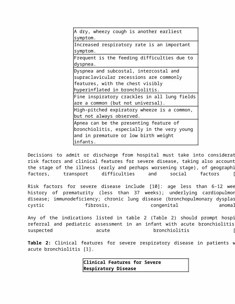
Clinical is the diagnosis of bronchiolitis, based on typical history and findings on physical examination. Clinical signs and symptoms of bronchiolitis consist of rhinorrhea, cough, wheezing, tachypnea, and increased respiratory effort manifested as grunting, nasal flaring, and intercostal and/or subcostal retractions. A coryzal phase for 2 to 3 days precedes the onset of other symptoms. Clinical conditions may deteriorate suddenly in the first 72 hours of the illness [10,25,26]. Clinical features of bronchiolitis are listed in the table 1 (Table 1). Table 1: Clinical features of bronchiolitis [1].
Clinical Features of BronchiolitisFever may be present, but high fever is uncommon and the absence of fever should not preclude the diagnosis.Rhinorrhea often precedes other symptoms such as cough, tachypnea, respiratory distress and feeding difficulties. A dry, wheezy cough is another earliest symptom.Increased respiratory rate is an important symptom.Frequent is the feeding difficulties due to dyspnea.Dyspnea and subcostal, intercostal and supraclavicular recessions are commonly features, with the chest visibly hyperinflated in bronchiolitis. Fine inspiratory crackles in all lung fields are a common (but not universal).High-pitched expiratory wheeze is a common, but not always observed.Apnea can be the presenting feature of bronchiolitis, especially in the very young and in premature or low birth weight infants.
Decisions to admit or discharge from hospital must take into consideration risk factors and clinical features for severe disease, taking also account of the stage of the illness (early and perhaps worsening stage), of geographical factors, transport difficulties and social factors [1].
Risk factors for severe disease include [10]: age less than 6-12 weeks; history of prematurity (less than 37 weeks); underlying cardiopulmonary disease; immunodeficiency; chronic lung disease (bronchopulmonary dysplasia, cystic fibrosis, congenital anomaly).
Any of the indications listed in table 2 (Table 2) should prompt hospital referral and pediatric assessment in an infant with acute bronchiolitis or suspected acute bronchiolitis [1].
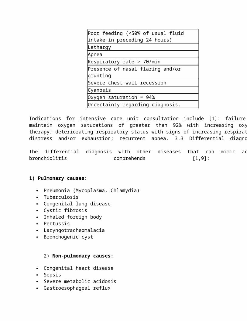
Table 2: Clinical features for severe respiratory disease in patients with acute bronchiolitis [1].
Clinical Features for Severe Respiratory DiseasePoor feeding (<50% of usual fluid intake in preceding 24 hours)LethargyApneaRespiratory rate > 70/minPresence of nasal flaring and/or gruntingSevere chest wall recessionCyanosisOxygen saturation = 94%Uncertainty regarding diagnosis.
Indications for intensive care unit consultation include [1]: failure to maintain oxygen saturations of greater than 92% with increasing oxygen therapy; deteriorating respiratory status with signs of increasing respiratory distress and/or exhaustion; recurrent apnea. 3.3 Differential diagnosis
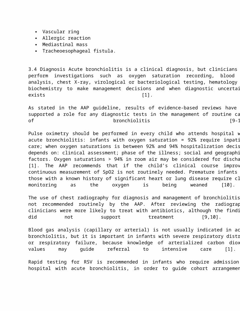
The differential diagnosis with other diseases that can mimic acute bronchiolitis comprehends [1,9]:
1) Pulmonary causes:
Pneumonia (Mycoplasma, Chlamydia) Tuberculosis Congenital lung disease Cystic fibrosis Inhaled foreign body Pertussis Laryngotracheomalacia Bronchogenic cyst
2) Non-pulmonary causes:
Congenital heart disease Sepsis Severe metabolic acidosis Gastroesophageal reflux Vascular ring Allergic reaction Mediastinal mass Tracheoesophageal fistula.
3.4 Diagnosis Acute bronchiolitis is a clinical diagnosis, but clinicians may perform investigations such as oxygen
saturation recording, blood gas analysis, chest X-ray, virological or bacteriological testing, hematology and biochemistry to make management decisions and when diagnostic uncertainty exists [1].
As stated in the AAP guideline, results of evidence-based reviews have not supported a role for any diagnostic tests in the management of routine cases of bronchiolitis [9-10].
Pulse oximetry should be performed in every child who attends hospital with acute bronchiolitis: infants with oxygen saturation = 92% require inpatient care; when oxygen saturations is between 92% and 94% hospitalization decision depends on: clinical assessment; phase of the illness; social and geographical factors. Oxygen saturations > 94% in room air may be considered for discharge [1]. The AAP recommends that if the child’s clinical course improves, continuous measurement of SpO2 is not routinely needed. Premature infants and those with a known history of significant heart or lung disease require close monitoring as the oxygen is being weaned [10].
The use of chest radiography for diagnosis and management of bronchiolitis is not recommended routinely by the AAP. After reviewing the radiographs, clinicians were more likely to treat with antibiotics, although the findings did not support treatment [9,10].
Blood gas analysis (capillary or arterial) is not usually indicated in acute bronchiolitis, but it is important in infants with severe respiratory distress or respiratory failure, because knowledge of arterialized carbon dioxide values may guide referral to intensive care [1].
Rapid testing for RSV is recommended in infants who require admission to hospital with acute bronchiolitis, in order to guide cohort arrangements.
Rapid viral antigen tests have variable sensitivity and specificity depending on the test and if used during the respiratory season [27], with a good predictive value during the peak viral season that decreases considerably at times of low prevalence. Most viruses have similar clinical courses, so the value of identifying the specific agent varies if the patients are managed as outpatient (virus identification may have little impact on management) or in the hospital setting, in which specific viral testing has been used as part of successful interventions to reduce nosocomial infections [9,28,29].
Routine bacteriological testing (of blood and urine) is not indicated in infants with typical acute bronchiolitis, but it should be considered in febrile infants less than 60 days old. Full blood count and measurement of urea and electrolytes are not indicated in assessment and management of infants with typical acute bronchiolitis, but the last ones should be considered in those with severe disease. Existing retrospective studies do not provide sufficient evidence to recommend C-reactive protein (CRP) measurement [1].
3.5 Therapy
The AAP recommends the following therapeutic strategy in infants with bronchiolitis [10]:
Antibacterial medications should be used only in children with bronchiolitis who have specific indications of the coexistence of a bacterial infection. When present, bacterial infection should be treated in the same manner as in the absence of bronchiolitis.
A carefully monitored trial of a-adrenergic or ß-adrenergic medication is an option. Inhaled bronchodilators should be continued only if there is a documented positive clinical response.
Clinicians should assess hydration and ability to take fluids orally.
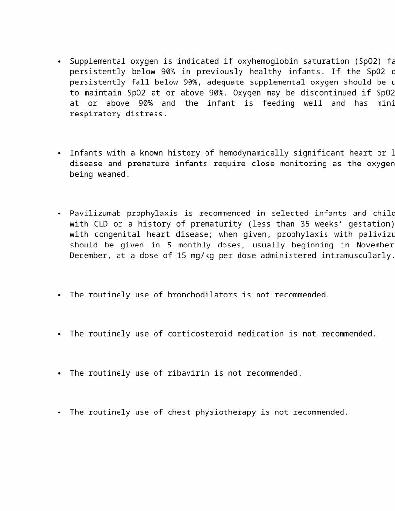
Supplemental oxygen is indicated if oxyhemoglobin saturation (SpO2) falls persistently below 90% in previously healthy infants. If the SpO2 does persistently fall below 90%, adequate supplemental oxygen should be used to maintain SpO2 at or above 90%. Oxygen may be discontinued if SpO2 is at or above 90% and the infant is feeding well and has minimal respiratory distress.
Infants with a known history of hemodynamically significant heart or lung disease and premature infants require close monitoring as the oxygen is being weaned.
Pavilizumab prophylaxis is recommended in selected infants and children with CLD or a history of prematurity (less than 35 weeks’ gestation) or with congenital heart disease; when given, prophylaxis with palivizumab should be given in 5 monthly doses, usually beginning in November or December, at a dose of 15 mg/kg per dose administered intramuscularly.
The routinely use of bronchodilators is not recommended.
The routinely use of corticosteroid medication is not recommended.
The routinely use of ribavirin is not recommended.
The routinely use of chest physiotherapy is not recommended.
Continuous measurement of SpO2 is not routinely needed, if the child’s clinical course improves.
Supplemental oxygen by nasal cannule or facemask is required in infants with oxygen saturation levels = 92% or who have severe respiratory distress or cyanosis [1].
In synthesis, bronchodilators use not improve in duration of illness or hospitalization, so the routine use is not recommended, but it may improve short-term clinical score in a subset of children, so the use is allowed only after proven benefit. Corticosteroids and leukotriene receptor antagonist not improve in duration of illness or hospitalization, so routine use is not recommended for the first, while the use is not recommended for the second ones. There are not recommendations about nebulized hypertonic saline, which however may reduce length of inpatients hospitalization [9].
3.6 Prognosis
Around half of infants without comorbidity are asymptomatic by 2 weeks after an acute bronchiolitis, but that a small proportion will still have symptoms after 4 weeks. Following acute bronchiolitis, cilial damage persists for 13-17 weeks [1].
In some children, intermittent symptoms may continue for several years, particularly with subsequent viral infections, and treatment is difficult, because no studies have shown efficacy of inhaled steroids; one randomized controlled trial found that the leukotriene receptor antagonist montelukast may give short term, minor symptomatic benefit after acute bronchiolitis, but widespread treatment with montelukast in this setting cannot be recommended [30,31].
An obliterative bronchiolitis, a rare complication in viral infections, may occur in infants with acute adenovirus bronchiolitis, in which there is a disease of the small and large airways associated with bronchiectasis. Symptoms as tachypnea, chronic cough, wheeze, chronic sputum productions may persist over the years, with a prolonged oxygen dependency. No effective specific treatment can be recommended: bronchiectasis is treated conventionally with chest physiotherapy and antibiotics [31].
The relation between RSV infection and subsequent asthma is hotly debated [32,33]. The best evidence is that RSV does not “cause” asthma; however, pre-existing atopy may be a marker for more severe bronchiolitis [34] and atopy itself predisposes to asthma. The separation of different phenotypes for preschool wheeze can be very difficult [31].
References
1. Scottish Intercollegiate Guidelines Network (SIGN) (2006) Bronchiolitis in children. A national clinical guideline. SIGN publication; no. 91 Scottish Intercollegiate Guidelines Network (SIGN): Edinburgh (Scotland.
2. http://www.monashhealth.org/icms_docs/2190_Bronchiolitis_-_full_evidence-based_guideline.pdf.
3. Worrall G (2008) Bronchiolitis. Can Fam Physician 54: 742-743.
4. Deshpande SA, Northern V (2003) The clinical and health economic burden of respiratory syncytial virus disease among children under 2 years of age in a defined geographical area. Arch Dis Child 88: 1065-1069.
5. Fleming DM, Pannell RS, Cross KW (2005) Mortality in children from influenza and respiratory syncytial virus. J Epidemiol Community Health 59: 586-590.
6. Hall CB (2001) Respiratory syncytial virus and parainfluenza virus. N Engl J Med 344: 1917-1928.
7. Wong JY, Rutman A, O'Callaghan C (2005) Recovery of the ciliated epithelium following acute bronchiolitis in infancy. Thorax 60: 582-587.
8. http://www.nottingham.ac.uk/paediatric-guideline/breathingguideline.pdf.
9. Zorc JJ, Hall CB (2010) Bronchiolitis: recent evidence on diagnosis and management. Pediatrics 125: 342-349.
10. American Academy of Pediatrics Subcommittee on Diagnosis and Management of Bronchiolitis (2006)
Diagnosis and management of bronchiolitis. Pediatrics 118: 1774-1793.
11. Dawson-Caswell M, Muncie HL Jr (2011) Respiratory syncytial virus infection in children. Am Fam Physician 83: 141-146.
12. Papadopoulos NG, Gourgiotis D, Javadyan A, Bossios A, Kallergi K, et al. (2004) Does respiratory syncytial virus subtype influences the severity of acute bronchiolitis in hospitalized infants? Respir Med 98: 879-882.
13. Gilca R, De Serres G, Tremblay M, Vachon ML, Leblanc E, et al. (2006) Distribution and clinical impact of human respiratory syncytial virus genotypes in hospitalized children over 2 winter seasons. J Infect Dis 193: 54-58.
14. Respiratory syncytial virus. In: Pickering LK, Baker CJ, Kimberlin DW, Long SS (eds.). Red Book: 2009 Report of the Committee on Infectious Diseases. (28thedn), American Academy of Pediatrics, Elk Grove Village, Ill.
15. Leung AK, Kellner JD, Davies HD (2005) Respiratory syncytial virus bronchiolitis. J Natl Med Assoc 97: 1708-1713.
16. Stempel HE, Martin ET, Kuypers J, Englund JA, Zerr DM (2009) Multiple viral respiratory pathogens in children with bronchiolitis. Acta Paediatr 98: 123-126.
17. Antunes H, Rodrigues H, Silva N, Ferreira C, Carvalho F, et al. (2010) Etiology of bronchiolitis in a
hospitalized pediatric population: prospective multicenter study. J Clin Virol 48: 134-136.
18. Jartti T, Korppi M (2011) Rhinovirus-induced bronchiolitis and asthma development. Pediatr Allergy Immunol 22: 350-355.
19. Kahn JS (2006) Epidemiology of human metapneumovirus. Clin Microbiol Rev 19: 546-557.
20. van den Hoogen BG, de Jong JC, Groen J, Kuiken T, de Groot R, et al. (2001) A newly discovered human pneumovirus isolated from young children with respiratory tract disease. Nat Med 7: 719-724.
21. Paranhos-Baccal? G, Komurian-Pradel F, Richard N, Vernet G, Lina B, et al. (2008) Mixed respiratory virus infections. J Clin Virol 43: 407-410.
22. Miller EK, Lu X, Erdman DD, Poehling KA, Zhu Y, et al. (2007) Rhinovirus-associated hospitalizations in young children. J Infect Dis 195: 773-781.
23. Peltola V, Waris M, Osterback R, Susi P, Hyypi? T, et al. (2008) Clinical effects of rhinovirus infections. J Clin Virol 43: 411-414.
24. Korppi M, Kotaniemi-Syrj?nen A, Waris M, Vainionp?? R, Reijonen TM (2004) Rhinovirus-associated wheezing in infancy: comparison with respiratory syncytial virus bronchiolitis. Pediatr Infect Dis J 23: 995-999.
25. Viswanathan M, King VJ, Bordley C, et al (2003) Management of bronchiolitis in infants and children. Rockville (MD): U.S. Department of Health and Human Services, Agency for Healthcare Research and Quality. Evidence Report/ Technology Assessment Number 69.
26. Paediatric Society of New Zealand (2005) Wheeze and chest infection in infants under 1 year. The Society, Wellington.
27. Henrickson KJ, Hall CB (2007) Diagnostic assays for respiratory syncytial virus disease. Pediatr Infect Dis J 26: S36-40.
28. Ralston S, Hill V (2009) Incidence of apnea in infants hospitalized with respiratory syncytial virus bronchiolitis: a systematic review. J Pediatr 155: 728-733.
29. Willwerth BM, Harper MB, Greenes DS (2006) Identifying hospitalized infants who have bronchiolitis and are at high risk for apnea. Ann Emerg Med 48: 441-447.
30. Bisgaard H (2003) Study Group on Montelukast and Respiratory Syncytial Virus. A randomized trial of montelukast in respiratory syncytial virus postbronchiolitis. Am J Respir Crit Care Med 167: 379-83.
31. Bush A, Thomson AH (2007) Acute bronchiolitis. BMJ 335: 1037-1041.
32. Stein RT, Sherrill D, Morgan WJ, Holberg CJ, Halonen M, et al. (1999) Respiratory syncytial virus in early
life and risk of wheeze and allergy by age 13 years. Lancet 354: 541-545.
33. Sigurs N, Bjarnason R, Sigurbergsson F, Kjellman B, Bj?rkst?n B (1995) Asthma and immunoglobulin E antibodies after respiratory syncytial virus bronchiolitis: a prospective cohort study with matched controls. Pediatrics 95: 500-505.
34. Henderson FW, Stewart PW, Burchinal MR, Voter KZ, Strope GL, et al. (1992) Respiratory allergy and the relationship between early childhood lower respiratory illness and subsequent lung function. Am Rev Respir Dis 145: 283-290.
4. Pneumonia
4.1 Definition
Pneumonia can be defined as an acute inflammation of the parenchyma of the lower respiratory tract, but this definition varies according to the organization, institution or health care setting. The World Health Organization (WHO) guidelines include a standardized definition of pneumonia based on clinical signs. The WHO definition of clinical pneumonia captures a broad spectrum of different pediatric respiratory diseases. The WHO criteria for non-severe pneumonia included: a history of cough and/or difficult breathing of less than 3 weeks duration, with (a) increased respiratory rate (Rate = 60/min if age <2 months, = 50/ min if age 2–11 months and = 40/min if age 12–59 months); (b) lower chest wall in drawing (severe pneumonia); or (c) cyanosis and/or inability to feed or drink (very severe pneumonia). The pneumonia can be divided into community-acquired pneumonia (CAP) and hospital-associated pneumonia. The CAP was defined as pneumonia with onset prior to or less than 72 hours following admission to hospital, while the hospital-associated pneumonia was defined as pneumonia with onset 48 hours after admission [1,2].
4.2 Epidemiology
Pneumonia contributes significantly to global childhood morbidity and mortality. According to a UNICEF-WHO report from 2006, over 2 million children die from pneumonia each year, accounting for almost one in five under-5 deaths worldwide [3]. This is more than the number of deaths associated with any other disease in the world, including acquired immune deficiency syndrome (AIDS), tuberculosis (TB), or malaria [4]. In 2010, pneumonia was ranked in the United States as the sixth leading cause of death for children one to 4 years of age and the 10th leading cause of death in adolescents [5]. Globally, the estimated incidence of clinical pneumonia in children aged < 5 years in developing countries is 0.28 episodes per child-year, whereas in developed countries it is 0.05 episodes per child-year [6,7]. Thus, ? 155 million episodes of clinical pneumonia occur in children <5 years of age annually, with 11-20 million of these needing hospital admission [8].
The WHO region with the highest number of pneumonia deaths was the African region (with 569,940 post-neonatal pneumonia deaths), whereas the regions with the highest percentage of post-neonatal pneumonia deaths were the East Mediterranean and South East Asian regions (with 30.69% and 31.45% of all post-neonatal deaths respectively). The countries with the highest number of pneumonia deaths were India, Nigeria, Democratic Republic of the Congo, Pakistan, Afghanistan and Ethiopia and these accounted for more than 55% of total pneumonia deaths [9].
National estimates of absolute and relative pneumonia mortality for post-neonatal children are presented in Figures 1 and 2.
Figure 1: National estimates of number of pneumonia deaths for children 1–59 months [9]
Figure 2: National estimates of % pneumonia deaths for children 1–59 months [9].
When comparing the number of estimated child pneumonia deaths between the years 2000–2003 [10] and 2008, a reduction in pneumonia mortality (27 %) is observed. This reduction may be explained by a general decrease in the overall child mortality in developing countries. In particular, in the years 2000–2003 the average annual total number of child deaths under five years old was 10.4 M, whereas it was 8.8 M deaths in 2008 [11], with a decline of 15%.
This reduction in pneumonia mortality is likely due in part to general socio-economic development (fertility, maternal education and empowerment) and in part due to development of general health services infrastructure and specific health programs [9].
Recently the introduction of effective new vaccines against bacterial pneumonia, such as Haemophilus influenzae type B (Hib) vaccine and pneumococcal conjugate vaccines (PCVs), performed mainly in countries of lower mortality, had an impact on global pneumonia mortality estimates [12]. Risk factors for CAP are shown in Table 1 (Table 1).
Medical Factors Environmental FactorsPrematurity Age < 1 yearImmunosuppressionMalnutrition
Passive tobacco smoke exposureWinter seasonOvercrowding and inadequate housingIndoor fuel exposure
4.3 Etiology
Rational treatment for pneumonia depends on knowing the most likely pathogens in each community, as the relative frequency of different agents may vary from one geographical region to another. However, identifying the causal pathogen, particularly bacteria, in children with lower respiratory tract infections is particularly difficult and does not establish causality. Conventional or experimental diagnostic tests identified pathogens in 42% to 85% of children with community-acquired pneumonia [13-15]. Mixed viral-bacterial infection is found in 23-33% of CAP cases [16].
Viruses, atypical and typical bacteria cause the vast majority of childhood pneumonia [17,18]. The distribution of
pathogens varies with age and clinical setting (Table 2).
Bacteria
Streptococcus pneumoniaeHaemophilus influenzaeStaphylococcus aureusMycobacterium tuberculosis
Atypical bacteria
Mycoplasma pneumoniaeChlamydia pneumoniae
Viruses
Respiratory syncytial virusHuman metapneumovirusParainfluenza virus types 1 and 3AdenovirusInfluenza A or BRhinovirusHuman bocavirus
The predominant pathogens vary for children of different age groups:
< 2 months: Gram-negative bacteria, Group B streptococcus, S. aureus, C. trachomatis, viruses (VRS, Adenovirus, Influenza e Parainfluenza);
2 months - 5 years: S. pneumoniae, H. influenzae, S. aureus, viruses. > 5 years: similar pathogens as 2 mounths - 5 years age group; in addition M. pneumoniae,
C. pneumonia
We describe below pneumonia caused by bacterial and non-bacterial causes.
4.4 Bacterial Pneumonia
Bacteria are the major cause of pneumonia mortality in both HIV-uninfected and HIV-infected children [19,20]. Streptococcus pneumoniae is the commonest cause of bacterial pneumonia in children of all ages and is found in 30-40 % of cases as a single or co-pathogen [21]. Group A streptococcal pneumonia is much less common and it contributes 1-7 % of cases. Although Staphylococcus aureus is not a common cause of pediatric pneumonia, it has been increasingly encountered in communities where methicillin-resistant Staphylococcus aureus (MRSA) is prevalent. Haemophilus influenza type b has almost disappeared because of vaccination. The overall incidence of pneumonia decreases with age, but it has been reported that the proportion of cases from atypical bacterial pathogens may increase among older children. Mycoplasma pneumoniae and Chlamydophila pneumoniae are more common causes of pneumonia among school-age children, [21,22]; in fact Mycoplasma infections have been common from the age of 5 years and Chlamydophila infections from the age of 10 years onwards [23].
The difference between the CAP and the hospital-associated pneumonia is in the causing organism: the pathogens causing hospital-associated pneumsonia are characteristically different from those causing CAP, with greater representation of gram-negative bacteria such as Klebsiella pneumoniae and Pseudomonas aeruginosa [24].
Culture-confirmed Mycobacterium tuberculosis has been identified in 8 % of HIV-infected and HIV-uninfected children hospitalized for acute pneumonia [25,26].
Among bacteria, newly other microbes have been discovered. Simkania negevensis, identified in early 1990s, is an intracellular bacterium that shares many characteristics with Chlamydophila species, such as the growth cycle,

genomic identity (80–87%) and antibiotic spectrum (susceptible to macrolides, tetracyclins and most fluoroquinolones but resistant to penicillins and cephalosporins). Primary infection seems to happen in early childhood; 30% of <2-year-old children are seropositive to S. negevensis, compared with only 2% to C. pneumoniae [27].
4.4.1 Pathophysiology: The pulmonary host defense is complex and includes mechanical barriers, humoral immunity, phagocytic cells and cell-mediated immunity [28,29].
Mechanical barriers are hairs from the nostrils that filter particles larger than 10 microns, mucociliary clearance, and sharp-angle branching of the central airways that helps the 5- to 10-micron particles to become impacted in the mucosa.
Humoral immunity is represented by mucosal immunoglobulin A (IgA), alveolar immunoglobulin M (IgM), and immunoglobulin G (IgG) present in transudates from the blood.
Phagocytic cells consist of polymorphonuclear (PMN) cells; alveolar, interstitial, and intravascular macrophages; and respiratory dendritic cells. Alveolar macrophages provide the first defense involved in internalizing and degrading the viral pathogens. They act as antigen-presenting and opsonin-producing cells.
Respiratory dendritic cells undergo maturation, activation, and early migration into the regional lymph nodes after the viral exposure. They act as antigen-presenting cells and are involved in the activation and differentiation of CD8+ T cells.
Cell-mediated immunity is the most important defense mechanism against the intracellular viral pathogens. This immunity is involved in antibody production, cytotoxic activity, and cytokine production. CD8+ memory or effector T cells tend to dominate the lymphocyte component of the virus-induced inflammatory component.
Pneumonia is characterized by inflammation of the alveoli and terminal airspaces in response to invasion by an infectious agent [30]. In non-hospitalized children, bacteria reach the lung by one of four routes [30]:
1. Inhalation of microorganisms that have been released into the air when an infected individual coughs or sneezes.
2. Aspiration of bacteria from the upper airways.3. Spread from contiguous infected sites.4. Hematogenous spread.
The activated inflammatory response often results in targeted migration of phagocytes, with the release of toxic substances from granules and the initiation of poorly regulated cascades (e.g., complement, coagulation, cytokines). These cascades may directly injure host tissues and adversely alter endothelial and epithelial integrity, vasomotor tone, intravascular hemostasis, and the activation state of fixed and migratory phagocytes at the inflammatory focus [31]. Pulmonary injuries are caused directly and/or indirectly by invading microorganisms [29,30].
Direct injury by the invading agent usually results from synthesis and secretion of microbial enzymes, proteins, toxic lipids, and toxins that disrupt host cell membranes, metabolic machinery, and the extracellular matrix that usually inhibits microbial migration [29,30].
Indirect injury is mediated by structural or secreted molecules, such as endotoxin, leukocidin, and toxic shock syndrome toxin-1 (TSST-1), which may alter local vasomotor tone and integrity, change the characteristics of the tissue perfused, and generally interfere with the delivery of oxygen and nutrients and removal of waste products from local tissues [32]. On a macroscopic level, the invading agents and the host defenses both tend to increase airway smooth muscle tone and resistance, mucus secretion, and the presence of inflammatory cells and debris in these secretions. These materials may further increase airway resistance and obstruct the airways, partially or totally, causing air trapping, atelectasis, and ventilatory dead space [30].
Four stages of lobar pneumonia have been described. In the first stage, which occurs within 24 hours of infection, the lung is characterized microscopically by vascular congestion and alveolar edema. Many bacteria and few neutrophils are present. The stage of red hepatization, so called because of its similarity to the consistency of liver, is characterized by the presence of many erythrocytes, neutrophils, desquamated epithelial cells, and fibrin within the alveoli. In the stage of gray hepatization, the lung is gray-brown to yellow because of fibrinopurulent exudate, disintegration of RBCs, and hemosiderin. The final stage of resolution is characterized by resorption and restoration of the pulmonary architecture. Fibrinous inflammation may lead to resolution or to organization and pleural adhesions [33].
When a bacterial infection is established, the diseases process varies according to the causative organism. Pneumococcal pneumonia remains the most common type of bacterial pneumonia and its pathophysiology has been extensively studied. The initial step in the development of this disease is the attachment of S. pneumoniae to cells of the nasopharynx and subsequent colonization. Colonization alone, however, does not cause clinical manifestations of illness because perfectly healthy people can harbor the microbe without evidence of infection. Factors that permit pneumococci to spread beyond the nasopharynx include the virulence of the strain, impaired host defense mechanisms, and viral infections of the respiratory tract. Viruses can damage respiratory tract lining cells, enhance bacterial adherence, and increase the production of mucus, which protects pneumococci from phagocytosis. In the alveoli, pneumococci infect type II alveolar cells and adhere to alveolar walls, causing an outpouring of fluid, red and white blood cells, and fibrin from the circulation, which, in turn, results in consolidation of the lung. Fluid in the lower airways creates a medium for further multiplication of bacteria and aids in the spread of infection through pores of Kohn into adjacent regions of the lung [34].
Instead the pathogenicity of M. pneumoniae is linked to the 2 properties. The first is a selective affinity for respiratory epithelial cells, and the second is the ability to produce hydrogen peroxide, which is responsible for much of the initial cell disruption in the respiratory tract and for damage to erythrocyte membranes. M. pneumoniae has a notable motility and specialized filamentous tips end that allows it to burrow between cilia within the respiratory epithelium, eventually causing sloughing of the respiratory epithelial cells [35].

4.4.2 Symptoms and signs: The clinical presentation of bacterial pneumonia varies from a mildly ill, ambulatory patient to a critically ill patient with respiratory failure or septic shock. The symptoms of pneumonia may be nonspecific, especially in infants and younger children. The sudden onset of symptoms with rapid progression of the illness is typical of bacterial pneumonia. A thorough past medical history and history of potential exposures are usually obtained. If the patient has been healthy previously, the cause of pneumonia is usually Mycoplasma or a gram-positive microbe. However, if the patient has been hospitalized, gram-negative microbes are suspected. Although not diagnostic of a particular causative agent, characteristics of the sputum may suggest a particular pathogen. Pneumococci may cause bloody or rust-colored sputum. Patients with Pseudomonas, Haemophilus, or pneumococcal infections are known to expectorate green sputum. Klebsiella and type 3 pneumococci cause the production of thick, dark red sputum [36].
In general, respiratory distress (tachypnea, nasal flaring, decreased breath sounds, cough, and rales) and fever are the prominent symptoms associated with pneumonia (Table 3) (36).
Table 3: Criteria for Respiratory Distress in Children with Pneumonia [22]
Signs of Respiratory Distress1.Tachypnea, respiratory rate, breaths/min* Age 0–2 months: > 60 Age 2–12 months: > 50 Age 1–5 Years: > 40 Age > 5 Years: > 202. Dyspnea 3. Retractions (suprasternal, intercostals, or subcostal)4. Grunting5. Nasal flaring6. Apnea7. Altered mental status8. Pulse oximetry measurement < 90% on room air
*Adapted from World Health Organization criteria. Infants and children with mild to moderate infection most commonly have a temperature < 38°C and a respiratory rate < 50 breaths per minute (bpm). Children with severe CAP commonly present with a temperature > 38°C, flaring of nostrils, grunting with breathing, tachypnea, tachycardia, and cyanosis. Although respiratory rate is a valuable clinical sign, the work of breathing (as evidenced by nasal flaring, breathlessness, cough, or wheeze) required by the infant or child may be more indicative of pneumonia [37].
Rigors or severe shaking chills may be observed with any infectious process. However, and for reasons that are not clear, the presence of rigors suggests pneumococcal pneumonia more often than pneumonia caused by other bacterial pathogens. A significant persistent cough may predominate in pneumonia caused by M. pneumoniae [38].
Patients may complain of non-specific symptoms that include headache, malaise, nausea, vomiting, and diarrhea, myalgia, shortness of breath, abdominal pain, loss of appetite and unintentional weight loss. Sharp or stabbing chest pain worsened by deep breathing or coughing and secondary to pleuritis is a common symptom of pneumococcal
infection, but may also occur with other types of pneumonia [39].
Pneumonia by typical respiratory bacterial pathogens have the greatest inflammation and disease severity, as evidenced by high temperature (> 38.4°C) within 72 hours after admission, association with pleural effusions, high percentage of band forms, elevated levels of procalcitonin, prolonged hospitalization, and a relatively high proportion of patients requiring assisted ventilation and readmission to hospital [40].
Leukocytosis (> 15.000 white blood cells/mm3) with a “shift to the left” and a predominance of neutrophils in the circulation may be observed with any bacterial infection. However, its absence, particularly in patients who are debilitated should not cause the clinician to discount the possibility of a bacterial infection. Leukopenia is a threatening sign of impending sepsis. An assessment of the arterial blood gases is essential to determine if hospital admission or oxygen supplementation is indicated and may reveal hypoxemia and respiratory acidosis. A pulse oximetric finding that is < 90% indicates significant hypoxemia [39].
Physical signs suggesting consolidation include dullness to percussion, increased tactile fremitus, reduced normal vesicular breath sounds and increased bronchial breath sounds, all of which can be difficult to detect in young children. Fine end-inspiratory crackles are typical for pneumonia in children [23]. Furthermore, the presence of wheezing should suggest the possibility that radiographic changes may be due to atelectasis and mucous plugging from asthma or bronchiolitis rather than pneumonia.
Finally, signs of an effusion are dullness to percussion, decreased tactile fremitus, and decreased or absent breath sounds, associated with, in some cases, signs of dehydration and/or sepsis [41].
4.4.3 Diagnostic tests: The etiology of pneumonia is difficult to determine in children because few children show bacteremia, and most cannot provide a sputum sample. If adequate sputum is available, it should be sent for Gram staining and subsequent culture [42]. Culture of pleural fluid is suggested if it can be sampled. Additional invasive or molecular testing should be pursued if the child fails to improve or worsens on therapy.
Specimens for culture from the lower respiratory tract can be obtained using sputum induction [43], endotracheal aspiration in intubated children and bronchoalveolar lavage (BAL). The isolation of bacteria from these samples may, however, represent contamination with bacteria that normally colonize the nasopharynx. A gram stain of expectorated sputum helps to distinguish bacterial from viral pneumonia and gram-negative from gram-positive microbes [42].
Blood culture may be useful to identify bacterial pathogens and their antimicrobial sensitivity, but only about 5% of blood cultures are positive in HIV-uninfected children with bacterial CAP. The sensitivity of blood cultures is greater in HIV-infected children, in whom approximately 18% of cultures are positive [44].
Blood cultures should not be routinely performed in nontoxic, fully immunized children with CAP managed in the outpatient setting. Blood cultures should be obtained in children who fail to demonstrate clinical improvement and in those who have progressive symptoms or clinical deterioration after initiation of antibiotic therapy. Blood cultures are not necessary repeated in children with clear clinical improvement to document resolution of pneumococcal bacteremia [22].
Urinary antigen detection tests are not recommended for the diagnosis of pneumococcal pneumonia in children because false-positive tests are common [22].
General tests of infection including acute phase reactants [erythrocyte sedimentation rate (ESR), C-reactive protein (CRP)], white cell count (WBC), neutrophil count and procalcitonin may not differentiate between bacterial and viral pneumonia [45-47]. In patients with more serious disease, such as those requiring hospitalization or those with pneumonia-associated complications, acute-phase reactants may be used in conjunction with clinical findings to assess response to therapy [22].
Pulse oximetry is performed in all children with pneumonia and suspected hypoxemia. The presence of hypoxemia guides decisions regarding site of care and further diagnostic testing [22].
Serological tests are performed for Chlamydia and Mycoplasma. The presence of Mycoplasma and Chlamydia immunoglobulin M and G antibodies contributes to the diagnosis; acute infection of Chlamydia is indicated by an IgM titer = 1:16 or by a = 4-fold rise in IgG titer, while an acute infection of Mycoplasma is indicated by an IgM titer = 1:10 or by a = 4-fold rise in IgG titer [48,49].
Tuberculin skin testing (Mantoux method) and induced sputum or gastric lavage are indicated when TB is suspected [50]. A Chest radiographs (CXRs) may be useful for confirming the presence of pneumonia and detecting complications such as a lung abscess or empyema. CXRs are however less useful for discriminating between causative pathogens and cannot accurately discriminate between viral and bacterial pneumonia [51].
CXRs reveal white shadows in the involved area indicative of an alveolar inflammatory process and may also indicate the following [39]:
A segmental or lobar opacity is observed with S. pneumonia. Cavitary lesions and bulging lung fissures are caused by K. pneumoniae and S. aureus. Cavitation and pleural effusions are caused by S. aureus and gram negative infections. Focal infiltrates are caused by atypical pathogens, M. pneumoniae or C. pneumonia.
Routine chest radiographs are not necessary for the confirmation of suspected CAP in patients well enough to be treated in the outpatient setting [40].
Indications for CXR include: clinical pneumonia unresponsive to standard ambulatory management; suspected pulmonary TB; suspected foreign body aspiration; hospitalized children to detect complications. CXRs may also be considered in children presenting with high fever, leukocytosis and no obvious focus of infections, since approximately 26% of such children may have radiographic evidence of pneumonia [22,52].
Follow-up films after acute uncomplicated pneumonia are of no value where the patient is asymptomatic [53]. A follow-up CXR is performed: in children with lobar collapse; to document resolution of a round pneumonia (as this may mimic the appearance of a Ghon focus); and in those with ongoing respiratory symptoms [22].
Bronchoscopic or blind protected specimen brush sampling, bronchoalveolar lavage (BAL), percutaneous lung aspiration, or open lung biopsy are reserved for the immunocompetent child with severe CAP if initial diagnostic tests are not positive [22].
4.4.4 Treatment: The choice of antibiotics is influenced by the epidemiology of the infecting organisms in the area,
prevalence of drug resistance, HIV prevalence and available resources. As it is difficult to distinguish between pneumonia caused by bacteria and that caused by viral infection, and because of the frequency of mixed bacterial-viral infections [54], children with pneumonia require an antibiotic. Antimicrobial therapy is not routinely required for preschool-aged children with CAP, because viral pathogens are responsible for the great majority of clinical disease. For patients with a suspected bacterial pathogen, start empiric antibiotic therapy as soon as possible (Table 4 and 5). Most bacterial pneumonia is responsive to amoxicillin, making it the antibiotic of choice. Amoxicillin should be used as first-line therapy for previously healthy, appropriately immunized infants and preschool children with mild to moderate CAP suspected to be of bacterial origin. Amoxicillin provides appropriate coverage for Streptococcus pneumoniae, the most prominent invasive bacterial pathogen [22,55].
Table 4: Guidelines for empirical antimicrobial therapy for previously healthy children three months to 17 years of age with community-acquired radiologically proven pneumonia of suspected bacterial etiology [132].
Step 1: assess severity and features of pneumonia: A. Most cases of nonsevere pneumonia ? high-dose amoxicillin oral or ampicillin IVB. Non-severe pneumonia with primary features of atypical pneumonia (subacute onset, prominent cough, minimal leukocytosis and a non-lobar infiltrate, usually in a school-age child) ? clarithromycin oral or azithromycin oral C. Severe pneumonia ? ceftriaxone IM/IV or cefotaxime IV plus clarithromycin oral or azithromycin oral/IVStep 2: assess whether child has proven or clinically suspected influenza plus evidence of secondary bacterial infection, consider adding an antiviral for influenza and use the following instead of the antibiotics from step 1: A. Non-severe pneumonia ? amoxicillin/clavulanate oral or cefuroxime IVB. Severe pneumonia ? ceftriaxone IM/IV or cefotaxime IV plus clarithromycin oral or azithromycin oral/IV. Some experts advise also adding cloxacillin IVStep 3: If the child also has a pleural effusion: A. Small effusion ? follow carefully for clinical deterioration and use antibiotics as directed in steps 1 and 2B. Moderate to large effusion ? consider pleural tap. Treat with ceftriaxone or cefotaxime instead of antibiotics in steps 1 and 2. Some experts recommend adding clindamycinStep 4: If the child has features that indicate pneumonia could be due to methicillin-resistant Staphylococcus aureus, add vancomycin or linezolid to the antibiotics chosen after steps 1, 2 and 3.
Ceftriaxone or cefotaxime may offer better coverage than amoxicillin or ampicillin for penicillin-resistant pneumococcus (although this remains controversial), and offer improved coverage for Haemophilus influenzae and Moraxella catarrhalis, but these are rare causes of pneumonia in healthy, immunized children. Clarithromycin and azithromycin do not always cover pneumococcus, so they should be reserved for children with suspected atypical pneumonia. Additionally, in patients who fail to improve after 48 h of therapy with a beta lactam alone (penicillin such as ampicillin or cephalosporins), one should first rule out an empyema and then consider adding clarithromycin or azithromycin for atypical pneumonia. Table 5: Doses of antimicrobials for suspected or proven bacterial pneumonia [132]
Antibiotic Route RegimenAmoxicillin PO 75–100 mg/kg/day divided tid* - Maximum 1 g tid
Amoxicillin clavulanate PO 75–100 mg/kg/day of amoxicillin component divided tid† Maximum 500 mg tid
Ampicillin IV 200 mg/kg/day divided q6h - Maximum 2 g q6hAzithromycin IV/PO 10 mg/kg day 1; 5 mg/kg days 2–5
Maximum 500 mg day 1; 250 mg days 2–5Cefprozil PO 30 mg/kg/day divided bid - Maximum 500 mg bidCefotaxime IV 200 mg/kg/day divided q6h -Maximum 1500 mg to 2 g q6hCeftriaxone IV 75–100 mg/kg/day divided q12h or q24h
Maximum 2 g dailyCefuroxime axetile PO 30 mg/kg/day divided tid - Maximum 500 mg tidCefuroxime IV 150 mg/kg/day divided q8h - Maximum 1.5 g q8hClarithromycin PO 15 mg/kg/day divided bid - Maximum 500 mg bidClindamycin PO 30–40 mg/kg/day divided tid - Maximum 450 mg tidClindamycin IV 40 mg/kg/day divided q8h - Maximum 600 mg q8hLinezolid IV/PO <40 kg: 30 mg/kg/day divided tid
12 years of age or older 600 mg bidVancomycin IV 40 mg/kg/day divided qid‡ - Maximum 500 mg qid‡
*Although twice daily (bid) dosing is adequate for otitis media, three times daily (tid) dosing is recommended for pneumonia; †Alternatively, one could supplement 50 mg/kg/day of amoxicillin-clavulanate with 25 mg/kg/day to 50 mg/kg/day of amoxicillin to reduce the risk of diarrhea with use of amoxicillin-clavulanate alone. ‡Higher doses may be indicated for highly resistant strains of methicillin-resistant Staphylococcus aureus. IV Intravenously; PO Orally; q6h Every 6 h; q8h Every 8 h; q12h Every 12 h; q24h Every 24 h; qid Four times daily. If atypical pathogens are suspected, then macrolide antibiotics become the antibiotic drug class of choice, with azithromycin being the preferred first-line agent [22,56-58].
When the previous penicillin allergic reaction is suspected, all beta-lactams should be avoided. For children with non-severe pneumonia treated as outpatients, clarithromycin and azithromycin are reasonable choices, while keeping in mind that pneumococcal resistance to antimicrobials is increasingly common [22].
The guidelines strongly recommend hospitalization for infants and children with respiratory distress or hypoxemia (oxygen saturation <90%); for suspicion of infection caused by community-acquired methicillin-resistant Staphylococcus aureus or any pathogen with high virulence; for infants 3 to 6 months old; or for family unable to provide appropriate care [22].
Treat with parenteral antibiotics to provide reliable blood and tissue concentrations [55]. Ampicillin or penicillin G may be given to fully immunized infants and school-aged child admitted to a hospital; however, take into account the local resistance pattern of S. pneumoniae to drugs within the penicillin class. For hospitalized children who are not yet fully immunized, who have life-threatening infections, or who are in a facility with a documented high rate of penicillin resistance, administer a third-generation parenteral cephalosporin such as ceftriaxone or cefotaxime empirically [59]. In monotherapy treatment of pneumococcal pneumonia, non-beta-lactam agents such as vancomycin have not been shown to be more effective than the third-generation cephalosporins. If S. aureus is the suspected microorganism or is confirmed with clinical, laboratory, or imaging characteristics, give vancomycin or clindamycin with a beta-lactam agent [60,61]. When atypical pathogens such as M. pneumoniae or C. pneumonia are
suspected, empiric therapy start with an oral or parenteral macrolide in combination with a beta-lactam [22].
Once a pathogen has been identified, adjust antimicrobial therapy as needed to target the specific microbe, to limit empiric antibiotic exposure, and to help limit the potential for antibiotic resistance. The recommended duration of treatment for CAP is 10 days, supported by clinical data and the practice guidelines [22,62-64]. Shorter treatment courses may be effective, especially in mild cases or outpatient treatment (5 days). Specific pathogens, such as MRSA, may need to be treated longer, exactly for 7-21 days [65]. Vancomycin has been the drug of choice for MRSA infections for many years. Recent data suggest that linezolid may be superior to vancomycin in the treatment of MRSA nosocomial pneumonia, but prospective, randomized studies are needed before linezolid is recommended as the preferred first-line therapy. Other approved agents for nosocomial MRSA infections, such as quinupristin/dalfopristin and daptomycin, should not be used in the treatment of MRSA pneumonia, as they were inferior in clinical trials. Tigecycline has excellent activity against MRSA in vitro, but should not be routinely used for the treatment of MRSA pneumonia, as clinical data are lacking [66]. If a patient is receiving intravenous antibiotics it is possible to switch to an oral agent when the clinical condition are improved to decrease risks from parenteral administration and to plan for the earliest possible discharge from the hospital. Hospital discharge may be considered when a child is clinically stable (improved appetite and activity level, afebrile for 24 hours, thoracic objectivity improved), mental status is back to baseline or stable, and the pulse oximetry level is > 90% on room air for at least 24 hours. Children receiving adequate therapy regimens should demonstrate both clinical and laboratory signs of improvement within 48 to 72 hours [22]. If improvement does not occur, further investigations should be carried, such as additional cultures to identify whether the original pathogen persists or it has developed resistance to the agent used, laboratory tests, and imaging evaluation to assess the extent and progression of the pneumonic or parapneumonic process.
In addition to antibiotics, careful supportive management is required for children with CAP. Oxygen therapy is used when there is: central cyanosis; lower chest in drawing; grunting; restlessness; inability to drink or feed; and respiratory rate > 70 breaths per minute [67-71]. Children with uncomplicated pneumonia should receive normal maintenance fluids. Appropriate rehydration is required in children who are dehydrated [22].
Children with pneumonia require a minimum of 50-60 kcal/kg/day, that needs to be considerably increased if malnutrition exist or following several days of poor nutrition. Vitamin A is not given to children with acute pneumonia unless this is measles associated [72-73]. There is no evidence that vitamin A improves outcome in non-measles pneumonia [72]. Therefore zinc is considered for use in children hospitalized with pneumonia, particularly if there is coexisting malnutrition [74].
Specific preventive strategies may reduce the incidence and the severity of pneumonia. All children should be immunized with vaccines for bacterial pathogens, including S. pneumoniae, Haemophilus influenza type b, and Pertussis to prevent CAP [22]. Parents and caretakers of infants, 6 months of age, including pregnant adolescents, should be immunized with vaccines for influenza virus and pertussis to protect the infants from exposure. Pneumococcal CAP after influenza virus infection is decreased by immunization against influenza virus [22].
Chest physiotherapy (CPT) is an important adjuvant in the treatment of most respiratory illnesses in children compared to adults. In general, it is accepted that application of manual techniques in patients with consolidation has no beneficial effect; however, the wider role that physiotherapy may play should be considered in terms of positioning to optimize ventilation and perfusion. Once the consolidatory phase begins to resolve, chest physiotherapy techniques might have some benefit in mobilizing and clearing secretions, especially in the weak or uncooperative child [75].
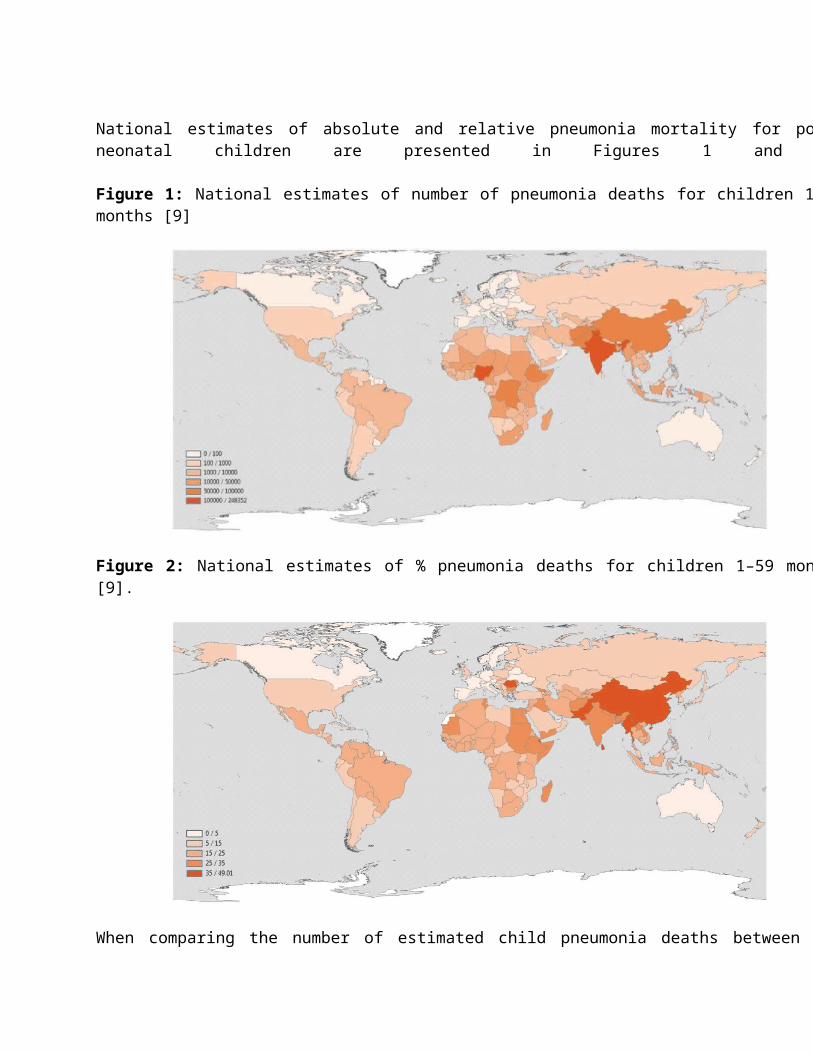
Chest physiotherapy is an airway clearance technique that consists of manual percussion of the chest wall by the caregiver, strategic positioning of the patient for mucus drainage with cough and breathing techniques. It is useful for individuals with copious mucus or thick secretions, those with weak respiratory mechanics or those with ineffective cough [76].
CPT consists of various manipulative procedures like postural drainage, chest percussion, vibration, thoracic squeezing and cough stimulation. Breathing exercise is an integral part of chest physiotherapy. It plays a significant role in airway clearance and parenchymal expansion by improving the efficiency of respiratory muscles. Postural drainage prevents the accumulation and enhances mobilization of bronchial secretion from the airway utilizing gravity to facilitate drainage. CPT should be done 1-4 times a day, preferably half an hour before meals or one and half hour after meals. The total duration should not exceed 30 minutes with 3-6 minutes in each position [76].
4.4.5 Complications: Complications of pneumonia are usually a consequence of direct spread of bacterial infection in the chest cavity (empyema, pleural effusion) or bacteremia with hematogenous spread (Table 6). Meningitis, suppurative arthritis and osteomyelitis are rare complications of hematogenous spread of infection by S. Pneumoniae [77] or by H. Influenzae. Complications of bacterial pneumonia in children include pleural effusion, empyema, lung abscess, pneumatocele and necrotizing pneumonia.
Empyema is associated with 3% of all pneumonia hospitalizations [78] and up to one third of pneumococcal pneumonia hospitalizations [79]. Empyema causes significant morbidity, with prolonged hospitalizations [80] and multiple invasive procedures. Occasionally, the infectious agent invades the pleura to cause pediatric paraneumonic empyema characterized by the presence of pus [81]. Empyema is a term derived from the Greek verb empyein (‘to suppurate’) and literally refers to frank pus in the pleural space. Empyema and parapneumonic effusion complicating pneumonia has increased dramatically in children in the UK over the last decade [82-83]. Empyema and complex parapneumonic effusion represent parts of a spectrum of disease, with three stages of progression (exudative, fibropurulent, organizational). Streptococcus pneumoniae has been found to be the principal pathogen in childhood empyema in the USA, with serotype 1 accounting for 24–50% of culture positive cases between 1993 and 2000 [84-85]. The treatment options for parapneumonic effusions range from non-invasive antibiotic therapy and observation, to semi-invasive techniques such as therapeutic aspiration, tube thoracostomy and intrapleural fibrinolytics, to invasive interventions such as thoracoscopy, thoracotomy or open drainage [86].
4.5 Non-Bacterial Pneumonia
Viruses may act as sole pathogens in pediatric CAP or predispose to bacterial pneumonia [87-89]. Viral pathogens are more common causes of CAP in children younger than 2 years, accounting for 80% of cases [22]. Viral etiologies vary by geography, season, and the age of patients studied, but Respiratory Syncytial Virus (RSV), Human Rhinovirus (hRV), Adenovirus, Human Bocavirus (hBoV), Human Metapneumovirus (hMPV), and Parainfluenza Viruses (PIV) are described consistently as the most common viruses associated with CAP in children. Viruses usually circulate in winter (e.g., RSV, influenza, PIV and hMPV) and as a sole cause of pneumonia are less common in older children with the exception of influenza [90].
RSV is the commonest cause of viral CAP, especially in the first 3 years of life. RSV causes significant mortality and morbidity in both HIV-infected and uninfected children, especially in children born prematurely and who are less than 6 months of age at the onset of the RSV season. HIV-infected children with RSV are more likely to develop pneumonia rather than bronchiolitis compared with HIV-uninfected children [26].
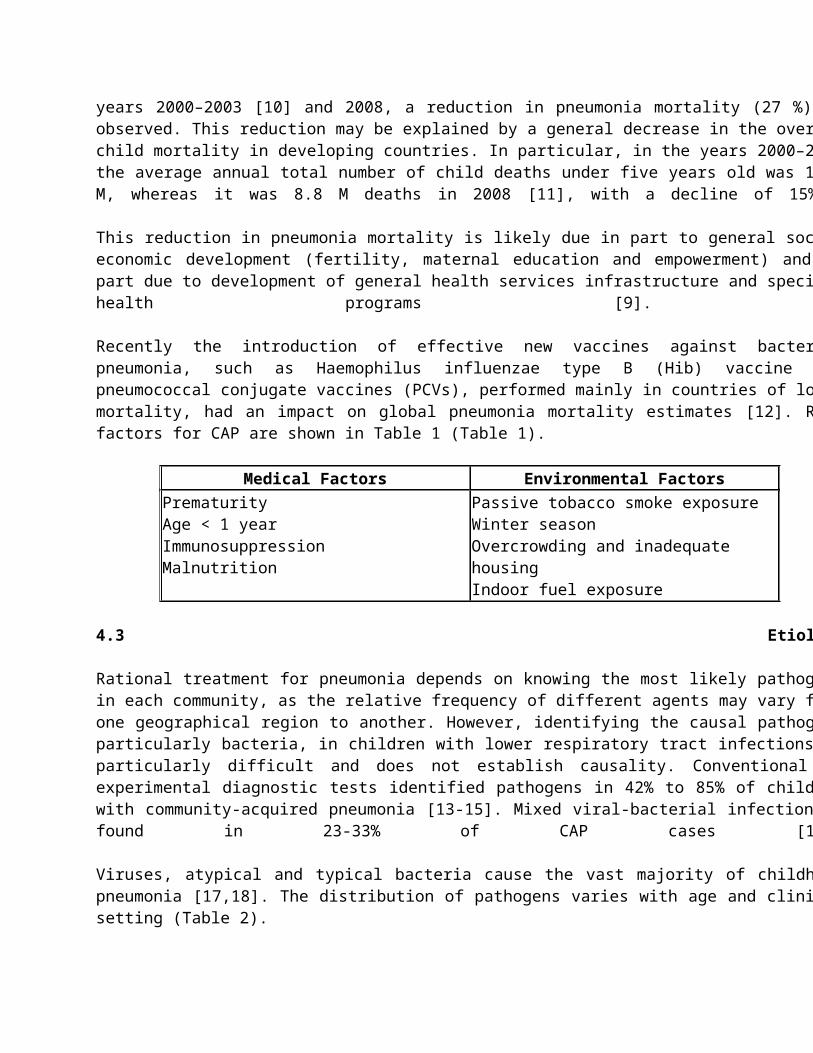
PIV is a common virus that infects most persons during childhood. PIV is second in importance to only RSV in causing lower respiratory tract disease in children and pneumonia and bronchiolitis in infants younger than 6 months [91]. Adenovirus accounts for 10% of pneumonias in children and can occur at any time of the year.
hMPV is a relatively newly discovered respiratory pathogen. It was recognized 10 years ago [92] and shares many properties with RSV, including human beings as the only hosts, a similar seasonality and nearly identical clinical and laboratory features [93-94]. hMPV is in the Paramyxoviridae family (like RSV and PIV) and is a pleomorphic-shaped virus surrounded by surface protein projections. Children aged <5 are susceptible to hMPV infection, and infants aged <2 with primary infection are at risk of severe infection [94-95]. hMPV and hBoV are found in 8-12% and ? 5%, respectively [16].
The fungal pneumonia in children is a rare condition, and is often seen in individuals with compromised immune system like AIDS. The most common fungal agents that cause pneumonia in children are Histoplasma capsulatum, Cryptococcus neoformans, Pneumocystis jiroveci, Blastomyces and Coccidioides immitis [96]. Pneumocystis jiroveci (previously P. carinii) pneumonia (PCP) is a common, serious infection among HIV-infected children and is associated with high mortality. Infants aged 6 weeks– 6 months are at highest risk for infection; PCP is the predominant cause of pneumonia mortality in HIV-infected children less than 6 months of age [97]. PCP has also been described in malnourished children and in young HIV-exposed uninfected infants.
4.5.1 Pathophysiology: After contamination, most respiratory viruses tend to multiply in the epithelium of the upper airway and secondarily infect the lung by means of airway secretions or hematogenous spread. Severe pneumonias may result in extensive consolidation of the lungs with varying degrees of hemorrhage. Some patients showed bloody effusions and diffuse alveolar damage [98].
The mechanism of damage to tissues depends on the virus involved. Viral infections are characterized by the accumulation of mononuclear cells in the submucosa and perivascular space, resulting in partial obstruction of the airway. Some viruses are mainly cytopathic, directly affecting the pneumocytes or the bronchial cells. With others, over exuberant inflammation from the immune response is the mainstay of the pathogenic process. Immune responses can be categorized according to patterns of cytokine production. Type 1 cytokines promote cell-mediated immunity, while type 2 cytokines mediate allergic responses [99].
In addition to humoral responses, cell-mediated immunity appears to be important for recovery from certain respiratory viral infections. Impaired type 1 response may explain why immunocompromised patients have more severe viral pneumonias. Respiratory viruses damage the respiratory tract and stimulate the host to release multiple humoral factors, including histamine, leukotriene C4, and virus-specific immunoglobulin E in RSV infection and bradykinin, interleukin 1, interleukin 6, and interleukin 8 in rhinovirus infections. RSV infections can also alter bacterial colonization patterns, increase bacterial adherence to respiratory epithelium, reduce mucociliary clearance, and alter bacterial phagocytosis by host cells.
The mechanism of viral transmission varies with the type of virus. Routes include large-droplet spread over short distances (<1 m), hand contact with contaminated skin and subsequent inoculation onto the nasal mucosa or conjunctiva (e. g., rhinovirus, RSV), and small-particle aerosol spread (e. g., influenza, adenovirus). Some viruses are extremely fastidious, whereas others have the capability of surviving on environmental surfaces for as long as 7 hours, on gloves for 2 hours, and on hands for 30 minutes.
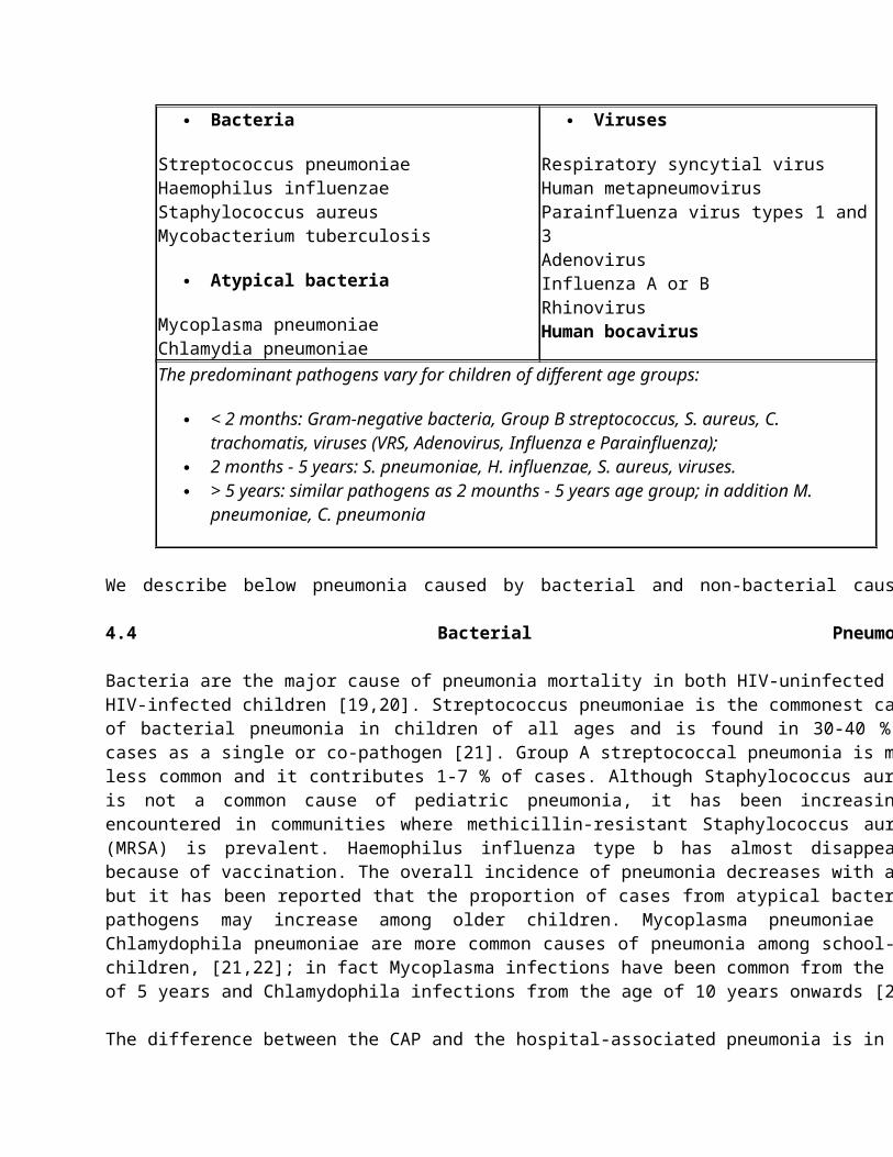
4.5.2 Signs and symptoms: The clinical manifestations of viral pneumonia vary because of the number of diverse etiologic agents. The common constitutional symptoms of all viral pneumonias are fever, chills, nonproductive cough, rhinitis, myalgias, headaches, and fatigue. Symptoms of viral pneumonia are similar to that of bacterial pneumonia, although studies have shown a lower probability of having chest pain and rigors in viral pneumonias [100]. Most patients have cough. Ascertaining immunization status, travel history, and possible exposure is important. During outbreaks with the usual respiratory viruses, the signs and symptoms can suggest the correct diagnosis in most cases.
The typical infection with influenza virus consists of a sudden onset of fever, chills, myalgia, arthralgia, cough, sore throat, and rhinorrhea. The incubation period is 1-2 days, and symptoms normally last 3-5 days. These symptoms are common to other respiratory viral infections but are highly suggestive of influenza virus infection when an outbreak is occurring in the community. Influenza is usually seen in epidemics and pandemics in late winter and early spring.
Peak attack rates for RSV occur in the winter in infants younger than 6 months. PIV infection most often occurs in the late fall or winter, although PIV-3 pneumonia is especially common in the spring [101].
Pneumonia caused by PCP is frequently (20-40 %) the initial presenting feature of AIDS in HIV-infected children not taking cotrimoxazole prophylaxis [102-103]. Although PCP may present with a tetrad of features comprising tachypnea, dyspnea, fever and cough, these are not specific for pneumonia caused by P. jiroveci. Hypoxia may be prominent and rapidly progressive. Other stigmata of AIDS such as hepatosplenomegaly and generalized lymphadenopathy are not always present and adventitious sounds in the chest may be absent despite clinical signs of severe respiratory distress.
4.5.3 Diagnostic tests: The currently validated methods to define the etiology of viral infection are serology, culture, cytological evaluation, rapid detection of antigens, and gene amplification techniques, even though they are not widely available and are often costly.
Virtually all viruses can be diagnosed through serology. However, it is necessary to collect paired blood samples (acute/convalescent phases), as a four-fold titer increase in relation to the first sampling is necessary to confirm the diagnosis. Therefore, serology is not routinely used, as it has been shown to be of little use in the acute phase of the disease, because titers rarely increase at this stage [22]. Serology is available for many of the community respiratory viruses such as Adenovirus, RSV, and seasonal Influenza.
Viral culture can also be employed for most of the respiratory viruses, with the long time necessary to obtain the results being a disadvantage, as well as the need for specific culture mediums. To perform the cultures, tissue samples from the upper and/or lower airways, sputum, and nasopharyngeal and bronchoalveolar lavage can be used. The cytopathic effects of the viruses are observed in cell cultures, such as the formation of syncytial collections of multinucleated giant cells, or evidence of viral growth. The subsequent identification of specific viruses in cell cultures may be accomplished by immunofluorescence techniques (direct or indirect), or nucleic acid probes. Other disadvantages of this method are high cost, low availability in clinical practice, and also the low yield for some specific agents such as RSV, hMPV, and Coronavirus [22].
Cytological assessment use samples from respiratory tissues and also secretions such as nasal and bronchoalveolar lavage. The technique aims at identifying nuclear (virus DNA) or cytoplasmic (virus RNA) inclusions, which are normally present in infected cells. The identification of the presence of such inclusions confirms the diagnosis [22].
These are rapid tests performed in easily obtainable specimens, such as nasal swab or wash. The enzyme-linked immunosorbent assay (ELISA) test is available for most pathogenic respiratory viruses; it is capable of detecting viral antigens, whereas the immunofluorescence requires intact infected cells [22].
The polymerase chain reaction (PCR) or reverse transcriptase-polymerase chain reaction (RT-PCR) techniques are extremely sensitive and specific to detect virus presence. It is the examination of choice for most respiratory viruses and, if available, should be employed together with the aforementioned diagnostic methods. The current development of this technique has allowed the knowledge of new causative agents of bronchiolitis and pneumonia in both pediatric and adult populations. This method can be applied to samples of nasopharyngeal or bronchial secretion swabs, and has the advantage that it can be performed in other body fluids [22].
A new molecular technique called multiplex reverse transcriptase polymerase chain reaction (MRT-PCR) allows for the rapid detection of several respiratory viruses such as influenza A and B; RSV A and B; HPIV 1, 2, and 3; hMPV and adenovirus [104].
Radiological confirmation is required whenever possible to support the clinical diagnosis and if the child has a worsening of the clinical condition. Poorly defined nodules and patchy areas of opacity with variable hyperinflation and without effusion are more indicative of a viral etiology [105], but some features are characteristic of individual viruses.
Radiographic findings in Influenza pneumonia are nonspecific and can be seen as perihilar and peribronchial opacities, consolidations, and diffuse bilateral interstitial opacities, especially in more severe forms of the disease. RSV pneumonia typically presents with patchy bilateral alveolar infiltrates and interstitial changes (similar to influenza) [106].
In PIV pneumonia chest radiographs may reveal findings ranging from focal infection to diffuse interstitial infiltrates or diffuse mixed alveolar-interstitial infiltrates consistent with acute lung injury. Adenovirus pneumonia usually presents with diffuse, bilateral and patchy, ground-glass infiltrates with a preference for lower lobes, although it can present with lobar consolidation [106]. Common, but no specific radiographic finding of severe PCP is diffuse or scattered ground-glass opacification [107].
4.5.4 Treatment: Children less than 2 years of age are commonly infected with viral pathogens. Those with mild cases of viral CAP do not require anti-microbial therapy. For children with moderate-to-severe CAP consistent with influenza infection, administer influenza antiviral therapy as soon as possible, especially during a widespread local circulation of influenza viruses (Table 7) [22]. Some influenza A strains will be susceptible to antiviral therapy, even though genetic variability is high each year. Early antiviral treatment has been shown to provide maximal benefit, therefore treatment should not be delayed until confirmation of positive influenza test results. Negative results of influenza diagnostic tests do not conclusively exclude influenza disease. Treatment after 48 hours of symptomatic infection may still provide clinical benefit to those with more severe disease [91].
Table 7: Influenza antiviral therapy in pediatric patients [22]
Drug (Brand name) Formulation DosingOseltamivir (Tamiflu) 75 mg capsule;
60 mg/5 mL suspension4-8 mo: 6 mg/kg/d in 2 doses9-23 mo: 7 mg/kg/d in 2 doses>24 mo: -4 mg/kg/d in 2 doses, for 5 days
Si 5 kg: 60 mg/d in 2 divided doses>15-23 kg: 90 mg/d in 2 divided doses>23-40 kg: 120 mg/d in 2 divided doses>40 kg: 150 mg/d in 2 divided doses
Zanamivir (Relenza) 5 mg per inhalation, using a Diskhaler
>7 y: 2 inhalations (10 mg total per dose), twice daily for 5 days
Amantadine (Symmetrel) 100 mg tablet; 50 mg/5 mL suspension
1-9 y: 5-8 mg/kg/d as single daily dose or in 2 doses; not to exceed 150 mg/d9-12 y: 200 mg/d in 2 doses(not studied as a single dose)
Rimantadine (Flumadine) 100 mg tablet; 50 mg/5 mL suspension
Not FDA approved for treatment in children, but published data exist on safety and efficacy in childrenSuspension: 1-9 y: 6.6 mg/kg/d (max 150 mg/kg/d) in 2 doses; >10 y: 200 mg/d, as single daily dose or in 2 doses
Children with chronic pulmonary, cardiovascular or immunosuppressive disease or those on aspirin should be vaccinated annually at the start of the influenza season. Children between 6 months and 9 years of age who have not been vaccinated previously require 2 immunizations of a single dose given 1 month apart; children who are older than 9 years or those who have been immunized previously require only a single immunization. Immune prophylaxis with RSV specific monoclonal antibody is also considered for premature infants or those with bronchopulmonary dysplasia, congenital heart disease, or immunodeficiency, to decrease the risk of severe pneumonia and hospitalization [91].
At last, the use of HAART to reconstitute immunity is very effective for decreasing the incidence of pneumonia and opportunistic infections in HIV-infected children [91].
4.5.5 Complications: Prompt diagnosis and treatment of lung infections are necessary to prevent complications (Table 6). The complications of viral pneumonia in children are: focal necrosis and airway plugging, atelectasis, bronchospasm, apnea spells, respiratory failure, bronchiectasis, bronchiolitis obliterans and pulmonary fibrosis [108].
Table 6: Complications associated with Community-Acquired Pneumonia [22]
Pulmonary Pleural effusion or empyema
Pneumothorax
Lung abscess
Bronchopleural fistula
Necrotizing pneumonia
Acute respiratory failure
Metastatic Meningitis
Central nervous system abscess
Pericarditis
Endocarditis
Osteomyelitis
Septic arthritis
Systemic Systemic inflammatory response syndrome or sepsis
Hemolytic uremic syndrome
4.6 Recurrent or Chronic Pneumonia
Recurrent pneumonia is defined as 2 episodes of pneumonia in a single year, or 3 episodes over any time period, and it is reported in 7-9% of children with pneumonia. The chest radiograph should show resolution of radiologic changes between episodes. It is common practice to investigate the immune and respiratory systems in children with recurrent pneumonia [109,110]. A rationale approaches to this problem first requires a clear definition concepts recurrent and persistent pneumonia. Unfortunately there is little uniformity among investigators. Although the chest radiographs are not universally obtained in all pediatric patients with suspected pneumonia, the presence of abnormal radiographic results must be considering as a crucial criterion for defining an episode of pneumonia in a child with recurrent or persistent lower respiratory infections [111].
Given the relatively common occurrence of recurrent pneumonia in children, it is suggestive that only four retrospective studies evaluate its causes [112-115]. An underlying cause was identified in more than 80% of patients, with large differences between studies as regards the prevalence of the most common causes.
It is useful to assess the etiology of recurrent or chronic pneumonia to classify the process according to the anatomic distribution in the lungs:
1. Pneumonias affecting a single anatomic region.2. Pneumonias affecting multiple anatomic regions.
In general, pneumonias affecting a single lobe are caused by obstruction of the lumen of the affected lobe. This may be caused by intraluminal obstruction, extraluminal compression and structural abnormalities of the airway and/or the lung parenchyma.
Aspiration of foreign bodies into the lung represents the most common cause of intraluminal airway obstruction in the pediatric population. Retained foreign bodies occur most commonly in the 6 months to 3 years age group. Most of the foreign bodies are localized in the right bronchus because it is slightly wider and originates at a less acute angle from the trachea. Symptoms of a bronchial foreign body include wheeze, cough, dyspnea, and occasionally hemoptysis. Physical signs vary in relation to the localization, size, composition, number of foreign bodies and in relation to the degree of resulting obstruction. Chest radiographs may be useful to diagnose the presence of inhaled foreign bodies. Sometimes, local atelectasis or air trapping may be shown [110].
Other causes of intraluminal obstruction are far less frequent in children, such as the active disease caused by Mycobacterium tuberculosis that it may appear with persistent focal infiltrates or atelectasis resulted by airway granulomas or bronchial adenomas [116] and endobronchial lipomas [117].
Enlarged lymph nodes are the most common causes of extraluminal airway compression. Infectious lymphadenopathy that causes airway compression is commonly caused by infection with M. tuberculosis. Lymphadenopathy caused by histoplasmosis and coccidiomycosis, in certain geographic areas where these fungal species may be endemic, causes similar symptoms, including compression and recurrent infectious pneumonias [118].
Non-infectious causes of pulmonary lymph nodes enlargement are less common and may also lead to extrinsic airway compression. Sarcoidosis is a rare multisystem disorder of unknown etiology that causes chronic, non-caseating, granulomatous lesions, in particular in lymphoid tissue of the mediastinal nodes [119]. Mediastinal malignancies may also occasionally lead extraluminal airway compression [110].
Congenital anomalies of heart and great vessels may also cause extrinsic compression of the airway [110]. Vascular rings and slings are a heterogeneous group of anomalies that result from abnormal development of the aortic arch. Anomalies include double aortic arch, vascular rings consisting of right aortic arch, anomalous origin of the left subclavian artery, and ligamentum arteriousus, innominate artery compression and pulmonary artery slings. Double aortic arch is the most common of these. Symptoms usually begin in early infancy and are characterized by wheeze, stridor, cough and infectious complications [110].
Bronchoscopy may define a focal area of large airway compression, suggesting the presence of a congenital vascular anomaly, but the definitive diagnosis requires using several imaging techniques such as chest computed tomography (CT), magnetic resonance imaging (MRI) with contrast, echocardiography and angiography [110].
Bronchial abnormalities may predispose to recurrent pneumonias, such as tracheal bronchus, bronchial stenosis, bronchomalacia and bronchiectasis. Tracheal bronchus is an extra or aberrant bronchus that usually originates in the right lateral wall of the trachea [110]. Congenital bronchomalacia is a rare cause of recurrent pneumonias that affects premature infants and children with Trisomia 21 [110].
Bronchiectasis is irreversible focal or generalized abnormal dilatation of the bronchial segments that occurs as congenital or acquired lesion caused by destruction of muscle and elastic tissue. Most cases of focal bronchiectasis occur after severe bacterial infection of the lung, which leads to localized airway damage. Measles virus, adenovirus,
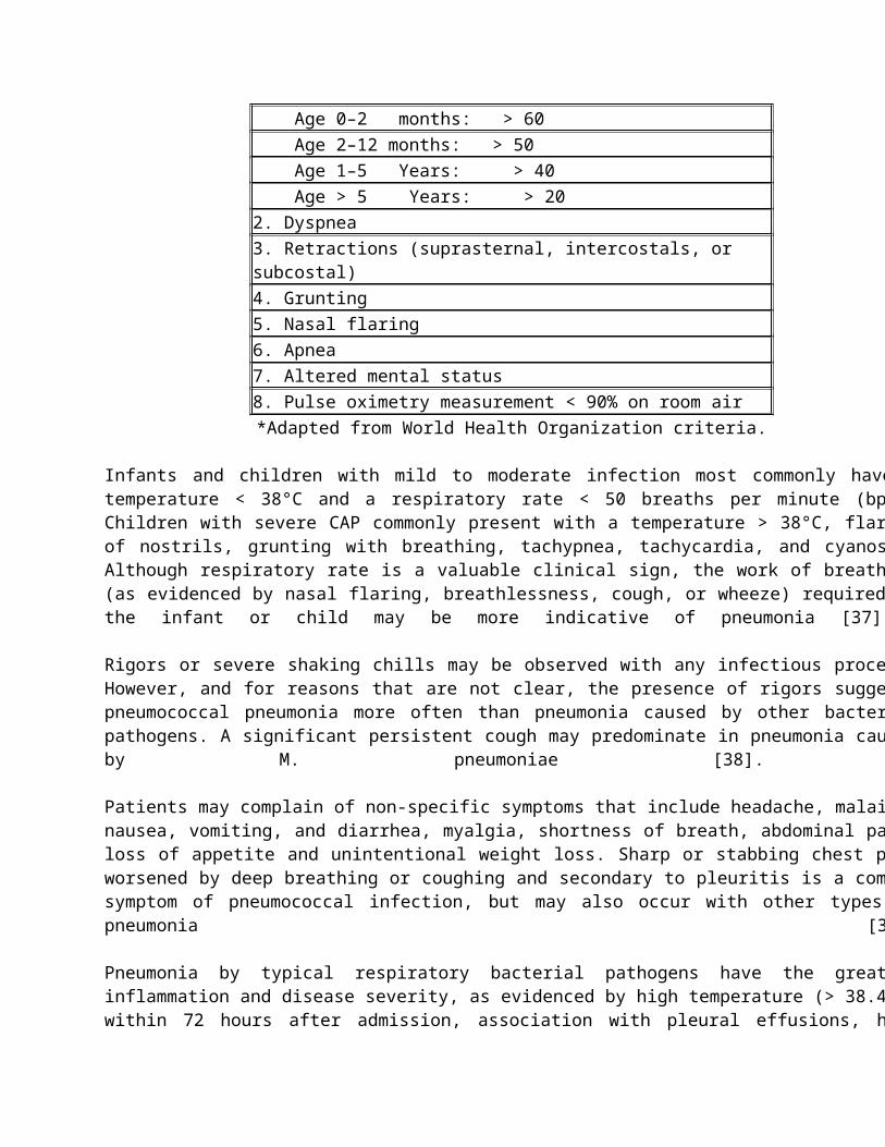
Bordetella pertussis and M. tuberculosis are often involved in the developmental of bronchiectasis. Haemophilus influenza and Staphylococcus aureus may contribute to the progression of the airway damage [110].
Bronchiectasis are divided into cylindric and saccular; in cylindric bronchiectasis the damage involves only the elastic and muscular supporting tissues of the airways, sparing the cartilage, while in the saccular bronchiectasis the damage extends to the cartilaginous structures of the airways. Another cause of focal recurrent atelectasis or pneumonia in children is the “right middle lobe syndrome”. The right middle lobe bronchus has a small diameter and a pliable wall and originates from the right brainstem bronchus with acute angle. These factors may contribute to the compression or collapse [110].
Pulmonary sequestrations, congenital cystic adenomatoid malformations and bronchogenic cysts are pulmonary lesions that may cause recurrent pneumonia. A wide variety of anatomic, immunologic and neurologic disorders can cause recurrent pneumonias affecting multiple lobes of the lung [110]. In one large study conducted on 238 children with recurrent pneumonia, Owayed and co-workers [112] have found that the oropharyngeal incoordination with aspiration syndrome is the commonest cause (48%), followed by immune disorders (10%), congenital heart disease (9%) and pulmonary abnormalities (8%).
Aspiration pneumonia develops after the inhalation of oropharyngeal contents into the lungs. It may be an acute or chronic event and it has multiple etiologies. The degree of pulmonary compromise caused by aspiration depends on the volume of aspirated material, the pH level of the material, and the type of material aspirated [120].
Impaired swallowing may be caused by central nervous system disorders, neuromuscular disease or anatomic abnormalities of the oropharynx. Drugs such as anesthetic agents and sedatives cause acute aspiration events because they may depress consciousness, the gag reflex and swallowing function. Aspiration may occur chronically in association with seizures in children with severe mental retardation and cerebral palsy [110].
Children with neuromuscular disorders, such as muscular and myotonic dystrophy, have a decreased or absent cough and gag reflexes and they produce a large amount of secretions, resulting in abnormalities of autonomic control; therefore they have abnormal swallowing mechanisms [110]. Although uncommon, some congenital or acquired anatomic lesions of the upper airway can be associated with recurrent pneumonia. Laryngeal and palatal abnormalities including cleft palate, laryngeal clefts and submucosal clefts are associated with uncoordinated swallowing reflexes [110].
On the other hand, recurrent pneumonia may be a complication of gastroesophageal reflux disease (GERD) due to aspiration of gastric juice [121]. GERD should always be considered a possible cause of recurrent pneumonia when children complain of typical symptoms (heartburn, regurgitation and dysphagia) [122-123]. However, the lack of gold standard diagnostic tests for both GERD and aspiration makes it difficult to clarify the contribution of GERD to recurrent pneumonia. Therefore markers of aspiration are needed for clinical practice to prove the causal relationship between gastroesophageal reflux and chronic aspiration [124].
Although asthma has been reported as a common and important underlying cause of recurrent pneumonia [109-110,122], data from the recent literature do not support this [125-127]; this is likely due to the diagnostic confusion between “asthma and recurrent pneumonia”. Therefore, it seems probably that very unusual and complicated cases of asthma may be considered a cause of recurrent pneumonia in children [125].
In order, asthma is a differential diagnostic consideration, and not an underlying cause of recurrent pneumonia [126].
There are several immunodeficiency disorders in which pneumonia occur as a main feature [128]. Most of these involve abnormalities of B-lymphocyte function, with associated abnormalities of immune globulin production and function. Patients with global deficiencies of immunoglobulin A (IgA) or IgG have increased susceptibility to bacterial pulmonary infections. Levels of IgG subclasses should also be evaluated because there are some patients who have normal levels of total IgG and decreased levels of IgG2 and/or IgG4 subclasses [110].
Patients with deficiencies of the complement pathway (C3, C4, properdin) or those with deficient numbers or function of phagocytic cells (Chediak-Higashi syndrome, Job’s syndrome or hyperimmunoglobulinemia E) can experience recurrent pneumonia [110].
Disorders of mucociliary function commonly present with recurrent pneumonia, sinusitis, otitis media and male infertility. Primary ciliary dyskinesia (PCD) is a rare autosomal recessive disease, caused by specific primary structural and/or functional abnormalities of the motile cilia with prevalence about 1/15.000 to 1/30.000 [129]. Approximately one half of the patients with PCD have situs inversus. Those patients having PCD with situs inversus are known to have Kartagener’s Syndrome.
Cystic fibrosis is caused by a mutation in a gene that encodes cystic fibrosis transmembrane conductance regulator (CFTR) protein, which is expressed in many epithelial cells and blood cells. Cystic fibrosis is most common in populations of northern European descent, among whom the disease occurs in approximately 1 in 3.000 births [130], and it presents with recurrent pneumonia, chronic sinusitis, steatorrhea and failure to thrive.
Other causes of recurrent pneumonia in childhood are congenital heart disease, bronchopulmonary dysplasia, toxicity by smoke inhalation, chronic ingestion of lipids and hypersensitivity pneumonitis (HP). The HP is a rare and heterogeneous group of conditions characterized by an allergic response to numerous environmental or occupational agents and it may occurs as an acute, subacute or chronic condition [109].
The initial clinical evaluation of a patient with chronic or recurrent pulmonary infections will provide the physician with information necessary to guide the choice of the laboratory investigations. A careful review of the patient’s history and physical examination will enable the clinician to proceed with appropriate diagnostic tools [110].
In case of suspected recurrent or chronic pneumonia, there are a number of aspects in the history that must be considered such as the frequency, duration and severity of all episodes of supposed pneumonia. Associated symptoms such as recurrent fever, weight loss, cough, wheeze, systemic illness, upper airway noises and labored breathing are important. Neonatal history and information about any serious infections, illnesses or surgical procedures are necessary for a correct diagnosis [110].
The timing and nature of onset of illness may provide valuable clues. A family history of early or unexplained deaths or chronic respiratory problems may suggest a diagnosis of immunodeficiency, or other diseases with genetic component. Important aspects of the social history are occupational exposure to potential hypersensitivity-inducing antigens, daycare attendance and the presence of siblings with similar problems [110].
The general examination should take into account of the dysmorphic features, which may be associated with structural airway abnormalities (trisomy 21) or immune disorders. Growth and development are important because a normal growth is a reassuring sign. The vital signs should be documented (oxyhemoglobin saturation) and the presence of eczema is often in association with asthma. Examination of the head and neck may document the presence of the signs of the chronic otitis or sinusitis. Periodontal disease and abnormal dentition may be indicators
of phagocytic cell dysfunction and hyper IgE syndrome, respectively [110].
Examination of the respiratory system consists of a careful evaluation of the mechanics of breathing with attention to symptoms such as shortness of breath, retractions, accessory muscle use, scoliosis and chest-wall deformity or asymmetry [110].
A careful cardiac examination can document undiagnosed cardiac lesions. Extremities should be carefully examined for signs of cyanosis and clubbing which may be seen in patients with cystic fibrosis, bronchiectasis and cyanotic heart disease. Chest radiograph is an important tool as laboratory evaluations that should be required in all cases of suspected recurrent pneumonia. All previous chest radiographes should review because this approach allows the physician to make a difference between recurrent and persistent pneumonia. The patient’s clinical status is very important to determine the urgency of the initial visit. If the patient is ill the priority is to establish adequacy of oxygenation and ventilation. Oxygen saturation is measured easily using pulse oximetry, while the method used to evaluate the ventilation is an arterial blood gas. If there is cough with sputum production should be collect a sample to be sent for gram stain and culture [110].
Bronchoscopy, chest CT, MRI or angiography should be performed to rule out foreign bodies or anatomic abnormalities in children with recurrent or persistent focal abnormalities on chest radiograph. Also a tuberculosis skin test (PPD) should be performed in any child with focal changes [110].
Neurologic dysfunction and swallowing difficulty or vomiting should be evaluated n children with abnormalities in diffuse or variable regions of the lungs on chest radiograph. In these cases a barium swallow should be required to assess the adequacy of the swallowing mechanism; it is also important to check for GERD. A 24-hour PH probe study may be the gold standard method of testing of GERD. Spirometry should be obtained before and after administration of an inhaled bronchodilator to evaluate if there is reversible airway obstruction. In patients with clinical history suggestive of asthma bronchial provocation tests (methacholine, mannitol, exercise, cold air inhalation) may also prove useful. In patients with signs and symptoms suggestive of cystic fibrosis a sweat test should be obtained. Immunologic function evaluation is also indicated by the clinical and radiographic history. Ciliary biopsy should be performed for children with recurrent sinusitis and otitis. A complete blood count with differential can provide important clues; anemia may suggest hemoglobinopathy or chonic disease and thrombocytopenia can be present in malignancy or Wiskott-Aldrich syndrome. The neutrophil number and function should be assessed in patients with recurrent abscesses of the skin or infections of the gums serial blood counts and T-lymphocyte function should be evaluated in patients with recurrent fungal infections or abnormal response to viral infections. A history of recurrent pyogenic infections requires a quantitative evaluation of immune globulins, including IgG subclasses, and an assessment of the classic complement pathways (total emolytic complement, C3 and C4 titers) [110].
Therapy of recurrent or persistent pneumonia includes nutritional support, exercise, chest physiotherapy and avoiding exposure to smoke or allergens, important factors implicated in the resolution of disease. The active cycle of breathing techniques used together with exercise training in clinically stable young cystic fibrosis patients increases thoracic mobility and the physical fitness parameters such as muscle endurance, strength, flexibility and speed [131].
In particular, the standard treatment for X-linked agammaglobulinemia (XLA) is intravenous immunoglobulin (IVIG). Despite apparently adequate treatment with IVIG, however, many patients still develop pansinusitis or post-infectious chronic lung diseases, most commonly bronchiectasis. Rotating antibiotics in treatment doses are then
needed in addition to monthly IVIG infusions [127]. Surgical intervention is used for patients with congenital anatomic abnormalities or acquired focal disease involving a single lobe or segment of lung. In most cases, a careful analysis of the etiology of recurrent or persistent pneumonia will direct the clinician to a correct therapeutic approach [110].
References
1. Taneli Puumalainen, Beatriz Quiambao, Erma Abucejo-Ladesma, Socorro Lupisan, Tarja Heiskanen-Kosma1, et al. (2008) Clinical case review: A method to improve identification of true clinical and radiographic pneumonia in children meeting the World Health Organization definition for pneumonia. BMC Infectious Diseases 8: 95.
2. Scott JA, Wonodi C, Mo?si JC, Deloria-Knoll M, DeLuca AN, et al. (2012) The definition of pneumonia, the assessment of severity, and clinical standardization in the Pneumonia Etiology Research for Child Health study. Clin Infect Dis 54 Suppl 2: S109-116.
3. http://www.unicef.org/publications/files/Pneumonia_The_Forgotten_Killer_of_Children.pdf.
4. http://www.cdc.gov/features/pneumonia/
5. Murphy SL, Xu IQ, Kochanek KD (2012) Deaths: Preliminary data for 2010. National vital statistics reports 60: 1-52. HyattsviUe, Md: National Center for Health Statistics.
6. Rudan I, Boschi-Pinto C, Biloglav Z, Mulholland K, Campbell H (2008) Epidemiology and etiology of childhood pneumonia. Bull World Health Organ 86: 408-416.
7. Rudan I, Tomaskovic L, Boschi-Pinto C, Campbell H; WHO Child Health Epidemiology Reference Group (2004) Global estimate of the incidence of clinical pneumonia among children under five years of age. Bull
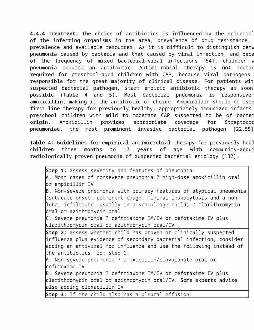
World Health Organ 82: 895-903.
8. Wardlaw T, Salama P, Johansson EW, Mason E (2006) Pneumonia: the leading killer of children. Lancet 368: 1048-1050.
9. Theodoratou E, Zhang JS, Kolcic I, Davis AM, Bhopal S, et al. (2011) Estimating pneumonia deaths of post-neonatal children in countries of low or no death certification in 2008. PLoS One 6: e25095.
10. Bryce J, Boschi-Pinto C, Shibuya K, Black RE; WHO Child Health Epidemiology Reference Group (2005) WHO estimates of the causes of death in children. Lancet 365: 1147-1152.
11. You D, Wardlaw T, Salama P, Jones G (2010) Levels and trends in under-5 mortality, 1990-2008. Lancet 375: 100-103.
12. Madhi SA, Levine OS, Hajjeh R, Mansoor OD, Cherian T (2008) Vaccines to prevent pneumonia and improve child survival. Bull World Health Organ 86: 365-372.
13. Wubbel L, Muniz L, Ahmed A, Trujillo M, Carubelli C, et al. (1999) Etiology and treatment of community-acquired pneumonia in ambulatory children. Pediatr Infect Dis J 18: 98-104.
14. Clements H, Stephenson T, Gabriel V, Harrison T, Millar M, et al. (2000) Rationalised prescribing for community acquired pneumonia: a closed loop audit. Arch Dis Child 83: 320-324.
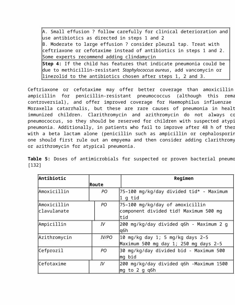
15. Juv?n T, Mertsola J, Waris M, Leinonen M, Meurman O, et al. (2000) Etiology of community-acquired pneumonia in 254 hospitalized children. Pediatr Infect Dis J 19: 293-298.
16. Cevey-Macherel M, Galetto-Lacour A, Gervaix A, Siegrist CA, Bille J, et al. (2009) Etiology of community-acquired pneumonia in hospitalized children based on WHO clinical guidelines. Eur J Pediatr 168: 1429-1436.
17. McIntosh K (2002) Community-acquired pneumonia in children. N Engl J Med 346: 429-437.
18. Shoham Y, Dagan R, Givon-Lavi N, Liss Z, Shagan T, et al. (2005) Community-acquired pneumonia in children: quantifying the burden on patients and their families including decrease in quality of life. Pediatrics 115: 1213-1219.
19. Sazawal S, Black RE; Pneumonia Case Management Trials Group (2003) Effect of pneumonia case management on mortality in neonates, infants, and preschool children: a meta-analysis of community-based trials. Lancet Infect Dis 3: 547-556.
20. Zar HJ (2004) Pneumonia in HIV-infected and HIV-uninfected children in developing countries: epidemiology, clinical features, and management. Curr Opin Pulm Med 10: 176-182.
21. Ostapchuk M, Roberts DM, Haddy R (2004) Community-acquired pneumonia in infants and children. Am Fam Physician 70: 899-908.
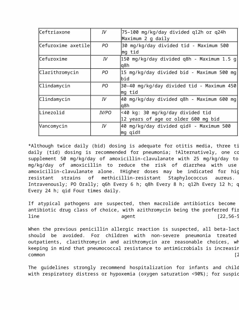
22. Bradley JS, Byington CL, Shah SS, Alverson B, Carter ER, et al. (2011) The management of community-acquired pneumonia in infants and children older than 3 months of age; clinical practice guidelines by the Pediatrie Infectious Diseases Society and the Infectious Diseases Society of America. Clin Infect Dis 53: e25-76.
23. Don M, Canciani M, Korppi M (2010) Community-acquired pneumonia in children: what's old? What's new? Acta Paediatr 99: 1602-1608.
24. Iregbu KC, Anwaal U (2007) Extended spectrum Beta-Lactamase-producing Klebsiella pneumoniae septicaemia outbreak in the Neonatal Intensive Care Unit of a tertiary hospital in Nigeria. Afr J Med Med Sci 36: 225-228.
25. Jeena PM, Pillay P, Pillay T, Coovadia HM (2002) Impact of HIV-1 co-infection on presentation and hospital-related mortality in children with culture proven pulmonary tuberculosis in Durban, South Africa. Int J Tuberc Lung Dis 6: 672-678.
26. Shabir A Madhi, Karen Petersen, Anas Madhi, Mannikhant Khoosal, Keith P Klugman1 (2000) Increased disease burden and antibiotic resistance of bacteria causing severe community-acquired lower respiratory tract infections in human immunodeficiency virus type 1-infected children. Clin Infect Dis 31: 170-176.
27. Friedman MG, Dvoskin B, Kahane S (2003) Infections with the chlamydia-like microorganism Simkania negevensis, a possible emerging pathogen. Microbes Infect 5: 1013-1021.
28. Metinko AP (2004) Neonatal pulmonary host defense mechanisms. In: Polin RA, Fox WW eds.). Fetal and Neonatal Physiology. (3rdedn), WB Saunders Co, Philadelphia.
29. Mizgerd JP (2008) Acute lower respiratory tract infection. N Engl J Med 358: 716-727.
30. Barnett ED, Klein JO (2006) Bacterial infections of the respiratory tract. In: Remington JS, Klein JO (eds.). Infectious Diseases of the Fetus and Newborn Infant. (6thedn), Elsevier Saunders Co, Philadelphia.
31. Zhang MZ, Wang LB, Chen C, Yang Y, Zhang LE (2005) [Role of dendritic cells in the pathogenesis of severe pneumonia in children]. Zhonghua Er Ke Za Zhi 43: 410-413.
32. Bone RC, Grodzin CJ, Balk RA (1997) Sepsis: a new hypothesis for pathogenesis of the disease process. Chest 112: 235-243.
33. Johnston RB Jr (1991) Pathogenesis of pneumococcal pneumonia. Rev Infect Dis 13 Suppl 6: S509-517.
34. van der Poll T, Opal SM (2009) Pathogenesis, treatment, and prevention of pneumococcal pneumonia. Lancet 374: 1543-1556.
35. Atkinson TP, Balish MF, Waites KB (2008) Epidemiology, clinical manifestations, pathogenesis and laboratory, detection of Mycoplasma pneumoniae infections. FEMS Microbiol Rev 32: 956-973.
36. Harris M, Clark J, Coote N, Fletcher P, Harnden A, et al. (2011) British Thoracic Society guidelines for the management of community acquired pneumonia in children: update 2011. Thorax 66 Suppl 2: ii1-23.
37. Chesnutt MS, Prendergast TJ (2007) Pneumonia. In: McPhee SJ, Papadakis MA, Tierney LM Jr (eds.). Current Medical Diagnosis and Treatment, 2007. (46thedn), McGraw-Hill, New York.
38. Michelow IC, Olsen K, Lozano J, Rollins NK, Duffy LB, et al. (2004) Epidemiology and clinical characteristics of community-acquired pneumonia in hospitalized children. Pediatrics 113: 701-707.
39. http://www.webmd.com/lung/tc/pneumonia-topic-overview
40. http://www.nlm.nih.gov/medlineplus/ency/article/000145.htm
41. Goldstein B, Giroir B, Randolph A; International Consensus Conference on Pediatric Sepsis (2005) International pediatric sepsis consensus conference: definitions for sepsis and organ dysfunction in pediatrics. Pediatr Crit Care Med 6: 2-8.
42. Low DE, Kellner JD, Allen U, Kovesi T, Reisman J, et al. (2004) Community-acquired pneumonia in children: A multidisciplinary consensus review. Can J Infect Dis 14: 3-11.
43. Zar HJ, Tannenbaum E, Hanslo D, Hussey G (2003) Sputum induction as a diagnostic tool for community-acquired pneumonia in infants and young children from a high HIV prevalence area. Pediatr Pulmonol 36: 58-62.
44. Madhi SA, Kuwanda L, Cutland C, Klugman KP (2005) The impact of a 9-valent pneumococcal conjugate vaccine on the public health burden of pneumonia in HIV-infected and -uninfected children. Clin Infect Dis
40: 1511-1518.
45. Nohynek H, Valkeila E, Leinonen M, Eskola J (1995) Erythrocyte sedimentation rate, white blood cell count and serum C-reactive protein in assessing etiologic diagnosis of acute lower respiratory infections in children. Pediatr Infect Dis J 14: 484-490.
46. Toikka P, Irjala K, Juv?n T, Virkki R, Mertsola J, et al. (2000) Serum procalcitonin, C-reactive protein and interleukin-6 for distinguishing bacterial and viral pneumonia in children. Pediatr Infect Dis J 19: 598-602.
47. Korppi M, Remes S, Heiskanen-Kosma T (2003) Serum procalcitonin concentrations in bacterial pneumonia in children: a negative result in primary healthcare settings. Pediatr Pulmonol 35: 56-61.
48. Ekman MR, Leinonen M, Syrj?l? H, Linnanm?ki E, Kujala P, et al. (1993) Evaluation of serological methods in the diagnosis of Chlamydia pneumoniae pneumonia during an epidemic in Finland. Eur J Clin Microbiol Infect Dis 12: 756-760.
49. Cassell GH, Gambill G, Duffy L (1996) ELISA in respiratory infections in humans. In: Tully J, Razin S (eds.). Molecular and Diagnostic Procedures in Mycoplasmology. Academic Press, San Diego, CA.
50. Zar HJ, Hanslo D, Apolles P, Swingler G, Hussey G (2005) Induced sputum versus gastric lavage for microbiological confirmation of pulmonary tuberculosis in infants and young children: a prospective study. Lancet 365: 130-134.
51. Swingler GH (2000) Radiologic differentiation between bacterial and viral lower respiratory infection in
children: a systematic literature review. Clin Pediatr (Phila) 39: 627-633.
52. Bachur R, Perry H, Harper MB (1999) Occult pneumonias: empiric chest radiographs in febrile children with leukocytosis. Ann Emerg Med 33: 166-173.
53. Gibson NA, Hollman AS, Paton JY (1993) Value of radiological follow up of childhood pneumonia. BMJ 307: 1117.
54. Madhi SA, Klugman KP; Vaccine Trialist Group (2004) A role for Streptococcus pneumoniae in virus-associated pneumonia. Nat Med 10: 811-813.
55. 0'Mara N (2011) Empiric treatment for pediatric community acquired pneumonia. Pharmacist's Letter 2011.
56. Morita JY, Kahn E, Thompson T, Laclaire L, Beall B, et al. (2000) Impact of azithromycin on oropharyngeal carriage of group A Streptococcus and nasopharyngeal carriage of macrolide-resistant Streptococcus pneumoniae. Pediatr Infect Dis J 19: 41-46.
57. Block S, Hedrick J, Hammerschlag MR, Cassell GH, Craft JC (1995) Mycoplasma pneumoniae and Chlamydia pneumoniae in pediatric community-acquired pneumonia: comparative efficacy and safety of clarithromycin vs. erythromycin ethylsuccinate. Pediatr Infect Dis J 14: 471-477.
58. Harris JA, Kolokathis A, Campbell M, Cassell GH, Hammerschlag MR (1998) Safety and efficacy of azithromycin in the treatment of community-acquired pneumonia in children. Pediatr Infect Dis J 17: 865-
871.
59. Pallares R, Capdevila O, Li?ares J, Grau I, Onaga H, et al. (2002) The effect of cephalosporin resistance on mortality in adult patients with nonmeningeal systemic pneumococcal infections. Am J Med 113: 120-126.
60. Ros?n B, Carratal? J, Tubau F, Dorca J, Li?ares J, et al. (2001) Usefulness of betalactam therapy for community-acquired pneumonia in the era of drug-resistant Streptococcus pneumoniae: a randomized study of amoxicillin-clavulanate and ceftriaxone. Microb Drug Resist 7: 85-96.
61. Miller LG, Kaplan SL (2009) Staphylococcus aureus: a community pathogen. Infect Dis Clin North Am 23: 35-52.
62. Haider BA, Saeed MA, Bhutta ZA (2008) Short-course versus long-course antibiotic therapy for non-severe community-acquired pneumonia in children aged 2 months to 59 months. Cochrane Database Syst Rev : CD005976.
63. Tice AD, Rehm SJ, Dalovisio JR, Bradley JS, Martinelli LP, et al. (2004) Practice guidelines for outpatient parenteral antimicrobial therapy. IDSA guidelines. Clin Infect Dis 38: 1651-1672.
64. Bradley JS, Ching DK, Hart CL (1987) Invasive bacterial disease in childhood: efficacy of oral antibiotic therapy following short course parenteral therapy in non-central nervous system infections. Pediatr Infect Dis J 6: 821-825.
65. Liu C, Bayer A, Cosgrove SE, Daum RS, Fridkin SK, et al. (2011) Clinical Practice Guidelines by the
Infectious Diseases Society of America for the Treatment of Methicillin- Resistant Staphylococcus aureus Infections in Adults and Children. Clin Infect Dis 52: e18-e55.
66. Maclayton DO, Hall RG 2nd (2007) Pharmacologic treatment options for nosocomial pneumonia involving methicillin-resistant Staphylococcus aureus. Ann Pharmacother 41: 235-244.
67. World Health Organization. (1993) Oxygen Therapy for acute respiratory infections in young children in developing countries. WHO.
68. Reuland DS, Steinhoff MC, Gilman RH, Bara M, Olivares EG, et al. (1991) Prevalence and prediction of hypoxemia in children with respiratory infections in the Peruvian Andes. J Pediatr 119: 900-906.
69. Campbell H, Byass P, Lamont AC, Forgie IM, O'Neill KP, et al. (1989) Assessment of clinical criteria for identification of severe acute lower respiratory tract infections in children. Lancet 1: 297-299.
70. Wang EE, Law BJ, Stephens D, et al. (1996) Study of inter observer reliability in clinical assessment of RSV lower respiratory illness: a Pediatric Investigators Collaborative Network for Infections in Canada (PICNIC) study. Pediatric Pulmonology 22: 23-27.
71. Onyango FE, Steinhoff MC, Wafula EM, Wariua S, Musia J, et al. (1993) Hypoxaemia in young Kenyan children with acute lower respiratory infection. BMJ 306: 612-615.
72. Brown N, Roberts C (2004) Vitamin A for acute respiratory infection in developing countries: a meta-
analysis. Acta Paediatr 93: 1437-1442.
73. D'Souza RM, D'Souza R (2002) Vitamin A for the treatment of children with measles--a systematic review. J Trop Pediatr 48: 323-327.
74. Shakur MS, Malek MA, Bano N, Islam K (2004) Zinc status in well nourished Bangladeshi children suffering from acute lower respiratory infection. Indian Pediatr 41: 478-481.
75. Wallis C, Prasad A (1999) Who needs chest physiotherapy? Moving from anecdote to evidence. Arch Dis Child 80: 393-397.
76. Balachandran A, Shivbalan S, Thangavelu S (2005) Chest physiotherapy in pediatric practice. Indian Pediatr 42: 559-568.
77. Song JY, Nahm MH, Moseley MA (2013) Clinical implications of pneumococcal serotypes: invasive disease potential, clinical presentations, and antibiotic resistance. J Korean Med Sci 28: 4-15.
78. Clark JE, Hammal D, Spencer D, Hampton F (2007) Children with pneumonia: how do they present and how are they managed? Arch Dis Child 92: 394-398.
79. Tan TQ, Mason EO Jr, Wald ER, Barson WJ, Schutze GE, et al. (2002) Clinical characteristics of children with complicated pneumonia caused by Streptococcus pneumoniae. Pediatrics 110: 1-6.
80. Li ST, Gates RL (2008) Primary operative management for pediatric empyema: decreases in hospital length of stay and charges in a national sample. Arch Pediatr Adolesc Med 162: 44-48.
81. Chonmaitree T, Powell KR (1983) Parapneumonic pleural effusion and empyema in children. Review of a 19-year experience, 1962-1980. Clin Pediatr (Phila) 22: 414-419.
82. Rees JH, Spencer DA, Parikh D, Weller P (1997) Increase in incidence of childhood empyema in West Midlands, UK. Lancet 349: 402.
83. Playfor SD, Smyth AR, Stewart RJ (1997) Increase in incidence of childhood empyema. Thorax 52: 932.
84. Tan TQ, Mason EO Jr, Wald ER, Barson WJ, Schutze GE, et al. (2002) Clinical characteristics of children with complicated pneumonia caused by Streptococcus pneumoniae. Pediatrics 110: 1-6.
85. Byington CL, Spencer LY, Johnson TA, Pavia AT, Allen D, et al. (2002) An epidemiological investigation of a sustained high rate of pediatric parapneumonic empyema: risk factors and microbiological associations. Clin Infect Dis 34: 434-440.
86. Islam S, Calkins CM, Goldin AB, Chen C, Downard CD, et al. (2012) The diagnosis and management of empyema in children: a comprehensive review from the APSA Outcomes and Clinical Trials Committee. J Pediatr Surg 47: 2101-2110.
87. Ampofo K, Bender J, Sheng X, Korgenski K, Daly J, et al. (2008) Seasonal invasive pneumococcal disease
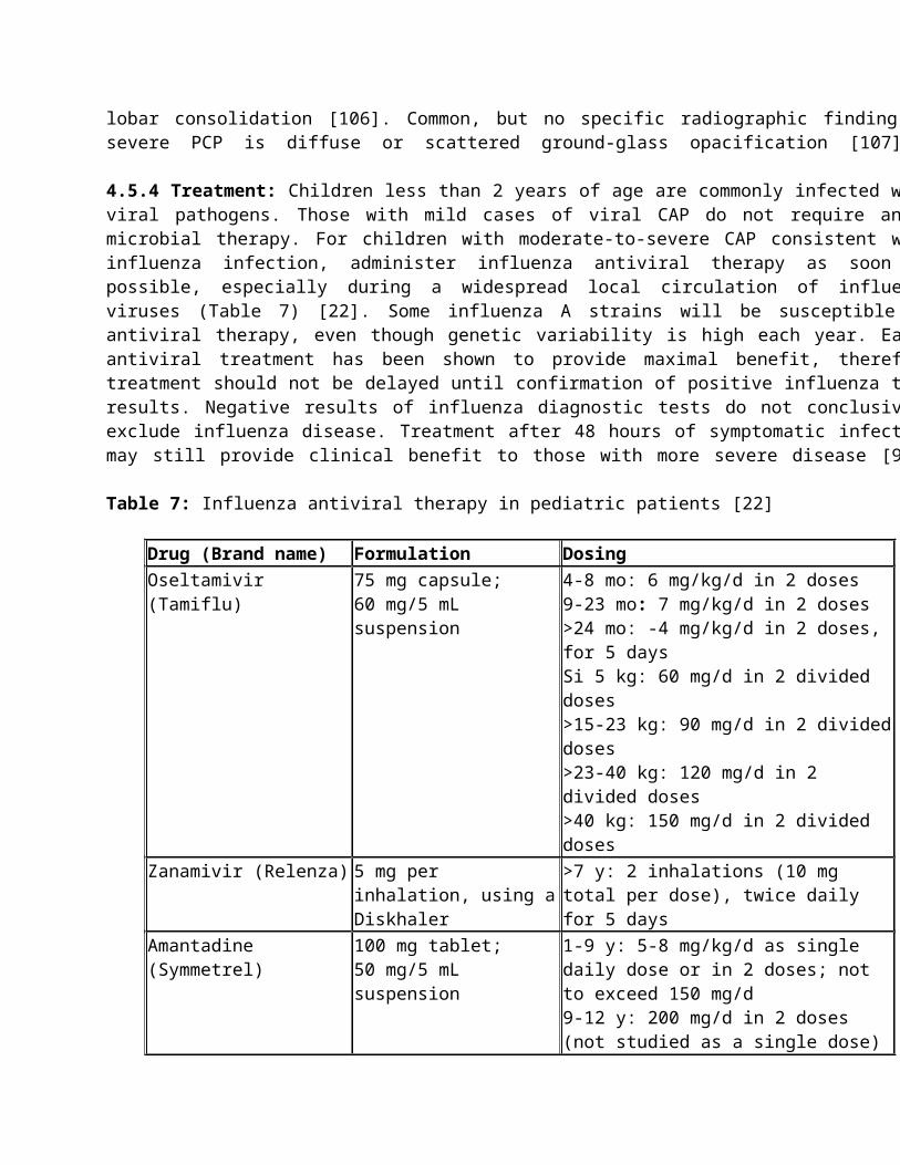
in children: role of preceding respiratory viral infection. Pediatrics 122: 229-237.
88. Talbot TR, Poehling KA, Hartert TV, Arbogast PG, Halasa NB, et al. (2005) Seasonality of invasive pneumococcal disease: temporal relation to documented influenza and respiratory syncytial viral circulation. Am J Med 118: 285-291.
89. Levin D, Tribuzio M, Green-Wrzesinki T, Ames B, Radwan S, et al. (2010) Empiric antibiotics are justified for infants with respiratory syncytial virus lower respiratory tract infection presenting with respiratory failure: a prospective study and evidence review. Pediatr Crit Care Med 11: 390-395.
90. Klugman KP, Chien YW, Madhi SA (2009) Pneumococcal pneumonia and influenza: a deadly combination. Vaccine 27 Suppl 3: C9-9C14.
91. Zar HJ, Jeena P, Argent A, Gie R, Madhi SA; Working Groups of the Paediatric Assembly of the South African Thoracic Society (2005) Diagnosis and management of community-acquired pneumonia in childhood--South African Thoracic Society Guidelines. S Afr Med J 95: 977-981, 984-90.
92. van den Hoogen BG, de Jong JC, Groen J, Kuiken T, de Groot R, et al. (2001) A newly discovered human pneumovirus isolated from young children with respiratory tract disease. Nat Med 7: 719-724.
93. Crowe JE Jr (2004) Human metapneumovirus as a major cause of human respiratory tract disease. Pediatr Infect Dis J 23: S215-221.
94. van den Hoogen BG, Osterhaus DM, Fouchier RA (2004) Clinical impact and diagnosis of human
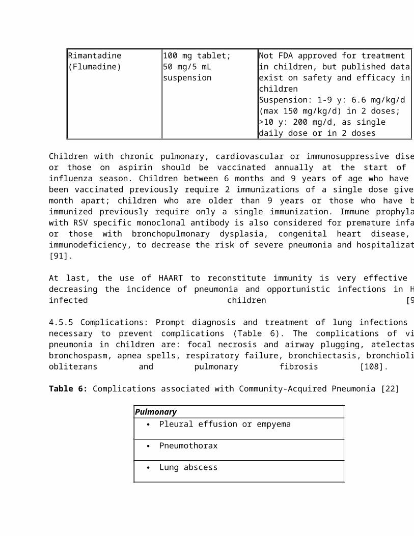
metapneumovirus infection. Pediatr Infect Dis J 23: S25-32.
95. van den Hoogen BG, van Doornum GJ, Fockens JC, Cornelissen JJ, Beyer WE, et al. (2003) Prevalence and clinical symptoms of human metapneumovirus infection in hospitalized patients. J Infect Dis 188: 1571-1577.
96. Maxson S, Jacobs RF (1996) Community-acquired fungal pneumonia in children. Semin Respir Infect 11: 196-203.
97. Chintu C, Mudenda V, Lucas S, Nunn A, Lishimpi K, et al. (2002) Lung diseases at necropsy in African children dying from respiratory illnesses: a descriptive necropsy study. Lancet 360: 985-990.
98. Levy MM, Baylor MS, Bernard GR, Fowler R, Franks TJ, et al. (2005) Clinical issues and research in respiratory failure from severe acute respiratory syndrome. Am J Respir Crit Care Med 171: 518-526.
99. Legg JP, Hussain IR, Warner JA, Johnston SL, Warner JO (2003) Type 1 and type 2 cytokine imbalance in acute respiratory syncytial virus bronchiolitis. Am J Respir Crit Care Med 168: 633-639.
100. Mathisen M, Strand TA, Sharma BN, Chandyo RK, Valentiner-Branth P, et al. (2010) Clinical presentation and severity of viral community-acquired pneumonia in young Nepalese children. Pediatr Infect Dis J 29: e1-6.
101. Pierangeli A, Scagnolari C, Selvaggi C, Monteleone K, Verzaro S, et al. (2012) Virological and clinical characterization of respiratory infections in children attending an emergency department during the
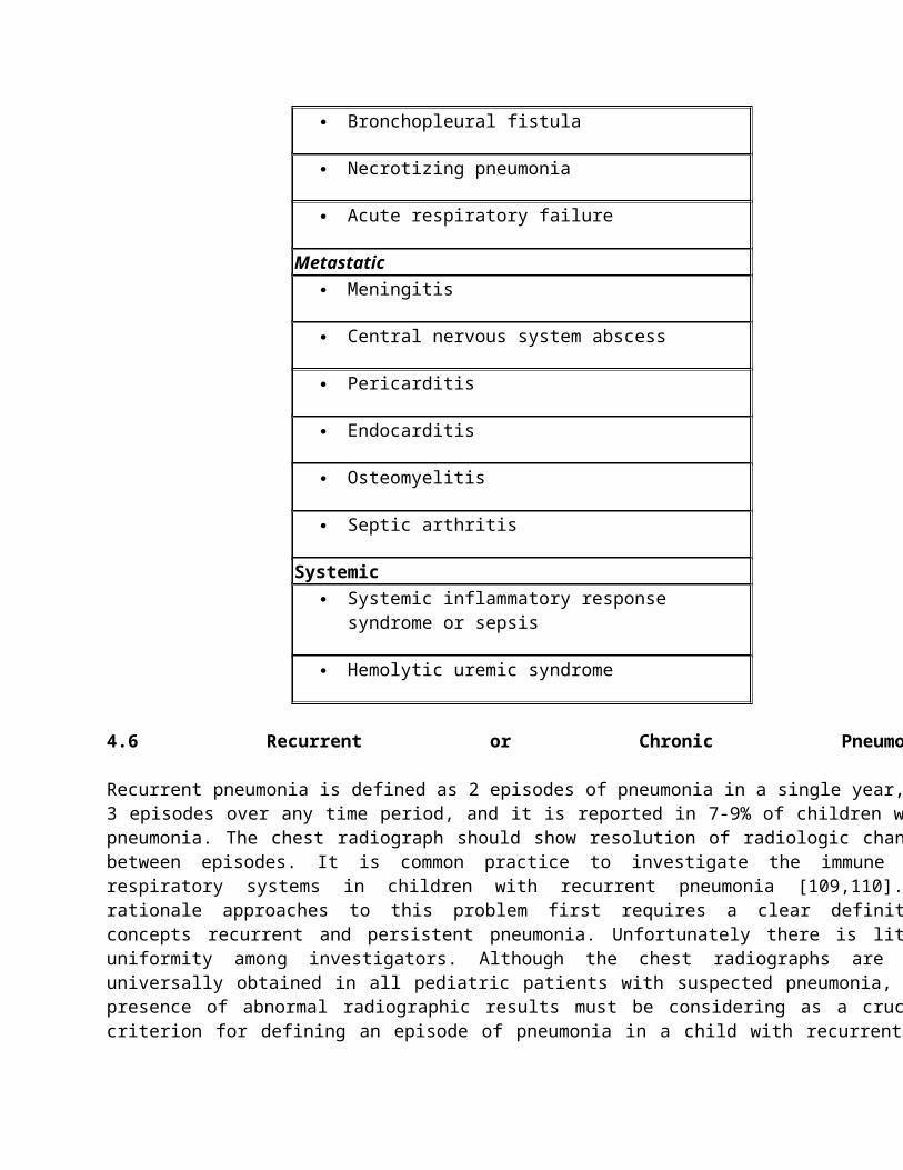
first autumn-winter circulation of pandemic A (H1N1) 2009 influenza virus. Clin Microbiol Infect 18: 366-373.
102. Zar HJ, Dechaboon A, Hanslo D, Apolles P, Magnus KG, et al. (2000) Pneumocystis carinii pneumonia in South African children infected with human immunodeficiency virus. Pediatr Infect Dis J 19: 603-607.
103. Ruffini DD, Madhi SA (2002) The high burden of Pneumocystis carinii pneumonia in African HIV-1-infected children hospitalized for severe pneumonia. AIDS 16: 105-112.
104. Pretorius MA, Madhi SA, Cohen C, Naidoo D, Groome M, et al. (2012) Respiratory viral coinfections identified by a 10-plex real-time reverse-transcription polymerase chain reaction assay in patients hospitalized with severe acute respiratory illness--South Africa, 2009-2010. J Infect Dis 206: 159-165.
105. Tew J, Calenoff L, Berlin BS (1977) Bacterial or nonbacterial pneumonia: accuracy of radiographic diagnosis. Radiology 124: 607-612.
106. da Rocha Neto OG, Leite RF, Baldi BG (2013) Update on viral community-acquired pneumonia. Rev Assoc Med Bras 59: 78-84.
107. Berdon WE, Mellins RB, Abramson SJ, Ruzal-Shapiro C (1993) Pediatric HIV infection in its second decade ? the changing pattern of lung involvement. Clinical, plain film, and computed tomographic findings. Radiol Clin North Am 31: 453-463.
108. Samransamruajkit R, Hiranrat T, Chieochansin T, Sritippayawan S, Deerojanawong J, et al. (2008) Prevalence, clinical presentations and complications among hospitalized children with influenza pneumonia. Jpn J Infect Dis 61: 446-449.
109. Panitch HB (2005) Evaluation of recurrent pneumonia. Pediatr Infect Dis J 24: 265-266.
110. Vaughan D, Katkin JP (2002) Chronic and recurrent pneumonias in children. Semin Respir Infect 17: 72-84.
111. Wald ER (1993) Recurrent and nonresolving pneumonia in children. Semin Respir Infect 8: 46-58.
112. Owayed AF, Campbell DM, Wang EE (2000) Underlying causes of recurrent pneumonia in children. Arch Pediatr Adolesc Med 154: 190-194.
113. Lodha R, Puranik M, Natchu UC, Kabra SK (2002) Recurrent pneumonia in children: clinical profile and underlying causes. Acta Paediatr 91: 1170-1173.
114. Ciftçi E, Günes M, Köksal Y, Ince E, Dogru U (2003) Underlying causes of recurrent pneumonia in Turkish children in a university hospital. J Trop Pediatr 49: 212-215.
115. Cabezuelo Huerta G, Vidal Mic? S, Abeledo G?mez A, Frontera Izquierdo P (2005) [Underlying causes of recurrent pneumonia]. An Pediatr (Barc) 63: 409-412.
116. Bradham RR (1981) Bronchial adenoma: a cause of recurrent and persistent pneumonia. J S C Med Assoc 77: 107-111.
117. Ovil Y, Schachner A, Schujman E, Spitzer SA, Levy MJ (1982) Benign endobronchial lipoma masquerading as recurrent pneumonia. Eur J Respir Dis 63: 481-483.
118. Strimlan CV, Dines DE, Payne WS (1975) Mediastinal granuloma. Mayo Clin Proc 50: 702-705.
119. Nathan N, Pautrat J, L'Hermine AC, Corvol H (2013) Pulmonary sarcoid-like granulomatous disease in an 11-month-old girl. BMJ Case Rep 2013.
120. Marik PE (2001) Aspiration pneumonitis and aspiration pneumonia. N Engl J Med 344: 665-671.
121. Saad K, Mohamed SA, Metwalley KA (2013) Recurrent/Persistent Pneumonia among Children in Upper Egypt. Mediterr J Hematol Infect Dis 5: e2013028.
122. Patria MF, Esposito S (2013) Recurrent lower respiratory tract infections in children: a practical approach to diagnosis. Paediatr Respir Rev 14: 53-60.
123. Berquist WE, Rachelefsky GS, Kadden M, Siegel SC, Katz RM, et al. (1981) Gastroesophageal reflux-associated recurrent pneumonia and chronic asthma in children. Pediatrics 68: 29-35.
124. Kitz R, Boehles HJ, Rosewich M, Rose MA (2012) Lipid-Laden Alveolar Macrophages and pH Monitoring in Gastroesophageal Reflux-Related Respiratory Symptoms. Pulm Med 2012: 673637.
125. Brand PL, Hoving MF, de Groot EP (2012) Evaluating the child with recurrent lower respiratory tract infections. Paediatr Respir Rev 13: 135-138.
126. Hoving MF, Brand PL (2013) Causes of recurrent pneumonia in children in a general hospital. J Paediatr Child Health 49: E208-212.
127. Shields MD, Doherty GM (2013) Chronic cough in children. Paediatr Respir Rev 14: 100-105.
128. Buckley RH (2004) Pulmonary complications of primary immunodeficiencies. Paediatr Respir Rev 5 Suppl A: S225-233.
129. Tamalet A, Blanchon S (2013) [Congenital ciliary dyskinesia. Focus]. Rev Pneumol Clin 69: 217-224.
130. Walters S, Mehta A (2007) Epidemiology of cystic fi brosis. In: Hodson M, Geddes DM, Bush A (eds.). Cystic fibrosis. (3rdedn), Edward Arnold Ltd, London.
131. Elbasan B, Tunali N, Duzgun I, Ozcelik U (2012) Effects of chest physiotherapy and aerobic exercise training on physical fitness in young children with cystic fibrosis. Ital J Pediatr 38: 2.
132. Le Saux N, Robinson JL (2011) Pneumonia in healthy Canadian children and youth: Practice points for management. Paediatr Child Health 16: 417-420.