Embed Size (px)
Citation preview
PEDIATRIC FRACTURES
Simon J. Hambidge, MD, PhD
April 5, 2004
Denver Health Pediatric Resident Noon Conference
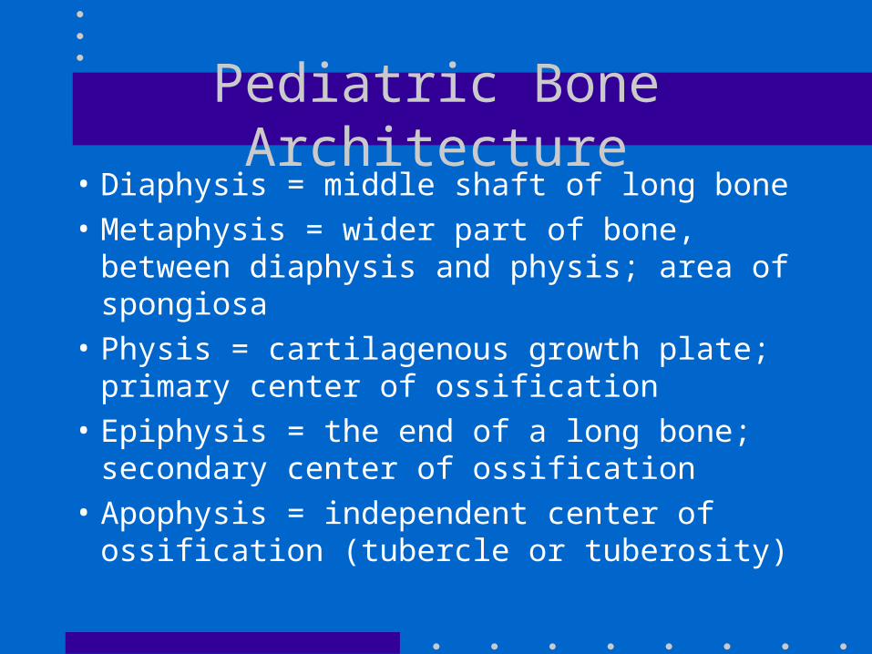
Pediatric Bone Architecture• Diaphysis = middle shaft of long bone
• Metaphysis = wider part of bone, between diaphysis and physis; area of spongiosa
• Physis = cartilagenous growth plate; primary center of ossification
• Epiphysis = the end of a long bone; secondary center of ossification
• Apophysis = independent center of ossification (tubercle or tuberosity)
Pediatric Bone - Unique Aspects
• More porous and pliable (larger Haversian canals); therefore more incomplete fractures
• Open growth plates
• Periosteum = thicker and more osteogenic potential
• Ligaments stronger than bone, and more flexible than in adults
• Rapid healing and remodeling potential
Fracture Definitions I
• Longitudinal = fracture along axis of bone
• Transverse = fracture line at right angle to bone
• Oblique = fracture at an angle to axis of bone
• Spiral = oblique Fx that encircles bone shaft
• Impacted = crushing, due to compression
• Comminuted = complex, multiple Fx fragments
Fractures Unique to Pediatrics• Plastic deformity: bending/bowing
• Greenstick: plastic deformity with partial Fx on the side of the bone opposite the impact
• Torus/Buckle/Cortical: occur at junction of metaphysis and diaphysis due to compressive forces (15% of all pediatric fractures)
• Avulsion Fractures (apophyseal fractures)
• Physeal Fractures
Fracture Definitions II
• Closed vs. Open (if communicates with air)
• Stress = Fx at microscopic level
• Displaced (expressed in percentage)
• Angulated (expressed in degrees)
• Compression = impacted or depressed
• Segmental = > 2 fractures in a single bone
Physeal Fractures - General
• “Weak link” of pediatric bone (cartilage)
• Adults - sprains & dislocations; children - physeal injuries
• Rapid healing (1/2 time of shaft fractures)
• Anatomic alignment critical for minimal deformity
• Tenderness over physis: suspect a fracture, even with normal radiographs!
Salter Harris Classification• I = “Same”: through the physis
• II = “Above”: from metaphysis into physis (75% of physeal injuries)
• III = “Lower”: from physis into epiphysis (more unstable; ensure good alignment)
• IV = “Through”: from metaphysis to epiphysis (surgical pinning usually indicated)
• V = “Everything Rong” (including the spelling): disruption of physis
Musculoskeletal Physical Exam• Observation: swelling, bruising, angulation,
deformity, shortening, or rotation
• Gentle Palpation: with focus on bony vs. soft tissue structures ($1,000,000 exam tool: finger to localize tenderness)
• Evaluation of ROM, distal motor function, vascular function, and sensory perception
• Beware of bony tenderness in the absence of any trauma history!
Splinting: General Principals
• Inspect for any open wound, swelling, or deformity
• Check distal pulse and neuro status
• In general, immobilize the joint above and below the fracture
• Pad all rigid splints (minimum 2 layers, with 3 around bony prominences)
• When in doubt, splint!
Clavicle Fractures• Dx: usually obvious based on PE and X-ray
• DDx: AC separation (sprain)
• Rx: simple arm sling for 3-4 weeks (4-6 weeks if > 12 yo); figure-of-8 sling outdated
• Education: – presence of callus (“lump”) after Fx is healed– ROM exercises (gentle) after 1-2 weeks
• Red Flag: nonunion after 4 months Rx– displaced Fx at AC joint may need surgery
Proximal Humerus Fractures
• DDx: AC separation, rotator cuff tear, rupture of long head of biceps, dislocation
• Rx: simple Fx = sling only for 3-6 weeks, ROM exercises after 1 week
• midshaft humeral fractures: similar, but check radial nerve, and may need coaptation splint for comfort
Elbow Fractures
• Dx: AP and lateral X-ray
• Small anterior fat pad is normal
• Posterior “fat pad” is always abnormal: suggests effusion and fracture
• Long axis of radius should bisect capitellum in any view
• Anterior line of humerus should transect capitellum (humeral epiphysis) in posterior 2/3
Elbow Ossification Centers
• Capitellum: appears by 1 year (unites at puberty)
• Radial head: by 4-5 years
• Medial Epicondyle: by 5 years (unites at age 20)
• Trochlea: by 9 years
• Olecranon: by 9 years
• Lateral Epicondyle: by 12 years

Elbow: Supracondylar Fractures• > 50% of all pediatric elbow fractures
• Mechanism = FOOSA with hyper-extension
• PE: careful NV exam (brachial artery)
• Can be occult: suspect if + fat pad, or displacement of AH line
• Cannot tolerate > 5 degrees angulation (can result in a varus “gunstock” deformity)
• Rx if not displaced or angulated: posterior 90o splint or LAC for 3-6 weeks
Elbow: Condyle Fractures
• Lateral: young children; Medial: teenagers
• May need oblique X-rays for Dx
• Rx: conservative only if < 2 mm displacement
• f/u X-ray within 3-5 days
• All lateral condyle fracture are SH IV and need ortho consult (can get a valgus deformity)
Elbow: Olecranon Fractures
• Mechamism = direct blow
• Relatively rare
• Don’t mistake ossification center for a fracture (can get comparison views with other elbow if unsure)
• Rx if nondisplaced: posterior 90o splint with rubber ball hand exercises
Elbow: Radial Head/Neck Fractures
• Dx = palpation of radial head with elbow at 90o; gentle pronation/supination of forearm
• Mechanism = FOOSH with supinated arm in a school aged child
• Rx if < 30o angulation: padded splint and sling for 3-4 weeks; early ROM
Nursemaid’s Elbow• Subluxation of the radial head (which slips through
the annular ligament)
• Mechanism = “POOSH”
• PE = toddler holding arm in pronation
• X-ray if any swelling or point tenderness (can have parent perform exam while you watch the child’s face)
• Rx = closed reduction (1 technique = flexion/supination)
Midshaft Forearm Fractures
• Often involve both radius and ulna
• Mechanism = FOOSH
• If angulated > 10-15o and/or displaced: consult ortho for closed reduction or internal fixation (then LAC for 6-10 weeks)
• Rx if not angulated or displaced: LAC until clinically and radiographically healed (6 weeks)
Monteggia Fracture
• Ulna fracture with dislocated radial head
• Check radial pulse
• Must recognize for adequate Rx (reduction of the dislocation as well as management of the fracture)
Fractures of the Distal Radius• Account for up to 1/4 of all pediatric Fx
• Mechanism = FOOSH
• Torus Fx: SAC or volar splint for 3-4 weeks
• SH II Fx common: need closed reduction if > 15o angulation
• Fx of distal radius and ulna or greenstick Fx of radius: closed reduction if > 15o angulation (have excellent remodeling potential)– Rx = LAC for 2-3 weeks, then SAC
Galeazzi Fracture
• Displaced fracture of the distal radius with disruption of the distal radioulnar joint
• Requires closed reduction and immobilization for 6 weeks
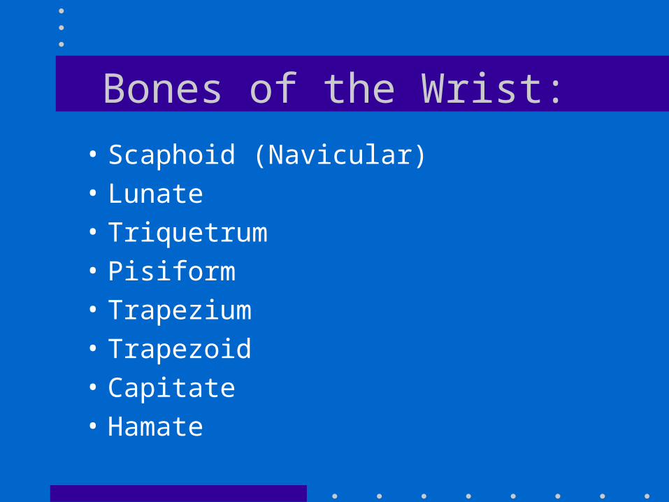
Bones of the Wrist:
• Scaphoid (Navicular)• Lunate• Triquetrum• Pisiform• Trapezium• Trapezoid• Capitate• Hamate
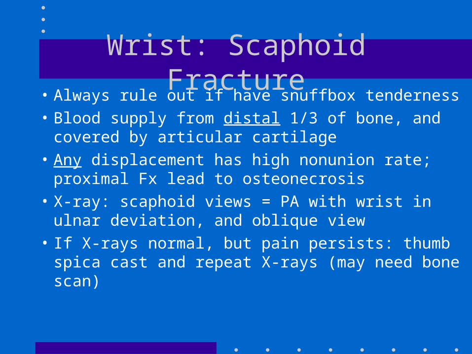
Wrist: Scaphoid Fracture• Always rule out if have snuffbox tenderness
• Blood supply from distal 1/3 of bone, and covered by articular cartilage
• Any displacement has high nonunion rate; proximal Fx lead to osteonecrosis
• X-ray: scaphoid views = PA with wrist in ulnar deviation, and oblique view
• If X-rays normal, but pain persists: thumb spica cast and repeat X-rays (may need bone scan)
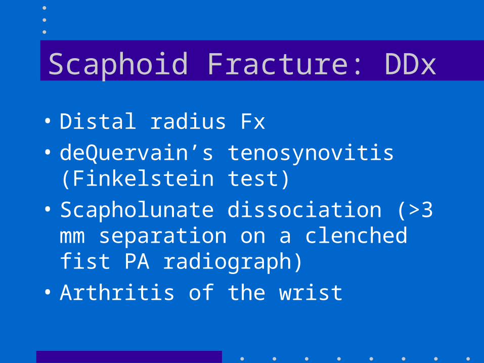
Scaphoid Fracture: DDx
• Distal radius Fx
• deQuervain’s tenosynovitis (Finkelstein test)
• Scapholunate dissociation (>3 mm separation on a clenched fist PA radiograph)
• Arthritis of the wrist
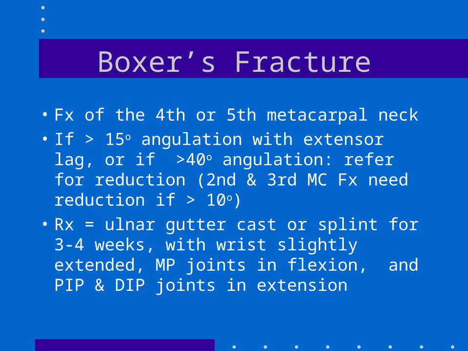
Boxer’s Fracture
• Fx of the 4th or 5th metacarpal neck
• If > 15o angulation with extensor lag, or if >40o angulation: refer for reduction (2nd & 3rd MC Fx need reduction if > 10o)
• Rx = ulnar gutter cast or splint for 3-4 weeks, with wrist slightly extended, MP joints in flexion, and PIP & DIP joints in extension
Phalangeal Fractures• Epiphyseal Fx common, usually no sequelae
• Rx if nondisplaced = Buddy Tape and finger splint for 3 weeks (early ROM)
• DDx: dislocation, Boutonniere deformity (tear of PIP extensor tendon), mallet or baseball finger (cannot extend DIP - splint 6 weeks in extension), rupture of profundus flexor tendon at DIP (surgical repair)
Skier’s (Gamekeeper’s) Thumb• Ulnar collateral ligament sprain +/- avulsion Fx• Mechanism: thumb forced radially by fall
while holding a ski pole• Complete tear (Dx = stress X-ray of MP joint):
surgical repair• Partial tear: thumb spica splint/cast with MP
joint at 20o flexion for 5-6 weeks (ROM after 3 weeks)
SCFE• Slipped Capital Femoral Epiphysis (a special
SH I Fracture)
• Hx: obese pre-adolescent/adolescent with leg pain (can be referred to knee!) & a limp
• Can be chronic or acute
• PE:loss of (and pain with) internal rotation with hip flexed
• X-ray: AP and frog-leg of both hips
• Rx: immediate surgical referral for pinning
Pelvic Avulsion Fractures
• Apophyseal avulsions: typically in muscular athletes aged 14 to 25
• ASIS: sartorius
• AIIS: rectus femoris (kicking)
• Ischial tuberosity: hamstring (hurdlers)
• Iliac crest: abdominal muscles
• Lesser trochanter: iliopsoas
• Rx: conservative - rest, ice, NSAIDS, PT
Fracture of the Patella
• PE: TTP over patella
• X-ray: AP, lateral, and sunrise
• Ensure there are not other injuries to the knee
• DDx: bipartite patella, patellar bursitis
• Rx: knee immobilizer X 6 weeks (ROM at 3-4 weeks)
Toddler’s Fracture• Spiral or oblique Fx of tibia
• Not suggestive of NAT in absence of other concerns
• Hx: toddler who limps or won’t walk (Hx of trauma is variable)
• Rx: posterior splint or cast; repeat X-rays @ 7-10 days
• Walking cast X 3-4 weeks (may need LLC for first 1-2 weeks)
Ankle Fractures • Most common in peds: SH1 avulsion fracture
of distal fibula (Rx = 3-6 weeks in SL walking cast)
• X-ray: AP, lateral, and oblique• Red flags for referral:
– widening or loss of medial clear space on mortise view
– isolated Fx of LM with tenderness of MM (bimalleolar injury with disruption of deltoid)
– Maisonneuve Fx (above + Fx of prox. fibula)
Fractures of the Hindfoot
• Talus and calcaneus
• Hx: major trauma (MVA or fall from a height)
• Many require surgical reduction and fixation: orthopedic referral on diagnosis
Metatarsal Fractures
• Rx: SLC or stiff-soled shoe, weightbearing as tolerated; repeat X-rays @ 3 weeks
• Referral red flags: multiple Fx, > 4 mm displacement, > 10o angulation, Lisfranc and Jones Fx, Fx of 1st metatarsal
• DDx: Lisfranc dislocation/sprain, Freiberg’s infarction (osteonecrosis of the 2nd metatarsal head), stress Fx
Proximal 5th Metatarsal Fx• Jones Fx: proximal metaphysis of 5th MT
– propensity for nonunion– Rx: referral, non-weightbearing cast for 6
weeks
• Tuberosity avulsion Fx– avulsion of very proximal tip of 5th MT
(insertion of peroneus brevis)– mechanism: inversion of ankle– Rx = gel/air splint & thick-soled shoes
Fracture of the Midfoot
• Lisfranc fracture-dislocation
• PE: most tender over tarso-MT joint
• Look for displacement of 2nd MT base from middle cuneiform = dislocation
• Rx: referral (may need surgery), 6-8 weeks of non-weightbearing cast
• high percentage of chronic midfoot pain