Embed Size (px)
Citation preview
REVIEWpublished: 30 June 2016
doi: 10.3389/fnins.2016.00310
Frontiers in Neuroscience | www.frontiersin.org 1 June 2016 | Volume 10 | Article 310
Edited by:
Kirsten R. Müller-Vahl,
Medical Doctor, Germany
Reviewed by:
Yang Xu,
Washington University, USA
James Leckman,
Yale University, USA
Kurt-Wolfram Sühs,
Hannover Medical School, Germany
*Correspondence:
Simone Macrì
Specialty section:
This article was submitted to
Child and Adolescent Psychiatry,
a section of the journal
Frontiers in Neuroscience
Received: 22 March 2016
Accepted: 20 June 2016
Published: 30 June 2016
Citation:
Spinello C, Laviola G and Macrì S
(2016) Pediatric Autoimmune
Disorders Associated with
Streptococcal Infections and
Tourette’s Syndrome in Preclinical
Studies. Front. Neurosci. 10:310.
doi: 10.3389/fnins.2016.00310
Pediatric Autoimmune DisordersAssociated with StreptococcalInfections and Tourette’s Syndromein Preclinical StudiesChiara Spinello, Giovanni Laviola and Simone Macrì *
Section of Behavioural Neuroscience, Department of Cell Biology and Neuroscience, Istituto Superiore di Sanità, Roma, Italy
Accumulating evidence suggests that Tourette’s Syndrome (TS) – a multifactorial
pediatric disorder characterized by the recurrent exhibition of motor tics and/or vocal
utterances – can partly depend on immune dysregulation provoked by early repeated
streptococcal infections. The natural and adaptive antibody-mediated reaction to
streptococcus has been proposed to potentially turn into a pathological autoimmune
response in vulnerable individuals. Specifically, in conditions of increased permeability of
the blood brain barrier (BBB), streptococcus-induced antibodies have been proposed
to: (i) reach neuronal targets located in brain areas responsible for motion control; and
(ii) contribute to the exhibition of symptoms. This theoretical framework is supported by
indirect evidence indicating that a subset of TS patients exhibit elevated streptococcal
antibody titers upon tic relapses. A systematic evaluation of this hypothesis entails
preclinical studies providing a proof of concept of the aforementioned pathological
sequelae. These studies shall rest upon individuals characterized by a vulnerable immune
system, repeatedly exposed to streptococcus, and carefully screened for phenotypes
isomorphic to the pathological signs of TS observed in patients. Preclinical animal
models may thus constitute an informative, useful tool upon which conducting targeted,
hypothesis-driven experiments. In the present review we discuss the available evidence
in preclinical models in support of the link between TS and pediatric autoimmune
neuropsychiatric disorders associated with streptococcus infections (PANDAS), and
the existing gaps that future research shall bridge. Specifically, we report recent
preclinical evidence indicating that the immune responses to repeated streptococcal
immunizations relate to the occurrence of behavioral and neurological phenotypes
reminiscent of TS. By the same token, we discuss the limitations of these studies: limited
evidence of behavioral phenotypes isomorphic to tics and scarce knowledge about
the immunological phenomena favoring the transition from natural adaptive immunity
to pathological outcomes.
Keywords: PANDAS, Tourette’s Syndrome, animal models, group A–beta hemolytic streptococcus, autoimmunity
Spinello et al. Preclinical Models of Tourette’s Syndrome
INTRODUCTION
Neuropsychiatric and neurological disorders are among theleading causes of disability worldwide (Silberberg et al., 2015).Several studies reported that people affected by neuropsychiatricillnesses show a set of psychosocial disturbances, rangingfrom difficulties in social interactions to emotional instability(Hartley et al., 2014; Cieza et al., 2015), ultimately resultingin difficulties in routine activities (Coenen et al., 2016). Sinceneuropsychiatric illnesses have a strong impact on the well-being of affected individuals, understanding the etiology ofthese diseases may beget remarkable heuristic advancements.Within this framework, epidemiological, clinical, and preclinicalstudies reveal that different determinants contribute to thepathogenesis of neuropsychiatric diseases. Among them, geneticfactors (Hyman, 2008) and several environmental risk factors,such as prenatal and perinatal injuries or stressors (Bronson andBale, 2016) and infectious phenomena (John et al., 2015), play akey role.
Autoimmunity, defined by Davison as “the failure of anorganism to recognize its own part as self, resulting in a series ofimmunological responses to its own cells and tissues” (Davison,2012) has emerged as a potential pathogenic factor in differenttypes of neuropsychiatric illnesses, including autoimmuneencephalitis (Höftberger, 2015), systemic lupus erythematosus(SLE; Podolska et al., 2015), or schizophrenia (Margari et al.,2013). Infectious phenomena constitute a vulnerability factorin the onset of autoimmune disorders. In particular, infectionsmay trigger the onset of autoimmune diseases in the presenceof vulnerability conditions. With respect to neuropsychiatricdisorders, these vulnerability conditions are represented, forexample, by an abnormal permeability of the blood brainbarrier (BBB; Almutairi et al., 2016). Hornig (2013) proposedthat microbes may contribute to the etiology of autoimmuneneurological and neuropsychiatric disorders by triggering theproduction of autoantibodies that directly bind brain targets.In susceptible individuals, these phenomena can result inthe appearance of behavioral and neurochemical abnormalities(Hornig, 2013).
Within this framework, streptococcal infections have beenlinked to a series of neuropsychiatric and movement disorders(Swedo et al., 1998). For example, different studies documentedthat the onset of Sydenham Chorea (SC), a variant ofrheumatic fever, is linked to group A β-hemolitic streptococcusinfections (Swedo et al., 1993; Cardoso et al., 1999). SC ischaracterized by choreiform movements that typically involveface and extremities and, in some cases, by behavioral difficultiesand emotional liability (Swedo et al., 1989; Marques-Diaset al., 1997). Besides SC, several authors proposed thatstreptococcus infections may constitute an etiological factor alsoin a series of illnesses that typically arise during childhood.In particular, Swedo and colleagues proposed the acronymPANDAS (Pediatric Autoimmune Neuropsychiatric DisorderAssociated with Streptococcal infections) to define a seriesof neurological and psychiatric disorders characterized by thepresence of antibodies produced in response to group A β-hemolytic streptococcus infections (Swedo et al., 1998). The
diagnostic criteria for PANDAS include: prepubertal onset;obsessive compulsive disorder (OCD) or chronic tic disorder;relapsing-remitting course of the disease; motor hyperactivityor reduced fine motor coordination; onset of the diseaseor symptoms exacerbation temporally related to streptococcalinfection (Swedo et al., 1998).
In PANDAS and SC, antibodies produced in response tostreptococcus have been proposed to be pathogenic in CNS inthe context of an increased BBB permeability. Since the BBB isthe primary protective barrier for neurons in central nervoussystem (CNS), BBB dysfunctions may contribute to the etiologyof several neuropsychiatric disorders (Almutairi et al., 2016). Inparticular, in PANDAS and SC, after crossing the damaged BBB,cross-reactive antibodies may bind specific brain targets at thelevel of Basal Ganglia (BG), a brain structure involved in motorcontrol (Martino et al., 2009; Murphy et al., 2010; Cutforth et al.,2016).
Streptococcal infections have also been suggested to relateto Tourette’s Syndrome (TS), a multifactorial and complexdisorder that may, in some cases, match the criteria for PANDAS(Hoekstra et al., 2013). TS is a childhood-onset disorder, inwhich chronic motor or phonic tics are the main symptoms.Tics are considered chronic if persist over a period longerthan 12 months (Lombroso and Scahill, 2008). Tic, accordingto the DSM-5, is defined as “a sudden, rapid, recurrent, non-rhythmic motor movement or vocalization” (APA, 2013). TSis more frequent in males than females, with a ratio of 4:1.Typically, symptoms occur during prepubertal age, between 5and 7 years, and have a waxing and waning course (Lombrosoand Scahill, 2008). A gradual increase in tic frequency andseverity is generally shown until 8–12 years, while a relevantreduction occurs in most patients at the end of adolescence(Leckman et al., 2010). Co-morbid conditions are typical inTS. In particular, obsessive-compulsive disorder (OCD) andattention-deficit/hyperactivity disorder (ADHD) are the mostcommon comorbidities (Leckman et al., 2010). The pathogenesisof TS is multifactorial, and include genetic vulnerability (Denget al., 2012), and several environmental risk factors suchas prenatal and perinatal stressors or injuries and bacterialand viral infections (Leckman et al., 1987, 1990; Leckmanand Peterson, 1993). With respect to precocious vulnerability,maternal factors (genetic or environmental) have been shown toincrease individual vulnerability to TS. For example, Dalsgaardet al. (2015) recently reported that maternal autoimmunediseases significantly increase vulnerability to TS in the progeny(Dalsgaard et al., 2015).
While maternal autoimmunity can influence vulnerability toTS, it is yet to be determined whether these effects are geneticor environmental. With respect to genetic predispositions,several authors identified a series of genes for which a directcontribution to TS can be reasonably proposed. Thus, geneticlinkage, cytogenetics and molecular genetic studies allowedidentifying a set of genes potentially involved in TS (State, 2011).Among them, contactin-associated protein-like 2 (CNTNAP2),SLIT and NTRK-like 1 (SLITRK1) or membrane peptidase2 like (IMMP2L) have been proposed as vulnerability genes.The proteic product of IMMP2L gene is a peptide with a
Frontiers in Neuroscience | www.frontiersin.org 2 June 2016 | Volume 10 | Article 310
Spinello et al. Preclinical Models of Tourette’s Syndrome
catalytic function that, in the dysfunctional form, may causethe activation of the cell apoptotic mechanism through anaberrant mithocondrial functionality (Ma et al., 2011). Severalauthors reported in some members of a family with TS thepresence of a translocation between chromosome 7 and 18 thatcauses the disruption of IMMP2L gene (Boghosian-Sell et al.,1996; Petek et al., 2001). However, the role of this gene inTS etiology remains unclear. CNTNAP2 is a transmembraneprotein of the family of neurexin, abundantly expressed atthe level of the axonal nodes of Ranvier, where it plays acrucial role in the cell-cell interaction. Poliak et al. (1999),hypothesized that this peptide may be involved in the positioningof K+ voltage-gated channel at the level of juxtaparanoderegion (Poliak et al., 1999). Verkerk et al. (2003) observed achromosomal translocation between chromosome 2 and 7, in theregion encoding CNTNAP2 protein, in a family of TS patients(Verkerk et al., 2003). The disruption of this region has beenproposed to affect brain areas involved in motor control, therebybeing responsible for the onset of tics (Verkerk et al., 2003).SLTRK1 is a member of a gene family that encodes a seriesof transmembrane proteins. The proteic product of SLTRK1gene is a peptide that contains two leucine-rich repeat (LRR)motive and an intracellular C terminus having similarities withthe tropomyosin-related kinase (Trk) neurotrophin’s receptor(Aruga and Mikoshiba, 2003). SLTRK1 favors the formation ofsynapses, neuritic outgrowth and neuronal survival (Kajiwaraet al., 2009). SLTRK1 transcription is regionally regulated inCNS; the pattern of expression is conserved among differentmammalian species, such as mouse, rhesus monkey and human,and shows a preferential expression in brain areas involvedin motor control, such as cortex, thalamus and basal ganglia(Stillman et al., 2009). In particular, SLRTK1 is expressed inthe body compartment of cortex pyramidal projection neuronsduring adult life, and is preferentially associated, in the striatum,with neurons of the direct circuit expressing substance P anddopamine receptor D1, that project to substantia nigra (SN)and to globus pallidus (GP; Stillman et al., 2009). Some TSpatients showed a missense mutation at the level of 3′ UTR ofthe SLTRK1 gene; this mutation leads to the production of aprotein with an altered capacity of binding the microRNA 189(Abelson et al., 2005). Moreover, an inversion in chromosome13 in proximity of the region of the gene has been reported inpatients with TS and ADHD (Proenca et al., 2011). Recently,Ercan-Sencicek et al. (2010) proposed that a mutation of thegene encoding for histidine decarboxylase (HDC) constitutesa rare genetic cause in TS (Ercan-Sencicek et al., 2010). Inparticular, the authors identified, through a study of a 2-generation pedigree in a family with a high incidence of TS, a raresegregating non-sense mutation in the l-hystidine decarboxylase(hdc) gene (Ercan-Sencicek et al., 2010). HDC is an enzymenecessary for the synthesis of histamine (HA) which, in turn, hasbeen hypothesized to modulate DA level in CNS (Haas et al.,2008). Subsequently, a reduced concentration of HA in CNS(caused by the non-sense hdc gene mutation) may result in analtered dopaminergic regulation at the level of the basal gangliacircuitry, thereby resulting in TS symptomatology (CastellanBaldan et al., 2014). In the same study, Castellan Baldan
and collaborators translated this evidence in an experimentalmodel (hdc knock-out mice, see discussion for additionaldetails). Moreover, an analysis of rare copy number variantsin TS conducted on 460 patients, revealed the presence ofa significant enrichment of genes involved in histaminergicpathways (Fernandez et al., 2012). In particular, the authorsreported an enrichment in striatum and cortex of HA coupledG receptors H2 and H3. Those receptors are located bothpresinaptically and postsinaptically: presynaptic HA receptorsare involved in the regulation not only of HA transmission,but also of dopamine (Fernandez et al., 2012). It is thustenable to propose that dysfunctions in histaminergic pathwaymay contribute to the onset of TS through the modulation ofdopaminergic transmission.
GAS infections, occurring after TS onset, have been proposedas a vulnerability factor potentially exacerbating symptoms(Martino et al., 2009; Landau et al., 2012). Additionally,in line with the possibility that altered immune capabilityconstitutes a predisposing factor, clinical data support anincreased vulnerability of the immune system in TS patients.For example, whilst Bos-Veneman et al. (2011) observed thatTS children were characterized by decreased levels of IgG3(Bos-Veneman et al., 2011), Kawikova et al. (2007) observedreduced concentrations of regulatory T cells in TS patientscompared to controls (Kawikova et al., 2007). Moreover, duringtic exacerbations, TS patients showed increased concentrationsof cytokines, interleukin 12 (IL-12) and tumor necrosis factoralpha (TFN-α) in serum (Leckman et al., 2005; Martino et al.,2015). Several authors reported the presence of peripheral anti-streptococcal antibodies and anti-BG antibodies in patientsaffected by TS. For example, Cardona and Orefici observed thata large cohort of TS patients showed significantly higher levelsof anti-streptococcal antibodies compared to control subjects;moreover, they reported that those patients had previouslybeen exposed to streptococcal infections (Cardona and Orefici,2001). Similarly, Rizzo and colleagues reported remarkablyhigher concentrations of anti-streptococcal antibody titers and asignificantly higher presence of anti-BG antibodies in TS patientscompared to control subjects (Rizzo et al., 2006). Martino andcolleagues reported a similar increase in anti-BG antibodies inTS patients compared to controls (Martino et al., 2011).
Although these studies support the existence of a link betweenstreptococcal infections and TS, several other studies failed toidentify a direct link between immunization and TS symptoms(Singer et al., 2005a; Dale et al., 2006; Morris et al., 2009; Brilotet al., 2011). In particular, Singer et al. (2005a) performed ELISAand Western blot analyses against several epitopes present inthe CNS (e.g., human postmortem caudate, putamen, prefrontalcortex) with sera obtained from PANDAS and TS patients,and controls. The authors did not detect differences in serumautoantibodies among groups (Singer et al., 2005a). Similarly,Morris et al. (2009), using a different experimental approach(immunofluorescence), failed to observe any difference amongPANDAS and TS patients, and controls in terms of serum anti-striatal antibody reactivity (Morris et al., 2009). Finally, Brilotet al. (2011) reported the presence of serum autoantibodiescapable of binding neuronal cell surface in SC patients, but not
Frontiers in Neuroscience | www.frontiersin.org 3 June 2016 | Volume 10 | Article 310
Spinello et al. Preclinical Models of Tourette’s Syndrome
in PANDAS or TS patients (Brilot et al., 2011). These resultsdemonstrate that the presence of autoimmune phenomena isneither a necessary nor a sufficient condition in the etiology ofTS. However, the evidence discussed above indicates that a subsetof TS cases may be dependent on autoimmune phenomena.Moreover, as already discussed, some cases of TS matchcriteria for PANDAS, suggesting that these two disorders mayshare — in specific circumstances — analogous etiopathologicalmechanisms.
Preclinical experimental models may constitute a valuablecomplement to clinical studies whereby they can aid thecomprehension of the fundamental mechanisms favoring diseaseonset. Animal models may allow testing different hypothesesregarding the role exerted by variable factors in the onset andcourse of a given disease, and to design innovative therapeuticapproaches (Rickard, 2004; van der Staay, 2006; van der Staayet al., 2009). Within this framework, several aspects of TS(symptomatology, genetic predisposition and environmental riskfactors) have been translated into preclinical animal models(Hallett et al., 2000; Yaddanapudi et al., 2010; Brimberg et al.,2012; Macrì et al., 2015; see Macrì et al., 2013 for a detailedreview).
Here, we will review preclinical data suggesting a link betweenautoimmunity and neurological diseases. In particular, we willdiscuss empirical evidence supporting the connection betweenTS and PANDAS, and the gaps of these studies that shall be filledin the future. Finally, in the light of the role of immunity inthe onset of psychiatric disturbance, we discuss the possibilitythat peripheral autoantibodies may constitute an innovativebiomarker of diagnostic use (Giana et al., 2015).
PRECLINICAL ANIMAL MODELS ANDAUTOIMMUNITY
Animal models constitute an important tool to aid theunderstanding of a given pathology and to potentiallyinform innovative therapeutic avenues. Thus, preclinicalexperimental models allow dissecting a given phenomenon intoits fundamental determinants (e.g., genetic vs. environmentalpredisposing factors) and addressing the role that each of themplays, either in isolation or in combination with each other. Thedevelopment of disease-related animal models rests upon severalstages: the generation of a disease model based on a theoreticalconstruct, the identification of abnormalities isomorphic to thesymptoms observed in the patient population and the studyof the efficacy of pharmacological treatments. The validity ofeach of these stages can be systematically scrutinized. Willnerproposed three validity criteria: construct, face, and predictivevalidity (Willner, 1984).
Construct validity can be defined as the etiological similaritybetween the disease in human population and the experimentalapproach attempting to model such disease.
Face validity relates to the degree of similarity between thesymptoms identified in the disorder examined and the phenotype(e.g., behavioral, physiological, immunological, neurobiological)in the experimental model (Willner and Mitchell, 2002). To
fulfill this criterion, a valid animal model shall resemble thesymptomatology observed in humans (van der Staay et al., 2009).
Predictive validity pertains to the therapeutic efficacy ofavailable treatments. Specifically, to possess an elevated degree ofpredictive validity, a given experimental disease model shall besensitive to the same available therapeutic approaches adopted inthe patients (Willner, 1984).
Within this framework, the use of preclinical modelshas been extensively applied to the study of autoimmuneneurological disorders (see Levite, 2014 and Hornig and Lipkin,2013 for detailed reviews). Several preclinical animal modelshave been developed to address the link between circulatingnatural antibodies (directed against specific brain targets),and behavioral and neurochemical abnormalities. For example,mice immunized with GluR1 peptide fragments (a subunit ofglutamate AMPA receptors) showed a significant elevation incirculating anti-GluR1 antibodies, marked hyperactivity, andincreased self-grooming (Capone et al., 2008), the latter beingassociated with repetitive behavior (Kalueff et al., 2016). Also,mice immunized with dopamine transporter (DAT) fragments,displayed spontaneous hyperactivity, reduced cognitive flexibilityand impulse control in operant behavioral paradigms. Moreover,the immunization protocol caused, as expected, an elevation inantibodies targeting dopamine transporter and a variation inbrain striatal concentrations of dopamine and its metabolites(Adriani et al., 2012).
Glutamate is the main excitatory neurotransmitter in CNS(Platt, 2007) and is crucial for several neuronal functions.Abnormalities in glutamatergic neurotransmission have beenshown to directly contribute to CNS disorders (Scoriels et al.,2015). The overactivation of glutamate receptors (excitotoxicity),induced by the excess of glutamate, may result in brain damageand neuronal death (Meldrum, 2000). Besides excitotoxicity,several types of anti-glutamate receptors antibodies are capableof inducing pathological effects in CNS (Levite, 2014). Theseautoantibodies emerged as one of the most widespread anddangerous pathogenic agents in CNS, causing impaired neuronalsignaling and brain damages and contributing to the onset of aseries of neuropsychiatric disorders (Levite, 2014). For example,patients affected by epilepsy and SLE, showed antibodies directedto different types of glutamate receptors, anti AMPA-GluR3B(Ganor et al., 2004, 2005a,b,c; Goldberg-Stern et al., 2014)and anti NMDA-NR2 (Borchers et al., 2005; Asano et al.,2013; Fanouriakis et al., 2013). From a translational perspective,antibodies against the same glutamate receptors have been shownto favor the onset of behavioral and neurochemical alterationsalso in preclinical models (see Levite, 2014 for a detailed review).These results have been observed in conditions of an increasedpermeability of the BBB (Kowal and Diamond, 2012). Severalauthors reported increased levels of anti-GluR3B antibodies indifferent mouse strains (specifically directed against peptide B ofsubunit R3 of glutamate AMPA receptors) after immunizationwith GluR3B peptide (Levite et al., 1999; Levite and Hermelin,1999; Ganor et al., 2014). Specifically, Ganor et al. (2014) reportedthat DBA/2J mice (genetically epilepsy-prone mice) developedelevated titers of GluR3B antibodies after immunization withGluR3B peptide emulsified in Complete Freund’s adjuvant
Frontiers in Neuroscience | www.frontiersin.org 4 June 2016 | Volume 10 | Article 310
Spinello et al. Preclinical Models of Tourette’s Syndrome
(CFA). The presence of these antibodies aggravated seizuresinduced by the administration of a chemoconvulsant agent,and caused abnormal behaviors in mice. With respect tobehavioral alterations, the authors observed, in mice positive toGluR3B antibodies, increased anxiety-like behaviors and motorimpairments (problems in balance, motor coordination andmuscle strength) compared to mice that did not show GluR3Bantibodies in serum (Ganor et al., 2014).
Kowal and colleagues developed an immune-mediated mousemodel of SLE (Kowal et al., 2004). These authors reportedthat the immunization of BALB/C mice with DNA peptidemimotope, arrayed as an octamer on a polylysine backbone(MAP peptide), induced the production of antibodies againstsubunit NR2 of NMDA glutamate receptor, associated withneuronal damages and cognitive impairments. In particular,following the administration of lipopolysaccharide (LPS, aprocedure known to increase the permeability of the BBB) toimmunized mice, NR2 antibodies bound neurons preferentiallyin hippocampus, inducing neuronal death and impaired memory(Kowal et al., 2004). NMDA-NR2 receptors are expressedthroughout the brain, but at highest density within hippocampus,hypothalamus, and amygdala. When the BBB damage wasinduced in mice by epinephrine administration, Huerta et al.(2006) showed that anti-NR2 antibodies bound preferentiallyamygdala’s neurons. Accordingly, immunized mice showedalteration in emotional behavior whereby they respondeddeficiently to fear-conditioning paradigms (Huerta et al., 2006).The latter has been shown to depend on an intact functionality ofthe amygdala (Sengupta et al., 2016).
Beside glutamate receptors, autoimmune phenomena inCNS involve other receptors, such as leucine-rich gliomainactivated 1 (LGI1), acquaporin-4 (AQP4), Gamma-AminoButyric Acid (GABAB), or myelin oligodendrocyte protein(MOG; see Irani et al., 2014 for a detailed review). Inpreclinical studies, immunization with MOG has been shown,in susceptible animals, to trigger the onset of a series ofinflammatory diseases and thereafter named experimentalautoimmune encephalomyelitis (EAE). EAE, considered as avalid animal model of multiple sclerosis (MS), are a groupof pathologies characterized by neurodegeneration, extensiveinflammation and demyelination in CNS. These neurochemicalalterations cause severe progressive motor impairments thatultimately result in flaccid paralysis of hind limbs (see Kippet al., 2012 for a detailed review). Beside MOG (Amor et al.,1994, 1996), other myelin antigens are capable of triggeringthe onset of EAE in rodents, such as myelin basic protein(MBP, see Swanborg, 2001 and Amor et al., 1996), proteolipidprotein (PLP, see Amor et al., 1993 and Amor et al., 1996)in presence of increased BBB permeability (Rabchevsky et al.,1999). Several preclinical studies showed that EAE are associatednot only with severe motor deficits, but also with behavioraland cognitive impairments (Mandolesi et al., 2010; Acharjeeet al., 2013; Olechowski et al., 2013). For example, Mandolesiet al. (2010) reported that EAE mice, compared to controls,showed hippocampal-dependent deficit in learning and memory(Mandolesi et al., 2010). Similarly, Acharjee and colleaguesobserved that EAE mice exhibited cognitive and behavioralimpairments in a precocious phase of the disease (Acharjee
et al., 2013). In particular, EAE mice exhibited reduction in thetime spent in the target quadrant of Morris Water maze andimpaired memory extinction in a fear-conditioning paradigm(Acharjee et al., 2013). Regarding behavioral impairments,authors observed in EAE mice increased anxiety-like behaviors.EAE mice showed, compared to controls, more time in themarginal zone of the apparatus during open field test andincreased time in the closed arm of plus maze test (Acharjee et al.,2013). Finally, Olechowski et al. (2013) observed impairments incognitive processes (assessed with a novel object recognition test)in a precocious phase of the disease (Olechowski et al., 2013).
The experimental evidence described above suggests thatautoimmune phenomena against CNS targets may trigger,in vulnerability conditions, the development of remarkablephenotypic abnormalities. The appearance of different behavioraland neurochemical impairments depends on the brain targetaffected by the autoimmune phenomena and, in some instances,by the tools adopted to modulate BBB integrity. As reportedabove (see Introduction), analogous mechanisms have beenproposed to contribute to the onset and exacerbation ofstreptococcal-related motor disturbances in clinical populations.Specifically, several authors (Martino et al., 2009; Murphyet al., 2010) proposed that antibodies produced in responseto streptococcal infections (STREP) may, in presence of anincreased vulnerability of the BBB, induce a pathologicalphenotype. In particular, these authors proposed that STREP-related antibodies may cross the damaged BBB and bindspecific brain targets at the level of Basal Ganglia (BG), abrain structure involved in motor control. This cascade ofevents may ultimately provoke a symptomatology typical ofstreptococcal-related motor disturbances. In the next section wewill describe some specific animal models developed with theaim of dissecting the mechanisms bridging the immunologicresponses to streptococcal infections to the onset of neurologicaland behavioral dysfunctions.
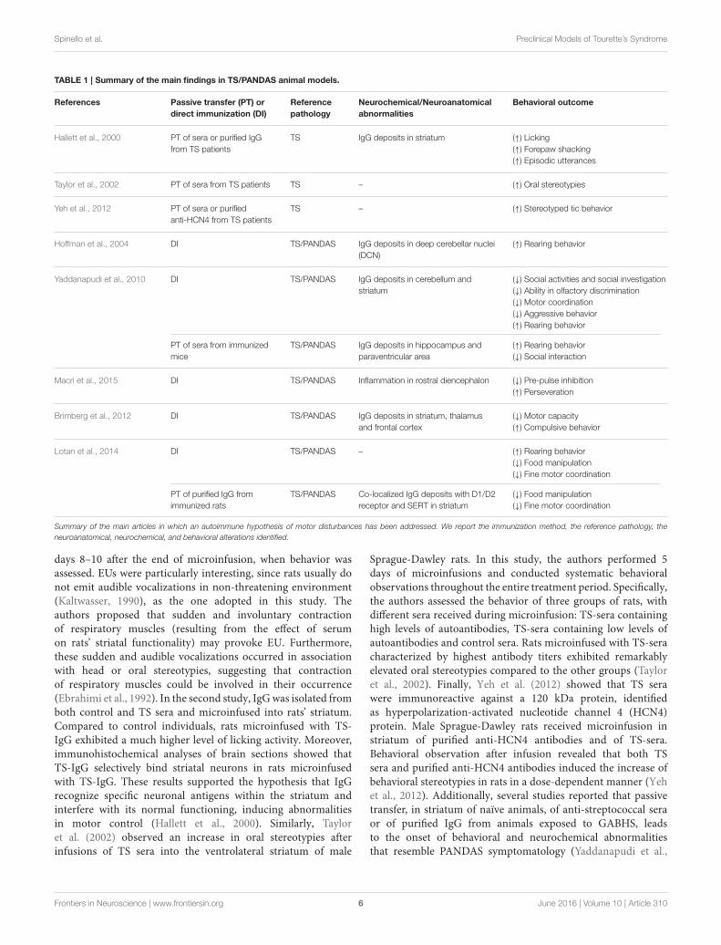
A Potential Link between StreptococcalInfections and TSPassive Transfer of Sera from TS PatientsDifferent approaches have been used to develop animal modelsaddressing the role of streptococcal infections in immune-mediated neuropsychiatric disorders (Table 1). The first line ofstudies entailed direct intracerebral administration, in rats, ofanti-neuronal antibodies sampled from TS patients (Hallett et al.,2000; Taylor et al., 2002; Loiselle et al., 2004; Singer et al.,2005b; Ben-Pazi et al., 2012; Yeh et al., 2012). Hallett et al.(2000) showed that brain intra-striatal microinfusions of TS serainduced behavioral stereotypies and episodic utterances (EU,repetitive, medium pitched sound of short duration) in maleFischer 344 rats. Stereotypies and EUs are considered analogousto involuntary movements observed in TS patients. In particular,the authors performed two separate studies, microinfusing eithersera obtained from TS children or Gamma Immunoglobulin(IgG) isolated from these sera. In the first study, compared tofacility-reared controls, rats microinfused with TS-sera showedexacerbated licking behavior, forepaw shaking and EU. Thoseabnormal behaviors were present during microinfusion, and on
Frontiers in Neuroscience | www.frontiersin.org 5 June 2016 | Volume 10 | Article 310
Spinello et al. Preclinical Models of Tourette’s Syndrome
TABLE 1 | Summary of the main findings in TS/PANDAS animal models.
References Passive transfer (PT) or
direct immunization (DI)
Reference
pathology
Neurochemical/Neuroanatomical
abnormalities
Behavioral outcome
Hallett et al., 2000 PT of sera or purified IgG
from TS patients
TS IgG deposits in striatum (↑) Licking
(↑) Forepaw shacking
(↑) Episodic utterances
Taylor et al., 2002 PT of sera from TS patients TS – (↑) Oral stereotypies
Yeh et al., 2012 PT of sera or purified
anti-HCN4 from TS patients
TS – (↑) Stereotyped tic behavior
Hoffman et al., 2004 DI TS/PANDAS IgG deposits in deep cerebellar nuclei
(DCN)
(↑) Rearing behavior
Yaddanapudi et al., 2010 DI TS/PANDAS IgG deposits in cerebellum and
striatum
(↓) Social activities and social investigation
(↓) Ability in olfactory discrimination
(↓) Motor coordination
(↓) Aggressive behavior
(↑) Rearing behavior
PT of sera from immunized
mice
TS/PANDAS IgG deposits in hippocampus and
paraventricular area
(↑) Rearing behavior
(↓) Social interaction
Macrì et al., 2015 DI TS/PANDAS Inflammation in rostral diencephalon (↓) Pre-pulse inhibition
(↑) Perseveration
Brimberg et al., 2012 DI TS/PANDAS IgG deposits in striatum, thalamus
and frontal cortex
(↓) Motor capacity
(↑) Compulsive behavior
Lotan et al., 2014 DI TS/PANDAS – (↑) Rearing behavior
(↓) Food manipulation
(↓) Fine motor coordination
PT of purified IgG from
immunized rats
TS/PANDAS Co-localized IgG deposits with D1/D2
receptor and SERT in striatum
(↓) Food manipulation
(↓) Fine motor coordination
Summary of the main articles in which an autoimmune hypothesis of motor disturbances has been addressed. We report the immunization method, the reference pathology, the
neuroanatomical, neurochemical, and behavioral alterations identified.
days 8–10 after the end of microinfusion, when behavior wasassessed. EUs were particularly interesting, since rats usually donot emit audible vocalizations in non-threatening environment(Kaltwasser, 1990), as the one adopted in this study. Theauthors proposed that sudden and involuntary contractionof respiratory muscles (resulting from the effect of serumon rats’ striatal functionality) may provoke EU. Furthermore,these sudden and audible vocalizations occurred in associationwith head or oral stereotypies, suggesting that contractionof respiratory muscles could be involved in their occurrence(Ebrahimi et al., 1992). In the second study, IgGwas isolated fromboth control and TS sera and microinfused into rats’ striatum.Compared to control individuals, rats microinfused with TS-IgG exhibited a much higher level of licking activity. Moreover,immunohistochemical analyses of brain sections showed thatTS-IgG selectively bind striatal neurons in rats microinfusedwith TS-IgG. These results supported the hypothesis that IgGrecognize specific neuronal antigens within the striatum andinterfere with its normal functioning, inducing abnormalitiesin motor control (Hallett et al., 2000). Similarly, Tayloret al. (2002) observed an increase in oral stereotypies afterinfusions of TS sera into the ventrolateral striatum of male
Sprague-Dawley rats. In this study, the authors performed 5days of microinfusions and conducted systematic behavioralobservations throughout the entire treatment period. Specifically,the authors assessed the behavior of three groups of rats, withdifferent sera received during microinfusion: TS-sera containinghigh levels of autoantibodies, TS-sera containing low levels ofautoantibodies and control sera. Rats microinfused with TS-seracharacterized by highest antibody titers exhibited remarkablyelevated oral stereotypies compared to the other groups (Tayloret al., 2002). Finally, Yeh et al. (2012) showed that TS serawere immunoreactive against a 120 kDa protein, identifiedas hyperpolarization-activated nucleotide channel 4 (HCN4)protein. Male Sprague-Dawley rats received microinfusion instriatum of purified anti-HCN4 antibodies and of TS-sera.Behavioral observation after infusion revealed that both TSsera and purified anti-HCN4 antibodies induced the increase ofbehavioral stereotypies in rats in a dose-dependent manner (Yehet al., 2012). Additionally, several studies reported that passivetransfer, in striatum of naïve animals, of anti-streptococcal seraor of purified IgG from animals exposed to GABHS, leadsto the onset of behavioral and neurochemical abnormalitiesthat resemble PANDAS symptomatology (Yaddanapudi et al.,
Frontiers in Neuroscience | www.frontiersin.org 6 June 2016 | Volume 10 | Article 310
Spinello et al. Preclinical Models of Tourette’s Syndrome
2010; Lotan et al., 2014). These studies are detailed in the nextsection.
Although these results support the existence of a link betweenautoimmune phenomena and behavioral stereotypies, analogoussubsequent studies failed to replicate these findings (Loiselle et al.,2004; Singer et al., 2005b; Ben-Pazi et al., 2012). In particular,Loiselle et al. (2004) performed microstriatal infusions of serumfrom TS and PANDAS patients in Fischer rats’ striatum. In thisstudy, rats received bilateral microinfusions of sera in ventral andventrolateral striatum. As in the experimental protocol describedin Hallett et al. (2000), sera were microinfused for 3 days, andanimal behavior was assessed during microinfusions and for 10days after the end of microinfusions. Unlike the two studiespreviously described (Hallett et al., 2000; Taylor et al., 2002),rats microinfused with TS sera or PANDAS sera did not showa significant increase in terms of motor or vocal stereotypies(Loiselle et al., 2004). Similarly, Singer et al. (2005b) observed thatinfusions of sera from patients with TS in ventrolateral striatumof Sprague-Dawley rats, did not significantly increase stereotypies(Singer et al., 2005b). A total of 16 rats received (for 4 days)sera containing elevated or low levels of antineural antibodies(ANAb), while eight control rats were infused with phosphatebuffered saline (PBS). Behavioral observations were performedfor 3 days before infusions, on days 2–4 during infusions,and for 3 days after the end of infusions. Stereotypies resultedsignificantly increased after serum infusion, but authors did notobserve significant differences between control group and groupstreated with low or elevated ANAb sera. Moreover, in contrastwith Taylor et al. (2002), this study suggests that the level ofantibodies in bloodmay have no influence on their pathogenicity;low or elevated titers of antineural antibodies in sera did notinduce a differential behavioral response in terms of stereotypies(Singer et al., 2005b). Finally, Ben-Pazi et al. (2012), did notobserve motor behavioral changes in rats after the injections ofsera from Sydenhams’s Chorea (SC) patients (Ben-Pazi et al.,2012). In particular, authors injected stereotaxically 6 µl of theIgG fraction of serum in rats’ left striatum, and induced rotationalbehavior administering amphetamine and apomorphine (after10 and 17 days from injections respectively). Authors observedthat the injections of SC-IgG in rats brain striatum did notinduce a significant increase in rotational behavior. Moreover,immunohistology staining, specific for dopaminergic or GABA-ergic markers, did not reveal cellular changes in rats injectedwith SDC-IgG compared to controls (Ben-Pazi et al., 2012).Although the reason for failure to detect stereotypies is unclear,Loiselle et al. (2004), proposed that methodological variationsmay constitute a possible explanation for the variable resultsobtained among different studies. These variations comprisedifferent methods to quantify antineural antibodies in sera, strainof rodents, timing of microinfusion, timing of observation, andconcentrations of microinfused sera (Loiselle et al., 2004).
Active Immunization with Group A Beta–Hemolytic
Streptococcus HomogenateOther experimental studies, using a different approach basedon active immunization, reported that streptococcal infectionsmay trigger, in the presence of a vulnerable BBB, basal ganglia
dysfunctions (Swerdlow and Sutherland, 2005). These studiesshow that streptococcus exposure may favor the onset ofbehavioral disturbances and neurochemical alterations, therebyproviding additional information regarding PANDAS etiology(Hoffman et al., 2004; Yaddanapudi et al., 2010; Brimberget al., 2012; Macrì et al., 2015). These results may support thehypothesis that antibodies produced in response to streptococcusinfections may bind, in a context of BBB permeability, braintargets at the level of basal ganglia, causing the onset of behavioraland motor disturbances and neurochemical alterations (Martinoet al., 2009).
For example, SJL/J mice (a mouse strain prone to theinduction of autoimmune encephalitis, see Korngold et al.,1986) repeatedly immunized with a group A beta—hemolyticstreptococcus (GABHS) homogenate emulsified in Freund’sadjuvant (FA), showed increased behavioral abnormalitiescompared to control subjects immunized with FA alone(Hoffman et al., 2004). Mice were screened in several behavioraltests to assess anxiety-like behavior, general behavioral responses,and exploratory behavior. Moreover, sera from all micewere tested for immunoreactivity to mouse brain, whilethe presence of IgG deposits has been assessed performingimmunohistochemistry on cerebral tissues. The authors reportedthat a subset of sera collected after the second boost fromGABHS mice were immunoreactive to several brain regions. Inparticular, GABHS sera labeled neurons in deep cerebellar nuclei(DCN), globus pallidus, and thalamus. GABHS immunizedmice, characterized by serum immunoreactivity to DCN, showedalso IgG deposits in the same brain area. Mice that showedserum immunoreactivity to DCN exhibited also increasedrearing behavior (considered as repetitive behavior) comparedto control mice and to GABHS subjects that did not showsera immunoreactivity to DCN. Moreover, the increase inrearing behavior correlated with DCN IgG deposits, and withserum IgG immunoreactivity to GABHS proteins. These resultspartially fulfill the criteria for PANDAS proposed by Swedoet al. (1998). In particular, the animal model described meetstwo criteria: the presence of chronic tic disorder and/or OCD;and the onset and exacerbation of symptoms associated withGABHS infections. Mice exposed to GABHS showed abnormalrepetitive behavior, partially reproducing OCD symptomatologyin humans. Moreover, the exhibition of repetitive behavior wastemporally related with the exposure to GABHS (Hoffman et al.,2004).
In a subsequent study, Yaddanapudi et al. (2010) showed thathumoral immunity is necessary and sufficient to induce PANDASrelated symptoms. The authors passively immunized SJL mice byexposing them to serum obtained from donor mice immunizedwith GABHS homogenate, and observed an abnormal behavioralphenotype (Yaddanapudi et al., 2010). Direct exposure toGABHS homogenate resulted in diminishedmotor coordination,increased rearing behavior, reduced social activities and socialinvestigation, inhibition of aggressive behavior and impairedability in olfactory discrimination. Passive transfer of GABHSsera reproduced the increment in rearing behavior and thealteration in social interaction, while did not have effects onmotor coordination. To demonstrate that the effects were due
Frontiers in Neuroscience | www.frontiersin.org 7 June 2016 | Volume 10 | Article 310
Spinello et al. Preclinical Models of Tourette’s Syndrome
to the immune response to the streptococcus immunization,the authors also performed a passive transfer study in whichsera of donor mice was depleted from Immunoglobulin G.IgG emerged as the active component of GABHS donor serawhereby its depletion abolished the behavioral abnormalitiesobserved in mice injected with non-depleted IgG GABHSsera. Consistently with what emerged regarding behavioralobservations, donor GABHS mice showed brain IgG deposits incerebellum and striatum and mice injected with non-depletedIgG GABHS sera showed brain IgG deposits in hippocampusand paraventricular area. Conversely, IgG-depleted GABHSmicedid not show brain deposits, confirming that IgG is the activecomponent of GABHS sera. The different localization of brainIgG deposits in donor mice and in mice that received non-depleted sera, may depend on the different approaches usedto increase the permeability of BBB (Freund’s adjuvant andLPS respectively). These results, together with what observedin experimental studies involving animal models of SLE (seeKowal et al., 2004 and Huerta et al., 2006), suggest that BBBpermeability is crucial inmediating the involvement of peripheralimmunity in PANDAS and, in general, in neuropsychiatricand neurological disorders (Almutairi et al., 2016). Recently,Dileepan et al. (2016), proposed a mechanism that allowsantibodies produced in response to streptococcal infections tocross the BBB and trigger autoimmune diseases of the CNS(Dileepan et al., 2016). In particular, they reported the presenceof group A streptococcal specific Th17 lymphocytes in tonsilsof humans previously exposed to natural GABHS infections(Dileepan et al., 2016). Repeated intranasal (i.n.) inoculationsof GABHS in mice triggered the expansion of Th17 cellsand the production of interleukin 17 (IL17), as shown in aprevious study (Dileepan et al., 2011). IL17 causes the damagingof BBB barrier through the production of reactive oxygenspecies (ROS) in endothelial cells (Kebir et al., 2007; Huppertet al., 2010). Dileepan et al. (2016) repeatedly inoculated micei.n. with GABHS to investigate if exposure to streptococcusinduces Th17 GABHS-specific cells enter the mice brain. Theyreported that group A streptococcal infections trigger in micea lymphocyte Th17 response together with the production ofIL-17A in nasal-associated lymphoid tissue (NALT). NALT isa tissue located in proximity of cribriform plate and has anequivalent functionality of palatine tonsils in humans (Parket al., 2003). Moreover, they reported the presence of GABHS-specific Th17 cells associated with damaged BBB; the damagedBBB allowed the deposition of serum IgG. Finally they reportedthe presence of activated microglia (neuroinflammation) andimpaired synaptic transmission. The authors suggested thatthe abnormal production of cytokine induced by infectionsmay disrupt the BBB, permitting autoantibodies to access thebrain and bind neural targets, ultimately causing the onset ofpathological phenotypes (Dileepan et al., 2016).
Recently, we repeatedly exposed developing male SJL/J miceto a GABHS homogenate, showing that a single exposure tostreptococcus is not sufficient to trigger behavioral abnormalitiesrelated to PANDAS (Macrì et al., 2015). In particular, we exposedmice to a primary immunization (GABHS homogenateemulsified in CFA), followed by three boosts (GABHS
homogenate emulsified in incomplete Freund’s adjuvant).We screened mice in two different behavioral test batteriesperformed between the primary immunization and the firstboost, and after the second boost. Mice exposed to a singleGABHS immunization did not show a differential phenotypecompared to controls. Conversely, after the second boost,GABHS mice showed increased repetitive and perseverativebehaviors and impaired sensorimotor gating. To evaluatesensorimotor gating, we measured their motor response in thePrepulse Inhibition of the startle reflex (PPI) task. PPI is anexperimental measure in which the startle reflex (response tosudden and intense stimulus) is inhibited by a weak stimulus.This task is of common use in human laboratory and holdsan elevated translational value (Swerdlow, 2013). In rodents,whole-body startle is measured by assessing the force resultingfrom the contraction of skeletal muscles (Swerdlow, 2013).PPI results impaired in a series of neuropsychiatric disorders,including schizophrenia (Swerdlow et al., 2006), Huntingtondisease (Swerdlow et al., 1995; Valls-Sole et al., 2004), OCD(Swerdlow et al., 1993; Hoenig et al., 2005; Ahmari et al., 2012),as well as TS (Castellanos et al., 1996; Swerdlow et al., 2001a,b;Zebardast et al., 2013). Preclinical evidence showed that inrodents experimental lesions of striatal circuits significantlyreduced PPI (Baldan Ramsey et al., 2011), and that theadministration of dopaminergic drugs modulated its expression(Mansbach et al., 1988; Russig et al., 2004). Therefore, impairedPPI observed in GABHS mice supports the hypothesis thatrepeated exposures to streptococcus may cause dysfunctions incortico-striatal-thalamocortical (CSTC) circuits, involved in TS(Swerdlow, 2013). A dysfunctional regulation of the CSTC hasbeen proposed to constitute a common factor among TS andcomorbid problems, such OCD (Berardelli et al., 2003; Leckmanet al., 2010). This hypothesis is supported by clinical evidencesuggesting the involvement of the central dopaminergic systemin TS: tics frequency is increased by dopamine (DA) D2 receptoragonists (Shprecher and Kurlan, 2009), and reduced by D2antagonists (Scahill et al., 2006).
The increased perseverative behavior observed in GABHSmice constitutes an additional evidence supporting thehypothesis that repeated exposure to streptococcus maycause dysfunctions in brain areas considered involved in TS.In particular, we assessed perseverative behavior in T-mazetest to measure spontaneous alternation, considered as anatural tendency to explore the environment (Deacon andRawlins, 2006). Lalonde (2002) showed that the exhibition ofspontaneous alternation depends on the integrity of severalbrain areas, including prefrontal cortex and dorsal striatum(Lalonde, 2002). Moreover, the administration of dopaminergicand serotoninergic drugs modulates spontaneous alternationbehavior (Irwin et al., 1983; Jaffard et al., 1991).
The fact that behavioral abnormalities have been observedafter repeated exposures to GABHS supports the hypothesis thata single immunization with streptococcus is not sufficientto trigger a pathological phenotype. The exhibition ofsymptoms may require a prolonged exposure, associatedwith the development of a high level of peripheral anti-GABHSantibodies. This hypothesis is supported by the fact that
Frontiers in Neuroscience | www.frontiersin.org 8 June 2016 | Volume 10 | Article 310
Spinello et al. Preclinical Models of Tourette’s Syndrome
we found elevated concentrations of antibodies in GABHS-mice sera after repeated injections, but not after the primaryimmunization. Mice repeatedly exposed to streptococcus showedalso neurochemical alterations (reduced serotonin and increasedlactate) in prefrontal cortex, a brain structure involved in thecontrol of the behavioral domains addressed in the study.Moreover, GABHS mice exhibited inflammatory processes(presence of infiltrates and active microglia) in the rostraldiencephalon. Thus, our study supports the hypothesis thatexposure to streptococcus is a vulnerability factor in the onsetof behavioral and neurochemical phenotypes homologous tosymptoms observed in PANDAS.
Brimberg et al. (2012) reported that male Lewis rats exposed toGABHS antigens, showed behavioral, immunological, and neuralcharacteristics resembling symptoms observed in PANDASpatients (Brimberg et al., 2012). Rats exposed to GABHSexhibit impaired motor capacity and compulsive behavior. Theadministration of haloperidol and paroxetine, both used totreat motor symptoms and compulsion in PANDAS, alleviatedsymptoms observed in GABHS mice. Importantly, this studywas the first reporting the presence of peripheral autoantibodiesagainst D1 and D2 receptors following active immunization withGABHS homogenate. Moreover, GABHS-exposed rats showedIgG deposits in striatum, thalamus and frontal cortex. Thisstudy supports the link between GABHS exposure and thedevelopment of anti-brain antibodies (in rats sera), specificallydirected against dopamine receptors. This evidence supports theidea that dopaminergic system has an important role in theonset of symptoms related to PANDAS (including TS). Finally,Lotan et al. (2014) extended these results by the identification ofthe serotonergic system as an additional mediator of the onsetof PANDAS related symptoms. Specifically, beside replicatingthe presence of antibodies against D1 and D2 receptors, theyobserved peripheral antibodies against serotonin (5HT-2A and5HT-2C) receptors in rats previously exposed to GABHS (Lotanet al., 2014). Furthermore, the active immunization of maleLewis rats resulted in a series of phenotypic abnormalitiesassociated with compulsive behavior and motor impairments:increased grooming; impairments in food manipulation; andimpairments in fine motor activity tested through walking ona narrow beam (Lotan et al., 2014). These observations arein line with pharmacological evidence indicating that severalserotonergic agonists may constitute an effective treatment forthe GABHS-dependent psychiatric symptoms (Swedo and Grant,2005; Murphy et al., 2010). Additionally, these results parallel ourstudy in whichwe showed that active streptococcal immunizationthroughout development may alter serotonergic transmission inthe adult brain (Macrì et al., 2015). In the same study, Lotan et al.(2014) addressed whether the antibodies produced in responseto GABHS were sufficient to induce an abnormal phenotype. Toinvestigate this aspect, the authors performed a passive transferexperiment in which they injected purified IgG from immunizedand control rats directly in the striatum of naïve rats (Lotanet al., 2014). In accordance with the predictions, microinfusionof IgG from immunized rats partially reproduced the phenotypeof rats exposed to the direct immunization: impairments infood manipulation and in beam walking test (Lotan et al.,
2014). Finally, immunoistochemical analysis of IgG deposits instriatum revealed the presence of IgG clusters in striatum of ratspassively exposed to GABHS; moreover, the authors observedthat these clusters co-localized with D1 and D2 receptors andwith serotonin transporter (SERT; Lotan et al., 2014).
LIMITATIONS OF THE STUDIES ANDFUTURE PERSPECTIVES
In the present manuscript, we aimed at describinganimal models developed to investigate the link betweenstreptococcus infections and the onset of autoimmune-mediatedneuropsychiatric disorder. Within this framework, animalmodels developed using active immunization constitute a validtool to investigate the etiological mechanisms of PANDAS andother related disorders, such as TS. Yet, these animal modelspresent a series of limitations that need to be addressed infuture experimental studies. Specifically, current experimentalmodels are limited in terms of the timing of symptomsobservation (prepubertal onset in humans in spite of the fact thatabnormalities in rodents are generally addressed in already adultsubjects) and in the limited exploitation of gene × environmentinteractions. In the following section, we discuss these limitationsand propose an approach to overcome them in the future (theseaspects are summarized in Table 2). As briefly mentioned, oneof the core limitations is represented by the timing of the onsetof PANDAS-like phenotype in preclinical models. PANDAS, asalready discussed, are a series of streptococcal-related disordersthat occur specifically in the pediatric population. Most of thePANDAS-related symptoms observed in animals (stereotypies,repetitive and perseverative behavior, impaired sensorimotorgating) have instead been addressed in late adolescent/adultindividuals. Such limitation is predominantly related to technicalconstraints associated with the immunization protocol. In all thestudies analyzed, the first immunization of a repeated protocolhas been performed at four (Hoffman et al., 2004; Yaddanapudiet al., 2010; Macrì et al., 2015), five (Brimberg et al., 2012), or6 weeks (Hoffman et al., 2004; Yaddanapudi et al., 2010) ofage, corresponding, in rodents, to puberty and adolescence.Moreover, the subsequent injections (performed to simulate arepeated exposure to streptococcus) were always interspaced by3 weeks. Thus, the consequences of the repeated exposure tostreptococcus have been evaluated in fully adult mice. To assessthe effects of streptococcus in younger individuals and comecloser to the specific characteristic of the pediatric population,future studies shall entail an earlier timing of the primaryinjection, and much shorter intervals between boosts.
Animal Models and Gene × EnvironmentInteractionsWith particular attention to TS, the utility of autoimmunemodelsshould be extended to investigate the role gene × environmentinteractions. Considering themultifactorial and complex etiologyof TS (entailing also genetic vulnerability), several experimentalmodels leveraged the use of genetically-engineered animals. Forexample, SLITRK knockout (ko) mice, have been developed
Frontiers in Neuroscience | www.frontiersin.org 9 June 2016 | Volume 10 | Article 310
Spinello et al. Preclinical Models of Tourette’s Syndrome
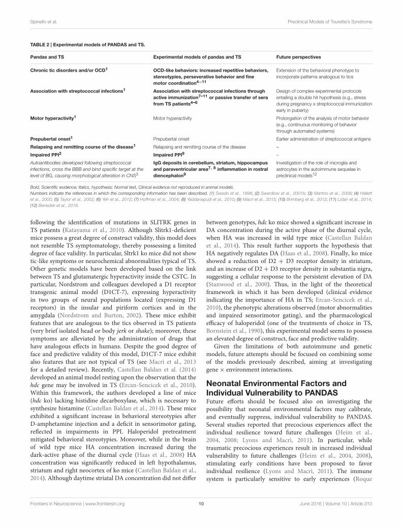
TABLE 2 | Experimental models of PANDAS and TS.
Pandas and TS Experimental models of pandas and TS Future perspectives
Chronic tic disorders and/or OCD1 OCD-like behaviors: increased repetitive behaviors,
stereotypies, perseverative behavior and fine
motor coordination4−11
Extension of the behavioral phenotype to
incorporate patterns analogous to tics
Association with streptococcal infections1 Association with streptococcal infections through
active immunization7–11 or passive transfer of sera
from TS patients4–6
Design of complex experimental protocols
entailing a double hit hypothesis (e.g., stress
during pregnancy x streptococcal immunization
early in puberty)
Motor hyperactivity1 Motor hyperactivity Prolongation of the analysis of motor behavior
(e.g., continuous monitoring of behavior
through automated systems)
Prepubertal onset1 Prepubertal onset Earlier administration of streptococcal antigens
Relapsing and remitting course of the disease1 Relapsing and remitting course of the disease –
Impaired PPI2 Impaired PPI9 –
Autoantibodies developed following streptococcal
infections, cross the BBB and bind specific target at the
level of BG, causing morphological alteration in CNS3
IgG deposits in cerebellum, striatum, hippocampus
and paraventricular area7, 8 inflammation in rostral
diencephalon9
Investigation of the role of microglia and
astrocytes in the autoimmune sequelae in
preclinical models12
Bold, Scientific evidence; Italics, hypothesis; Normal text, Clinical evidence not reproduced in animal models.
Numbers indicate the references in which the corresponding information has been described. (1) Swedo et al., 1998; (2) Swerdlow et al., 2001b; (3) Martino et al., 2009; (4) Hallett
et al., 2000; (5) Taylor et al., 2002; (6) Yeh et al., 2012; (7) Hoffman et al., 2004; (8) Yaddanapudi et al., 2010; (9) Macrì et al., 2015; (10) Brimberg et al., 2012; (11) Lotan et al., 2014;
(12) Benedek et al., 2016.
following the identification of mutations in SLITRK genes inTS patients (Katayama et al., 2010). Although Slitrk1-deficientmice possess a great degree of construct validity, this model doesnot resemble TS symptomatology, thereby possessing a limiteddegree of face validity. In particular, Sltrk1 ko mice did not showtic-like symptoms or neurochemical abnormalities typical of TS.Other genetic models have been developed based on the linkbetween TS and glutamatergic hyperactivity inside the CSTC. Inparticular, Nordstrom and colleagues developed a D1 receptortransgenic animal model (D1CT-7), expressing hyperactivityin two groups of neural populations located (expressing D1receptors) in the insular and piriform cortices and in theamygdala (Nordstrom and Burton, 2002). These mice exhibitfeatures that are analogous to the tics observed in TS patients(very brief isolated head or body jerk or shake); moreover, thesesymptoms are alleviated by the administration of drugs thathave analogous effects in humans. Despite the good degree offace and predictive validity of this model, D1CT-7 mice exhibitalso features that are not typical of TS (see Macrì et al., 2013for a detailed review). Recently, Castellan Baldan et al. (2014)developed an animal model resting upon the observation that thehdc gene may be involved in TS (Ercan-Sencicek et al., 2010).Within this framework, the authors developed a line of mice(hdc ko) lacking histidine decarboxylase, which is necessary tosynthesize histamine (Castellan Baldan et al., 2014). These miceexhibited a significant increase in behavioral stereotypies afterD-amphetamine injection and a deficit in sensorimotor gating,reflected in impairments in PPI. Haloperidol pretreatmentmitigated behavioral stereotypies. Moreover, while in the brainof wild type mice HA concentration increased during thedark-active phase of the diurnal cycle (Haas et al., 2008) HAconcentration was significantly reduced in left hypothalamus,striatum and right neocortex of ko mice (Castellan Baldan et al.,2014). Although daytime striatal DA concentration did not differ
between genotypes, hdc ko mice showed a significant increase inDA concentration during the active phase of the diurnal cycle,when HA was increased in wild type mice (Castellan Baldanet al., 2014). This result further supports the hypothesis thatHA negatively regulates DA (Haas et al., 2008). Finally, ko miceshowed a reduction of D2 + D3 receptor density in striatum,and an increase of D2 + D3 receptor density in substantia nigra,suggesting a cellular response to the persistent elevation of DA(Stanwood et al., 2000). Thus, in the light of the theoreticalframework in which it has been developed (clinical evidenceindicating the importance of HA in TS; Ercan-Sencicek et al.,2010), the phenotypic alterations observed (motor abnormalitiesand impaired sensorimotor gating), and the pharmacologicalefficacy of haloperidol (one of the treatments of choice in TS,Bornstein et al., 1990), this experimental model seems to possessan elevated degree of construct, face and predictive validity.
Given the limitations of both autoimmune and geneticmodels, future attempts should be focused on combining someof the models previously described, aiming at investigatinggene× environment interactions.
Neonatal Environmental Factors andIndividual Vulnerability to PANDASFuture efforts should be focused also on investigating thepossibility that neonatal environmental factors may calibrate,and eventually suppress, individual vulnerability to PANDAS.Several studies reported that precocious experiences affect theindividual resilience toward future challenges (Heim et al.,2004, 2008; Lyons and Macrì, 2011). In particular, whiletraumatic precocious experiences result in increased individualvulnerability to future challenges (Heim et al., 2004, 2008),stimulating early conditions have been proposed to favorindividual resilience (Lyons and Macrì, 2011). The immunesystem is particularly sensitive to early experiences (Roque
Frontiers in Neuroscience | www.frontiersin.org 10 June 2016 | Volume 10 | Article 310
Spinello et al. Preclinical Models of Tourette’s Syndrome
et al., 2014). For example, several studies conducted in rodentsshowed that maternal separation (daily 3–6 h mother-offspringseparations during the first 2 weeks of life) or exposure toearly physiological stressors result in increased susceptibilitytoward viral infections (Meagher et al., 2010) or vulnerabilitytoward autoimmune phenomena (Bakker et al., 2000). Individualreactivity to immune challenge has been proposed to dependon the functionality of hypothalamic-pituitary-adrenocortical(HPA) axis (Bakker et al., 2000; Meagher et al., 2010). Inparticular, the differential response to immune challenge dependson the modulation of the functionality of immune system exertedby elevations in levels of corticosterone (Laban et al., 1995).Several studies showed that circulating corticosteroids have adirect effect on T-cell, suppressing immune responses (Wicket al., 1993). Thus, it is tenable to propose that the modulationof corticosterone reactivity through experimental stressors, maycalibrate individual susceptibility toward phenomena that relateto immunity (immune and autoimmune phenomena). Withinthis framework, Levine and Saltzman (1987) reported thatexperimental stressors favoring an upregulation of the HPA-axis alter individual vulnerability to EAE (Levine and Saltzman,1987). Moreover, Levine and colleagues showed that stressreduces (Levine et al., 1962a) and adrenalectomy enhances(Levine et al., 1962b) vulnerability to EAE. Thus, a persistentupregulation of HPA axis induced by neonatal stressor mayconsistently prevent some of the consequences of experimentalmodels of autoimmunity, such as PANDAS. Beside analyzing therole of environmental factors in modulating the functionalityof the immune system, future studies shall thoroughly detailwhich portions of the immune system are involved in theautoimmune sequelae (Benedek et al., 2016). An interestingtarget to be contemplated in future studies is the activation ofmicroglia and the role exerted by astrocytes (Benedek et al., 2016;Lécuyer et al., 2016). These targets appear particularly relevantwhereby their involvement has already been demonstrated inexperimental models of autoimmunity (Correale, 2014; Shemerand Jung, 2015; Benedek et al., 2016; Lécuyer et al., 2016). Whilethese studies addressed the role of astrocytes and microglia inexperimental models of MS, it may be important to evaluatewhether these outcomes translate to experimental models ofPANDAS or TS. This need is also corroborated by clinicalevidence indicating that microglia can be activated in TS andPANDAS patients (Kumar et al., 2015), see below for a detaileddescription.
Peripheral Autoantibodies As DiagnosticBiomarker in TSFinally, we emphasize the value of the detection of peripheralautoantibodies as a reliable method potentially aiding thediagnosis of neurological disorders. The search of biologicalmarkers measurable and detectable using non-invasiveapproaches constitutes an important tool in the diagnosisof these diseases (Damoiseaux et al., 2015). With particularattention to autoimmune disorders, the measurement ofautoantibodies has been proposed as a valuable tool not only inthe diagnosis, but also in the prediction and in the prognosis of
autoimmune diseases (Harel and Shoenfeld, 2006; Shepshelovichand Shoenfeld, 2006; Bizzaro et al., 2007; see Damoiseaux et al.,2015 for a detailed review). Within this framework, severalperipheral autoantibodies emerged as clinically relevant inseveral neurological and neuropsychiatric disorders, such asmultiple sclerosis (Comabella and Montalban, 2014), limbicencephalitis (Beck et al., 2009; Schlumberger et al., 2014),myasthenia gravis (Verschuuren et al., 2013), or ADHD (Gianaet al., 2015). However, the debate on the efficacy of serumautoantibodies as diagnostic markers is still open. For exampleHöftberger (2015) reported that in the case of autoimmuneencephalitis (AIE), serum did not contain antineural antibodiesin the 14% of patients, while autoantibodies were alwaysdetected in patients’ cerebrospinal fluid (CSF). Thus, theabsence of autoantibodies in serum may not be sufficient toexclude AIE (Höftberger, 2015). With respect to TS, as alreadydiscussed (see Introduction), several clinical studies reportedthe presence of anti-BG antibodies in sera of TS patients (Rizzoet al., 2006; Martino et al., 2011). Moreover, the injectionsof TS sera (containing autoantibodies) directly into rodents’striatum, result, in some cases, in behavioral and neurochemicalalterations that partially resemble PANDAS symptomatology(Hallett et al., 2000; Yeh et al., 2012). In addition, autoantibodiesin TS sera seem to induce PANDAS-like behavioral phenotypesin a concentration-dependent manner (high or low level ofautoantibodies titers, see Taylor et al., 2002). These results seemto support the hypothesis that anti-BG antibodies may constitutea valid biomarker in the diagnosis of some cases of TS. However,results reported in subsequent studies, where the passive transferof TS sera in rodents’ striatum failed to induce PANDAS-likephenotypes (Loiselle et al., 2004; Ben-Pazi et al., 2012), suggestthat additional studies are necessary to investigate the diagnosticvalue of the detection of peripheral BG-antigens in TS. Furtherefforts may be focused, for example, on the standardizationof the assays used to quantify antineural antibodies in sera(Jacobs et al., 2015). In particular, further studies should befocused on investigating the use of immunohistochemistry asa method for the detection of anti-neuronal autoantibodies inCNS disorders. Hachiya et al. (2013) showed, in a recent study,that immunohistochemistry may constitute a reliable methodfor the detection of autoantibodies in serum of patients affectedby CNS disorders associated with GABHS infections (Hachiyaet al., 2013). In particular, they assessed immunoreactivity ofsera (obtained during the acute phase of disease or duringremission or convalescence) from patients affected by threeCNS disorders linked to GABHS infections (acute disseminatedencephalomyelitis, PANDAS and subacute encephalitis). Theauthors performed immunohistochemistry on brain sectionsof hippocampus, basal ganglia, cerebellar cortex and midbrainobtained from male controls (aged 5 and 9 years) that didnot present CNS alterations. Sera obtained from patientsaffected by acute disseminated encephalomyelitis and PANDASshowed immunoreactivity in globus pallidus neurons, whilesera obtained from patients affected by subacute encephalitisshowed immunoreactivity in the extra pyramidal cell layersin the temporal cortex. Conversely, sera obtained duringremission or convalescence did not show immunoreactivity
Frontiers in Neuroscience | www.frontiersin.org 11 June 2016 | Volume 10 | Article 310
Spinello et al. Preclinical Models of Tourette’s Syndrome
(Hachiya et al., 2013). With particular attention to PANDASand TS, future efforts should be focused, for example, onanalyzing immunoreactivity of sera from patients towarddopamine D2 receptors. In the context of the identification ofimmune-related diagnostic biomarkers, Kumar et al. (2015)recently evaluated neuroinflammation in TS and PANDASchildren. Specifically, the authors performed a Positron EmissionTomographic (PET) study to identify markers of activatedmicroglia (Kumar et al., 2015). Activated microglia has beenproposed to constitute a valid indicator of the presence ofneuroinflammation (Kreutzberg, 1996). To address this aspect,the authors exploited the capacity of activated microglia toexpress the translocator protein receptor (TSPO). TSPO, in turn,can be selectively identified through the radioactive tracer 11C-[R]-PK11195 (PK) (Cagnin et al., 2007). Using this approach,the authors analyzed neuroinflammation in basal ganglia andthalamus and observed increased binding potential in bilateralcaudate and bilateral lentiform nucleus in PANDAS patients.TS children exhibited neuroinflammation in bilateral lentiformnucleus only, suggesting possible neuroanatomical differencesbetween PANDAS and TS diseases (Kumar et al., 2015). Thus, the
monitoring of neuroinflammation through PET may constitutea potential method to clarify pathophysiological mechanisms inTS and PANDAS.
AUTHOR CONTRIBUTIONS
CS and SM wrote the first draft of the manuscript; CS, SM, andGL worked on the subsequent versions of the manuscript.
ACKNOWLEDGMENTS
The authors of this manuscript received funding from theEuropean Community’s Seventh Framework Programme(FP7/2007-2013) under grant agreements n◦ 278367 (ProjectEMTICS) and n◦ 603016 (Project MATRICS). This paper reflectsonly the authors’ views and the European Union is not liable forany use that may be made of the information contained therein.We sincerely thank Prof. Francesco Cardona for critical readingof a previous version of this manuscript, and Prof. GraziellaOrefici for constant and constructive feedback on the topic of thepresent manuscript.
REFERENCES
Abelson, J. F., Kwan, K. Y., O’Roak, B. J., Baek, D. Y., Stillman, A. A., Morgan, T.
M., et al. (2005). Sequence variants in SLITRK1 are associated with Tourette’s
syndrome. Science 310, 317–320. doi: 10.1126/science.1116502
Acharjee, S., Nayani, N., Tsutsui, M., Hill, M. N., Ousman, S. S., and Pittman,
Q. J. (2013). Altered cognitive-emotional behavior in early experimental
autoimmune encephalitis–cytokine and hormonal correlates. Brain Behav.
Immun. 33, 164–172. doi: 10.1016/j.bbi.2013.07.003
Adriani, W., Koot, S., Columba-Cabezas, S., Romano, E., Travaglini, D., van den
Bos, R., et al. (2012). Immunization with DAT fragments is associated with
long-term striatal impairment, hyperactivity and reduced cognitive flexibility
in mice. Behav. Brain Funct. 8:54. doi: 10.1186/1744-9081-8-54
Ahmari, S. E., Risbrough, V. B., Geyer, M. A., and Simpson, H. B. (2012). Impaired
sensorimotor gating in unmedicated adults with obsessive-compulsive
disorder. Neuropsychopharmacology 37, 1216–1223. doi: 10.1038/npp.2011.308
Almutairi, M. M., Gong, C., Xu, Y. G., Chang, Y., and Shi, H. (2016). Factors
controlling permeability of the blood-brain barrier. Cell. Mol. Life Sci. 73,
57–77. doi: 10.1007/s00018-015-2050-8
Amor, S., Baker, D., Groome, N., and Turk, J. L. (1993). Identification of a
major encephalitogenic epitope of proteolipid protein (residues 56–70) for
the induction of experimental allergic encephalomyelitis in Biozzi AB/H and
nonobese diabetic mice. J. Immunol. 150, 5666–5672.
Amor, S., Groome, N., Linington, C., Morris, M. M., Dornmair, K., Gardinier,
M. V., et al. (1994). Identification of epitopes of myelin oligodendrocyte
glycoprotein for the induction of experimental allergic encephalomyelitis in SJL
and Biozzi AB/H mice. J. Immunol. 153, 4349–4356.
Amor, S., O’Neill, J. K., Morris, M. M., Smith, R. M., Wraith, D. C.,
Groome, N., et al. (1996). Encephalitogenic epitopes of myelin basic protein,
proteolipid protein, myelin oligodendrocyte glycoprotein for experimental
allergic encephalomyelitis induction in Biozzi ABH (H-2Ag7) mice share an
amino acid motif. J. Immunol. 156, 3000–3008.
APA (2013). Diagnostic and Statistical Manual of Mental Disorders. Arlington, VA:
American Psychiatric Association.
Aruga, J., and Mikoshiba, K. (2003). Identification and characterization of Slitrk, a
novel neuronal transmembrane protein family controlling neurite outgrowth.
Mol. Cell. Neurosci. 24, 117–129. doi: 10.1016/S1044-7431(03)00129-5
Asano, N. M., Coriolano, M., Asano, B. J., and Lins, O. G. (2013). Psychiatric
comorbidities in patients with systemic lupus erythematosus: a systematic
review of the last 10 years. Rev. Bras. Reumatol. 53, 431–437. doi:
10.1016/S2255-5021(13)70114-7
Bakker, J. M., Kavelaars, A., Kamphuis, P. J., Cobelens, P. M., van Vugt, H. H., van
Bel, F., et al. (2000). Neonatal dexamethasone treatment increases susceptibility
to experimental autoimmune disease in adult rats. J. Immunol. 165, 5932–5937.
doi: 10.4049/jimmunol.165.10.5932
Baldan Ramsey, L. C., Xu, M., Wood, N., and Pittenger, C. (2011). Lesions of the
dorsomedial striatum disrupt prepulse inhibition. Neuroscience 180, 222–228.
doi: 10.1016/j.neuroscience.2011.01.041
Beck, L. H. Jr., Bonegio, R. G., Lambeau, G., Beck, D. M., Powell, D. W., Cummins,
T. D., et al. (2009). M-type phospholipase A2 receptor as target antigen
in idiopathic membranous nephropathy. N. Engl. J. Med. 361, 11–21. doi:
10.1056/NEJMoa0810457
Benedek, G., Zhang, J., Bodhankar, S., Nguyen, H., Kent, G., Jordan,
K., et al. (2016). Estrogen induces multiple regulatory B cell subtypes
and promotes M2 microglia and neuroprotection during experimental
autoimmune encephalomyelitis. J. Neuroimmunol. 293, 45–53. doi: 10.1016/
j.jneuroim.2016.02.009
Ben-Pazi, H., Sadan, O., and Offen, D. (2012). Striatal microinjection of Sydenham
chorea antibodies: using a rat model to examine the dopamine hypothesis. J.
Mol. Neurosci. 46, 162–166. doi: 10.1007/s12031-011-9559-6
Berardelli, A., Currà, A., Fabbrini, G., Gilio, F., and Manfredi, M. (2003).
Pathophysiology of tics and Tourette syndrome. J. Neurol. 250, 781–787. doi:
10.1007/s00415-003-1102-4
Bizzaro, N., Tonutti, E., Visentini, D., Alessio, M. G., Platzgummer, S., Morozzi,
G., et al. (2007). Antibodies to the lens and cornea in anti-DFS70-
positive subjects. Ann. N.Y. Acad. Sci. 1107, 174–183. doi: 10.1196/annals.
1381.019
Boghosian-Sell, L., Comings, D. E., and Overhauser, J. (1996). Tourette syndrome
in a pedigree with a 7;18 translocation: identification of a YAC spanning the
translocation breakpoint at 18q22.3. Am. J. Hum. Genet. 59, 999–1005.
Borchers, A. T., Aoki, C. A., Naguwa, S. M., Keen, C. L., Shoenfeld, Y.,
and Gershwin, M. E. (2005). Neuropsychiatric features of systemic lupus
erythematosus. Autoimmun. Rev. 4, 329–344. doi: 10.1016/j.autrev.2005.01.008
Bornstein, R. A., Stefl, M. E., and Hammond, L. (1990). A survey of Tourette
syndrome patients and their families: the 1987 Ohio Tourette Survey. J.
Neuropsychiatry Clin. Neurosci. 2, 275–281.
Bos-Veneman, N. G., Olieman, R., Tobiasova, Z., Hoekstra, P. J., Katsovich, L.,
Bothwell, A. L., et al. (2011). Altered immunoglobulin profiles in children
Frontiers in Neuroscience | www.frontiersin.org 12 June 2016 | Volume 10 | Article 310
Spinello et al. Preclinical Models of Tourette’s Syndrome
with Tourette syndrome. Brain Behav. Immun. 25, 532–538. doi: 10.1016/
j.bbi.2010.12.003
Brilot, F., Merheb, V., Ding, A., Murphy, T., and Dale, R. C. (2011).
Antibody binding to neuronal surface in Sydenham chorea, but not in
PANDAS or Tourette syndrome. Neurology 76, 1508–1513. doi: 10.1212/
WNL.0b013e3182181090
Brimberg, L., Benhar, I., Mascaro-Blanco, A., Alvarez, K., Lotan, D., Winter, C.,
et al. (2012). Behavioral, pharmacological, and immunological abnormalities
after streptococcal exposure: a novel rat model of Sydenham chorea and
related neuropsychiatric disorders. Neuropsychopharmacology 37, 2076–2087.
doi: 10.1038/npp.2012.56
Bronson, S. L., and Bale, T. L. (2016). The Placenta as a mediator of stress effects on
neurodevelopmental reprogramming. Neuropsychopharmacology 41, 207–218.
doi: 10.1038/npp.2015.231
Cagnin, A., Kassiou, M., Meikle, S. R., and Banati, R. B. (2007). Positron emission
tomography imaging of neuroinflammation.Neurotherapeutics 4, 443–452. doi:
10.1016/j.nurt.2007.04.006
Capone, F., Adriani,W., Shumilina,M., Izykenova, G., Granstrem, O., Dambinova,
S., et al. (2008). Autoantibodies against opioid or glutamate receptors are
associated with changes in morphine reward and physical dependence
in mice. Psychopharmacology 197, 535–548. doi: 10.1007/s00213-007-
1062-y
Cardona, F., and Orefici, G. (2001). Group A streptococcal infections and tic
disorders in an Italian pediatric population. J. Pediatr. 138, 71–75. doi:
10.1067/mpd.2001.110325
Cardoso, F., Vargas, A. P., Oliveira, L. D., Guerra, A. A., and Amaral, S. V. (1999).
Persistent Sydenham’s chorea.Mov. Disord. 14, 805–807.
Castellan Baldan, L., Williams, K. A., Gallezot, J. D., Pogorelov, V., Rapanelli, M.,
Crowley, M., et al. (2014). Histidine decarboxylase deficiency causes tourette
syndrome: parallel findings in humans and mice. Neuron 81, 77–90. doi:
10.1016/j.neuron.2013.10.052
Castellanos, F. X., Fine, E. J., Kaysen, D., Marsh, W. L., Rapoport, J. L., and
Hallett, M. (1996). Sensorimotor gating in boys with Tourette’s syndrome
and ADHD: preliminary results. Biol. Psychiatry 39, 33–41. doi: 10.1016/0006-
3223(95)00101-8
Cieza, A., Anczewska, M., Ayuso-Mateos, J. L., Baker, M., Bickenbach, J., Chatterji,
S., et al. (2015). Understanding the impact of brain disorders: towards a
‘horizontal epidemiology’ of psychosocial difficulties and their determinants.
PLoS ONE 10:e0136271. doi: 10.1371/journal.pone.0136271
Coenen, M., Cabello, M., Umlauf, S., Ayuso-Mateos, J. L., Anczewska, M.,
Tourunen, J., et al. (2016). Psychosocial difficulties from the perspective of
persons with neuropsychiatric disorders. Disabil. Rehabil. 38, 1134–1145. doi:
10.3109/09638288.2015.1074729
Comabella, M., and Montalban, X. (2014). Body fluid biomarkers in multiple
sclerosis. Lancet Neurol. 13, 113–126. doi: 10.1016/S1474-4422(13)70233-3
Correale, J. (2014). The role of microglial activation in disease progression. Mult.
Scler. 20, 1288–1295. doi: 10.1177/1352458514533230
Cutforth, T., DeMille, M. M., Agalliu, I., and Agalliu, D. (2016). CNS autoimmune
disease after infections: animal models, cellular mechanisms and genetic
factors. Future Neurol. 11, 63–76. doi: 10.2217/fnl.16.4
Dale, R. C., Church, A. J., Candler, P. M., Chapman, M., Martino, D., and
Giovannoni, G. (2006). Serum autoantibodies do not differentiate PANDAS
and Tourette syndrome from controls. Neurology 66:1612. doi: 10.1212/
01.wnl.0000226832.36908.4c
Dalsgaard, S., Waltoft, B. L., Leckman, J. F., and Mortensen, P. B. (2015). Maternal
history of autoimmune disease and later development of tourette syndrome
in offspring. J. Am. Acad. Child Adolesc. Psychiatry 54, 495–501.e1. doi:
10.1016/j.jaac.2015.03.008
Damoiseaux, J., Andrade, L. E., Fritzler, M. J., and Shoenfeld, Y. (2015).
Autoantibodies 2015: from diagnostic biomarkers toward prediction,
prognosis and prevention. Autoimmun. Rev. 14, 555–563. doi: 10.1016/
j.autrev.2015.01.017
Davison, K. (2012). Autoimmunity in psychiatry. Br. J. Psychiatry 200, 353–355.
doi: 10.1192/bjp.bp.111.104471
Deacon, R. M., and Rawlins, J. N. (2006). T-maze alternation in the rodent. Nat.
Protoc. 1, 7–12. doi: 10.1038/nprot.2006.2
Deng, H., Gao, K., and Jankovic, J. (2012). The genetics of Tourette syndrome.Nat.
Rev. Neurol. 8, 203–213. doi: 10.1038/nrneurol.2012.26
Dileepan, T., Linehan, J. L., Moon, J. J., Pepper, M., Jenkins, M. K., and Cleary, P. P.
(2011). Robust antigen specific th17 T cell response to group A Streptococcus is
dependent on IL-6 and intranasal route of infection. PLoS Pathog. 7:e1002252.
doi: 10.1371/journal.ppat.1002252
Dileepan, T., Smith, E. D., Knowland, D., Hsu, M., Platt, M., Bittner-Eddy,
P., et al. (2016). Group A Streptococcus intranasal infection promotes CNS
infiltration by streptococcal-specific Th17 cells. J. Clin. Invest. 126, 303–317.
doi: 10.1172/JCI80792
Ebrahimi, A., Pochet, R., and Roger, M. (1992). Topographical organization of
the projections from physiologically identified areas of the motor cortex to the
striatum in the rat. Neurosci. Res. 14, 39–60.
Ercan-Sencicek, A. G., Stillman, A. A., Ghosh, A. K., Bilguvar, K., O’Roak, B. J.,
Mason, C. E., et al. (2010). L-histidine decarboxylase and Tourette’s syndrome.
N. Engl. J. Med. 362, 1901–1908. doi: 10.1056/NEJMoa0907006
Fanouriakis, A., Boumpas, D. T., and Bertsias, G. K. (2013). Pathogenesis
and treatment of CNS lupus. Curr. Opin. Rheumatol. 25, 577–583. doi:
10.1097/BOR.0b013e328363eaf1
Fernandez, T. V., Sanders, S. J., Yurkiewicz, I. R., Ercan-Sencicek, A. G.,
Kim, Y. S., Fishman, D. O., et al. (2012). Rare copy number variants in
tourette syndrome disrupt genes in histaminergic pathways and overlap
with autism. Biol. Psychiatry 71, 392–402. doi: 10.1016/j.biopsych.2011.
09.034
Ganor, Y., Freilinger, M., Dulac, O., and Levite, M. (2005a). Monozygotic
twins discordant for epilepsy differ in the levels of potentially
pathogenic autoantibodies and cytokines. Autoimmunity 38, 139–150.
doi: 10.1080/08916930500100825
Ganor, Y., Goldberg-Stern, H., Amrom, D., Lerman-Sagie, T., Teichberg, V.
I., Pelled, D., et al. (2004). Autoimmune epilepsy: some epilepsy patients
harbor autoantibodies to glutamate receptors and dsDNA on both sides
of the blood-brain barrier, which may kill neurons and decrease in
brain fluids after hemispherotomy. Clin. Dev. Immunol. 11, 241–252. doi:
10.1080/17402520400001736
Ganor, Y., Goldberg-Stern, H., Blank, M., Shoenfeld, Y., Dobrynina, L. A.,
Kalashnikova, L., et al. (2005c). Antibodies to glutamate receptor subtype
3 (GluR3) are found in some patients suffering from epilepsy as the main
disease, but not in patients whose epilepsy accompanies antiphospholipid
syndrome or Sneddon’s syndrome. Autoimmunity 38, 417–424. doi: 10.1080/
08916930500246339
Ganor, Y., Goldberg-Stern, H., Cohen, R., Teichberg, V., and Levite, M. (2014).
Glutamate receptor antibodies directed against AMPA receptors subunit 3
peptide B (GluR3B) can be produced in DBA/2J mice, lower seizure threshold
and induce abnormal behavior. Psychoneuroendocrinology 42, 106–117. doi:
10.1016/j.psyneuen.2014.01.005
Ganor, Y., Goldberg-Stern, H., Lerman-Sagie, T., Teichberg, V. I., and Levite, M.
(2005b). Autoimmune epilepsy: distinct subpopulations of epilepsy patients
harbor serum autoantibodies to either glutamate/AMPA receptor GluR3,
glutamate/NMDA receptor subunit NR2A or double-stranded DNA. Epilepsy
Res. 65, 11–22. doi: 10.1016/j.eplepsyres.2005.03.011
Giana, G., Romano, E., Porfirio, M. C., D’Ambrosio, R., Giovinazzo, S.,
Troianiello, M., et al. (2015). Detection of auto-antibodies to DAT in
the serum: interactions with DAT genotype and psycho-stimulant therapy
for ADHD. J. Neuroimmunol. 278, 212–222. doi: 10.1016/j.jneuroim.2014.
11.008
Goldberg-Stern, H., Ganor, Y., Cohen, R., Pollak, L., Teichberg, V., and
Levite, M. (2014). Glutamate receptor antibodies directed against
AMPA receptors subunit 3 peptide B (GluR3B) associate with some
cognitive/psychiatric/behavioral abnormalities in epilepsy patients.
Psychoneuroendocrinology 40, 221–231. doi: 10.1016/j.psyneuen.2013.11.007
Haas, H. L., Sergeeva, O. A., and Selbach, O. (2008). Histamine in the nervous
system. Physiol. Rev. 88, 1183–1241. doi: 10.1152/physrev.00043.2007
Hachiya, Y., Miyata, R., Tanuma, N., Hongou, K., Tanaka, K., Shimoda, K.,
et al. (2013). Autoimmune neurological disorders associated with group-
A beta-hemolytic streptococcal infection. Brain Dev. 35, 670–674. doi:
10.1016/j.braindev.2012.10.003
Hallett, J. J., Harling-Berg, C. J., Knopf, P. M., Stopa, E. G., and Kiessling,
L. S. (2000). Anti-striatal antibodies in Tourette syndrome cause
neuronal dysfunction. J. Neuroimmunol. 111, 195–202. doi: 10.1016/
S0165-5728(00)00320-9
Frontiers in Neuroscience | www.frontiersin.org 13 June 2016 | Volume 10 | Article 310
Spinello et al. Preclinical Models of Tourette’s Syndrome
Harel, M., and Shoenfeld, Y. (2006). Predicting and preventing autoimmunity,
myth or reality? Ann. N.Y. Acad. Sci. 1069, 322–345. doi: 10.1196/
annals.1351.031
Hartley, S., McArthur, M., Coenen, M., Cabello, M., Covelli, V., Roszczynska-
Michta, J., et al. (2014). Narratives reflecting the lived experiences of people
with brain disorders: common psychosocial difficulties and determinants. PLoS
ONE 9:e96890. doi: 10.1371/journal.pone.0096890
Heim, C., Newport, D. J., Mletzko, T., Miller, A. H., and Nemeroff, C. B.
(2008). The link between childhood trauma and depression: insights from
HPA axis studies in humans. Psychoneuroendocrinology 33, 693–710. doi:
10.1016/j.psyneuen.2008.03.008
Heim, C., Plotsky, P. M., and Nemeroff, C. B. (2004). Importance of
studying the contributions of early adverse experience to neurobiological
findings in depression. Neuropsychopharmacology 29, 641–648. doi:
10.1038/sj.npp.1300397
Hoekstra, P. J., Dietrich, A., Edwards, M. J., Elamin, I., and Martino, D. (2013).
Environmental factors in Tourette syndrome. Neurosci. Biobehav. Rev. 37,
1040–1049. doi: 10.1016/j.neubiorev.2012.10.010
Hoenig, K., Hochrein, A., Quednow, B. B., Maier, W., and Wagner, M. (2005).
Impaired prepulse inhibition of acoustic startle in obsessive-compulsive
disorder. Biol. Psychiatry 57, 1153–1158. doi: 10.1016/j.biopsych.2005.01.040
Hoffman, K. L., Hornig, M., Yaddanapudi, K., Jabado, O., and Lipkin, W. I.
(2004). A murine model for neuropsychiatric disorders associated with group
A beta-hemolytic streptococcal infection. J. Neurosci. 24, 1780–1791. doi:
10.1523/JNEUROSCI.0887-03.2004
Höftberger, R. (2015). Neuroimmunology: an expanding frontier in autoimmunity.
Front. Immunol. 6:206. doi: 10.3389/fimmu.2015.00206
Hornig, M. (2013). The role of microbes and autoimmunity in the pathogenesis
of neuropsychiatric illness. Curr. Opin. Rheumatol. 25, 488–795. doi:
10.1097/BOR.0b013e32836208de
Hornig, M., and Lipkin, W. I. (2013). Immune-mediated animal models
of Tourette syndrome. Neurosci. Biobehav. Rev. 37, 1120–1138. doi:
10.1016/j.neubiorev.2013.01.007
Huerta, P. T., Kowal, C., DeGiorgio, L. A., Volpe, B. T., and Diamond, B. (2006).
Immunity and behavior: antibodies alter emotion. Proc. Natl. Acad. Sci. U.S.A.
103, 678–683. doi: 10.1073/pnas.0510055103
Huppert, J., Closhen, D., Croxford, A., White, R., Kulig, P., Pietrowski, E., et al.
(2010). Cellular mechanisms of IL-17-induced blood-brain barrier disruption.
FASEB J. 24, 1023–1034. doi: 10.1096/fj.09-141978
Hyman, S. E. (2008). A glimmer of light for neuropsychiatric disorders.Nature 455,
890–893. doi: 10.1038/nature07454
Irani, S. R., Gelfand, J. M., Al-Diwani, A., and Vincent, A. (2014). Cell-
surface central nervous system autoantibodies: clinical relevance and emerging
paradigms. Ann. Neurol. 76, 168–184. doi: 10.1002/ana.24200
Irwin, J., Tombaugh, T. N., Zacharko, R. M., and Anisman, H. (1983). Alteration
of exploration and the response to food associated cues after treatment with
pimozide. Pharmacol. Biochem. Behav. 18, 235–246.
Jacobs, J. F., van der Molen, R. G., Bossuyt, X., and Damoiseaux, J. (2015). Antigen
excess in modern immunoassays: to anticipate on the unexpected. Autoimmun.
Rev. 14, 160–167. doi: 10.1016/j.autrev.2014.10.018
Jaffard, R., Mocaer, E., Poignant, J. C., Micheau, J., Marighetto, A., Meunier, M.,
et al. (1991). Effects of tianeptine on spontaneous alternation, simple and
concurrent spatial discrimination learning and on alcohol-induced alternation
deficits in mice. Behav. Pharmacol. 2, 37–46.
John, C. C., Carabin, H., Montano, S. M., Bangirana, P., Zunt, J. R., and Peterson,
P. K. (2015). Global research priorities for infections that affect the nervous
system. Nature 527, S178–S186. doi: 10.1038/nature16033
Kajiwara, Y., Buxbaum, J. D., and Grice, D. E. (2009). SLITRK1 binds 14-3-3
and regulates neurite outgrowth in a phosphorylation-dependent manner. Biol.
Psychiatry 66, 918–925. doi: 10.1016/j.biopsych.2009.05.033
Kaltwasser, M. T. (1990). Acoustic signaling in the black rat (Rattus rattus). J.
Comp. Psychol. 104, 227–232.
Kalueff, A. V., Stewart, A. M., Song, C., Berridge, K. C., Graybiel, A. M., and
Fentress, J. C. (2016). Neurobiology of rodent self-grooming and its value
for translational neuroscience. Nat. Rev. Neurosci. 17, 45–59. doi: 10.1038/
nrn.2015.8
Katayama, K., Yamada, K., Ornthanalai, V. G., Inoue, T., Ota, M., Murphy, N.
P., et al. (2010). Slitrk1-deficient mice display elevated anxiety-like behavior
and noradrenergic abnormalities. Mol. Psychiatry 15, 177–184. doi: 10.1038/
mp.2008.97
Kawikova, I., Leckman, J. F., Kronig, H., Katsovich, L., Bessen, D. E.,
Ghebremichael, M., et al. (2007). Decreased numbers of regulatory
T cells suggest impaired immune tolerance in children with tourette
syndrome: a preliminary study. Biol. Psychiatry 61, 273–278. doi: 10.1016/
j.biopsych.2006.06.012
Kebir, H., Kreymborg, K., Ifergan, I., Dodelet-Devillers, A., Cayrol, R., Bernard,
M., et al. (2007). Human TH17 lymphocytes promote blood-brain barrier
disruption and central nervous system inflammation.Nat. Med. 13, 1173–1175.
doi: 10.1038/nm1651
Kipp, M., van der Star, B., Vogel, D. Y., Puentes, F., van der Valk, P., Baker, D.,
et al. (2012). Experimental in vivo and in vitro models of multiple sclerosis:
EAE and beyond.Mult. Scler. Relat. Disord. 1, 15–28. doi: 10.1016/j.msard.2011.
09.002
Korngold, R., Feldman, A., Rorke, L. B., Lublin, F. D., and Doherty, P. C. (1986).
Acute experimental allergic encephalomyelitis in radiation bone marrow
chimeras between high and low susceptible strains of mice. Immunogenetics 24,
309–315.
Kowal, C., and Diamond, B. (2012). Aspects of CNS lupus: mouse models of
anti-NMDA receptor antibody mediated reactivity. Methods Mol. Biol. 900,
181–206. doi: 10.1007/978-1-60761-720-4_9
Kowal, C., DeGiorgio, L. A., Nakaoka, T., Hetherington, H., Huerta, P. T.,
Diamond, B., et al. (2004). Cognition and immunity; antibody impairs memory.
Immunity 21, 179–188. doi: 10.1016/j.immuni.2004.07.011
Kreutzberg, G. W. (1996). Microglia: a sensor for pathological events in the CNS.
Trends Neurosci. 19, 312–318.
Kumar, A., Williams, M. T., and Chugani, H. T. (2015). Evaluation of basal
ganglia and thalamic inflammation in children with pediatric autoimmune
neuropsychiatric disorders associated with streptococcal infection and
tourette syndrome: a positron emission tomographic (PET) study using
11C-[R]-PK11195. J. Child Neurol. 30, 749–756. doi: 10.1177/0883073814
543303
Laban, O., Dimitrijevic, M., von Hoersten, S., Markovic, B. M., and Jankovic, B. D.
(1995). Experimental allergic encephalomyelitis in adult DA rats subjected to
neonatal handling or gentling. Brain Res. 676, 133–140.
Lalonde, R. (2002). The neurobiological basis of spontaneous alternation.Neurosci.
Biobehav. Rev. 26, 91–104. doi: 10.1016/S0149-7634(01)00041-0
Landau, Y. E., Steinberg, T., Richmand, B., Leckman, J. F., and Apter, A. (2012).
Involvement of immunologic and biochemical mechanisms in the pathogenesis
of Tourette’s syndrome. J. Neural Transm. 119, 621–626. doi: 10.1007/s00702-
011-0739-x
Leckman, J. F., and Peterson, B. S. (1993). The pathogenesis of Tourette’s
syndrome: epigenetic factors active in early CNS development. Biol. Psychiatry
34, 425–427.
Leckman, J. F., Bloch, M. H., Smith, M. E., Larabi, D., and Hampson, M.
(2010). Neurobiological substrates of Tourette’s disorder. J. Child Adolesc.
Psychopharmacol. 20, 237–247. doi: 10.1089/cap.2009.0118
Leckman, J. F., Dolnansky, E. S., Hardin, M. T., Clubb, M., Walkup, J. T.,
Stevenson, J., et al. (1990). Perinatal factors in the expression of Tourette’s
syndrome: an exploratory study. J. Am. Acad. Child Adolesc. Psychiatry 29,
220–226. doi: 10.1097/00004583-199003000-00010
Leckman, J. F., Katsovich, L., Kawikova, I., Lin, H., Zhang, H., Krönig, H.,
et al. (2005). Increased serum levels of interleukin-12 and tumor necrosis
factor-alpha in Tourette’s syndrome. Biol. Psychiatry 57, 667–673. doi:
10.1016/j.biopsych.2004.12.004
Leckman, J. F., Price, R. A., Walkup, J. T., Ort, S., Pauls, D. L., and Cohen, D.
J. (1987). Nongenetic factors in Gilles de la Tourette’s syndrome. Arch. Gen.
Psychiatry 44:100.
Lécuyer, M. A., Kebir, H., and Prat, A. (2016). Glial influences on BBB functions
and molecular players in immune cell trafficking. Biochim. Biophys. Acta 1862,
472–482. doi: 10.1016/j.bbadis.2015.10.004
Levine, S., and Saltzman, A. (1987). Nonspecific stress prevents relapses of
experimental allergic encephalomyelitis in rats. Brain Behav. Immun. 1,
336–341.
Levine, S., Strebel, R., Wenk, E. J., and Harman, P. J. (1962a). Suppression of
experimental allergic encephalomyelitis by stress. Proc. Soc. Exp. Biol. Med. 109,
294–298.
Frontiers in Neuroscience | www.frontiersin.org 14 June 2016 | Volume 10 | Article 310
Spinello et al. Preclinical Models of Tourette’s Syndrome
Levine, S., Wenk, E. J., Muldoon, T. N., and Cohen, S. G. (1962b). Enhancement of
experimental allergic encephalomyelitis by adrenalectomy. Proc. Soc. Exp. Biol.
Med. 111, 383–385.
Levite, M. (2014). Glutamate receptor antibodies in neurological diseases: anti-
AMPA-GluR3 antibodies, anti-NMDA-NR1 antibodies, anti-NMDA-NR2A/B
antibodies, anti-mGluR1 antibodies or anti-mGluR5 antibodies are present
in subpopulations of patients with either: epilepsy, encephalitis, cerebellar
ataxia, systemic lupus erythematosus (SLE) and neuropsychiatric SLE, Sjogren’s
syndrome, schizophrenia, mania or stroke. These autoimmune anti-glutamate
receptor antibodies can bind neurons in few brain regions, activate glutamate
receptors, decrease glutamate receptor’s expression, impair glutamate-induced
signaling and function, activate blood brain barrier endothelial cells,
kill neurons, damage the brain, induce behavioral/psychiatric/cognitive
abnormalities and ataxia in animal models, and can be removed or silenced
in some patients by immunotherapy. J. Neural Transm. 121, 1029–1075. doi:
10.1007/s00702-014-1193-3
Levite, M., and Hermelin, A. (1999). Autoimmunity to the glutamate receptor
in mice–a model for Rasmussen’s encephalitis? J. Autoimmun. 13, 73–82. doi:
10.1006/jaut.1999.0297
Levite, M., Fleidervish, I. A., Schwarz, A., Pelled, D., and Futerman, A. H. (1999).
Autoantibodies to the glutamate receptor kill neurons via activation of the
receptor ion channel. J. Autoimmun. 13, 61–72. doi: 10.1006/jaut.1999.0301
Loiselle, C. R., Lee, O., Moran, T. H., and Singer, H. S. (2004). Striatal
microinfusion of Tourette syndrome and PANDAS sera: failure to induce
behavioral changes.Mov. Disord. 19, 390–396. doi: 10.1002/mds.10522
Lombroso, P. J., and Scahill, L. (2008). Tourette syndrome and
obsessive-compulsive disorder. Brain Dev. 30, 231–237. doi: 10.1016/
j.braindev.2007.09.001
Lotan, D., Benhar, I., Alvarez, K., Mascaro-Blanco, A., Brimberg, L., Frenkel,
D., et al. (2014). Behavioral and neural effects of intra-striatal infusion of
anti-streptococcal antibodies in rats. Brain Behav. Immun. 38, 249–262. doi:
10.1016/j.bbi.2014.02.009
Lyons, D. M., and Macrì, S. (2011). Resilience and adaptive aspects of stress
in neurobehavioral development. Neurosci. Biobehav. Rev. 35:1451. doi:
10.1016/j.neubiorev.2010.09.004
Ma, Y., Mehta, S. L., Lu, B., and Li, P. A. (2011). Deficiency in the inner
mitochondrial membrane peptidase 2-like (Immp21) gene increases ischemic
brain damage and impairs mitochondrial function.Neurobiol. Dis. 44, 270–276.
doi: 10.1016/j.nbd.2011.06.019
Macrì, S., Ceci, C., Onori, M. P., Invernizzi, R. W., Bartolini, E., Altabella, L., et al.
(2015). Mice repeatedly exposed to Group-A beta-Haemolytic Streptococcus
show perseverative behaviors, impaired sensorimotor gating, and immune
activation in rostral diencephalon. Sci. Rep. 5:13257. doi: 10.1038/srep13257
Macrì, S., Proietti Onori, M., and Laviola, G. (2013). Theoretical and
practical considerations behind the use of laboratory animals for the
study of Tourette syndrome. Neurosci. Biobehav. Rev. 37, 1085–1100. doi:
10.1016/j.neubiorev.2013.03.014
Mandolesi, G., Grasselli, G., Musumeci, G., and Centonze, D. (2010). Cognitive
deficits in experimental autoimmune encephalomyelitis: neuroinflammation
and synaptic degeneration. Neurol. Sci. 31(Suppl. 2), S255–S259. doi:
10.1007/s10072-010-0369-3
Mansbach, R. S., Geyer, M. A., and Braff, D. L. (1988). Dopaminergic stimulation
disrupts sensorimotor gating in the rat. Psychopharmacology 94, 507–514.
Margari, F., Petruzzelli, M. G., Mianulli, R., Toto, M., Pastore, A., Bizzaro,
N., et al. (2013). Anti-brain autoantibodies in the serum of schizophrenic
patients: a case-control study. Psychiatry Res. 210, 800–805. doi:
10.1016/j.psychres.2013.09.006
Marques-Dias, M. J., Mercadante, M. T., Tucker, D., and Lombroso, P. (1997).
Sydenham’s chorea. Psychiatr. Clin. North Am. 20, 809–820.
Martino, D., Chiarotti, F., Buttiglione,M., Cardona, F., Creti, R., Nardocci, N., et al.
(2011). The relationship between groupA streptococcal infections and Tourette
syndrome: a study on a large service-based cohort. Dev. Med. Child Neurol. 53,
951–957. doi: 10.1111/j.1469-8749.2011.04018.x
Martino, D., Dale, R. C., Gilbert, D. L., Giovannoni, G., and Leckman, J. F. (2009).
Immunopathogenic mechanisms in tourette syndrome: a critical review. Mov.
Disord. 24, 1267–1279. doi: 10.1002/mds.22504
Martino, D., Zis, P., and Buttiglione, M. (2015). The role of immune
mechanisms in Tourette syndrome. Brain Res. 1617, 126–143. doi:
10.1016/j.brainres.2014.04.027
Meagher, M. W., Sieve, A. N., Johnson, R. R., Satterlee, D., Belyavskyi, M., Mi,
W., et al. (2010). Neonatal maternal separation alters immune, endocrine, and
behavioral responses to acute Theiler’s virus infection in adult mice. Behav.
Genet. 40, 233–249. doi: 10.1007/s10519-010-9333-5
Meldrum, B. S. (2000). Glutamate as a neurotransmitter in the brain: review of
physiology and pathology. J. Nutr. 130(4S Suppl), 1007S–1015S.
Morris, C. M., Pardo-Villamizar, C., Gause, C. D., and Singer, H. S. (2009).
Serum autoantibodies measured by immunofluorescence confirm a failure to
differentiate PANDAS and Tourette syndrome from controls. J. Neurol. Sci. 276,
45–48. doi: 10.1016/j.jns.2008.08.032
Murphy, T. K., Kurlan, R., and Leckman, J. (2010). The immunobiology
of Tourette’s disorder, pediatric autoimmune neuropsychiatric disorders
associated with Streptococcus, and related disorders: a way forward. J. Child
Adolesc. Psychopharmacol. 20, 317–331. doi: 10.1089/cap.2010.0043
Nordstrom, E. J., and Burton, F. H. (2002). A transgenic model of comorbid
Tourette’s syndrome and obsessive-compulsive disorder circuitry. Mol.
Psychiatry 7, 617–625, 524. doi: 10.1038/sj.mp.4001144
Olechowski, C. J., Tenorio, G., Sauve, Y., and Kerr, B. J. (2013). Changes
in nociceptive sensitivity and object recognition in experimental
autoimmune encephalomyelitis (EAE). Exp. Neurol. 241, 113–121. doi:
10.1016/j.expneurol.2012.12.012
Park, H. S., Francis, K. P., Yu, J., and Cleary, P. P. (2003). Membranous cells
in nasal-associated lymphoid tissue: a portal of entry for the respiratory
mucosal pathogen group A streptococcus. J. Immunol. 171, 2532–2537. doi:
10.4049/jimmunol.171.5.2532
Petek, E., Windpassinger, C., Vincent, J. B., Cheung, J., Boright, A. P., Scherer,
S. W., et al. (2001). Disruption of a novel gene (IMMP2L) by a breakpoint in
7q31 associated with Tourette syndrome. Am. J. Hum. Genet. 68, 848–858. doi:
10.1086/319523
Platt, S. R. (2007). The role of glutamate in central nervous system health and
disease–a review. Vet. J. 173, 278–286. doi: 10.1016/j.tvjl.2005.11.007
Podolska, M. J., Biermann, M. H., Maueröder, C., Hahn, J., and Herrmann, M.
(2015). Inflammatory etiopathogenesis of systemic lupus erythematosus: an
update. J. Inflamm. Res. 8, 161–171. doi: 10.2147/JIR.S70325
Poliak, S., Gollan, L., Martinez, R., Custer, A., Einheber, S., Salzer, J. L., et al.
(1999). Caspr2, a new member of the neurexin superfamily, is localized at the
juxtaparanodes of myelinated axons and associates with K+ channels. Neuron
24, 1037–1047.
Proenca, C. C., Gao, K. P., Shmelkov, S. V., Rafii, S., and Lee, F. S. (2011). Slitrks
as emerging candidate genes involved in neuropsychiatric disorders. Trends
Neurosci. 34, 143–153. doi: 10.1016/j.tins.2011.01.001
Rabchevsky, A. G., Degos, J. D., and Dreyfus, P. A. (1999). Peripheral injections
of Freund’s adjuvant in mice provoke leakage of serum proteins through the
blood-brain barrier without inducing reactive gliosis. Brain Res. 832, 84–96.
Rickard, M. D. (2004). The use of animals for research on animal diseases:
its impact on the harm-benefit analysis. Altern. Lab. Anim. 32(Suppl. 1A),
225–227.
Rizzo, R., Gulisano, M., Pavone, P., Fogliani, F., and Robertson, M. M. (2006).
Increased antistreptococcal antibody titers and anti-basal ganglia antibodies
in patients with Tourette syndrome: controlled cross-sectional study. J. Child
Neurol. 21, 747–753. doi: 10.1177/08830738060210091001
Roque, S., Mesquita, A. R., Palha, J. A., Sousa, N., and Correia-Neves, M. (2014).
The behavioral and immunological impact of maternal separation: a matter of
timing. Front. Behav. Neurosci. 8:192. doi: 10.3389/fnbeh.2014.00192
Russig, H., Spooren, W., Durkin, S., Feldon, J., and Yee, B. K. (2004).
Apomorphine-induced disruption of prepulse inhibition that can be
normalised by systemic haloperidol is insensitive to clozapine pretreatment.
Psychopharmacology 175, 143–147. doi: 10.1007/s00213-004-1810-1
Scahill, L., Erenberg, G., Berlin, C. M. Jr., Budman, C., Coffey, B. J., Jankovic,
J., et al. (2006). Contemporary assessment and pharmacotherapy of Tourette
syndrome. NeuroRx 3, 192–206. doi: 10.1016/j.nurx.2006.01.009
Schlumberger, W., Hornig, N., Lange, S., Probst, C., Komorowski, L., Fechner,
K., et al. (2014). Differential diagnosis of membranous nephropathy with
autoantibodies to phospholipase A2 receptor 1. Autoimmun. Rev. 13, 108–113.
doi: 10.1016/j.autrev.2013.09.005
Scoriels, L., Salek, R. M., Goodby, E., Grainger, D., Dean, A. M., West, J. A.,
et al. (2015). Behavioural andmolecular endophenotypes in psychotic disorders
reveal heritable abnormalities in glutamatergic neurotransmission. Transl.
Psychiatry 5:e540. doi: 10.1038/tp.2015.26
Frontiers in Neuroscience | www.frontiersin.org 15 June 2016 | Volume 10 | Article 310
Spinello et al. Preclinical Models of Tourette’s Syndrome
Sengupta, A., Winters, B., Bagley, E. E., and McNally, G. P. (2016). Disrupted
prediction error links excessive amygdala activation to excessive fear. J.
Neurosci. 36, 385–395. doi: 10.1523/JNEUROSCI.3670-15.2016
Shemer, A., and Jung, S. (2015). Differential roles of resident microglia and
infiltrating monocytes in murine CNS autoimmunity. Semin. Immunopathol.
37, 613–623. doi: 10.1007/s00281-015-0519-z
Shepshelovich, D., and Shoenfeld, Y. (2006). Prediction and prevention of
autoimmune diseases: additional aspects of the mosaic of autoimmunity. Lupus
15, 183–190. doi: 10.1191/0961203306lu2274rr
Shprecher, D., and Kurlan, R. (2009). The management of tics. Mov. Disord. 24,
15–24. doi: 10.1002/mds.22378
Silberberg, D., Anand, N. P., Michels, K., and Kalaria, R. N. (2015). Brain and
other nervous system disorders across the lifespan - global challenges and
opportunities. Nature 527, S151–S154. doi: 10.1038/nature16028
Singer, H. S., Hong, J. J., Yoon, D. Y., and Williams, P. N. (2005a). Serum
autoantibodies do not differentiate PANDAS and Tourette syndrome from
controls. Neurology 65, 1701–1707. doi: 10.1212/01.wnl.0000183223.69946.f1
Singer, H. S., Mink, J. W., Loiselle, C. R., Burke, K. A., Ruchkina, I., Morshed, S.,
et al. (2005b). Microinfusion of antineuronal antibodies into rodent striatum:
failure to differentiate between elevated and low titers. J. Neuroimmunol. 163,
8–14. doi: 10.1016/j.jneuroim.2005.02.018
Stanwood, G. D., Lucki, I., and McGonigle, P. (2000). Differential regulation of
dopamine D2 and D3 receptors by chronic drug treatments. J. Pharmacol. Exp.
Ther. 295, 1232–1240.
State, M. W. (2011). The genetics of Tourette disorder. Curr. Opin. Genet. Dev. 21,
302–309. doi: 10.1016/j.gde.2011.01.007
Stillman, A. A., Krsnik, Z., Sun, J., Rasin, M. R., State, M. W., Sestan, N., et al.
(2009). Developmentally regulated and evolutionarily conserved expression of
SLITRK1 in brain circuits implicated in Tourette syndrome. J. Comp. Neurol.
513, 21–37. doi: 10.1002/cne.21919
Swanborg, R. H. (2001). Experimental autoimmune encephalomyelitis in the rat:
lessons in T-cell immunology and autoreactivity. Immunol. Rev. 184, 129–135.
doi: 10.1034/j.1600-065x.2001.1840112.x
Swedo, S. E., and Grant, P. J. (2005). Annotation: PANDAS: a model for
human autoimmune disease. J. Child Psychol. Psychiatry 46, 227–234. doi:
10.1111/j.1469-7610.2004.00386.x
Swedo, S. E., Leonard, H. L., Garvey, M., Mittleman, B., Allen, A. J., Perlmutter,
S., et al. (1998). Pediatric autoimmune neuropsychiatric disorders associated
with streptococcal infections: clinical description of the first 50 cases. Am. J.
Psychiatry 155, 264–271.
Swedo, S. E., Leonard, H. L., Schapiro, M. B., Casey, B. J., Mannheim, G. B., Lenane,
M. C., et al. (1993). Sydenham’s chorea: physical and psychological symptoms
of St Vitus dance. Pediatrics 91, 706–713.
Swedo, S. E., Rapoport, J. L., Cheslow, D. L., Leonard, H. L., Ayoub, E. M.,
Hosier, D.M., et al. (1989). High prevalence of obsessive-compulsive symptoms
in patients with Sydenham’s chorea. Am. J. Psychiatry 146, 246–249. doi:
10.1176/ajp.146.2.246
Swerdlow, N. R. (2013). Update: studies of prepulse inhibition of startle, with
particular relevance to the pathophysiology or treatment of Tourette Syndrome.
Neurosci. Biobehav. Rev. 37, 1150–1156. doi: 10.1016/j.neubiorev.2012. 09.002
Swerdlow, N. R., and Sutherland, A. N. (2005). Using animal models to develop
therapeutics for Tourette Syndrome. Pharmacol. Ther. 108, 281–293. doi:
10.1016/j.pharmthera.2005.05.003
Swerdlow, N. R., Benbow, C. H., Zisook, S., Geyer, M. A., and Braff, D. L. (1993).
A preliminary assessment of sensorimotor gating in patients with obsessive
compulsive disorder. Biol. Psychiatry 33, 298–301.
Swerdlow, N. R., Geyer, M. A., and Braff, D. L. (2001a). Neural circuit regulation
of prepulse inhibition of startle in the rat: current knowledge and future
challenges. Psychopharmacology 156, 194–215. doi: 10.1007/s002130100799
Swerdlow, N. R., Karban, B., Ploum, Y., Sharp, R., Geyer, M. A., and Eastvold,
A. (2001b). Tactile prepuff inhibition of startle in children with Tourette’s
syndrome: in search of an “fMRI-friendly” startle paradigm. Biol. Psychiatry 50,
578–585. doi: 10.1016/S0006-3223(01)01164-7
Swerdlow, N. R., Light, G. A., Cadenhead, K. S., Sprock, J., Hsieh, M. H.,
and Braff, D. L. (2006). Startle gating deficits in a large cohort of patients
with schizophrenia: relationship to medications, symptoms, neurocognition,
and level of function. Arch. Gen. Psychiatry 63, 1325–1335. doi: 10.1001/
archpsyc.63.12.1325
Swerdlow, N. R., Paulsen, J., Braff, D. L., Butters, N., Geyer, M. A., and Swenson,M.
R. (1995). Impaired prepulse inhibition of acoustic and tactile startle response
in patients with Huntington’s disease. J. Neurol. Neurosurg. Psychiatr. 58,
192–200.
Taylor, J. R., Morshed, S. A., Parveen, S., Mercadante, M. T., Scahill, L., Peterson,
B. S., et al. (2002). An animal model of Tourette’s syndrome. Am. J. Psychiatry
159, 657–660. doi: 10.1176/appi.ajp.159.4.657
Valls-Sole, J., Munoz, J. E., and Valldeoriola, F. (2004). Abnormalities of prepulse
inhibition do not depend on blink reflex excitability: a study in Parkinson’s
disease and Huntington’s disease. Clin. Neurophysiol. 115, 1527–1536. doi:
10.1016/j.clinph.2004.02.014
van der Staay, F. J. (2006). Animal models of behavioral dysfunctions: basic
concepts and classifications, and an evaluation strategy. Brain Res. Rev. 52,
131–159. doi: 10.1016/j.brainresrev.2006.01.006
van der Staay, F. J., Arndt, S. S., and Nordquist, R. E. (2009). Evaluation of
animal models of neurobehavioral disorders. Behav. Brain Funct. 5:11. doi:
10.1186/1744-9081-5-11
Verkerk, A. J., Mathews, C. A., Joosse, M., Eussen, B. H., Heutink, P., Oostra,
B. A., et al. (2003). CNTNAP2 is disrupted in a family with Gilles de la
Tourette syndrome and obsessive compulsive disorder. Genomics 82, 1–9. doi:
10.1016/S0888-7543(03)00097-1
Verschuuren, J. J., Huijbers, M. G., Plomp, J. J., Niks, E. H., Molenaar, P. C.,
Martinez-Martinez, P., et al. (2013). Pathophysiology of myasthenia gravis
with antibodies to the acetylcholine receptor, muscle-specific kinase and low-
density lipoprotein receptor-related protein 4. Autoimmun. Rev. 12, 918–923.
doi: 10.1016/j.autrev.2013.03.001
Wick, G., Hu, Y., Schwarz, S., and Kroemer, G. (1993). Immunoendocrine
communication via the hypothalamo-pituitary-adrenal axis in autoimmune
diseases. Endocr. Rev. 14, 539–563. doi: 10.1210/edrv-14-5-539
Willner, P. (1984). The validity of animal models of depression.
Psychopharmacology 83, 1–16.
Willner, P., and Mitchell, P. J. (2002). The validity of animal models of
predisposition to depression. Behav. Pharmacol. 13, 169–188. doi: 10.1097/
00008877-200205000-00001
Yaddanapudi, K., Hornig, M., Serge, R., De Miranda, J., Baghban, A., Villar,
G., et al. (2010). Passive transfer of streptococcus-induced antibodies
reproduces behavioral disturbances in a mouse model of pediatric autoimmune
neuropsychiatric disorders associated with streptococcal infection. Mol.
Psychiatry 15, 712–726. doi: 10.1038/mp.2009.77
Yeh, C. B., Shui, H. A., Chu, T. H., Chen, Y. A., Tsung, H. C., and Shyu, J.
F. (2012). Hyperpolarisation-activated cyclic nucleotide channel 4 (HCN4)
involvement in Tourette’s syndrome autoimmunity. J. Neuroimmunol. 250,
18–26. doi: 10.1016/j.jneuroim.2012.05.009
Zebardast, N., Crowley, M. J., Bloch, M. H., Mayes, L. C., Wyk, B. V., Leckman,
J. F., et al. (2013). Brain mechanisms for prepulse inhibition in adults
with Tourette syndrome: initial findings. Psychiatry Res. 214, 33–41. doi:
10.1016/j.pscychresns.2013.05.009
Conflict of Interest Statement: The authors declare that the research was
conducted in the absence of any commercial or financial relationships that could
be construed as a potential conflict of interest.
Copyright © 2016 Spinello, Laviola and Macrì. This is an open-access article
distributed under the terms of the Creative Commons Attribution License (CC BY).
The use, distribution or reproduction in other forums is permitted, provided the
original author(s) or licensor are credited and that the original publication in this
journal is cited, in accordance with accepted academic practice. No use, distribution
or reproduction is permitted which does not comply with these terms.
Frontiers in Neuroscience | www.frontiersin.org 16 June 2016 | Volume 10 | Article 310