Embed Size (px)
Citation preview
To order: www.abgent.com Email: [email protected] or [email protected]: 858.875.1900 Fax: 858.622.0609
Purified Mouse Monoclonal Antibody (Mab) Catalog # AW5698
PDL1 Monoclonal Antibody
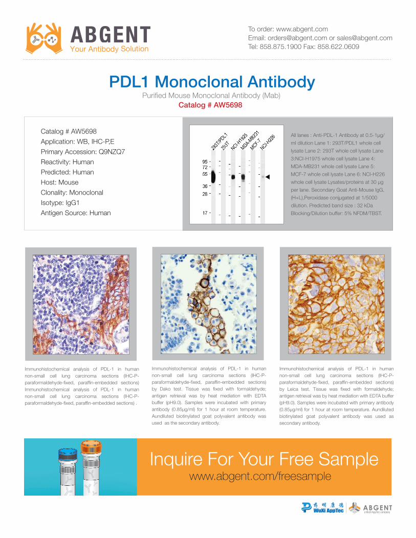
Immunohistochemical analysis of PDL-1 in human non-small cell lung carcinoma sections (IHC-P- paraformaldehyde-fixed, paraffin-embedded sections) Immunohistochemical analysis of PDL-1 in human non-small cell lung carcinoma sections (IHC-P- paraformaldehyde-fixed, paraffin-embedded sections) .
Immunohistochemical analysis of PDL-1 in human non-small cell lung carcinoma sections (IHC-P- paraformaldehyde-fixed, paraffin-embedded sections) by Dako test. Tissue was fixed with formaldehyde; antigen retrieval was by heat mediation with EDTA buffer (pH9.0). Samples were incubated with primary antibody (0.85µg/ml) for 1 hour at room temperature. Aundiluted biotinylated goat polyvalent antibody was used as the secondary antibody.
Immunohistochemical analysis of PDL-1 in human non-small cell lung carcinoma sections (IHC-P- paraformaldehyde-fixed, paraffin-embedded sections) by Leica test. Tissue was fixed with formaldehyde; antigen retrieval was by heat mediation with EDTA buffer (pH9.0). Samples were incubated with primary antibody (0.85µg/ml) for 1 hour at room temperature. Aundiluted biotinylated goat polyvalent antibody was used as secondary antibody.
Catalog # AW5698
Application: WB, IHC-P,E
Primary Accession: Q9NZQ7
Reactivity: Human
Predicted: Human
Host: Mouse
Clonality: Monoclonal
Isotype: IgG1
Antigen Source: Human
All lanes : Anti-PDL-1 Antibody at 0.5-1µg/
ml dilution Lane 1: 293T/PDL1 whole cell
lysate Lane 2: 293T whole cell lysate Lane
3:NCI-H1975 whole cell lysate Lane 4:
MDA-MB231 whole cell lysate Lane 5:
MCF-7 whole cell lysate Lane 6: NCI-H226
whole cell lysate Lysates/proteins at 30 µg
per lane. Secondary Goat Anti-Mouse IgG,
(H+L),Peroxidase conjµgated at 1/5000
dilution. Predicted band size : 32 kDa
Blocking/Dilution buffer: 5% NFDM/TBST.
Inquire For Your Free Samplewww.abgent.com/freesample
To order: www.abgent.com Email: [email protected] or [email protected]: 858.875.1900 Fax: 858.622.0609
• 20,000+ antibody projects completed
• 30,000+ peptides synthesized
• WB, IHC, IF validation available
• AAALAC and ISO accredited facility
Custom Services
• Cancer • Cardiovascular • Cell Biology • Developmental Biology • Immunology
• Metabolism • Microbiology • Neuroscience • Signal Transduction • Stem Cells
PRIMARY ANTIBODIES (55,000+)
SECONDARY ANTIBODIES CELL/TISSUE/LYSATES PEPTIDES FL cDNA CLONES
Catalog Products
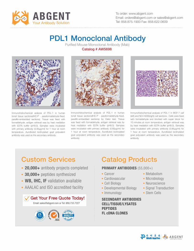
Immunohistochemical analysis of PDL-1 in human tonsil tissue sections(IHC-P - paraformaldehyde-fixed, paraffin-embedded sections). Tissue was fixed with formaldehyde; antigen retrieval was by heat mediation with EDTA buffer (pH9.0). Samples were incubated with primary antibody (0.85µg/ml) for 1 hour at room temperature. Aundiluted biotinylated goat polyvalent antibody was used as the secondary antibody.
Immunohistochemical analysis of PDL-1 in human tonsil tissue sections(IHC-P - paraformaldehyde-fixed, paraffin-embedded sections) by Dako test. Tissue was fixed with formaldehyde; antigen retrieval was by heat mediation with EDTA buffer (pH9.0). Samples were incubated with primary antibody (0.85µg/ml) for 1 hour at room temperature. Aundiluted biotinylated goat polyvalent antibody was used as the secondary antibody.
Immunohistochemical analysis of PDL-1 in MCF-7 cell (left) and NCI-H226(right) cell sections . Cells were fixed with formaldehyde and blocked with super block for 10 minutes at room temperature; antigen retrieval was by heat mediation with EDTA buffer (pH9.0). Samples were incubated with primary antibody (0.85µg/ml) for 1 hour at room temperature. Aundiluted biotinylated goat polyvalent antibody was used as the secondary antibody.
Purified Mouse Monoclonal Antibody (Mab) Catalog # AW5698
PDL1 Monoclonal Antibody