Embed Size (px)
Citation preview
journal of orthopaedic & sports physical therapy | volume 40 | number 2 | february 2010 | 103
[ case report ]
Edwards14 defines piriformis syndrome as neuritis of branches of the sciatic nerve caused by pressure of an injured or irri-tated piriformis muscle. Symptoms asso-ciated with piriformis syndrome typically consist of buttock pain that radiates into the hip, posterior aspect of the thigh, and the proximal portion of the lower leg.28,29 In general, pain increases with sitting or squatting, but persons with piriformis syndrome may experience difficulty with walking or other functional activities.12,29 Piriformis syndrome typically does not result in neurological deficits such as de-creased deep tendon reflexes and myoto-mal weakness.29
Several authors attribute piriformis syndrome to a shortening or “spasm” of the piriformis that results in compression of the sciatic nerve.6,10,15,27,29,33 The cause of spasm of the piriformis muscle has been most attributed to direct trauma, postsur-gical injury, lumbar and sacroiliac joint pathologies, and overuse.15-17,27,29 Given as such, the standard treatment for piri-formis syndrome has focused on decreas-ing spasm or shortening of the piriformis muscle and any associated inflamma-tion. Medical management of piriformis
The piriformis originates from the anterior aspect of sacral vertebrae 2 through 4 and inserts on the greater trochanter. The sciatic nerve typically exits the greater sciatic notch just below the inferior border of the piriformis muscle.
In about 7% to 21% of studied populations, the sciatic nerve (or a division of it), actually penetrates the muscle.3,12,15,30,32,33
t STUDY DESIGN: Case report.
t OBJECTIVE: To describe an alternative treat-ment approach for piriformis syndrome using a hip muscle strengthening program with movement reeducation.
t BACKGROUND: Interventions for piriformis syndrome typically consist of stretching and/or soft tissue massage to the piriformis muscle. The premise underlying this approach is that a short-ening or “spasm” of the piriformis is responsible for the compression placed upon the sciatic nerve.
t CASE DESCRIPTION: The patient was a 30-year-old male with right buttock and posterior thigh pain for 2 years. Clinical findings upon examination included reproduction of symptoms with palpation and stretching of the piriformis. Movement analysis during a single-limb step-down revealed excessive hip adduction and internal rota-tion, which reproduced his symptoms. Strength assessment revealed weakness of the right hip abductor and external rotator muscles. The pa-tient’s treatment was limited to hip-strengthening
exercises and movement reeducation to correct the excessive hip adduction and internal rotation during functional tasks.
t OUTCOMES: Following the intervention, the patient reported 0/10 pain with all activities. The initial Lower Extremity Functional Scale question-naire score of 65/80 improved to 80/80. Lower extremity kinematics for peak hip adduction and internal rotation improved from 15.9° and 12.8° to 5.8° and 5.9°, respectively, during a step-down task.
t DISCUSSION: This case highlights an alterna-tive view of the pathomechanics of piriformis syndrome (overstretching as opposed to over-shortening) and illustrates the need for functional movement analysis as part of the examination of these patients.
t LEVEL OF EVIDENCE: Therapy, level 4. J Orthop Sports Phys Ther 2010;40(2):103-111. doi:10.2519/jospt.2010.3108
t KEY WORDS: biomechanics, gluteus, hip pain, radiculopathy, sciatica
1 Physical Therapist, Department of Physical Medicine and Rehabilitation, Kaiser Permanente West Los Angeles, Los Angeles, CA. 2 Physical Therapist, Department of Physical Medicine and Rehabilitation, Kaiser Permanente Los Angeles, Los Angeles, CA. 3 Assistant Professor, Department of Physical Therapy, University of Pittsburgh, Pittsburgh, PA. 4 Associate Professor and Co-Director, Musculoskeletal Biomechanics Research Laboratory, Division of Biokinesiology and Physical Therapy, University of Southern California, Los Angeles, CA. Jason Tonley and Steven Yun completed this case report as a requirement for the Kaiser Permanente Los Angeles Movement Science Fellowship. Dr. Powers acknowledges a financial interest in the SERF Strap that was used as part of this case report. Address correspondence to Dr Christopher M. Powers, Division of Biokinesiology and Physical Therapy, University of Southern California, 1540 E Alcazar St, CHP-155, Los Angeles, CA 90089-9006. E-mail: [email protected]
JASON C. TONLEY, DPT, OCS1 • Steven M. Yun, MPT, OCS1 • RONALD J. KOChEVAR, DPT, OCS2 JEREMY A. DYE, MPT, OCS2 • Shawn Farrokhi, PT, PhD, DPT3 • ChriStopher M. powerS, PT, PhD4
Treatment of an Individual With Piriformis Syndrome Focusing on Hip Muscle Strengthening and Movement
Reeducation: A Case Report
40-02 Tonley_folio.indd 103 1/20/10 4:10:09 PM
104 | february 2010 | volume 40 | number 2 | journal of orthopaedic & sports physical therapy
[ case report ]syndrome is reported in the literature to include injections,1,3,5,8,11,15,16,26,27,30 prescrip-tion of nonsteroidal anti-inflammatory drugs and muscle relaxants,1,5,8,11,15,16,26,27 surgical release,1,5,8,11,14,16 and referral to physical therapy.1,5,11,15,20,27,30 The most commonly reported physical therapy interventions include ultrasound,1,5,11,16,19 soft tissue mobilization,1,5,11,15,16,19,30 piri-formis stretching,1,11,15,16,19,26,27,30 hot packs or cold spray,5,11,15,16,19 and various lumbar spine treatments.1,5,8,10,13,26,30
As noted above, a common assumption guiding physical therapy intervention for piriformis syndrome is that the piriformis is shortened or in spasm, creating com-pression of the sciatic nerve. Our alter-nate theory is that the piriformis muscle may be functioning in an elongated posi-tion or subjected to high eccentric loads during functional activities secondary to weak agonist muscles. For example, if the hip excessively adducts and internally ro-tates during weight-bearing tasks due to weakness of the gluteus maximus and/or gluteus medius, a greater eccentric load may be shifted to the piriformis muscle. Perpetual loading of the piriformis mus-cle through overlengthening and eccen-tric demand may result in sciatic nerve compression or irritation.
Interestingly, many authors have rec-ognized hip abductor weakness as an associated finding with piriformis syn-drome.1,2,3,5,8,13,17,27,32 Yet only 2 of these reports included hip abduction strength-ening as part of the treatment program,1,17 with 1 of the 2 authors noting that hip abduction exercises “seemed to hasten recovery.”17 Therefore, a treatment pro-gram addressing hip strength and move-ment reeducation to control the femur in the frontal and transverse planes during functional activities may play a role in the treatment of patients with piriformis syn-drome who demonstrate excessive frontal and transverse plane motions at the hip.
The purpose of this case report was to describe an alternative treatment ap-proach for piriformis syndrome that em-phasizes hip muscle strengthening and movement reeducation. The patient in
this case had symptoms consistent with piriformis syndrome, weak hip abductors and external rotators, and excessive hip adduction and internal rotation during functional lower extremity activities.
CASE DESCRIPTION
General Demographics
The patient was a 30-year-old male who worked as a real estate agent. He also reported that he was
a part-time tennis instructor and partici-pated in a weekly basketball league. Apart from his current symptoms, the patient also had a 15-year history of intermittent low back pain.
history of Presenting ConditionThe patient was initially seen by an or-thopaedic surgeon and given a diagno-sis of sciatica. At this visit, radiographs were obtained of his lumbar spine and hip, which revealed no abnormalities. The patient was subsequently referred to physical therapy.
The patient presented to physical therapy with a 2-year history of deep right buttock pain that radiated to the posterior thigh. The patient stated that the onset of his pain was insidious and denied any trauma that contributed to his current symptoms. The patient did not report any prior treatment for his but-tock and thigh pain, but stated that he received intermittent chiropractic treat-ment for his low back symptoms.
Presenting ComplaintsThe patient reported that his symptoms were exaggerated by playing basketball or tennis for 30 to 60 minutes. Stairs and squatting activities also were noted as aggravating activities. He reported that his pain was alleviated when he stopped participating in sports, but that it would be 4 to 6 days before symptoms would completely resolve. The patient’s current activity level included participation in a weekly basketball league and teaching tennis 6 to 12 hours per week. He report-ed having to modify his tennis instruction
to avoid running activities. The patient’s symptoms were limiting the number of tennis lessons he could give per week and his ability to play basketball. The pa-tient’s stated goals were to participate in his weekly basketball league and return to his normal regime of tennis lessons of 3 nights per week for 2 to 4 hours.
Test and MeasuresPain and Functional Status The patient completed a visual analog scale (VAS), where 0 is no pain and 10 is the most pain possible, to assess his current level of pain.34 The patient’s baseline pain in his buttock and posterior thigh was 3/10 and reached a level of 9/10 after participation in 1 game of basketball. Prior to treat-ment, the patient completed the Lower Extremity Functional Scale to evaluate his functional status with regards to his buttock and posterior thigh symptoms. This self-assessment functional tool has been shown to be valid and reliable.4 The patient’s score on the Lower Extrem-ity Functional Scale Questionnaire was 65/80, with 80 representing maximum function.Differential Diagnosis Screening Active and passive examination of the lumbar spine and sacroiliac joint were performed to rule out spine and pelvic contributions to his symptoms. Active examination tests for the lumbar spine consisted of ac-tive range of motion (AROM), followed by AROM with overpressure in flexion, extension, and sidebending and rotation to the left and right. Passive testing was performed using both central and lateral posterior-to-anterior spring tests from the fifth lumbar to the tenth thoracic spi-nal segments. There was no reproduction of his buttock or thigh symptoms with ac-tive or passive testing to the lumbar spine. The sacroiliac joint was assessed using a cluster of tests, as described by Laslett.21 All tests were negative with respect to the reproduction of symptoms.Hip Joint Examination Passive mo-tion assessment of the hip joint revealed normal ranges for hip flexion, internal rotation, and external rotation without
40-02 Tonley_folio.indd 104 1/20/10 4:10:10 PM
journal of orthopaedic & sports physical therapy | volume 40 | number 2 | february 2010 | 105
reproduction of symptoms. Hip flexor muscle length was assessed using the Thomas test.18 No restrictions were noted when the 1-joint hip flexor was assessed (ie, iliopsoas); however, restrictions (–12°) were evident when evaluating the 2-joint hip flexor (ie, rectus femoris).
Clinical tests were performed to as-sess for intra-articular hip joint pathol-ogy. The scour test, hip quadrant test, log roll test, FABER, and active straight leg test23 were performed, and all were nega-tive with respect to reproduction of the patient’s symptoms. Clinical tests also were performed to assess for piriformis syndrome. These tests included the piri-formis stretch test above and below 60° of hip flexion,2,5,15,16 the flexion/adduc-tion/internal rotation (FAIR) test,5,15,16 and piriformis contraction test.2 The piriformis stretch test below 60° was performed by maximally adducting and internally rotating the hip, with the hip flexed to 45°. The piriformis stretch test above 60° was performed by maximally adducting and externally rotating the hip, with the hip flexed to 90°. The FAIR test was performed by placing the patient in a sidelying position on the unaffected side, with the affected (superior) hip being moved passively into flexion, adduction, and internal rotation.15,16 The piriformis contraction test was performed by plac-ing the patient in the FAIR test position and having the patient elevate the knee off the table for 5 seconds.2 The patient was found to have reproduction of but-tock and thigh symptoms with both of the piriformis stretch tests, the FAIR test, and the piriformis contraction test.
Neurodynamic testing, as described by Butler,7 was performed to assess the sci-atic nerve. Reproduction of buttock pain occurred at 50° of hip flexion during the straight leg raise test. Assessment of the straight leg raise test with ankle dorsi-flexion resulted in reproduction of lower back, buttock, and posterior thigh pain, with only 10° of hip flexion. Symptoms resolved with ankle plantar flexion.
Soft tissue palpation revealed tender-ness of the piriformis muscle and tro-
chanteric bursa. Palpation also resulted in the reproduction of buttock and pos-terior thigh pain.Muscle Strength Manual muscle testing of the hip musculature was performed as described by Kendall et al.18 Hip ex-tensor strength was tested in the modi-fied position, due to hip flexor tightness. Manual muscle test grades of 3+/5, 3+/5, and 4–/5 were given for the hip exten-sors, hip abductors, and external rotators, respectively.Dynamic Assessment An observational gait analysis was performed as the pa-
tient walked at a self-selected pace. The patient demonstrated decreased hip ex-tension bilaterally in terminal stance. He also demonstrated abnormal movements in both the frontal and transverse planes during the stance phase on the right: (a) right trunk lean during single-limb sup-port, (b) increased hip adduction dur-ing loading response through terminal stance, (c) increased hip internal rotation during weight acceptance through termi-nal stance, and (d) contralateral pelvic drop during single-limb support.
The patient also was evaluated while
FIGURE 1. Patient performing the step-down maneuver pretreatment (A) and posttreatment (B). Both photos were taken at the same knee flexion angle (as assessed by motion analysis). Pretreatment, the patient demonstrates a greater amount of hip internal rotation and adduction and contralateral hip drop.
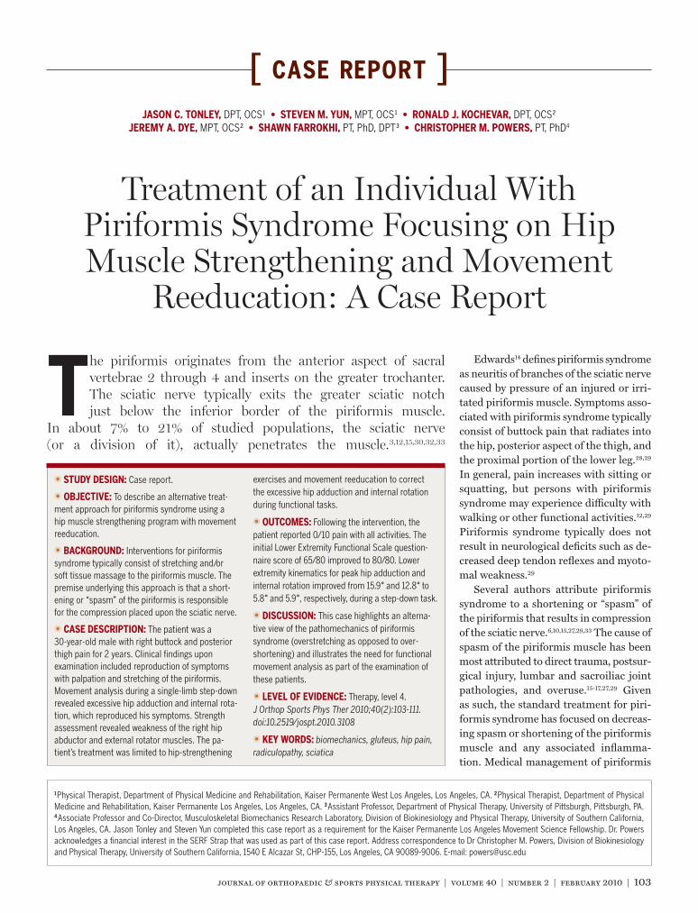
FIGURE 2. The SERF Strap consist of a thin, elastic material that is secured to the proximal aspect of the leg, wraps in a spiral fashion around the thigh, and is anchored around the pelvis. The line of action of the SERF Strap pulls the hip into external rotation.
40-02 Tonley_folio.indd 105 1/20/10 4:10:13 PM
106 | february 2010 | volume 40 | number 2 | journal of orthopaedic & sports physical therapy
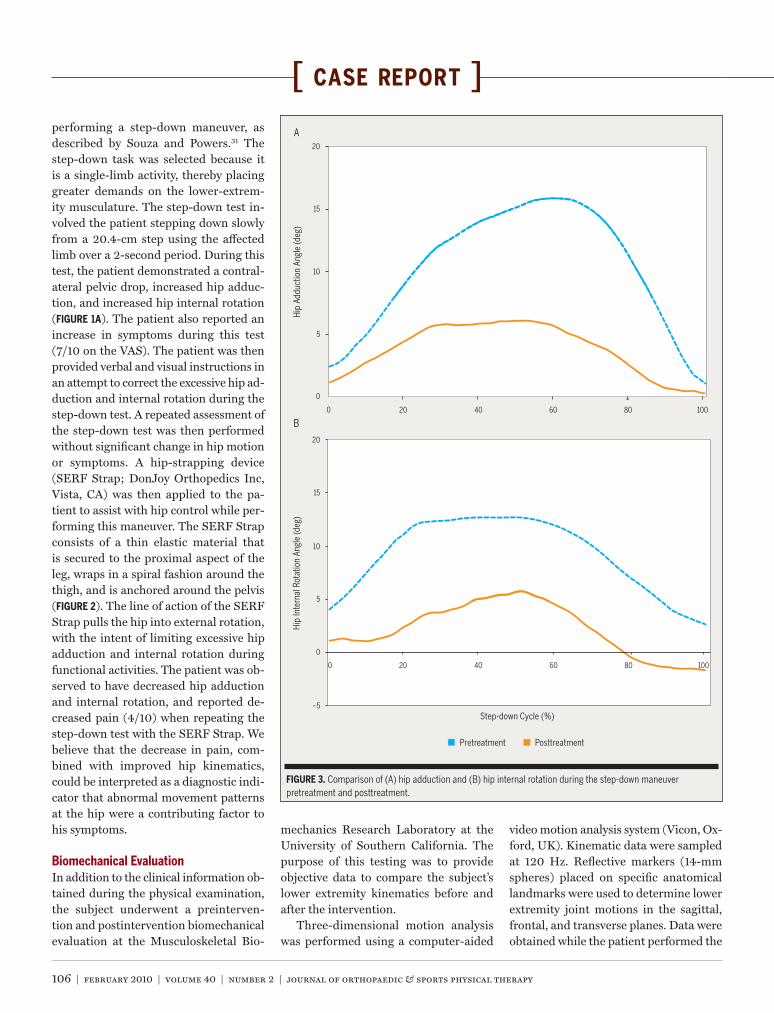
[ case report ]performing a step-down maneuver, as described by Souza and Powers.31 The step-down task was selected because it is a single-limb activity, thereby placing greater demands on the lower-extrem-ity musculature. The step-down test in-volved the patient stepping down slowly from a 20.4-cm step using the affected limb over a 2-second period. During this test, the patient demonstrated a contral-ateral pelvic drop, increased hip adduc-tion, and increased hip internal rotation (FIGURE 1A). The patient also reported an increase in symptoms during this test (7/10 on the VAS). The patient was then provided verbal and visual instructions in an attempt to correct the excessive hip ad-duction and internal rotation during the step-down test. A repeated assessment of the step-down test was then performed without significant change in hip motion or symptoms. A hip-strapping device (SERF Strap; DonJoy Orthopedics Inc, Vista, CA) was then applied to the pa-tient to assist with hip control while per-forming this maneuver. The SERF Strap consists of a thin elastic material that is secured to the proximal aspect of the leg, wraps in a spiral fashion around the thigh, and is anchored around the pelvis (FIGURE 2). The line of action of the SERF Strap pulls the hip into external rotation, with the intent of limiting excessive hip adduction and internal rotation during functional activities. The patient was ob-served to have decreased hip adduction and internal rotation, and reported de-creased pain (4/10) when repeating the step-down test with the SERF Strap. We believe that the decrease in pain, com-bined with improved hip kinematics, could be interpreted as a diagnostic indi-cator that abnormal movement patterns at the hip were a contributing factor to his symptoms.
Biomechanical EvaluationIn addition to the clinical information ob-tained during the physical examination, the subject underwent a preinterven-tion and postintervention biomechanical evaluation at the Musculoskeletal Bio-
mechanics Research Laboratory at the University of Southern California. The purpose of this testing was to provide objective data to compare the subject’s lower extremity kinematics before and after the intervention.
Three-dimensional motion analysis was performed using a computer-aided
video motion analysis system (Vicon, Ox-ford, UK). Kinematic data were sampled at 120 Hz. Reflective markers (14-mm spheres) placed on specific anatomical landmarks were used to determine lower extremity joint motions in the sagittal, frontal, and transverse planes. Data were obtained while the patient performed the
10
15
20
0
5
Hip
Add
uctio
n An
gle
(deg
)
0 20 40 60 80 100
10
15
20
0
5
Hip
Inte
rnal
Rot
atio
n An
gle
(deg
)
0 20 40 60 80 100
–5Step-down Cycle (%)
Pretreatment Posttreatment
A
B
FIGURE 3. Comparison of (A) hip adduction and (B) hip internal rotation during the step-down maneuver pretreatment and posttreatment.
40-02 Tonley_folio.indd 106 1/20/10 4:10:15 PM
journal of orthopaedic & sports physical therapy | volume 40 | number 2 | february 2010 | 107
step-down maneuver. The subject was in-structed to stand on his right lower ex-tremity while performing a step-down in 2 seconds and to return to the starting position in 2 seconds. The depth of the step for the step-down maneuver was set at 18 cm (approximately 10% of the patient’s total body height). The speed of the movement was guided with a metro-nome set at 60 beats per minute. When averaged across 5 repetitions, peak hip adduction and internal rotation were 15.9° and 12.8°, respectively (FIGURE 3). These values were considered excessive, based on the normative data previously published by Souza and Powers.31
AssessmentGiven the subjective and objective in-formation obtained during our exami-nation, it was our impression that the patient demonstrated significant impair-ments specific to the hip region. More specifically, our patient presented with weakness of the hip extensors, abductors, and external rotators, limited control of the hip and pelvis during functional movement testing, and reproduction of symptoms with passive stretching and activation of the piriformis muscle. We theorized that weakness of the gluteus maximus and gluteus medius was con-tributing to abnormal movement pat-terns at the hip, thereby subjecting the piriformis to excessive lengthening or eccentric loading during functional ac-tivities. In turn, we believed that the ex-cessive lengthening of the piriformis was compressing the sciatic nerve. The fact that the application of the SERF Strap resulted in a simultaneous improvement in hip motion and buttock pain sup-ported our hypothesis. Therefore, it was our belief that an intervention focused on addressing the hip muscle weakness and abnormal movement patterns would alleviate the patient’s pain and improve his functional status.
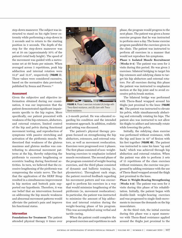
InterventionFoundations for Treatment The patient attended physical therapy 8 times over
a 3-month period. He was educated re-garding his condition and the intended treatment approach. In addition, realistic goal setting was discussed.
The patient’s physical therapy pro-gram focused on strengthening the hip abductors, extensors, and external rota-tors, as well as movement reeducation. Exercises were progressed over 3 phases. The first phase consisted of non–weight-bearing exercises to emphasize isolated muscle recruitment. The second phase of the program consisted of weight-bearing exercises, and the third phase consisted of dynamic and ballistic training (ie, plyometrics). Throughout each stage, the patient received feedback regarding his movement pattern and was encour-aged to perform his exercises in a way that would minimize lengthening of the piriformis (ie, movement reeducation). In particular, the patient was instructed to minimize the amount of hip adduc-tion and internal rotation during the weight-bearing phase of his program. This was accomplished using verbal and tactile cueing.
When the patient could complete the proposed exercises and repetitions in each
phase, the program would progress to the next phase. The patient was given a home exercise program that he was instructed to perform once a day. The home exercise program paralleled the exercises given in the clinic. The patient was instructed to perform all exercises in a manner that would not reproduce his symptoms.Phase 1: Isolated Muscle Recruitment (Weeks 0-4) The patient was seen for 2 visits during this period. He was given 2 exercises: bilateral bridging to target his hip extensors and sidelying clams to tar-get his hip abductors and external rota-tors. For all exercises during this phase the patient was instructed to emphasize motion at the hip joint and to avoid ex-cessive pelvis/trunk motion.
The bilateral bridge was performed with Thera-Band wrapped around his thighs just proximal to the knee (FIGURE
4A). The patient was instructed to elevate his pelvis, while simultaneously abduct-ing and externally rotating his hips. The patient also was instructed to not allow his thighs to adduct and internally rotate while lowering the pelvis.
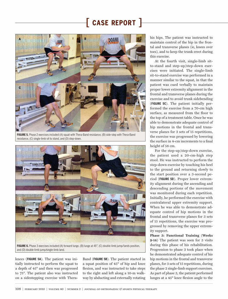
Initially, the sidelying clam exercise was performed without resistance, with the hip and knee in 45° of flexion with his feet together (FIGURE 4B). The patient was instructed to raise his knee “up and back,” which was achieved through hip abduction and external rotation. When the patient was able to perform 3 sets of 15 repetitions of the clam exercises without resistance, the exercise was pro-gressed by adding resistance with the use of Thera-Band wrapped around the thigh just proximal to the knee.Phase 2: Weight-Bearing Strengthening (Weeks 4-9) The patient was seen for 3 visits during this phase of his rehabili-tation. Initially, the patient began with double-limb weight-bearing exercises and was progressed to single-limb move-ments to increase the demands on the hip musculature.
At the third visit, the initial exercise during this phase was a squat maneu-ver with Thera-Band resistance applied around the thighs just proximal to the
FIGURE 4. Phase 1 exercises included (A) bridge with Thera-Band resistance, and (B) clam with Thera-Band resistance.
40-02 Tonley_folio.indd 107 1/20/10 4:10:16 PM
108 | february 2010 | volume 40 | number 2 | journal of orthopaedic & sports physical therapy
[ case report ]
knees (FIGURE 5A). The patient was ini-tially instructed to perform the squat to a depth of 45° and then was progressed to 75°. The patient also was instructed on a sidestepping exercise with Thera-
Band (FIGURE 5B). The patient started in a squat position of 45° of hip and knee flexion, and was instructed to take steps to the right and left along a 10-m walk-way by abducting and externally rotating
his hips. The patient was instructed to maintain control of the hip in the fron-tal and transverse planes (ie, knees over toes), and to keep the trunk erect during this exercise.
At the fourth visit, single-limb sit-to-stand and step-up/step-down exer-cises were initiated. The single-limb sit-to-stand exercise was performed in a manner similar to the squat, in that the patient was cued verbally to maintain proper lower extremity alignment in the frontal and transverse planes during the exercise and to avoid trunk sidebending (FIGURE 5C). The patient initially per-formed the exercise from a 70-cm high surface, as measured from the floor to the top of a treatment table. Once he was able to demonstrate adequate control of hip motions in the frontal and trans-verse planes for 3 sets of 15 repetitions, the exercise was progressed by lowering the surface in 4-cm increments to a final height of 58 cm.
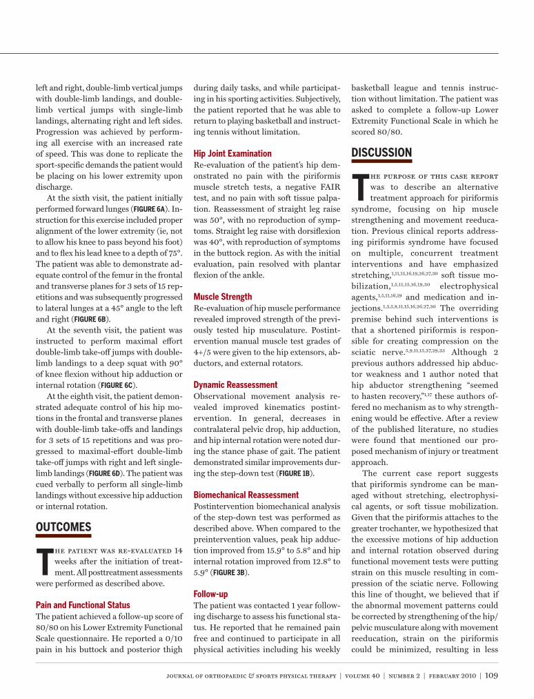
For the step-up/step-down exercise, the patient used a 20-cm-high step stool. He was instructed to perform the step-down exercise by touching his heel to the ground and returning slowly to the start position over a 3-second pe-riod (FIGURE 5D). Proper lower extrem-ity alignment during the ascending and descending portions of the movement was monitored during each repetition. Initially, he performed the exercise with contralateral upper extremity support. When he was able to demonstrate ad-equate control of hip motions in the frontal and transverse planes for 3 sets of 15 repetitions, the exercise was pro-gressed by removing the upper extrem-ity support.Phase 3: Functional Training (Weeks 9-14) The patient was seen for 3 visits during this phase of his rehabilitation. Progression to phase 3 took place when he demonstrated adequate control of his hip motions in the frontal and transverse planes, for 3 sets of 15 repetitions, during the phase 2 single-limb support exercises. As part of phase 3, the patient performed lunges at a 45° knee flexion angle to the
FIGURE 5. Phase 2 exercises included (A) squat with Thera-Band resistance, (B) side-step with Thera-Band resistance, (C) single-limb sit to stand, and (D) step-down.
FIGURE 6. Phase 3 exercises included (A) forward lunge, (B) lunge at 45°, (C) double-limb jump/lands position, and (D) double-limb jump/single-limb land.
40-02 Tonley_folio.indd 108 1/20/10 4:10:19 PM
journal of orthopaedic & sports physical therapy | volume 40 | number 2 | february 2010 | 109
left and right, double-limb vertical jumps with double-limb landings, and double-limb vertical jumps with single-limb landings, alternating right and left sides. Progression was achieved by perform-ing all exercise with an increased rate of speed. This was done to replicate the sport-specific demands the patient would be placing on his lower extremity upon discharge.
At the sixth visit, the patient initially performed forward lunges (FIGURE 6A). In-struction for this exercise included proper alignment of the lower extremity (ie, not to allow his knee to pass beyond his foot) and to flex his lead knee to a depth of 75°. The patient was able to demonstrate ad-equate control of the femur in the frontal and transverse planes for 3 sets of 15 rep-etitions and was subsequently progressed to lateral lunges at a 45° angle to the left and right (FIGURE 6B).
At the seventh visit, the patient was instructed to perform maximal effort double-limb take-off jumps with double-limb landings to a deep squat with 90° of knee flexion without hip adduction or internal rotation (FIGURE 6C).
At the eighth visit, the patient demon-strated adequate control of his hip mo-tions in the frontal and transverse planes with double-limb take-offs and landings for 3 sets of 15 repetitions and was pro-gressed to maximal-effort double-limb take-off jumps with right and left single-limb landings (FIGURE 6D). The patient was cued verbally to perform all single-limb landings without excessive hip adduction or internal rotation.
OUTCOMES
The patient was re-evaluated 14 weeks after the initiation of treat-ment. All posttreatment assessments
were performed as described above.
Pain and Functional StatusThe patient achieved a follow-up score of 80/80 on his Lower Extremity Functional Scale questionnaire. He reported a 0/10 pain in his buttock and posterior thigh
during daily tasks, and while participat-ing in his sporting activities. Subjectively, the patient reported that he was able to return to playing basketball and instruct-ing tennis without limitation.
hip Joint ExaminationRe-evaluation of the patient’s hip dem-onstrated no pain with the piriformis muscle stretch tests, a negative FAIR test, and no pain with soft tissue palpa-tion. Reassessment of straight leg raise was 50°, with no reproduction of symp-toms. Straight leg raise with dorsiflexion was 40°, with reproduction of symptoms in the buttock region. As with the initial evaluation, pain resolved with plantar flexion of the ankle.
Muscle StrengthRe-evaluation of hip muscle performance revealed improved strength of the previ-ously tested hip musculature. Postint-ervention manual muscle test grades of 4+/5 were given to the hip extensors, ab-ductors, and external rotators.
Dynamic ReassessmentObservational movement analysis re-vealed improved kinematics postint-ervention. In general, decreases in contralateral pelvic drop, hip adduction, and hip internal rotation were noted dur-ing the stance phase of gait. The patient demonstrated similar improvements dur-ing the step-down test (FIGURE 1B).
Biomechanical ReassessmentPostintervention biomechanical analysis of the step-down test was performed as described above. When compared to the preintervention values, peak hip adduc-tion improved from 15.9° to 5.8° and hip internal rotation improved from 12.8° to 5.9° (FIGURE 3B).
Follow-upThe patient was contacted 1 year follow-ing discharge to assess his functional sta-tus. He reported that he remained pain free and continued to participate in all physical activities including his weekly
basketball league and tennis instruc-tion without limitation. The patient was asked to complete a follow-up Lower Extremity Functional Scale in which he scored 80/80.
DISCUSSION
The purpose of this case report was to describe an alternative treatment approach for piriformis
syndrome, focusing on hip muscle strengthening and movement reeduca-tion. Previous clinical reports address-ing piriformis syndrome have focused on multiple, concurrent treatment interventions and have emphasized stretching,1,11,15,16,19,26,27,30 soft tissue mo-bilization,1,5,11,15,16,19,30 electrophysical agents,1,5,11,16,19 and medication and in-jections.1,3,5,8,11,15,16,26,27,30 The overriding premise behind such interventions is that a shortened piriformis is respon-sible for creating compression on the sciatic nerve.5,9,11,15,27,29,33 Although 2 previous authors addressed hip abduc-tor weakness and 1 author noted that hip abductor strengthening “seemed to hasten recovery,”1,17 these authors of-fered no mechanism as to why strength-ening would be effective. After a review of the published literature, no studies were found that mentioned our pro-posed mechanism of injury or treatment approach.
The current case report suggests that piriformis syndrome can be man-aged without stretching, electrophysi-cal agents, or soft tissue mobilization. Given that the piriformis attaches to the greater trochanter, we hypothesized that the excessive motions of hip adduction and internal rotation observed during functional movement tests were putting strain on this muscle resulting in com-pression of the sciatic nerve. Following this line of thought, we believed that if the abnormal movement patterns could be corrected by strengthening of the hip/pelvic musculature along with movement reeducation, strain on the piriformis could be minimized, resulting in less
40-02 Tonley_folio.indd 109 1/20/10 4:10:20 PM
110 | february 2010 | volume 40 | number 2 | journal of orthopaedic & sports physical therapy
[ case report ]compression on the sciatic nerve. By ad-dressing the proposed functional cause of the piriformis irritation, as opposed to focusing on the muscle itself, we were able to achieve a long-term successful outcome for this patient.
The intervention described in this case report focused on functional ex-ercises aimed at strengthening the hip extensors, abductors, and external ro-tators, as well as correction of faulty movement patterns. In this respect, we feel that the key muscle was the gluteus maximus. Apart from being a strong hip extensor and external rotator, the up-per half of this muscle also functions as a hip abductor.22 Given that the gluteus maximus is a primary external rotator of the hip,26 we feel that improved per-formance of this muscle served 2 func-tions: (1) to decrease the demand on the piriformis through agonist activity and (2) to prevent hip motion that would cause increased strain on the piriformis. At discharge, observed improvements in hip strength corresponded to improve-ments in hip kinematics. In particular, kinematic data revealed a reduction in the amount of hip internal rotation (6.9°) and hip adduction (10.1°) with the step-down test. In addition, the patient demonstrated a 30° improvement in the straight leg raise test with ankle dorsi-flexion (10°-40°). We speculate that this may have been the result of decreased ir-ritability of the sciatic nerve.
Our patient reached his goal of return-ing to his prior level of sport involvement with no complaints of pain during or af-ter his basketball games or tennis. In ad-dition to the cessation of his buttock and thigh pain, the patient reported full reso-lution of his low back pain, a compelling outcome that may or may not have any correlation to his increase in functional hip stability.
Despite these outcomes, care must be taken in establishing cause and ef-fect based on a single case report. That being stated, the likelihood of post-treatment outcomes being the result of spontaneous time-related recovery are
unlikely, given the chronicity of the pa-tient’s symptoms. Although we hypothe-size that improved gluteus maximus and gluteus medius strength and movement reeducation were the primary reasons underlying the clinical outcomes noted in this case report, it is possible that the patient’s improvements were the result of an increase in piriformis strength, thereby allowing this muscle to better withstand the loads placed upon it dur-ing functional activities. Furthermore, care must be taken in assuming the changes observed in our patient were due solely to increases in hip muscle strength. First, strength assessments were made using subjective measures of muscle performance testing as op-posed to more objective methods (ie, dynamometry). Second, a recent study by Mizner and colleagues suggests that changes in movement patterns may be independent of muscle strength.25 As such, future research should be directed at better understanding the relationship between muscle performance deficits and altered movement patterns in rela-tionship to piriformis syndrome.
CONCLUSION
This case report describes the management of a patient with complaints and findings consis-
tent with piriformis syndrome, who responded favorably to an intervention program focused on strengthening of the hip musculature and correction of faulty lower extremity movement pat-terns. Clinically relevant improvements were observed without treatment strate-gies commonly used to treat piriformis syndrome (stretching, soft tissue mobi-lization, injections). Therefore, physi-cal therapy interventions focusing on strengthening of hip musculature to reduce excessive hip motions may be in-dicated for patients presenting with piri-formis syndrome. Despite the outcomes presented in this case report, care must be taken in establishing cause and effect based on a single patient. t
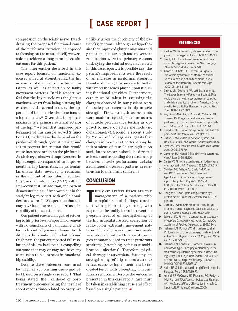
REFERENCES
1. Barton PM. Piriformis syndrome: a rational ap-proach to management. Pain. 1991;47:345-352.
2. Beatty RA. The piriformis muscle syndrome: a simple diagnostic maneuver. Neurosurgery. 1994;34:512-514; discussion 514.
3. Benzon HT, Katz JA, Benzon HA, Iqbal MS. Piriformis syndrome: anatomic consider-ations, a new injection technique, and a review of the literature. Anesthesiology. 2003;98:1442-1448.
4. Binkley JM, Stratford PW, Lott SA, Riddle DL. The Lower Extremity Functional Scale (LEFS): scale development, measurement properties, and clinical application. North American Ortho-paedic Rehabilitation Research Network. Phys Ther. 1999;79:371-383.
5. Boyajian-O’Neill LA, McClain RL, Coleman MK, Thomas PP. Diagnosis and management of piriformis syndrome: an osteopathic approach. J Am Osteopath Assoc. 2008;108:657-664.
6. Broadhurst N. Piriformis syndrome and buttock pain. Aust Fam Physician. 1990;19:1754.
7. Butler D. The Sensitive Nervous System. Ad-elaide, Australia: NOI Group Publications; 2000.
8. Byrd JW. Piriformis syndrome. Oper Tech Sports Med. 2005;13:71-79.
9. Cameron HU, Noftal F. The piriformis syndrome. Can J Surg. 1988;31:210.
10. Carter AT. Piriformis syndrome: a hidden cause of sciatic pain. Athl Training. 1988;23:243-245.
11. Childers MK, Wilson DJ, Gnatz SM, Con-way RR, Sherman AK. Botulinum toxin type A use in piriformis muscle syndrome: a pilot study. Am J Phys Med Rehabil. 2002;81:751-759. http://dx.doi.org/10.1097/01.PHM.0000027426.98000.57
12. Douglas S. Sciatic pain and piriformis syn-drome. Nurse Pract. 1997;22:166-168, 170, 172 passim.
13. Durrani Z, Winnie AP. Piriformis muscle syn-drome: an underdiagnosed cause of sciatica. J Pain Symptom Manage. 1991;6:374-379.
14. Edwards FO. Piriformis syndrome. In: Academy of Applied Osteopathy Yearbook. Carmel, CA: Academy of Applied Osteopathy; 1962:39-41.
15. Fishman LM, Dombi GW, Michaelsen C, et al. Piriformis syndrome: diagnosis, treatment, and outcome--a 10-year study. Arch Phys Med Reha-bil. 2002;83:295-301.
16. Fishman LM, Konnoth C, Rozner B. Botulinum neurotoxin type B and physical therapy in the treatment of piriformis syndrome: a dose-find-ing study. Am J Phys Med Rehabil. 2004;83:42-50; quiz 51-43. http://dx.doi.org/10.1097/01.PHM.0000104669.86076.30
17. Hallin RP. Sciatic pain and the piriformis muscle. Postgrad Med. 1983;74:69-72.
18. Kendall FP, McCreary EK, Provance PG, Rodgers MM, Romani WA. Muscles: Testing and Function with Posture and Pain. 5th ed. Baltimore, MD: Lippincott, Williams, & Wilkins; 2005.
40-02 Tonley_folio.indd 110 1/20/10 4:10:22 PM
journal of orthopaedic & sports physical therapy | volume 40 | number 2 | february 2010 | 111
@ MORE INFORMATIONwww.jospt.org
MC. Diagnosis and management of posttrau-matic piriformis syndrome: a case study. J Manipulative Physiol Ther. 2006;29:486-491. http://dx.doi.org/10.1016/j.jmpt.2006.06.006
25. Mizner RL, Kawaguchi JK, Chmielewski TL. Muscle strength in the lower extremity does not predict postinstruction improvements in the landing patterns of female athletes. J Orthop Sports Phys Ther. 2008;38:353-361. http://dx.doi.org/10.2519/jospt.2008.2726
26. Neumann DA. Kinesiology of the Musculoskel-etal System: Foundations for Physical Rehabili-tation. St Louis, MO: Mosby; 2002.
27. Pace JB, Nagle D. Piriformis syndrome. Western J Med. 1976;124:435-439.
28. Papadopoulos SM, McGillicuddy JE, Albers JW. Unusual cause of ‘piriformis muscle syndrome’. Arch Neurol. 1990;47:1144-1146.
29. Parziale JR, Hudgins TH, Fishman LM. The piri-formis syndrome: a review paper. Am J Orthop. 1996;25:819-823.
30. Retzlaff EW, Berry AH, Haight AS, et al. The
piriformis muscle syndrome. J Am Osteopath Assoc. 1974;73:799-807.
31. Souza RB, Powers CM. Differences in hip kine-matics, muscle strength, and muscle activation between subjects with and without patellofemo-ral pain. J Orthop Sports Phys Ther. 2009;39:12-19. http://dx.doi.org/10.2519/jospt.2009.2885
32. Steiner C, Staubs C, Ganon M, Buhlinger C. Piri-formis syndrome: pathogenesis, diagnosis, and treatment. J Am Osteopath Assoc. 1987;87:318-323.
33. TePoorten BA. The piriformis muscle. J Am Os-teopath Assoc. 1969;69:150-160.
34. Williamson A, Hoggart B. Pain: a review of three commonly used pain rating scales. J Clin Nurs. 2005;14:798-804. http://dx.doi.org/10.1111/j.1365-2702.2005.01121.x
19. Keskula DR, Tamburello M. Conservative Man-agement of Piriformis Syndrome. J Athl Train. 1992;27:102-110.
20. Kuncewicz E, Gajewska E, Sobieska M, Sambo-rski W. Piriformis muscle syndrome. Ann Acad Med Stetin. 2006;52:99-101; discussion 101.
21. Laslett M, Aprill CN, McDonald B, Young SB. Diagnosis of sacroiliac joint pain: validity of individual provocation tests and composites of tests. Man Ther. 2005;10:207-218. http://dx.doi.org/10.1016/j.math.2005.01.003
22. Lyons K, Perry J, Gronley JK, Barnes L, Antonelli D. Timing and relative intensity of hip extensor and abductor muscle action during level and stair ambulation. An EMG study. Phys Ther. 1983;63:1597-1605.
23. Martin RL, Enseki KR, Draovitch P, Trapuzzano T, Philippon MJ. Acetabular labral tears of the hip: examination and diagnostic challenges. J Or-thop Sports Phys Ther. 2006;36:503-515. http://dx.doi.org/10.2519/jospt.2006.2135
24. Mayrand N, Fortin J, Descarreaux M, Normand
EARN CEUs With JOSPT’s Read for Credit Program
JOSPT’s Read for Credit (RFC) program invites Journal readers to study and analyze selected JOSPT articles and successfully complete online quizzes about them for continuing education credit. To participate in the program:
1. Go to www.jospt.org and click on “Read for Credit” in the left-hand navigation column that runs throughout the site or on the link in the “Read for Credit” box in the right-hand column of the home page. 2. Choose an article to study and when ready, click “Take Exam” for that article. 3. Login and pay for the quiz by credit card. 4. Take the quiz. 5. Evaluate the RFC experience and receive a personalized certificate of continuing education credits.
The RFC program o�ers you 2 opportunities to pass the quiz. You may review all of your answers—including the questions you missed. You receive 0.2 CEUs, or 2 contact hours, for each quiz passed. The Journal website maintains a history of the quizzes you have taken and the credits and certificates you have been awarded in the “My CEUs” section of your “My JOSPT” account.
40-02 Tonley_folio.indd 111 1/20/10 4:10:24 PM