Embed Size (px)
Citation preview


EDITORMZ Khan, SENRA Academic PublishersBurnaby, British Columbia, Canada
ASSOCIATE EDITORSDongmei Zhou, Institute of Soil ScienceChines Academy of Sciences, China
Errol Hassan, University of QueenslandGatton, Australia
Paul CH Li, Simon Fraser UniversityBurnaby, British Columbia, Canada
EDITORIAL STAFFJasen NelsonWalter LeungSara AliHao-Feng (howie) LaiBen ShiehAlvin Louie
MANAGING DIRECTORMak, SENRA Academic PublishersBurnaby, British Columbia, Canada
Board of Editorial Advisors
Richard CallaghanUniversity of Calgary, AB, CanadaDavid T CrambUniversity of Calgary, AB, CanadaMatthew CooperGrand Valley State University, AWRI, Muskegon, MI, USAAnatoly S BorisovKazan State University, Tatarstan, RussiaRon ColeyColey Water Resource & Environment Consultants, MB, CanadaChia-Chu ChiangUniversity of Arkansas at Little Rock, Arkansas, USAMichael J DreslikIllinois Natural History, Champaign, IL, USADavid FederUniversity of Calgary, AB, CanadaDavid M GardinerUniversity of California, Irvine, CA, USAGeoffrey J HayUniversity of Calgary, AB, CanadaChen HaoanGuangdong Institute for drug control, Guangzhou, ChinaHiroyoshi ArigaHokkaido University, JapanGongzhu HuCentral Michigan University, Mount Pleasant, MI, USAMoshe InbarUniversity of Haifa at Qranim, Tivon, IsraelSA IsiorhoIndiana University - Purdue University, (IPFW), IN, USABor-Luh LinUniversity of Iowa, IA, USAJinfei LiGuangdong Coastal Institute for Drug Control, Guangzhou, ChinaCollen KellyVictoria University of Wellington, New ZealandHamid M.K.AL-NaimiyUniversity of Sharjah, UAEEric L PetersChicago State University, Chicago, IL, USARoustam LatypovKazan State University, Kazan, RussiaFrances CP LawSimon Fraser University, Burnaby, BC, CanadaGuangchun LeiRamsar Convention Secretariat, SwitzerlandAtif M MemonUniversity of Maryland, MD, USASR NasyrovKazan State University,Kazan, RussiaRussell A NicholsonSimon Fraser University, Burnaby, BC, CanadaBorislava GutartsCalifornia State University, CA, USASally PowerImperial College London, UK
Volume 8, Number 1Feb 2014
Editorial OfficeE-mail: [email protected]
The Canadian Journal of Pure and AppliedSciences (CJPAS-ISSN 1715-9997) is a peerreviewed multi-disciplinary specialist journalaimed at promoting research worldwide inAgricultural Sciences, Biological Sciences,Chemical Sciences, Computer and MathematicalSciences, Engineering, Environmental Sciences,Medicine and Physics (al l subjects).
Every effort is made by the editors, board ofeditorial advisors and publishers to see that noinaccurate or misleading data, opinions, orstatements appear in this journal, they wish tomake clear that data and opinions appearingin the articles are the sole responsibility of thecontributor concerned. The CJPAS accept noresponsibility for the misleading data, opinionor statements.
5919 129 B Street SurreyBritish Columbia V3X 0C5 Canadawww.cjpas.netE-mail: [email protected]
Academic PublishersSENRA
Print ISSN 1715-9997Online ISSN 1920-3853
CJPAS is Abstracted/Indexed in:
CANADIAN JOURNAL OFPURE AND APPLIED SCIENCES
MemberCANADIAN ASSOCIATION OF LEARNED JOURNALS
Gordon McGregor ReidNorth of England Zoological Society, UKPratim K ChattarajIndian Institute of Technology, Kharagpur, IndiaAndrew Alek TuenInstitute of Biodiversity, Universiti Malaysia Sarawak, MalaysiaDale WrubleskiInstitute for Wetland and Waterfowl Research, Stonewall, MB, CanadaDietrich Schmidt-VogtAsian Institute of Technology, ThailandDiganta GoswamiIndian Institute of Technology Guwahati, Assam, IndiaM Iqbal ChoudharyHEJ Research Institute of Chemistry, KarachiDaniel Z SuiTexas A&M University, TX, USASS AlamIndian Institute of Technology Kharagpur, IndiaBiagio RicceriUniversity of Catania, ItalyZhang HemingChemistry & Environment College, Normal University, ChinaC VisvanathanAsian Institute of Technology, ThailandIndraneil DasUniversiti Malaysia, Sarawak, MalaysiaGopal DasIndian Institute of Technology , Guwahati, IndiaMelanie LJ StiassnyAmerican Museum of Natural History, New York, NY, USAKumlesh K DevBio-Sciences Research Institute, University College Cork, Ireland.Shakeel A KhanUniversity of Karachi, KarachiXiaobin ShenUniversity of Melbourne, AustraliaMaria V KalevitchRobert Morris University, PA, USAXing JinHong Kong University of Science & Tech.Leszek CzuchajowskiUniversity of Idaho, ID, USABasem S AttiliUAE University, UAEDavid K ChiuUniversity of Guelph, Ontario, CanadaGustavo DavicoUniversity of Idaho, ID, USAAndrew V SillsGeorgia Southern University Statesboro, GA, USACharles S. WongUniversity of Alberta, CanadaGreg GastonUniversity of North Alabama, USA
Thomson Reuters, EBSCO, Ulrich'sPeriodicals Directory, Scirus, CiteSeerX,Index Copernicus, Directory of OpenAccess Journals, Google Scholar, CABI,Chemical Abstracts, Zoological Records,Global Impact Factor Australia, J-Gate,HINARI, WorldCat, British Library,European Library, Biblioteca Central, TheI n t u t e Conso r t i um, Genam icsJournalSeek, bibliotek.dk, OAJSE, ZurichOpen Repository and Archive JournalDatabase. CJPAS has received:
Global Impact Factor for 2012 = 2.657Index Copernicus Journals Evaluation for2011 = 4.63Frequency:3 times a year (Feb, June and Oct.)
SENRA Academic Publishers, British Columbia Vol. 8, No. 1, pp. 2663-2669, February 2014 Online ISSN: 1920-3853; Print ISSN: 1715-9997
CYTOSOLIC CALCIUM MEASUREMENT FOR SINGLE-CELL DRUG EFFICACY AND CARDIOTOXICITY EVALUATIONS
USING MICROFLUIDIC BIOCHIPS
*XiuJun Li1 and *Paul CH Li2 1Department of Chemistry, University of Texas at El Paso, El Paso, TX 79912, USA
2Department of Chemistry, Simon Fraser University, Burnaby, BC, V5A 1S6, Canada
ABSTRACT Intracellular calcium ([Ca2+]i) regulates a diverse range of cellular functions and signaling pathways. This review article aims to highlight applications of microfluidic single-cell analysis in drug discovery including drug efficacy test and drug side-effect test, based on intracellular calcium measurement. Keywords: Cytosolic calcium, microfluidic chip, cytotoxicity, drug efficacy, drug cardiotoxicity, single-cell analysis, herbal ingredients, licorice. INTRODUCTION Although medicinal herbs have been used in folk medicine with a long history, only recently has the screening of natural anticancer drugs from herbs gained much interest (Lee et al., 2007; Kanazawa et al., 2003; Srivastava and Gupta, 2007). Colorimetric cytotoxicity assays, such as the one using MTT (3-[4,5-dimethylthiazol-2-yl]-2,5-diphenyl tetrazolium bromide), are widely used for drug screening. However, these conventional assays usually require substantial amounts of herbal ingredients which are often expensive and limited in amount. In addition, these assays are time-consuming, usually taking ~ 4 days for experiments (Dell'Erba et al., 2005). Moreover, when screening for herbal compounds, the reliability and sensitivity of this assay are sometimes affected by the presence of antioxidants and colored substances that may lead to chemical and color interferences, respectively (Wang et al., 2006). Therefore, a new cell-based technique is needed to test the drug efficacy of herbal compounds. It is well-known that intracellular calcium acts as a universal second messenger to regulate a diverse range of cellular functions (e.g., cell death and myocyte contraction) (Dorsam and Gutkind, 2007). In addition, the elevation of cytosolic Ca2+ concentration or [Ca2+]i is associated with the activation of cell membrane-bound G-protein-coupled receptors (GPCRs), which represent the drug targets of 50-60% of current therapeutic agents (Dorsam and Gutkind, 2007). Therefore, the cytosolic Ca2+ measurement is now one of the most important cell-based assays in screening for new drug candidates (Monteith and Bird, 2005; Worley and Main, 2002). For
example, it is found that various stimuli (e.g., anti-cancer drugs) can cause sustained [Ca2+]i elevations, which disrupt the Ca2+ homeostasis and result in cytotoxicity, and even cell death (Orrenius et al., 2003). Moreover, this disruption is believed to be an early event of cytotoxicity (Orrenius et al., 2003; Gurfinkel et al., 2006). Therefore, the effect of anticancer herbal compounds on cancer cells can be rapidly evaluated by [Ca2+]i measurement. During the process of drug discovery, drug biosafety (e.g. cardiac effect) must also be evaluated. It is known that any disturbance to cytosolic Ca2+ may cause adverse effects on the contractility of myocytes, leading to cardiotoxicity (Missiaen et al., 2000). For instance, many chemicals or drugs (e.g., caffeine and cocaine) have undesirable side-effects on the heart as measured by their effects on the [Ca2+]i mobilization (Sardao et al., 2002). A high plasma caffeine concentration (> 1-2 mM) is known to be lethal for adult humans, in which a large increase in cytosolic calcium occurs by emptying the internal store sarcoplasmic reticulum (SR) of heart muscle cells (Sardao et al., 2002). In addition, many anticancer drugs (e.g., daunorubicin, or DNR) have serious toxic effects on the heart (Olson and Mushlin, 1990). Therefore, the [Ca2+]i measurements can provide useful information for cardiotoxicity of drug candidates before their animal tests and human trials. Miniaturized microfluidic devices have been used for cellular analysis (see Fig. 1). They require much less reagent consumption when compared with the traditional microtiter plate-based assays. In addition, the small dimensions of microfluidic devices, which are compatible with the sizes of biological cells, allow for the study of a
*Corresponding author emails: [email protected]; [email protected]

Li and Li
2664
small number of cells, e.g., rare cells (Li et al., 2008). This has made the cell-based assay one of the most popular micro total analysis system (µTAS) applications (Dittrich et al., 2006; Li et al., 2012; Li et al., 2011; Kovarik et al., 2012). Among the microfluidics-based cellular applications, much emphasis has been placed on single-cell analysis (Sims and Allbritton, 2007; Di Carlo and Lee, 2006), which helps observe cellular heterogeneity and can provide information about cell-to-cell variations (Teruel and Meyer, 2002; Wheeler et al., 2003). For instance, Wheeler et al. (2003), Peng and Li (2004, 2005), Li et al. (2009), Yin et al. (2008), Yang et al. (2002), and Zhang et al. (2006) have measured calcium changes of spherical cells due to chemical stimuli using fluorescence detection. Furthermore, single cylindrically shaped cardiomyocytes (heart muscle cells) have been measured for calcium changes (Li and Li, 2005; Li et al., 2007; Li and Li, 2006; Kaji et al., 2003; Klauke et al., 2003; Cheng et al., 2006; Klauke et al., 2007; Klauke et al., 2006). Among these microfluidic measurements on calcium, Li et al. quantified [Ca2+]i of single cells (Li and Li, 2005), showing that only quantified [Ca2+]i, but not fluorescence intensity, can accurately represent the [Ca2+]i changes of cells in response to drug stimulation. This quantitative method was applied to single cancer cells using a microfluidic biochip for drug efficacy test (see Fig. 2) (Li et al., 2009). This method quickly detected the sustained [Ca2+]i increase, an early event of cytotoxicity, caused by different reagents on leukemia cancer cells. Meanwhile, by using a microfluidic chip with improved cell retention of cardiomyocytes (see Fig. 3), drug cardiotoxicity test on calcium mobilization of single cardiomyocytes from various chemicals (e.g. caffeine), the chemotherapeutic
drugDNR, and anti-cancer drug candidate isoliquiritigenin (IQ) were studied (Li et al., 2007). Drug efficacy evaluations on cancer cells Daunorubicin, a highly effective chemotherapy drug of the anthracycline family, has been reported to cause apoptosis in leukemic cell lines, such as U937 and HL60 (Vial et al., 1997; Durrieu et al., 1998). Since [Ca2+]i mobilization precedes the caspase activation in the apoptosis pathway (Durrieu et al., 1998), the [Ca2+]i measurement method can reveal the early-stage information of cell death (Li et al., 2009). The schematic diagram of the chip, the cell retention structure and single RAW cell retained within the structure are shown in figure 2A, 2B and 2C, respectively. Figure 2D shows the [Ca2+]i mobilization of a single RAW cell (cell 1) by the DNR treatment. After DNR was added, [Ca2+]i did not increase very much during the first 1200 s (see curve 1). Then a sustained [Ca2+]i increase up to ~420 nM was observed after ~3800 s of the DNR treatment. A control experiment on another individual cell (cell 2) without Fluo-4 loading was performed, showing no obvious cellular fluorescence increase observed due to the DNR accumulation into the cell (see curve 2). This confirmed that DNR accumulation did not interfere with the fluorescent measurement of [Ca2+]i because of the different emission wavelengths of DNR (585 nm) and Fluo 4-Ca2+ (525 nm) (Li et al., 2008; Li and Li, 2005). At a higher concentration of DNR (35 µM), the [Ca2+]i increase was even faster, and it was observed that some RAW cells died within ~4500 s. Isoliquiritigenin (IQ), a flavonoid ingredient from licorice, was found to exhibit cytotoxic effects on human prostate
A
B
Fig. 1. Different microstructures for single-cell capture (A) from (Wheeler et al., 2003) and multiple single-cell capture (B) from (Di Carlo et al., 2006).

Canadian Journal of Pure and Applied Sciences
2665
(Kanazawa et al., 2003), gastric (Ma et al., 2001), heptoma (Hsu et al., 2005) and breast cancer cells (Maggiolini et al., 2002), and also on mouse renal (Yamazaki et al., 2002) and melanoma cells (Iwashita et al., 2000). As it was reported that the cytotoxic effect of IQ on gastric cancer cells may involve a calcium-dependent pathway (Ma et al., 2001), the anticancer effect of IQ on leukemia cells was investigated by monitoring [Ca2+]i mobilization (Li et al., 2009). It was shown that 50 µM IQ has caused a sustained [Ca2+]i increase on a single cell reaching a level of 342±39 nM (n=3) after 1.5 h (Li et al., 2009). Glycyrrhizin (GL), which is a major ingredient of licorice (Pompei et al., 1979; Sung and Li, 2004), has various desirable pharmacological properties such as anti-viral (Cinatl et al., 2003), anti-inflammatory (Tanaka et al., 1987), has also been studied to see whether this ingredient has anticancer or cytotoxic effects on leukemia cells. But no sustained increase of [Ca2+]i on single RAW cells from GL (up to 100 µM) was observed (Li et al., 2009). Drug cardiotoxicity evaluations on cardiomyocytes In drug development, after efficacy test of drug candidates, it is important to evaluate their cardiovascular safety. Figure 3C shows a single cardiomyocyte retained in the cell retention chamber of a chip with improved cell
retention (Fig. 3A and B) for cylindrical cardiomyocytes as described elsewhere (Li et al., 2007). The known cardiac effect of caffeine was first verified by the real-time monitoring of the [Ca2+]i change on the contraction of a single cardiomyocyte (Li et al., 2007). After 40 mM caffeine was added, a rapid [Ca2+]i increase of 580 ± 65 nM was observed, which was due to the transient release of Ca2+ from SR (Bers, 1987). This [Ca2+]i increase induced the cardiomyocyte to quickly contract by ~40% afterward. These [Ca2+]i changes in cardiomyocytes indicate the early event of cardiotoxicity, as compared to the previous observations of cytotoxicity in RAW cells. The effect of IQ on [Ca2+]i of a single cardiomyocyte is shown in figure 3D (curve 1). Before drug treatment, the resting [Ca2+]i was ~118 nM. After 100 µM IQ was introduced, [Ca2+]i showed a slow increase after ~20 min (at ~4400 s), until it flattened off at ~279 nM after ~80 min (at ~8000 s). The results showed that IQ did increase the [Ca2+]i of cardiomyocyte in a time-dependent manner, but the change was not dramatic, which indicated that IQ might have a low toxic effect on cardiomyocytes. For comparison, the cellular response of 3.5 µM of DNR was also measured, as shown in figure 3D as curve 2. It can be seen that the [Ca2+]i increase due to DNR is much higher
Fig. 2. Drug efficacy evaluations on cancer cells. (A) Microchip layout. (B) Single-cell capture structure. (C) A captured RAW 264.7 cell in the microstructure. (D) DNR-induced [Ca2+]i mobilization on single leukemia RAW cells. The figure is adapted from Ref (Li et al., 2009).
A
B D
C
Li and Li
2666
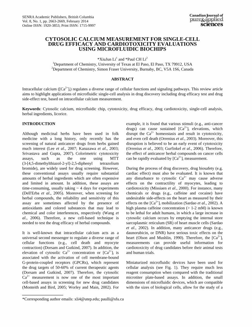
than that resulted from IQ. For instance, DNR caused [Ca2+]i to increase up to 1563 nM, even when a much lower DNR concentration (3.5 µM) was used, while 100 µM IQ increased [Ca2+]i only up to 279 nM. The very high [Ca2+]i concentration of 1563 nM is detrimental to the cells (Olson and Mushlin, 1990), as it has been reported that [Ca2+]i above ~10-6 M or 1000 nM is lethal to cells (Hesketh et al., 1983). More experiments show that the maximum [Ca2+]i due to DNR and IQ within 90 min are 1507±80 nM and 255±35 nM (n=3), respectively. These data suggest that DNR has a much greater cytotoxic effect than IQ on cardiomyocytes. In addition to the Ca2+ data, the microfluidic single-cell method provided the data of the cell morphology changes before and after the drug treatment (Fig. 3D). For instance, after DNR treatment, cell 2 showed cell shortening and membrane blebbing, which is indicative of an unhealthy or dying cell, while cell 1 still showed healthy status after the IQ treatment. Although DNR is widely used in treatment of leukemia, the use of DNR is unfortunately limited by its potentially fatal cardiotoxicity (Olson and Mushlin, 1990; Olson et al., 2000). As compared with DNR, IQ has less effect on
[Ca2+]i of heart muscle cells, and hence less cardiotoxicity. In conjunction with the finding from the previous section that 50 µM IQ has a cytotoxic effect on leukemia cells, IQ can be a potential drug candidate with less cardiotoxicity. But the findings of these cell-based in vitro tests are subject to the confirmation by in vivo assays. SUMMARY Intracellular calcium measurement using microfluidic devices provides a versatile platform for drug discovery from drug efficacy test to side-effect test. Such an approach, together with the effort to develop high throughput capability and automation, will be useful to evaluate drug efficacy and cardiosafety, without the 4-day wait when using the conventional assays. ACKNOWLEDGEMENTS Financial support from Natural Science and Engineering Research Council (NSERC) of Canada (Discovery Grant), UT STARS Award, UTEP IDR and URI programs is gratefully acknowledged.
Fig. 3. Microfluidic single cell analysis for drug cardiotoxicity evaluation. (A) Chip layout. (B) Single-cell capture structure. (C) A single cardiomyocyte retained in the capture structure. (D) Cardiotoxicity evaluations on cardiomyocytes as depicted by the [Ca2+]i responses after drug treatment of DNR and IQ. The figure is adapted from Li et al. (2007).
A
B
C
D

Canadian Journal of Pure and Applied Sciences
2667
REFERENCES Bers, DM. 1987. Ryanodine and the calcium content of cardiac SR assessed by caffeine and rapid cooling contractures. Am. J. Physiol. 253:C408-15.
Cheng, W., Klauke, N., Sedgwick, H., Smith, GL. and Cooper, JM. 2006. Metabolic monitoring of the electrically stimulated single heart cell within a microfluidic platform. Lab Chip. 6:1424-31.
Cinatl, J., Morgenstern, B., Bauer, G., Chandra, P., Rabenau, H. and Doerr, HW. 2003. Treatment of SARS with human interferons. Lancet. 362:293-4.
Dell'Erba, C., Chiavarina, B., Fenoglio, C., Petrillo, G., Cordazzo, C., Boncompagni, E., Spinelli, D., Ognio, E., Aiello, C., Mariggio, MA. and Viale, M. 2005. Inhibition of cell proliferation, cytotoxicity and induction of apoptosis of 1,4-bis(1-naphthyl)-2,3-dinitro-1,3-butadiene in gastrointestinal tumour cell lines and preliminary evaluation of its toxicity in vivo. Pharmacol. Res. 52:271-82.
Di Carlo, D., Aghdam, N. and Lee, LP. 2006. Single-cell enzyme concentrations, kinetics, and inhibition analysis using high-density hydrodynamic cell isolation arrays. Anal. Chem. 78:4925-30.
Di Carlo, D. and Lee, LP. 2006. Dynamic single-cell analysis for quantitative biology. Anal. Chem. 78: 7918-25.
Dittrich, PS., Tachikawa, K. and Manz, A. 2006. Micro total analysis systems. Latest advancements and trends. Anal Chem. 78:3887-908.
Dorsam, RT. and Gutkind, JS. 2007. G-protein-coupled receptors and cancer. Nat. Rev. Cancer. 7:79-94
Durrieu, F., Belloc, F., Lacoste, L., Dumain, P., Chabrol, J., Dachary-Prigent, J., Morjani, H., Boisseau, MR., Reiffers, J., Bernard, P. and Lacombe, F. 1998. Caspase activation is an early event in anthracycline-induced apoptosis and allows detection of apoptotic cells before they are ingested by phagocytes. Exp. Cell Res. 240: 165-75.
Gurfinkel, DM., Chow, S., Hurren, R., Gronda, M., Henderson, C., Berube, C., Hedley, DW. and Schimmer, AD. 2006. Disruption of the endoplasmic reticulum and increases in cytoplasmic calcium are early events in cell death induced by the natural triterpenoid Asiatic acid. Apoptosis. 11:1463-71.
Hesketh, TR., Smith, GA., Moore, JP., Taylor, MV. and Metcalfe, JC. 1983. Free cytoplasmic calcium concentration and the mitogenic stimulation of lymphocytes. J. Biol. Chem. 258:4876-82.
Hsu, YL., Kuo, PL. and Lin, CC. 2005. Isoliquiritigenin induces apoptosis and cell cycle arrest through p53-dependent pathway in Hep G2 cells. Life Sci. 77:279-92.
Iwashita, K., Kobori, M., Yamaki, K. and Tsushida, T. 2000. Flavonoids inhibit cell growth and induce apoptosis in B16 melanoma 4A5 cells. Biosci. Biotechnol. Biochem. 64:1813-20.
Kaji, H., Nishizawa, M. and Matsue, T. 2003. Localized chemical stimulation to micropatterned cells using multiple laminar fluid flows. Lab Chip. 3:208-11.
Kanazawa, M., Satomi, Y., Mizutani, Y., Ukimura, O., Kawauchi, A., Sakai, T., Baba, M., Okuyama, T., Nishino, H. and Miki, T. 2003. Isoliquiritigenin inhibits the growth of prostate cancer. Eur. Urol. 43:580-6.
Klauke, N., Smith, G. and Cooper, JM. 2007. Microfluidic systems to examine intercellular coupling of pairs of cardiac myocytes. Lab Chip. 7:731-9.
Klauke, N., Smith, GL. and Cooper, J. 2003. Stimulation of single isolated adult ventricular myocytes within a low volume using a planar microelectrode array. Biophys. J. 85:1766-74.
Klauke, N., Smith, GL. and Cooper, J. 2006. Extracellular recordings of field potentials from single cardiomyocytes. Biophys. J. 91:2543-51.
Kovarik, M. L., Gach, PC., Ornoff, DM., Wang, Y., Balowski, J., Farrag, L. and Allbritton, NL. 2012. Micro total analysis systems for cell biology and biochemical assays. Anal Chem. 84:516-40.
Lee, C. K., Park, KK., Lim, SS., Park, JH. and Chung, WY. 2007. Effects of the licorice extract against tumor growth and cisplatin-induced toxicity in a mouse xenograft model of colon cancer. Biol. Pharm. Bull. 30:2191-5.
Li, XJ., Chen, YC. and Li, PCH. 2011. A simple and fast microfluidic approach of same-single-cell analysis (SASCA) for the study of multidrug resistance modulation in cancer cells. Lab on a Chip. 11:1378-1384.
Li, XJ., Huang, J., Tibbits, GF. and Li, PCH. 2007. Real-time monitoring of intracellular calcium dynamic mobilization of a single cardiomyocyte in a microfluidic chip pertaining to drug discovery. Electrophoresis 28:4723-4733.
Li, XJ. and Li, PCH. 2005. Microfluidic selection and retention of a single cardiac myocyte, on-chip dye loading, cell contraction by chemical stimulation, and quantitative fluorescent analysis of intracellular calcium. Anal. Chem. 77:4315-4322.
Li and Li
2668
Li, XJ. and Li, PCH. 2006. Contraction study of a single cardiac muscle cell in a microfluidic chip. Methods Mol. Biol. 321:199-225.
Li, XJ., Ling, V. and Li, PCH. 2008. Same-single-cell analysis for the study of drug efflux modulation of multidrug resistant cells using a microfluidic chip. Anal. Chem. 80:4095-4102.
Li, XJ., Valadez, AV., Zuo, P. and Nie, Z. 2012. Microfluidic 3D cell culture: potential application for tissue-based bioassays. Bioanalysis 4:1509-1525.
Li, XJ., Xue, X. and Li, PCH. 2009. Real-time detection of the early event of cytotoxicity of herbal ingredients on single leukemia cells studied in a microfluidic biochip. Integr. Biol. 1:90-98.
Ma, J., Fu, NY., Pang, DB., Wu, WY. and Xu, AL. 2001. Apoptosis induced by isoliquiritigenin in human gastric cancer MGC-803 cells. Planta Med. 67:754-7.
Maggiolini, M., Statti, G., Vivacqua, A., Gabriele, S., Rago, V., Loizzo, M., Menichini, F. and Amdo, S. 2002. Estrogenic and antiproliferative activities of isoliquiritigenin in MCF7 breast cancer cells. J. Steroid Biochem. Mol. Biol. 82:315-22.
Missiaen, L., Robberecht, W., van den Bosch, L., Callewaert, G., Parys, JB., Wuytack, F., Raeymaekers, L., Nilius, B., Eggermont, J. and De Smedt, H. 2000. Abnormal intracellular ca(2+)homeostasis and disease. Cell Calcium 28:1-21.
Monteith, GR. and Bird, GS. 2005. Techniques: high-throughput measurement of intracellular Ca(2+) -- back to basics. Trends Pharmacol. Sci. 26:218-23.
Olson, RD., Li, X., Palade, P., Shadle, SE., Mushlin, PS., Gambliel, HA., Fill, M., Boucek, RJ. Jr. and Cusack, BJ. 2000. Sarcoplasmic reticulum calcium release is stimulated and inhibited by daunorubicin and daunorubicinol. Toxicol. Appl. Pharmacol. 169:168-76.
Olson, RD. and Mushlin, PS. 1990. Doxorubicin cardiotoxicity: analysis of prevailing hypotheses. Faseb J 4: 3076-86.
Olson, RD. and Mushlin, PS. 1990. Doxorubicin cardiotoxicity: analysis of prevailing hypotheses. Faseb J. 4: 3076-86.
Orrenius, S., Zhivotovsky, B. and Nicotera, P. 2003. Regulation of cell death: the calcium-apoptosis link. Nat. Rev. Mol. Cell Biol. 4:552-65.
Peng, XY. and Li, PCH. 2005. Extraction of pure cellular fluorescence by cell scanning in a single-cell microchip. Lab Chip 5:1298-302.
Peng, XY. and Li, PCH. 2004. A three-dimensional flow
control concept for single-cell experiments on a microchip. 2. Fluorescein diacetate metabolism and calcium mobilization in a single yeast cell as stimulated by glucose and pH changes. Anal. Chem. 76:5282-92.
Pompei, R., Flore, O., Marccialis, MA., Pani, A. and Loddo, B. 1979. Glycyrrhizic acid inhibits virus growth and inactivates virus particles. Nature. 281:689-90.
Sardao, VA., Oliveira, PJ. and Moreno, AJ. 2002. Caffeine enhances the calcium-dependent cardiac mitochondrial permeability transition: relevance for caffeine toxicity. Toxicol Appl Pharmacol. 179:50-6.
Sims, CE. and. Allbritton, NL. 2007. Analysis of single mammalian cells on-chip. Lab Chip. 7(4):423-40.
Srivastava, JK. and Gupta, S. 2007. Antiproliferative and apoptotic effects of chamomile extract in various human cancer cells. J. Agric. Food Chem. 55:9470-8.
Sung, MW. and Li, PCH. 2004. Chemical analysis of raw, dry-roasted, and honey-roasted licorice by capillary electrophoresis. Electrophoresis. 25:3434-40
Tanaka, H., Hasegawa, T., Matsushita, M., Miichi, H. and Hayashi, S. 1987. Quantitative-evaluation of ocular antiinflammatory drugs based on measurements of corneal temperature in rabbits - dexamethasone and glycyrrhizin. Ophthalmic Res. 19: 213-220.
Teruel, MN. and Meyer, T. 2002. Parallel single-cell monitoring of receptor-triggered membrane translocation of a calcium-sensing protein module. Science. 295:1910-2.
Vial, JP., Belloc, F., Dumain, P., Besnard, S., Lacombe, F., Boisseau, MR., Reiffers, J. and Bernard, P. 1997. Study of the apoptosis induced in vitro by antitumoral drugs on leukaemic cells. Leuk. Res. 21:163-172.
Wang, X., Ge, J., Wang, K., Qian, J. and Zou, Y. 2006. Evaluation of MTT assay for measurement of emodin-induced cytotoxicity. Assay Drug Dev. Technol. 4:203-7.
Wheeler, AR., Throndset, WR., Whelan, RJ., Leach, AM., Zare, RN., Liao, YH., Farrell, K., Manger, ID. and Daridon, A. 2003. Microfluidic device for single-cell analysis. Anal. Chem. 75:3581-6.
Worley, JF. 3rd, and Main, MJ. 2002. An industrial perspective on utilizing functional ion channel assays for high throughput screening. Receptors Channels. 8:269-82.
Yamazaki, S., Morita, T., Endo, H., Hamamoto, T., Baba, M., Joichi, Y., Kaneko, S., Okada, Y., Okuyama, T., Nishino, H. and Tokue, A. 2002. Isoliquiritigenin suppresses pulmonary metastasis of mouse renal cell carcinoma. Cancer Lett. 183:23-30.

Canadian Journal of Pure and Applied Sciences
2669
Yang, M., Li, CW. and Yang, J. 2002. Cell docking and on-chip monitoring of cellular reactions with a controlled concentration gradient on a microfluidic device. Anal Chem. 74:3991-4001.
Yin, H., Pattrick, N., Zhang, X., Klauke, N., Cordingley, HC., Haswell, SJ. and Cooper, JM. 2008. Quantitative comparison between microfluidic and microtiter plate formats for cell-based assays. Anal. Chem. 80: 179-85.
Zhang, X., Yin, H., Cooper, JM. and Haswell, SJ. 2006. A microfluidic-based system for analysis of single cells based on Ca2+ flux. Electrophoresis. 27:5093-100
Received: Oct 17, 2013; Accepted: Dec 5, 2013