Embed Size (px)
Citation preview

ORIGINAL ARTICLE – BONE AND SOFT TISSUE SARCOMAS
Management of Recurrent Retroperitoneal Sarcoma (RPS)in the Adult: A Consensus Approach from the Trans-AtlanticRPS Working Group
Trans-Atlantic RPS Working Group
ABSTRACT
Introduction. Retroperitoneal soft tissue sarcomas (RPS)
are rare tumors. Surgery is the mainstay of curative ther-
apy, but local recurrence is common. No recommendations
concerning the best management of recurring disease have
been developed so far. Although every effort should be
made to optimize the initial approach, recommendations to
treat recurring RPS will be helpful to maximize disease
control at recurrence.
Methods. An RPS transatlantic working group was estab-
lished in 2013. The goals of the group were to share
institutional experiences, build large multi-institutional case
series, and develop consensus documents on the approach to
this difficult disease. The outcome of this document applies
to recurrent RPS that is nonvisceral in origin. Included are
sarcomas of major veins, undifferentiated pleomorphic
sarcoma of psoas, ureteric leiomyosarcoma (LMS). Exclu-
ded are desmoids-type fibromatosis, angiomyolipoma,
gastrointestinal stromal tumors, sarcomas arising from the
gut or its mesentery, uterine LMS, prostatic sarcoma,
paratesticular/spermatic cord sarcoma, Ewing sarcoma,
alveolar/embryonal rhabdomyosarcoma, sarcoma arising
from teratoma, carcinosarcoma, sarcomatoid carcinoma,
clear cell sarcoma, radiation-induced sarcoma, paragan-
glioma, and malignant pheochromocytoma.
Results. Recurrent RPS management was evaluated from
diagnosis to follow-up. It is a rare and complex malignancy
that is best managed by an experienced multidisciplinary
team in a specialized referral center. The best chance of
cure is at the time of primary presentation, but some
patients may experience prolonged disease control also at
recurrence, when the approach is optimized and follows the
recommendations contained herein.
Conclusions. International collaboration is critical for
adding to the present knowledge. A transatlantic prospec-
tive registry has been established.
Trans-Atlantic RPS Working Group
Jan Ahlen, Department of Surgery, Karolinska Hospital,
Stockholm, Sweden
Nita Ahuja, Department of Surgery, The Sidney Kimmel
Comprehensive Cancer Center at Johns Hopkins, The Johns
Hopkins University School of Medicine, Baltimore, MD
Robert Antbacka, Department of Surgery, Huntsman
Cancer Institute, University of Utah, Salt Lake City, UT
Sanjay Bagaria, Department of Surgery, Mayo Clinic
Florida, Jacksonville, FL
Jean-Yves Blay, Department of Cancer Medicine, Centre
Leon Berard, Lyon, France
Sylvie Bonvalot, Department of Surgery, Institute Curie,
Paris, France
Dario Callegaro, Department of Surgery, Fondazione
IRCCS Istituto Nazionale dei Tumori, Milan, Italy
Robert J. Canter, Division of Surgical Oncology,
Department of Surgery, UC Davis School of Medicine,
Sacramento, CA
Kenneth Cardona, Division of Surgical Oncology,
Department of Surgery, Winship Cancer Institute, Emory
University, Atlanta, GA
Paolo G. Casali, Department of Cancer Medicine – Fon-
dazione IRCCS Istituto Nazionale dei Tumori – Milan, Italy
Chiara Colombo, Department of Surgery, Fondazione
IRCCS Istituto Nazionale dei Tumori, Milan, Italy
Angelo P. Dei Tos, Department of Pathology, Treviso
General Hospital – Treviso, Italy
Correspondence to: Alessandro Gronchi, MDDepartment of Surgery, Fondazione IRCCS Istituto Nazionale dei
Tumori, Milan, Italy, e-mail: [email protected]
Correspondence to: Carol J. Swallow, MD, PhDDepartment of Surgical Oncology, Mount Sinai Hospital and Princess
Margaret Cancer Center, and Department of Surgery University of
Toronto, Toronto, Canada, e-mail: [email protected]
� Society of Surgical Oncology 2016
First Received: 22 March 2016;
Published Online: 1 August 2016
Ann Surg Oncol (2016) 23:3531–3540
DOI 10.1245/s10434-016-5336-7

Antonino De Paoli, Department of Radiation Oncology,
Centro di Riferimento Oncologico, Aviano, Italy
Anant Desai, Department of Surgery, Queen Elizabeth
Hospital, Birmingham, United Kingdom
Brendan C. Dickson, Departments of Pathology and Lab-
oratory Medicine, Mount Sinai Hospital, Toronto, Canada
Fritz C. Eilber, Department of Surgery, University of
California - Los Angeles, Division of Surgical Oncology,
Los Angeles, CA
Marco Fiore, Department of Surgery, Fondazione IRCCS
Istituto Nazionale dei Tumori, Milan, Italy
Cristopher D. Fletcher, Department of Pathology, Brig-
ham and Women’s Hospital, Harvard Medical School,
Boston, MA
Samuel J. Ford, Department of Surgery, Queen Elizabeth
Hospital, Birmingham, United Kingdom
Hans J. Gelderblom, Department of Clinical Oncology,
Leiden University Medical Center, Leiden, The
Netherlands
Ricardo Gonzalez, Department of Surgery, Moffitt Cancer
Center and Research Institute, Tampa, FL
Giovanni Grignani, Department of Cancer Medicine,
Istituto Candiolo – Turin, Italy
Valerie Grignol, Department of Surgery, Division of
Surgical Oncology, Ohio State University Medical Center,
Columbus, OH
Alessandro Gronchi, Department of Surgery, Fondazione
IRCCS Istituto Nazionale dei Tumori, Milan, Italy
Rick L. Haas, Department of Radiation Oncology,
Netherlands Cancer Institute, Amsterdam, The Netherlands
Andrew J. Hayes, Department of Surgery, Royal Marsden
Hospital NHS Foundation Trust, London, United Kingdom
Wolfgang Hartmann, Department of Pathology, Gerhard-
Domagk-Institute of Pathology University Hospital Mun-
ster, Germany
Thomas Henzler, Institute of Clinical Radiology and
Nuclear Medicine, University Medical Center Mannheim,
Heidelberg University, Mannheim, Germany
Peter Hohenberger, Department of Surgical Oncology &
Thoracic Surgery, Mannheim, Germany
Antoine Italiano, Department of Cancer Medicine, Insti-
tute Bergonie, Bordeaux, France
Jens Jakob, Department of Surgical Oncology & Thoracic
Surgery, Mannheim, Germany
Robin L. Jones, Department of Cancer Medicine, Royal
Marsden Hospital, London, United Kingdom
Ian Judson, Department of Cancer Medicine, Royal
Marsden Hospital, London, United Kingdom
John M. Kane, 3rd, Department of Surgical Oncology,
Roswell Park Cancer Institute, Buffalo, NY
Guy Lahat, Department of Surgery, Sourasky Medical
Center, Tel Aviv, Israel
Andrea J. MacNeill, Department of Surgery, Vancouver
General Hospital, University of British Columbia, Van-
couver, Canada
Roberta Maestro, Department of Experimental Oncology,
Centro di Riferimento Oncologico, Aviano, Italy
Christina Messiou, Department of Radiology, Royal
Marsden Hospital NHS Foundation Trust, London, United
Kingdom
Pierre Meeus, Department of Surgery, Centre Leon Ber-
ard, Lyon, France
Rosalba Miceli, Department of Biostatistics, Fondazione
IRCCS Istituto Nazionale dei Tumori, Milan, Italy
John T. Mullen, Department of Surgery, Massachusetts
General Hospital, Boston, MA
Carolyn Nessim, Department of Surgery, The Ottawa
Hospital, Ottawa, Canada
Elisabetta Pennacchioli, Department of Surgery, Euro-
pean Institute of Oncology, Milan, Italy
Vinu G. Pillarisetty, Department of Surgery, University of
Washington, Seattle, WA
Raphael E. Pollock, Department of Surgery, Division of
Surgical Oncology, Ohio State University Medical Center,
Columbus, OH
Vittorio Quagliuolo, Department of Surgery, Humanitas
Cancer Center, Rozzano, Italy
Stefano Radaelli, Department of Surgery, Fondazione
IRCCS Istituto Nazionale dei Tumori, Milan, Italy
Chandrajit P. Raut, Department of Surgery, Division of
Surgical Oncology, Brigham and Women’s Hospital,
Harvard Medical School, Boston, MA
Piotr Rutkowski, Department of Soft Tissue/Bone Sar-
coma and Melanoma, Maria Sklodowska-Curie Memorial
Cancer Center and Institute of Oncology, Warsaw, Poland
Sergio Sandrucci, Department of Surgery, S Giovanni
Battista Hospital, University of Turin, Turin, Italy
Yvonne M. Schrage, Department of Surgery, Leiden
University Medical Center, Leiden, The Netherlands
Jason K. Sicklick, Division of Surgical Oncology,
Department of Surgery, University of California, Moores
Cancer Center, San Diego, CA
Myles J. Smith, Department of Surgery, Royal Marsden
Hospital NHS Foundation Trust, London, United Kingdom
Silvia Stacchiotti, Department of Cancer Medicine, Fon-
dazione IRCCS Istituto Nazionale dei Tumori, Milan, Italy
Eberhardt Stoeckle, Department of Surgery, Institute
Bergonie, Bordeaux, France
Dirk C. Strauss, Department of Surgery, Royal Marsden
Hospital NHS Foundation Trust, London, United Kingdom
3532 Trans-Atlantic RPS Working Group

Carol J. Swallow, Department of Surgery, University of
Toronto, Canada
William Tseng, Department of Surgery, University of
Southern California, Keck School of Medicine, Los
Angeles, CA
Eva Wardelmann, Department of Pathology, Gerhard-
Domagk-Institute of Pathology University Hospital Mun-
ster, Germany
Frits van Coevorden, Department of Surgical Oncology,
Netherlands Cancer Institute, Amsterdam, The Netherlands
Winan J. van Houdt, Department of Surgical Oncology,
Netherlands Cancer Institute, Amsterdam, The Netherlands
Nabil Wasif, Department of Surgery, Mayo Clinic Ari-
zona, Phoenix, AZ
INTRODUCTION
Retroperitoneal sarcomas (RPS) are rare tumors, with an
expected incidence of .5-1 new case/100,000 inhabitants
per year.1 Surgery is the mainstay of curative therapy and
local control is critical for cure.2–13
Overall local/abdominal recurrence is common (&50 %
at 5 years overall) following resection of primary
retroperitoneal sarcoma (RPS).2–13 A high proportion of
recurrences occur late (after 5 years), mandating ongoing
follow-up.13–15
Different histologic subtypes have different patterns and
timing of recurrence following resection of primary RPS.2,3,16
The distribution of histological subtypes in recurrent RPS is
distinct from that of primary RPS.2,3,16,17 Local recurrence,
and/or its treatment, is the most common cause of death from
RPS. In general, recurrent RPS is associated with a worse
prognosis than primary RPS.3,11,18–20
A trans-Atlantic RPS working group (TARPSWG) was
established in 2013. Included in the working group were
North-American and European centers who accepted the
invitation. The group has published a consensus approach
to primary RPS and a large, multicenter, retrospective case
series analysis on primary RPS.2,13 As a follow-up of the
consensus approach to primary RPS, the group expanded
substantially, including several more European and North
American centers, and developed this consensus on recur-
rent RPS. A larger, multicenter, retrospective review on
recurrent RPS is presently ongoing and a prospective reg-
istry, just finalized, is starting soon.
METHODS
An extensive literature search of published evidence was
made and—given the paucity of available data—the
TARPSWG developed guidelines on recurrent RPS mainly
based on expert agreements. The working group initially
met during the 2015 American Society of Clinical
Oncology Annual Meeting in Chicago, IL. A document
was drafted and circulated in the following months. The
group then formally convened during the 2015 Connective
Tissue Oncology Society Annual Meeting in Salt-Lake
City, Utah. The document was finally approved at a
meeting during the 2016 Society of Surgical Oncology
Annual Meeting in Boston, Massachusetts. All group
members reviewed and agreed to the present version of the
recommendations.
The following statements apply to recurrent RPS that is
nonvisceral in origin.
• Included are ‘‘usual’’ recurrent retroperitoneal sarcoma
(liposarcoma, leiomyosarcoma, solitary fibrous tumor,
malignant peripheral nerve sheath tumors, synovial
sarcoma, etc.)
• Included are sarcomas of major veins (inferior vena
cava, renal vein, ovarian/testicular vein), undifferenti-
ated pleomorphic sarcoma of psoas, ureteric
leiomyosarcoma (LMS).
• Excluded are benign entities, such as desmoids and
angiomyolipoma.
• Excluded are gastrointestinal stromal tumors, visceral
sarcomas, such as those arising from the gut or its
mesentery, uterine LMS, prostatic sarcoma, paratestic-
ular/spermatic cord sarcoma.
• Excluded are Ewing sarcoma, alveolar/embryonal
rhabdomyosarcoma, sarcoma arising from teratoma,
carcinosarcoma, sarcomatoid carcinoma, clear cell
sarcoma, radiation-induced sarcoma.
• Excluded are paraganglioma and malignant
pheochromocytoma.
RESULTS
Principles of recommended practice from diagnosis to
follow-up are summarized in 42 statements. Each statement
has been attributed a level of evidence according to the
scale reported in Table 1.21
1. Prior to undertaking a management decision, patients
with recurrent RPS should be presented at a Multi-
Disciplinary Case Conference with participation by
surgical oncologists, medical oncologists, radiation
oncologists, pathologists and radiologists with exper-
tise in sarcoma (VA).
Pretreatment Assessment
Imaging
2. All relevant imaging studies performed prior to
resection of the primary RPS should be obtained and
Management of Recurrent Retroperitoneal Sarcoma (RPS) in the Adult 3533

reviewed, as should all subsequent imaging studies, in
particular the initial postoperative baseline imaging to
determine whether prior resection was in fact grossly
incomplete (R2) (VA).
3. Current extent of local and distant disease should be
determined using oral and intravenous (IV) contrast
enhanced computed tomography of the chest, abdomen
and pelvis (CT-CAP) (IVA).22–24
4. Current imaging should be compared to all prior imaging
to ascertain the extent and progression of recurrent
disease, with attention to pattern of relapse (locoregional
vs. peritoneal) and rate of progression (IVB).25
5. The risk of invasion into adjacent organ(s)/critical
structure(s) with further progression should be evalu-
ated, and taken into account when deciding on the best
management approach (VB).
6. MRI may be a useful ancillary modality in selected
cases to define extent of adjacent organ/structure
involvement that is not clear on CT but is not required
in most cases (IVB).26
MRI may be helpful in operative planning for pelvic
tumors or tumors that abut/involve bone or psoas or
oblique muscles or vertebral foramen.23 MRI also is an
option in patients with IV contrast allergy or other
serious contraindication to CT (IVB).23
7. PET scan is rarely indicated; it may be indicated if the
extent of active abdominal disease is difficult to
evaluate and/or there is uncertainty about different
growth characteristics in multifocal lesions that would
alter management (IVB).27,28
8. Abdominal (nonhepatic parenchymal) recurrence
should be categorized as
(a) locoregional (at the site of the primary RPS or
within the ipsilateral RP);
(b) multifocal/contralateral RP;
(c) both (VB).
An example is shown in Fig. 1.
Pathology
9. Histopathology of the primary tumor should be
reviewed by a pathologist specialized in the
evaluation of soft tissue tumors; molecular subtyping
should be performed where appropriate (e.g., Mdm2 in
lipomatous tumors) (VA).
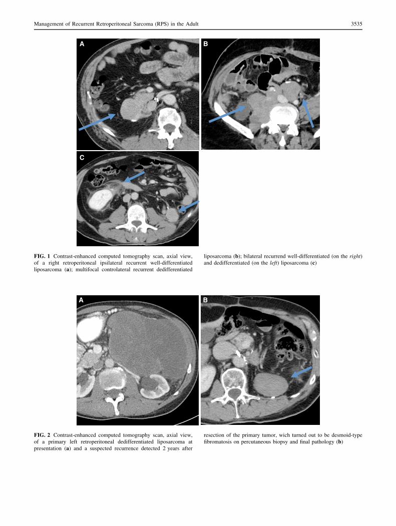
10. Percutaneous core biopsy confirmation of recurrence
is often useful, for a number of reasons: 1) to provide
a definitive diagnosis, because a variety of other
entities can be mistaken for recurrence of the original
primary RPS, for example desmoid fibromatosis
(Fig. 2), radiation-associated osteosarcoma, or
angiosarcoma in the bed of original LPS; 2) to guide
selection of preoperative therapies, including poten-
tial targeted therapies; 3) as part of a translational
research program or clinical trial; 4) because resec-
tion often is challenging and may be morbid and
should not be undertaken without due cause (VB).
Patient Evaluation
11. The patient’s symptoms, and pace of symptom
progression, should be noted
The patient’s current performance status should be
recorded. Physical examination should include
notation of previous incisions, and the relationship
of new tumor mass(es) to previous incisions,
including port sites (VB).
12. Renal function and nutritional status must be assessed
(VB).
TABLE 1 Level of Evidence (LOE) and Grade of Recommendation (GOR) adapted from the Infectious Diseases Society of American-United
States Public Health Service Grading System
I Evidence from at least one large randomized control trial of good methodological quality (low potential for bias) or meta-analyses of well-
conducted randomized trials without heterogeneity
II Small randomized trials or large randomized trials with a suspicion of bias (lower methodological quality) or meta-analyses of such trials or
of trials with demonstrated heterogeneity
III Prospective cohort studies
IV Retrospective cohort studies or case-control studies
V Studies without control group, case reports, experts opinions
A Strong evidence for efficacy with a substantial clinical benefit, strongly recommended
B Strong or moderate evidence for efficacy but with a limited clinical benefit, generally recommended
C Insufficient evidence for efficacy or benefit does not outweigh the risk or the disadvantages (adverse events, costs,), optional
D Moderate evidence against efficacy or for adverse outcome, generally not recommended
E Strong evidence against efficacy or for adverse outcome, never recommended
3534 Trans-Atlantic RPS Working Group
A B
C
FIG. 1 Contrast-enhanced computed tomography scan, axial view,
of a right retroperitoneal ipsilateral recurrent well-differentiated
liposarcoma (a); multifocal controlateral recurrent dedifferentiated
liposarcoma (b); bilateral recurrend well-differentiated (on the right)
and dedifferentiated (on the left) liposarcoma (c)
A B
FIG. 2 Contrast-enhanced computed tomography scan, axial view,
of a primary left retroperitoneal dedifferentiated liposarcoma at
presentation (a) and a suspected recurrence detected 2 years after
resection of the primary tumor, wich turned out to be desmoid-type
fibromatosis on percutaneous biopsy and final pathology (b)
Management of Recurrent Retroperitoneal Sarcoma (RPS) in the Adult 3535

Review of Previous Treatment
13. The operative note describing the resection of the
primary tumor should be obtained and reviewed, as
should notes describing any subsequent operative or
other interventional procedures (VB).
14. Timing from previous surgery should be noted, as
well as factors that precluded macroscopically com-
plete resection; if not described, these should be
elicited through personal communication with the
referring surgeon (VB).
15. Previous pathology reports, and if possible any tissue
slides/blocks, from previous resection(s) should be
obtained and reviewed (VB).
16. The nature of previous surgery should be categorized
as
(1) Macroscopically complete (en bloc resection).
(2) Macroscopically incomplete (gross residual dis-
ease as noted on operative report or on immediate
postoperative cross-sectional imaging, Fig. 3)
(3) Piecemeal and/or associated with tumor rupture
or morcellation (VB).
17. Details of any previously administered radiotherapy
or systemic therapy should be reviewed (VA).
Patient Selection for Resection of Recurrence
18. While complete gross resection can be viewed as the
only curative-intent option in patients with recurrent
RPS, the chance of long term disease-free survival is
limited and this must be recognized in multidisci-
plinary evaluation and planning of management for
the individual patient (IVA).3,29
The decision to pursue curative resection is multifacto-
rial and often nuanced. A nomogram to assist in this
decision is presently lacking, the available ones mainly
built to predict the risk of primary disease.30 Variables to
be considered are discussed below.
Abdominal Recurrence
19. In the case of isolated locoregional recurrence,
especially if the previous resection was incomplete,
the goal of resection should be curative, and the
extent of resection should be as required to achieve
complete gross resection (IVB).31–34
19.1. For local recurrence of WD-LPS within the
field of previous resection(s), the surgeon may
rationally wait for an increase in tumor size in
order to space out the interval between
operations (IVB).35
20. Multifocal intra-abdominal disease is difficult to resect
completely, will almost certainly recur again, and
carries a poor prognosis particularly with resection
alone. The prognosis following resection worsens with
increasing number of intraperitoneal deposits.34 Any
resection should be with the goal of avoiding compli-
cations of progression and preserving function (i.e.,
limited resection) or with palliative intent as discussed
below. Because the oncologic benefit of surgery is
likely to be limited and the risk of morbidity may be
substantial, a very careful approach to patient selection
for surgical intervention is required (IVA).32–36
21. A history of previous piecemeal resection/tumor
rupture indicates a strong potential for multifocal
peritoneal recurrence and curative-intent resection is
A B
FIG. 3 Contrast-enhanced computed tomography scan, axial view, of a primary right retroperitoneal dedifferentiated liposarcoma at
presentation (a) and a persistent perirenal disease detected at the first postoperative assessment after resection of the primary tumor (b)
3536 Trans-Atlantic RPS Working Group

generally not appropriate; resection should be with
the goals of avoiding complications of progression
and preserving function (limited resection); a possible
exception is a previous flank/lumbar approach, where
piecemeal resection may not have been associated
with peritoneal contamination (IVB).4,5
22. Histopathologic subtype should factor into the decision to
pursue re-resection, because outcomes differ (e.g., WD-
LPS would be favored for re-resection) (IVA).3,29–36
23. A period of observation and reimaging may help to
select more favorable candidates for resection, par-
ticularly for asymptomatic WD-LPS (rate of growth
of less than 1 cm per month is associated with better
duration of disease control) (IVB).35
24. Long-term survival may be achieved in well-selected
patients (e.g. no history of tumor rupture, low grade,
long DFI, solitary local recurrence), however there is a
very limited chance of cure and this must be considered
when offering aggressive therapy 33,36 (IVB).
25. Risk of mortality and serious morbidity after radical
resection of recurrent RPS are significant; good
performance status is important to recovery.
In general, with each serial resection, the oncologic
outcome deteriorates, and the chance of mortality and
serious morbidity are magnified (IVB).32 The often indo-
lent course of recurrent disease (particularly LPS), even in
the presence of a bulky mass, should be considered when
major morbidity is expected following resection and the
patient is asymptomatic (IVB).35
Distant Recurrence
26. Selected patients with limited oligo-metastatic
recurrence of RPS may have prolonged survival
following metastasectomy (e.g., LMS) (IVB).37
27. In general, synchronous abdominal and distant
recurrence should not be resected, and the patient
should be considered for systemic therapy as part of a
multimodality approach based on tumor histol-
ogy/molecular subtyping (IVB).37
Preoperative Therapy
28. For patients being considered for resection of recurrent
RPS, neoadjuvant therapy should be considered (VB).
28.1. Cytotoxic and/or targeted systemic therapies
may be of benefit in downsizing recurrent
disease to improve resectability, especially in
the case of LMS, DD-LPS, UPS, SFT, and
synovial sarcoma, and also may facilitate
assessment of tumor biology/prognosis, espe-
cially when a high grade RPS has recurred
after a short disease-free interval, and when
resection of locally recurrent disease is antic-
ipated to be morbid (VB).
28.2. Preoperative XRT should be considered, par-
ticularly if no previous XRT has been
administered and the recurrence is isolated,
although its value in improving disease control
has not been studied, and toxicity may be
magnified in the setting of prior resection (VB).
29. Response to treatment varies by histological subtype
of RPS and the management plan should be devel-
oped in recognition of this and in conjunction with a
multidisciplinary care team (VB).
Preoperative treatment of recurrent RPS should ideally
be given within the context of a clinical trial.
Resection
30. The technical challenge of resection is typically
compounded following curative-intent en bloc
resection of primary RPS (VA).
30.1. Loss of the original planes increases the
difficulty of determining the extent of disease
and the optimal extent of resection (VA).
30.2 Distortion of anatomical relationships due to prior
resection can lead to injury of important structures
(e.g., IVC, duodenum, femoral nerve) (VA).
30.3 Vascular involvement by recurrent disease
may necessitate vessel resection and recon-
struction; this should be done in a planned
manner and as part of curative-intent resection,
at a specialized center (VA).
31. The use of intraoperative frozen sections on marginal
tissues as a guide to extent of resection is generally not
advised. Frozen-section analysis of soft-tissue tumors
is frequently problematic, and particularly so in the
setting of reoperative surgery and previous receipt of
adjuvant therapies (VA). Result of frozen-section
analysis may be misleading. In some specific scenarios,
such as LMS of a major vein, frozen-section analysis of
a vascular margin may be useful if additional tissue at
the relevant site can be removed (VB).
32. Technical and non-technical peri-operative consider-
ations are as enunciated in the TARPSWG consensus
statement on management of primary RPS (IVA).13,38
Postoperative Systemic Therapy and Other
Locoregional Therapies
33. There is no proven role for prophylactic systemic
therapy after complete resection of recurrent RPS
(IVC).39,40
Management of Recurrent Retroperitoneal Sarcoma (RPS) in the Adult 3537

34. There is no proven role for brachytherapy or postop-
erative XRT after resection of recurrent RPS, and
each may lead to increased morbidity (IVE).41,42
34.1 IORT may be considered following resection
of recurrent RPS, although the evidence is
weak (IVD).43
35. There is no proven role for intraperitoneal chemother-
apy, but it could be considered for highly selected
patients and histologies (IVD).44–49 Ideally intraperi-
toneal therapy for these specific histologies should be
administered within the context of a clinical trial. For
usual histological subtypes, the results of debulking
surgery and intraperitoneal chemotherapy for multi-
focal intraperitoneal sarcoma have been uniformly
poor with prohibitive morbidity.
36. There is no proven role for regional hyperthermia
(IIID).50
37. After resection of recurrent RPS, patients should be
followed with regular cross-sectional imaging, given
the high risk for further relapse (IVB).3,29,31–36
Treatment of Patients Who are Not Eligible for
Curative Resection
38. In selected patients, cytotoxic and/or targeted sys-
temic therapies may be of benefit in achieving and
prolonging improved quality of life (IVB).51–53
38.1. In selected patients, a significant response to
cytotoxic and/or targeted systemic therapies
may prompt reconsideration of resection
(VB).
39. Patients should be considered for palliative proce-
dures as symptoms arise. Early referral for symptom
management is desirable (VA).
40. XRT may be of palliative benefit, in particular with
pain control related to nerve compression or infiltra-
tion (VB).
41. In select patients, survival with recurrent RPS may be
prolonged (e.g., WD-LPS). The potential to live
many years despite the ongoing presence of recurrent
disease should be recognized and adequately com-
municated to the patient (VB).
42. R2 resection may be indicated for symptom control in
highly selected patients, particularly if WD-LPS; in
contrast R2 resection for more aggressive histologies
may increase the risk for peritoneal dissemination/
sarcomatosis (VB).
CONCLUSIONS
The available evidence on the approach to recurrent RPS is
clearly limited. This is the largest consensus document on this
topic and follows the one on primary RPS. An effort to merge
and formally report the retrospective experience of the par-
ticipating institutions, which backs its development, is
presently ongoing. The two documents will help to harmonize
the approach to this difficult disease. Another major effort to
enter all eligible patients onto an international collaborative
prospective registry is being finalized and will be active soon.
We anticipate that this registry will be the basis of much
further understanding of this difficult disease and will serve as
a platform for collaborative translational research.
Bio-banking of fresh/frozen tissue should be encouraged
to facilitate future research. Evaluations of long-term
function and quality-of-life following therapy for recurrent
RPS are lacking. Ideally quality-of-life should be assessed
preoperatively, as well as postoperatively. The poor prog-
nosis of recurrent RPS highlights the importance of optimal
management of primary RPS.13
REFERENCES
1. Cormier JN, Gronchi A, Pollock RE. Soft tissue sarcomas. In:
Brunicardi F, Andersen D, Billiar T, Dunn D, Hunter J, Matthews
J, Pollock RE (eds.) Schwartz’s principles of surgery. McGraw
Hill, New York; 2015:1465–94.
2. Gronchi A, Strauss DC, Miceli R, et al. Variability in patterns
of recurrence after resection of primary retroperitoneal sar-
coma (RPS): A report on 1007 patients from the multi-
institutional collaborative RPS Working Group. Ann Surg
2016;263:1002–9.
3. Gronchi A, Miceli R, Allard MA, et al. Personalizing the
approach to retroperitoneal soft tissue sarcoma: histology-specific
patterns of failure and post-relapse outcome after primary
extended resection. Ann Surg Oncol 2015;22(5):1447–54.
4. Toulmonde M, Bonvalot S, Meeus P, et al. Retroperitoneal sar-
comas: patterns of care at diagnosis, prognostic factors and focus
on main histological subtypes: a multicenter analysis of the
French Sarcoma Group. Ann Oncol 2014;25:735–42.
5. Bonvalot S, Rivoire M, Castaing M, et al. Primary retroperitoneal
sarcomas: a multivariate analysis of surgical factors associated
with local control. J Clin Oncol 2009;27:31–7.
6. Gronchi A, Lo Vullo S, Fiore M, et al. Aggressive surgical
policies in a retrospectively reviewed single-institution case ser-
ies of retroperitoneal soft tissue sarcoma patients. J Clin Oncol
2009;27:24–30.
7. Hassan I, Park SZ, Donohue JH, et al. Operative management of
primary retroperitoneal sarcomas: a reappraisal of an institutional
experience. Ann Surg 2004;239:244–50.
8. Stoeckle E, Coindre JM, Bonvalot S, et al. Prognostic factors in
retroperitoneal sarcoma: a multivariate analysis of a series of 165
patients of the French Cancer Center Federation Sarcoma Group.
Cancer 2001;92:359–68.
9. Strauss DC, Hayes AJ, Thway K, et al. Surgical management of
primary retroperitoneal sarcoma. Br J Surg 2010;97:698–706.
3538 Trans-Atlantic RPS Working Group

10. Lehnert T, Cardona S, Hinz U, et al. Primary and locally recur-
rent retroperitoneal soft-tissue sarcoma: local control and
survival. Eur J Surg Oncol 2009;35:986–93.
11. Lewis JJ, Leung D, Woodruff JM, et al. Retroperitoneal soft-
tissue sarcoma: analysis of 500 patients treated and followed at a
single institution. Ann Surg 1998;228:355–365.
12. Gronchi A, Miceli R, Colombo C, et al. Frontline extended sur-
gery is associated with improved survival in retroperitoneal low-
intermediate grade soft tissue sarcomas. Ann Oncol
2012;23(4):1067–73.
13. Management of primary retroperitoneal sarcoma (RPS) in the
adult: a consensus approach from the Trans-Atlantic RPS
Working Group. Ann Surg Oncol 2015;22:256–63.
14. Heslin MJ, Lewis JJ, Nadler E, et al. Prognostic factors associated
with long-term survival for retroperitoneal sarcoma: implications
for management. J Clin Oncol 1997;15:2832–9.
15. Canter RJ, Qin LX, Ferrone CR, et al. Why do patients with low-grade
soft tissue sarcoma die? Ann Surg Oncol 2008;15(12):3550–60.
16. Tan MC, Brennan MF, Kuk D, et al. Histology-based classifi-
cation predicts pattern of recurrence and improves risk
stratification in primary retroperitoneal sarcoma. Ann Surg
2016;263:593–600.
17. Singer S, Antonescu CR, Riedel E, et al. Histologic subtype and
margin of resection predict pattern of recurrence and survival for
retroperitoneal liposarcoma. Ann Surg 2003;238:358–70; dis-
cussion 370–1.
18. Gronchi A, Casali PG, Fiore M, et al. Retroperitoneal soft tissue
sarcomas: patterns of recurrence in 167 patients treated at a single
institution. Cancer 2004;100:2448–55.
19. Anaya DA, Lahat G, Wang X, et al. Postoperative nomogram for
survival of patients with retroperitoneal sarcoma treated with
curative intent. Ann Oncol 2010;21:397–402.
20. Gyorki DE, Brennan MF. Management of recurrent retroperi-
toneal sarcoma. J Surg Oncol. 2014;109:53-9.
21. Dykewicz CA. Summary of the guidelines for preventing
opportunistic infections among hematopoietic stem cell transplant
recipients. Clin Infect Dis 2001;33:139–44.
22. Lahat G, Madewell JE, Anaya DA, et al. Computed tomography
scan-driven selection of treatment for retroperitoneal liposarcoma
histologic subtypes. Cancer 2009;115:1081–90.
23. Morosi C, Stacchiotti S, Marchiano A, et al. Correlation between
radiological assessment and histopathological diagnosis in
retroperitoneal tumors: analysis of 291 consecutive patients at a
tertiary reference sarcoma center. Eur J Surg Oncol 2014. 10.
1016/j.ejso.2014.10.005.
24. Tzeng CW, Smith JK, Heslin MJ. Soft tissue sarcoma: preoper-
ative and postoperative imaging for staging. Surg Oncol Clin N
Am 2007;16:389–402.
25. Tseng WW, Madewell JE, Wei W, et al. Locoregional disease
patterns in well-differentiated and dedifferentiated retroperitoneal
liposarcoma: implications for the extent of resection? Ann Surg
Oncol 2014;21:2136–43.
26. Shiraev T, Pasricha SS, Choong P, et al. Retroperitoneal sarco-
mas: a review of disease spectrum, radiological features,
characterisation and management. J Med Imaging Radiat Oncol
2013;57:687–700.
27. Niccoli-Asabella A, Altini C, Notaristefano A, et al. A retro-
spective study comparing contrast-enhanced computed
tomography with 18F-FDG-PET/CT in the early follow-up of
patients with retroperitoneal sarcomas. Nucl Med Commun
2013;34:32–9.
28. Alford S, Choong P, Chander S, et al. Value of PET scan in
patients with retroperitoneal sarcoma treated with preoperative
radiotherapy. Eur J Surg Oncol 2012;38:176–80.
29. Grobmyer SR, Wilson JP, Apel B, et al. Recurrent retroperitoneal
sarcoma: impact of biology and therapy on outcomes. J Am Coll
Surg 2010;210:602–8, 608–10.
30. Gronchi A, Miceli R, Shurell E, et al. Outcome prediction in pri-
mary resected retroperitoneal soft tissue sarcoma: histology-
specific overall survival and disease-free survival nomograms built
on major sarcoma center datasets. J Clin Oncol 2013;31:1649–55.
31. van Dalen T, Hoekstra HJ, van Geel AN, et al. Locoregional
recurrence of retroperitoneal soft tissue sarcoma: second chance
of cure for selected patients. Eur J Surg Oncol 2001;27:564–8.
32. Neuhaus SJ, Barry P, Clark MA, et al. Surgical management of
primary and recurrent retroperitoneal liposarcoma. Br J Surg
2005;92:246–52.
33. Lochan R, French JJ, Manas DM. Surgery for retroperitoneal soft
tissue sarcomas: aggressive re-resection of recurrent disease is
possible. Ann R Coll Surg Engl 2011;93:39–43.
34. Anaya DA, Lahat G, Liu J, et al. Multifocality in retroperitoneal
sarcoma: a prognostic factor critical to surgical decision-making.
Ann Surg 2009;249:137–42.
35. Park JO, Qin LX, Prete FP, et al. Predicting outcome by growth
rate of locally recurrent retroperitoneal liposarcoma: the one
centimeter per month rule. Ann Surg 2009;250:977–82.
36. Yang JY, Kong SH, Ahn HS, et al. Prognostic factors for reop-
eration of recurrent retroperitoneal sarcoma: the role of
clinicopathological factors other than histologic grade. J Surg
Oncol 2015;111:165–72.
37. Toulmonde M, Bonvalot S, Ray-Coquard I, et al. Retroperitoneal
sarcomas: patterns of care in advanced stages, prognostic factors
and focus on main histological subtypes: a multicenter analysis of
the French Sarcoma Group. Ann Oncol 2014;25:730–4.
38. Bonvalot S, Raut CP, Pollock RE, et al. Technical considerations
in surgery for retroperitoneal sarcomas: position paper from E-
Surge, a master class in sarcoma surgery, and EORTC-STBSG.
Ann Surg Oncol 2012;19:2981–91.
39. Pervaiz N, Colterjohn N, Farrokhyar F, et al. A systematic meta-
analysis of randomized controlled trials of adjuvant chemother-
apy for localized resectable soft-tissue sarcoma. Cancer
2008;113:573–81.
40. Woll PJ, Reichardt P, Le Cesne A, et al. Adjuvant chemotherapy
with doxorubicin, ifosfamide, and lenograstim for resected soft-
tissue sarcoma (EORTC 62931): a multicentre randomised con-
trolled trial. Lancet Oncol 2012;13:1045–54.
41. Smith MJ, Ridgway PF, Catton CN, et al. Combined management
of retroperitoneal sarcoma with dose intensification radiotherapy
and resection: long-term results of a prospective trial. Radiother
Oncol 2014;110:165–71.
42. Tseng WH, Martinez SR, Do L, et al. Lack of survival benefit
following adjuvant radiation in patients with retroperitoneal sar-
coma: a SEER analysis. J Surg Res 2011;168:e173–80.
43. Roeder F, Ulrich A, Habl G, et al. Clinical phase I/II trial to
investigate preoperative dose-escalated intensity-modulated
radiation therapy (IMRT) and intraoperative radiation therapy
(IORT) in patients with retroperitoneal soft tissue sarcoma:
interim analysis. BMC Cancer 2014;14:617-2407-14-617.
44. Hayes-Jordan A, Green HL, Lin H, et al. Complete cytoreduction
and HIPEC improves survival in desmoplastic small round cell
tumor. Ann Surg Oncol 2014;21:220–4.
45. Hayes-Jordan A. Cytoreductive surgery followed by hyperther-
mic intraperitoneal chemotherapy in DSRCT: progress and
pitfalls. Curr Oncol Rep 2015;17:38-015-0461-1.
46. Sugarbaker P, Ihemelandu C, Bijelic L. Cytoreductive surgery
and HIPEC as a treatment option for laparoscopic resection of
uterine leiomyosarcoma with morcellation: early results. Ann
Surg Oncol 2015.
Management of Recurrent Retroperitoneal Sarcoma (RPS) in the Adult 3539

47. Baratti D, Pennacchioli E, Kusamura S, et al. Peritoneal sarco-
matosis: is there a subset of patients who may benefit from
cytoreductive surgery and hyperthermic intraperitoneal
chemotherapy? Ann Surg Oncol 2010;17:3220–8.
48. Bonvalot S, Cavalcanti A, Le Pechoux C, et al. Randomized trial
of cytoreduction followed by intraperitoneal chemotherapy ver-
sus cytoreduction alone in patients with peritoneal sarcomatosis.
Eur J Surg Oncol 2005;31:917–23.
49. Rossi CR, Deraco M, De Simone M, et al. Hyperthermic
intraperitoneal intraoperative chemotherapy after cytoreductive
surgery for the treatment of abdominal sarcomatosis: clinical
outcome and prognostic factors in 60 consecutive patients.
Cancer 2004;100:1943–50.
50. Angele MK, Albertsmeier M, Prix NJ, Hohenberger P, et al.
Effectiveness of regional hyperthermia with chemotherapy for
high-risk retroperitoneal and abdominal soft-tissue sarcoma after
complete surgical resection: a subgroup analysis of a randomized
phase-III multicenter study. Ann Surg 2014;260(5):749–54.
51. Sanfilippo R, Bertulli R, Marrari A, et al. High-dose continuous-
infusion ifosfamide in advanced well-differentiated/dedifferenti-
ated liposarcoma. Clin Sarcoma Res 2014;4(1):16.
52. Martin-Liberal J, Alam S, Constantinidou A, et al. Clinical
activity and tolerability of a 14-day infusional Ifosfamide
schedule in soft-tissue sarcoma. Sarcoma. 2013;2013:868973.
doi:10.1155/2013/868973.
53. Radaelli S, Stacchiotti S, Casali PG, et al. Emerging therapies for
adult soft tissue sarcoma. Expert Rev Anticancer Ther
2014;14.6:689–704.
3540 Trans-Atlantic RPS Working Group