Embed Size (px)
Citation preview

Dr. Cathy Stern, OD, FCOVD, FCSO, FNORA Behavioral, Developmental and Neuro-Optometry
Concussion – Look into the Eyes
BIA-MA 36th Annual Brain Injury Conference March 30, 2017
Cathy Stern, OD, FCOVD, FCSO, FNORA Neuro-Optometric Rehabilitation

Concussion The American Academy of Neurology defines concussion as “ a pathophysiological disturbance in neurological function characterized by clinical symptoms induced by biomechanical forces”
75% of TBI is mTBI
Those most likely to sustain a TBI are:
younger than age 4 age 15-19 older than 65
Cathy Stern, OD, FCOVD, FCSO, FNORA Neuro-Optometric Rehabilitation

Models of Vision
Medical/Optical Model
detection of eye disease and visual acuity
Behavioral Model
vision guides movement and directs action
vision is learned
visual skills can be improved with neuro-optometric rehabilitation
Cathy Stern, OD, FCOVD, FCSO, FNORA
Neuro-Optometric Rehabilitation

Models of Vision
Ocular Visual Impairment
rooted in the eyeball
Cerebral Visual Impairment (CVI)
in the visual pathways/connections through the brain
and connecting with the rest of the body
Cathy Stern, OD, FCOVD, FCSO, FNORA
Neuro-Optometric Rehabilitation

Models of Vision
Vision Involves Two Mechanisms
focal: identification, eyesight
ambient: spatial orientation & localization in space
Visual acuity is to vision as the alphabet is to reading
Cathy Stern, OD, FCOVD, FCSO, FNORA
Neuro-Optometric Rehabilitation

Cathy Stern, OD, FCOVD, FCSO, FNORA Neuro-Optometric Rehabilitation

The Visual Process
To enhance vision for both:
visual input (sensory) and visual output (motor)
Optimal visual processing leads to:
better sustained attention
faster processing speed
superior comprehension/perception
Cathy Stern, OD, FCOVD, FCSO, FNORA
Neuro-Optometric Rehabilitation

Cathy Stern, OD, FCOVD, FCSO, FNORA
Neuro-Optometric Rehabilitation
Why Evaluate Vision after Closed Head Injury
Routine vision evaluation detects eyesight ability and eye disease damage
Neuro-Optometric Vision Evaluation assesses visual efficiency and visual processing critical for reading, using your cell phone, computer use, activities of daily living, recreation, driving and mobility

Cathy Stern, OD, FCOVD, FCSO, FNORA
Neuro-Optometric Rehabilitation
Why Evaluate Vision after Closed Head Injury
Vision plays a significant role for abilities that require sustained visual attention or judgment of direction, distance and space
Vision skill plays an important role for the success of activities being addressed in therapies including occupational, physical, speech and cognitive rehab.
Vision is also a critical skill for balance, walking and driving

Visual Dysfunction in Concussion
Increased vulnerability 50% of the brain is involved in visual information processing
Cathy Stern, OD, FCOVD, FCSO, FNORA Neuro-Optometric Rehabilitation
Cathy Stern, OD, FCOVD, FCSO, FNORA
Neuro-Optometric Rehabilitation
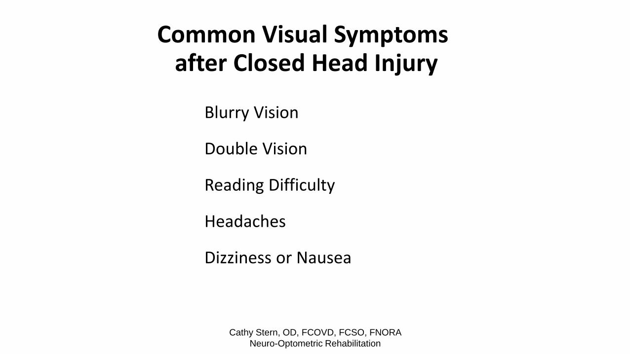
Common Visual Symptoms after Closed Head Injury
Blurry Vision
Double Vision
Reading Difficulty
Headaches
Dizziness or Nausea

Cathy Stern, OD, FCOVD, FCSO, FNORA
Neuro-Optometric Rehabilitation
Common Visual Symptoms after Closed Head Injury
Attention Deficit
Hemispatial Inattention (Visual Neglect)
Light Sensitivity
Low Blink Rate
Spatial Disorientation
direction/distance

Cathy Stern, OD, FCOVD, FCSO, FNORA
Neuro-Optometric Rehabilitation

Cathy Stern, OD, FCOVD, FCSO, FNORA
Neuro-Optometric Rehabilitation
Double Vision is extremely disruptive to reading Double Vision is extremely disruptive to reading

Cathy Stern, OD, FCOVD, FCSO, FNORA
Neuro-Optometric Rehabilitation
Functional Vision Changes associated With Closed Head Injury
Oculomotor Dysfunction (eye tracking)
Convergence Insufficiency
Accommodative (focusing) Deficit
Depth Perception Deficiency

Cathy Stern, OD, FCOVD, FCSO, FNORA
Neuro-Optometric Rehabilitation
Functional Vision Changes associated With Closed Head Injury
Visual Field Loss
Hemispatial Inattention (Visual Neglect)
Strabismus (eye turn)

Cathy Stern, OD, FCOVD, FCSO, FNORA
Neuro-Optometric Rehabilitation
Functional Vision Changes associated With Closed Head Injury
Visual Midline Shift Syndrome (balance and posture)
Visual Memory and Visual Perceptual
Visual Motor Integration
Decreased Blink Rate
Visual Acuity (eyesight) Loss

Cathy Stern, OD, FCOVD, FCSO, FNORA
Neuro-Optometric Rehabilitation
POST TRAUMA VISION SYNDROME
The Characteristics of the PTVS include:
Exotropia or High Exophoria
Convergence Insufficiency
Accommodative Insufficiency
Spatial Disorientation
Unstable Peripheral Vision

Cathy Stern, OD, FCOVD, FCSO, FNORA
Neuro-Optometric Rehabilitation
POST TRAUMA VISION SYNDROME
The Symptoms of the PTVS include:
Possible Diplopia
Poor Concentration and Attention
Objects Appear to Move
Asthenopic Symptoms

Cathy Stern, OD, FCOVD, FCSO, FNORA
Neuro-Optometric Rehabilitation
Vision Evaluation
What You Often Get
History
Visual Acuity (eyesight)
Eye Health
Refractive Status

Cathy Stern, OD, FCOVD, FCSO, FNORA
Neuro-Optometric Rehabilitation
Neuro-Optometric Vision Evaluation
What Should You Expect
Oculomotor (visual tracking)
Pursuits - smooth tracking movements
Saccades - visual fixations from one point to another point
Binocular Eye Coordination – how the two eyes work together
Strabismus or high phoria ( “tendency” for the eye to turn)
Suppression is the "ignoring or turning off vision" of an eye
Double vision is the inability to ignore a second image
a dark patch should not be used to treat diplopia

Cathy Stern, OD, FCOVD, FCSO, FNORA
Neuro-Optometric Rehabilitation
Neuro-Optometric Vision Evaluation
What Should You Expect
Vergence Facility or stamina of the ability to converge/ diverge
Accommodation or focus flexibility
hold focus at near
shift focus from distance to near and back
Depth Perception or Stereopsis

Cathy Stern, OD, FCOVD, FCSO, FNORA
Neuro-Optometric Rehabilitation
Visual Acuity and Refractive Status 50% of ABI patients in one study required a prescription for spectacles
These patients included
those who needed glasses for the first time.
those who needed a replacement for lost eyeglasses.
those requiring a change in their prescription.
Progressive no-line bifocals may exacerbate symptoms

Cathy Stern, OD, FCOVD, FCSO, FNORA
Neuro-Optometric Rehabilitation
Neuro-Optometric Vision Evaluation
Visual Field – affects mobility and attention
Types of visual field assessment
1. confrontation
2. automated perimetry (better for disease)
3. kinetic perimetry (more sensitive)
4. line bisection or letter cancellation
for unilateral spatial inattention (neglect)
5. observation of performance during daily activities

Cathy Stern, OD, FCOVD, FCSO, FNORA
Neuro-Optometric Rehabilitation

Cathy Stern, OD, FCOVD, FCSO, FNORA
Neuro-Optometric Rehabilitation
Neuro-Optometric Vision Evaluation Balance and Postural Status
Visual Midline Shift Syndrome (VMSS) The examiner moves a wand laterally in front of the person and
asks the person to state when the wand appears to be directly in front of the person's nose. This indicates the position of the midline as either centered or with a lateral shift. When the wand is passed vertically in front of the person's face, the person then must respond when the wand appears to be at eye level. This indicates the anterior-posterior relationship of the visual midline. This is the objective analysis to evaluate for Visual Midline Shift Syndrome (VMSS).

Cathy Stern, OD, FCOVD, FCSO, FNORA
Neuro-Optometric Rehabilitation
Visual Midline Shift Syndrome (VMSS)
The patient is leaning away from the affected side, complaining that the floor
looks tilted, and that the wall or floor may be appearing to shift or move.

Cathy Stern, OD, FCOVD, FCSO, FNORA
Neuro-Optometric Rehabilitation
DO YOU CURRENTLY EXPERIENCE ANY OF THE FOLLOWING:
Prior to
Yes No Injury?
• Eyes ache
• Difficulty moving or turning eyes
• Pain with movement of eyes
• Eyes twitch
• Pain in or around eyes
• Eye redness
• Burning eyes
• Watery eyes
• Itchy eyes
• Brightness is bothersome
• Motion sickness / car sickness
• Headaches
• Blurred vision
• Difficulty changing focus far to near
• Double vision

Cathy Stern, OD, FCOVD, FCSO, FNORA
Neuro-Optometric Rehabilitation
DO YOU CURRENTLY EXPERIENCE ANY OF THE FOLLOWING:
Prior to
Yes No Injury?
• One eye turns in, out, up or down
• Squinting, covering or closing one eye
• Movement of objects in the environment
is bothersome
• Fluorescent light is bothersome
• Lose place often when reading
• Words jump or move around when reading
• Short attention span for reading or writing
• Skip words frequently when reading
• Discomfort when reading
Cathy Stern, OD, FCOVD, FCSO, FNORA
Neuro-Optometric Rehabilitation
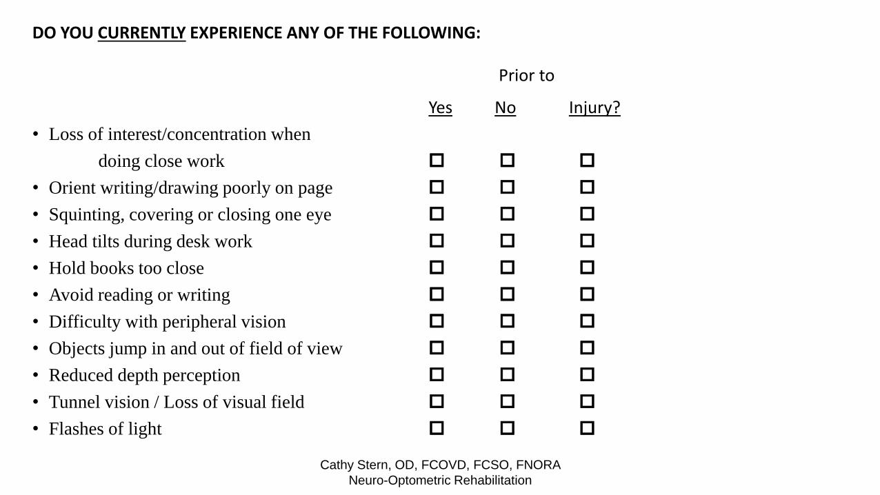
DO YOU CURRENTLY EXPERIENCE ANY OF THE FOLLOWING:
Prior to
Yes No Injury?
• Loss of interest/concentration when
doing close work
• Orient writing/drawing poorly on page
• Squinting, covering or closing one eye
• Head tilts during desk work
• Hold books too close
• Avoid reading or writing
• Difficulty with peripheral vision
• Objects jump in and out of field of view
• Reduced depth perception
• Tunnel vision / Loss of visual field
• Flashes of light

Cathy Stern, OD, FCOVD, FCSO, FNORA
Neuro-Optometric Rehabilitation
Neuro-Optometric Rehabilitation vs. Low Vision Rehabilitation
Neuro Rehabilitation is relatively new and practiced primarily by optometrists
with more specialized training (FNORA)
Visual processing may be affected even when eyesight is good and it can
interfere with the rehabilitative process
It affects a person’s ability to integrate visual, kinesthetic, proprioceptive and
vestibular input and therefore affects output - walking, reading, writing, driving
Low Vision management is directed to sight improvement or environmental
accommodations – larger print, magnifiers, hand-held telescopes
Some patients will need both services

Cathy Stern, OD, FCOVD, FCSO, FNORA
Neuro-Optometric Rehabilitation
Vision Screening
Stereopsis – depth perception
Worth 4 dot test - a test for suppression

Cathy Stern, OD, FCOVD, FCSO, FNORA
Neuro-Optometric Rehabilitation
Randot Stereo Test Stereo Fly, Stereo Animals
and Wirt Circles

Cathy Stern, OD, FCOVD, FCSO, FNORA
Neuro-Optometric Rehabilitation
Worth Four Dot Test (W4D)

Cathy Stern, OD, FCOVD, FCSO, FNORA
Neuro-Optometric Rehabilitation

Vision Screening
Visual Tracking and Locating
Convergence Ability (convergence insufficiency)
Eye Teaming – accurate, stamina, stability
Cathy Stern, OD, FCOVD, FCSO, FNORA Neuro-Optometric Rehabilitation

Cathy Stern, OD, FCOVD, FCSO, FNORA
Neuro-Optometric Rehabilitation
NSUCO Maples Oculomotor Test
Pursuits Have the patient stand (if possible) with their arms at their
sides and instruct them to follow a fixation target with
their eyes. Hold the target approximately 16 inches from
the patient and trace a circle with an 8 inch radius from
primary gaze at least two times in a clockwise direction
and two times in the counter-clockwise direction. Monitor
and record the patient’s ability to complete each rotation,
to maintain fixation of the target, and any head or body
movements made during the testing.

Cathy Stern, OD, FCOVD, FCSO, FNORA
Neuro-Optometric Rehabilitation
NSUCO Maples Oculomotor Test
Saccades Have the patient stand (if possible)with their arms at their sides
and feet at shoulder’s width. The procedure involves two
targets. Hold the two fixation targets 16 inches from the
patient with a target positioned horizontally 4 inches from the
right and left of the midline, respectively. Have the patient
fixate one target and then instruct them to quickly look at the
other target. Repeat each cycle four more times.[It is reasonable
to test saccades in vertical and diagonal orientations if more
data is desired]. Monitor and record any over- or under-
shooting of fixation, the ability to complete the task, and any
head or body movements.

Cathy Stern, OD, FCOVD, FCSO, FNORA
Neuro-Optometric Rehabilitation
Southern California College of Optometry (SCCO)
4+ System for Evaluating Oculomotility
Pursuit ability: test binocularly
• 4+ Smooth and accurate
• 3+ One fixation loss
• 2+ Two fixation losses
• 1+ More than two fixation losses or any uncontrolled head movements
Saccadic ability: test binocularly
• 4+ Smooth and accurate
• 3+ Some slight undershooting
• 2+ Gross undershooting or overshooting or increased latency
• 1+ Inability to do task or any uncontrolled head movement
• Normal latency of initiating a saccade is 120 to 180 milliseconds

Cathy Stern, OD, FCOVD, FCSO, FNORA
Neuro-Optometric Rehabilitation
Assessing Convergence
A good screening test is Near Point of Convergence.
This provides quantitative and qualitative information.
• Quantitative or inches fusion breaks from the
nose.
• Qualitative or the ease, comfort and stability with
which the patient holds fusion.

Nearpoint of Convergence (NPC)
Requires a small near picture target
Ask the student to look at the target as you move it toward his/her nose
Record the distance in inches from the nose an eye drifts outward and/or the student reports seeing two images. This should be no greater than 3 inches
Reverse direction and record the distance in inches from the nose when the student regains fusion of the eyes. This should be no greater than 6 inches
Cathy Stern, OD, FCOVD, FCSO, FNORA Neuro-Optometric Rehabilitation

Brock String
Cathy Stern, OD, FCOVD, FCSO, FNORA Neuro-Optometric Rehabilitation

Important Visual Factors
Fixation / Visual Tracking / Locating
Focus Flexibility
Near Point of Convergence and Eye Teaming
Visual – Vestibular
These skills lead to sustained visual attention
over time and faster processing speed
Cathy Stern, OD, FCOVD, FCSO, FNORA Neuro-Optometric Rehabilitation

Cognitive Load
1. basic pursuit – follow the tip of my pen
2. now ask the person to repeat the task while
spelling or trying simple math problems
this is more representative of performance in the real world
Cathy Stern, OD, FCOVD, FCSO, FNORA Neuro-Optometric Rehabilitation

Start with 10 flash cards of tic-tac-toe boards each with one letter X and
one letter O. Flash each card quickly and have try to reproduce the
image on a page of blank tic-tac-toe grids.
Allow the person to compare the images and point out any mismatches.
For higher level visualization, number the tic-tac-toe board 1-9 from upper left to lower right. Play tic-tac-toe by having each player say the number of the square they wish to place their X or O. See if the child or adult can tell if and when there is a winner.
TIC-TAC-TOE FLASH
Cathy Stern, OD, FCOVD, FCSO, FNORA Neuro-Optometric Rehabilitation

X
o
Cathy Stern, OD, FCOVD, FCSO, FNORA Neuro-Optometric Rehabilitation

Cathy Stern, OD, FCOVD, FCSO, FNORA Neuro-Optometric Rehabilitation

Cathy Stern, OD, FCOVD, FCSO, FNORA
Neuro-Optometric Rehabilitation
Treatment Alternatives Visual field expanding devices
Prism expansion system
Yoked prisms
Vision therapy for fixation and scanning
Optical and non-optical aids for Low Vision
Reading - markers ("L")
Turn book 90 degrees
Watch sizing/ spacing of print and lighting
Management or monitoring of eye disease

Cathy Stern, OD, FCOVD, FCSO, FNORA
Neuro-Optometric Rehabilitation
Treatment Alternatives
Neuro-Optometric Vision Rehabilitation - an individualized treatment
The goal is the treatment of visual processing and the enhancement of visual performance to meet the needs of each individual
Neuro-Optometric Vision Rehabilitation should be the recommended treatment for visual conditions that include ocular motor and binocular vision dysfunctions, amblyopia, strabismus and visual motor/visual perceptual/visual processing disorders
The use of lenses, prisms, filters and tints along with active neuro-optometric vision rehabilitation is integral to treating post-concussion. The goal of the treatment is to alleviate symptoms, achieve desired visual outcomes, meet the patient’s needs and improve the patient’s quality of life.

Cathy Stern, OD, FCOVD, FCSO, FNORA
Neuro-Optometric Rehabilitation
1. How soon after my brain injury is diagnosed should I seek a vision evaluation?
2. What type of vision examination is necessary?
3. Will all eye doctors give the same examination?
4. How long will vision rehabilitation take?
5. What is Neuro-Optometric Vision Rehabilitation?
6. How do you find an optometrist specializing in neuro- optometric vision rehabilitation?

Web Sites
Neuro Optometric Rehabilitation Association - www.nora.cc
College of Optometrists in Vision Development - www.covd.org
Optometric Extension Program Foundation - www.oepf.org
College of Syntonic Optometry - www.syntonicphototherapy.com
American Optometric Association - www.aoa.org
Dr. Cathy Stern, OD, FCSO, FCOVD - www.MyVisionDoc.com
Cathy Stern, OD, FCOVD, FCSO, FNORA Neuro-Optometric Rehabilitation

Storm King Art Center Cathy Stern, OD, FCOVD, FCSO, FNORA
Neuro-Optometric Rehabilitation

Cathy Stern, OD, FCOVD, FCSO, FNORA
Neuro-Optometric Rehabilitation
![Bryan Concussion General Audience - 2015.pptx [Read-Only] · 2015-09-03 · CONCUSSION ‐16,400,000 MTBI and Post‐Concussion Syndrome ‐ 141,000 Concussion Management ‐1,550,000](https://img.pdfslide.us/doc/110x75/5fb548e39d237d0cb0684f4f/bryan-concussion-general-audience-2015pptx-read-only-2015-09-03-concussion.jpg)