Embed Size (px)
Citation preview
Glencoe Science
Chapter Resources
Cell Reproduction
Includes:
Reproducible Student Pages
ASSESSMENT
✔ Chapter Tests
✔ Chapter Review
HANDS-ON ACTIVITIES
✔ Lab Worksheets for each Student Edition Activity
✔ Laboratory Activities
✔ Foldables–Reading and Study Skills activity sheet
MEETING INDIVIDUAL NEEDS
✔ Directed Reading for Content Mastery
✔ Directed Reading for Content Mastery in Spanish
✔ Reinforcement
✔ Enrichment
✔ Note-taking Worksheets
TRANSPARENCY ACTIVITIES
✔ Section Focus Transparency Activities
✔ Teaching Transparency Activity
✔ Assessment Transparency Activity
Teacher Support and Planning
✔ Content Outline for Teaching
✔ Spanish Resources
✔ Teacher Guide and Answers
404-i-vi-MSS05-000000_CR 19.04.2004 12:08 Page i tammyb 301:goscanc:scanc404:layouts:
Glencoe Science
Photo CreditsSection Focus Transparency 1: (t) Royalty Free/CORBIS, (b) Dwight R. Kuhn;Section Focus Transparency 2: (tl) William Hamilton/SuperStock, (tr) Animals Animals/Lynn Stone,(b) Animals Animals/Bates Littlehales; Section Focus Transparency 3: Yann Arthus-Bertrand/CORBIS
Copyright © by The McGraw-Hill Companies, Inc. All rights reserved.Permission is granted to reproduce the material contained herein on the conditionthat such material be reproduced only for classroom use; be provided to students,teachers, and families without charge; and be used solely in conjunction with theCell Reproduction program. Any other reproduction, for use or sale, is prohibitedwithout prior written permission of the publisher.
Send all inquiries to:Glencoe/McGraw-Hill8787 Orion Place Columbus, OH 43240-4027
ISBN 0-07-867094-2
Printed in the United States of America.
1 2 3 4 5 6 7 8 9 10 024 09 08 07 06 05 04
404-i-vi-MSS05-000000_CR 3/20/04 8:42AM Pageiiimpos03301:goscanc:scanc404:layouts:
iii
To the Teacher iv
Reproducible Student Pages■ Hands-On Activities
MiniLAB: Modeling Mitosis . . . . . . . . . . . . . . . . . . . . . . . . . . . . . . . . . . . . . . . . . . . . . . . . . . . . . . . . . 3MiniLAB: Try at Home Modeling DNA Replication . . . . . . . . . . . . . . . . . . . . . . . . . . . . . . . . . . . . . . 4Lab: Mitosis in Plant Cells . . . . . . . . . . . . . . . . . . . . . . . . . . . . . . . . . . . . . . . . . . . . . . . . . . . . . . . . . . 5Lab: Use the Internet Mutations . . . . . . . . . . . . . . . . . . . . . . . . . . . . . . . . . . . . . . . . . . . . . . . . . . . . . 7Laboratory Activity 1: Modeling Cell Division in Early Development . . . . . . . . . . . . . . . . . . . . . . . . . 9Laboratory Activity 2: Examining Models of Chromosomes. . . . . . . . . . . . . . . . . . . . . . . . . . . . . . . . 11Foldables: Reading and Study Skills . . . . . . . . . . . . . . . . . . . . . . . . . . . . . . . . . . . . . . . . . . . . . . . . . 15
■ Meeting Individual NeedsExtension and Intervention
Directed Reading for Content Mastery . . . . . . . . . . . . . . . . . . . . . . . . . . . . . . . . . . . . . . . . . . . . . . . 17Directed Reading for Content Mastery in Spanish . . . . . . . . . . . . . . . . . . . . . . . . . . . . . . . . . . . . . . 21Reinforcement . . . . . . . . . . . . . . . . . . . . . . . . . . . . . . . . . . . . . . . . . . . . . . . . . . . . . . . . . . . . . . . . . . 25Enrichment . . . . . . . . . . . . . . . . . . . . . . . . . . . . . . . . . . . . . . . . . . . . . . . . . . . . . . . . . . . . . . . . . . . . 28Note-taking Worksheet . . . . . . . . . . . . . . . . . . . . . . . . . . . . . . . . . . . . . . . . . . . . . . . . . . . . . . . . . . . 31
■ AssessmentChapter Review . . . . . . . . . . . . . . . . . . . . . . . . . . . . . . . . . . . . . . . . . . . . . . . . . . . . . . . . . . . . . . . . . 35Chapter Test . . . . . . . . . . . . . . . . . . . . . . . . . . . . . . . . . . . . . . . . . . . . . . . . . . . . . . . . . . . . . . . . . . . . 37
■ Transparency ActivitiesSection Focus Transparency Activities. . . . . . . . . . . . . . . . . . . . . . . . . . . . . . . . . . . . . . . . . . . . . . . . 42Teaching Transparency Activity . . . . . . . . . . . . . . . . . . . . . . . . . . . . . . . . . . . . . . . . . . . . . . . . . . . . . 45Assessment Transparency Activity . . . . . . . . . . . . . . . . . . . . . . . . . . . . . . . . . . . . . . . . . . . . . . . . . . . 47
Teacher Support and PlanningContent Outline for Teaching . . . . . . . . . . . . . . . . . . . . . . . . . . . . . . . . . . . . . . . . . . . . . . . . . . . . . . T2Spanish Resources . . . . . . . . . . . . . . . . . . . . . . . . . . . . . . . . . . . . . . . . . . . . . . . . . . . . . . . . . . . . . . T5Teacher Guide and Answers . . . . . . . . . . . . . . . . . . . . . . . . . . . . . . . . . . . . . . . . . . . . . . . . . . . . . . . T9
Table of Contents
Additional Assessment Resources available with Glencoe Science:
• ExamView® Pro Testmaker• Assessment Transparencies• Performance Assessment in the Science Classroom• Standardized Test Practice Booklet• MindJogger Videoquizzes• Vocabulary PuzzleMaker at msscience.com• Interactive Chalkboard• The Glencoe Science Web site at: msscience.com• An interactive version of this textbook along with assessment resources are available
online at: mhln.com
404-i-vi-MSS05-000000_CR 3/20/04 8:42AM Pageiiiimpos03301:goscanc:scanc404:layouts:
Cop
yrig
ht ©
Gle
ncoe
/McG
raw
-Hill
,a d
ivis
ion
of t
he M
cGra
w-H
ill C
ompa
nies
,Inc
.
iv
This chapter-based booklet contains all of the resource materials to help you teachthis chapter more effectively. Within you will find:
Reproducible pages for ■ Student Assessment■ Hands-on Activities■ Meeting Individual Needs (Extension and Intervention)■ Transparency Activities
A teacher support and planning section including ■ Content Outline of the chapter■ Spanish Resources■ Answers and teacher notes for the worksheets
Hands-On ActivitiesMiniLAB and Lab Worksheets: Each of these worksheets is an expanded version of each laband MiniLAB found in the Student Edition. The materials lists, procedures, and questionsare repeated so that students do not need their texts open during the lab. Write-on rules areincluded for any questions. Tables/charts/graphs are often included for students to recordtheir observations. Additional lab preparation information is provided in the Teacher Guideand Answers section.
Laboratory Activities: These activities do not require elaborate supplies or extensive pre-labpreparations. These student-oriented labs are designed to explore science through a stimu-lating yet simple and relaxed approach to each topic. Helpful comments, suggestions, andanswers to all questions are provided in the Teacher Guide and Answers section.
Foldables: At the beginning of each chapter there is a Foldables: Reading & Study Skillsactivity written by renowned educator Dinah Zike that provides students with a tool thatthey can make themselves to organize some of the information in the chapter. Students maymake an organizational study fold, a cause and effect study fold, or a compare and contraststudy fold, to name a few. The accompanying Foldables worksheet found in this resourcebooklet provides an additional resource to help students demonstrate their grasp of theconcepts. The worksheet may contain titles, subtitles, text, or graphics students need tocomplete the study fold.
Meeting Individual Needs (Extension and Intervention)Directed Reading for Content Mastery: These worksheets are designed to provide studentswith learning difficulties with an aid to learning and understanding the vocabulary andmajor concepts of each chapter. The Content Mastery worksheets contain a variety of formatsto engage students as they master the basics of the chapter. Answers are provided in theTeacher Guide and Answers section.
To the Teacher
404-i-vi-MSS05-000000_CR 3/20/04 8:42 AM Page iv impos03 301:goscanc:scanc404:layouts:
Cop
yrig
ht ©
Gle
ncoe
/McG
raw
-Hill
,a d
ivis
ion
of t
he M
cGra
w-H
ill C
ompa
nies
,Inc
.
v
Directed Reading for Content Mastery (in Spanish): A Spanish version of the DirectedReading for Content Mastery is provided for those Spanish-speaking students who are learning English.
Reinforcement: These worksheets provide an additional resource for reviewing the con-cepts of the chapter. There is one worksheet for each section, or lesson, of the chapter.The Reinforcement worksheets are designed to focus primarily on science content and lesson vocabulary, although knowledge of the section vocabulary supports understanding ofthe content. The worksheets are designed for the full range of students; however, they willbe more challenging for your lower-ability students. Answers are provided in the TeacherGuide and Answers section.
Enrichment: These worksheets are directed toward above-average students and allow themto explore further the information and concepts introduced in the section. A variety offormats are used for these worksheets: readings to analyze; problems to solve; diagrams to examine and analyze; or a simple activity or lab which students can complete in the classroom or at home. Answers are provided in the Teacher Guide and Answers section.
Note-taking Worksheet: The Note-taking Worksheet mirrors the content contained in theteacher version—Content Outline for Teaching. They can be used to allow students to takenotes during class, as an additional review of the material in the chapter, or as study notesfor students who have been absent.
AssessmentChapter Review: These worksheets prepare students for the chapter test. TheChapter Review worksheets cover all major vocabulary, concepts, and objectives
of the chapter. The first part is a vocabulary review and the second part is a concept review.Answers and objective correlations are provided in the Teacher Guide and Answers section.
Chapter Test: The Chapter Test requires students to use process skills and understand content.Although all questions involve memory to some degree, you will find that your students willneed to discover relationships among facts and concepts in some questions, and to use higherlevels of critical thinking to apply concepts in other questions. Each chapter test normallyconsists of four parts: Testing Concepts measures recall and recognition of vocabulary andfacts in the chapter; Understanding Concepts requires interpreting information and morecomprehension than recognition and recall—students will interpret basic information anddemonstrate their ability to determine relationships among facts, generalizations, definitions,and skills; Applying Concepts calls for the highest level of comprehension and inference;Writing Skills requires students to define or describe concepts in multiple sentence answers.Answers and objective correlations are provided in the Teacher Guide and Answers section.
Transparency ActivitiesSection Focus Transparencies: These transparencies are designed to generate interestand focus students’ attention on the topics presented in the sections and/or to assess
prior knowledge. There is a transparency for each section, or lesson, in the Student Edition.The reproducible student masters are located in the Transparency Activities section. Theteacher material, located in the Teacher Guide and Answers section, includes TransparencyTeaching Tips, a Content Background section, and Answers for each transparency.
404-i-vi-MSS05-000000_CR 3/20/04 8:42 AM Page v impos03 301:goscanc:scanc404:layouts:
Cop
yrig
ht ©
Gle
ncoe
/McG
raw
-Hill
,a d
ivis
ion
of t
he M
cGra
w-H
ill C
ompa
nies
,Inc
.
vi
Teaching Transparencies: These transparencies relate to major concepts that will benefitfrom an extra visual learning aid. Most of these transparencies contain diagrams/photosfrom the Student Edition. There is one Teaching Transparency for each chapter. The TeachingTransparency Activity includes a black-and-white reproducible master of the transparencyaccompanied by a student worksheet that reviews the concept shown in the transparency.These masters are found in the Transparency Activities section. The teacher material includesTransparency Teaching Tips, a Reteaching Suggestion, Extensions, and Answers to StudentWorksheet. This teacher material is located in the Teacher Guide and Answers section.
Assessment Transparencies: An Assessment Transparency extends the chapter content andgives students the opportunity to practice interpreting and analyzing data presented incharts, graphs, and tables. Test-taking tips that help prepare students for success on stan-dardized tests and answers to questions on the transparencies are provided in the TeacherGuide and Answers section.
Teacher Support and PlanningContent Outline for Teaching: These pages provide a synopsis of the chapter by section,including suggested discussion questions. Also included are the terms that fill in the blanksin the students’ Note-taking Worksheets.
Spanish Resources: A Spanish version of the following chapter features are included in thissection: objectives, vocabulary words and definitions, a chapter purpose, the chapter Activi-ties, and content overviews for each section of the chapter.
404-i-vi-MSS05-000000_CR 3/20/04 8:42 AM Page vi impos03 301:goscanc:scanc404:layouts:
Cell Reproduction 1
ReproducibleStudent Pages
Reproducible Student Pages■ Hands-On Activities
MiniLAB: Modeling Mitosis . . . . . . . . . . . . . . . . . . . . . . . . . . . . . . . . . 3MiniLAB: Try at Home Modeling DNA Replication. . . . . . . . . . . . . . . 4Lab: Mitosis in Plant Cells. . . . . . . . . . . . . . . . . . . . . . . . . . . . . . . . . . . 5Lab: Use the Internet Mutations. . . . . . . . . . . . . . . . . . . . . . . . . . . . . . 7Laboratory Activity 1: Modeling Cell Division in Early Development . 9Laboratory Activity 2: Examining Models of Chromosomes . . . . . . . . 11Foldables: Reading and Study Skills. . . . . . . . . . . . . . . . . . . . . . . . . . 15
■ Meeting Individual NeedsExtension and Intervention
Directed Reading for Content Mastery . . . . . . . . . . . . . . . . . . . . . . . 17Directed Reading for Content Mastery in Spanish . . . . . . . . . . . . . . 21Reinforcement . . . . . . . . . . . . . . . . . . . . . . . . . . . . . . . . . . . . . . . . . . 25Enrichment. . . . . . . . . . . . . . . . . . . . . . . . . . . . . . . . . . . . . . . . . . . . . 28Note-taking Worksheet . . . . . . . . . . . . . . . . . . . . . . . . . . . . . . . . . . . 31
■ AssessmentChapter Review . . . . . . . . . . . . . . . . . . . . . . . . . . . . . . . . . . . . . . . . . 35Chapter Test . . . . . . . . . . . . . . . . . . . . . . . . . . . . . . . . . . . . . . . . . . . . 37
■ Transparency ActivitiesSection Focus Transparency Activities . . . . . . . . . . . . . . . . . . . . . . . . 42Teaching Transparency Activity . . . . . . . . . . . . . . . . . . . . . . . . . . . . . 45Assessment Transparency Activity . . . . . . . . . . . . . . . . . . . . . . . . . . . 47
404-1-48-MSS05-000000_CR 3/20/04 8:43 AM Page 1 impos03 301:goscanc:scanc404:layouts:
2 Cell Reproduction
Hands-OnActivities
Hands-On Activities
404-1-48-MSS05-000000_CR 3/20/04 8:43 AM Page 2 impos03 301:goscanc:scanc404:layouts:
Cop
yrig
ht ©
Gle
ncoe
/McG
raw
-Hill
,a d
ivis
ion
of t
he M
cGra
w-H
ill C
ompa
nies
,Inc
.
Cell Reproduction 3
Name Date Class
Modeling MitosisProcedure1. Make models of cell division using materials supplied by your teacher.
2. Use four chromosomes in your model.
3. When finished, arrange the models in the order in which mitosis occurs.
Analysis1. In which steps is the nucleus visible?
2. How many cells does a dividing cell form?
Hand
s-On
Act
iviti
es
404-1-48-MSS05-000000_CR 3/20/04 8:43 AM Page 3 impos03 301:goscanc:scanc404:layouts:
4 Cell Reproduction
Name Date Class
Modeling DNA ReplicationProcedure1. Suppose you have a segment of DNA that is six nitrogen base pairs in
length. In the space below, using the letters A, T, C, and G, write a combina-tion of six pairs, remembering that A and T are always a pair and C and Gare always a pair.
2. Duplicate your segment of DNA. In the space below, diagram how thishappens and show the new DNA segments.
AnalysisCompare the order of bases of the original DNA to the new DNA molecules.
Hands-On Activities
Cop
yrig
ht ©
Gle
ncoe
/McG
raw
-Hill
,a d
ivis
ion
of t
he M
cGra
w-H
ill C
ompa
nies
,Inc
.
404-1-48-MSS05-000000_CR 3/20/04 8:43 AM Page 4 impos03 301:goscanc:scanc404:layouts:
Cop
yrig
ht ©
Gle
ncoe
/McG
raw
-Hill
,a d
ivis
ion
of t
he M
cGra
w-H
ill C
ompa
nies
,Inc
.
Cell Reproduction 5
Name Date Class
Lab PreviewDirections: Answer these questions before you begin the Lab.
1. Why should you examine the root tip under high and low power?
2. What is the root cap?
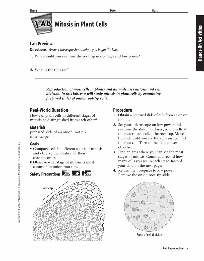
Reproduction of most cells in plants and animals uses mitosis and cell division. In this lab, you will study mitosis in plant cells by examiningprepared slides of onion root-tip cells.
Real-World QuestionHow can plant cells in different stages ofmitosis be distinguished from each other?
Materialsprepared slide of an onion root tipmicroscope
Goals■ Compare cells in different stages of mitosis
and observe the location of theirchromosomes.
■ Observe what stage of mitosis is most common in onion root tips.
Safety Precautions
Procedure1. Obtain a prepared slide of cells from an onion
root tip.2. Set your microscope on low power and
examine the slide. The large, round cells atthe root tip are called the root cap. Movethe slide until you see the cells just behindthe root cap. Turn to the high-power objective.
3. Find an area where you can see the moststages of mitosis. Count and record howmany cells you see in each stage. Recordyour data on the next page.
4. Return the nosepiece to low power.Remove the onion root-tip slide.
Mitosis in Plant Cells
Hand
s-On
Act
iviti
es
Root cap
Zone of cell division
404-1-48-MSS05-000000_CR 3/20/04 8:43 AM Page 5 impos03 301:goscanc:scanc404:layouts:
6 Cell Reproduction
Cop
yrig
ht ©
Gle
ncoe
/McG
raw
-Hill
,a d
ivis
ion
of t
he M
cGra
w-H
ill C
ompa
nies
,Inc
.
Name Date Class
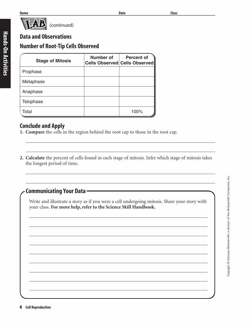
Data and Observations
Number of Root-Tip Cells Observed
Hands-On Activities
Communicating Your Data
Write and illustrate a story as if you were a cell undergoing mitosis. Share your story withyour class. For more help, refer to the Science Skill Handbook.
Conclude and Apply1. Compare the cells in the region behind the root cap to those in the root cap.
2. Calculate the percent of cells found in each stage of mitosis. Infer which stage of mitosis takesthe longest period of time.
(continued)
Stage of Mitosis
Prophase
Metaphase
Anaphase
Telophase
Total
Number of Cells Observed
Percent of Cells Observed
100%
404-1-48-MSS05-000000_CR 3/20/04 8:43 AM Page 6 impos03 301:goscanc:scanc404:layouts:
Cop
yrig
ht ©
Gle
ncoe
/McG
raw
-Hill
,a d
ivis
ion
of t
he M
cGra
w-H
ill C
ompa
nies
,Inc
.
Cell Reproduction 7
Name Date Class
Mutations can result in dominant or recessive genes. A recessive characteristic can appear onlyif an organism has two recessive genes for that characteristic. However, a dominant characteristic can appear if an organism has one or two dominant genes for that characteristic.Why do some mutations result in more common traits while others do not?
Real-World QuestionForm a hypothesis about how a mutation canbecome a common trait.
Goals■ Observe traits of various animals.■ Research how mutations become traits.■ Gather data about mutations.■ Make a frequency table of your findings and
communicate them to other students.
Data SourceVisit msscience.com formore information on
common genetic traits in different animals,recessive and dominant genes, and data fromother students.
Make a Plan1. Observe common traits in various ani-
mals, such as household pets or animalsyou might see in a zoo.
2. Learn what genes carry these traits in eachanimal.
3. Research the traits to discover which ones areresults of mutations. Are all mutations domi-nant? Are any of these mutations beneficial?
Follow Your Plan1. Make sure your teacher approves your plan
before you start.2. Visit the link shown below to access differ-
ent Web sites for information about muta-tions and genetics.
3. Decide if a mutation is beneficial, harmful,or neither. Record your data in your Sci-ence Journal.
Analyze Your Data1. Record in your Science Journal a list of
traits that are results of mutations.2. Describe an animal, such as a pet or an
animal you’ve seen in the zoo. Point outwhich traits are known to be the result of amutation.
3. Make a chart that compares recessivemutations to dominant mutations. Whichare more common?
4. Share your data with other students byposting it at the link shown on the nextpage.
Use the Internet
Mutations
Hand
s-On
Act
iviti
es
404-1-48-MSS05-000000_CR 3/20/04 8:43 AM Page 7 impos03 301:goscanc:scanc404:layouts:
Conclude and Apply1. Compare your findings to those of your classmates and other data at the link shown below.
What were some of the traits your classmates found that you did not? Which were the mostcommon?
2. Look at your chart of mutations. Are all mutations beneficial? When might a mutation beharmful to an organism?
3. Predict how your data would be affected if you had performed this lab when one of these com-mon mutations first appeared. Do you think you would see more or less animals with this trait?
4. Mutations occur every day but we only see a few of them. Infer how many mutations overmillions of years can lead to a new species.
8 Cell Reproduction
Cop
yrig
ht ©
Gle
ncoe
/McG
raw
-Hill
,a d
ivis
ion
of t
he M
cGra
w-H
ill C
ompa
nies
,Inc
.
Name Date Class
Hands-On Activities
(continued)
Communicating Your Data
Find this lab using the link below. Post your data in the table provided. Combine your datawith that of other students and make a chart that shows all of the data.
msscience.com
404-1-48-MSS05-000000_CR 3/20/04 8:43 AM Page 8 impos03 301:goscanc:scanc404:layouts:
Cop
yrig
ht ©
Gle
ncoe
/McG
raw
-Hill
,a d
ivis
ion
of t
he M
cGra
w-H
ill C
ompa
nies
,Inc
.
Cell Reproduction 9
Name Date Class
Modeling Cell Division in EarlyDevelopment
Every person starts out as a single cell. Cell division is responsible for the development of ababy from the single cell. As the single cell begins developing, cell division results in exponentialgrowth in the number of cells present. Exponential growth is growth that occurs at an ever-increasing rate. On a graph, exponential growth is represented by a J-shaped curve.
StrategyYou will model how cell division results in exponential growth in the number of cells in a
developing human.You will determine why exponential growth cannot continue indefinitely during human
development.You will infer why uncontrolled cell division, which occurs in cancer, can be so harmful to
human health.
Materialsuncooked ricepaper cups (11)graph paper
Procedure1. Obtain a container of uncooked rice from
your teacher. Each grain of rice representsone human cell.
2. Place one grain of rice in a paper cup. Thisgrain of rice represents the single cell thatresults when sperm and egg unite.
3. Label paper cups 1 through 10, and placethem in a row next to the cup containingthe original cell. During the first 10 celldivisions, the cells in the developinghuman all have the same cell-cycle length.
4. Place two grains of rice into cup 1 to represent the number of cells present afterthe original cell undergoes the first roundof mitosis. Record the number 2 in thetable in the Data and Observations section.
5. Place grains of rice into cup 2 to representthe number of cells that will be presentafter the second round of mitosis. Recordthe number of cells in your data table.
6. Repeat step 5 for cups 3 through 10.7. Using your data and graph paper, make a
line graph that shows the growth in thenumbers of cells. Label the x-axis Numberof cell divisions and the y-axis Number ofcells.
LaboratoryActivity11
Hand
s-On
Act
iviti
es
404-1-48-MSS05-000000_CR 3/20/04 8:43 AM Page 9 impos03 301:goscanc:scanc404:layouts:
10 Cell Reproduction
Cop
yrig
ht ©
Gle
ncoe
/McG
raw
-Hill
,a d
ivis
ion
of t
he M
cGra
w-H
ill C
ompa
nies
,Inc
.
Laboratory Activity 1 (continued)
Name Date Class
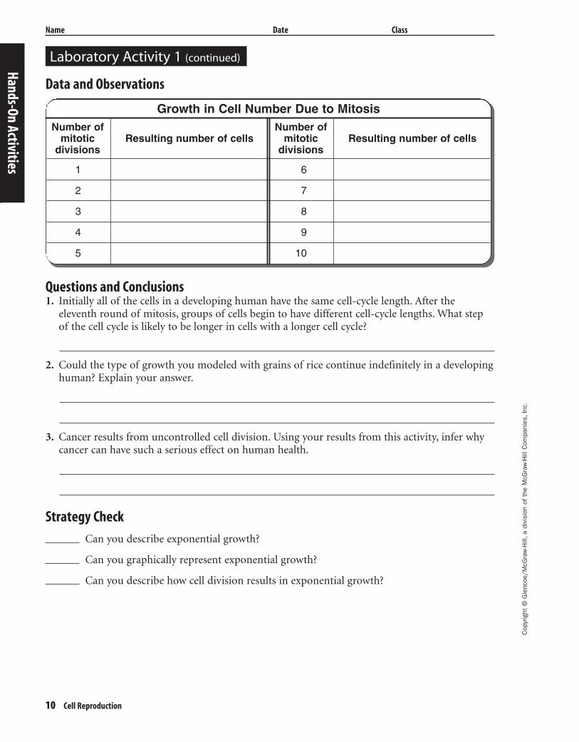
Data and Observations
Questions and Conclusions1. Initially all of the cells in a developing human have the same cell-cycle length. After the
eleventh round of mitosis, groups of cells begin to have different cell-cycle lengths. What stepof the cell cycle is likely to be longer in cells with a longer cell cycle?
2. Could the type of growth you modeled with grains of rice continue indefinitely in a developinghuman? Explain your answer.
3. Cancer results from uncontrolled cell division. Using your results from this activity, infer whycancer can have such a serious effect on human health.
Strategy Check
Can you describe exponential growth?
Can you graphically represent exponential growth?
Can you describe how cell division results in exponential growth?Hands-On Activities
Growth in Cell Number Due to MitosisNumber of Number of
mitotic Resulting number of cells mitotic Resulting number of cellsdivisions divisions
1 6
2 7
3 8
4 9
5 10
404-1-48-MSS05-000000_CR 3/20/04 8:43 AM Page 10 impos03 301:goscanc:scanc404:layouts:
Cop
yrig
ht ©
Gle
ncoe
/McG
raw
-Hill
,a d
ivis
ion
of t
he M
cGra
w-H
ill C
ompa
nies
,Inc
.
Cell Reproduction 11
Name Date Class
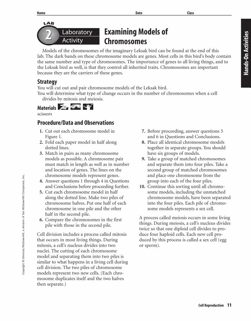
Examining Models ofChromosomes
Models of the chromosomes of the imaginary Leksak bird can be found at the end of this lab. The dark bands on these chromosome models are genes. Most cells in this bird’s body containthe same number and type of chromosomes. The importance of genes to all living things, and tothe Leksak bird as well, is that they control all inherited traits. Chromosomes are importantbecause they are the carriers of these genes.
StrategyYou will cut out and pair chromosome models of the Leksak bird.You will determine what type of change occurs in the number of chromosomes when a cell
divides by mitosis and meiosis.
Materials scissors
Procedure/Data and Observations
LaboratoryActivity22
Hand
s-On
Act
iviti
es
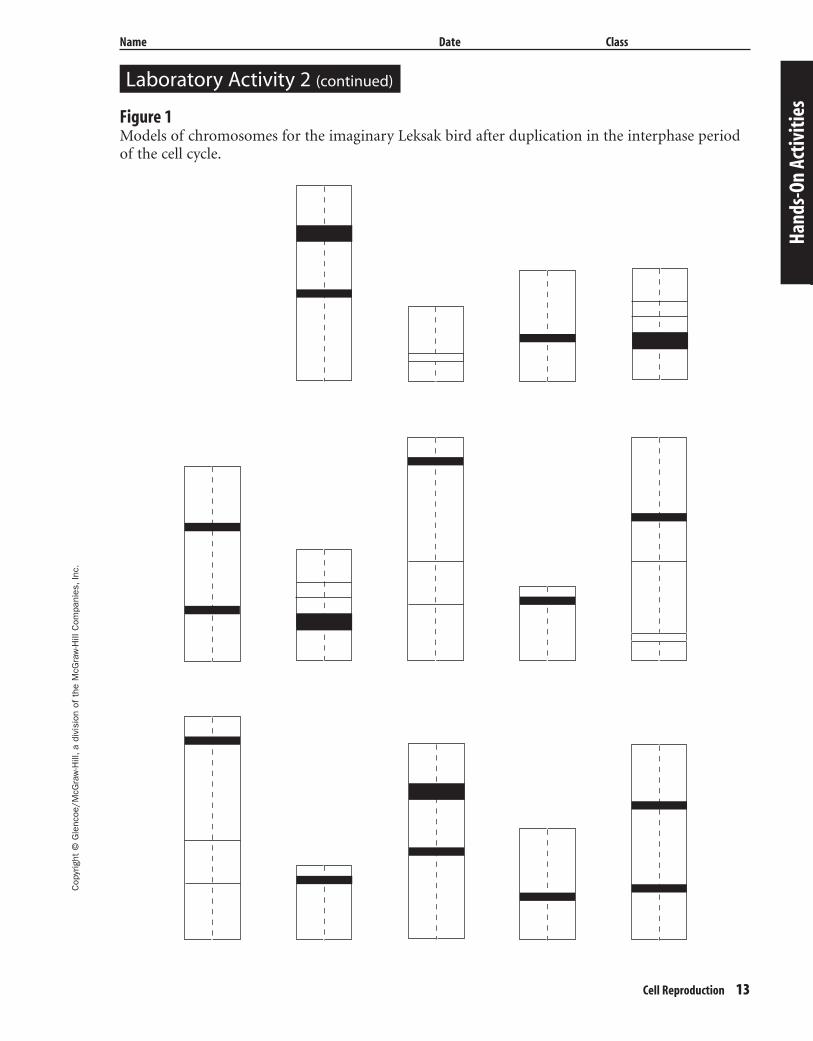
1. Cut out each chromosome model in Figure 1.
2. Fold each paper model in half along dotted lines.
3. Match in pairs as many chromosomemodels as possible. A chromosome pairmust match in length as well as in numberand location of genes. The lines on thechromosome models represent genes.
4. Answer questions 1 through 4 in Questionsand Conclusions before proceeding further.
5. Cut each chromosome model in halfalong the dotted line. Make two piles ofchromosome halves. Put one half of eachchromosome in one pile and the otherhalf in the second pile.
6. Compare the chromosomes in the firstpile with those in the second pile.
Cell division includes a process called mitosisthat occurs in most living things. Duringmitosis, a cell’s nucleus divides into twonuclei. The cutting of each chromosomemodel and separating them into two piles issimilar to what happens in a living cell duringcell division. The two piles of chromosomemodels represent two new cells. (Each chro-mosome duplicates itself and the two halvesthen separate.)
7. Before proceeding, answer questions 5and 6 in Questions and Conclusions.
8. Place all identical chromosome modelstogether in separate groups. You shouldhave six groups of models.
9. Take a group of matched chromosomesand separate them into four piles. Take asecond group of matched chromosomesand place one chromosome from thegroup into each of the four piles.
10. Continue this sorting until all chromo-some models, including the unmatchedchromosome models, have been separatedinto the four piles. Each pile of chromo-some models represents a sex cell.
A process called meiosis occurs in some livingthings. During meiosis, a cell’s nucleus dividestwice so that one diploid cell divides to pro-duce four haploid cells. Each new cell pro-duced by this process is called a sex cell (eggor sperm).
404-1-48-MSS05-000000_CR 3/20/04 8:43 AM Page 11 impos03 301:goscanc:scanc404:layouts:
12 Cell Reproduction
Cop
yrig
ht ©
Gle
ncoe
/McG
raw
-Hill
,a d
ivis
ion
of t
he M
cGra
w-H
ill C
ompa
nies
,Inc
.
Laboratory Activity 2 (continued)
Name Date Class
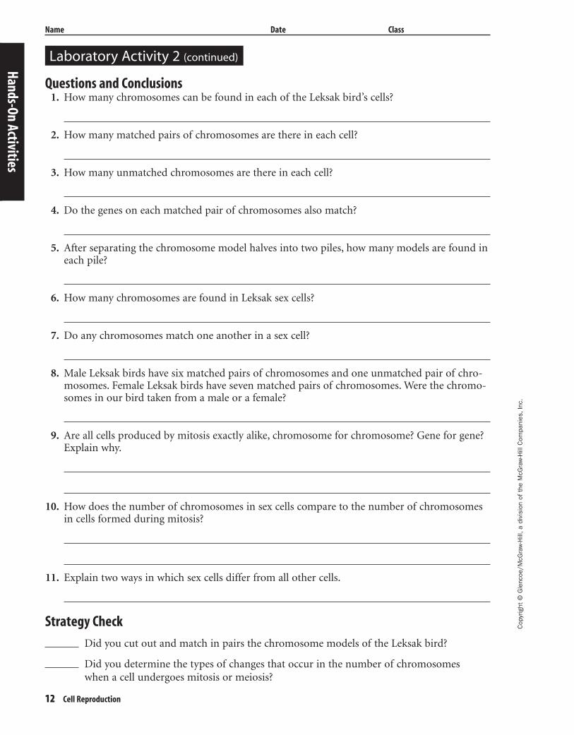
Questions and Conclusions1. How many chromosomes can be found in each of the Leksak bird’s cells?
2. How many matched pairs of chromosomes are there in each cell?
3. How many unmatched chromosomes are there in each cell?
4. Do the genes on each matched pair of chromosomes also match?
5. After separating the chromosome model halves into two piles, how many models are found ineach pile?
6. How many chromosomes are found in Leksak sex cells?
7. Do any chromosomes match one another in a sex cell?
8. Male Leksak birds have six matched pairs of chromosomes and one unmatched pair of chro-mosomes. Female Leksak birds have seven matched pairs of chromosomes. Were the chromo-somes in our bird taken from a male or a female?
9. Are all cells produced by mitosis exactly alike, chromosome for chromosome? Gene for gene?Explain why.
10. How does the number of chromosomes in sex cells compare to the number of chromosomesin cells formed during mitosis?
11. Explain two ways in which sex cells differ from all other cells.
Strategy Check
Did you cut out and match in pairs the chromosome models of the Leksak bird?
Did you determine the types of changes that occur in the number of chromosomes when a cell undergoes mitosis or meiosis?
Hands-On Activities
404-1-48-MSS05-000000_CR 3/20/04 8:43 AM Page 12 impos03 301:goscanc:scanc404:layouts:
Cop
yrig
ht ©
Gle
ncoe
/McG
raw
-Hill
,a d
ivis
ion
of t
he M
cGra
w-H
ill C
ompa
nies
,Inc
.
Cell Reproduction 13
Name Date Class
Hand
s-On
Act
iviti
es
Laboratory Activity 2 (continued)
Figure 1Models of chromosomes for the imaginary Leksak bird after duplication in the interphase periodof the cell cycle.
404-1-48-MSS05-000000_CR 3/20/04 8:43 AM Page 13 impos03 301:goscanc:scanc404:layouts:

404-1-48-MSS05-000000_CR 3/20/04 8:43 AM Page 14 impos03 301:goscanc:scanc404:layouts:
Cop
yrig
ht ©
Gle
ncoe
/McG
raw
-Hill
,a d
ivis
ion
of t
he M
cGra
w-H
ill C
ompa
nies
,Inc
.
Cell Reproduction 15
Cell Reproduction
Directions: Use this page to label your Foldable at the beginning of the chapter.
Chromatid pairs line up in the center of the cell.
Each day, cells in your body wear out and are replaced.
The chromatid pairs are visible and the spindle begins to form.
The chromosomes duplicate.
The chromosomes separate.
The cytoplasm begins to separate.
You grow because cell division increases the total number of cells in your body.
Name Date Class
Hand
s-On
Act
iviti
es
404-1-48-MSS05-000000_CR 3/20/04 8:43 AM Page 15 impos03 301:goscanc:scanc404:layouts:
16 Cell Reproduction
Meeting IndividualNeeds
Meeting Individual Needs
404-1-48-MSS05-000000_CR 3/20/04 8:43 AM Page 16 impos03 301:goscanc:scanc404:layouts:
Cop
yrig
ht ©
Gle
ncoe
/McG
raw
-Hill
,a d
ivis
ion
of t
he M
cGra
w-H
ill C
ompa
nies
,Inc
.
Name Date Class
Cell Reproduction 17
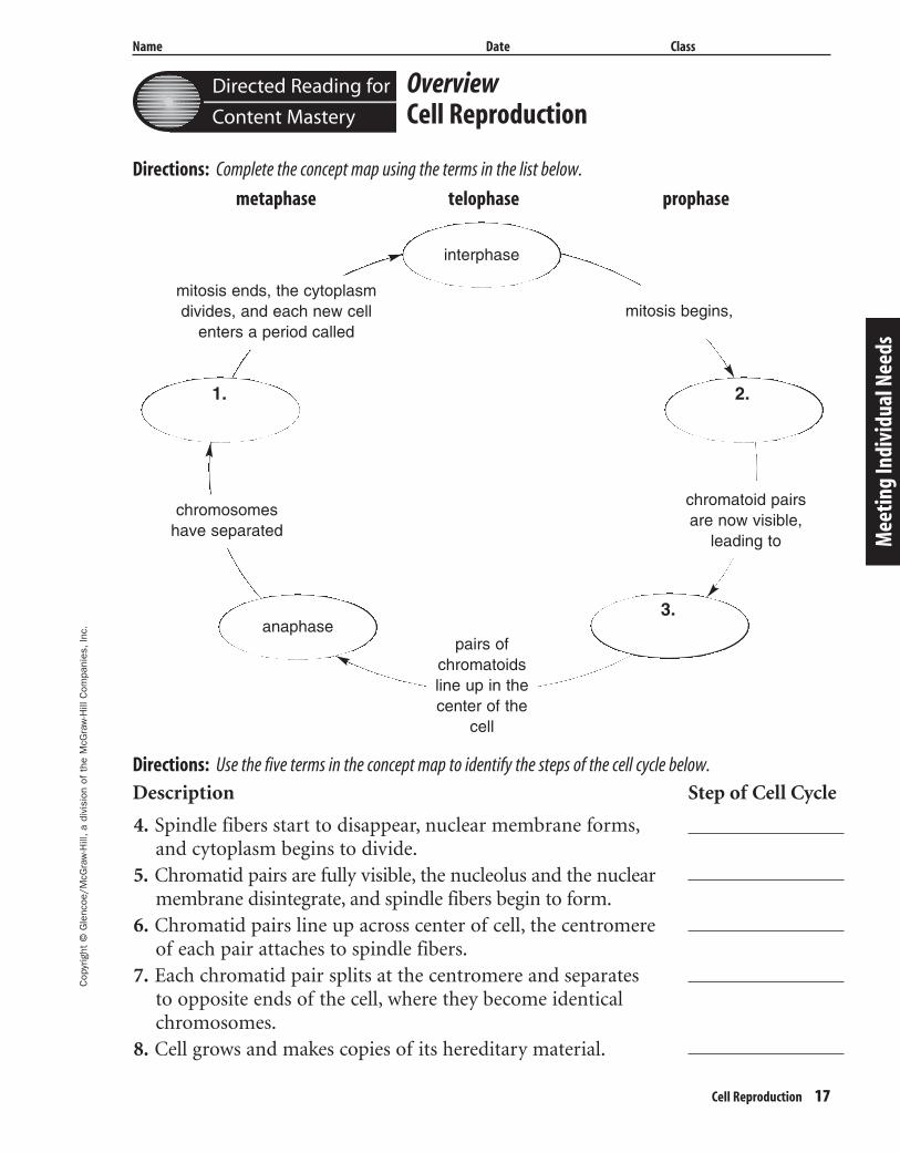
OverviewCell Reproduction
Directions: Complete the concept map using the terms in the list below.
metaphase telophase prophase
Directions: Use the five terms in the concept map to identify the steps of the cell cycle below.
Description
4. Spindle fibers start to disappear, nuclear membrane forms,and cytoplasm begins to divide.
5. Chromatid pairs are fully visible, the nucleolus and the nuclearmembrane disintegrate, and spindle fibers begin to form.
6. Chromatid pairs line up across center of cell, the centromereof each pair attaches to spindle fibers.
7. Each chromatid pair splits at the centromere and separatesto opposite ends of the cell, where they become identicalchromosomes.
8. Cell grows and makes copies of its hereditary material.
Step of Cell Cycle
Mee
ting
Indi
vidu
al N
eeds
Directed Reading for
Content Mastery
1. 2.
3.
interphase
anaphase
chromosomeshave separated
chromatoid pairsare now visible,
leading to
mitosis begins,mitosis ends, the cytoplasmdivides, and each new cell
enters a period called
pairs of chromatoidsline up in thecenter of the
cell
404-1-48-MSS05-000000_CR 3/20/04 8:43 AM Page 17 impos03 301:goscanc:scanc404:layouts:
Name Date Class
18 Cell Reproduction
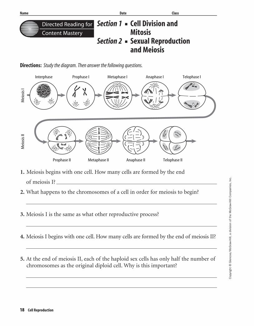
Section 1 ■ Cell Division andMitosis
Section 2 ■ Sexual Reproductionand Meiosis
Directions: Study the diagram. Then answer the following questions.
1. Meiosis begins with one cell. How many cells are formed by the end
of meiosis I?
2. What happens to the chromosomes of a cell in order for meiosis to begin?
3. Meiosis I is the same as what other reproductive process?
4. Meiosis I begins with one cell. How many cells are formed by the end of meiosis II?
5. At the end of meiosis II, each of the haploid sex cells has only half the number ofchromosomes as the original diploid cell. Why is this important?
Cop
yrig
ht ©
Gle
ncoe
/McG
raw
-Hill
,a d
ivis
ion
of t
he M
cGra
w-H
ill C
ompa
nies
,Inc
.
Interphase
Mei
osis
IM
eios
is II
Prophase I Metaphase I Anaphase I Telophase I
Prophase II Metaphase II Anaphase II Telophase II
Directed Reading for
Content Mastery
404-1-48-MSS05-000000_CR 3/20/04 8:43 AM Page 18 impos03 301:goscanc:scanc404:layouts:
Cop
yrig
ht ©
Gle
ncoe
/McG
raw
-Hill
,a d
ivis
ion
of t
he M
cGra
w-H
ill C
ompa
nies
,Inc
.
Name Date Class
Cell Reproduction 19
Section 3 ■ DNA
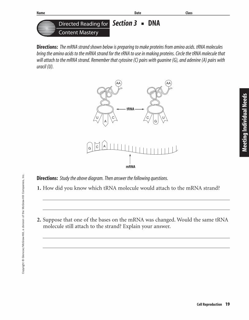
Directions: The mRNA strand shown below is preparing to make proteins from amino acids. tRNA moleculesbring the amino acids to the mRNA strand for the rRNA to use in making proteins. Circle the tRNA molecule thatwill attach to the mRNA strand. Remember that cytosine (C) pairs with guanine (G), and adenine (A) pairs withuracil (U).
Directions: Study the above diagram. Then answer the following questions.
1. How did you know which tRNA molecule would attach to the mRNA strand?
2. Suppose that one of the bases on the mRNA was changed. Would the same tRNAmolecule still attach to the strand? Explain your answer.
Mee
ting
Indi
vidu
al N
eeds
ACU
AA AA
CG
U
tRNA
mRNA
ACG
Directed Reading for
Content Mastery
404-1-48-MSS05-000000_CR 3/20/04 8:43 AM Page 19 impos03 301:goscanc:scanc404:layouts:
Name Date Class
20 Cell Reproduction
Key TermsCell Reproduction
Directions: Select the term from the following list that matches each description.
asexual chromosome diploid DNA eggs
genes haploid meiosis mitosis mutation
RNA sexual sperm zygote fertilization
1. Many cells in your body grow and divide every day bywhat process?
2. What structure in a cell’s nucleus holds the hereditaryinformation?
3. term for the joining of an egg and sperm
4. the sections of DNA that contain instructions for producing specific proteins
5. What are male sex cells called?
6. What cell forms when an egg and a sperm join?
7. the term for any permanent change in a gene or chromosome
8. the type of reproduction that produces a new organism;with identical chromosomes to those of the parent organism.
9. the process that produces haploid sex cells
10. an organism grows and functions by following the information in this code
11. the term for female sex cells
12. Cells with pairs of chromosomes are this.
13. type of reproduction that requires the joining of two sex cells
14. This type of nucleic acid carries the information neededto make proteins.
15. cells that do not have pairs of chromosomes (sex cells)
Cop
yrig
ht ©
Gle
ncoe
/McG
raw
-Hill
,a d
ivis
ion
of t
he M
cGra
w-H
ill C
ompa
nies
,Inc
.
Directed Reading for
Content Mastery
404-1-48-MSS05-000000_CR 3/20/04 8:43 AM Page 20 impos03 301:goscanc:scanc404:layouts:
Cop
yrig
ht ©
Gle
ncoe
/McG
raw
-Hill
,a d
ivis
ion
of t
he M
cGra
w-H
ill C
ompa
nies
,Inc
.
Nombre Fecha Clase
Reproducción celular 21
Satis
face
las n
eces
idad
es in
divi
dual
es
Lectura dirigida para
Dominio del contenido
Cop
yrig
ht ©
Gle
ncoe
/McG
raw
-Hill
,a d
ivis
ion
of t
he M
cGra
w-H
ill C
ompa
nies
,Inc
.SinopsisReproducción celular
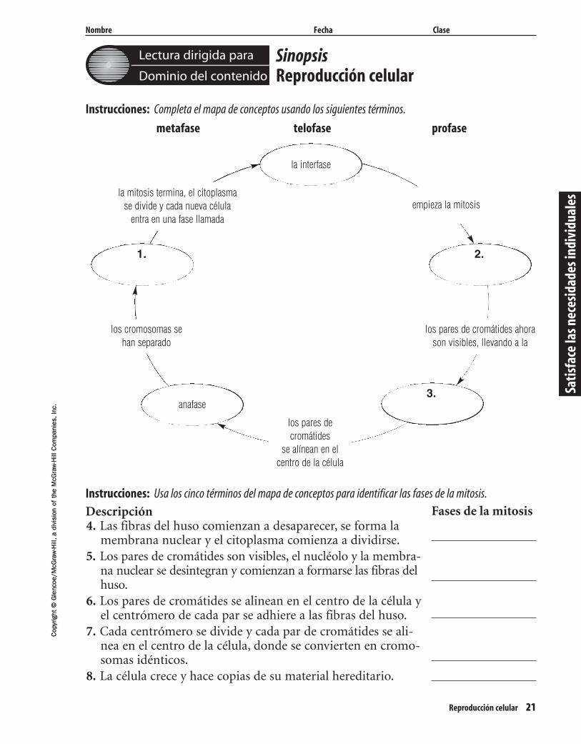
Instrucciones: Completa el mapa de conceptos usando los siguientes términos.
metafase telofase profase
Instrucciones: Usa los cinco términos del mapa de conceptos para identificar las fases de la mitosis.
Descripción4. Las fibras del huso comienzan a desaparecer, se forma la
membrana nuclear y el citoplasma comienza a dividirse.5. Los pares de cromátides son visibles, el nucléolo y la membra-
na nuclear se desintegran y comienzan a formarse las fibras delhuso.
6. Los pares de cromátides se alinean en el centro de la célula yel centrómero de cada par se adhiere a las fibras del huso.
7. Cada centrómero se divide y cada par de cromátides se ali-nea en el centro de la célula, donde se convierten en cromo-somas idénticos.
8. La célula crece y hace copias de su material hereditario.
Fases de la mitosis
1. 2.
3.
la interfase
anafase
los cromosomas sehan separado
los pares de cromátides ahorason visibles, llevando a la
empieza la mitosisla mitosis termina, el citoplasma
se divide y cada nueva célulaentra en una fase llamada
los pares decromátides
se alínean en elcentro de la célula
404-1-48-MSS05-000000_CR 3/20/04 8:43 AM Page 21 impos03 301:goscanc:scanc404:layouts:
Nombre Fecha Clase
Cop
yrig
ht ©
Gle
ncoe
/McG
raw
-Hill
,a d
ivis
ion
of t
he M
cGra
w-H
ill C
ompa
nies
,Inc
.
Satisface las necesidades individuales
Lectura dirigida para
Dominio del contenido
22 Reproducción celular
Sección 1 ■ División celular y mitosis
Sección 2 ■ Reproducción sexual y meiosis
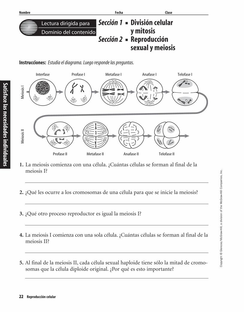
Instrucciones: Estudia el diagrama. Luego responde las preguntas.
1. La meiosis comienza con una célula. ¿Cuántas células se forman al final de lameiosis I?
2. ¿Qué les ocurre a los cromosomas de una célula para que se inicie la meiosis?
3. ¿Qué otro proceso reproductor es igual la meiosis I?
4. La meiosis I comienza con una sola célula. ¿Cuántas células se forman al final de lameiosis II?
5. Al final de la meiosis II, cada célula sexual haploide tiene sólo la mitad de cromo-somas que la célula diploide original. ¿Por qué es esto importante?
Interfase
Mei
osis
IM
eios
is II
Profase I Metafase I Anafase I Telofase I
Profase II Metafase II Anafase II Telofase II
404-1-48-MSS05-000000_CR 3/20/04 8:43 AM Page 22 impos03 301:goscanc:scanc404:layouts:
Cop
yrig
ht ©
Gle
ncoe
/McG
raw
-Hill
,a d
ivis
ion
of t
he M
cGra
w-H
ill C
ompa
nies
,Inc
.
Nombre Fecha Clase
Reproducción celular 23
Satis
face
las n
eces
idad
es in
divi
dual
es
Lectura dirigida para
Dominio del contenido
Sección 3 ■ DNA
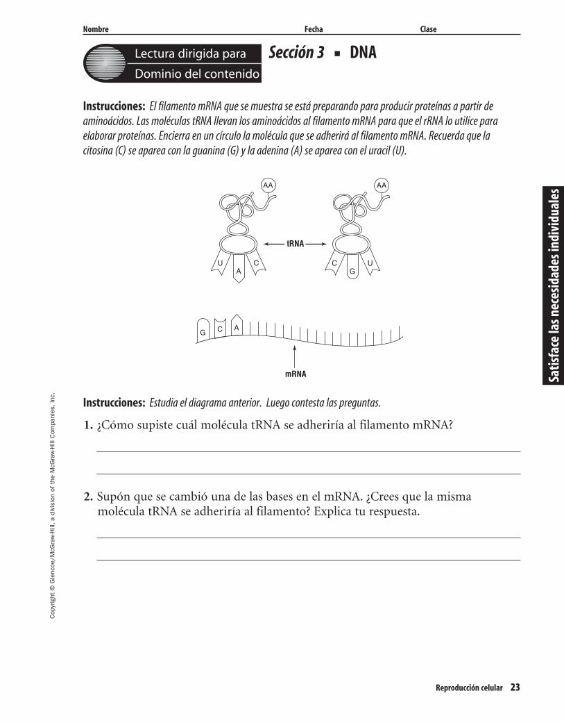
Instrucciones: El filamento mRNA que se muestra se está preparando para producir proteínas a partir deaminoácidos. Las moléculas tRNA llevan los aminoácidos al filamento mRNA para que el rRNA lo utilice para elaborar proteínas. Encierra en un círculo la molécula que se adherirá al filamento mRNA. Recuerda que lacitosina (C) se aparea con la guanina (G) y la adenina (A) se aparea con el uracil (U).
Instrucciones: Estudia el diagrama anterior. Luego contesta las preguntas.
1. ¿Cómo supiste cuál molécula tRNA se adheriría al filamento mRNA?
2. Supón que se cambió una de las bases en el mRNA. ¿Crees que la mismamolécula tRNA se adheriría al filamento? Explica tu respuesta.
ACU
AA AA
CG
U
tRNA
mRNA
ACG
404-1-48-MSS05-000000_CR 3/20/04 8:43 AM Page 23 impos03 301:goscanc:scanc404:layouts:
Nombre Fecha Clase
24 Reproducción celular
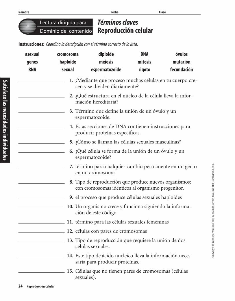
Instrucciones: Coordina la descripción con el término correcto de la lista.
asexual cromosoma diploide DNA óvulos
genes haploide meiosis mitosis mutación
RNA sexual espermatozoide cigoto fecundación
1. ¿Mediante qué proceso muchas células en tu cuerpo cre-cen y se dividen diariamente?
2. ¿Qué estructura en el núcleo de la célula lleva la infor-mación hereditaria?
3. Término que define la unión de un óvulo y un espermatozoide.
4. Estas secciones de DNA contienen instrucciones paraproducir proteínas específicas.
5. ¿Cómo se llaman las células sexuales masculinas?
6. ¿Qué célula se forma de la unión de un óvulo y un espermatozoide?
7. término para cualquier cambio permanente en un gen oen un cromosoma
8. Tipo de reproducción que produce nuevos organismos;con cromosomas idénticos al organismo progenitor.
9. el proceso que produce células sexuales haploides
10. Un organismo crece y funciona siguiendo la informa-ción de este código.
11. término para las células sexuales femeninas
12. células con pares de cromosomas
13. Tipo de reproducción que requiere la unión de doscélulas sexuales.
14. Este tipo de ácido nucleico lleva la información nece-saria para producir proteínas.
15. Células que no tienen pares de cromosomas (células sexuales).
Cop
yrig
ht ©
Gle
ncoe
/McG
raw
-Hill
,a d
ivis
ion
of t
he M
cGra
w-H
ill C
ompa
nies
,Inc
.
Satisface las necesidades individuales
Lectura dirigida para
Dominio del contenido
Términos clavesReproducción celular
404-1-48-MSS05-000000_CR 3/20/04 8:43 AM Page 24 impos03 301:goscanc:scanc404:layouts:
Cop
yrig
ht ©
Gle
ncoe
/McG
raw
-Hill
,a d
ivis
ion
of t
he M
cGra
w-H
ill C
ompa
nies
,Inc
.
Name Date Class
Cell Reproduction 25
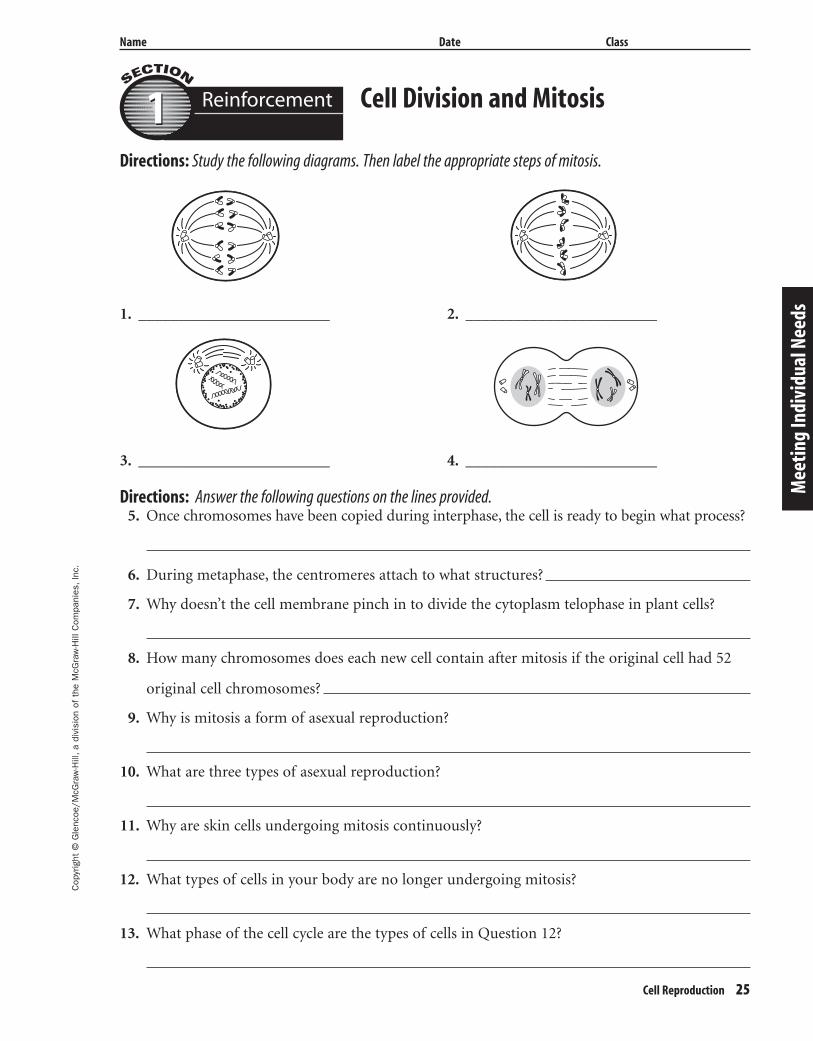
Directions: Study the following diagrams. Then label the appropriate steps of mitosis.
1. ________________________ 2. ________________________
3. ________________________ 4. ________________________
Directions: Answer the following questions on the lines provided.5. Once chromosomes have been copied during interphase, the cell is ready to begin what process?
6. During metaphase, the centromeres attach to what structures?
7. Why doesn’t the cell membrane pinch in to divide the cytoplasm telophase in plant cells?
8. How many chromosomes does each new cell contain after mitosis if the original cell had 52
original cell chromosomes?
9. Why is mitosis a form of asexual reproduction?
10. What are three types of asexual reproduction?
11. Why are skin cells undergoing mitosis continuously?
12. What types of cells in your body are no longer undergoing mitosis?
13. What phase of the cell cycle are the types of cells in Question 12?
Cell Division and Mitosis
Mee
ting
Indi
vidu
al N
eeds
Reinforcement11
404-1-48-MSS05-000000_CR 3/20/04 8:43 AM Page 25 impos03 301:goscanc:scanc404:layouts:
26 Cell Reproduction
Cop
yrig
ht ©
Gle
ncoe
/McG
raw
-Hill
,a d
ivis
ion
of t
he M
cGra
w-H
ill C
ompa
nies
,Inc
.
Name Date Class
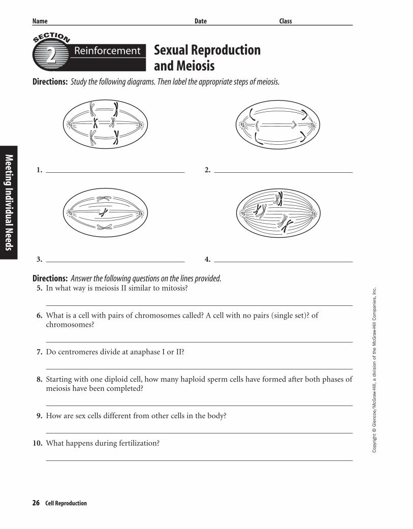
Sexual Reproduction and Meiosis
Directions: Study the following diagrams. Then label the appropriate steps of meiosis.
1. 2.
3. 4.
Reinforcement22
Meeting Individual Needs
Directions: Answer the following questions on the lines provided.5. In what way is meiosis II similar to mitosis?
6. What is a cell with pairs of chromosomes called? A cell with no pairs (single set)? ofchromosomes?
7. Do centromeres divide at anaphase I or II?
8. Starting with one diploid cell, how many haploid sperm cells have formed after both phases ofmeiosis have been completed?
9. How are sex cells different from other cells in the body?
10. What happens during fertilization?
404-1-48-MSS05-000000_CR 3/20/04 8:43 AM Page 26 impos03 301:goscanc:scanc404:layouts:
Cop
yrig
ht ©
Gle
ncoe
/McG
raw
-Hill
,a d
ivis
ion
of t
he M
cGra
w-H
ill C
ompa
nies
,Inc
.
Name Date Class
Cell Reproduction 27
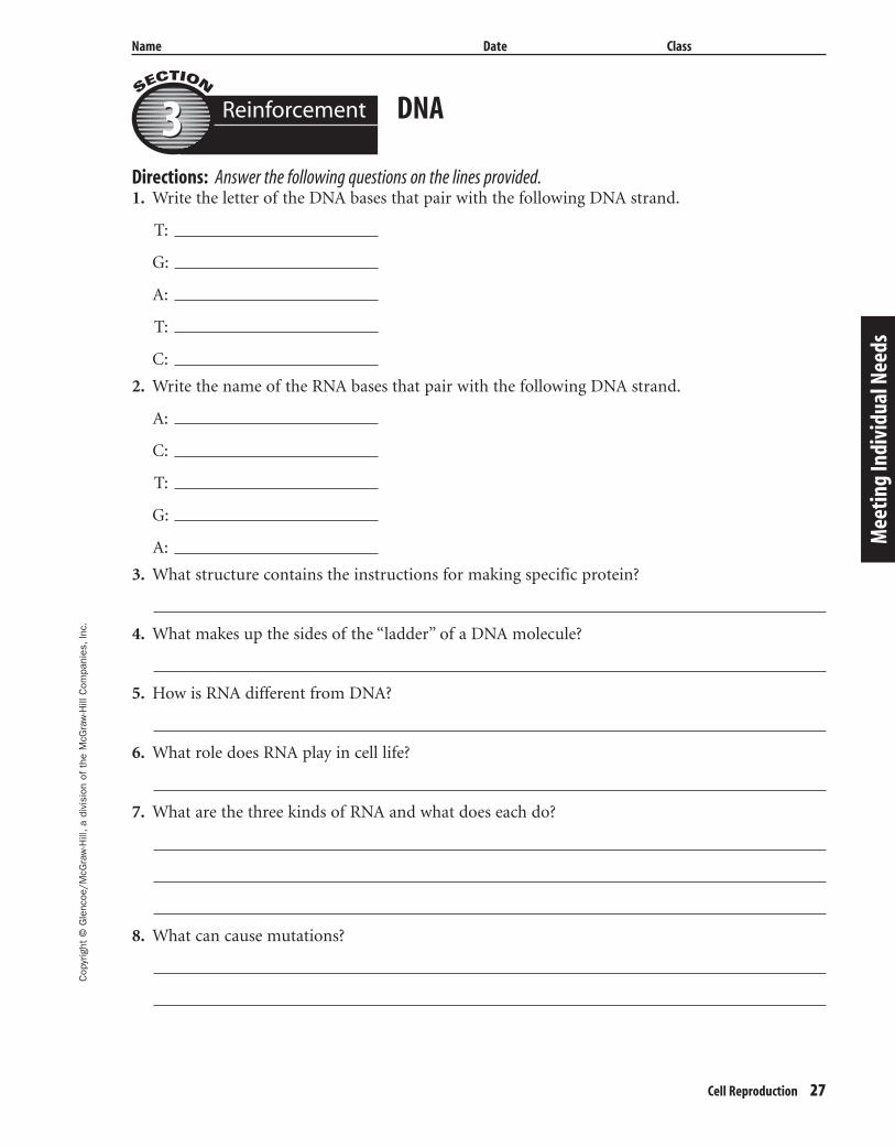
Directions: Answer the following questions on the lines provided.1. Write the letter of the DNA bases that pair with the following DNA strand.
T:
G:
A:
T:
C:
2. Write the name of the RNA bases that pair with the following DNA strand.
A:
C:
T:
G:
A:
3. What structure contains the instructions for making specific protein?
4. What makes up the sides of the “ladder” of a DNA molecule?
5. How is RNA different from DNA?
6. What role does RNA play in cell life?
7. What are the three kinds of RNA and what does each do?
8. What can cause mutations?
DNA
Mee
ting
Indi
vidu
al N
eeds
Reinforcement33
404-1-48-MSS05-000000_CR 3/20/04 8:43 AM Page 27 impos03 301:goscanc:scanc404:layouts:
28 Cell Reproduction
Cop
yrig
ht ©
Gle
ncoe
/McG
raw
-Hill
,a d
ivis
ion
of t
he M
cGra
w-H
ill C
ompa
nies
,Inc
.
Name Date Class
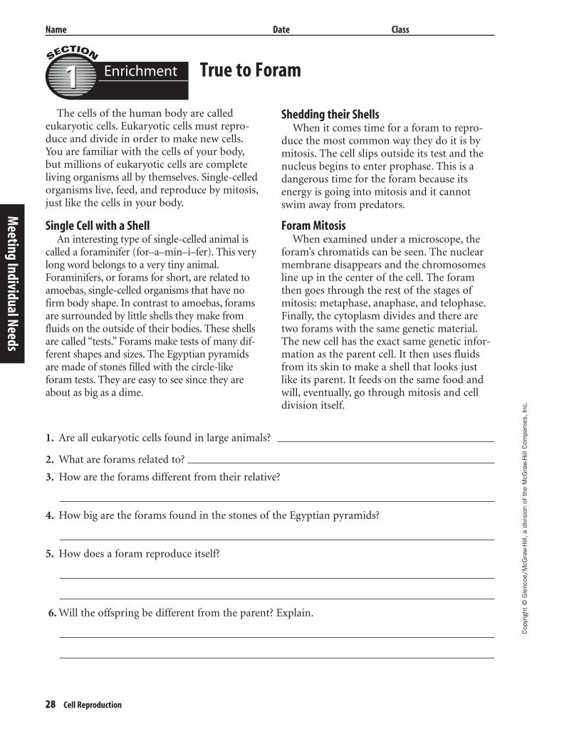
True to Foram
The cells of the human body are calledeukaryotic cells. Eukaryotic cells must repro-duce and divide in order to make new cells.You are familiar with the cells of your body,but millions of eukaryotic cells are completeliving organisms all by themselves. Single-celledorganisms live, feed, and reproduce by mitosis,just like the cells in your body.
Single Cell with a ShellAn interesting type of single-celled animal is
called a foraminifer (for–a–min–i–fer). This verylong word belongs to a very tiny animal.Foraminifers, or forams for short, are related toamoebas, single-celled organisms that have nofirm body shape. In contrast to amoebas, foramsare surrounded by little shells they make fromfluids on the outside of their bodies. These shellsare called “tests.” Forams make tests of many dif-ferent shapes and sizes. The Egyptian pyramidsare made of stones filled with the circle-likeforam tests. They are easy to see since they areabout as big as a dime.
Shedding their ShellsWhen it comes time for a foram to repro-
duce the most common way they do it is bymitosis. The cell slips outside its test and thenucleus begins to enter prophase. This is adangerous time for the foram because itsenergy is going into mitosis and it cannotswim away from predators.
Foram MitosisWhen examined under a microscope, the
foram’s chromatids can be seen. The nuclearmembrane disappears and the chromosomesline up in the center of the cell. The foramthen goes through the rest of the stages ofmitosis: metaphase, anaphase, and telophase.Finally, the cytoplasm divides and there aretwo forams with the same genetic material.The new cell has the exact same genetic infor-mation as the parent cell. It then uses fluidsfrom its skin to make a shell that looks justlike its parent. It feeds on the same food andwill, eventually, go through mitosis and celldivision itself.
1. Are all eukaryotic cells found in large animals?
2. What are forams related to?
3. How are the forams different from their relative?
4. How big are the forams found in the stones of the Egyptian pyramids?
5. How does a foram reproduce itself?
6. Will the offspring be different from the parent? Explain.
Meeting Individual Needs
Enrichment11
404-1-48-MSS05-000000_CR 3/20/04 8:43 AM Page 28 impos03 301:goscanc:scanc404:layouts:
Cop
yrig
ht ©
Gle
ncoe
/McG
raw
-Hill
,a d
ivis
ion
of t
he M
cGra
w-H
ill C
ompa
nies
,Inc
.
Name Date Class
Cell Reproduction 29
Crossing Over
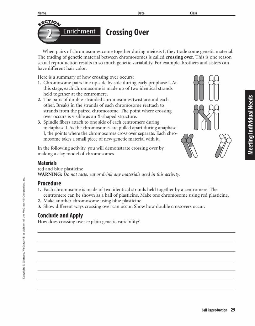
When pairs of chromosomes come together during meiosis I, they trade some genetic material.The trading of genetic material between chromosomes is called crossing over. This is one reasonsexual reproduction results in so much genetic variability. For example, brothers and sisters canhave different hair color.
In the following activity, you will demonstrate crossing over by making a clay model of chromosomes.
Materialsred and blue plasticineWARNING: Do not taste, eat or drink any materials used in this activity.
Procedure1. Each chromosome is made of two identical strands held together by a centromere. The
centromere can be shown as a ball of plasticine. Make one chromosome using red plasticine.2. Make another chromosome using blue plasticine.3. Show different ways crossing over can occur. Show how double crossovers occur.
Conclude and ApplyHow does crossing over explain genetic variability?
Mee
ting
Indi
vidu
al N
eeds
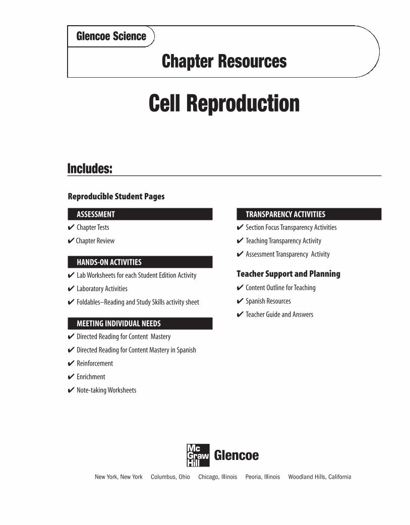
Here is a summary of how crossing over occurs:1. Chromosome pairs line up side by side during early prophase I. At
this stage, each chromosome is made up of two identical strandsheld together at the centromere.
2. The pairs of double-stranded chromosomes twist around eachother. Breaks in the strands of each chromosome reattach tostrands from the paired chromosome. The point where crossingover occurs is visible as an X-shaped structure.
3. Spindle fibers attach to one side of each centromere duringmetaphase I. As the chromosomes are pulled apart during anaphaseI, the points where the chromosomes cross over separate. Each chro-mosome takes a small piece of new genetic material with it.
Enrichment22
404-1-48-MSS05-000000_CR 3/20/04 8:43 AM Page 29 impos03 301:goscanc:scanc404:layouts:
30 Cell Reproduction
Cop
yrig
ht ©
Gle
ncoe
/McG
raw
-Hill
,a d
ivis
ion
of t
he M
cGra
w-H
ill C
ompa
nies
,Inc
.
Name Date Class
Radiation, Genes, and Mutations
Radiation is known to be dangerous tohuman bodies. Millions of body cells exposedto high-energy waves from X rays, radon gas,and ultra-violet radiation have been perma-nently harmed by these emissions. The DNA ofthe individual cells is too delicate to withstandthe energy produced by these kinds of radia-tion. The DNA molecules are torn apart orsuffer drastic changes in their genetic sequenc-ing which can lead to mutations.
Under normal conditions, DNA moleculesroutinely undergo some sort of genetic alter-ation. During replication, or copying of thecell, mistakes in gene sequencing often occur.However, the cell contains many repair mech-anisms that continually monitor and repairdamage to DNA strands.
Radiation and Gene DamageWhen cells are exposed to radiation, how-
ever, several types of molecular destructionare possible. The DNA is both physically andchemically broken (cleaved) by the highenergy waves. Often the repair of the DNAstrand by enzymes or other chemicals is not
adequate enough to put the DNA moleculeback together in its proper sequence.When replication occurs, the new strands ofDNA carry the new altered sequence of genes.As each generation of cells is produced the mutations continue to show up in thereplicated cells. These cells are often non-functional and become tumorous growthssuch as skin cancer.
RadonRadon gas is especially harmful to lung tis-
sues because it enters the body through regu-lar breathing in a building contaminated withradon. The emissions easily damage fragilelung tissues. Not only do the high-energyradioactive emissions destroy cellular DNA,but other large particles tear the cell mem-branes apart leaving the body strained to con-stantly repair the damaged tissues.
Fortunately, many forms of genetic alter-ation by radiation are preventable. Limitingexposure to X rays, using sunscreen, and test-ing buildings for radon levels can help preventdamage to a cell’s genetic makeup.
1. What makes radiation dangerous to cells?
2. How are skin cells damaged by exposure to ultra-violet radiation?
3. In what two ways does radon destroy genetic information in lung tissue cells?
4. Why do you think it’s important that a pregnant woman always tell her healthcare provider sheis pregnant before receiving any X ray examination?
Meeting Individual Needs
Enrichment33
404-1-48-MSS05-000000_CR 3/20/04 8:43 AM Page 30 impos03 301:goscanc:scanc404:layouts:
Cop
yrig
ht ©
Gle
ncoe
/McG
raw
-Hill
,a d
ivis
ion
of t
he M
cGra
w-H
ill C
ompa
nies
,Inc
.
Name Date Class
Cell Reproduction 31
Cell Reproduction
Section 1 Cell Division and Mitosis
A. Cell division—increases the number of cells and causes many-celled _____________ to grow
B. The Cell Cycle—series of events that takes place from one _____________ to the next
1. Cells have periods of formation, growth and development, and death called _______________.
2. Interphase—most of the life of any eukaryotic cell, or cell with a nucleus, is spent in a
period of __________ and _______________.
a. During interphase, a cell duplicates its _______________ and prepares for cell division.
b. After interphase, the nucleus divides, and then the ______________ separates to formtwo new cells.
C. Mitosis—process in which the nucleus divides to form two identical __________
1. Chromosome—structure in the nucleus that contains ______________ material
2. Prophase
a. Nucleolus and ____________________ disintegrate.
b. __________ move to opposite ends of the cell.
c. ______________ begin to stretch across the cell.
3. Metaphase—pairs of ______________ line up across the center of the cell.
4. Anaphase
a. Each ______________ divides.
b. Each pair of chromatids _____________ and moves to opposite ends of the cell.
5. _________—spindle fibers disappear and a new nucleus forms.
D. Division of the Cytoplasm—for most cells, the _____________ separates after the nucleusdivides.
1. In ______________ cells, the cell membrane pinches in the middle and the cytoplasm
divides.
2. In ______________ cells, a cell plate forms.
E. Results of mitosis
1. Each cell in your body, except sex cells, has a nucleus with ______ chromosomes.
2. Allows growth and ____________ worn out or damaged cells
Mee
ting
Indi
vidu
al N
eeds
Note-takingWorksheet
404-1-48-MSS05-000000_CR 3/20/04 8:43 AM Page 31 impos03 301:goscanc:scanc404:layouts:
32 Cell Reproduction
Cop
yrig
ht ©
Gle
ncoe
/McG
raw
-Hill
,a d
ivis
ion
of t
he M
cGra
w-H
ill C
ompa
nies
,Inc
.
Name Date Class
F. ____________________—a new organism is produced from one parent organism.
1. An organism with no nucleus divides into two identical organisms by ___________.
2. _______—a small, exact copy of the adult grows from the body of the parent.
3. In ________________, a whole new organism grows from each piece of the parent.
Section 2 Sexual Reproduction and Meiosis
A. ______ reproduction—two sex cells, usually an egg and a sperm, come together.
1. Fertilization—the joining of an _______ and a _________, generally from two differentorganisms of the same species
a. Sperm are formed in the ________ reproductive organs.
b. Eggs are formed in the __________ reproductive organs.
c. A cell that forms from fertilization is a __________.
2. Following fertilization, ___________ begins and a new organism develops.
3. Human body cells are ___________, because they have 23 pairs of similar chromosomes.
4. Human sex cells are ___________, because they have 23 single chromosomes.
B. _______—a process that produces haploid sex cells and ensures that offspring have the same
___________ number as its parent.
1. In meiosis I, the nucleus divides and produces two new cells with one duplicated
______________ each.
2. In meiosis II, the nuclei divide and the chromatids separate, producing ________ cells withhalf the number of chromosomes of the original nucleus.
Section 3 DNA
A. DNA—a ____________ that contains information that an organism needs to grow and function
1. Watson and _________ made an accurate model of DNA in 1953.
2. The structure of DNA is similar to a __________________.
a. The sides of the ladder are made up of _____________________________.
b. The rungs of the ladder are made up of __________________.
3. Before a cell divides, its DNA duplicates itself by unwinding and separating its sides, then
each side becomes a pattern on which a _____________ forms.
Meeting Individual Needs
Note-taking Worksheet (continued)
404-1-48-MSS05-000000_CR 3/20/04 8:43 AM Page 32 impos03 301:goscanc:scanc404:layouts:
Cop
yrig
ht ©
Gle
ncoe
/McG
raw
-Hill
,a d
ivis
ion
of t
he M
cGra
w-H
ill C
ompa
nies
,Inc
.
Name Date Class
Cell Reproduction 33
B. Genes—sections of _______ on a chromosome
1. Contain instructions for making specific ____________
2. RNA carries the _________ for making proteins from the nucleus to the ribosomes in thecytoplasm.
a. Messenger RNA carries the code that directs the order in which the _______________bond.
b. Ribosomal RNA makes up _____________, where proteins are built.
c. ________ RNA brings amino acids to the ribosomes to build the protein.
3. Cells use only the _________ that direct the making of proteins needed by that cell.
C. Mutations—any permanent __________ in the DNA sequence of a cell’s gene or chromosome
1. Can be caused by outside factors like X rays, ____________, and some chemicals
2. A change in a gene or chromosome can change the __________ of an organism.
Mee
ting
Indi
vidu
al N
eeds
Note-taking Worksheet (continued)
404-1-48-MSS05-000000_CR 3/20/04 8:43 AM Page 33 impos03 301:goscanc:scanc404:layouts: