Embed Size (px)
Citation preview

Developmental Biology 415 (2016) 24–32
Contents lists available at ScienceDirect
Developmental Biology
http://d0012-16
n CorrE-m
journal homepage: www.elsevier.com/locate/developmentalbiology
An unregulated regulator: Vasa expression in the development ofsomatic cells and in tumorigenesis
Jessica Poon, Gary M. Wessel, Mamiko Yajima n
MCB Department, Brown University, 185 Meeting Street, BOX-GL173, Providence, RI 02912, USA
a r t i c l e i n f o
Article history:Received 5 December 2015Received in revised form9 May 2016Accepted 11 May 2016Available online 11 May 2016
Keywords:VasaDDX4Germ line factorsTumorigenesisTranslation
x.doi.org/10.1016/j.ydbio.2016.05.01206/& 2016 Elsevier Inc. All rights reserved.
esponding author.ail address: [email protected] (M. Y
a b s t r a c t
Growing evidence in diverse organisms shows that genes originally thought to function uniquely in thegerm line may also function in somatic cells, and in some cases even contribute to tumorigenesis. Herewe review the somatic functions of Vasa, one of the most conserved “germ line” factors among me-tazoans. Vasa expression in somatic cells is tightly regulated and often transient during normal devel-opment, and appears to play essential roles in regulation of embryonic cells and regenerative tissues. Itsdysregulation, however, is believed to be an important element of tumorigenic cell regulation. In thisperspectives paper, we propose how some conserved functions of Vasa may be selected for somatic cellregulation, including its potential impact on efficient and localized translational activities and in somecases on cellular malfunctioning and tumorigenesis.
& 2016 Elsevier Inc. All rights reserved.
1. Introduction
More than 40 genes that are normally expressed selectively inthe germ line of many model organisms are also expressed invarious tumor types in humans (Simpson et al., 2005). Re-capitulating functions of a germ line program thus has been hy-pothesized to contribute to the characteristic features of neoplasticdiseases, including immortality, hypomethylation of DNA, andmetastatic migrations (Hanahan and Weinberg, 2011). Indeed, inCaenorhabditis elegans, mutations of the retinoblastoma (Rb) tu-mor suppressor complex induce somatic cells to express germ linegenes, leading cells to revert to patterns of gene expression nor-mally restricted to germ cells (Wang et al., 2005). Further, C. ele-gans mutants with decreased insulin-like signaling (e.g. daf-2, theinsulin-like receptor, and age-1, the downstream PI3K mutantstrains) cause mis-expression of the germ line factors pie-1 and pglin the somatic cells. These altered somatic cells are then moreresistant to stress-induced damage and have increased cellularlifespans (or negligible senescence) (Curran et al., 2009). This re-sult is particularly noteworthy considering C. elegans somatic cellsare genetically programmed to cease cell divisions after the animalreaches adulthood. These observations suggest that the acquisitionof germ line elements may contribute to somatic phenotypes, andeven of tumorgenic cells.
Vasa was first identified in Drosophila (Hay et al., 1988; Lasko
ajima).
and Ashburner, 1988) as a polar granule component consisting ofcytoplasmic granules inherited by posterior pole cells, which di-rects the fate of the cell to the germ line (Illmensee and Mahowald,1974). It is a member of the DEAD-box RNA helicase family (for themolecular classification and characterization of Vasa, see reviewsof Raz (2000), Linder (2006), Gustafson and Wessel (2010), andLasko (2013)), similar in sequence to eukaryotic initiation factor 4A(eIF4A) (Hay et al., 1988; Lasko and Ashburner, 1988). Vasa isconsidered to function as a translational regulator in the germ line(Hay et al., 1988; Lasko and Ashburner, 1988; Linder et al., 1989;Sengoku et al., 2006). Follow-up reports in Drosophila suggest thatVasa associates with eIF5B, an essential translation initiation factorrequired for joining of ribosomal subunits (Carrera et al., 2000;Pestova et al., 2000), and is involved in translation of gurken inoocytes and mei-P26 mRNA in germ line stem cells (Johnstone andLasko, 2004; Liu et al., 2010). A recent report in insect ovarian cellsalso suggests that Vasa binds to mRNAs with its DEAD-box domainand functions in the generation of germ line-specific, Piwi-inter-acting RNAs (piRNAs) that protect animal genomes against trans-posons and are essential for fertility (Xiol et al., 2014). Vasa thusappears to have broad functions, including translational regulationof specific mRNAs and piRNA generation, which contribute to cellfate determination within the germ line.
Vasa is one of the most conserved germ line factors in variousmetazoan organisms, based on sequence, expression patterns, andfunctions in the germ line. Its knockdown in major model organ-isms compromises the animal's reproductive capabilities withvarying severity between different organisms, or even between
J. Poon et al. / Developmental Biology 415 (2016) 24–32 25
sexes of the same species. For instance, Vasa knockdown in Dro-sophila results in sterility in females but not in males (Styhler et al.,1998; Lasko and Ashburner, 1990; Renault, 2012), whereas in miceit causes sterility in males but not in females (Tanaka et al., 2000).In C. elegans, GLH1, one of four Vasa homologs in this organism, isimportant both for germ cell development and for gamete ma-turation (Roussell and Bennett, 1993; Gruidl et al., 1996; Kuznickiet al., 2000; Spike et al., 2008). In zebrafish, maternal Vasa is stablethroughout early development and is necessary for initial germcell formation, and Vasa produced embryonically is required forgerm cell maintenance (Braat et al., 2001; Hartung et al., 2014). InMedaka, on the other hand, Vasa protein synthesized in the em-bryo is important for germ cell migration but not for survival (Liet al., 2009). The mechanism of these variable Vasa inputs amongdifferent species and sexes is unclear. It was recently proposedthat Vasa might have multiple different functions in each cell typeor organism by using different partner proteins (Dehghani andLasko, 2015). The identity of such distinct interactors though, re-mains unclear.
Recent reports in more diverse organisms suggest that Vasamight also function outside of the germ line. These somaticfunctions of Vasa include various types of embryonic cells (Yajimaand Wessel, 2011a; Schwager et al., 2014), regenerative tissues(Wagner et al., 2012; Yajima and Wessel, 2015), and even in tu-morigenic cells (Janic et al., 2010). These reports cast potentialadditional functions of Vasa and/or of Vasa's interacting proteinsin the soma. In this Perspectives paper, we speculate on thefunctional impact of Vasa in somatic cells as well as on potentialcontributions during tumorigenesis.
2. Vasa's contributions as a mitotic factor duringembryogenesis
Although expression and activities of Vasa are most prominentin the germ line, Vasa protein and/or mRNA is often widely ex-pressed in somatic lineages during embryogenesis of many or-ganisms (summarized in Table 1). Proteins and mRNAs are oftenstored maternally in the oocyte and embryo, and thus Vasa pre-sence in the resultant somatic cells does not necessarily indicate afunctional role. Initial observations in Drosophila showed thatmaternal Vasa contributes to the formation of both the germ cellsand the embryonic abdominal segments (Schupbach and Wie-schaus, 1986a, 1986b; Hay et al., 1990), implying a broad impact onembryogenesis. Flies mutant for Vasa embryonically, however, areviable as homozygotes, suggesting no essential zygotic roles insomatic development of Drosophila. Recent studies in the seaurchin and spider, on the other hand, demonstrated that Vasa doesfunction in somatic cells during embryogenesis. In the sea urchin,Vasa was found to localize to the nuclear periphery at S-phase ofthe cell cycle in blastomeres and accumulate on the mitotic ap-paratus at M-phase of every blastomere during early embryogen-esis. The knockdown of Vasa in these embryos resulted in defectsof proper spindle organization and chromosome segregation, cellcycle delays and often death of the embryo (Yajima and Wessel,2011a, 2011b). In spider embryogenesis, Vasa is localized to smallpuncta ubiquitously distributed throughout the cortex before PGCsemerged at the late germ band stage and its knockdown by RNAinterference resulted in defects of mitotic progression in the em-bryo (Schwager et al., 2014). Importantly, this function of Vasa as amitotic regulator was also reported in Drosophila germ line stemcells (Pek and Kai, 2011). In those cells, Vasa concentrated in theperinuclear region during S-phase and to the spindle and chro-mosome region during M-phase. Its functionality was highlightedin mitotic progression by regulating the localization of chromo-some-associated proteins, which in turn mediated chromosome
condensation and segregation (Pek and Kai, 2011). These reportssuggest a conserved function of Vasa as a mitotic regulator both inthe germ line and in the soma (Fig. 1).
Detailed functional contributions of Vasa in mitotic regulationof somatic cells are yet to be identified but it may “simply” functionto increase translation locally. A recent report in the sea urchinembryo suggests that Vasa interacts with most mRNAs in the celland its knockdown results in inhibition of overall protein synthesisdown to approximately 20% that of the normal embryo (Yajimaand Wessel, 2015). Vasa may thus function by contributing totranslation of a large number of mRNAs, which allows cells toproduce sufficient amounts of proteins for rapid cell replicationand differentiation during embryogenesis, especially in a large cellwith rapid cell fate transitions.
3. Vasa's function in the soma during regeneration andtumorigenesis
Vasa also appears to function within the soma during re-generation and somatic tumorigenesis. In planarian regenerativetissues, Vasa expression is upregulated (Wagner et al., 2012), as itis in Drosophila brain tumor cells (Janic et al., 2010), and even inhuman cancer cells (Hashimoto et al., 2008; Nguyen et al., 2011;Kim et al., 2014). In planarians, transcriptional profiling identifiedthat expression of several germ line genes are upregulated in adultproliferating neoblasts during regeneration. Among those genes,the vasa-like gene, vasa-1 was identified to be essential for pro-liferative expansion to promote the process of differentiation inneoblast descendants, suggesting its functional role in stem cellregulation (Wagner et al., 2012).
When a malignant brain tumor was induced in Drosophila (byinactivation of a polycomb repressor gene, lethal (3) malignantbrain tumor [l(3)mbt]; Gateff et al., 1993), 102 genes were found toincrease transcript levels specific to this mutant (Janic et al., 2010).Among these genes, a quarter of them (26 of 102) are knownfactors required for the Drosophila germ line. In addition to vasa,these genes included, for instance, nanos, necessary for pole plasmdevelopment (Wong et al., 2005) and piwi and aubergine, involvedin the biogenesis of Piwi interacting RNAs (piRNAs) that protectgerm line cells against transposable elements and viruses (Klat-tenhoff and Theurkauf, 2008; Patil and Kai, 2010). Importantly, notonly were these genes expressed in the tumor cells, but inhibitionof vasa, piwi, aubergine, or nanos halted tumor growth, suggestingthat the expression of these factors in the somatic tumor had anessential function. In this process of tumorigenesis regulation,interestingly, vasa was not upregulated at the mRNA level but itsprotein expression was increased. This is distinct from other germline factors such as piwi and aubergine, which were up-regulatedboth at the mRNA and protein levels during tumorigenesis. Thisfinding suggests that vasa mRNA (and potentially Vasa protein aswell) is expressed at low levels during normal soma development,and can be differentially regulated and utilized upon appropriatestimulation. This post-transcriptional regulation of Vasa is acommon theme (e.g. Wagner et al., 2012; Yajima andWessel, 2015)likely important for this gene function.
DDX4 (the Vasa ortholog in humans) expression was also foundin various types of cancer tissues in humans. In ovarian cancercells, DDX4 expression was significantly increased together withCD133, a cancer stem cell marker, whereas almost all CD133-ne-gative cells had no DDX4 expression, implying that DDX4 may playan important role in cancer stem cells (Kim et al., 2014). Further, inthe SKOV-3 (epithelial ovarian cancer) cell line, artificially forced-expression of DDX4 resulted in up-regulation of 10 proteins anddown-regulation of 6 proteins. Among these proteins altered byDDX4, 14-3-3s protein was down-regulated but its mRNA level

Table 1Summary of Vasa expression and functional contributions in the soma.
Organism mRNA Protein Functional contribution References
Fly (Drosophila melanogaster) Uniformly distributed during syncytial blastodermstage.
Uniformly distributed during syncytial blastodermstage.
Abdominal segment formation. Schupbach and Wieschaus, 1986;Lasko and Ashburner, 1988, 1990;Hay et al., 1988
Spider (Parasteatoda tepidariorum) Uniformly distributed in the early embryo, and be-comes highly expressed in mesoderm and ectodermafter germ band formation.
Uniformly distributed in the early embryo, and be-comes undetectable after germ band formation.
Early embryonic mitotic divisions andspindle integrity.
Schwager et al., 2014
Planarians (Dugesia japonica andSchmidtea mediterranea)
Expressed at the lower level in the neoblasts andhighly expressed in the growing blastema duringregeneration.
ND Neoblast proliferation and differ-entiation during regeneration.
Shibata et al., 1999; Wagner et al.,2012
Hydra (Hydra magnipapillata) Expressed in multipotent stem cells (i-cells) and ec-todermal epithelial cells.
ND ND Mochizuki et al., 2001
C. elegans Expressed in all cells of young embryos and reducedafter the 8- to 10-cell stage.
Background level ND Gruidl et al., 1996; Kuznicki et al.,2000
Polychaete (Platynereis dumeriliiand Capitella sp. I)
Uniformly distributed until the four cell stage, andbecomes confined to the ventral plate and the meso-dermal bands at 15 pf and to mesodermal posteriorgrowth zone (MPGZ) during larval and adult stages.
Distributed in the yolk free cytoplasm of all blas-tomeres at 4-cell stage, and becomes enriched tosecondary mesoblast cells and MPGZ during larvaland adult stages.
ND Rebscher et al., 2007; Dill andSeaver, 2008
Sea urchin (Strongylocentrotuspurpuratus)
Uniformly distributed in every cell until gastrula stage,and transiently expressed in the cells of the adultprecursor (rudiment).
Enriched on the mitotic apparatus of every earlyembryonic cell, and transiently expressed in the adultprecursor cells and wound-healing tissues.
Early embryonic mitotic cell cycleprogression, wound-healing, and de-velopmental reprograming.
Yajima and Wessel, 2011b, 2015
Ascidians (Ciona intestinalis) Uniformly distributed in each cell until 16-cell stageand widely expressed in the postplasm and the trunkregion.
Concentrated at the postplasm at 64-cell and en-riched in the posterior-most cells (the B7.6 cells) andthe trunk region.
ND Shirae-Kurabayashi et al., 2006
Zebrafish Localized to the distal ends of the first and secondcleavage planes at 4-cell stage.
Distributed throughout the embryo duringembryogenesis.
ND Yoon et al., 1997; Braat et al., 1999,2000; Kosaka et al., 2007
Frog (Xenopus laevis) Expressed during early embryogenesis by RT-PCRanalysis.
Uniformly expressed until tailbud stage, and becomesenriched in the mesoderm during early tadpole stage.
ND Ikenishi et al., 1996; Li et al., 2010
Chick ND Localized in the basal cleavage furrow in the earlyembryo, and becomes scattered in the hypoblast layerafter stage X.
ND Tsunekawa et al., 2000
ND, no data.
J.Poonet
al./Developm
entalBiology
415(2016)
24–32
26

Fig. 1. Vasa is involved in mitotic progression. A. Vasa localizes on the mitotic apparatus and is essential for cell cycle progression of sea urchin embryos (Adapted fromYajima and Wessel (2011a)) and Drosophila germ line stem cells (Adapted from Pek and Kai (2011)). Vasa is enriched in peri-nuclear regions prior to mitotic entry (Inter-phase), localizes on the mitotic apparatus during M-phase, and disappears at the end of mitosis, followed by peri-nuclear localization at the beginning of next mitotic entry.Vasa knockdown (right panels) in those cells result in defects of spindle organization and chromosome segregation. The knockdown image of sea urchin is an embryo at 16-cell stage, and that of Drosophila is a germ line stem cell at Anaphase. B. A cartoon diagram of Vasa protein (red) localization in the sea urchin embryonic cells (left panel) andDrosophila germline stem cells (middle panel). Both in sea urchin embryonic cells and Drosophila germ line stem cells, Vasa is evenly distributed over the spindle (green)and DNA (blue) during mitosis, and its knockdown (right panel) results in cell division defects, including abnormal chromosomal segregation. Further, in the sea urchin, Vasabecomes more enriched toward the vegetal blastomere (a future germ line) of the spindle at 16-cell stage by an unknown mechanism.
J. Poon et al. / Developmental Biology 415 (2016) 24–32 27
was not changed, suggesting that DDX4 may be involved in post-transcriptional regulation of 14-3-3s (Hashimoto et al., 2008).Importantly, 14-3-3s is frequently suppressed in human breast,gastric, lung and ovarian cancers (Ferguson et al., 2000; Iwata
et al., 2000; Osada et al., 2002; Akahira et al., 2004). 14-3-3s isknown to sequester the mitotic initiation complex (cdk1-cyclin B1)to the cytoplasm in response to the DNA damage, and induces cellcycle arrest for the G2 checkpoint (Chan et al., 1999). With the

J. Poon et al. / Developmental Biology 415 (2016) 24–3228
forced expression of DDX4 in which 14-3-3s is down-regulated,the SKOV-3 ovarian cancer cells appeared to ignore the G2checkpoint and underwent rapid cell cycle progression even in thepresence of DNA damage (Hashimoto et al., 2008). In these cells,DDX4 may have precociously nucleated the mitotic initiationcomplex and induced pre-mature mitotic entry by down-regulat-ing 14-3-3s activities. If this speculation is correct, DDX4 over-expressing cells could then continue to proliferate with damagedgenetic materials, which can eventually result in tumor formation.These speculations need to be tested through further experiments.One could test, for instance, if forced expression of 14-3-3s wouldrescue DDX4-overexpression phenotypes, slow down cell cycleprogression and enable repair of DNA damage in the genome. TheSKOV cell line used here is derived from ovarian carcinomas whosemolecular status and cellular dynamics may be quite differentfrom normal cells or even non-ovarian cancer cells. Another im-portant experiment is to test the protein content of cells with orwithout DDX4 present to determine if the proteome varies byDDX4's functionality in non-ovarian cells. Since Vasa protein levelin the cell is thought to be regulated by the Ubiquitin proteasome-dependent protein degradation mechanism (Liu et al., 2003;Styhler et al., 1998, 2002; Kugler et al., 2010; Woo et al., 2006a,2006b), introducing constitutively active forms of DDX4 into thecell would be important to identify its functional roles. For theseexperiments, one could mutate putative protein degradation re-cognition sites, or may target DDX4 expression to an ectopic sub-cellular region where ‘Vasa-regulators’ may be absent. These ex-periments both in non-oncogenic and in non-ovarian-cancer cellswill potentially reveal actual and conserved contributions of DDX4to cancer cell regulation.
These recent results in tumorgenic and cancerous cells suggestthat somatic cells that have been considered Vasa-null in theirnormal state could potentially induce Vasa expression throughpost-transcriptional regulation under certain stimuli. Indeed, inthe sea urchin embryo, Vasa mRNA and protein appears to beconsistently expressed at low levels in various types of somaticcells during normal development, but the protein can be up-regulated ‘transiently’ from the same amount of mRNA during theformation of adult precursor tissues, wound-healing, or develop-mental re-programming, processes which require cells to undergoactive proliferation and differentiation (Voronina et al., 2008; Ya-jima and Wessel, 2015). During these regulatory processes, im-portantly, the number of cells that entered into M-phase was in-creased 2-fold (Yajima and Wessel, 2015). Vasa might thus assistcells to temporarily up-regulate translation of molecules essentialfor cell proliferation. In somatic lineages, therefore, Vasa might beessential for ‘a transient induction’ of active cell proliferationthrough its translational activities that normally occur in the germline.
In the germ line and gonads, on the other hand, Vasa protein isenriched and functional, yet these cells typically divide slowly.This may be because of different binding partners of Vasa or othermore dominating factors that have inhibitory effects on the Vasa'sfunction or cell cycle control in these cells. For instance, Nanos,another germ line factor, is known to inhibit cyclin-B mRNAtranslation and functions as a cell cycle repressor, preventing thegerm cells from cell cycle progression (Barker et al., 1992; Asaokaet al., 1999; Kadyrova et al., 2007; Juliano et al., 2010). Whether thesomatic cells described above also recover this regulatory me-chanism ‘transiently’ to manipulate the level of Vasa activities forproper embryogenesis or regeneration has yet to be tested. An-other possibility is that Vasa needs to be modified in order to befunctional in some somatic cells (as described in the next section).In tumorigenic cells, however, this regulatory mechanism may beinactivated. Vasa over-expression will then not adequately limitthe continual translation of Vasa's interactors such as cyclin-B and
other embryonic mRNAs (Liu et al., 2009; Yajima and Wessel,2015). This may result in over expression of embryonic proteinsand a recapitulation of the embryonic molecular status in the cells,but only in an “unregulated” manner. This could lead to un-controlled cellular activities, which may in some cases enable theretention of continuously proliferating tumorgenic cells.
4. Vasa function is tightly regulated by post-transcriptionalmechanisms
Knockdown or overexpression of genes of interest has been agold standard to allow researchers to identify the gene's functionalcontributions as well as its surrounding molecular network duringdevelopment. Vasa over-expression, however, often results in nodramatic phenotype in an organism's normal development (Ike-nishi and Yamakita, 2003; Orsborn et al., 2007; Kugler et al., 2010;Gustafson et al., 2011), suggesting again that the presence of VasamRNA and/or protein in the soma does not immediately indicateits function in those cells, and that Vasa function is tightly regu-lated by post-transcriptional mechanisms. In Drosophila embryos,it is thought that Vasa protein is actively degraded in the somathrough E3 Ubiquitin ligase pathways, resulting in embryos withVasa enriched only in the germ line (Liu et al., 2003; Styhler et al.,1998, 2002; Kugler et al., 2010; Woo et al., 2006a, 2006b). Theknockdown of these Ubiquitin pathways in the embryo, however,resulted only in higher expression of Vasa in the germ line but didnot appear to induce Vasa accumulation in the soma evenwhen itsmRNA was present. These facts suggest that a differential me-chanism of Vasa regulation in the germ line and the soma. In thesea urchin embryo, similarly, Vasa protein over-expression did nothave a significant phenotype (Gustafson et al., 2011). Therefore,introducing excessive amounts of Vasa protein usually “simply”results in extra degradation of Vasa protein at each cell cycle, andno developmental defects in these embryos. It should be notedthat Vasa does not appear to be sufficient for translational activityon its own; it may be rate limiting in some cells, and/or be part of acomplex of translational regulators that are upregulated uponstimuli that leads to increased translation of cellular mRNAs. Fur-ther, a recent report in Drosophila with a series of deletions/mu-tations in the vasa gene demonstrated that multiple domains inthe N- and C-terminal regions of Vasa protein are important for itslocalization in the germ line and for its functions in transposonrepression, posterior patterning, and pole cell specification. No-tably, some of these engineered mutant -Vasa proteins resulted inthe increase or decrease of Vasa protein accumulation in the germline but not in the somatic cells, suggesting again a simple mu-tation of the gene or inhibition of degradation pathway is in-sufficient to induce Vasa accumulation in the soma (Dehghani andLasko, 2015). Embryos thus appear to have a high fidelity system tokeep titrated levels of Vasa protein, especially in the somatic cells.This strong post-transcriptional and -translational regulation ofVasa makes it difficult to artificially over-express Vasa proteins tothe level where a phenotype may be apparent.
Further, modifications of Vasa also appear critical for its dif-ferential activities in some cases. In Drosophila oogenesis, it hasbeen reported that phosphorylated-Vasa activity is required fortranslation of Gurken (Grk), which is responsible for axis specifi-cation of oocytes (Ghabrial and Schupbach, 1999; Klattenhoff et al.,2007). In the DNA-repair mutants (e.g. spindle-B/DMC1/RAD51-like, spindle-C and okra/Rad54) or the repeat-associated smallinterfering RNA (rasiRNA) pathway mutants (e.g. armitage, spin-dle-E, aubergine), Vasa protein appears to migrate distinctively ona poly-acrylamide gel relative to wild-type ovary samples. Thisretardation was reversed after phosphatase treatment, indicatingthat the retarded mobility resulted from a phosphorylated form of
J. Poon et al. / Developmental Biology 415 (2016) 24–32 29
Vasa. This retardation was also restored by an additional mutationin the double-strand-break signaling (e.g. mei-41, mnk), which isactivated in the presence of DNA-damage or -double-strandbreaks. Therefore, in the DNA-repair and rasiRNA mutants, per-sistence of double-strand break activates the DNA-damagecheckpoint pathway, which results in phosphorylation of Vasa andthen blocks efficient translation of Grk, leading to axial patterningdefects. These results suggest that Vasa may be differentiallymodified in each lineage or developmental stage for its differentialactivities. In support of this idea, recent efforts in Drosophila toidentify functional regions of Vasa in vivo (Dehghani and Lasko,2015) demonstrated that many of the Vasa mutations that wouldbe expected to abrogate DEAD-box helicase function localized andfunctioned appropriately in oogenesis, but mis-functioned in em-bryonic patterning, pole cell specification, grk activation, ortransposon repression. Therefore, Vasa uses different domains andmodifications for each of its differential activities during devel-opment. This fact implies that post-transcriptional modificationsof Vasa are essential for its differential activities in each lineageand/or role. Vasa (DDX4) protein also appears phosphorylated insea urchins (Guo et al., 2015), and potentially in mice and humansbased on the search in the PhosphoSite proteomic database(http://www.phosphosite.org; Gustafson and Wessel, 2010), yet nodetailed analysis has been provided especially in somatic cells. Inthe future, accomplishing the phosphor-proteomics analysis in thegerm line, embryonic cells, regenerative tissues, and in tumor cellswill likely reveal if Vasa undergoes differential modifications foreach cellular process and for each development stage. The iden-tified Vasa phosphorylation sites could then be mutated to test ifany induce differential functions in each cell type. These detailedfunctional assays will be essential to reveal differential functionalmechanisms of Vasa.
5. Hypothesis: potential functions of Vasa in the soma
Both in germ line and somatic lineages, Vasa is sometimesfound in granule-like aggregations. Recent work in the insectovarian cell line suggests that Vasa functions as a transient am-plifier complex that mediates biogenesis of secondary piRNAs withPiwi proteins (Xiol et al., 2014). Vasa forms a tight clamp (inter-action) on the RNA to anchor the amplifier onto transposon tran-scripts to inhibit their activities in the germ line at the perinuclearregion. Although this RNA binding activity of Vasa has not beentested in the soma, members of this amplifier complex, Piwi pro-teins, have been also recently proposed to work in somatic stemcells (Ross et al., 2014). This casts an interesting insight that Vasaand Piwi may function in a similar manner for quality control ofthe genomes in stem cells. In the sea urchin embryo, similarly, itwas found that Vasa is essential for the clustering of variousmRNAs to the nuclear envelope and to the mitotic apparatus.Further, Vasa knockdown results in �80% reduction of total pro-tein synthesis in these embryos (Yajima and Wessel, 2015). Theseresults suggest again that Vasa's primary function may be inclustering of various mRNAs to a proper sub-cellular localization(e.g. mitotic apparatus) for precise and efficient translation. This islikely essential for the quality control of embryonic developmentin which each blastomere in a single embryo undergoes robust butdifferent translation for differential cell fate determination. Indeed,temporally and spatially controlled mRNA translation on the mi-totic apparatus has been proposed as an important biologicalevent for proper embryonic cell regulation (Raff et al., 1990; Grossand Fry, 1966; Blower et al., 2007).
In Xenopus and sea urchin embryos, ribosomes and translationfactors are reported to be tightly associated to the mitotic appa-ratus (Suprenant, 1993; Liska et al., 2004; Mitchison et al., 2004;
Blower et al., 2007), suggesting potential translational activities onthe mitotic apparatus during M-phases. The large cells of 100–1000 mm such as eggs require distinct mechanisms of molecularregulation; simple expression and diffusion of molecules may notaccomplish sufficient distribution of molecules to each daughtercell during rapid cell divisions of a large-sized cell (e.g. every 30–40 min per cell division for Xenopus and sea urchin embryos).Localized translation on the mitotic apparatus is therefore con-sidered to provide a mechanism important for efficient and preciseprotein production necessary for rapid cell divisions and differ-ential cell fate determinations during embryogenesis. In theseembryonic cells, Vasa may recruit various mRNAs to the mitoticapparatus and help their interactions with translational regulatorsduring M-phase. By doing so, Vasa could regulate translation in atemporally and spatially controlled-manner during embryogen-esis. This hypothesis has to be tested by analyzing the subcellularfunction of Vasa on the mitotic apparatus in developing embryos.This can be accomplished by recruiting Vasa localization to theectopic sub-cellular region to sequester Vasa from the mitoticapparatus. One could then analyze if the sequestered Vasa inducesectopic localized translation, and how that could result in changesof overall translation efficiency, cell divisions, and embryonic de-velopment. This sub-cellular level of analysis will be essential inthe future to delineate the potential functions of Vasa as a loca-lized translational regulator during embryogenesis.
These actively dividing embryonic cells as well as tumorgeniccells are also known to use distinct mechanisms of energy meta-bolism. Differentiated cells with slower cell cycling typically useoxidative phosphorylation for energy generation, which produces32–38 mol ATP/mol glucose. Rapidly dividing embryonic cells, onthe other hand, have altered metabolisms that rely more onanaerobic glycolysis (Warburg-like metabolism; Warburg, 1930,1956a, 1956b; Kondoh et al., 2007; Hsu and Sabatini, 2008), whichproduces only 2 mol ATP/mol glucose. This altered pathway ishypothesized to allow the diversion of glycolytic intermediatesinto various biosynthetic pathways, including those generatingnucleotides and amino acids, facilitating the biosynthesis of themacromolecules and organelles required for assembly of new cellproliferation (DeBerardinis et al., 2008; Vander Heiden et al.,2009). Perhaps, in the environment of anaerobic glycolysis withlow-ATP production, Vasa may be essential for efficient translationusing minimal amounts of ATP to meet the required level of pro-tein synthesis during rapid embryonic cell divisions. A recent re-port in Drosophila demonstrated a functional involvement of germline factors in the glycolytic pathway; glycolytic enzymes (e.g.Aldolase, GAPDH, Phosphoglycerate Kinase, Enolase, and PyruvateKinase) were found to be components of germ plasm. Importantly,GAPDH was enriched with a germ line specific Tudor/Vasa com-plex, and Pyruvate Kinase directly interacted with Tudor in germcells. Further, knockdown of Enolase or Pyruvate Kinase resulted inreduction of piRNAs, and compromised germ cell formation.Through the interaction with germ line factors, therefore, glyco-lytic pathway components appear to function in protecting thegenome from transposable elements and in germ cell formation(Gao et al., 2015). Since Tudor and Vasa are reported to functiontogether in the germ cells (Arkov et al., 2006), it will be intriguingto test if Vasa is involved in this process of metabolic pathway.Further, it is important to identify if Vasa is in any way associatedwith a particular metabolic state of the cell (e.g. glucose con-sumption rate, and generation of lactate and ATP) during regula-tion of embryonic and tumorgenic cells. This can be measured byconventional HPLC analysis and enzymatic assays, or by Massspectrometry-based metabolomics that can provide broad in-formation on the chemical composition of complex (metabolomes)in cell extracts. The outcomes of these experiments may reflectVasa's “employment” during minimal ATP-generation, perhaps for

J. Poon et al. / Developmental Biology 415 (2016) 24–3230
more efficient translational contributions during embryogenesisand tumorigenesis.
6. Additional future questions
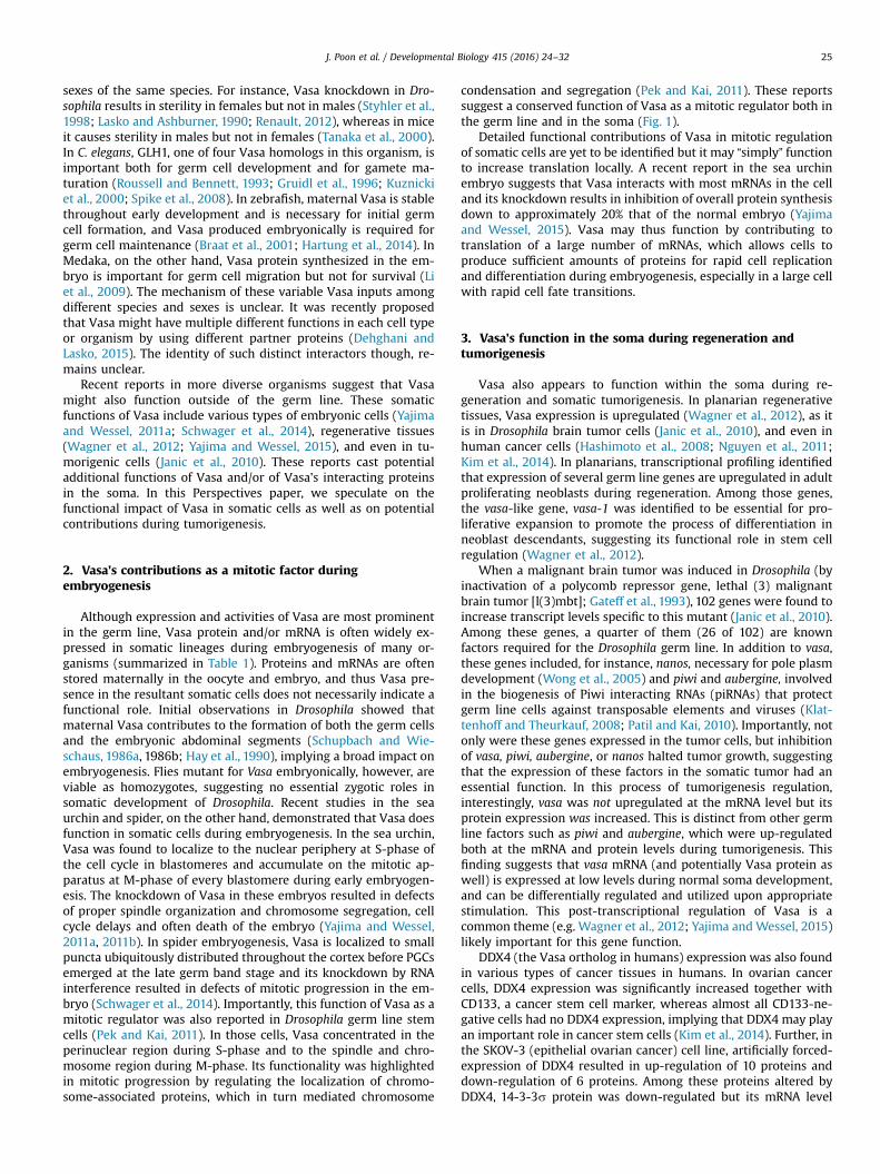
Many cells and organisms appear to have a tightly regulatedsystem for Vasa expression, especially in the soma, implying abroad biological importance. In tumorgenic cells where cellulardynamics are often altered, on the other hand, this tight controlover Vasa may be lacking, allowing cells to accumulate Vasa andits target factors (e.g. cell cycle materials) more effectively tocontribute to a part of tumorigenic processes. Investigating themechanisms of Vasa regulation in naturally Vasa-expressing tu-morgenic cells may be thus helpful to understand the nature ofthis molecule function in the cells. Further, identifying “upstreamregulator(s)” of Vasa in these tumorigenic cells could also decipherhow this molecule may be tightly regulated for its transientfunction during normal development, especially in the soma.DDX4 (Vasa ortholog in humans) mutations in the cancer cellgenome have been reported so far in 55 cancer cell types, whichinclude prostate, ovarian, breast, glioma, melanoma, uterine, andmany other cancer cell lines or tissues (Fig. 2, Identified by thecBioPortal search; Gao et al., 2013; Cerami et al., 2012). Throughthis genomic mutation-based search, however, the cause or con-sequence of these DDX4 mutations in cancer cells appears ratherrandom; some cancer cell lines such as prostate cancer cells lackddx4, whereas other cells such as breast cancer cell lines duplicateddx4 genes in the genome. It is thus unlikely that DDX4 is a causeof cancer initiation nor is consistently involved in a major stepof cancer cell development, yet rather might contribute to
Fig. 2. The cBioPortal (http://www.cbioportal.org/) database search result. Cancer cell tylists cancer type, and Y-axis indicates the frequency of the alterations in the cancers. Mutred, and multiple alterations in gray colored balls indicate cancer cell types. The text in
tumorigenesis as an important translation factor when expressedin the context of other translation factors of the cancer cell phe-notype. Considering that Vasa's post-transcriptional modificationsare critical to development of various somatic cells, it is importantto rely on a protein level database for analyzing Vasa function incancer cells. Other germ line factors may also be resolved withsuch an approach. In concert with metabolomics studies, onecould then potentially predict even more close associations withtumorigenesis.
In conclusion, most cells of the body do not have detectablelevel of Vasa, but when Vasa protein is expressed in somatic and/or tumorgenic cells, Vasa is essential for cellular regulation. Wethink of this as a Vasa addiction that perhaps alters the cellularmetabolism, including translational regulation. Were Vasa proteinaltered in those addicted cells, the cell then crashes and burns.Such theoretical compensatory regulation within these cells willinform us as to how Vasa fundamentally functions in the cell.Further study of the determinants of Vasa localization and activityin various somatic cells including cancerous cells should helpelucidate why ectopic Vasa expression can be detrimental.
Acknowledgements
This work was supported by the American Heart AssociationScientist Development Grant-14SDG18350021 to M.Y. and by theNational Institutes of Health-2R01HD028152 and the NationalScience Foundation- IOS-1120972. grants to G.M.W.
pes that contain mutations in the DDX4 (Vasa) gene in the genome are listed. X-axisations of DDX4 in the genome is indicated in green, deletion in blue, amplification inparenthesis indicates the original data source of each cancer cell.

J. Poon et al. / Developmental Biology 415 (2016) 24–32 31
References
Akahira, J., Sugihashi, Y., Suzuki, T., Ito, K., Niikura, H., Moriya, T., Nitta, M., Okamura,H., Inoue, S., Sasano, H., Okamura, K., Yaegashi, N., 2004. Decreased expressionof 14-3-3 sigma is associated with advanced disease in human epithelialovarian cancer: its correlation with aberrant DNA methylation. Clin. Cancer Res.10, 2687–2693.
Arkov, A.L., Wang, J.Y., Ramos, A., Lehmann, R., 2006. The role of Tudor domains ingermline development and polar granule architecture. Development 133,4053–4062.
Asaoka, M.T., Yamada, M., Nakamura, A., Hanyu, K., Kobayashi, S., 1999. MaternalPumilio acts together with Nanos in germline development in Drosophilaembryos. Nat. Cell Biol. 1, 431–437.
Barker, D.D., Wang, C., Moore, J., Dickinson, L.K., Lehmann, R., 1992. Pumilio is es-sential for function but not for distribution of the Drosophila abdominal de-terminant Nanos. Genes Dev. 6, 2312–2326.
Blower, M., Feric, E., Weis, K., Heald, R., 2007. Genome-wide analysis demonstratesconserved localization of messenger RNAs to mitotic microtubules. J. Cell Biol.179, 1365–1373.
Braat, A., Zandbergen, T., van de Water, S., Goos, H., Zivkovic, D., 1999. Character-ization of zebrafish primordial germ cells: morphology and early distribution ofvasa RNA. Dev. Dyn. 216, 153–167.
Braat, A.K., van de Water, S., Korving, J., Zivkovic, D., 2001. A zebrafish vasa mor-phant abolishes vasa protein but does not affect the establishment of thegermline. Genesis 30, 183–185.
Braat, A.K., van de Water, S., Goos, H., Bogerd, J., Zivkovic, D., 2000. Vasa proteinexpression and localization in the zebrafish. Mech. Dev. 95 (1–2), 271–274.
Carrera, P., Johnstone, O., Nakamura, A., Casanova, J., Jackle, H., Lasko, P., 2000. Vasamediates translation through interaction with a Drosophila yIF2 homolog. Mol.Cell 5, 181–187.
Cerami, E., Gao, J., Dogrusoz, U., Gross, B.E., Sumer, S.O., Aksoy, B.A., Jacobsen, A.,Byrne, C.J., Heuer, M.L., Larsson, E., Antipin, Y., Reva, B., Goldberg, A.P., Sander,C., Schultz, N., 2012. The cBio cancer genomics portal: an open platform forexploring multidimensional cancergenomics data. Cancer Discov. 2 (5),401–404.
Chan, T.A., Hermeking, H., Lengauer, C., Kinzler, K.W., Vogelstein, B., 1999. 14-3-3sigma is required to prevent mitotic catastrophe after DNA damage. Nature401, 616–620.
Curran, S.P., Wu, X., Riedel, C.G., Ruvkun, G., 2009. A soma-to-germline transfor-mation in long-lived Caenorhabditis elegans mutants. Nature 459, 1079–1084.
DeBerardinis, R.J., Lum, J.J., Hatzivassiliou, G., Thompson, C.B., 2008. The biology ofcancer: metabolic reprogramming fuels cell growth and proliferation. CellMetab. 7, 11–20.
Dehghani, M., Lasko, P., 2015. In vivo mapping of the functional regions of theDEAD-box helicase Vasa. Biol. Open 4 (4), 450–462.
Dill, K.K., Seaver, E.C., 2008. Vasa and nanos are coexpressed in somatic and germline tissue from early embryonic cleavage stages through adulthood in thepolychaete Capitella sp. I. Dev. Genes Evol. 218, 453–463.
Ferguson, A.T., Evron, E., Umbricht, C.B., Pandita, T.K., Chan, T.A., Hermeking, H.,Marks, J.R., Lambers, A.R., Futreal, P.A., Stampfer, M.R., Sukumar, S., 2000. Highfrequency of hypermethylation at the 14-3-3 sigma locus leads to gene silen-cing in breast cancer. Proc. Natl. Acad. Sci. USA 97, 6049–6054.
Gao, J., Aksoy, B.A., Dogrusoz, U., Dresdner, G., Gross, B., Sumer, S.O., Sun, Y., Ja-cobsen, A., Sinha, R., Larsson, E., Cerami, E., Sander, C., Schultz, N., 2013. In-tegrative analysis of complex cancer genomics and clinical profiles using thecBioPortal. Sci. Signal. 6 (269), pl1.
Gao, M., Thomson, T.C., Creed, T.M., Tu, S., Loganathan, S.N., Jackson, C.A., McClus-key, P., Lin, Y., Collier, S.E., Weng, Z., Lasko, P., Ohi, M.D., Arkov, A.L., 2015.Glycolytic enzymes localize to ribonucleoprotein granules in Drosophila germcells, bind Tudor and protect from transposable elements. EMBO Rep. 16,379–386.
Gateff, E., Löffler, T., Wismar, J., 1993. A temperature-sensitive brain tumor sup-pressor mutation of Drosophila melanogaster: developmental studies and mo-lecular localization of the gene. Mech. Dev. 41 (1), 15–31.
Ghabrial, A., Schüpbach, T., 1999. Activation of a meiotic checkpoint regulatestranslation of Gurken during Drosophila oogenesis. Nat. Cell Biol. 1 (6),354–357.
Gross, P.R., Fry, B.J., 1966. Continuity of protein synthesis through cleavage meta-phase. Science 153 (3737), 749–751.
Gruidl, M.E., Smith, P.A., Kuznicki, K.A., McCrone, J.S., Kirchner, J., Roussell, D.L.,Strome, S., Bennett, K.L., 1996. Multiple potential germline helicases are com-ponents of the germ-line-specific P granules of Caenorhabditis elegans. Proc.Natl. Acad. Sci. USA 93, 13837–13842.
Guo, H., Garcia-Vedrenne, A.E., Isserlin, R., Lugowski, A., Morada, A., Sun, A., Miao,Y., Kuzmanov, U., Wan, C., Ma, H., Foltz, K., Emili, A., 2015. Phosphoproteomicnetwork analysis in the sea urchin Strongylocentrotus purpuratus reveals newcandidates in egg activation. Proteomics 15 (23–24), 4080–4095.
Gustafson, E.A., Wessel, G.M., 2010. DEAD-box helicases: posttranslational regula-tion and function. Biochem. Biophys. Res. Commun. 395, 1–6.
Gustafson, E.A., Yajima, M., Juliano, C.E., Wessel, G.M., 2011. Post-translationalregulation by gustavus contributes to selective Vasa protein accumulation inmultipotent cells during embryogenesis. Dev. Biol. 349, 440–450.
Hanahan, D., Weinberg, R.A., 2011. Hallmarks of cancer: the next generation. Cell144, 646–674.
Hartung, O., Forbes, M.M., Marlow, F.L., 2014. Zebrafish vasa is required for germ
cell differentiation and maintenance. Mol. Reprod. Dev. 81 (10), 946–961.Hashimoto, H., Sudo, T., Mikami, Y., Otani, M., Takano, M., Tsuda, H., Itamochi, H.,
Katabuchi, H., Ito, M., Nishimura, R., 2008. Germ cell specific protein VASA isover-expressed in epithelial ovarian cancer and disrupts DNA damage-inducedG2 checkpoint. Gynecol. Oncol. 111, 312–319.
Hay, B., Jan, L.Y., Jan, Y.N., 1988. A protein component of Drosophila polar granules isencoded by vasa and has extensive sequence similarity to ATP-dependent he-licases. Cell 55, 577–587.
Hay, B., Jan, L.Y., Jan, Y.N., 1990. Localization of vasa, a component of Drosophilapolar granules, in maternal-effect mutants that alter embryonic anteroposteriorpolarity. Development 109, 425–433.
Hsu, P.P., Sabatini, D.M., 2008. Cancer cell metabolism: Warburg and beyond. Cell134, 703–707.
Ikenishi, K., Yamakita, S., 2003. A trial for induction of supernumerary primordialgerm cells in Xenopustadpoles by injecting RNA of Xenopus vasa homologueinto germline cells of 32-cell embryos. Dev. Growth Differ. 45 (5–6), 417–426.
Ikenishi, K., Tanaka, T., Komiya, T., 1996. Spatio-temporal distribution of the proteinof the Xenopus vasa homologue (Xenopus vasalike gene 1, XVLG1) in embryos.Dev. Growth Differ. 38, 527–535.
Illmensee, K., Mahowald, A.P., 1974. Transplantation of posterior polar plasm inDrosophila. Induction of germ cells at the anterior pole of the egg. Proc. Natl.Acad. Sci. USA 71 (4), 1016–1020.
Iwata, N., Yamamoto, H., Sasaki, S., Itoh, F., Suzuki, H., Kikuchi, T., Kaneto, H., Iku, S.,Ozeki, I., Karino, Y., Satoh, T., Toyota, J., Satoh, M., Endo, T., Imai, K., 2000. Fre-quent hypermethylation of CpG islands and loss of expression of the 14-3-3sigma gene in human hepatocellular carcinoma. Oncogene 19, 5298–5302.
Janic, A., Mendizabal, L., Llamazares, S., Rossell, D., Gonzalez, C., 2010. Ectopic ex-pression of germline genes drives malignant brain tumor growth in Drosophila.Science 330, 1824–1827.
Johnstone, O., Lasko, P., 2004. Interaction with eIF5B is essential for Vasa functionduring development. Development 131, 4167–4178.
Juliano, C.E., Yajima, M., Wessel, G.M., 2010. Nanos functions to maintain the fate ofthe small micromere lineage in the sea urchin embryo. Dev. Biol. 337, 220–232.
Kadyrova, L.Y., Habara, Y., Lee, T.H., Wharton, R.P., 2007. Translational control ofmaternal Cyclin B mRNA by Nanos in the Drosophila germline. Development134 (8), 1519–1527.
Kim, K.H., Kang, Y.J., Jo, J.O., Ock, M.S., Moon, S.H., Suh, D.S., Yoon, M.S., Park, E.S.,Jeong, N., Eo, W.K., Kim, H.Y., Cha, H.J., 2014. DDX4 (DEAD box polypeptide 4)colocalizes with cancer stem cell marker CD133 in ovarian cancers. Biochem.Biophys. Res. Commun. 447, 315–322.
Klattenhoff, C., Theurkauf, W., 2008. Biogenesis and germline functions of piRNAs.Development 135, 3–9.
Klattenhoff, C., Bratu, D.P., McGinnis-Schultz, N., Koppetsch, B.S., Cook, H.A.,Theurkauf, W.E., 2007. Drosophila rasiRNA pathway mutations disrupt em-bryonic axis specification through activation of an ATR/Chk2 DNA damage re-sponse. Dev. Cell 2 (1), 45–55.
Kondoh, H., Lleonart, M.E., Nakashima, Y., Yokode, M., Tanaka, M., Bernard, D., Gil, J.,Beach, D., 2007. A high glycolytic flux supports the proliferative potential ofmurine embryonic stem cells. Antioxid. Redox Signal. 9, 293–299.
Kosaka, K., Kawakami, K., Sakamoto, H., Inoue, K., 2007. Spatiotemporal localizationof germ plasm RNAs during zebrafish oogenesis. Mech. Dev. 124 (4), 279–289.
Kugler, J.M., Woo, J.S., Oh, B.H., Lasko, P., 2010. Regulation of Drosophila Vasa in vivothrough paralogous Cullin-RING E3 ligase specificity receptors. Mol. Cell Biol.30, 1769–1782.
Kuznicki, K.A., Smith, P.A., Leung-Chiu, W.M., Estevez, A.O., Scott, H.C., Bennett, K.L.,2000. Combinatorial RNA interference indicates GLH-4 can compensate forGLH-1; these two P granule components are critical for fertility in C. elegans.Development 127, 2907–2916.
Lasko, P.F., Ashburner, M., 1988. The product of the Drosophila gene vasa is verysimilar to eukaryotic initiation factor-4A. Nature 335, 611–617.
Lasko, P.F., Ashburner, M., 1990. Posterior localization of vasa protein correlateswith, but is not sufficient for, pole cell development. Genes Dev. 4, 905–921.
Lasko, P., 2013. The DEAD-box helicase Vasa: evidence for a multiplicity offunctionsin RNA processes and developmental biology. Biochem. Biophys. Acta 1829,810–816.
Li, C.J., Liu, L., Chen, X.H., Zhang, T., Gan, F., Cheng, B.L., 2010. Identification of a vasahomologue gene in grass carp and its expression pattern in tissues and duringembryogenesis. Comp. Biochem. Physiol. B Biochem. Mol. Biol. 157 (2), 159–166.
Li, M., Hong, N., Xu, H., Yi, M., Li, C., Guia, J., Hong, Y., 2009. Medaka vasa is requiredfor migration but not survival of primordial germ cells. Mech. Dev. 126,366–381 (å).
Linder, P., 2006. Dead-box proteins: a family affair–active and passive players inRNP-remodeling. Nucleic Acids Res. 34, 4168–4180.
Linder, P., Lasko, P.F., Ashburner, M., Leroy, P., Nielsen, P.J., Nishi, K., Schnier, J.,Slonimski, P.P., 1989. Birth of the D-E-A-D box. Nature 337, 121–122.
Liska, A.J., Popov, A.V., Sunyaev, S., Coughlin, P., Habermann, B., Shevchenko, A.,Bork, P., Karsenti, E., Shevchenko, A., 2004. Homology-based functional pro-teomics by mass spectrometry: application to the Xenopus microtubule asso-ciated proteome. Proteomics 4, 2707–2721.
Liu, N., Dansereau, D.A., Lasko, P., 2003. Fat facets interacts with vasa in the Dro-sophila pole plasm and protects it from degradation. Curr. Biol. 13, 1905–1909.
Liu, N., Han, H., Lasko, P., 2009. Vasa promotes Drosophila germline stem cell dif-ferentiation by activating mei-P26 translation by directly interacting with a(U)-rich motif in its 3′ UTR. Genes Dev. 23, 2742–2752.
Mitchison, T.J., Maddox, P., Groen, A., Cameron, L., Perlman, Z., Ohi, R., Desai, R.,Salmon, E.D., Kapoor, T.M., 2004. Bipolarization and poleward flux correlate

J. Poon et al. / Developmental Biology 415 (2016) 24–3232
during Xenopus extract spindle assembly. Mol. Biol. Cell 15, 5603–5615.Mochizuki, K., Nishimiya-Fujisawa, C., Fujisawa, T., 2001. Universal occurrence of
the vasa-related genes among metazoans and their germline expression inHydra. Dev. Genes Evol. 211 (6), 299–308.
Nguyen, L.B., Diskin, S.J., Capasso, M., Wang, K., Diamond, M.A., Glessner, J., Kim, C.,Attiyeh, E.F., Mosse, Y.P., Cole, K., Iolascon, A., Devoto, M., Hakonarson, H., Li, H.K., Maris, J.M., 2011. Phenotype restricted genome-wide association study usinga gene-centric approach identifies three low-risk neuroblastoma susceptibilityLoci. PLoS Genet. 7, e1002026.
Orsborn, A.M., Li, W., McEwen, T.J., Mizuno, T., Kuzmin, E., Matsumoto, K., Bennett,K.L., 2007. GLH-1, the C. elegans P granule protein, is controlled by the JNK KGB-1 and by the COP9 subunit CSN-5. Development 134 (18), 3383–3392.
Osada, H., Tatematsu, Y., Yatabe, Y., Nakagawa, T., Konishi, H., Harano, T., Tezel, E.,Takada, M., Takahashi, T., 2002. Frequent and histological type-specific in-activation of 14-3-3sigma in human lung cancers. Oncogene 21, 2418–2424.
Patil, V.S., Kai, T., 2010. Repression of retroelements in Drosophila germline viapiRNA pathway by the Tudor domain protein Tejas. Curr. Biol. 20, 724–730.
Pek, J.W., Kai, T., 2011. A role for vasa in regulating mitotic chromosome con-densation in Drosophila. Curr. Biol. 21, 39–44.
Pestova, T.V., Lomakin, I.B., Lee, J.H., Choi, S.K., Dever, T.E., Hellen, C.U.T., 2000. Thejoining of ribosomal subunits in eukaryotes requires eIF5B. Nature 403,332–335.
Raff, J.W., Whitfield, W.G., Glover, D.M., 1990. Two distinct mechanisms localizecyclin B transcripts in syncytial Drosophila embryos. Development 110,1249–1261.
Raz, E., 2000. The function and regulation of vasa-like genes in germ-cell devel-opment. Genome Biol. 1 (3), Reviews 1017.1.
Rebscher, N., Zelada-Gonza lez, F., Banisch, T.U., Raible, F., Arendt, D., 2007. Vasaunveils a common origin of germ cells and of somatic stem cells from theposterior growth zone in the polychaete Platynereis dumerilii. Dev. Biol. 306,599–611.
Renault, A.D., 2012. vasa is expressed in somatic cells of the embryonic gonad in asex-specificmanner in Drosophila melanogaster. Biol. Open 1 (10), 1043–1048.
Ross, R.J., Weiner, M.M., Lin, H., 2014. PIWI proteins and PIWI-interacting RNAs inthe soma. Nature 505, 353–359.
Roussell, D.L., Bennett, K.L., 1993. glh-1, a germ-line putative RNA helicase fromCaenorhabditis, has four zinc fingers. Proc. Natl. Acad. Sci. USA 90, 9300–9304.
Schupbach, T., Wieschaus, E., 1986. Maternal-effect mutations altering the anterior-posterior pattern of the Drosophila embryo. Dev. Biol. 113, 443–448.
Schwager, E.E., Meng, Y., Extavour, C.G., 2014. Vasa and piwi are required for mitoticintegrity in early embryogenesis in the spider Parasteatoda tepidariorum. Dev.Biol. 402 (2), 276–290.
Sengoku, T., Nureki, O., Nakamura, A., Kobayashi, S., Yokoyama, S., 2006. Structuralbasis for RNA unwinding by the DEAD-box protein Drosophila Vasa. Cell 125,287–300.
Shibata, N., Umesono, Y., Orii, H., Sakurai, T., Watanabe, K., Agata, K., 1999. Ex-pression of vasa(vas)-related genes in germline cells and totipotent somaticstem cells of planarians. Dev. Biol. 206, 73–87.
Shirae-Kurabayashi, M., Nishikata, T., Takamura, K., Tanaka, K.J., Nakamoto, C., Na-kamura, A., 2006. Dynamic redistribution of vasa homolog and exclusion ofsomatic cell determinants during germ cell specification in Ciona intestinalis.Development 133, 2683–2693.
Simpson, A.J., Caballero, O.L., Jungbluth, A., Chen, Y.T., Old, L.J., 2005. Cancer/testisantigens, gametogenesis and cancer. Nat. Rev. Cancer 5, 615–625 (Review).
Spike, C., Meyer, N., Racen, E., Orsborn, A., Kirchner, J., Kuznicki, K., Yee, C., Bennett,K., Strome, S., 2008. Genetic analysis of the Caenorhabditis elegans GLH familyof P-granule proteins. Genetics 178, 1973–1987.
Styhler, S., Nakamura, A., Lasko, P., 2002. VASA localization requires the SPRY-do-main and SOCS-box containing protein. GUSTAVUS. Dev. Cell 3, 865–876.
Styhler, S., Nakamura, A., Swan, A., Suter, B., Lasko, P., 1998. vasa is required forGURKEN accumulation in the oocyte, and is involved in oocyte differentiationand germline cyst development. Development 125, 1569–1578.
Suprenant, K.A., 1993. Microtubules, ribosomes, and RNA: evidence for cytoplasmiclocalization and translational regulation. Cell Motil. Cytoskelet. 25 (1), 1–9.
Tanaka, S.S., Toyooka, Y., Akasu, R., Katoh-Fukui, Y., Nakahara, Y., Suzuki, R., Yo-koyama, M., Noce, T., 2000. The mouse homolog of Drosophila Vasa is requiredfor the development of male germ cells. Genes Dev. 14, 841–853.
Tsunekawa, N., Naito, M., Sakai, Y., Nishida, T., Noce, T., 2000. Isolation of chickenvasa homolog gene and tracing the origin of primordial germ cells. Develop-ment 127, 2741–2750.
Vander Heiden, M.G., Cantley, L.C., Thompson, C.B., 2009. Understanding theWarburg effect: the metabolic requirements of cell proliferation. Science 324,1029–1033.
Voronina, E., Lopez, M., Juliano, C.E., Gustafson, E., Song, J.L., Extavour, C., George, S.,Oliveri, P., McClay, D., Wessel, G.M., 2008. Vasa protein expression is restrictedto the small micromeres of the sea urchin, but is inducible in other lineagesearly in development. Dev. Biol. 314, 276–286.
Wagner, D.E., Ho, J.J., Reddien, P.W., 2012. Genetic regulators of a pluripotent adultstem cell system in planarians identified by RNAi and clonal analysis. Cell StemCell 10, 299–311.
Wang, D., Kennedy, S., Conte, D.J., Kim, J.K., Gabel, H.W., Kamath, R.S., Mello, C.C.,Ruvkun, G., 2005. Somatic misexpression of germline P granules and enhancedRNA interference in retinoblastoma pathway mutants. Nature 436, 593–597.
Warburg, O.H., 1956a. On the origin of cancer cells. Science 123, 309–314.Warburg, O.H., 1956b. On respiratory impairment in cancer cells. Science 124,
269–270.Warburg, O.H., 1930. The Metabolism of Tumours: Investigations from the Kaiser
Wilhelm Institute for Biology, Berlin-Dahlem, London, UK, Arnold Constable.Wong, M.D., Jin, X., Xie, T., 2005. Molecular mechanisms of germline stem cell
regulation. Annu. Rev. Genet. 39, 173–195.Woo, J.S., Suh, H.Y., Park, S.Y., Oh, B.H., 2006b. Structural basis for protein re-
cognition by B30.2/SPRY domains. Mol. Cell 24, 967–976.Woo, J.S., Imm, J.H., Min, C.K., Kim, K.J., Cha, S.S., Oh, B.H., 2006a. Structural and
functional insights into the B30.2/SPRY domain. EMBO J. 25, 1353–1363.Xiol, J., Spinelli, P., Laussmann, M.A., Homolka, D., Yang, Z., Cora, E., Couté, Y., Conn,
S., Kadlec, J., Sachidanandam, R., Kaksonen, M., Cusack, S., Ephrussi, A., Pillai, R.S., 2014. RNA clamping by Vasa assembles a piRNA amplifier complex ontransposon transcripts. Cell 157, 1698–1711.
Yajima, M., Wessel, G.M., 2011a. The DEAD-box RNA helicase Vasa functions inembryonic mitotic progression in the sea urchin. Development 138, 2217–2222.
Yajima, M., Wessel, G.M., 2011b. The multiple hats of Vasa: its functions in thegermline and in cell cycle progression. Mol. Reprod. Dev. 78, 861–867.
Yajima, M., Wessel, G.M., 2015. The germ line factor Vasa functions broadly in so-matic cells: mRNA clustering, translational regulation, and wound healing.Development 142 (11), 1960–1970.
Yoon, C., Kawakami, K., Hopkins, N., 1997. Zebrafish vasa homologue RNA is loca-lized to the cleavage planes of 2- and 4-cell-stage embryos and is expressed inthe primordial germ cells. Development 124, 3157–3165.