Embed Size (px)
Citation preview
One in a Million - A Case of Back Pain Matea Orlovic D.O., Kyle Ransom D.O., Joel Stillings, D.O.
Desert Regional Medical Center
Questions and Answers Questions: 1. Name a few differential diagnoses that come to mind
regarding the HPI? 2. What is the best imaging modality to make the diagnosis? 3. Why did the CT scan miss the findings seen on the MRI?
Answers: 1. AAA, aortic dissection, saddle PE, spinal epidural
hematoma. 2. MRI with and without contrast is the gold standard
imaging modality for diagnosing spinal epidural hematoma.
3. CT scans can miss a spinal epidural hematoma because blood has similar attenuation as bone. Also, as x-ray beams pass through bone, this may cause streak artifact resulting in dark bands and lack of visualization of blood. In this case, the streak artifact appears to be obscuring the area of the hematoma.
Case History Chief Complaint: Back Pain History of Present Illness: 52 y/o male with PMH of HIV, HTN and diabetes BIBA complaining of severe upper back pain. Patient is a math teacher, he had a sudden onset of severe interscapular back pain radiating to the right arm and down right leg while sitting on the computer during a zoom meeting. He denies associated injury, falls, or trauma. He denies associated weakness, chest pain, nausea, vomiting, abdominal pain. He denies any history of similar symptoms or history of back surgery in the past. He takes a baby Aspirin daily, but denies any anticoagulant use. Physical Exam: General: In distress, writhing in pain, having difficulty lying still on gurney. Head & Neck: Atraumatic. Neck supple, normal ROM. CV: RRR, 2+ bilateral radial and DP pulses. Resp: Lungs clear to auscultation, respirations are non-labored. Abdomen: Protuberant but soft, non-tender, no pulsatile mass. Back: No reproducible midline tenderness to palpation of the cervical, thoracic, or lumbar spine, no overlying erythema. Musculoskeletal: No deformity. Neuro: A&Ox4, moving all extremities, subjective paresthesias of right arm and right leg.
Clinical Pearls• The incidence of spinal epidural hematoma is a 1 per 1,000,000
people annually. • MRI with and without contrast is the gold standard imaging
modality for diagnosing spinal epidural hematoma. • Early diagnosis and surgical decompression is vital in preventing
severe neurologic sequelae.
Image 1
DiscussionDue to the patient’s presentation of sudden onset of severe interscapular pain, a CT angiogram of chest, abdomen and pelvis was ordered but did not show evidence of AAA or aortic dissection. Throughout the ED stay, the patient’s paresthesias progressed to numbness, then weakness of the right arm and right leg. Neurology team was consulted, CT brain and C-spine were normal. The patient was scheduled for an MRI which showed a C4-C7 intraspinal epidural hematoma. He was immediately taken to OR by neurosurgery for bilateral decompressive laminectomy and evacuation of epidural hematoma.
The incidence of spinal epidural hematoma is a 1 per 1,000,000 people annually. It can occur spontaneously, from trauma, or from iatrogenic sources such as lumbar puncture. Risk factors for spontaneous spinal epidural hematoma include vascular malformations, anticoagulant use, coagulopathy, blood disorder, tumors, alcohol use and more. The bleeding is usually venous but can be from an arterial source as well. The accumulation of blood can lead to compression of the spinal cord and may lead to significant neurological damage if not timely evacuated.
A cranial epidural hematoma can be seen on computerized tomography (CT) scan, however, CT can miss a spinal epidural hematoma due to blood having similar attenuation as bone. As the Xray beams pass through bone, it may cause streak artifacts resulting in dark bands seen above. Due to these factors, MRI is the gold standard imaging modality to diagnose epidural hematomas. A CT myelography may also be used when MRI is unavailable.
This condition is rare, but if correctly identified and treated surgically within 36 hours of full paralysis or 48 hours of partial motor deficits, the patient can potentially have a full recovery of function. This makes it worth considering as a differential diagnosis of back pain.
Epidural fluid collection measuring 1 cm with low signal rim identified posterior to the cord at the C4-C7 levels compatible with epidural hematoma. The cord appears compressed most pronounced at the C6 and C7 levels with mild increased cord signal. Increased STIR signal may suggest interspinous ligamentous injuries at the C5-C6, and C6-C7 levels.
Radiology
Image 2
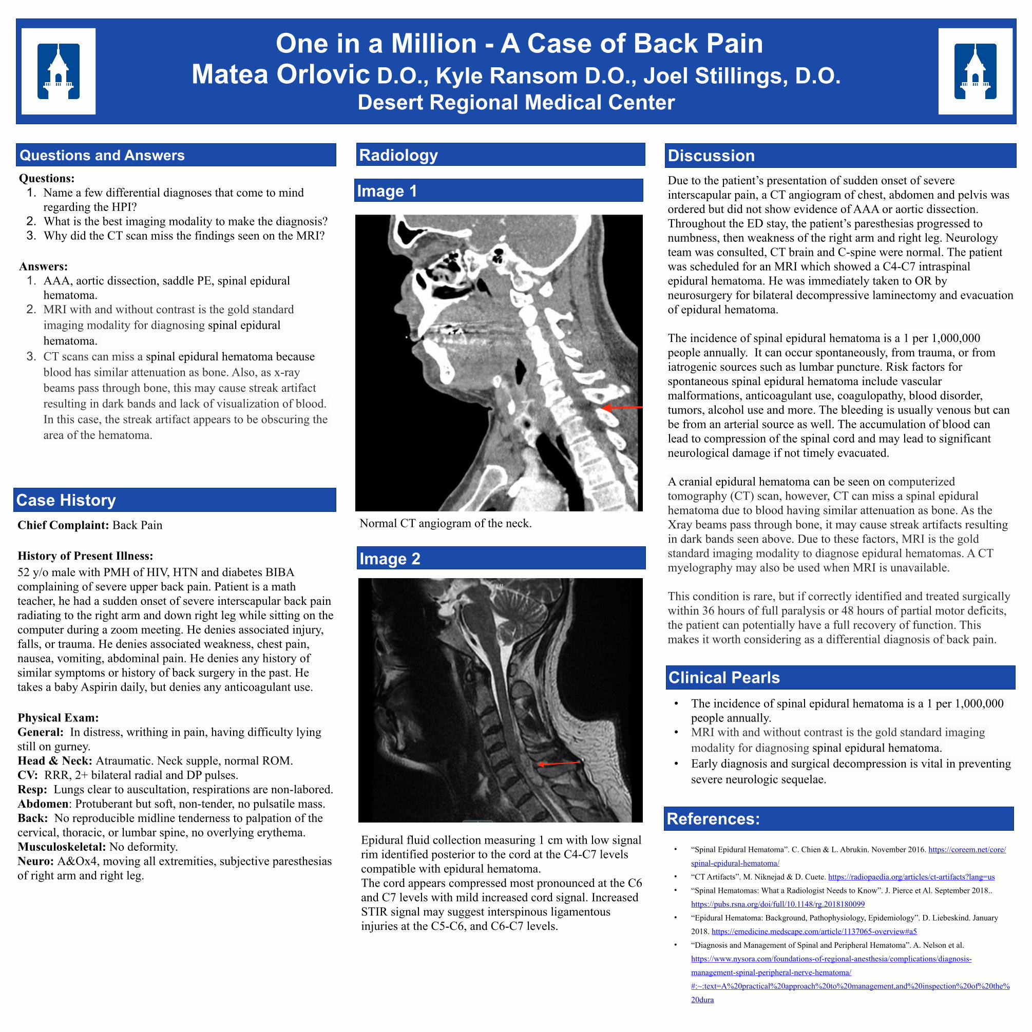
Normal CT angiogram of the neck.
References:• “Spinal Epidural Hematoma”. C. Chien & L. Abrukin. November 2016. https://coreem.net/core/
spinal-epidural-hematoma/ • “CT Artifacts”. M. Niknejad & D. Cuete. https://radiopaedia.org/articles/ct-artifacts?lang=us • “Spinal Hematomas: What a Radiologist Needs to Know”. J. Pierce et Al. September 2018..
https://pubs.rsna.org/doi/full/10.1148/rg.2018180099 • “Epidural Hematoma: Background, Pathophysiology, Epidemiology”. D. Liebeskind. January
2018. https://emedicine.medscape.com/article/1137065-overview#a5 • “Diagnosis and Management of Spinal and Peripheral Hematoma”. A. Nelson et al.
https://www.nysora.com/foundations-of-regional-anesthesia/complications/diagnosis-
management-spinal-peripheral-nerve-hematoma/
#:~:text=A%20practical%20approach%20to%20management,and%20inspection%20of%20the%
20dura


![Back Talk - Back Pain Rescue[1]](https://img.pdfslide.us/doc/110x75/577d35821a28ab3a6b90a19c/back-talk-back-pain-rescue1.jpg)