Embed Size (px)
Citation preview
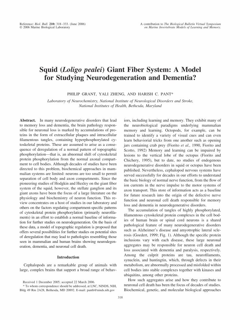
Squid (Loligo pealei) Giant Fiber System: A Modelfor Studying Neurodegeneration and Dementia?
PHILIP GRANT, YALI ZHENG, AND HARISH C. PANT*
Laboratory of Neurochemistry, National Institute of Neurological Disorders and Stroke,National Institutes of Health, Bethesda, Maryland
Abstract. In many neurodegenerative disorders that leadto memory loss and dementia, the brain pathology respon-sible for neuronal loss is marked by accumulations of pro-teins in the form of extracellular plaques and intracellularfilamentous tangles, containing hyperphosphorylated cy-toskeletal proteins. These are assumed to arise as a conse-quence of deregulation of a normal pattern of topographicphosphorylation—that is, an abnormal shift of cytoskeletalprotein phosphorylation from the normal axonal compart-ment to cell bodies. Although decades of studies have beendirected to this problem, biochemical approaches in mam-malian systems are limited: neurons are too small to permitseparation of cell body and axon compartments. Since thepioneering studies of Hodgkin and Huxley on the giant fibersystem of the squid, however, the stellate ganglion and itsgiant axons have been the focus of a large literature on thephysiology and biochemistry of neuron function. This re-view concentrates on a host of studies in our laboratory andothers on the factors regulating compartment-specific patternsof cytoskeletal protein phosphorylation (primarily neurofila-ments) in an effort to establish a normal baseline of informa-tion for further studies on neurodegeneration. On the basis ofthese data, a model of topographic regulation is proposed thatoffers several possibilities for further studies on potential sitesof deregulation that may lead to pathologies resembling thoseseen in mammalian and human brains showing neurodegen-eration, dementia, and neuronal cell death.
Introduction
Cephalopods are a remarkable group of animals withlarge, complex brains that support a broad range of behav-
iors, including learning and memory. They exhibit many ofthe neurobiological paradigms underlying mammalianmemory and learning. Octopods, for example, can betrained to identify a variety of visual cues and can evenlearn behaviorial tricks from one another such as openingjars containing crab prey (Fiorito et al., 1990, Fiorito andScotto, 1992) Memory and learning can be impaired bylesions to the vertical lobe of the octopus (Fiorito andChichery, 1995), but to date, no studies of endogenousneurodegenerative disorders in squid or octopus have beenpublished. Nevertheless, cephalopod nervous systems haveserved successfully for decades in our efforts to understandthe basic biology of normal nerve function, from the flow ofion currents in the nerve impulse to the motor systems ofaxon transport. This store of information acts as a baselinefor future research into the origin of the defective nervefunction and neuronal cell death responsible for memoryloss and dementia in neurodegenerative disorders.
The accumulation of tangles of highly phosphorylated,filamentous cytoskeletal protein complexes in the cell bod-ies of human brain or spinal cord neurons is a sharedpathological feature of many neurodegenerative disorderssuch as Alzheimer’s disease and amyotrophic lateral scle-rosis (Goedert, 1999; Fig. 1). Although the specific proteininclusions vary with each disease, these large neuronalaggregates may be responsible for neuron cell death andloss associated with dementia and paralysis, respectively.Among the culprit proteins are tau, neurofilaments,synuclein, and huntingtin, which, through defects in theirmetabolism, are abnormally processed and misfolded withincell bodies into stable complexes together with kinases andubiquitins, among other proteins.
How such aggregates arise and how they contribute toneuronal cell death has been the focus of decades of studies.Biochemical, genetic, and molecular biological approaches
Received 1 December 2005; accepted 22 March 2006.* To whom correspondence should be addressed, at LNC, NINDS, NIH,
Bldg 49 Rm 2A28, Bethesda, MD 20892. E-mail: [email protected]
Reference: Biol. Bull. 210: 318–333. (June 2006) A contribution to The Biological Bulletin Virtual Symposium© 2006 Marine Biological Laboratory on Marine Invertebrate Models of Learning and Memory.
318

have furnished an understanding of some of the factorscontributing to the pathology. One common thread emerg-ing, however, has been the recognition that the normallytight compartment-specific regulation of post-translationalprocessing (phosphorylation, glycosylation, etc.) and as-sembly have been compromised in these disorders. Forexample, neurofilaments, a major cytoskeletal component oflarge axons, are normally assembled from subunit proteins(NF-L, NF-M, and NF-H). Upon entry into the axon, thelatter two are highly phosphorylated in lysine-serine-proline(KSP) repeats in their C-terminal tail domains (Sternbergerand Sternberger, 1983; Nixon et al., 1987, 1989; Pant et al.,2000). This results in the formation of sidearms, cross-bridges between NFs and microtubules, that create a cy-toskeletal scaffold sustaining large axonal diameters (deWaegh et al., 1992; Sanchez et al., 2000). Cell bodies, onthe other hand, contain only hypophosphorylated NF pro-teins. Tail domain phosphorylation does not occur in cellbodies except in pathological situations when KSP repeatsare hyperphosphorylated into filamentous aggregates. Thefactors responsible for topographic regulation/deregulationof NF phosphorylation are not understood, in part becauseseparation of cell bodies from axons in sufficient quantitiesfor biochemical studies is difficult in most mammaliannervous systems. Fortunately, the stellate ganglion of thesquid giant fiber system, which shares many neuronal prop-erties with mammalian systems, including a robust NFmetabolism, is readily separated into giant cell bodies and
pure axoplasm in quantities sufficient for diverse biochem-ical studies. This system has been the focus of our studiesfor several years as we attempted to sort out the factorsunderlying the compartmental-specific patterns of neurofila-ment phosphorylation.
The Squid Giant Fiber System
It was J. Z. Young who first described the structure andorigin of the giant fiber system of the squid Loligo (Young,1939). Since then a vast literature on the physiology, be-havior, and biochemistry of the giant fiber system has beenpublished, including the Nobel-prize-winning work ofHodgkin and Huxley (see also, Gilbert et al., 1990). The jetpropulsion locomotory behavior of this remarkable organ-ism depends upon the signaling evoked by a system of giantneurons beginning with two large “command neurons” inthe brain. The command neurons integrate most sensoryinput to transmit excitatory impulses to the dorsal stellateganglia and giant axons that innervate the muscles of themantle. The diameter of the giant axon can vary between300 and 800 micrometers (Adelman and Gilbert, 1990), andits length may extend several centimeters depending onspecies and size of the animal. Accordingly, ganglia andgiant axons are easily dissected from the mantle and pre-pared for electrophysiological and biochemical studies orfor studies of mechanisms of axon transport (Fig. 2). Thegiant axon owes its large size to the fact that it arises as a
Figure 1. Example of aggregate expression in cervical spinal cord ganglion neurons in amyotrophic lateralsclerosis (ALS). (A) Normal neuron. (B) Neuron showing ALS pathology as a result of deregulated topographicneurofilament phosphorylation. Both expressing an antibody to phosphorylated NF-H.
319GIANT AXON AND NEURODEGENERATION?

fusion of many individual axons from the large neurons(25–40 �m) of the giant fiber lobe (GFL). About 3–10 �l ofpure axoplasm, uncontaminated by sheath or glial cells, canbe collected from a single giant axon, depending on size,making it possible to collect up to 300–1000 �l of axoplasmfrom 10 squid (2 axons per squid). Together with GFL cellbodies dissected from the same stellate ganglia, the twotissue preparations can be compared biochemically. Usingthis system, we have studied the factors regulating thetopographic phosphorylation of NF proteins.
Squid Neurofilament Proteins
Squid neurofilaments in the giant axon, like those inmammalian axons, contribute to the large axon caliber re-sponsible for rapid conduction velocity, a property essentialto the jet propulsive function of mantle muscle. Neurofila-ments, together with microtubules and associated proteins,are organized into a hexagonal array that makes up a three-dimensional cytoskeletal network in the giant axon(Metuzals and Izzard, 1969; Hodge and Adelman, 1980;
Martin et al., 1990). NFs make up about 13% of totalaxoplasm protein (Brown and Lasek, 1990) and consist ofthree subunit proteins—a large, highly phosphorylated NF-220 and two smaller subunits, NF60 and NF70. These havebeen cloned and shown to arise by alternative splicing froma single gene, in contrast to the three independent genes thatcode for mammalian NF subunits (Szaro et al., 1991; Wayet al., 1992). Biochemically and immunocytochemically,the NF-220 is phosphorylated only in the axon, whereas anon-phosphorylated NF-180, together with NF60/70, is de-tected in the perikarya of the stellate ganglia (Tytell et al.,1990; Cohen et al., 1987). Phosphorylation occurs predom-inantly on the multiple K/RSP, SAR/K, and SEK/R repeatsites (approximately 47) on the C-terminal tail of the NF-220 subunit (Jaffe et al., 2001). This topographic pattern ofNF phosphorylation resembles that seen in mammalian neu-rons, with the axon as the principal site of NF phosphory-lation, as described above.
A comparison of kinase assays of axoplasm and GFLlysates illustrates the differences in compartment activities.
Figure 2. Stellate ganglion and giant axon isolated from squid (Loligo pealei). (A) Intact preparationshowing giant axon (a) in its sheath and stellate ganglion (b) with stubs of other giant axons. About 2� normalsize. (B) Collection of pure axoplasm: (a) Droplet of axoplasm extruded from giant axon; (b) droplet squeezedfrom axon, leaving empty collapsed sheath (c); (d) intact axon with sheath. (C) Isolated stellate ganglion showingsite of cut to collect giant fiber lobe cell bodies. “a” are giant axons.
320 P. GRANT ET AL.
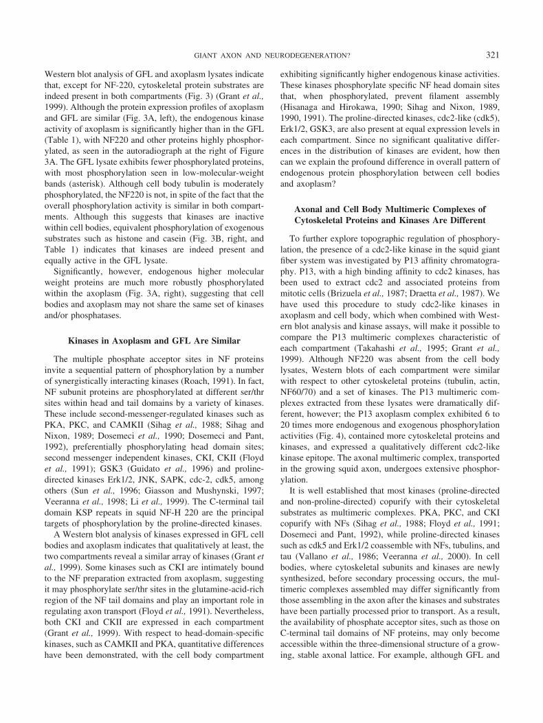
Western blot analysis of GFL and axoplasm lysates indicatethat, except for NF-220, cytoskeletal protein substrates areindeed present in both compartments (Fig. 3) (Grant et al.,1999). Although the protein expression profiles of axoplasmand GFL are similar (Fig. 3A, left), the endogenous kinaseactivity of axoplasm is significantly higher than in the GFL(Table 1), with NF220 and other proteins highly phosphor-ylated, as seen in the autoradiograph at the right of Figure3A. The GFL lysate exhibits fewer phosphorylated proteins,with most phosphorylation seen in low-molecular-weightbands (asterisk). Although cell body tubulin is moderatelyphosphorylated, the NF220 is not, in spite of the fact that theoverall phosphorylation activity is similar in both compart-ments. Although this suggests that kinases are inactivewithin cell bodies, equivalent phosphorylation of exogenoussubstrates such as histone and casein (Fig. 3B, right, andTable 1) indicates that kinases are indeed present andequally active in the GFL lysate.
Significantly, however, endogenous higher molecularweight proteins are much more robustly phosphorylatedwithin the axoplasm (Fig. 3A, right), suggesting that cellbodies and axoplasm may not share the same set of kinasesand/or phosphatases.
Kinases in Axoplasm and GFL Are Similar
The multiple phosphate acceptor sites in NF proteinsinvite a sequential pattern of phosphorylation by a numberof synergistically interacting kinases (Roach, 1991). In fact,NF subunit proteins are phosphorylated at different ser/thrsites within head and tail domains by a variety of kinases.These include second-messenger-regulated kinases such asPKA, PKC, and CAMKII (Sihag et al., 1988; Sihag andNixon, 1989; Dosemeci et al., 1990; Dosemeci and Pant,1992), preferentially phosphorylating head domain sites;second messenger independent kinases, CKI, CKII (Floydet al., 1991); GSK3 (Guidato et al., 1996) and proline-directed kinases Erk1/2, JNK, SAPK, cdc-2, cdk5, amongothers (Sun et al., 1996; Giasson and Mushynski, 1997;Veeranna et al., 1998; Li et al., 1999). The C-terminal taildomain KSP repeats in squid NF-H 220 are the principaltargets of phosphorylation by the proline-directed kinases.
A Western blot analysis of kinases expressed in GFL cellbodies and axoplasm indicates that qualitatively at least, thetwo compartments reveal a similar array of kinases (Grant etal., 1999). Some kinases such as CKI are intimately boundto the NF preparation extracted from axoplasm, suggestingit may phosphorylate ser/thr sites in the glutamine-acid-richregion of the NF tail domains and play an important role inregulating axon transport (Floyd et al., 1991). Nevertheless,both CKI and CKII are expressed in each compartment(Grant et al., 1999). With respect to head-domain-specifickinases, such as CAMKII and PKA, quantitative differenceshave been demonstrated, with the cell body compartment
exhibiting significantly higher endogenous kinase activities.These kinases phosphorylate specific NF head domain sitesthat, when phosphorylated, prevent filament assembly(Hisanaga and Hirokawa, 1990; Sihag and Nixon, 1989,1990, 1991). The proline-directed kinases, cdc2-like (cdk5),Erk1/2, GSK3, are also present at equal expression levels ineach compartment. Since no significant qualitative differ-ences in the distribution of kinases are evident, how thencan we explain the profound difference in overall pattern ofendogenous protein phosphorylation between cell bodiesand axoplasm?
Axonal and Cell Body Multimeric Complexes ofCytoskeletal Proteins and Kinases Are Different
To further explore topographic regulation of phosphory-lation, the presence of a cdc2-like kinase in the squid giantfiber system was investigated by P13 affinity chromatogra-phy. P13, with a high binding affinity to cdc2 kinases, hasbeen used to extract cdc2 and associated proteins frommitotic cells (Brizuela et al., 1987; Draetta et al., 1987). Wehave used this procedure to study cdc2-like kinases inaxoplasm and cell body, which when combined with West-ern blot analysis and kinase assays, will make it possible tocompare the P13 multimeric complexes characteristic ofeach compartment (Takahashi et al., 1995; Grant et al.,1999). Although NF220 was absent from the cell bodylysates, Western blots of each compartment were similarwith respect to other cytoskeletal proteins (tubulin, actin,NF60/70) and a set of kinases. The P13 multimeric com-plexes extracted from these lysates were dramatically dif-ferent, however; the P13 axoplasm complex exhibited 6 to20 times more endogenous and exogenous phosphorylationactivities (Fig. 4), contained more cytoskeletal proteins andkinases, and expressed a qualitatively different cdc2-likekinase epitope. The axonal multimeric complex, transportedin the growing squid axon, undergoes extensive phosphor-ylation.
It is well established that most kinases (proline-directedand non-proline-directed) copurify with their cytoskeletalsubstrates as multimeric complexes. PKA, PKC, and CKIcopurify with NFs (Sihag et al., 1988; Floyd et al., 1991;Dosemeci and Pant, 1992), while proline-directed kinasessuch as cdk5 and Erk1/2 coassemble with NFs, tubulins, andtau (Vallano et al., 1986; Veeranna et al., 2000). In cellbodies, where cytoskeletal subunits and kinases are newlysynthesized, before secondary processing occurs, the mul-timeric complexes assembled may differ significantly fromthose assembling in the axon after the kinases and substrateshave been partially processed prior to transport. As a result,the availability of phosphate acceptor sites, such as those onC-terminal tail domains of NF proteins, may only becomeaccessible within the three-dimensional structure of a grow-ing, stable axonal lattice. For example, although GFL and
321GIANT AXON AND NEURODEGENERATION?
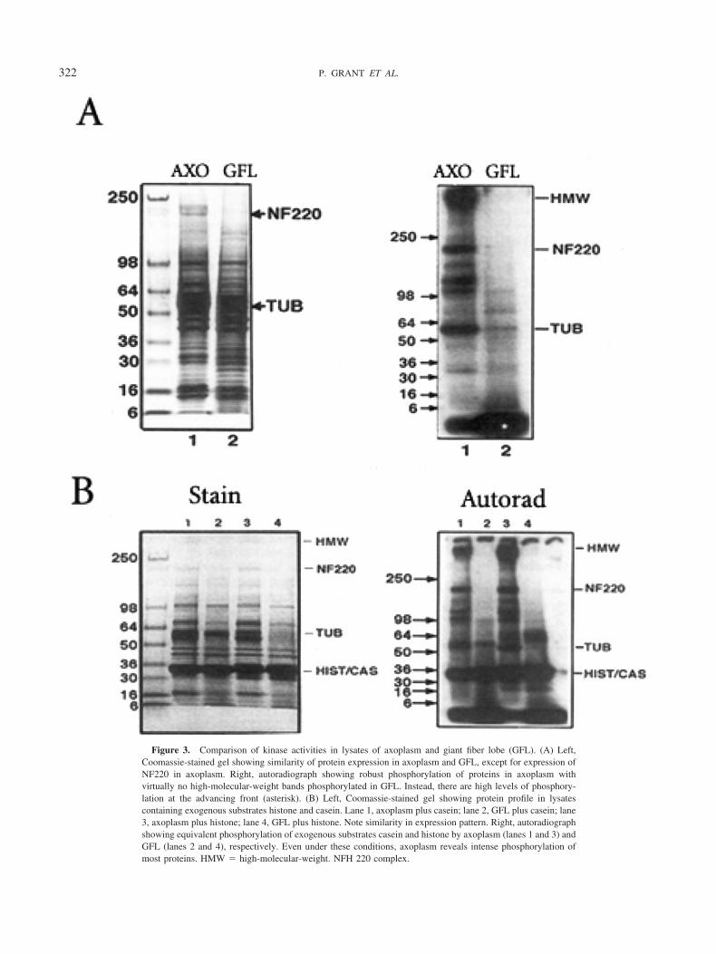
Figure 3. Comparison of kinase activities in lysates of axoplasm and giant fiber lobe (GFL). (A) Left,Coomassie-stained gel showing similarity of protein expression in axoplasm and GFL, except for expression ofNF220 in axoplasm. Right, autoradiograph showing robust phosphorylation of proteins in axoplasm withvirtually no high-molecular-weight bands phosphorylated in GFL. Instead, there are high levels of phosphory-lation at the advancing front (asterisk). (B) Left, Coomassie-stained gel showing protein profile in lysatescontaining exogenous substrates histone and casein. Lane 1, axoplasm plus casein; lane 2, GFL plus casein; lane3, axoplasm plus histone; lane 4, GFL plus histone. Note similarity in expression pattern. Right, autoradiographshowing equivalent phosphorylation of exogenous substrates casein and histone by axoplasm (lanes 1 and 3) andGFL (lanes 2 and 4), respectively. Even under these conditions, axoplasm reveals intense phosphorylation ofmost proteins. HMW � high-molecular-weight. NFH 220 complex.
322 P. GRANT ET AL.

axoplasm lysates expressed CKI and CKII, only the axonalP13 complex expressed CKI, a result that correlated with itshigh level of casein phosphorylation. This is consistent withprevious observations of the high affinity of CKI to theaxonal NF preparations and the possible role of CKI inregulation of slow axonal transport of NFs. Moreover, elec-tron microscopy studies of filamentous structures in cellbody and axoplasm reveal differences reflecting the phos-phorylation-driven sidearm extension of the NF220 C-ter-minal tail domains as NF polymers slowly migrate down theaxon (Cohen et al., 1987). The NF subunits predominate incell body NFs in a network of smooth filaments withoutsidearms. In axoplasm, however, NFs exhibited a morecomplex beaded structure arising from accumulations offixed phosphorylated NF220 sidearms associated with ki-nases and other proteins.
Macromolecular complexes of kinases and scaffoldingproteins are common in signal transduction systems (Rob-inson and Cobb, 1997), or in integrin-based focal adhesioncomplexes (Miyamoto et al., 1995). Specific targeting pro-teins, as part of such complexes, localize kinases to differentcellular compartments or organelles evoking activity (Fauxand Scott, 1996). A mammalian scaffold protein, AKAP79,acts as a platform for the assembly of PKA, PKC, andcalcineurin, a complex that is targeted to neuronal postsyn-aptic membranes where they colocalize with substrates(Klauck et al., 1996).
Such localization mechanisms may account for theperikaryal and axonal differences in phosphorylation activ-ity characteristic of the squid giant fiber system. Does this,however, fully explain topographic regulation? It may be apartial answer, since the role of phosphatases in regulatingphosphorylation has not been explored.
Phosphatases as Regulators of TopographicPhosphorylation
In an early study of kinase phosphorylation of squid NFs,preliminary evidence was obtained suggesting that the lowlevel of endogenous NF phosphorylation by GFL extracts
was due to the presence of an active inhibitor. GFL extractsadded to an active axonal-derived NF preparation dramati-cally inhibited phosphorylation (Pant et al., 1986). Heatingthe GFL lysate eliminated the inhibitory effect, suggestingthat an enzyme—possibly a phosphatase—might be respon-sible. Using phosphatase inhibitors, okadaic acid for theser/thr phosphatases, and vanadate for specific inhibition oftyrosine phosphatases (Brautigan and Shriner, 1988; Gor-don, 1991), phosphatase activities were compared in ex-tracts from both compartments. Endogenous phosphoryla-tion of GFL lysates was insensitive to okadaic treatment,suggesting the relative inactivity of ser/thr phosphatases(Grant and Pant, 2004). Surprisingly, ser/thr phosphatasesseem to be more abundant in axoplasm, suggesting that theiractivities may be inhibited within the multimeric complex.However, the response to vanadate points to the GFL cellbodies as relatively enriched with tyrosine phosphatases andtyrosine kinases compared to axoplasm, particularly thoseassociated with a membrane fraction. Localization of ty-rosine kinases and phosphatases within membranes of thecell body compartment may implicate signal transductionpathways as additional negative regulators of cytoskeletalprotein phosphorylation. Western blot analysis, using mam-malian-derived antibodies specific for catalytic groups onmammalian and human phosphatase epitopes, did revealthat the GFL membrane fraction expressed several tyrosinephosphatases at higher levels together with tyrosine phos-phatase assays (Grant and Pant, 2004) (Fig. 5). Can suchdifferences in tyrosine phosphatase activities account fortopographic regulation of phosphorylation? If so, how?
If we assume that the equilibria between kinase andphosphatase activities within compartment-specific multi-
Figure 4. Autoradiographs showing a comparison of phosphorylationactivities in PI3 multimeric complexes extracted from axoplasm (lanes 1and 3) and GFL, (lanes 2 and 4). Lane I, endogenous activity in axoplasm;lane 2, endogenous activity in GFL; lane 3, PI3 axoplasm activity in thepresence of exogenous casein; lane 4, PI3 GFL activity in the presence ofcasein. HMW � high molecular wt. NF220 complex.
Table 1
Comparison of total kinase activities (in pmol/�g P � 10�4) ofaxoplasm and giant fiber lobe (GFL) lysates
Tissue End Hist Cas
AXO 10.2 � 3.7 11.6 � 3.8 6.0 � 6.0GFL 5.9 � 3.1 15.2 � 4.9 12.6 � 10.0
Although endogenous activity of axoplasm is about 2� that in GFL, thephosphorylation of exogenous substrates is about the same for histone andcasein in the GFL, suggesting that kinases are indeed present and active inthe GFL. End � endogenous activity; Hist � activity with histone assubstrate; Cas � activity with casein as substrate.
323GIANT AXON AND NEURODEGENERATION?
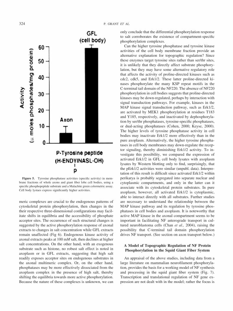
meric complexes are crucial to the endogenous patterns ofcytoskeletal protein phosphorylation, then changes in thetheir respective three-dimensional configurations may facil-itate shifts in equilibria and the accessibility of phosphateacceptor sites. The occurrence of such structural changes issuggested by the active phosphorylation response of axonalextracts to changes in salt concentration while GFL extractsremain unaffected (Fig 6). Endogenous kinase activity ofaxonal extracts peaks at 100 mM salt, then declines at highersalt concentrations. On the other hand, with an exogenoussubstrate such as histone, no robust salt effect is noted inaxoplasm or in GFL extracts, suggesting that high saltreadily exposes acceptor sites on endogenous substrates inthe axonal multimeric complex. Or, on the other hand,phosphatases may be more effectively dissociated from theaxoplasm complex in the presence of high salt, therebyshifting the equilibria towards more active phosphorylation.Because the nature of these complexes is unknown, we can
only conclude that the differential phosphorylation responseto salt corroborates the existence of compartment-specificphosphorylation complexes.
Can the higher tyrosine phosphatase and tyrosine kinaseactivities of the cell body membrane fraction provide analternative explanation for topographic regulation? Sincethese enzymes target tyrosine sites rather than ser/thr sites,it is unlikely that they directly affect substrate phosphory-lation, but they may have some alternative regulatory rolethat affects the activity of proline-directed kinases such ascdc2, cdk5, and Erk1/2. These latter proline-directed ki-nases phosphorylate the many KSP repeat motifs in theC-terminal tail domain of the NF220. The absence of NF220phosphorylation in cell bodies suggests that proline-directedkinases may be down-regulated, perhaps by interaction withsignal transduction pathways. For example, kinases in theMAP kinase signal transduction pathway, such as Erk1/2,are activated by MEK1 phosphorylation at residues T183and Y185, respectively, and inactivated by dephosphoryla-tion by ser/thr phosphatases, tyrosine-specific phosphatases,or dual-acting phosphatases (Cohen, 2000; Keyse, 2000).The higher levels of tyrosine phosphatase activity in cellbodies may inactivate Erk1/2 more effectively than in thepure axoplasm. Alternatively, the higher tyrosine phospha-tases in cell body membranes may down-regulate the recep-tor signaling, thereby diminishing Erk1/2 activity. To in-vestigate this possibility, we compared the expression ofactivated Erk1/2 in GFL cell body lysates with axoplasmlysates by Western blotting only to find, surprisingly, thatthe pErk1/2 activities were similar (unpubl. data). Interpre-tation of this result is difficult since activated Erk1/2 withinperikarya is probably segregated into separate nuclear andcytoplasmic compartments, and only in the latter can itassociate with its cytoskeletal protein substrates. In pureaxoplasm, however, all activated Erk1/2 is cytoplasmic,able to interact directly with all substrates. Further studiesare necessary to understand the relationship between theMAP kinase pathway and its regulation by tyrosine phos-phatases in cell bodies and axoplasm. It is noteworthy thatactive MAP kinase in the axonal compartment seems to beimportant in facilitating NF anterograde transport in cul-tured neuroblastoma cells (Chan et al., 2004), raising thepossibility that C-terminal tail domain phosphorylationdrives NF transport. (See section on axon transport below.)
A Model of Topographic Regulation of NF ProteinPhosphorylation in the Squid Giant Fiber System
An appraisal of the above studies, including data from alarge literature on mammalian neurofilament phosphoryla-tion, provides the basis for a working model of NF synthesisand processing in the squid giant fiber system (Fig. 7).Transcription and translational regulation of NF gene ex-pression are not dealt with in the model; rather the focus is
Figure 5. Tyrosine phosphatase activities (specific activity) in mem-brane fractions of whole axons and giant fiber lobe cell bodies, using aspecific phosphopeptide substrate and a Malachite green colormetric assay.Cell body lystaes express significantly higher activities.
324 P. GRANT ET AL.
on post-translational processing, NF assembly, and trans-port from the cell body compartment to the axon. It isassumed that all “players” in these processes—kinases,phosphatases, regulators and cytoskeletal substrates—aresynthesized in cell bodies of the GFL, simultaneously withthe initial processing events. According to the model, po-lymerization of NF monomers and oligomers is transiently
affected by phosphorylation of NF60-kd and NF 180-kdhead domain sites by PKA, PKC, or both. A cell-body-specific multimeric complex of “incomplete” filaments anddense bodies is assembled with little or no evidence ofsidearm formation (Cohen et al., 1987). The complex con-sists of substrates; kinases, including proline-directed ki-nases (Erk1/2, cdc2-like kinase, SAP kinase); and phospha-
Figure 6. Effect of varying concentrations of NaCl on endogenous kinase activities of axoplasm and giantfiber lobe (GFL) extracts in the presence of phosphatase inhibitors. (A) Graph showing the response of axoplasmextracts to increasing concentrations of NaCl, with peak activity at 100 mM, while GFL lysates exhibit no changeover the range of concentrations. (B) Autoradiograph of gels prepared from above lysates, showing the dramaticdifferences in sensitivity of axoplasm and GFL extracts. Arrow points to low-molecular-weight region whereGFL activity is almost exclusively expressed. HMW � high-molecular-weight, NF220 complex.
325GIANT AXON AND NEURODEGENERATION?
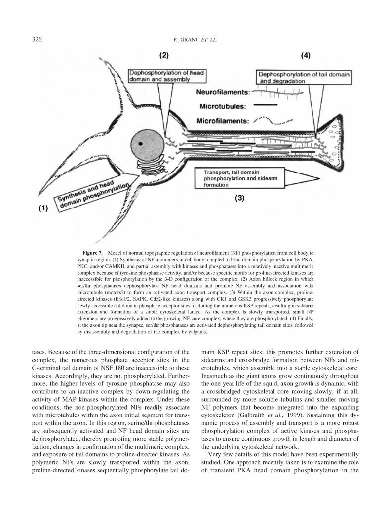
tases. Because of the three-dimensional configuration of thecomplex, the numerous phosphate acceptor sites in theC-terminal tail domain of NSF 180 are inaccessible to thesekinases. Accordingly, they are not phosphorylated. Further-more, the higher levels of tyrosine phosphatase may alsocontribute to an inactive complex by down-regulating theactivity of MAP kinases within the complex. Under theseconditions, the non-phosphorylated NFs readily associatewith microtubules within the axon initial segment for trans-port within the axon. In this region, serine/thr phosphatasesare subsequently activated and NF head domain sites aredephosphorylated, thereby promoting more stable polymer-ization, changes in confirmation of the multimeric complex,and exposure of tail domains to proline-directed kinases. Aspolymeric NFs are slowly transported within the axon,proline-directed kinases sequentially phosphorylate tail do-
main KSP repeat sites; this promotes further extension ofsidearms and crossbridge formation between NFs and mi-crotubules, which assemble into a stable cytoskeletal core.Inasmuch as the giant axons grow continuously throughoutthe one-year life of the squid, axon growth is dynamic, witha crossbridged cytoskeletal core moving slowly, if at all,surrounded by more soluble tubulins and smaller movingNF polymers that become integrated into the expandingcytoskeleton (Galbraith et al., 1999). Sustaining this dy-namic process of assembly and transport is a more robustphosphorylation complex of active kinases and phospha-tases to ensure continuous growth in length and diameter ofthe underlying cytoskeletal network.
Very few details of this model have been experimentallystudied. One approach recently taken is to examine the roleof transient PKA head domain phosphorylation in the
Figure 7. Model of normal topographic regulation of neurofilament (NF) phosphorylation from cell body tosynaptic region. (1) Synthesis of NF monomers in cell body, coupled to head domain phosphorylation by PKA,PKC, and/or CAMKII, and partial assembly with kinases and phosphatases into a relatively inactive multimericcomplex because of tyrosine phosphatase activity, and/or because specific motifs for proline-directed kinases areinaccessible for phosphorylation by the 3-D configuration of the complex. (2) Axon hillock region in whichser/thr phosphatases dephosphorylate NF head domains and promote NF assembly and association withmicrotubule (motors?) to form an activated axon transport complex. (3) Within the axon complex, proline-directed kinases (Erk1/2, SAPK, Cdc2-like kinases) along with CK1 and GSK3 progressively phosphorylatenewly accessible tail domain phosphate acceptor sites, including the numerous KSP repeats, resulting in sidearmextension and formation of a stable cytoskeletal lattice. As the complex is slowly transported, small NFoligomers are progressively added to the growing NF-core complex, where they are phosphorylated. (4) Finally,at the axon tip near the synapse, ser/thr phosphatases are activated dephosphorylating tail domain sites, followedby disassembly and degradation of the complex by calpains.
326 P. GRANT ET AL.

Figure 8. Test of the model. Does PKA phosphorylation of specific serine sites in the head domain of NFMinhibit phosphorylation of tail domain sites by proline-directed kinase Erk1/2? (A) Diagram of the sequencestructure of NF-M showing the C-terminal tail domain KSP sites for proline-directed kinases, the N-terminalhead domain PKA and PKC sites ser1, ser23, and ser46. The three serine sites were mutated to alanine(muNFM), which prevents their phosphorylation. (B) NIH3T3 cells were transiently transfected with eitherwild-type NF-M (wtNFM) or mutant NF-M and treated with EGF, which stimulates Erk1/2 activity andphosphorylation of wt NF-M (lane 4), as seen with an antibody specific for tail domain phosphorylated KSP sites.Prior stimulation of PKA kinase with forskolin (lane 2) inhibits NF-M phosphorylation without affecting Erk1/2activity, suggesting that NF-M tail domain sites were made inaccessible by an intramolecular reconfiguration ofNF-M. Inhibition of PKA activity with a specific inhibitor (KT5720) restores NF-M tail domain phosphorylation(lane 3). On the other hand, in response to EGF, cells transfected with the mutant NF-M exhibit levels of NF-Mtail domain phosphorylation similar to that seen in wtNF-M transfected cells (lane 6), and they are completelyunaffected by prior forskolin treatment and PKA activation; i.e., head domain sites are not phosphorylated (lane5, compare to lane 2). Finally, activation of Erk1/2 is essential since an inhibitor of MEKI (PD98059), theupstream kinase that activates Erk1/2, significantly down-regulates NF-M tail domain phosphorylation (lane 7).(C) An immunocytochemical assay of the transfected cells using the same specific phosphorylated NF-Mantibody illustrates the effect. (a) Transfected 3T3 cells stimulated with EGF show robust expression of NF-Mtail domain phosphorylation. (b) Prior treatment of transfected cells with forskolin inhibits tail domainphosphorylation. (c) Forskolin/EGF treated cells exposed to the PKA inhibitor KT5720 show normal NF-M taildomain phosphorylation, while in (d), cells transfected with the muNF-M fail to respond to forskolin pretreat-ment and express normal levels of NF-M tail domain phosphorylation.
327GIANT AXON AND NEURODEGENERATION?
phosphorylation of KSP residues in C-terminal tail domainsof mammalian NF-M (Zheng et al., 2003). When corticalneurons were treated with forskolin, which specifically ac-tivates PKA phosphorylation, the phosphorylation of en-dogenous NF-M tail domain was inhibited, as determinedby antibodies specific for the phosphorylated tail domainepitopes (Fig. 8). Moreover, the sites of PKA phosphoryla-tion were shown to be in the NFM head domain by trans-fecting NIH3T3 cells with wild-type cells and NF-M mu-tated at specific PKA head domain residues. Stimulation ofthe cells expressing the mutant NF-M with epidermalgrowth factor in the presence of forskolin failed to inhibitNFM tail domain phosphorylation, but did inhibit tail do-main phosphorylation in wild-type transfected cells. Thissuggests an intramolecular model of inhibition; head do-main phosphorylation, by preventing assembly of the NFpolymer from oligomers, evokes conformational changes intail domains such that they are inaccessible to proline-directed kinases. Tail domains, in the absence of assembly,may retain a globular, collapsed structure as seen in cellbody filaments. Such conformational changes could explainthe formation of an inactive, cell-body-specific multimericcomplex.
The mechanisms of slow and fast axon transport havebeen extensively studied in the squid giant fiber system(Weiss et al., 1990). Originally, it was believed that neuro-filaments were transported as part of the slow transportsystem, along with tubulin and actin. However, recent stud-ies of green fluorescent protein (GFP)-labeled neurofila-ments transfected into neurons or injected into axoplasmhave shown that axonal transport of neurofilaments is morecomplex than previously assumed (Galbraith et al., 1999;Prahlad et al., 2000; Wang et al., 2000; Yabe et al.,2001a,b). Apparently, both NF-M and NF-H move at fastrates followed by very long pauses, or by “fits and starts,”giving the impression that most neurofilaments are station-ary but a few move rapidly in bursts, in both directions.Some studies suggest that neurofilaments move as poly-mers, while others invoke smaller oligomers. To complicatematters, rates of transport seem to relate to the extent of taildomain phosphorylation in NF-M and NF-H (Yabe et al.,2001b). C-terminal tail phosphorylation of NF-H and side-arm formation seem to slow transport, but complete re-moval of the NF-H tail domain in a transfected mousemodel had no apparent effect on axonal transport in theoptic nerve, nor on axonal caliber in motor and sensorynerves (Rao et al., 2002). These contradictory results aredifficult to explain. In neurons without NF-H tail domains,there is, however, a compensatory increase in phosphoryla-tion of NF-M that may sustain the transport rate. We shallsee later that factors interfering with NF transport may beresponsible for some of the pathologies found in certainneurodegenerative disorders.
Other features of the model, such as the role of phospha-
tases, are also under investigation. The complexity of theprocesses involved in compartmental-specific phosphoryla-tion implies a system of interactive networks with manyregulatory “nodes” at structural as well as molecular sites.Any one of these might lead to deregulation and chronicstimulation of cellular pathology, resulting in cell death.
Does the Model Offer Clues as to the FactorsResponsible for Some Forms of Neurodegeneration?
Squid, to our knowledge, do not suffer neurodegenera-tion; perhaps they don’t live long enough. Nevertheless, thegiant axon of Loligo pealei has served as the generic modelof neural function in most organisms, and it is likely that theunderlying biochemical mechanisms are also shared. Doesthe model described above offer any insights into the originof some neurodegenerative disorders that might inform aworking experimental approach?
For many neurodegenerative disorders, including Alzhei-mer’s disease (AD), amyotrophic lateral sclerosis (ALS),and Parkinson’s disease (PD), one of the principal neuronalpathologies is the accumulation of abnormal aggregates andneurofibrillary tangles within the cell body; these oftencontain, among many other proteins, extensively phosphor-ylated cytoskeletal proteins, such as PHF tau in AD and NFsin ALS (Sternberger et al., 1985; Lee, 1995; Julien andMushynski, 1998; Al-Chalabi and Miller, 2003). Evidentlysuch accumulations are toxic to cells, which accounts for theneuronal loss that leads to memory defects and dementia.For decades, much effort has been directed to studying thefactors responsible for the abnormal phosphorylation andaggregation of these elements within cell bodies. The resultsof this effort, according to the model above, suggest thattopographic phosphorylation of cytoskeletal proteins hasbeen deregulated.
Can our studies on the squid axon, a large motor axon,and its numerous cell bodies in the GFL provide any in-sights into ALS pathology? In ALS, motor neurons withsomatal aggregates enriched in phosphorylated neurofila-ments usually degenerate (see Fig. 1). The mechanismsunderlying this pathology and the factors responsible forcell death have been intensively studied in a number ofmodel systems (Bruijn et al., 2004). A major lead was theidentification of several dominant mutations in the super-oxide dismutase gene SOD1 responsible for familial-linkedALS, which represents only 10% of all ALS cases (Rosen etal., 1993; Sapp et al., 1995). The pathology and course ofthe disease are similar to that seen in the majority ofsporadic ALS cases. The mutations result in a gain of toxicproperties leading to cell death. Transgenic mouse modelswith these human SOD1 mutations gradually develop theweakening disease and display the characteristic abnormalmotor neuron inclusions consisting of phosphorylatedNF-H, SOD1, and ubiquitin, among other proteins. How
328 P. GRANT ET AL.

such stable aggregates induce neuronal death is not under-stood, but the presence of ubiquitin suggests that abnormalproteins are in the proteosome degradation pathway. Toxiceffects of such aggregates may clog proteosome pathwayswith abnormal proteins, or they may alter mitochondrialfunction, leading to apoptosis (Bruijn et al., 2004). It is notclear whether the abnormal hyperphosphorylation of NFwithin the cell body is the principal factor driving aggregateformation, or whether abnormally phosphorylated proteinsresult from conformational changes in the folding of sub-strates within cell bodies (Kosik and Shimura, 2005). Thelatter would suggest that exogenous stress factors (oxygenradicals, excitatory stress factors, toxic substances producedby the abnormal activity of the SOD1 mutant enzyme)induce subtle modifications of a cell body’s multimericphosphorylation complex, activating kinases, inactivatingphosphatases, or both (Johnson et al., 1996). For example,tyrosine phosphorylation of tau can induce significant con-formational changes (Fabian et al., 1994). In the model, thissuggests that sites previously inaccessible to proline-di-rected kinases such as cdc-2 like kinases (cdk5), and MAPkinases, together with CK1, a non-proline-directed kinase(Kuret et al., 1997), may become available for aberranthyperphosphorylation. It should be noted that abnormallyactivated cdk5 kinase, a principal mammalian neuronal ki-nase regulating a range of neuronal functions from migra-tion during neurogenesis to mature synaptic function andneuronal survival, has been implicated in tau hyperphos-phorylation and tangle formation in AD (Patrick et al.,1999). MAP kinase pathways, which are essential duringgrowth and differentiation in the developing nervous sys-tem, may, when deregulated by stress, also lead to tauhyperphosphorylation and cell death (Cheung and Slack,2004; Subramaniam et al., 2004). Abnormal aggregates andtangles found in perikarya of brain neurons in such disor-ders as AD, Parkinson’s, or ALS are deregulated “multi-meric” complexes of cytoskeletal proteins, active kinases(Erk1/2, Cdk5, GSK3, PKA), other proteins (�-synuclein),and ubiquitin, suggesting that some abnormal proteinswithin the complex are undergoing degradation (Pallares-Trujillo et al., 1998).
Phosphorylated tangles and aggregates may hamper orprevent axon transport of transmitter-filled vesicles to syn-apses and induce cell death. It has been suggested thathighly phosphorylated NFs significantly slow axonal trans-port: the more NF-H tail domain sites phosphorylated, thegreater the delay in transport (Ackerley et al., 2003, Jung etal., 2000a,b). Moreover, two proline-directed kinases, im-plicated as the principal players in NF-H C-terminal taildomain phosphorylation in the squid giant axon, seem tohave conflicting effects on axonal transport in mammalianneurons. On the one hand, cdk5 phosphorylation of NFsseems to block axonal transport and induce the accumula-tion of phosphorylated NFs within perikarya, presumably
by increased pausing during transport (Ackerley et al.,2003; Shea et al., 2004). On the other hand, results from thesame laboratory indicate that NF C-terminal phosphoryla-tion by MAP kinase (Erk1/2) facilitates and regulates ax-onal transport (Chan et al., 2004). Each kinase seems tohave opposing effects on transport, perhaps indicating dif-ferential effects on NF binding to the transport motorskinesin and dynein, altering retrograde and anterogradetransport differentially. Nevertheless, the conundrum is fur-ther complicated by observations, mentioned above, thatthere were no effects on transport in axons transfected with“tailess” NF-H mutant subunits (Rao et al., 2002).
Significantly, studies of axonal transport in axoplasmextruded from the squid giant axon suggest that the effect ofcdk5 phosphorylation may be mediated indirectly by virtueof its regulation of GSK3 activity and the kinesin motor(Morfini et al., 2004). Sustained cdk5 activity is essentialfor promoting kinesin-driven fast axon transport; specificinhibition of cdk5 slows transport indirectly, by activating aphosphatase PP1 that dephosphorylates and activatesGSK3, which in turn, phosphorylates the kinesin motor,slowing anterograde but not retrograde transport. Since ki-nesin is a motor responsible for the fast transport of NFs(Yabe et al., 1999), such indirect effects on motors byderegulated proline-directed kinases may contribute to theperikaryal accumulation of aggregates by blocking axontransport.
An alternative site for deregulation may be overexpres-sion of one or another NF subunit precursor, or deletion ofan NF subunit as in NF null mice, thus disrupting subunitstoichiometry, affecting NF assembly and axon diameter orfilament spacing (Elder et al., 1998; Rao et al., 1998; Zhu etal., 1998). This, too, may alter the structure of phosphory-lation complexes, activating kinases and NF phosphoryla-tion, with subsequent effects on axon transport.
Finally, stress-induced abnormalities in signal transduc-tion pathways that down-regulate ser/thr and/or tyrosinephosphatases may disrupt the fine-tuned equilibria thattightly regulate topographic phosphorylation between cellbody and axonal compartments. An increase in the numberof phosphorylated sites on cytoskeletal proteins within thecell body could evoke conformational changes that result inaggregates, tangles, obstructed transport, and eventually celldeath. Contrary to expectations, however, an increase ofassembled NF-M and NF-H in perikarya of SOD1 mutantmice seems to ameliorate the disease, increasing life spanfor many months (Couillard-Despres et al., 1998). In fact,the perikaryal accumulations of hyperphosphorylated NF-Hinduced by a deregulated cdk5 in SOD mutant ALS micealleviates the toxicity and improves the life span of the mice(Nguyen et al., 2001), an unexpected result in view of thepresumed role of deregulated cdk5 and tau hyperphospho-rylation in AD. It is proposed that the excess NF-H, with itsnumerous KSP sites, acts as a sink for cdk5 activity, thereby
329GIANT AXON AND NEURODEGENERATION?

reducing toxicity levels. This may, however, reflect only ashort-term adaptation since neurodegeneration is a long-term process of accumulation of insults, so that an NF-Hsink may eventually become a burden if it is in the wrongplace. It is also possible that such conflicting results maysimply reflect differences in the affected neuronal pheno-types—large motor neurons with very long, large-diameter,myelinated axons compared to the smaller pyramidal neu-rons of the cortex and hippocampus.
Another observation to confound our attempts to under-stand ALS is the demonstration that the motor neuronpathology in SOD mutant mice is not cell-autonomous butseems to depend on interactions with surrounding cells,perhaps other neurons or glia (Clement et al., 2003). Whenchimeric mice that included both normal and SOD mutantneurons in the same brain were used, those SOD1 neuronsthat were surrounded by many normal non-neuronal cellsescaped the pathology and did not degenerate, while otherSOD1 neurons that were surrounded by mutant neurons ornonneuronal neurons invariably degenerated. Such chimericmutant mice survived for much longer than the ALS modelmouse. Even more striking was the fact that normal neuronssurrounded by SOD1 mutant neurons developed theperikaryal filamentous deposits and degenerated, evidently“infected” by the abnormal neurons. At least in these modelALS mice, glial interactions play an important role in thedevelopment of the disease. In this connection, the squidgiant fiber system may offer an excellent opportunity tostudy glial-axonal interactions because the axoplasm can bereadily separated from its glial-rich sheath, and both can bestudied biochemically under various experimental condi-tions.
Such speculations serve primarily as working models forfurther experimentation. Although we do not have squidmodels for neurodegeneration, we do have the giant axonsystem which, under appropriate experimental manipula-tions, may be induced to undergo controlled pathologiesresembling those seen in neurodegenerative disorders.These, in turn, might be more amenable to biochemicalstudies.
It is noteworthy that another large motor neuron system,that of the anterior bulbar cells (ABC cells) in the hindbrainof the lamprey, a most primitive vertebrate, provides an-other useful model for studying neuronal cytoskeletal reg-ulation, particularly in experimentally induced neurodegen-eration (Hall et al., 2000, 2001). The large ABC axonsresemble the squid giant axon in that they are not myelin-ated. The principal, and only, NF subunit in the lampreyneurons is NF-180, which is most homologous to mamma-lian NF-M but has many more glutamic acid residues andonly two tail domain non-KSP phosphorylation sites. Nev-ertheless, it forms NFs that function as they do in mamma-lian neurons. Here, the effects of NF sidearm extension onaxon caliber are probably facilitated by the charge-charge
repulsions of the glutamic acid residues instead of highlyphosphorylated KSP sites. Compared to the squid giantaxon system, however, lamprey ABC neurons are moreamenable to experimental manipulation, such as direct cellbody injection of NF180 to study the effect of overexpres-sion, or transfection of ABC cells by injection with GFP-labeled human tau constructs to examine the time course oftau-induced neural degeneration (Hall et al., 2001). Forexample, in the latter study, the authors demonstrated thatthe earliest stages of pathology were seen in swollen distaldendrites of ABC neurons with accumulations of paired helicalfilament (PHF1) antigen expression. In time, these accumula-tions swept progressively into proximal dendrites, somata, andaxons, accompanied by tau hyperphosphorylation and degen-eration. This study could provide some details of the progres-sive stages of tau-induced neurodegeneration.
We see that these two unusual motor neuron systems—squid giant fibers and lamprey ABC neurons—offer novelexperimental approaches to our efforts to understand neu-rodegeneration: the former biochemically, because largecell bodies can be separated from pure axoplasm; the latterbecause of the ease of direct experimental manipulation bytransfection. It would be useful if the technology of thelatter could be successfully applied to the former.
Acknowledgments
This research was supported by the Intramural ResearchPrograms of the NIH, National Institute of NeurologicalDisorders and Stroke.
Literature Cited
Ackerley, S., P. Thornhill, A. J. Grierson, J. Brownlees, B. H. Ander-ton, P. N. Leigh, C. E. Shaw, and C. C. Miller. 2003. Neurofila-ment heavy chain side arm phosphorylation regulates axonal transportof neurofilaments. J. Cell Biol. 161: 489–495.
Adelman, W. J., and D. L. Gilbert. 1990. Electrophysiology and bio-physics of the squid giant axon, Pp. 93–132 in Squid as ExperimentalAnimals, D.L. Gilbert, W.J. Adelman, and J.M. Arnold eds. PlenumPress, New York.
Al-Chalabi, A., and C. C. J. Miller. 2003. Neurofilaments and neuro-logical disease. BioEssays 25: 346–355.
Brautigan, D. L., and C. L. Shriner. 1988. Methods to distinguishvarious types of protein phosphatase activity. Methods Enzymol. 159:339–346.
Brizuela, L., G. Draetta, and D. Beach. 1987. p13sucl acts in the fissionyeast cell division cycle as a component of the p34cdc2 protein kinase.EMBO J. 6: 3507–3514.
Brown, A., and R. J. Lasek. 1990. The cytoskeleton of the squid giantaxon, Pp. 235–302 in Squid as Experimental Animals, D.L. Gilbert,W. J. Adelman, and J. M. Arnold, eds. Plenum Press, New York.
Bruijn, L. I., T. M. Miller, and D. W. Cleveland. 2004. Unraveling themechanisms involved in motor neuron degeneration in ALS. Annu.Rev. Neurosci. 27: 723–749.
Chan, W. K., A. Dickerson, D. Ortiz, A. F. Pimenta, C. M. Moran, J.Motil, S. J. Snyder, K. Malik, H. C. Pant, and T. B. Shea. 2004.Mitogen-activated protein kinase regulates neurofilament axonal trans-port. J. Cell Sci. 117: 4629–4642.
330 P. GRANT ET AL.
Cheung, E. C., and R. S. Slack. 2004. Emerging role for ERK as a keyregulator of neuronal apoptosis. Sci STKE 2004: PE45.
Clement, A. M., M. D. Nguyen, E. A. Roberts, M. L. Garcia, S. Boillee,et al. 2003. Wild-type nonneuronal cells extend survival of SOD1mutant motor neurons in ALS mice. Science 302: 113–117.
Cohen, P. 2000. The regulation of protein function by multisite phos-phorylation—a 25-year update. Trends Biochem. Sci. 25: 596–601.
Cohen, R. S., H. C. Pant, S. House, and H. Gainer. 1987. Biochemicaland immunocytochemical characterization and distribution of phos-phorylated and nonphosphorylated subunits of neurofilaments in squidgiant axon and stellate ganglion. J. Neurosci. 7: 2056–2074.
Couillard-Despres, S., Q. Zhu, P. C. Wong, D. L. Price, D. W. Cleve-land, and J. P. Julien. 1998. Protective effect of neurofilamentheavy gene overexpression in motor neuron disease induced by mutantsuperoxide dismutase. Proc. Natl. Acad. Sci. USA 95: 9626–9630.
de Waegh, S. M., V. M. Lee, and S. T. Brady. 1992. Local modulationof neurofilament phosphorylation, axonal caliber, and slow axonaltransport by myelinating Schwann cells. Cell 68: 451–463.
Dosemeci, A., and H. C. Pant. 1992. Association of cyclic-AMP-de-pendent protein kinase with neurofilaments. Biochem. J. 282: 477–481.
Dosemeci, A., C. C. Floyd, and H. C. Pant. 1990. Characterization ofneurofilament-associated protein kinase activities from bovine spinalcord. Cell Mol. Neurobiol. 10: 369–382.
Draetta, G., L. Brizuela, J. Potashkin, and D. Beach. 1987. Identifi-cation of p34 and p13 human homologues of the cell cycle regulatorsof fission yeast encoded by cdc2 and suc1. Cell 50: 319–325.
Elder, G. A., V. L. Friedrich, Jr., P. Bosco, C. Kang, A. Gourov, P. H.Tu, V. M. Lee, and R. A. Lazzarini. 1998. Absence of the mid-sizedneurofilament subunit decreases axonal calibers, levels of light neuro-filament (NF-L), and neurofilament content. J. Cell Biol. 141: 727–739.
Fabian, H., L. Otvos, Jr., G. I. Szendrei, E. Lang, and H. H. Mantsch.1994. Tyrosine- versus serine-phosphorylation leads to conforma-tional changes in a synthetic tau peptide. J. Biomol. Struct. Dyn. 12:573–579.
Faux, M. C., and J. D. Scott. 1996. More on target with proteinphosphorylation: conferring specificity by location. Trends Biol. Sci.21: 312–315.
Fiorito, G., and R. Chichery. 1995. Lesions of the vertical lobe impairvisual discrimination learning by observation in Octopus vulgaris.Neurosci. Lett. 192: 117–120.
Fiorito, G., and P. Scotto. 1992. Observational learning in Octopusvulgaris. Science 256: 545–547.
Fiorito, G., C. von Planta, and P. Scotto. 1990. Problem solving abilityof Octopus vulgaris Lamarck (Mollusca, Cephalopoda). Behav. NeuralBiol. 53: 217–230.
Floyd, C. C., P. Grant, P. E. Gallant, and H. C. Pant. 1991. Principalneurofilament-associated protein kinase in squid axoplasm is related tocasein kinase I. J. Biol. Chem. 266: 4987–4994.
Galbraith, J. A., T. S. Reese, M. L. Schlief, and P. E. Gallant. 1999.Slow transport of unpolymerized tubulin and polymerized neurofila-ment in the squid giant axon. Proc. Natl. Acad. Sci. USA 96: 11589–11594.
Giasson, B. I., and W. E. Mushynski. 1997. Study of proline-directedprotein kinases involved in phosphorylation of the heavy neurofilamentsubunit. J. Neurosci. 17: 9466–9472.
Gilbert, D. L., W. J. Adelman, and J. M. Arnold. 1990. Squid asExperimental Animals. Plenum Press, New York.
Goedert, M. 1999. Filamentous nerve cell inclusions in neurodegenera-tive diseases: tauopathies and alpha-synucleinopathies. Philos. Trans.R. Soc. Lond. B Biol. Sci. 354: 1101–1118.
Gordon, J. A. 1991. Use of vanadate as protein-phosphotyrosine phos-phatase inhibitor. Methods Enzymol. 20: 477–482.
Grant, P., and H. C. Pant. 2004. Topographic regulation of phosphor-
ylation in giant neurons of the squid, Loligo pealei: role of phospha-tases. J. Neurobiol. 58: 514–528.
Grant, P., M. Diggins, and H. C. Pant. 1999. Topographic regulation ofcytoskeletal protein phosphorylation by multimeric complexes in thesquid giant fiber system. J. Neurobiol. 40: 89–102.
Guidato, S., L. H. Tsai, J. Woodgett, and C. C. Miller. 1996. Differ-ential cellular phosphorylation of neurofilament heavy side-arms byglycogen synthase kinase-3 and cyclin-dependent kinase-5. J. Neuro-chem. 66: 1698–1706.
Hall, G. F., B. Chu, S. Lee. Y. Liu, and J. Yao. 2000. The singleneurofilament subunit of the lamprey forms filaments and regulatesaxonal caliber and neuronal size in vivo. Cell Motil. Cytoskelet. 46:166–182.
Hall, G. H., V. M.-Y. Lee, G. Lee, and J. Yao. 2001. Staging ofneurofibrillary degeneration caused by human tau overexpression in aunique cellular model of human tauopathy. Am. J. Pathol. 158: 235–246.
Hisanaga, S., and N. Hirokawa. 1990. Molecular architecture of theneurofilament. II. Reassembly process of neurofilament L protein invitro. J. Mol. Biol. 211: 871–882.
Hodge, A. J., and W. J. Adelman, Jr. 1980. The neuroplasmic networkin Loligo and Hermissenda neurons. J. Ultrastruct. Res. 70: 220–241.
Jaffe, H., P. Sharma, P. Grant, and H. Pant. 2001. Characterization ofthe phosphorylation sites of the squid (Loligo pealei) high-molecular-weight neurofilament protein from giant axon axoplasm. J. Neurochem.76: 1022–1031.
Johnson, L. N., M. E. Noble, and D. J. Owen. 1996. Active andinactive protein kinases: structural basis for regulation. Cell 85: 149–158.
Julien, J. P., and W. E. Mushynski. 1998. Neurofilaments in health anddisease. Prog. Nucleic Acid Res. Mol. Biol. 61: 1–23.
Jung, C., J. T. Yabe, S. Lee, and T. B. Shea. 2000a. Hypophosphor-ylated neurofilament subunits undergo axonal transport more rapidlythan more extensively phosphorylated subunits in situ. Cell Motil.Cytoskelet. 47: 120–129.
Jung, C., J. T. Yabe, and T. B. Shea. 2000b. C-terminal phosphoryla-tion of the high molecular weight neurofilament subunit correlates withdecreased neurofilament axonal transport velocity. Brain Res. 856:12–19.
Keyse, S. M. 2000. Protein phosphatases and the regulation of mitogen-activated protein kinase signalling. Curr. Opin. Cell Biol. 12: 186–192.
Klauck, T. M., M. C. Faux, K. Labudda, L. K. Langeberg, S. Jaken,and J. D. Scott. 1996. Coordination of three signaling enzymes byAKAP79, a mammalian scaffold protein. Science 271: 1589–1592.
Kosik, K. S., and H. Shimura. 2005. Phosphorylated tau and the neu-rodegenerative foldopathies. Biochim. Biophys. Acta 1739: 298–310.
Kuret, J., G. S. Johnson, D. Cha, E. R. Christenson, A. J. DeMaggio,and M. F. Hoekstra. 1997. Casein kinase 1 is tightly associated withpaired-helical filaments isolated from Alzheimer’s disease brain.J. Neurochem. 69: 2506–2515.
Lee, V. M. 1995. Disruption of the cytoskeleton in Alzheimer’s disease.Curr. Opin. Neurobiol. 5: 663–668.
Li, B.-S., Veeranna, P. Grant, and H. C. Pant. 1999. Activation ofmitogen-activated protein kinases (Erk1 and Erk2) cascade results inphosphorylation of NF-M tail domains in transfected NIH 3T3 cells.Eur. J. Biochem. 262: 211–217.
Martin, R., K. Schilling, W. Fritz, and A. Giuditta. 1990. Visualiza-tion of differential neurofilament phosphorylation in the pre- andpostsynaptic axoplasm of the squid giant synapse: an electron spectro-scopic study. Neuroscience 37: 553–562.
Metuzals, J., and C. S. Izzard. 1969. Spatial patterns of threadlikeelements in the axoplasm of the giant nerve fiber of the squid (Loligopealii L.) as disclosed by differential interference microscopy and byelectron microscopy. J. Cell Biol. 43: 456–479.
331GIANT AXON AND NEURODEGENERATION?
Miyamoto, S., H. Teramoto, O. A. Coso, J. S. Gutkind, P. D. Burbelo,S. K. Akiyama, and K. M. Yamada. 1995. Integrin function: mo-lecular hierarchies of cytoskeletal and signaling molecules. J. Cell Biol.131: 791–805.
Morfini, G., G. Szebenyi, H. Brown, H. C. Pant, G. Pigino, S. DeBoer,U. Beffert, and S. T. Brady. 2004. A novel CDK5-dependent path-way for regulating GSK3 activity and kinesin-driven motility in neu-rons. EMBO J. 23: 2235–2245.
Nguyen, M. D., R. C. Lariviere, and J. P. Julien. 2001. Deregulationof Cdk5 in a mouse model of ALS: toxicity alleviated by perikaryalneurofilament inclusions. Neuron 30: 135–147.
Nixon, R. A., S. E. Lewis, and C. A. Marotta. 1987. Posttranslationalmodification of neurofilament proteins by phosphate during axoplasmictransport in retinal ganglion cell neurons. J. Neurosci. 7: 1145–1158.
Nixon, R. A., S. E. Lewis, D. Dahl, C. A. Marotta, and U. C. Drager.1989. Early posttranslational modifications of the three neurofilamentsubunits in mouse retinal ganglion cells: neuronal sites and time coursein relation to subunit polymerization and axonal transport. Brain Res.Mol. Brain Res. 5: 93–108.
Pallares-Trujillo, J., F. J. Lopez-Soriano, and J. M. Argiles. 1998.The involvement of the ubiquitin system in Alzheimer’s disease Int. J.Mol. Med. 2: 3–15.
Pant, H. C., P. E. Gallant, and H. Gainer. 1986. Characterization of acyclic nucleotide- and calcium-independent neurofilament protein ki-nase activity in axoplasm from the squid giant axon. J. Biol. Chem.261: 2968–2977.
Pant, H. C., Veeranna, and P. Grant. 2000. Regulation of axonalneurofilament phosphorylation. Curr. Top. Cell. Regul. 36: 133–150.
Patrick, G. N., L. Zukerberg, M. Nikolic, S. de la Monte, P. Dikkes,and L. H. Tsai. 1999. Conversion of p35 to p25 deregulates Cdk5activity and promotes neurodegeneration. Nature 402: 615–622.
Prahlad, V., B. T. Helfand, G. M. Langford, R. D. Vale, and R. D.Goldman. 2000. Fast transport of neurofilament protein along mi-crotubules in squid axoplasm. J. Cell. Sci. 113: 3939–3946.
Rao, M. V., M. K. Houseweart, T. L. Williamson, T. O. Crawford, J.Folmer and D. W. Cleveland. 1998. Neurofilament-dependent ra-dial growth of motor axons and axonal organization of neurofilamentsdoes not require the neurofilament heavy subunit (NF-H) or its phos-phorylation J. Cell Biol. 143: 171–181.
Rao, M. V., M. L. Garcia, Y. Miyazaki, T. Gotow, A. Yuan, S. Mattina,C. M. Ward, N. A. Calcutt, Y. Uchiyama, R. A. Nixon, and D. W.Cleveland. 2002. Gene replacement in mice reveals that the heavilyphosphorylated tail of neurofilament heavy subunit does not affectaxonal caliber or the transit of cargoes in slow axonal transport. J. CellBiol. 158: 681–693.
Roach, P. J. 1991. Multisite and hierarchal protein phosphorylation.J. Biol. Chem. 266: 14139–14142.
Robinson, M. J., and M. H. Cobb. 1997. Mitogen-activated proteinkinase pathways. Curr. Opin. Cell Biol. 9: 180–186.
Rosen, D. R., T. Siddique, D. Patterson, D. A. Figlewicz, P. Sapp, A.Hentati, D. Donaldson, J. Goto, J. P. O’Regan, H. X. Deng, et al.1993. Mutations in Cu/Zn superoxide dismutase gene are associatedwith familial amyotrophic lateral sclerosis. Nature 362: 59–62.
Sanchez, I., L. Hassinger, R. K. Sihag, D. W. Cleveland, P. Mohan, andR. A. Nixon. 2000. Local control of neurofilament accumulationduring radial growth of myelinating axons in vivo: selective role ofsite-specific phosphorylation. J. Cell Biol. 151: 1013–1024.
Sapp, P. C., D. R. Rosen, B. A. Hosler, J. Esteban, D. McKenna-Yasek,J. P. O’Regan, H. R. Horvitz, and R. H. Brown, Jr. 1995. Identi-fication of three novel mutations in the gene for Cu/Zn superoxidedismutase in patients with familial amyotrophic lateral sclerosis. Neu-romuscul. Disord. 5: 353–357.
Shea, T. B., J. T. Yabe, D. Ortiz, A. Pimenta, P. Loomis, R. D.Goldman, N. Amin, and H. C. Pant. 2004. Cdk5 regulates axonal
transport and phosphorylation of neurofilaments in cultured neurons.J. Cell Sci. 117: 933–941.
Sihag, R. K., and R. A. Nixon. 1989. In vivo phosphorylation of distinctdomains of the 70-kilodalton neurofilament subunit involves differentprotein kinases. J. Biol. Chem. 264: 457–464.
Sihag, R. K., and R. A. Nixon. 1990. Phosphorylation of the amino-terminal head domain of the middle molecular mass 145-kDa subunitof neurofilaments: evidence for regulation by second messenger-depen-dent protein kinases. J. Biol. Chem. 265: 4166–4171.
Sihag, R. K., and R. A. Nixon. 1991. Identification of Ser-55 as a majorprotein kinase A phosphorylation site on the 70-kDa subunit of neu-rofilaments: early turnover during axonal transport. J. Biol. Chem. 266:18861–18867.
Sihag, R. K., A. Y. Jeng, and R. A. Nixon. 1988. Phosphorylation ofneurofilament proteins by protein kinase C. FEBS Lett. 233: 181–185.
Sternberger, L. A., and N. H. Sternberger. 1983. Monoclonal antibod-ies distinguish phosphorylated and non-phosphorylated forms of neu-rofilaments in situ. Proc. Natl. Acad. Sci. USA 80: 6126–6130.
Sternberger, N. H., L. A. Sternberger, and J. Ulrich. 1985. Aberrantneurofilament phosphorylation in Alzheimer disease. Proc. Natl. Acad.Sci. USA 82: 4274–4276.
Subramaniam, S., U. Zirrgiebel, O. von Bohlen und Halbach, J. Stre-lau, C. Laliberte, D. R. Kaplan, and K. Unsicker. 2004. ERKactivation promotes neuronal degeneration predominantly throughplasma membrane damage and independently of caspase-3. J. Cell Biol.165: 357–369.
Sun, D., C. L. Leung, and R. K. H. Liem. 1996. Phosphorylation of thehigh molecular weight neurofilament protein (NF-H) by Cdk5 and p35.J. Biol. Chem. 271: 14245–14251.
Szaro, B. G., H. C. Pant, J. Way, and J. Battey. 1991. Squid lowmolecular weight neurofilament proteins are a novel class of neurofila-ment protein. A nuclear lamin-like core and multiple distinct proteinsformed by alternative RNA processing. J. Biol. Chem. 266: 15035–15041.
Takahashi, M., N. Amin, P. Grant, and H. C. Pant. 1995. P13suclassociates with a cdc2-like kinase in a multimeric cytoskeletal complexin squid axoplasm. J. Neurosci. 15: 6222–6229.
Tytell, M., H. C. Pant, H. Gainer, and W. D. Hill. 1990. Character-ization of the distinctive neurofilament subunits of the soma and axoninitial segments in the squid stellate ganglion. J. Neurosci. Res. 25:153–161.
Vallano, M. L., J. R. Goldenring, R. S. Lasher, and R. J. DeLorenzo.1986. Association of calcium/calmodulin-dependent kinase with cy-toskeletal preparations: phosphorylation of tubulin, neurofilament, andmicrotubule associated proteins. Ann. N. Y. Acad. Sci. 466: 357–374.
Veeranna, N. D. Amin, N. G. Ahn, H. Jaffe, C. A. Winters, P. Grant,and H. C. Pant. 1998. Mitogen-activated protein kinases (Erk1,2)phosphorylate Lys-Ser-Pro (KSP) repeats in neurofilament proteinsNF-H and NF-M. J. Neurosci. 18: 4008–4021.
Veeranna, K. T. Shetty, M. Takahashi, P. Grant, and H. C. Pant. 2000.Cdk5 and MAPK are associated with complexes of cytoskeletal pro-teins in rat brain. Brain Res. Mol. Brain Res. 76: 229–236.
Wang, L., C. L. Ho, D. Sun, R. K. Liem, and A. Brown. 2000. Rapidmovement of axonal neurofilaments interrupted by prolonged pauses.Nat. Cell Biol. 2: 137–141.
Way, J., M. R. Hellmich, H. Jaffe, B. Szaro, H. C. Pant, H. Gainer, andJ. Battey. 1992. A high-molecular-weight squid neurofilament pro-tein contains a lamin-like rod domain and a tail domain with Lys-Ser-Pro repeats. Proc. Natl. Acad. Sci. USA 89: 6963–6967.
Weiss, D. G., M. A. Meyer, and G. M. Langford. 1990. Studyingaxoplasmic transport by video microscopy and using the squid giantaxon as a model system. Pp. 303–321 in Squid as ExperimentalAnimals, D. L. Gilbert, W. J. Adelman, and J. M. Arnold, eds.Plenum Press, New York.
332 P. GRANT ET AL.
Yabe, J. T., A. Pimenta, and T. B. Shea. 1999. Kinesin-mediatedtransport of neurofilament protein oligomers in growing axons. J. CellSci. 112 (Pt 21): 3799–3814.
Yabe, J. T., W. K. Chan, T. M. Chylinski, S. Lee, A. F. Pimenta, andT. B. Shea. 2001a. The predominant form in which neurofilamentsubunits undergo axonal transport varies during axonal initiation, elon-gation, and maturation. Cell Motil. Cytoskelet. 48: 61–83.
Yabe, J. T., T. Chylinski, F. S. Wang, A. Pimenta, S. D. Kattar, M. D.Linsley, W. K. Chan, and T. B. Shea. 2001b. Neurofilaments con-sist of distinct populations that can be distinguished by C-terminalphosphorylation, bundling, and axonal transport rate in growing axonalneurites. J. Neurosci. 21: 2195–2205.
Young, J. Z. 1939. Fused neurons and synaptic contacts in the giantnerve fibres of cephalopods. Philos. Trans. R. Soc. Lond. B Biol. Sci.229: 465–503.
Zheng, Y. L., B. S. Li, Veeranna, and H. C. Pant. 2003. Phosphory-lation of the head domain of neurofilament protein (NF-M): a factorregulating topographic phosphorylation of NF-M tail domain KSP sitesin neurons. J. Biol. Chem. 278: 24026–24032.
Zhu, Q., M. Lindenbaum, F. Levavasseur, H. Jacomy, and J. P. Julien.1998. Disruption of the NF-H gene increases axonal microtubulecontent and velocity of neurofilament transport: relief of axonopathyresulting from the toxin �,��-iminodipropionitrile. J. Cell Biol. 143:183–193.
333GIANT AXON AND NEURODEGENERATION?