Embed Size (px)
Citation preview
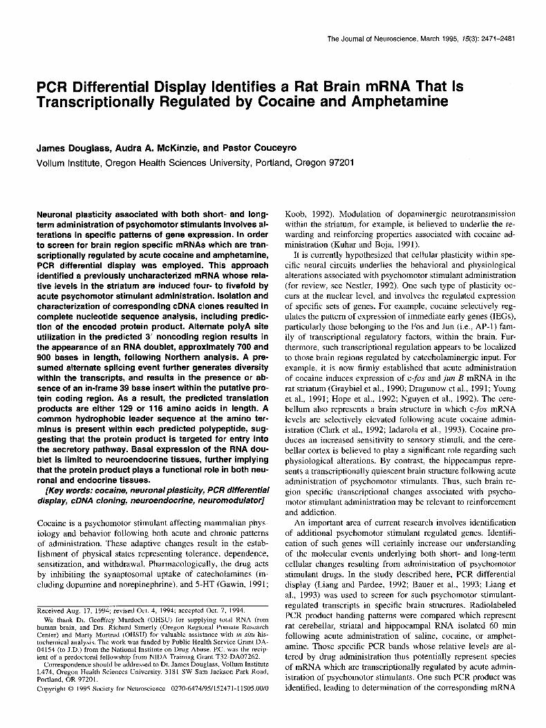
The Journal of Neuroscience, March 1995, 15(3): 2471-2481
PCR Differential Display Identifies a Rat Brain mRNA That Is Transcriptionally Regulated by Cocaine and Amphetamine
James Douglass, Audra A. McKinzie, and Pastor Couceyro
Vellum Institute, Oregon Health Sciences University, Portland, Oregon 97201
Neuronal plasticity associated with both short- and long- term administration of psychomotor stimulants involves al- terations in specific patterns of gene expression. In order to screen for brain region specific mRNAs which are tran- scriptionally regulated by acute cocaine and amphetamine, PCR differential display was employed. This approach identified a previously uncharacterized mFlNA whose rela- tive levels in the striatum are induced four- to fivefold by acute psychomotor stimulant administration. Isolation and characterization of corresponding cDNA clones resulted in complete nucleotide sequence analysis, including predic- tion of the encoded protein product. Alternate polyA site utilization in the predicted 3’ noncoding region results in the appearance of an RNA doublet, approximately 700 and 900 bases in length, following Northern analysis. A pre- sumed alternate splicing event further generates diversity within the transcripts, and results in the presence or ab- sence of an in-frame 39 base insert within the putative pro- tein coding region. As a result, the predicted translation products are either 129 or 116 amino acids in length. A common hydrophobic leader sequence at the amino ter- minus is present within each predicted polypeptide, sug- gesting that the protein product is targeted for entry into the secretory pathway. Basal expression of the RNA dou- blet is limited to neuroendocrine tissues, further implying that the protein product plays a functional role in both neu- ronal and endocrine tissues.
[Key words: cocaine, neuronal plasticity, PCR differential display, cDNA cloning, neuroendocrine, neuromodulator]
Cocaine is a psychomotor stimulant affecting mammalian phys- iology and behavior following both acute and chronic patterns of administration. These adaptive changes result in the estab- lishment of physical states representing tolerance, dependence, sensitization, and withdrawal. Pharmacologically, the drug acts by inhibiting the synaptosomal uptake of catecholamines (in- cluding dopamine and norepinephrine), and 5-HT (Gawin, 1991;
Received Aug. 17, 1994; revised Oct. 4, 1994; accepted Oct. 7, 1994.
We thank Dr. Geoffrey Murdoch (OHSU) for supplying total RNA from human brain, and Drs. Richard Simerly (Oregon Regional Primate Research Center) and Marty Mortrud (OHSU) for valuable assistance with in situ his- tochemical analysis. The work was funded by Public Health Service Grant DA- 04154 (to J.D.) from the National Institute on Drug Abuse. PC. was the recip- ient of a predoctoral fellowship from NIDA Training Grant T32-DA07262.
Correspondence should be addressed to Dr. James Douglass, Vellum Institute L474, Oregon Health Sciences University, 3181 SW Sam Jackson Park Road, Portland, OR 9720 1.
Copyright 0 I995 Society for Neuroscience 0270-6474/95/152471-l 1$05.00/O
Koob, 1992). Modulation of dopaminergic neurotransmission within the striatum, for example, is believed to underlie the re- warding and reinforcing properties associated with cocaine ad- ministration (Kuhar and Boja, 1991).
It is currently hypothesized that cellular plasticity within spe- cific neural circuits underlies the behavioral and physiological alterations associated with psychomotor stimulant administration (for review, see Nestler, 1992). One such type of plasticity oc- curs at the nuclear level, and involves the regulated expression of specific sets of genes. For example, cocaine selectively reg- ulates the pattern of expression of immediate early genes (IEGs), particularly those belonging to the Fos and Jun (i.e., AP-1) fam- ily of transcriptional regulatory factors, within the brain. Fur- thermore, such transcriptional regulation appears to be localized to those brain regions regulated by catecholaminergic input. For example, it is now firmly established that acute administration of cocaine induces expression of c-fos and jun B mRNA in the rat striatum (Graybiel et al., 1990; Dragunow et al., 1991; Young et al., 1991; Hope et al., 1992; Nguyen et al., 1992). The cere- bellum also represents a brain structure in which c-fos mRNA levels are selectively elevated following acute cocaine admin- istration (Clark et al., 1992; Iadarola et al., 1993). Cocaine pro- duces an increased sensitivity to sensory stimuli, and the cere- bellar cortex is believed to play a significant role regarding such physiological alterations. By contrast, the hippocampus repre- sents a transcriptionally quiescent brain structure following acute administration of psychomotor stimulants. Thus, such brain re- gion specific transcriptional changes associated with psycho- motor stimulant administration may be relevant to reinforcement and addiction.
An important area of current research involves identification of additional psychomotor stimulant regulated genes. Identifi- cation of such genes will certainly increase our understanding of the molecular events underlying both short- and long-term cellular changes resulting from administration of psychomotor stimulant drugs. In the study described here, PCR differential display (Liang and Pardee, 1992; Bauer et al., 1993; Liang et al., 1993) was used to screen for such psychomotor stimulant- regulated transcripts in specific brain structures. Radiolabeled PCR product banding patterns were compared which represent rat cerebellar, striatal and hippocampal RNA isolated 60 min following acute administration of saline, cocaine, or amphet- amine. Those specific PCR bands whose relative levels are al- tered by drug administration thus potentially represent species of mRNA which are transcriptionally regulated by acute admin- istration of psychomotor stimulants. One such PCR product was identified, leading to determination of the corresponding mRNA
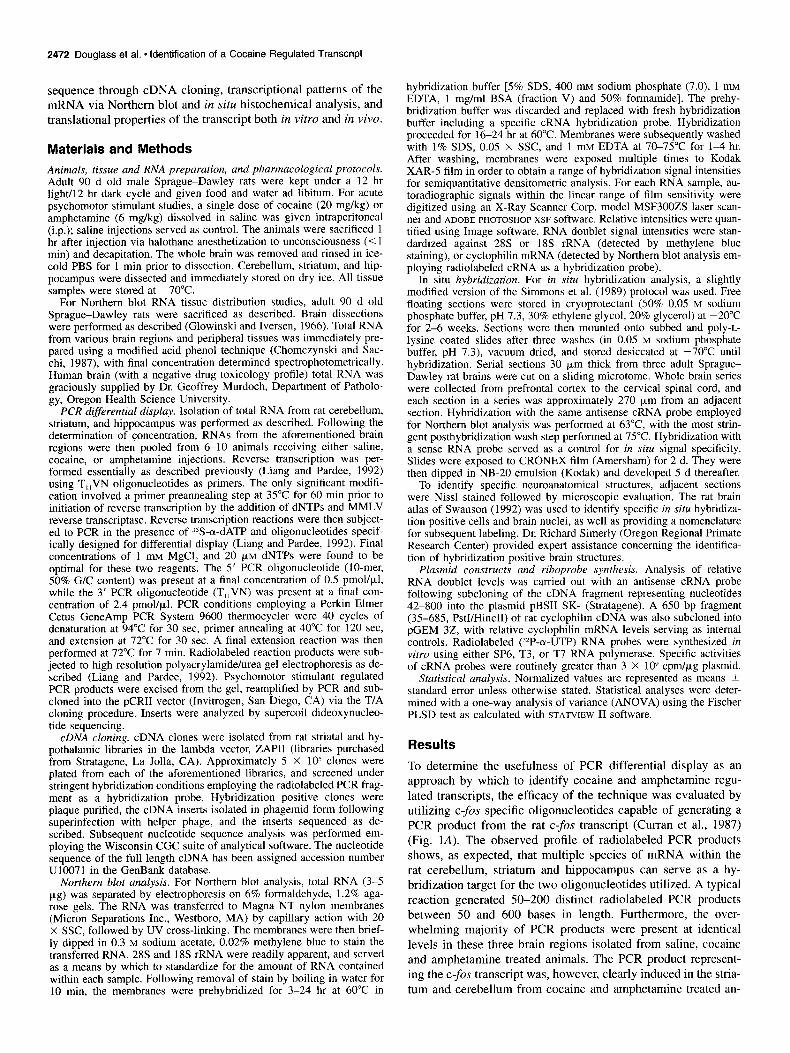
2472 Douglass et al. - identification of a Cocaine Regulated Transcript
sequence through cDNA cloning, transcriptional patterns of the mRNA via Northern blot and in situ histochemical analysis, and translational properties of the transcript both in vitro and in vivo.
Materials and Methods
Animals, tissue and RNA preparation, and pharmacological protocols. Adult 90 d old male Sprague-Dawley rats were kept under a 12 hr light/l2 hr dark cycle and given food and water ad libitum. For acute psychomotor stimulant studies, a single dose of cocaine (20 mg/kg) or amphetamine (6 mg/kg) dissolved in saline was given intraperitoneal (i.p.); saline injections served as control. The animals were sacrificed 1 hr after injection via halothane anesthetization to unconsciousness (<l min) and decapitation. The whole brain was removed and rinsed in ice- cold PBS for 1 min prior to dissection. Cerebellum, striatum, and hip- pocampus were dissected and immediately stored on dry ice. All tissue samples were stored at -70°C.
For Northern blot RNA tissue distribution studies, adult 90 d old Sprague-Dawley rats were sacrificed as described. Brain dissections were uerformed as described (Glowinski and Iversen, 1966). Total RNA from barious brain regions and peripheral tissues was immediately pre- pared using a modified acid phenol technique (Chomczynski and Sac- chi, 1987), with final concentration determined spectrophotometrically. Human brain (with a negative drug toxicology profile) total RNA was graciously supplied by Dr. Geoffrey Murdoch, Department of Patholo- gy, Oregon Health Science University.
PCR differential display. Isolation of total RNA from rat cerebellum, striatum, and hippocampus was performed as described. Following the determination of concentration, RNAs from the aforementioned brain regions were then pooled from 6-10 animals receiving either saline, cocaine, or amphetamine injections. Reverse transcription was per- formed essentially as described previously (Liang and Pardee, 1992) using T,,VN oligonucleotides as primers. The only significant modifi- cation involved a primer preannealing step at 35°C for 60 min prior to initiation of reverse transcription by the addition of dNTPs and MMLV reverse transcriptase. Reverse transcription reactions were then subject- ed to PCR in the presence of ?-a-dATP and oligonucleotides specif- ically designed for differential display (Liang and Pardee, 1992). Final concentrations of 1 mu M&l, and 20 &M dNTPs were found to be optimal for these two reag&tsI The 5’ PCR oligonucleotide (IO-mer, 5b% G/C content) was present at a final concentration of 0.5 pmollpl, while the 3’ PCR oligonucleotide (T,,VN) was present at a final con- centration of 2.4 pmol/p,l. PCR conditions employing a Perkin Elmer Cetus GeneAmp PCR System 9600 thermocycler were 40 cycles of denaturation at 94°C for 30 set, primer annealing at 40°C for 120 set, and extension at 72°C for 30 sec. A final extension reaction was then performed at 72°C for 7 min. Radiolabeled reaction products were sub- jected to high resolution polyacrylamide/urea gel electrophoresis as de- scribed (Liang and Pardee, 1992). Psychomotor stimulant regulated PCR products-were excised from the gel, reamplified by PCR and sub- cloned into the DCRII vector (Invitrogen, San Diego, CA) via the T/A
1
cloning procedure. Inserts were analyzed by supercoil dideoxynucleo- tide sequencing.
cDNA cloning. cDNA clones were isolated from rat striatal and hy- pothalamic libraries in the lambda vector, ZAP11 (libraries purchased from Stratagene, La Jolla, CA). Approximately 5 X lo5 clones were plated from each of the aforementioned libraries, and screened under stringent hybridization conditions employing the radiolabeled PCR frag- ment as a hybridization probe. Hybridization positive clones were plaque purified, the cDNA inserts isolated in phagemid form following superinfection with helper phage, and the inserts sequenced as de- scribed. Subsequent nucleotide sequence analysis was performed em- ploying the Wisconsin CGC suite of analytical software. The nucleotide sequence of the full length cDNA has been assigned accession number U1007 1 in the GenBank database.
Northern blot analysis. For Northern blot analysis, total RNA (3-5 pg) was separated by electrophoresis on 6% formaldehyde, 1.2% aga- rose gels. The RNA was transferred to Magna NT nylon membranes (Micron Separations Inc., Westboro, MA) by capillary action with 20 X SSC, followed by UV cross-linking. The membranes were then brief- ly dipped in 0.3 M sodium acetate, 0.02% methylene blue to stain the transferred RNA. 28s and 18s rRNA were readily apparent, and served as a means by which to standardize for the amount of RNA contained within each sample. Following removal of stain by boiling in water for 10 min, the membranes were prehybridized for 3-24 hr at 60°C in
hybridization buffer [5% SDS, 400 mu sodium phosphate (7.0) 1 mM EDTA, 1 mglml BSA (fraction V) and 50% formamide]. The prehy- bridization buffer was discarded and replaced with fresh hybridization buffer including a specific cRNA hybridization probe. Hybridization proceeded for 16-24 hr at 60°C. Membranes were subsequently washed with 1% SDS, 0.05 X SSC, and 1 mtvr EDTA at 70-75°C for l-4 hr. After washing, membranes were exposed multiple times to Kodak XAR-5 film in order to obtain a range of hybridization signal intensities for semiquantitative densitometric analysis. For each RNA sample, au- toradiographic signals within the linear range of film sensitivity were digitized using an X-Ray Scanner Corp. model MSF300ZS laser scan- ner and ADOBE PHOTOSHOP XSF software. Relative intensities were quan- tified using Image software. RNA doublet signal intensities were stan- dardized against 28s or 18s rRNA (detected bv methvlene blue staining), o;cyclophilin mRNA (detected’by Northern blot analysis em- ploying radiolabeled cRNA as a hybridization probe).
In situ hybridization. For in situ hybridization analysis, a slightly modified version of the Simmons et al. (1989) protocol was used. Free floating sections were stored in cryoprotectant (50% 0.05 M sodium phosphate buffer, pH 7.3, 30% ethylene glycol, 20% glycerol) at -20°C for 2-6 weeks. Sections were then mounted onto subbed and poly-L- lysine coated slides after three washes (in 0.05 M sodium phosphate buffer. nH 7.3). vacuum dried, and stored desiccated at -70°C until hybridization. Serial sections 30 pm thick from three adult Sprague- Dawley rat brains were cut on a sliding microtome. Whole brain series were collected from prefrontal cortex to the cervical spinal cord, and each section in a series was approximately 270 km from an adjacent section. Hybridization with the same antisense cRNA probe employed for Northern blot analysis was performed at 63°C with the most strin- gent posthvbridization wash step performed at 75°C. Hybridization with a sense RNA probe served as-a-control for in situ signal specificity. Slides were exuosed to CRONEX film (Amersham) for 2 d. Thev were then dipped in’NB-20 emulsion (Kodak) and developed 5 d thereafter.
To identify specific neuroanatomical structures, adjacent sections were Nissl stained followed by microscopic evaluation. The rat brain atlas of Swanson (1992) was used to identify specific in situ hybridiza- tion positive cells and brain nuclei, as well as providing a nomenclature for subsequent labeling. Dr. Richard Simerly (Oregon Regional Primate Research Center) provided expert assistance concerning the identifica- tion of hybridization positive brain structures.
Plasmid constructs and riboprobe synthesis. Analysis of relative RNA doublet levels was carried out with an antisense cRNA probe following subcloning of the cDNA fragment representing nucleotides 42-800 into the plasmid pBSI1 SK- (Stratagene). A 650 bp fragment (35-685, PstI/HincII) of rat cyclophilin cDNA was also subcloned into pGEM 32, with relative cyclophilin mRNA levels serving as internal controls. Radiolabeled (‘2P-a-UTP) RNA probes were synthesized in vitro using either SP6, T3, or T7 RNA polymerase. Specific activities of cRNA probes were routinely greater than 3 X lo9 cpm/ug plasmid.
Statistical analysis. Normalized values are represented as means ? standard error unless otherwise stated. Statistical analyses were deter- mined with a one-way analysis of variance (ANOVA) using the Fischer PLSD test as calculated with STATVIEW II software.
Results
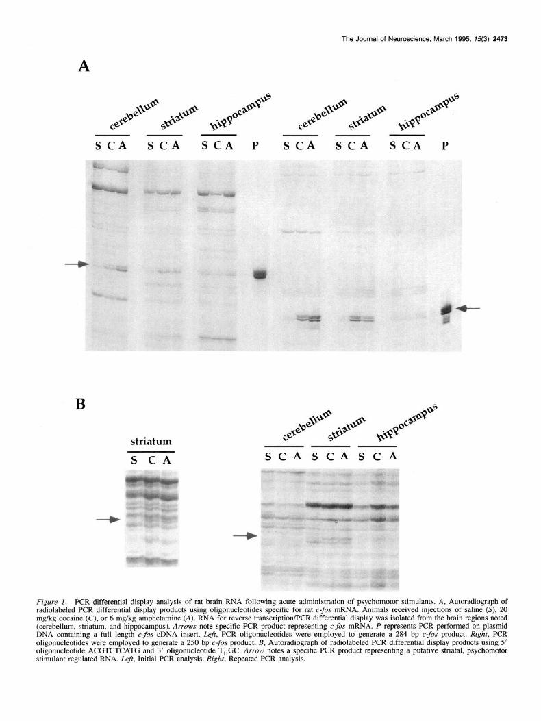
To determine the usefulness of PCR differential display as an approach by which to identify cocaine and amphetamine regu- lated transcripts, the efficacy of the technique was evaluated by utilizing c-fos specific oligonucleotides capable of generating a PCR product from the rat c-fos transcript (Curran et al., 1987) (Fig. 1A). The observed profile of radiolabeled PCR products shows, as expected, that multiple species of mRNA within the rat cerebellum, striatum and hippocampus can serve as a hy- bridization target for the two oligonucleotides utilized. A typical reaction generated 50-200 distinct radiolabeled PCR products between 50 and 600 bases in length. Furthermore, the over- whelming majority of PCR products were present at identical levels in these three brain regions isolated from saline, cocaine and amphetamine treated animals. The PCR product represent- ing the c-fos transcript was, however, clearly induced in the stria- turn and cerebellum from cocaine and amphetamine treated an-
The Journal of Neuroscience, March 1995, 15(3) 2473
s CA SCA SCA P S CA SCA SCA P
B
striatum
S CA SCASCASCA
Figure 1. PCR differential display analysis of rat brain RNA following acute administration of psychomotor stimulants. A, Autoradiograph of radiolabeled PCR differential display products using oligonucleotides specific for rat c-&r mRNA. Animals received injections of saline (S), 20 mg/kg cocaine (C), or 6 mg/kg amphetamine (A). RNA for reverse transcription/PCR differential display was isolated from the brain regions noted (cerebellum, striatum, and hippocampus). Arrows note specific PCR product representing c-fos mRNA. P represents PCR performed on plasmid DNA containing a full length c-fos cDNA insert. Z&t, PCR oligonucleotides were employed to generate a 284 bp c-fos product. Right, PCR oligonucleotides were employed to generate a 250 bp c-fos product. B, Autoradiograph of radiolabeled PCR differential display products using 5’ oligonucleotide ACGTCTCATG and 3’ oligonucleotide T,,GC. Arrow notes a specific PCR product representing a putative striatal, psychomotor stimulant regulated RNA. Left, Initial PCR analysis. Right, Repeated PCR analysis.
2474 Douglass et al. l Identification of a Cocaine Regulated Transcript
A cerebellum striatum hippocampus
SCA SCA S C A
f 28s
Kb 1.77
q 1.52 - 1.28
- 0.78
- 0.63 - 0.40
B
arbitrary OD
units
&O i 5.0
4.0
3.0
2.0
1.0
saline acute acute cocaine amphetamine
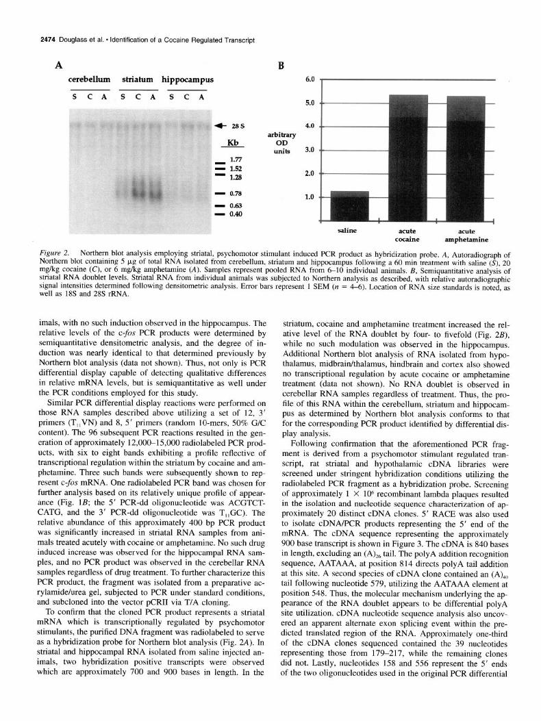
Figure 2. Northern blot analysis employing striatal, psychomotor stimulant induced PCR product as hybridization probe. A, Autoradiograph of Northern blot containing 5 kg of total RNA isolated from cerebellum. striatum and hionocamnu< fnllowin~ a 60 min treatment with saline (S), 20 1~1~ ~~~~~~=.~. ~..._ ..-... _ mg/kg cocaine (C), or 6 mikg amphetamine (A). Samples represent ‘pooled RNA from 6-10 individual animals. B, Semiquantitative anal&& of striatal RNA doublet levels. Striatal RNA from individual animals was subjected to Northern analysis as described, with relative autoradiographic signal intensities determined following densitometric analvsis. Error bars represent 1 SEM (n = 4-6). Location of RNA size standards is noted, as well as 1% and 2% rRNA.
imals, with no such induction observed in the hippocampus. The relative levels of the c-fos PCR products were determined by semiquantitative densitometric analysis, and the degree of in- duction was nearly identical to that determined previously by Northern blot analysis (data not shown). Thus, not only is PCR differential display capable of detecting qualitative differences in relative mRNA levels, but is semiquantitative as well under the PCR conditions employed for this study.
Similar PCR differential display reactions were performed on those RNA samples described above utilizing a set of 12, 3’ primers (T,,VN) and 8, 5’ primers (random lo-mers, 50% G/C content). The 96 subsequent PCR reactions resulted in the gen- eration of approximately 12,00&15,000 radiolabeled PCR prod- ucts, with six to eight bands exhibiting a profile reflective of transcriptional regulation within the striatum by cocaine and am- phetamine. Three such bands were subsequently shown to rep- resent C--OS mRNA. One radiolabeled PCR band was chosen for further analysis based on its relatively unique profile of appear- ance (Fig. 1B; the 5’ PCR-dd oligonucleotide was ACGTCT- CATG, and the 3’ PCR-dd oligonucleotide was T,,GC). The relative abundance of this approximately 400 bp PCR product was significantly increased in striatal RNA samples from ani- mals treated acutely with cocaine or amphetamine. No such drug induced increase was observed for the hippocampal RNA sam- ples, and no PCR product was observed in the cerebellar RNA samples regardless of drug treatment. To further characterize this PCR product, the fragment was isolated from a preparative ac- rylamide/urea gel, subjected to PCR under standard conditions, and subcloned into the vector pCRI1 via T/A cloning.
To confirm that the cloned PCR product represents a striatal mRNA which is transcriptionally regulated by psychomotor stimulants, the purified DNA fragment was radiolabeled to serve as a hybridization probe for Northern blot analysis (Fig. 2A). In striatal and hippocampal RNA isolated from saline injected an- imals, two hybridization positive transcripts were observed which are approximately 700 and 900 bases in length. In the
striatum, cocaine and amphetamine treatment increased the rel- ative level of the RNA doublet by four- to fivefold (Fig. 2B), while no such modulation was observed in the hippocampus. Additional Northern blot analysis of RNA isolated from hypo- thalamus, midbrainkhalamus, hindbrain and cortex also showed no transcriptional regulation by acute cocaine or amphetamine treatment (data not shown). No RNA doublet is observed in cerebellar RNA samples regardless of treatment. Thus, the pro- file of this RNA within the cerebellum, striatum and hippocam- pus as determined by Northern blot analysis conforms to that for the corresponding PCR product identified by differential dis- play analysis.
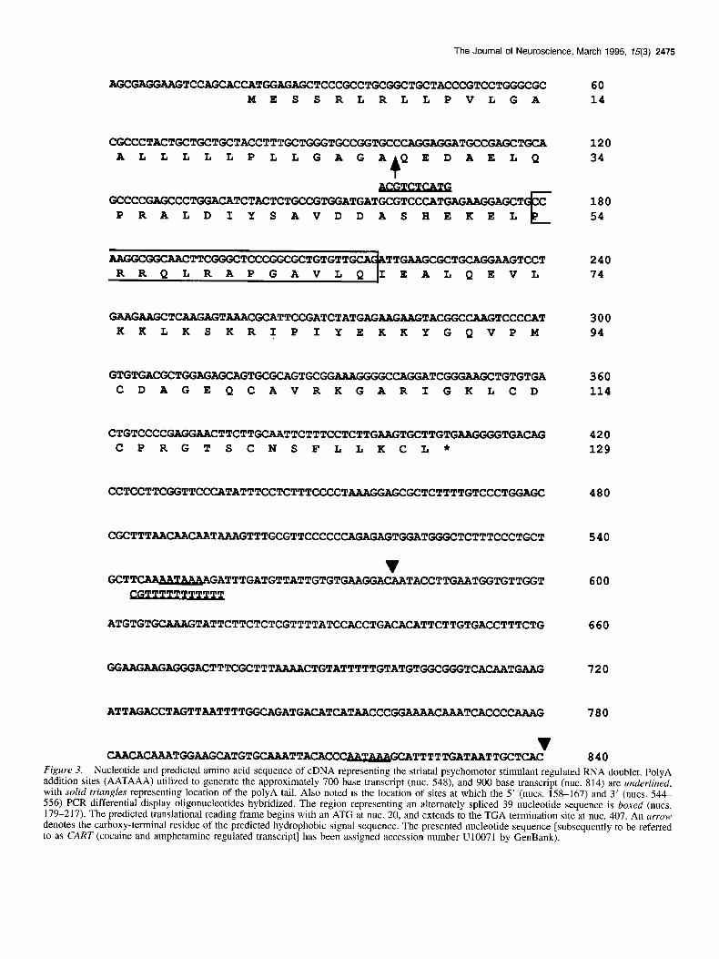
Following confirmation that the aforementioned PCR frag- ment is derived from a psychomotor stimulant regulated tran- script, rat striatal and hypothalamic cDNA libraries were screened under stringent hybridization conditions utilizing the radiolabeled PCR fragment as a hybridization probe. Screening of approximately 1 X lo6 recombinant lambda plaques resulted in the isolation and nucleotide sequence characterization of ap- proximately 20 distinct cDNA clones. 5’ RACE was also used to isolate cDNA/PCR products representing the 5’ end of the mRNA. The cDNA sequence representing the approximately 900 base transcript is shown in Figure 3. The cDNA is 840 bases in length, excluding an (A),, tail. The polyA addition recognition sequence, AATAAA, at position 814 directs polyA tail addition at this site. A second species of cDNA clone contained an (A),, tail following nucleotide 579, utilizing the AATAAA element at position 548. Thus, the molecular mechanism underlying the ap- pearance of the RNA doublet appears to be differential polyA site utilization. cDNA nucleotide sequence analysis also uncov- ered an apparent alternate exon splicing event within the pre- dicted translated region of the RNA. Approximately one-third of the cDNA clones sequenced contained the 39 nucleotides representing those from 179-217, while the remaining clones did not. Lastly, nucleotides 158 and 556 represent the 5’ ends of the two oligonucleotides used in the original PCR differential
The Journal of Neuroscience, March 1995, 15(3) 2475
AGCGAGGMGTCCAGCAC CATGGAGAGCTCCCGCCTGCGGCTGCTACCCGTCCTGGGCGC 60 MESSRLRLLPVLGA 14
CGCCCTACTGCTGCTGCTACCTTTGCTGGGTGCCGGTGCCCAGGAGGATGCCGAGCTGCA ALLLLLPLLGAGAQEDAELQ
4 GCCCCGAGCCCTGGACATCTACTCTGCCGTGGATGATGCGTCCCATGAGAAGGmXT C
PRALDIYSAVDDASHEKEL E
AAGGCGGCAACTTCGGGCTCCCGGCGCTGTGTT TTGAAGCGCT-ZUiGTCCT EALQEVL
120 34
180 54
240 74
-TCAAGAGTAAACGCATTCCGATCTATGAGMU3AGT~~CAAGTCCC~T KKLKSKRIPIYEKKYGQVPM
300 94
GTGTGACGCTGGAGAGCAG TGCGCAGTGCGG AAAGGGGCCAGGATCGGGAAGCTGTGTGA 360 CDAGEQCAVRKGARIGKLCD 114
CTGTCCCCGAGGM~CTTCTTGCAATTCTTTCCTCTTGAAGTGCTTGTG~TGACJ~G 420 CPRGTSCNSFLLKCL* 129
CCTCCTTCGGTTCCCATATTTCCTCTTTCCCCTAAAGGAG 480
CGCTTTAACAACAA TAAAGTTTGC!GTTCCCCCCAGAGAGTGGATGGGCTCTTTCCCTGCT
v GCTTwATTTGATGTTATTGTGTGAAGGAmTACCTTGAATGGTGTTGGT
540
600
ATGTGTGCAAAGTATTCTTCTCTCGTTTTATCCACCTGACCTTTCTG 660
GGAAG?dGiAGGGACTTTCGCTTTAAAACTGTATTTTTGTATTTTTGTATGTt3GC~T~~TG~ 720
ATTAGACCTAGTTAATTTTGGCAGATGACATCATAACCCGGWWTCACCC~ 780
v CAACACAAATGGAAGCATGTGCAAATTACACC ~TTTTTGATAATTGCTCAC 840
Figure 3. Nucleotide and predicted amino acid sequence of cDNA representing the striatal psychomotor stimulant regulated RNA doublet. PolyA addition sites (AATAAA) utilized to generate the approximately 700 base transcript (nut. 548), and 900 base transcript (nut. 814) are underlined, with solid triangles representing location of the polyA tail. Also noted is the location of sites at which the 5’ (nuts. 158-167) and 3’ (nuts. 544 556) PCR differential display oligonucleotides hybridized. The region representing an alternately spliced 39 nucleotide sequence is boxed (nuts. 179-217). The predicted translational reading frame begins with an ATG at nut. 20, and extends to the TGA termination site at nut. 407. An arrow denotes the carboxy-terminal residue of the predicted hydrophobic signal sequence. The presented nucleotide sequence [subsequently to be referred to as CART (cocaine and amphetamine regulated transcript] has been assigned accession number U10071 by GenBank).
2476 Douglass et al. - Identification of a Cocaine Regulated Transcript
display analysis. Several mismatches are present between the PCR differential display oligonucleotides and the complemen- tary cDNA sequence, as anticipated. The sequence of the orig- inal PCR differential display fragment also matches that located between nucleotides 168 and 543 in the cDNA clones.
The first potential translation initiation codon is located at nucleotide 20. In 90-95% of the vertebrate transcripts analyzed to date, the first 5’ AUG represents the site for translation ini- tiation (Kozak, 1989). Furthermore, the nucleotide sequence in which this ATG is embedded is similar to that representing a consensus Kozak translation initiation sequence element (GCCGCCRCCATGG). The predicted encoded polypeptide is either 129 or 116 residues in length, depending on the presence or absence of nucleotides 179-217 within the putative protein coding region, respectively. The first 27 residues represent an amino terminal hydrophobic domain conforming to the motif of a signal peptide (Lingappa and Blobel, 1980; van Heijne, 1990), suggesting that the polypeptide may be targeted for entry into the secretory pathway. There are also multiple paired basic ami- no acid residues located throughout the molecule which could theoretically serve as target sites for posttranslation proteolytic cleavage events (Steiner et al., 1992). One such site is present within the alternately spliced domain encoding residues 54-66, further suggesting that if proteolytic cleavage events did occur, then different peptide end products would be produced from the two distinct primary translation products.
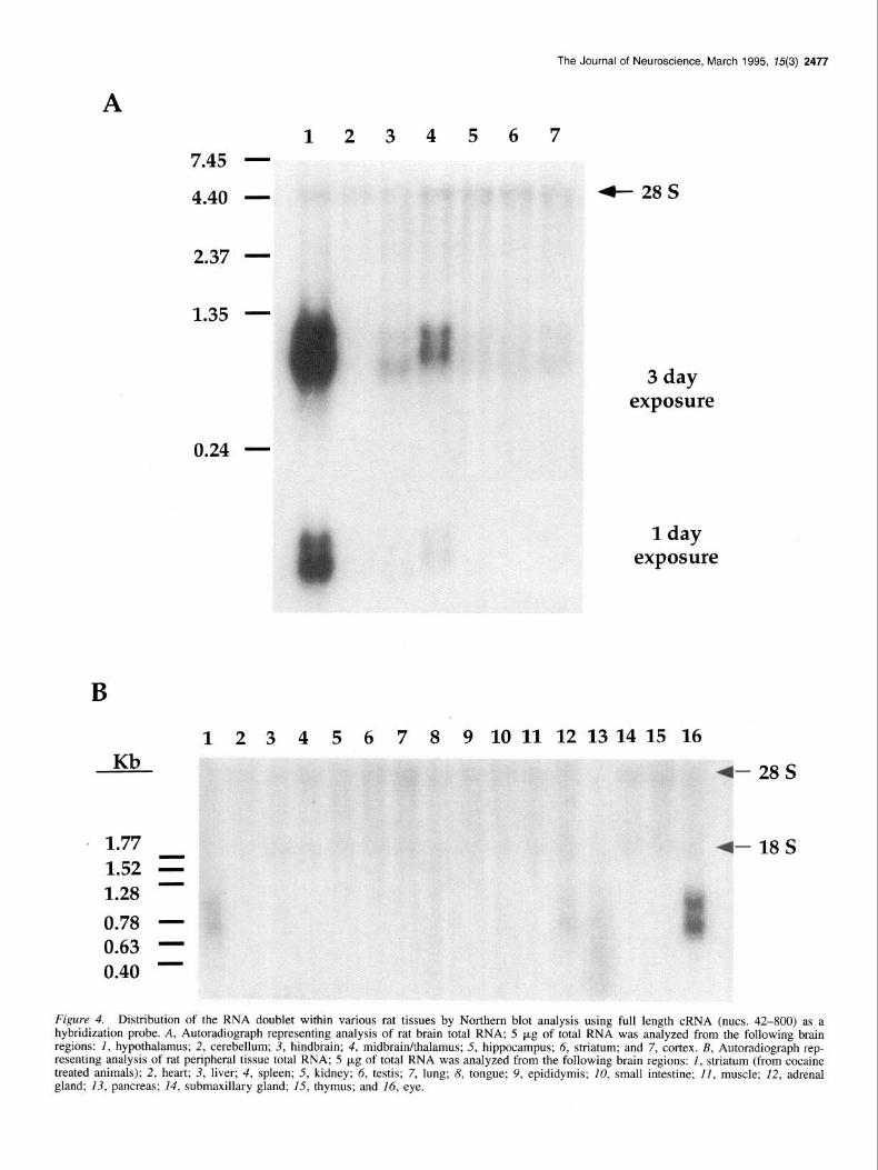
Additional Northern blot analysis was performed to determine the tissue distribution of the RNA doublet. In Figure 4A, RNA from various rat brain regions was analyzed employing a full length radiolabeled antisense cRNA hybridization probe. The major site of synthesis of the RNA doublet is the hypothalamus. Relatively abundant RNA levels are also observed in the thal- amuslmidbrain, with significantly lower expression observed in the cortex, striatum, hippocampus, and hindbrain. Semiquanti- tative densitometric analysis of the hybridization signals re- vealed that hypothalamic RNA levels are approximately 300- fold greater, and midbrain/thalamus RNA levels IO-fold greater than striatal levels. Analysis of RNA isolated from peripheral tissues (Fig. 4B) also detected the presence of the RNA doublet in rat eye and adrenal, while no hybridization signal was ob- served in the other 13 tissues examined. Levels of the RNA doublet in the eye approximate those of the midbrain/thalamus, while adrenal levels are similar to those observed in the striatum. Several additional endocrine tissues were also evaluated for ex- pression of the mRNA doublet (data not shown). No hybridiza- tion signal was observed in rat ovary and uterus RNA. However, a robust hybridization signal was observed when pituitary RNA was examined; the relative intensity of the pituitary hybridiza- tion signal approximates that observed for the hypothalamus. Expression of the RNA doublet is thus limited to neuroendocrine tissues, with dramatically varied levels of transcriptional ex- pression observed throughout the brain.
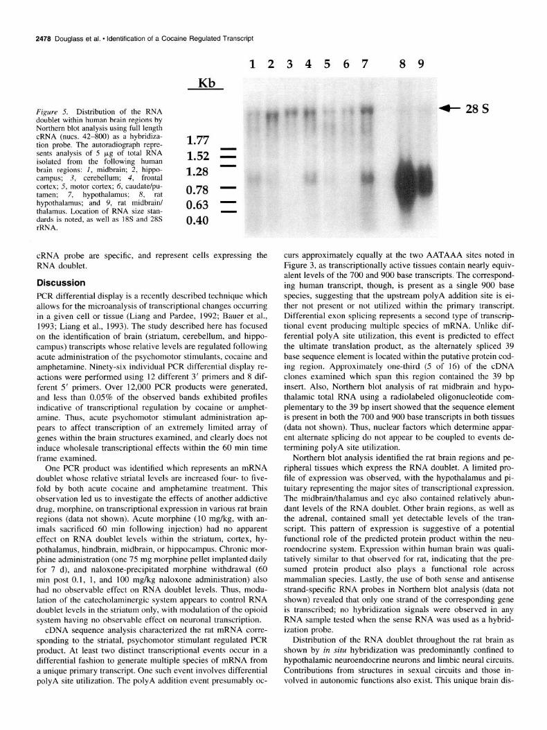
Further Northern blot analysis also confirmed that the tran- script is present in RNA isolated from human brain (Fig. 5). Relatively abundant levels of an approximately 900 base hy- bridizing transcript are observed in RNA from human hypo- thalamus, frontal cortex, and midbrain, with lower levels seen in the hippocampus, motor cortex, and striatum. No such hy- bridization signal was observed in cerebellar RNA. Thus, the pattern of distribution of the transcript in human brain is rela- tively conserved to that observed for rat brain. However, only a single species of hybridizing RNA is present in human, in con-
trast to the doublet RNA band which is consistently observed in rat. The likely explanation for this difference is the lack of a polyA addition site, or recognition of the site, in the human transcript.
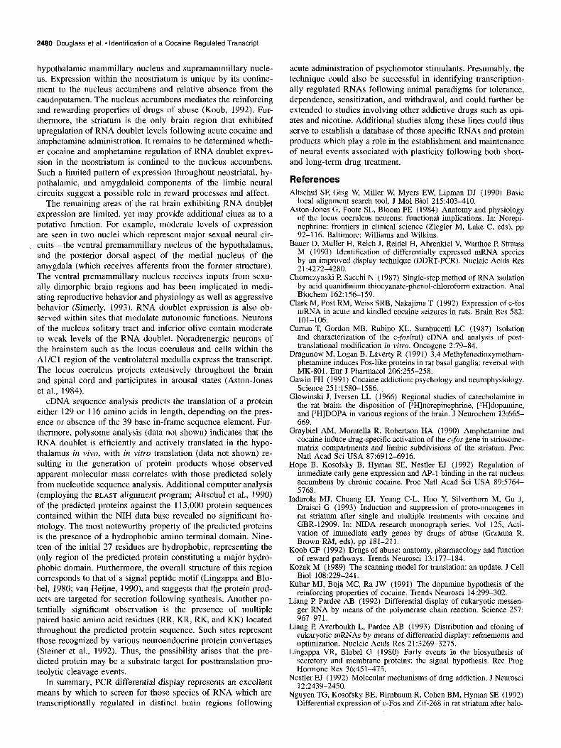
Lastly, regional expression of the RNA doublet throughout the adult rat brain was more precisely determined by in situ histochemical analysis. A subset of hybridization positive regions are presented in Figure 6. For these studies a 790 base cRNA hybridization probe spanning the putative coding region (including the alternately spliced 39 base insert) and 3’ noncod- ing region was utilized. The most intense hybridization signals were observed in the Edinger-Westphal nucleus and induseum griseum. Major expression was also seen in the periventricular zone of the hypothalamus (especially the paraventricular and arcuate nuclei) and cells throughout the medial hypothalamus (typified by labeling of the supraoptic nucleus and perifornical region). Within these structures, intense hybridization signals were observed at the single cell level.
Telencephalic hybridization signals were limited to a few ar- eas. Of note was the distribution in the neostriatum where mod- erate labeling was evident in the ventral region, consisting of the nucleus accumbens and olfactory tubercle. This area of hy- bridization positive cells stretched along the nucleus accumbens border within striosomes of the ventrolateral caudoputamen. Within the neocortex, moderate labeling was seen only in the primary somatosensory area (layer 4) and the piriform area. In- tense labeling was observed in the induseum griseum, with the bed nuclei of the stria terminalis and the dorsal blade of the dentate gyrus of the rostra1 hippocampus exhibiting moderate to weak labeling. Moderately intense hybridization signals were also seen in the amygdaloid complex, particularly in the medial part of the septal nucleus, posterior dorsal part of the medial nucleus, posterior cortical nucleus, and posterior basolateral nu- cleus.
In the diencephalon, the hypothalamus exhibited the most ex- tensive labeling distribution of any brain region examined. In- tense to moderate labeling of cells was evident throughout entire nuclei such as the paraventricular (particularly the parvocellular region), arcuate, and supraoptic nuclei. Moderate labeling was seen in the posterior periventricular nucleus, ventral and medial premammillary nucleus, and lateral aspect of the supermammil- lary nucleus. Scattered cells in the perifornical region, lateral hypothalamus, and posterior hypothalamus exhibited intense to moderate signals. Within the thalamus, the medial aspect of the zona incerta, ventral half of nucleus reuniens, and lateral haben- ula were the only structures exhibiting moderate to weakly in- tense hybridization signals.
The mesencephalon contained the fewest number of hybrid- ization positive cells. However, within this region the Edinger- Westphal nucleus exhibited intense labeling. Scattered cells with moderate to weak labeling were also noted in the dorsal peria- queductal gray. The rhombencephalon also contained regions of hybridization positive cells. Both the inferior olive and nucleus of the solitary tract exhibited moderate labeling, where as the locus coeruleus and nucleus incertus showed weak labeling. Scattered cells throughout the Al/Cl region of the nucleus am- biguus further exhibited moderate labeling.
Hybridization of all aforementioned brain sections with a sense cRNA probe under identical conditions showed no label- ing of any of the structures described above. Thus, the hybrid- ization signals which were observed employing the antisense
The Journal of Neuroscience, March 1995, 75(3) 2477
A 1234567
7.45 -
4- 28s 4.40 -
2.37 -
0.24 -
‘ 1.77 1.52 =
- 1.28 0.78 - 0.63 - 0.40 -
1 day exposure
6 7 8 9 10 11 12 13 14 15 16
- 28s
-18s
Figure 4. Distribution of the RNA doublet within various rat tissues by Northern blot analysis using full length cRNA (nuts. 42-800) as a hybridization probe. A, Autoradiograph representing analysis of rat brain total RNA; 5 kg of total RNA was analyzed from the following brain regions: I, hypothalamus; 2, cerebellum; 3, hindbrain; 4, midbrainkhalamus; 5, hippocampus; 6, striatum; and 7, cortex. B, Autoradiograph rep- resenting analysis of rat peripheral tissue total RNA; 5 p,g of total RNA was analyzed from the following brain regions: I, striatum (from cocaine treated animals); 2, heart; 3, liver; 4, spleen; 5, kidney; 6, testis; 7, lung; 8, tongue; 9, epididymis; 10, small intestine; II, muscle; 12, adrenal gland; 13, pancreas; 14, submaxillary gland; 15, thymus; and 16, eye.
2478 Douglass et al. l identification of a Cocaine Regulated Transcript
1234567 89 l/L
Figure 5. Distribution of the RNA doublet within human brain regions by Northern blot analysis using full length cRNA (nuts. 42-800) as a hybridiza- tion probe. The autoradiograph repre- 1.77 sents analysis of 5 pg of total RNA isolated from the following human 1.52 = brain regions: 1, midbrain; 2, hippo- campus; 3, cerebellum; 4, frontal 1.28 - cortex; 5, motor cortex; 6, caudate/pu- tamen; 7, hypothalamus; 8, rat 0.78 - hypothalamus; and 9, rat midbrain/ thalamus. Location of RNA size stan- 0.63 1 dards is noted, as well as 18s and 28s rRNA.
0.40
cRNA probe are specific, and represent cells expressing the RNA doublet.
Discussion PCR differential display is a recently described technique which allows for the microanalysis of transcriptional changes occurring in a given cell or tissue (Liang and Pardee, 1992; Bauer et al., 1993; Liang et al., 1993). The study described here has focused on the identification of brain (striatum, cerebellum, and hippo- campus) transcripts whose relative levels are regulated following acute administration of the psychomotor stimulants, cocaine and amphetamine. Ninety-six individual PCR differential display re- actions were performed using 12 different 3’ primers and 8 dif- ferent 5’ primers. Over 12,000 PCR products were generated, and less than 0.05% of the observed bands exhibited profiles indicative of transcriptional regulation by cocaine or amphet- amine. Thus, acute psychomotor stimulant administration ap- pears to affect transcription of an extremely limited array of genes within the brain structures examined, and clearly does not induce wholesale transcriptional effects within the 60 min time frame examined.
One PCR product was identified which represents an mRNA doublet whose relative striatal levels are increased four- to five- fold by both acute cocaine and amphetamine treatment. This observation led us to investigate the effects of another addictive drug, morphine, on transcriptional expression in various rat brain regions (data not shown). Acute morphine (10 mg/kg, with an- imals sacrificed 60 min following injection) had no apparent effect on RNA doublet levels within the striatum, cortex, hy- pothalamus, hindbrain, midbrain, or hippocampus. Chronic mor- phine administration (one 75 mg morphine pellet implanted daily for 7 d), and naloxone-precipitated morphine withdrawal (60 min post 0.1, 1, and 100 mg/kg naloxone administration) also had no observable effect on RNA doublet levels. Thus, modu- lation of the catecholaminergic system appears to control RNA doublet levels in the striatum only, with modulation of the opioid system having no observable effect on neuronal transcription.
cDNA sequence analysis characterized the rat mRNA corre- sponding to the striatal, psychomotor stimulant regulated PCR product. At least two distinct transcriptional events occur in a differential fashion to generate multiple species of mRNA from a unique primary transcript. One such event involves differential polyA site utilization. The polyA addition event presumably oc-
4- 28s
curs approximately equally at the two AATAAA sites noted in Figure 3, as transcriptionally active tissues contain nearly equiv- alent levels of the 700 and 900 base transcripts. The correspond- ing human transcript, though, is present as a single 900 base species, suggesting that the upstream polyA addition site is ei- ther not present or not utilized within the primary transcript. Differential exon splicing represents a second type of transcrip- tional event producing multiple species of mRNA. Unlike dif- ferential polyA site utilization, this event is predicted to effect the ultimate translation product, as the alternately spliced 39 base sequence element is located within the putative protein cod- ing region. Approximately one-third (5 of 16) of the cDNA clones examined which span this region contained the 39 bp insert. Also, Northern blot analysis of rat midbrain and hypo- thalamic total RNA using a radiolabeled oligonucleotide com- plementary to the 39 bp insert showed that the sequence element is present in both the 700 and 900 base transcripts in both tissues (data not shown). Thus, nuclear factors which determine appar- ent alternate splicing do not appear to be coupled to events de- termining polyA site utilization.
Northern blot analysis identified the rat brain regions and pe- ripheral tissues which express the RNA doublet. A limited pro- file of expression was observed, with the hypothalamus and pi- tuitary representing the major sites of transcriptional expression. The midbrain/thalamus and eye also contained relatively abun- dant levels of the RNA doublet. Other brain regions, as well as the adrenal, contained small yet detectable levels of the tran- script. This pattern of expression is suggestive of a potential functional role of the predicted protein product within the neu- roendocrine system. Expression within human brain was quali- tatively similar to that observed for rat, indicating that the pre- sumed protein product also plays a functional role across mammalian species. Lastly, the use of both sense and antisense strand-specific RNA probes in Northern blot analysis (data not shown) revealed that only one strand of the corresponding gene is transcribed; no hybridization signals were observed in any RNA sample tested when the sense RNA was used as a hybrid- ization probe.
Distribution of the RNA doublet throughout the rat brain as shown by in situ hybridization was predominantly confined to hypothalamic neuroendocrine neurons and limbic neural circuits. Contributions from structures in sexual circuits and those in- volved in autonomic functions also exist. This unique brain dis-
The Journal of Neuroscience, March 1995, 75(3) 2479
Figure 6. In situ hybridization anal- ysis of RNA doublet expression throughout the adult rat brain. Dark- field photomicrographs are shown from coronal (30 pm thick) sections from one animal (n = 3) probed with an ?S- antisense cRNA hybridization probe. A section at the level of the prefrontal cortex hybridized with a sense cRNA control probe is also shown. Sections were developed 5 d after emulsion. ZG, induseum griseum; ACB, nucleus ac- cumbens; OT, olfactory tubercle; uco, anterior commissure; PVH, paraven- tricular nucleus; SO, supraoptic nucle- us; ARH, arcuate nucleus; DC, dentate gyrus; LHA, lateral hypothalamic area; MM, mammillary nucleus; SUM, su- pramammillary nucleus; PAG, peria- queductal gray; EW, Edinger-Westphal nucleus; LC, locus coeruleus; NI, nu- cleus incertus; NTS, nucleus of the sol- itary tract; IO, inferior olive; AMB, nu- cleus ambiguus. The rat brain atlas of Swanson (1992) was used to identify specific in situ hybridization positive cells and brain nuclei, as well as pro- viding a nomenclature for labeling.
tribution may provide some insights into a possible functional role for the encoded protein product. For example, expression of the RNA doublet is robust in the hypothalamus. Its abundance in this brain region is due, in part, to expression within neu- roendocrine neurons such as the paraventricular, arcuate and su- praoptic nuclei. Many neurons within these regions send affer- ents to, and release neuropeptides which modulate pituitary hormone secretion and regulate osmolality (Simerly, 1993).
Thus, localization of high levels of the RNA doublet to these neurons, coupled with the predicted structure of the encoded protein product(s) suggest that the transcript may encode a neu- roendocrine secretogogue.
Another possible role for the putative encoded protein is sug- gested by expression of the RNA doublet in limbic neural cir- cuits. For example, the transcript is present within the amyg- daloid complex, dentate gyms of the hippocampus, and the
2480 Douglass et al. * Identification of a Cocaine Regulated Transcript
hypothalamic mammillary nucleus and supramammillary nucle- us. Expression within the neostriatum is unique by its confine- ment to the nucleus accumbens and relative absence from the caudoputamen. The nucleus accumbens mediates the reinforcing and rewarding properties of drugs of abuse (Koob, 1992). Fur- thermore, the striatum is the only brain region that exhibited upregulation of RNA doublet levels following acute cocaine and amphetamine administration. It remains to be determined wheth- er cocaine and amphetamine regulation of RNA doublet expres- sion in the neostriatum is confined to the nucleus accumbens. Such a limited pattern of expression throughout neostriatal, hy- pothalamic, and amygdaloid components of the limbic neural circuits suggest a possible role in reward processes and affect.
The remaining areas of the rat brain exhibiting RNA doublet expression are limited, yet may provide additional clues as to a putative function. For example, moderate levels of expression are seen in two nuclei which represent major sexual neural cir- cuits-the ventral premammillary nucleus of the hypothalamus, and the posterior dorsal aspect of the medial nucleus of the amygdala (which receives afferents from the former structure). The ventral premammillary nucleus receives inputs from sexu- ally dimorphic brain regions and has been implicated in medi- ating reproductive behavior and physiology as well as aggressive behavior (Simerly, 1993). RNA doublet expression is also ob- served within sites that modulate autonomic functions. Neurons of the nucleus solitary tract and inferior olive contain moderate to weak levels of the RNA doublet. Noradrenergic neurons of the brainstem such as the locus coeruleus and cells within the Al/Cl region of the ventrolateral medulla express the transcript. The locus coeruleus projects extensively throughout the brain and spinal cord and participates in arousal states (Aston-Jones et al., 1984).
cDNA sequence analysis predicts the translation of a protein either 129 or 116 amino acids in length, depending on the pres- ence or absence of the 39 base in-frame sequence element. Fur- thermore, polysome analysis (data not shown) indicates that the RNA doublet is efficiently and actively translated in the hypo- thalamus in vivo, with in vitro translation (data not shown) re- sulting in the generation of protein products whose observed apparent molecular mass correlates with those predicted solely from nucleotide sequence analysis. Additional computer analysis (employing the BLAST alignment program; Altschul et al., 1990) of the predicted proteins against the 113,000 protein sequences contained within the NIH data base revealed no significant ho- mology. The most noteworthy property of the predicted proteins is the presence of a hydrophobic amino terminal domain. Nine- teen of the initial 27 residues are hydrophobic, representing the only region of the predicted protein constituting a major hydro- phobic domain. Furthermore, the overall structure of this region corresponds to that of a signal peptide motif (Lingappa and Blo- bel, 1980; van Heijne, 1990), and suggests that the protein prod- ucts are targeted for secretion following synthesis. Another po- tentially significant observation is the presence of multiple paired basic amino acid residues (RR, KR, RK, and KK) located throughout the predicted protein sequence. Such sites represent those recognized by various neuroendocrine protein convertases (Steiner et al., 1992). Thus, the possibility arises that the pre- dicted protein may be a substrate target for posttranslation pro- teolytic cleavage events.
In summary, PCR differential display represents an excellent means by which to screen for those species of RNA which are transcriptionally regulated in distinct brain regions following
acute administration of psychomotor stimulants. Presumably, the technique could also be successful in identifying transcription- ally regulated RNAs following animal paradigms for tolerance, dependence, sensitization, and withdrawal, and could further be extended to studies involving other addictive drugs such as opi- ates and nicotine. Additional studies along these lines could thus serve to establish a database of those specific RNAs and protein products which play a role in the establishment and maintenance of neural events associated with plasticity following both short- and long-term drug treatment.
References
Altschul SE Gisg W, Miller W, Myers EW, Lipman DJ (1990) Basic local alignment search tool. J Mol Biol 215:403-410.
Aston-Jones G, Foote SL, Bloom FE (1984) Anatomy and physiology of the locus coeruleus neurons: functional implications. In: Norepi- nephrine: frontiers in clinical science (Ziegle; M, Lake C, eds), bp 92-l 16. Baltimore: Williams and Wilkins.
Bauer D, Muller H, Reich J, Reidel H, Ahrenkiel V, Warthoe P, Strauss M (1993) Identification of differentially expressed mRNA species by an improved display technique (DDRT-PCR). Nucleic Acids Res 2 1:4272-42X0.
Chomczynski P, Sacchi N (1987) Single-step method of RNA isolation by acid quanidinium thiocyanate-phenol-chloroform extraction. Anal Biochem 162:156-159.
Clark M, Post RM, Weiss SRB, Nakajima T (1992) Expression of c-fos mRNA in acute and kindled cocaine seizures in rats. Brain Res 582: 101-106.
Curran T, Gordon MB, Rubino KL, Sambucetti LC (1987) Isolation and characterization of the c-fos(rat) cDNA and analysis of post- translational modification in vitro. Oncogene 2:79-X4.
Dragunow M, Logan B, Laverty R (1991) 3,4 Methylenedioxymetham- phetamine induces Fos-like proteins in rat basal ganglia: reversal with MK-801. Eur J Pharmacol 206:255-258.
Gawin FH (199 1) Cocaine addiction: psychology and neurophysiology. Science 251:1580--15X6.
Glowinski J, Iversen LL (1966) Regional studies of catecholamine in the rat brain: the disposition of [3H]norepinephrine, [3H]dopamine, and [3H]DOPA in various regions of the brain. J Neurochem 13:665- 669.
Graybiel AM, Moratella R, Robertson HA (1990) Amphetamine and cocaine induce drug-specific activation of the c-fos gene in striosome- matrix compartments and limbic subdivisions of the striatum. Proc Nat1 Acad sci USA 87:6912-6916.
Hone B. Kosofskv B. Hvman SE. Nestler EJ (1992) Regulation of I , . I v
immediate early gene expression and AP-1 binding in the rat nucleus accumbens by chronic cocaine. Proc Nat1 Acad Sci USA X9:5764- 5768.
Iadarola MJ, Chuang EJ, Yeung C-L, Hoo Y, Silverthorn M, Gu J, Draisci G (1993) Induction and suppression of proto-oncogenes in rat striatum after single and multi& treatments with co&e and GBR-12909. In: NIDA research monograuh series. Vol 125, Acti- vation of immediate early genes by &is of abuse (Grzanna R, Brown RM, eds), pp 181-211.
Koob GF (1992) Drugs of abuse: anatomy, pharmacology and function of reward pathways. Trends Neurosci 13: 177-184.
Kozak M (1989) The scanning model for translation: an update. J Cell Biol 10X:229-241.
Kuhar MJ, Boia MC, Ra JW (1991) The dopamine hypothesis of the reinforcing properties of cocaine. Trends Nkurosci l&:299-302.
Liang I? Pardee AB (1992) Differential disulav of eukarvotic messen- ge; RNA by means of the polymerase &air; reaction.<Science 257: 967-97 1.
Liang P, Averboukh L, Pardee AB (1993) Distribution and cloning of eukaryotic mRNAs by means of differential display: refinements and optimization. Nucleic- Acids Res 21:3269-3275: -
Lingauua VR. Blobel G (1980) Earlv events in the biosvnthesis of secretory and membrane proteins: <he signal hypothesis: Ret Prog Hormone Res 36:45 1475.
Nestler EJ (1992) Molecular mechanisms of drug addiction. J Neurosci 12~2439-2450.
Nguyen TG, Kosofsky BE, Bimbaum R, Cohen BM, Hyman SE (1992) Differential expression of c-Fos and Zif-268 in rat striatum after halo-
The Journal of Neuroscience, March 1995, 15(3) 2481
peridol, clozapine and amphetamine. Proc Nat1 Acad Sci USA 89: 4210-4274.
Simerly RB (1994) Anatomical substrates of hypothalamic integration. In: The rat nervous system (Paxinos G, ed), in press. New York: Academic.
Simmons DM, Arriza JL, Swanson LW (1989) A complete protocol for in situ hybridization of messenger RNAs in brain and other tissues with radiolabelled single-stranded RNA probes. J Histotechnol 12: 169-181.
Steiner DP, Smeekens SP, Ohagi S, Chan SJ (1992) The new enzy- mology of precursor processing endoproteases. J Biol Chem 267: 23435-23438.
Swanson LW (1992) Brain maps: structure of the rat brain. New York: Elsevier.
von Heijne G (1990) The signal peptide. J Membr Biol, 115:195-201. Young ST, Porrino LJ, Iadarola MJ (1991) Cocaine induces striatal
c-fos-immunoreactive proteins via dopaminergic Dl receptors. Proc Nat1 Acad Sci USA 88:1291-1295.