Embed Size (px)
Citation preview
Developmental Cell, Volume 41
Supplemental Information
PCP and SAX-3/Robo Pathways Cooperate to Regulate
Convergent Extension-Based Nerve Cord Assembly
in C. elegans
Pavak K. Shah, Matthew R. Tanner, Ismar Kovacevic, Aysha Rankin, Teagan E.Marshall, Nathaniel Noblett, Nhan Nguyen Tran, Tony Roenspies, Jeffrey Hung, ZheqianChen, Cristina Slatculescu, Theodore J. Perkins, Zhirong Bao, and Antonio Colavita
slt-1(eh15)
wt
slt-1(ok255)
fmi-1(rh308)
wt
prkl-1(ok3182)
prkl-1(zy11)
vang-1(tm1422)
vang-1(ok1142)
sax-3(zy5)
sax-3(ky123)
DD1 DD2 DD3 DD4 DD5 DD6
Relative position along AP axis0 20 40 60 80 100%
DD1 DD2 DD3 DD4 DD5 DD6
prkl-1(ok3182)
vang-1(ok1142); prkl-1(ok3182)
vang-1(tm1422); prkl-1(ok3182)
Relative position along AP axis0 20 40 60 80 100%
vang-1(ok1142)
vang-1(ok1142); fmi-1(rh308)
vang-1(ok1142); slt-1(eh15)
prkl-1(ok3182)
prkl-1(ok3182); fmi-1(rh308)
prkl-1(ok3182); slt-1(eh15)
sax-3(zy5)
sax-3(zy5);fmi-1(rh308)
sax-3(zy5); slt-1(eh15)
A
C
B
vang-1(ok1142) sax-3(zy5)
sax-3(zy5); prkl-1(ok3182)
vang-1(zy60 [gfp::vang-1])
prkl-1(ok3182); zyIs33[gfp::prkl-1]
vang-1(ok1142) sax-3(zy5)
vang-1(ok1142) sax-3(zy5); zyIs43[sax-3::gfp]
Figure S1
Relative position along AP axis0 20 40 60 80 100%
DD1 DD2 DD3 DD4 DD5 DD6
N38
44
39
39
44
46
55
50
51
N40
33
41
45
46
53
50
48
50
50
50
52
52
50
49
48
50
50
52
56
N

0 20 40 60 80
100
zy5 [Q987stop]
TM
wt zy5 ky123
A
B C
***
0 20 40 60 80
100
% A
nte
rio
r n
erv
e r
ing
0
20
40
60
80
100
% L
eth
alit
y
% N
otc
he
d h
ea
ds
0 20 40 60 80
100
% A
VM
g
uid
an
ce d
efe
cts
0 20 40 60 80
100
%
HS
N
gu
ida
nce
de
fect
s
wt zy5 ky123 wt zy5 ky123
wt zy5 ky123 wt zy5 ky123
***
***
ns ns
D
E F
0
20
40
60
80
100
Figure S2
N=322 N=278 N=265 N=316 N=274 N=66 N=122 N=114 N=31
N=59 N=144 N=112 N=137 N=161 N=119
A
PDA
VD
VNC PAGRVG
VNC PAGRVG
wdIs6; ynIs37 sax-3(zy5); prkl-1(ok3182); wdIs6; ynIs37
VB
VA
wdIs6 sax-3(zy5); prkl-1(ok3182); wdIs6
C
D
E
gfp
rfpm
erge
B wyIs75; ynIs37 sax-3(zy5); prkl-1(ok3182); wyIs75; ynIs37
Figure S3

cp13
(NM
Y-2:
:GFP
)m
erge
cnd-
1p::P
H::R
FP
prkl-1(ok3182)vang-1(ok1142) sax-3(zy5)C D E
Contracting edge
Wild-type posterior contraction
Ectopic anterior contractionFailed posterior contraction
Ectopic anterior contractionPosterior contraction
A
-2’ 0’ -2’ 0’ -3’ 0’
-2’ 0’cp
13(N
MY-
2::G
FP)
mer
gecn
d-1p
::PH
::RFP
wt B
Figure S4
SUPPLEMENTAL INFORMATION
Supplemental Figure Legends
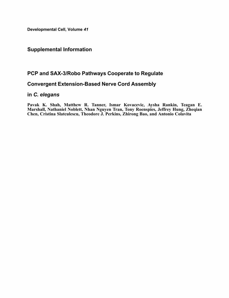
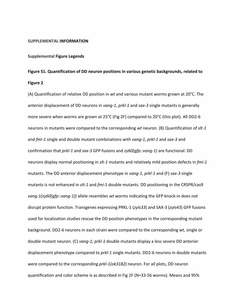
Figure S1. Quantification of DD neuron positions in various genetic backgrounds, related to
Figure 2
(A) Quantification of relative DD position in wt and various mutant worms grown at 20°C. The
anterior displacement of DD neurons in vang-1, prkl-1 and sax-3 single mutants is generally
more severe when worms are grown at 25°C (Fig 2F) compared to 20°C (this plot). All DD2-6
neurons in mutants were compared to the corresponding wt neuron. (B) Quantification of slt-1
and fmi-1 single and double mutant combinations with vang-1, prkl-1 and sax-3 and
confirmation that prkl-1 and sax-3 GFP fusions and zy60(gfp::vang-1) are functional. DD
neurons display normal positioning in slt-1 mutants and relatively mild position defects in fmi-1
mutants. The DD anterior displacement phenotype in vang-1, prkl-1 and (F) sax-3 single
mutants is not enhanced in slt-1 and fmi-1 double mutants. DD positioning in the CRSPR/cas9
vang-1(zy60[gfp::vang-1]) allele resembles wt worms indicating the GFP knock-in does not
disrupt protein function. Transgenes expressing PRKL-1 (zyIs33) and SAX-3 (zyIs43) GFP fusions
used for localization studies rescue the DD position phenotypes in the corresponding mutant
background. DD2-6 neurons in each strain were compared to the corresponding wt, single or
double mutant neuron. (C) vang-1; prkl-1 double mutants display a less severe DD anterior
displacement phenotype compared to prkl-1 single mutants. DD2-6 neurons in double mutants
were compared to the corresponding prkl-1(ok3182) neuron. For all plots, DD neuron
quantification and color scheme is as described in Fig 2F (N=33-56 worms). Means and 95%

confidence intervals are shown for each DD neuron. *p≤0.01; **p≤0.001, using the two-tailed t-
test. Worms quantified in (B and C) were grown at 25°C.
Figure S2. Characterization of sax-3(zy5) phenotypes, related to Figure 2
(A) Schematic of SAX-3 protein structure and location of zy5 mutation. TM indicates position of
transmembrane domain. (B-F) Quantification of (B) embryonic lethality (N=265-322 embryos),
(C) notched head (N=66-316 worms), (D) anterior nerve ring (N=31-122), (E) AVM (N=59-144)
and (F) HSN (N=119-161) axon guidance phenotypes in wt, sax-3(zy5), and sax-3(ky123) null
mutants. Error bars indicate standard error of proportion. Significance was assessed using
Fisher exact tests, ***<0.001, ns, not significant.
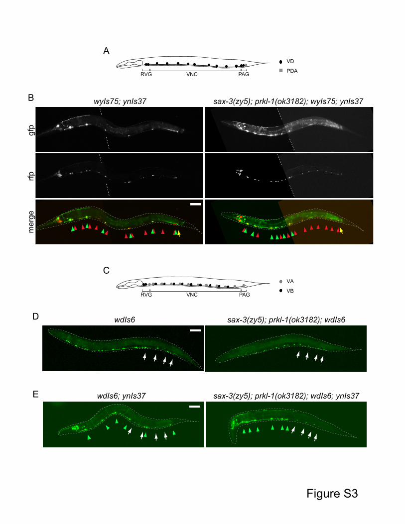
Figure S3. VNC positioning of post-embryonically-derived VD, VA and VB neurons in sax-
3(zy5); prkl-1(ok3182) double mutants, related to Figure 2
(A and B) Characterization of post-embryonically derived VD neurons. (A) Schematic showing
VD and PDA neuron position in C. elegans. (B) Representative images of wt and sax-3(zy5); prkl-
1(ok3182) L4 stage worms in which DD (RFP) and VD (RFP and red arrowheads) neurons are
labeled with a wyIs75[unc-47p::mCherry; exp-1p::gfp] and DD (GFP and green arrowheads)
neurons with ynIs37 reporters. DD neurons are identified by their overlap when GFP and RFP
channels are merged. The embryonically-derived PDA (GFP and yellow arrow) neuron is also
labeled by exp-1p::gfp in wyIs75. VD neurons in sax-3(zy5); prkl-1(ok3182) mutants display
relatively minor positioning errors compared to the severe anterior displacement of DD

neurons. The position of the PDA neuron which is located in the preanal ganglion (PAG) is not
affected in sax-3(zy5); prkl-1(ok3182) mutants (0% position defect, N=50 worms). (C-E)
Characterization of post-embryonically derived VA and VB neurons. (C) Schematic showing VA
and VB neuron position in C. elegans. (D) Representative images of wt and sax-3(zy5); prkl-
1(ok3182) L4 stage worms in which VA and VB neurons are labeled with a wdIs6[del-1p::gfp]
reporter. The posterior most del-1p::gfp labeled neurons (VB9, VB10, VB11, and VA11) are
marked with white arrows. (E) Representative images of wt and sax-3(zy5); prkl-1(ok3182) L4
stage worms in which VA, VB and DD neurons are labeled with wdIs6 and ynIs37 reporters. VB9,
VB10, VB11, and VA11 (white arrows) neurons in sax-3(zy5); prkl-1(ok3182) mutants display
relatively minor positioning errors compared to the severe anterior displacement of DD (green
arrowheads) neurons. DD neurons were identified by their brighter GFP compared to VA and
VB neurons. All scale bars are 50 μm.
Figure S4. NMY-2 Localization During Contraction of Cell Contacts, related to Figure 4
(A) Single slices showing NMY-2::GFP localization in a representative wt embryo during the
contraction of the cell contact between posterior DA and DD neurons (dashed oval) and a non-
contracting cell contact (dashed box). (B) Schematics illustrating wt and mutant patterns of
intercalation. Single slices showing NMY-2 localization during ectopic cell contact contraction
(dashed oval) and failed contraction (dashed box) in (C) vang-1(ok1142), (D) prkl-1(ok3182) and
(E) sax-3(zy5) mutants. Scale bars are 2 μm. Red dot marks DA5, blue dot marks DD4 and purple
dot marks DD6.