Embed Size (px)
Citation preview

Supplementary Figure 1. S. cerevisiae PTS1 tags are necessary to localise pclA and penDE to peroxisomes. (a) S. cerevisiae peroxisomal protein CIT2 with an mRuby2 fluorescence tag co-localises with venus-fluorescence-tagged pclA and penDE proteins additionally tagged with the S. cerevisiae Peroxisome Targeting Sequence 1 (PTS1) tripeptide SKL at the C-terminus. For this and subsequent parts, the promoter driving expression of pclA and penDE with the venus fluorescence tag is the strong constitutive promoter pTDH3. (b) Venus-fluorescence-tagged pclA and penDE proteins without the S. cerevisiae PTS1 but tagged with the native P. chrysogenum PTS1 tripeptides SKI and ARL do not co-localise with mRuby2-tagged CIT2. (c) Plasmids from part (a) transformed into an S. cerevisiae strain harbouring a delete of the peroxisomal importer pex5 prevent peroxisomal localisation of CIT2 and the S. cerevisiae PTS1-tagged pclA and penDE proteins.
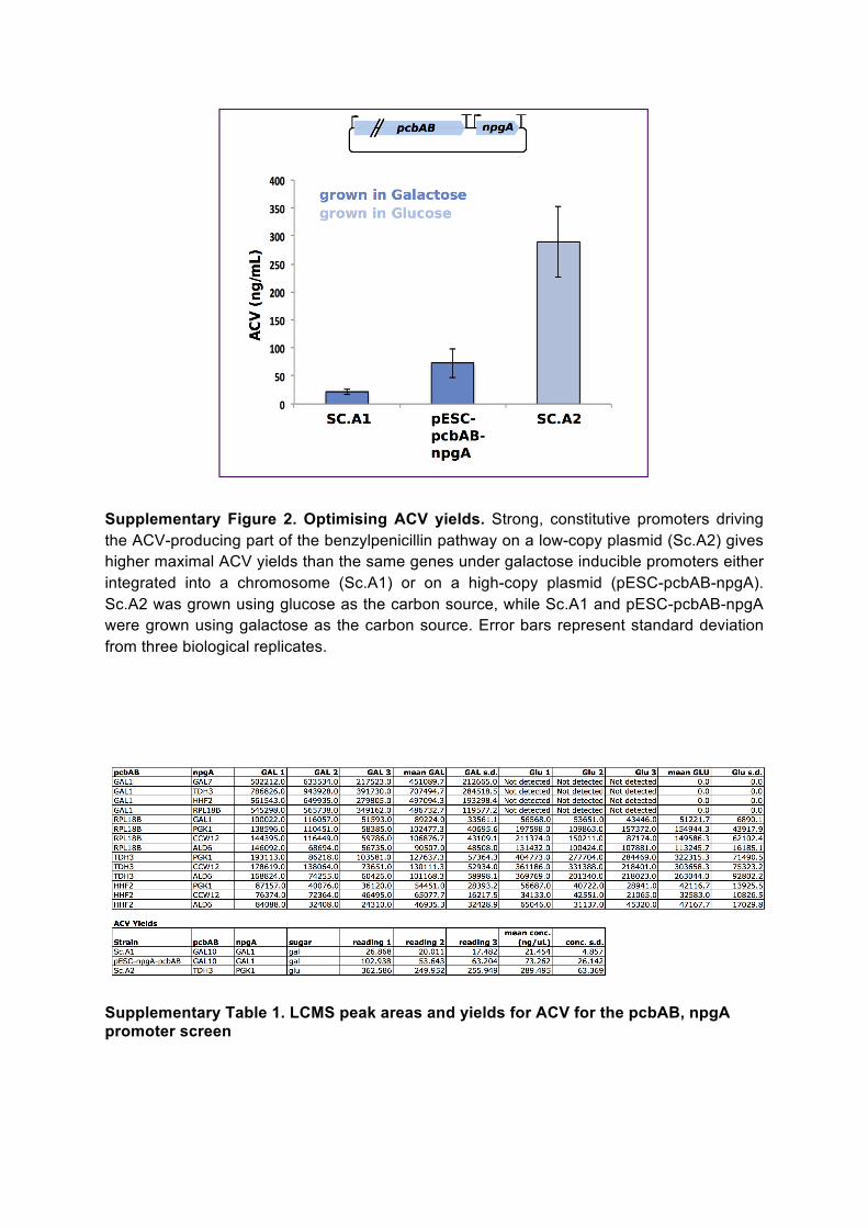
Supplementary Figure 2. Optimising ACV yields. Strong, constitutive promoters driving the ACV-producing part of the benzylpenicillin pathway on a low-copy plasmid (Sc.A2) gives higher maximal ACV yields than the same genes under galactose inducible promoters either integrated into a chromosome (Sc.A1) or on a high-copy plasmid (pESC-pcbAB-npgA). Sc.A2 was grown using glucose as the carbon source, while Sc.A1 and pESC-pcbAB-npgA were grown using galactose as the carbon source. Error bars represent standard deviation from three biological replicates.
Supplementary Table 1. LCMS peak areas and yields for ACV for the pcbAB, npgA promoter screen

Supplementary Table 2. LCMS peak areas and yields for ACV for the pcbAB, npgA promoter screen

Supplementary Table 3. Read counts and statistics for nanopore and Sanger sequencing

Supplementary Table 4. Strains and plasmids used in this paper * same as pAA179, but with promoters driving pcbC, pclA and penDE as specified by Table S12
Supplementary Table 5. ACV and benzylpenicillin production media composition
Supplementary Table 6. LCMS specifications

Supplementary Table 7. Promoters used in first promoter screen to optimise conversion of ACV to benzylpenicillin.
* Based on fold-change expression over background of fluorescent reporter Venus protein under each promoter, cells grown in glucose. Taken from Lee, M.E., DeLoache, W.C., Cervantes, B. & Dueber, J.E. ACS Synth Biol 4, 975-986 (2015).
** Uncharacterised in the above study, but should be similar to value from pGAL1, as these promoters naturally drive similar levels of expression when uninduced.
Supplementary Table 8. Relationship between promoter strength and benzylpenicillin yield in first promoter screen
Carbon source for these experiments is glucose, with promoter strengths that are indicated those for growth in glucose. Blue = strains featured in Figure 2. Red = strains with low penicillin production, not featured in Figure 2.
Supplementary Table 9. Golden Gate Assembly reaction parts for promoter screen to optimise conversion of ACV to benzylpenicillin and for nanopore sequencing

Supplementary Table 10. Promoters used in second promoter screen to optimise conversion of ACV to benzylpenicillin
* Based on fold-change expression over background of fluorescent reporter Venus protein under each promoter, cells grown in glucose. Taken from Lee, M.E., DeLoache, W.C., Cervantes, B. & Dueber, J.E. ACS Synth Biol 4, 975-986 (2015).
Supplementary Table 11. Golden Gate Assembly reaction parts for second promoter screen to optimise conversion of ACV to benzylpenicillin
Supplementary Table 12. Promoter strengths for strains shown in Figure 3d