Embed Size (px)
DESCRIPTION
PCC Case Presentation 2/8/06. History of Case. - PowerPoint PPT Presentation
Citation preview

PCC Case Presentation 2/8/06
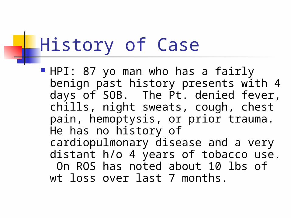
History of Case HPI: 87 yo man who has a fairly
benign past history presents with 4 days of SOB. The Pt. denied fever, chills, night sweats, cough, chest pain, hemoptysis, or prior trauma. He has no history of cardiopulmonary disease and a very distant h/o 4 years of tobacco use. On ROS has noted about 10 lbs of wt loss over last 7 months.
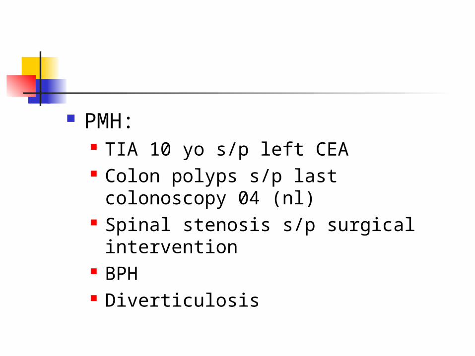
PMH:
TIA 10 yo s/p left CEA Colon polyps s/p last colonoscopy 04
(nl) Spinal stenosis s/p surgical
intervention BPH Diverticulosis
SH: lives in
Middleton with wife on a farm. Nonsmoker for >50y, no ETOH, worked as a farmer all his life
FH: no h/o lung disease. Noncontributory.
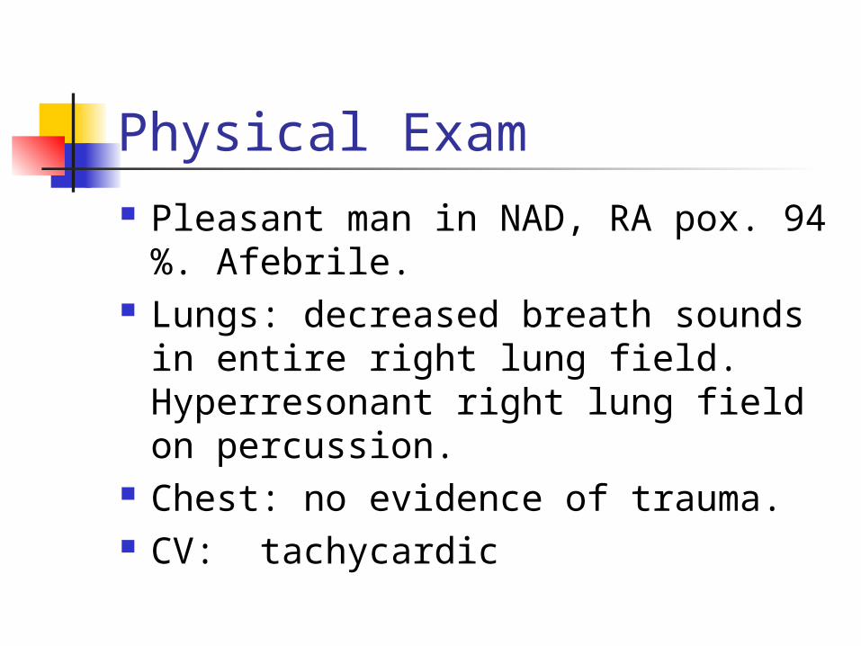
Physical Exam Pleasant man in NAD, RA pox. 94 %.
Afebrile. Lungs: decreased breath sounds in
entire right lung field. Hyperresonant right lung field on percussion.
Chest: no evidence of trauma. CV: tachycardic
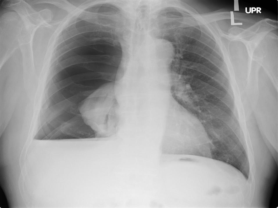
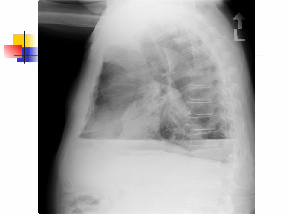
Dx: Spontaneous Pneumothorax Pt. was transferred to ED at UW
and had a Cook catheter inserted without complication and patient admitted for further evaluation.
Next step was to determine etiology and keep lung expanded.
Spontaneous Pneumothorax Definition: No preexisting obvious
cause, as compared to traumatic pneumothorax or iatrogenic pneumothorax.
Iatrogenic may be more common than spontaneous. In one study at the VA in Long Beach, CA over 5 years there were 108 iatrogenic versus 90 spontaneous pneumothoraces.
Spontaneous pneumothorax Primary versus secondary
Primary-No obvious underlying cause. (although many primary cases actually have an underlying cause if more closely evaluated)Secondary: multiple underlying causes.
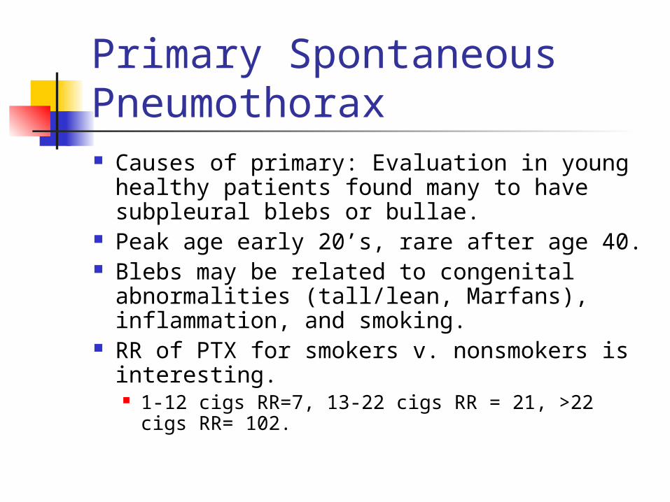
Primary Spontaneous Pneumothorax Causes of primary: Evaluation in young
healthy patients found many to have subpleural blebs or bullae.
Peak age early 20’s, rare after age 40. Blebs may be related to congenital
abnormalities (tall/lean, Marfans), inflammation, and smoking.
RR of PTX for smokers v. nonsmokers is interesting. 1-12 cigs RR=7, 13-22 cigs RR = 21, >22 cigs
RR= 102.
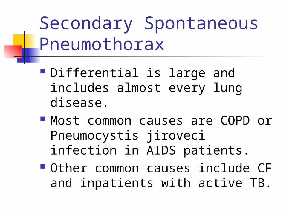
Secondary Spontaneous Pneumothorax Differential is large and includes
almost every lung disease. Most common causes are COPD or
Pneumocystis jiroveci infection in AIDS patients.
Other common causes include CF and inpatients with active TB.
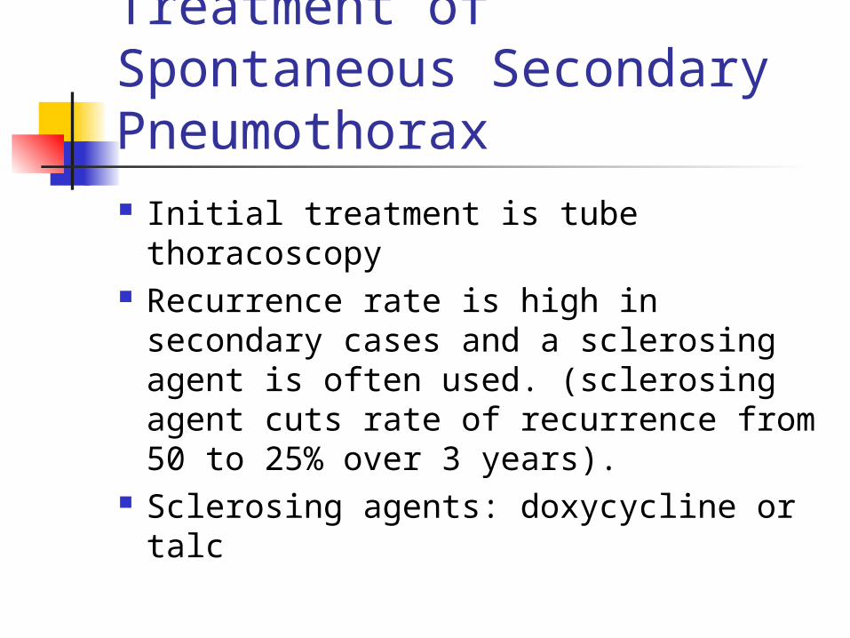
Treatment of Spontaneous Secondary Pneumothorax Initial treatment is tube thoracoscopy Recurrence rate is high in secondary
cases and a sclerosing agent is often used. (sclerosing agent cuts rate of recurrence from 50 to 25% over 3 years).
Sclerosing agents: doxycycline or talc
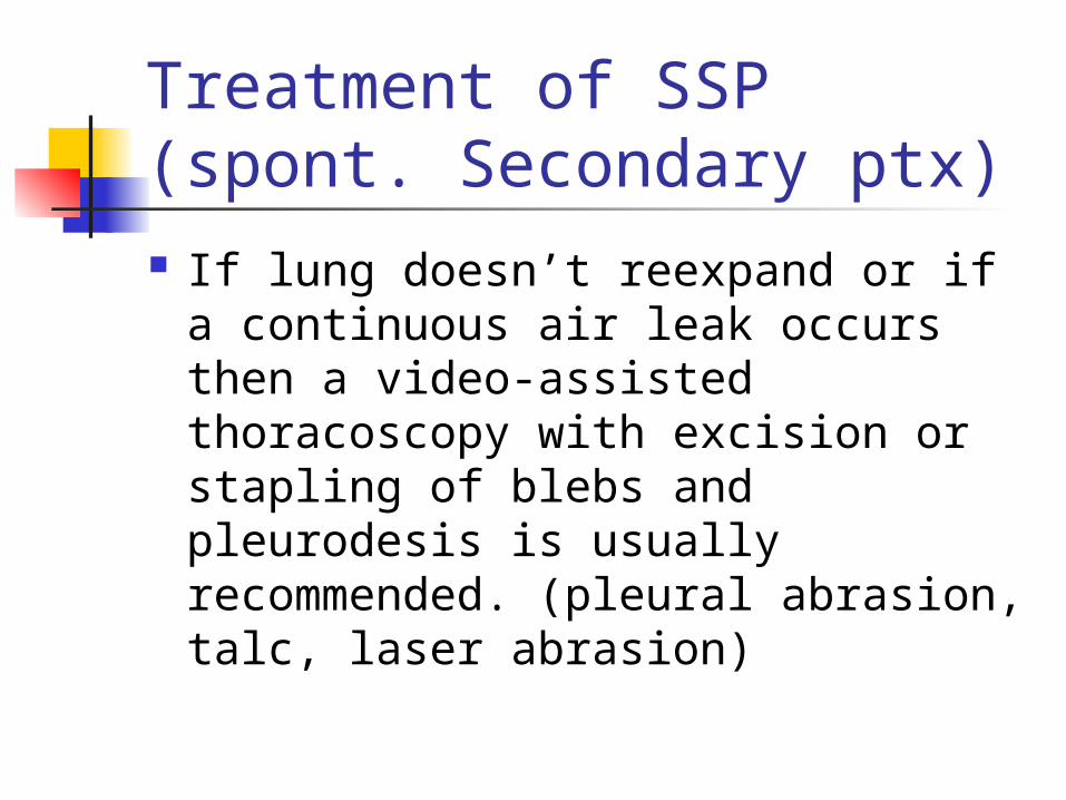
Treatment of SSP (spont. Secondary ptx) If lung doesn’t reexpand or if a
continuous air leak occurs then a video-assisted thoracoscopy with excision or stapling of blebs and pleurodesis is usually recommended. (pleural abrasion, talc, laser abrasion)
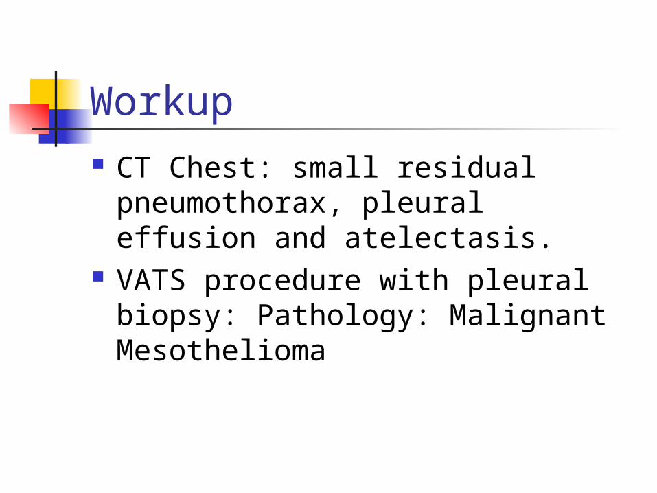
Workup CT Chest: small residual
pneumothorax, pleural effusion and atelectasis.
VATS procedure with pleural biopsy: Pathology: Malignant Mesothelioma
Hospital Course since DX Chest tube placed X 3. Failure X 3 Admitted 3 times over 3 months for
procedures to reexpand his right lung. First procedure after initial chest tube was
VATS with pleurodesis with talc. This was repeated twice. Then he had an attempt at a Heimlich valve which failed. Finally he had a right sided thoracotomy with decortication and pleural tenting performed.
Final procedure was successful and lung has stayed expanded since.
Malignant Mesothelioma This was final diagnosis in this
patient and the cause of his secondary spontaneous pneumothorax.
Aggressive tumor of the serosal surfaces.
Incidence increasing worldwide, as a result of prior asbestos exposure.
Malignant Mesothelioma Recent Review article in NEJM
10/13/05 immediately followed this patients diagnosis.
80% of patients are male and usually present with pleural effusion.
Peak incidence is expected to occur in 10-20 years worldwide. Although in US it may be already reaching its peak.
Malignant Mesothelioma 3 common exposures:
1. People directly exposed at work. E.g. Miners of blue asbestos. Playgrounds covered with asbestos tailings.
2. Workers exposed later in the use of asbestos products. E.g. plumbers, carpenters, defense personnel and installers of insulation.
3. The rest (20-30% of cases) were exposed to end product.
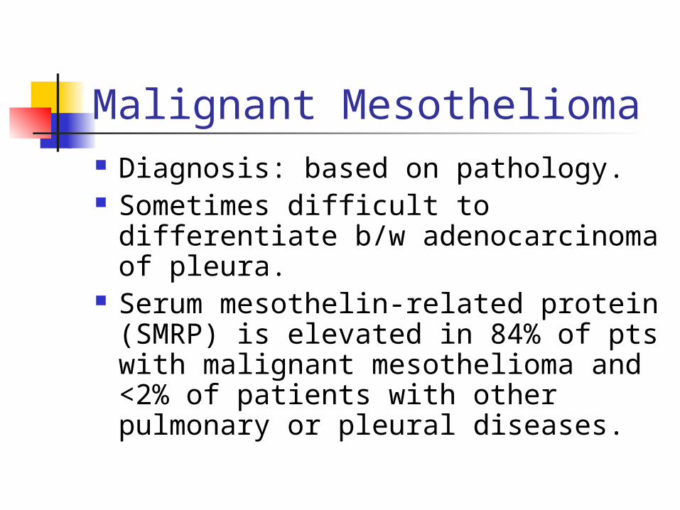
Malignant Mesothelioma Diagnosis: based on pathology. Sometimes difficult to differentiate
b/w adenocarcinoma of pleura. Serum mesothelin-related protein
(SMRP) is elevated in 84% of pts with malignant mesothelioma and <2% of patients with other pulmonary or pleural diseases.
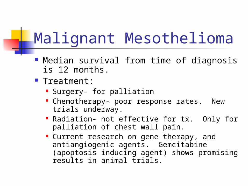
Malignant Mesothelioma Median survival from time of diagnosis is 12
months. Treatment:
Surgery- for palliation Chemotherapy- poor response rates. New trials
underway. Radiation- not effective for tx. Only for palliation
of chest wall pain. Current research on gene therapy, and
antiangiogenic agents. Gemcitabine (apoptosis inducing agent) shows promising results in animal trials.
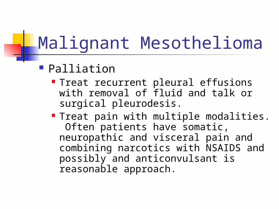
Malignant Mesothelioma Palliation
Treat recurrent pleural effusions with removal of fluid and talk or surgical pleurodesis.
Treat pain with multiple modalities. Often patients have somatic, neuropathic and visceral pain and combining narcotics with NSAIDS and possibly and anticonvulsant is reasonable approach.
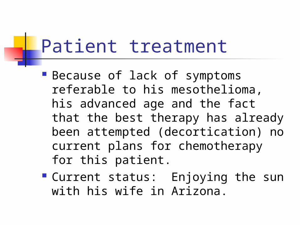
Patient treatment Because of lack of symptoms referable
to his mesothelioma, his advanced age and the fact that the best therapy has already been attempted (decortication) no current plans for chemotherapy for this patient.
Current status: Enjoying the sun with his wife in Arizona.
References Robinson and Lake. Advances in
Malignant Mesothelioma NEJM, Oct. 13, 2005, 353;14
Mason: Murray and Nadels Textbook of Respiratory Medicine, 4th ed., 2005
Sahn, SA, Heffner, JE. Spontaneous Pneumothorax NEJM, 2000; 342:868