Embed Size (px)
Citation preview
WHITEPAPER
PBMC Tips and Tricks
MK1193 | Feb 2019 NanoString Technologies®, Inc.
Authors: Emer Clarke, Ph.D., Gary dos Santos
2 | PBMC Tips and Tricks FEB RUARY 2019
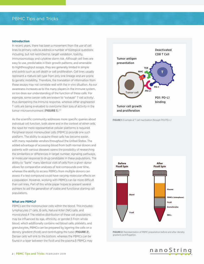
IntroductionIn recent years, there has been a movement from the use of cell lines to primary cells to address a number of biological questions including, but not restricted to, target validation, toxicity, immunooncology and cytokine storm risk. Although cell lines are easy to use, predictable in their growth patterns, and amenable to highthroughput assays, they are generally limited in simple end points such as cell death or cell proliferation. Cell lines usually represent a mature cell type from only one lineage and are prone to genetic instability. Therefore, the translation of information from these assays may not correlate well with the in vivo situation. As our awareness increases as to the many players in the immune system, so too does our understanding of the function of these cells. For example, some cancer cells are known to “exhaust” T cell activity1, thus dampening the immune response, whereas other engineered T cells are being evaluated to overcome their loss of activity in the tumor microenvironment (FIGURE 1).2,3
As the scientific community addresses more specific queries about individual cell function, both alone and in the context of other cells, the need for more representative cellular platforms is required. Peripheral blood mononuclear cells (PBMCs) provide one such platform. The ability to acquire these cells has become easier, with many reputable vendors throughout the United States. The added advantage of accessing blood from both normal donors and patients with various diseases opens the possibility of researching the similarities or differences in target number, signaling pathways, or molecular response to drug candidates in these populations. The ability to “bank” many identical vials of cells from a given donor allows for comparative analyses of test compounds over time, whereas the ability to access PBMCs from multiple donors can assess if a test compound could have varying molecular effects on a population. However, working with PBMCs can be more difficult than cell lines. Part of this white paper hopes to present several pointers to aid the generation of viable and functional starting cell populations.
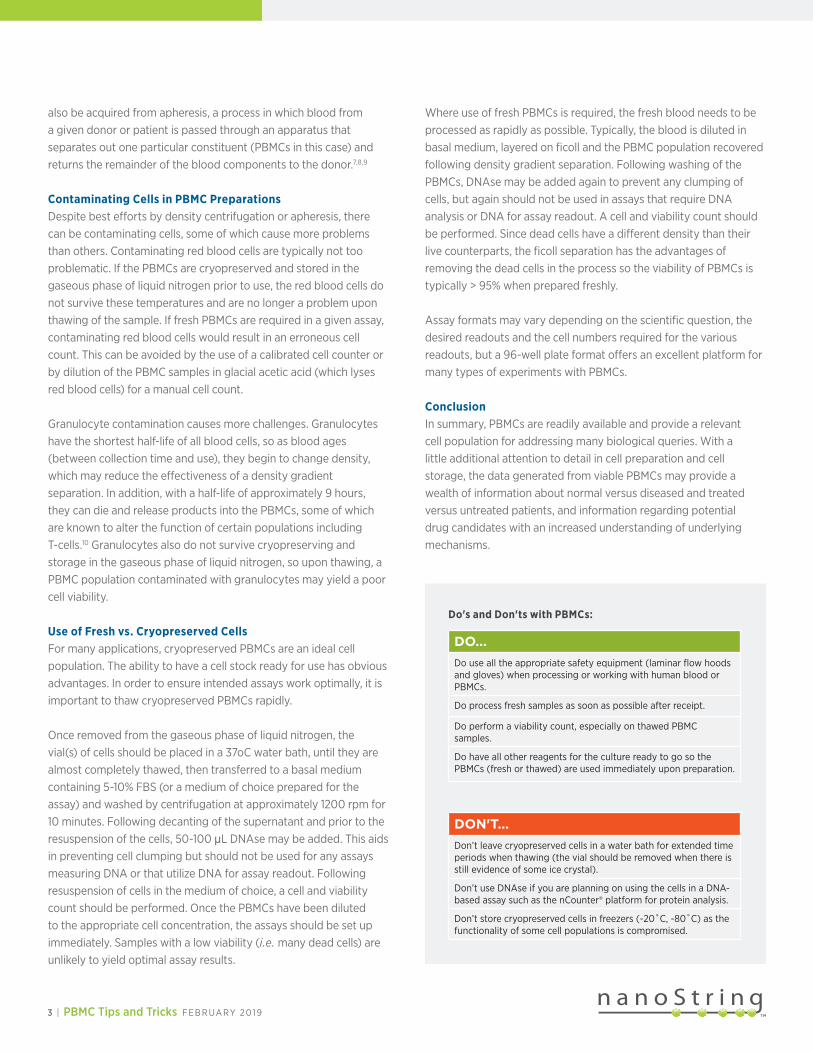
What are PBMCs?PBMCs are the mononuclear cells within the blood. This includes: lymphocytes (T cells, B cells, Natural Killer (NK) cells, and monocytes).4 The relative distribution of these cell populations may be influenced by age, ethnicity, or gender.5 From whole blood, which additionally contains red blood cells, platelets, and granulocytes, PBMCs can be prepared by layering the cells on a density gradient (ficoll) and centrifuging the tube (FIGURE 2). Denser cells will sink to the bottom, whereas the PBMCs can be found in a layer between the ficoll and the plasma.6 PBMCs may
PBMC Tips and Tricks
Tumor cell
TCR
PD-1
PD-L1
MHC T
Tumor cell growth and proliferation
PD1: PD-L1 binding
Deactivated CD8 T Cell
Tumor antigen presentation
FIGURE 1: Example of T cell inactivation through PD1/PD-L1
Plasma
PBMCs (interphase)
Ficoll
Granulocytes
RBCs
Blood
Ficoll
BeforeFicoll Spin
AfterFicoll Spin
FIGURE 2: Representation of PBMC preparation before and after density gradient centrifugation.
Do's and Don'ts with PBMCs:
DO...Do use all the appropriate safety equipment (laminar flow hoods and gloves) when processing or working with human blood or PBMCs.
Do process fresh samples as soon as possible after receipt.
Do perform a viability count, especially on thawed PBMC samples.
Do have all other reagents for the culture ready to go so the PBMCs (fresh or thawed) are used immediately upon preparation.
DON'T...Don’t leave cryopreserved cells in a water bath for extended time periods when thawing (the vial should be removed when there is still evidence of some ice crystal).
Don’t use DNAse if you are planning on using the cells in a DNA-based assay such as the nCounter® platform for protein analysis.
Don’t store cryopreserved cells in freezers (-20˚C, -80˚C) as the functionality of some cell populations is compromised.
3 | PBMC Tips and Tricks FEB RUARY 2019
also be acquired from apheresis, a process in which blood from a given donor or patient is passed through an apparatus that separates out one particular constituent (PBMCs in this case) and returns the remainder of the blood components to the donor.7,8,9
Contaminating Cells in PBMC PreparationsDespite best efforts by density centrifugation or apheresis, there can be contaminating cells, some of which cause more problems than others. Contaminating red blood cells are typically not too problematic. If the PBMCs are cryopreserved and stored in the gaseous phase of liquid nitrogen prior to use, the red blood cells do not survive these temperatures and are no longer a problem upon thawing of the sample. If fresh PBMCs are required in a given assay, contaminating red blood cells would result in an erroneous cell count. This can be avoided by the use of a calibrated cell counter or by dilution of the PBMC samples in glacial acetic acid (which lyses red blood cells) for a manual cell count.
Granulocyte contamination causes more challenges. Granulocytes have the shortest half-life of all blood cells, so as blood ages (between collection time and use), they begin to change density, which may reduce the effectiveness of a density gradient separation. In addition, with a half-life of approximately 9 hours, they can die and release products into the PBMCs, some of which are known to alter the function of certain populations including T-cells.10 Granulocytes also do not survive cryopreserving and storage in the gaseous phase of liquid nitrogen, so upon thawing, a PBMC population contaminated with granulocytes may yield a poor cell viability.
Use of Fresh vs. Cryopreserved CellsFor many applications, cryopreserved PBMCs are an ideal cell population. The ability to have a cell stock ready for use has obvious advantages. In order to ensure intended assays work optimally, it is important to thaw cryopreserved PBMCs rapidly.
Once removed from the gaseous phase of liquid nitrogen, the vial(s) of cells should be placed in a 37oC water bath, until they are almost completely thawed, then transferred to a basal medium containing 5-10% FBS (or a medium of choice prepared for the assay) and washed by centrifugation at approximately 1200 rpm for 10 minutes. Following decanting of the supernatant and prior to the resuspension of the cells, 50-100 μL DNAse may be added. This aids in preventing cell clumping but should not be used for any assays measuring DNA or that utilize DNA for assay readout. Following resuspension of cells in the medium of choice, a cell and viability count should be performed. Once the PBMCs have been diluted to the appropriate cell concentration, the assays should be set up immediately. Samples with a low viability (i.e. many dead cells) are unlikely to yield optimal assay results.
Where use of fresh PBMCs is required, the fresh blood needs to be processed as rapidly as possible. Typically, the blood is diluted in basal medium, layered on ficoll and the PBMC population recovered following density gradient separation. Following washing of the PBMCs, DNAse may be added again to prevent any clumping of cells, but again should not be used in assays that require DNA analysis or DNA for assay readout. A cell and viability count should be performed. Since dead cells have a different density than their live counterparts, the ficoll separation has the advantages of removing the dead cells in the process so the viability of PBMCs is typically > 95% when prepared freshly.
Assay formats may vary depending on the scientific question, the desired readouts and the cell numbers required for the various readouts, but a 96-well plate format offers an excellent platform for many types of experiments with PBMCs.
ConclusionIn summary, PBMCs are readily available and provide a relevant cell population for addressing many biological queries. With a little additional attention to detail in cell preparation and cell storage, the data generated from viable PBMCs may provide a wealth of information about normal versus diseased and treated versus untreated patients, and information regarding potential drug candidates with an increased understanding of underlying mechanisms.
REFERENCES
FOR RESEARCH USE ONLY. Not for use in diagnostic procedures. © 2019 NanoString Technologies, Inc. All rights reserved. NanoString, NanoString Technologies, the NanoString logo and nCounter are trademarks or registered trademarks of NanoString Technologies, Inc., in the United States and/or other countries. All other trademarks and/or service marks not owned by NanoString that appear in this document are the property of their respective owners.
For more information, please visit nanostring.com
FEB 2019 MK1193
Sales ContactsUnited States [email protected]: [email protected]
NanoString Technologies, Inc. 530 Fairview Avenue NorthSeattle, Washington 98109
Asia Pacific & Japan [email protected] Regions [email protected]
T (888) 358-6266F (206) 378-6288
nanostring.com [email protected]
1 Sugimoto, T. & Watanabe T. (2016). Follicular Lymphoma: The
Role of the Tumor Microenvironment in Prognosis. J Clin Exp
Hematop 56(1):1-19.
2 Davila M.L. & Sadelain M. Biology and Clinical Application
of CAR T Cells for B cell malignancies. Int. J. Hematol 104(1):
6-17.
3 Cherkassy L. et al (2016). Human CAR T Cells with
cellintrinsic PD-1 checkpoints blockade resist tumor-
mediated inhibition. J. Clin Invest. 126 (8): 3130-3144.
4 Goff L. et al (1985). Normal values for the different classes
of cenous blood mononuclear cells defined by monoclonal
antibodies. J Clin Path 38(1): 54-59.
5 Tollerud D.J. et al (1989). The influence of age, race, and
gender on peripheral blood mononuclear-cell subsets in
healthy non-smokers. J Clin Immunol 9(3): 214-222
6 Casale T.B. & KAliner M. (1982). A rapid method for isolation
of human mononuclear cells free of significant platelet
contamination. J Immunol Methods 55(3): 347-353.
7 Mookerjee B.K. (1076). Influence of separation techniques
on the distribution and function of lymphocyte population.
A comparison on three techniques. Transplantation 22 (2);
101-107.
8 Snyder E.L. et al (2001). Ex vivo evaluation of PBMNCs
collected with a new separator. Transfusion 41(7): 940-949.
9 Steininger P.A. et al (2014). First comparative evaluation of
a new leukapheresis technology in non-cytokine stimulated
donors. Vox Sang 106(3): 248-255.
10 McKenna K.C.et al (2009). Delayed processing of blood
increases the frequency of activated CD11b+ CD15+
granulocytes which inhibit T cell function. J Immunol Methods
(341 (1-2): 68-75.