Embed Size (px)
Citation preview
1
(PBL) Deep Vein Thrombosis and Pulmonary Embolism Management:
The Clot Thickens
David Schneider, MD, FAAFP
ACTIVITY DISCLAIMERThe material presented here is being made available by the American Academy of Family Physicians for educational purposes only. Please note that medical information is constantly changing; the information contained in this activity was accurate at the time of publication. This material is not intended to represent the only, nor necessarily best, methods or procedures appropriate for the medical situations discussed. Rather, it is intended to present an approach, view, statement, or opinion of the faculty, which may be helpful to others who face similar situations.
The AAFP disclaims any and all liability for injury or other damages resulting to any individual using this material and for all claims that might arise out of the use of the techniques demonstrated therein by such individuals, whether these claims shall be asserted by a physician or any other person. Physicians may care to check specific details such as drug doses and contraindications, etc., in standard sources prior to clinical application. This material might contain recommendations/guidelines developed by other organizations. Please note that although these guidelines might be included, this does not necessarily imply the endorsement by the AAFP.
This live CME session is supported by an educational grant from the Bristol-Myers Squibb and Pfizer Alliance.
2
DISCLOSUREIt is the policy of the AAFP that all individuals in a position to control content disclose any relationships with commercial interests upon nomination/invitation of participation. Disclosure documents are reviewed for potential conflict of interest (COI), and if identified, conflicts are resolved prior to confirmation of participation. Only those participants who had no conflict of interest or who agreed to an identified resolution process prior to their participation were involved in this CME activity.
All individuals in a position to control content for this session have indicated they have no relevant financial relationships to disclose.
The content of my material/presentation in this CME activity will include discussion of unapproved or investigational uses of products or devices as indicated: Consider anticoagulation of distal deep venous thrombosis under certain circumstances, per recommendations.
David Schneider, MD, FAAFPFaculty physician/Didactics Director/Procedures Director, Santa Rosa Family Medicine Residency, California; Professor of Family and Community Medicine, University of California, San Francisco (UCSF) School of Medicine
Dr. Schneider cares for the underserved in Santa Rosa, California, serving Latino, Southeast Asian, and Eritrean populations. He has taught the breadth and depth of family medicine for more than 20 years, and his professional interests include the physician-patient relationship and clinical skills. Cardiovascular system conditions are one of his specialty topics, and he points to "the growing body of evidence suggesting that lifestyle is as effective as, or more effective than, pharmacologic interventions in primary prevention." Dr. Schneider also focuses on conditions of the endocrine system (especially thyroid); skin conditions and dermatology; primary prevention, with a focus on lifestyle; and procedures. Board certified in both family medicine and integrative holistic medicine, he produces Dr. Dave's To Your Health segments for Wine Country Radio and BlogTalkRadio.com.
3
Learning Objectives1. Practice applying new knowledge and skills gained from
Venous Thromboembolism Management sessions, through collaborative learning with peers and expert faculty.
2. Identify strategies that foster optimal management of venous thromboembolism, within the context of professional practice.
3. Formulate an action plan to implement practice changes, aimed at improving patient care.
Associated Sessions
• Deep Vein Thrombosis and Pulmonary Embolism Management: The Clot Thickens
4
Chief Complaint
• 47 yo WF presents to office w/LLE pain X5 days. She thinks it getting a bit worse.
History of Present Illness
• L lower leg pain started ~ 5 days ago, maybe walking too much lately (trying to lose wt & get healthier).
• No improvement w/ibuprofen or naproxen Na.• No F/C, no N/V. No bowel/bladder sx.• No back pain.• Normal activity level.
5
Past Medical History
• HTN, controlled – 12 years.
• Former smoker, 1 ppd age 19 – 41.
• Reports some mild varicose veins in recent years, asymptomatic.
Medications, Allergies
• HCTZ 25 mg daily, lisinopril 10 mg daily.– BP has been controlled on this regimen.
• NKDA,
6
Family History
• Both parents have HTN.
• No known VTE in family.
• Father w/COPD (chronic smoker).
Social History
• Former smoker, 1 ppd age 19 – 41.
• Occasional ETOH.
• No pets. 2 high school aged kids.
7
Review of Systems
• No HA, visual sx.
• No CP/SOB.
• No F/C, no N/V. No bowel/bladder sx.
• No back pain.
• Normal activity level.
Physical Examination
• BP 142/82, T98.7, P 88, R 16/reg. NAD.
• HEENT & neck WNL.
• H—RRR, no m or g. Lungs clear.
• Ext: LLE tender & bluish cord over calf, reproducing pain; lower leg w/pitting edema.
8
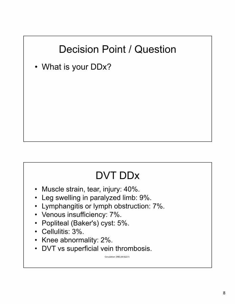
Decision Point / Question
• What is your DDx?
DVT DDx• Muscle strain, tear, injury: 40%.• Leg swelling in paralyzed limb: 9%.• Lymphangitis or lymph obstruction: 7%.• Venous insufficiency: 7%.• Popliteal (Baker's) cyst: 5%.• Cellulitis: 3%.• Knee abnormality: 2%.• DVT vs superficial vein thrombosis.
Circulation 1981;64:622‐5
9
Laboratory/Radiology
• You send her to ED for eval.– CBC WBC 11.2, o/w WNL.
– Comp met panel WNL.
Decision Point / Question
• What’s your workup?
10
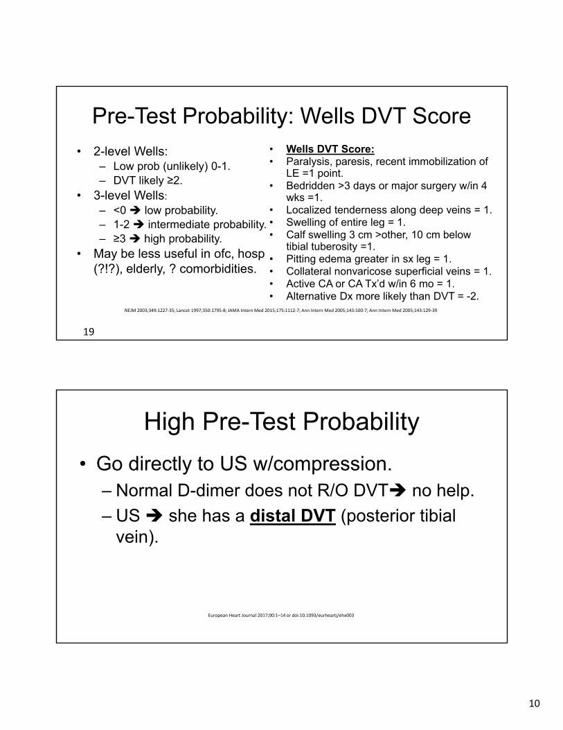
Pre-Test Probability: Wells DVT Score
• 2-level Wells:– Low prob (unlikely) 0-1.– DVT likely ≥2.
• 3-level Wells:
– <0 low probability.– 1-2 intermediate probability.– ≥3 high probability.
• May be less useful in ofc, hosp (?!?), elderly, ? comorbidities.
• Wells DVT Score:• Paralysis, paresis, recent immobilization of
LE =1 point.• Bedridden >3 days or major surgery w/in 4
wks =1.• Localized tenderness along deep veins = 1.• Swelling of entire leg = 1.• Calf swelling 3 cm >other, 10 cm below
tibial tuberosity =1.• Pitting edema greater in sx leg = 1.• Collateral nonvaricose superficial veins = 1.• Active CA or CA Tx’d w/in 6 mo = 1.• Alternative Dx more likely than DVT = -2.
19
NEJM 2003;349:1227‐35; Lancet 1997;350:1795‐8; JAMA Intern Med 2015;175:1112‐7; Ann Intern Med 2005;143:100‐7; Ann Intern Med 2005;143:129‐39
High Pre-Test Probability
• Go directly to US w/compression.– Normal D-dimer does not R/O DVT no help.
– US she has a distal DVT (posterior tibial vein).
European Heart Journal 2017;00:1–14 or doi:10.1093/eurheartj/ehx003
11
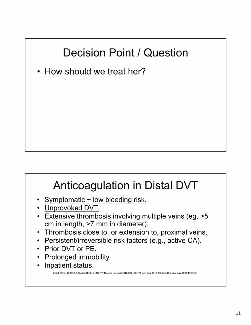
Decision Point / Question
• How should we treat her?
Anticoagulation in Distal DVT• Symptomatic + low bleeding risk.• Unprovoked DVT.• Extensive thrombosis involving multiple veins (eg, >5
cm in length, >7 mm in diameter).• Thrombosis close to, or extension to, proximal veins.• Persistent/irreversible risk factors (e.g., active CA).• Prior DVT or PE.• Prolonged immobility.• Inpatient status.
Chest 2016;149:315‐52; Chest 2014;146:1468‐77; Thromb Haemost 2010;104:1063‐70; Ann Surg 2010;251:735‐42; J Vasc Surg 2007;46:513‐9
12
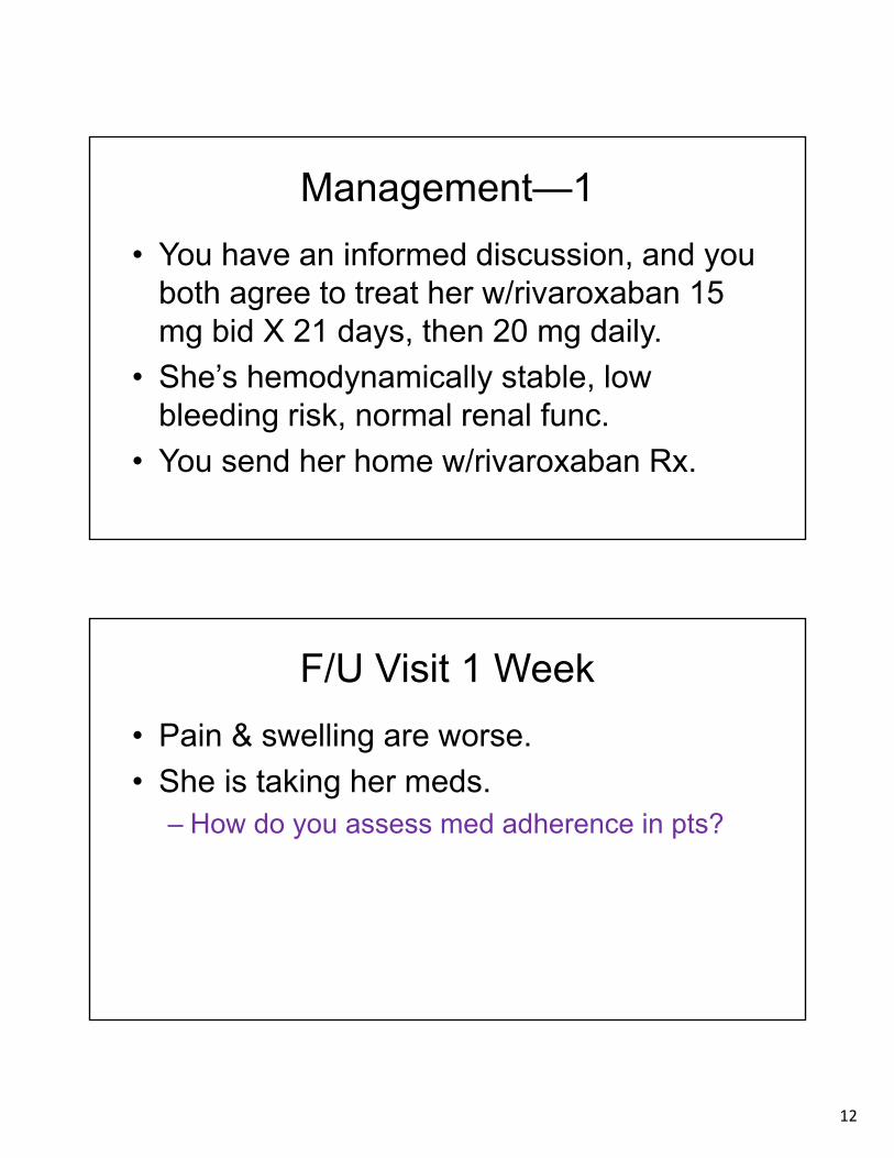
Management—1
• You have an informed discussion, and you both agree to treat her w/rivaroxaban 15 mg bid X 21 days, then 20 mg daily.
• She’s hemodynamically stable, low bleeding risk, normal renal func.
• You send her home w/rivaroxaban Rx.
F/U Visit 1 Week
• Pain & swelling are worse.
• She is taking her meds.– How do you assess med adherence in pts?
13
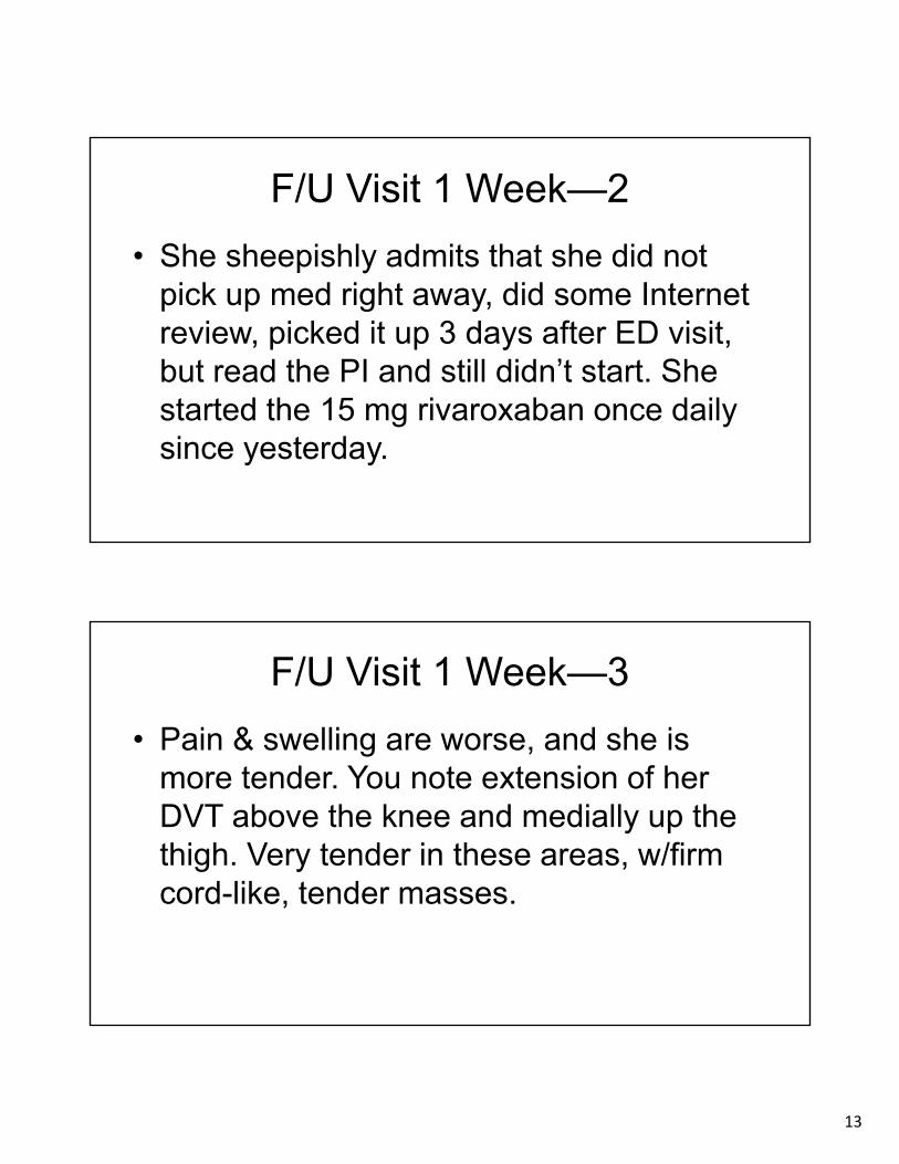
F/U Visit 1 Week—2
• She sheepishly admits that she did not pick up med right away, did some Internet review, picked it up 3 days after ED visit, but read the PI and still didn’t start. She started the 15 mg rivaroxaban once daily since yesterday.
F/U Visit 1 Week—3
• Pain & swelling are worse, and she is more tender. You note extension of her DVT above the knee and medially up the thigh. Very tender in these areas, w/firm cord-like, tender masses.
14
Decision Point / Question
• Next steps?
Worsening Clinical Status
• You send her by ambulance to ED, call ED physician &/or charge nurse.
• You are on call for admitting your own pts today, anyway.

15
Wells DVT Scores• 2-level Wells:
– Low prob (unlikely) 0-1.– DVT likely ≥2.
• 3-level Wells:
– <0 low probability.– 1-2 intermediate probability.– ≥3 high probability.
• May be less useful in ofc, hosp (?!?), elderly, ? comorbidities.
• Wells DVT Score:• Paralysis, paresis, recent immobilization of
LE =1 point.• Bedridden >3 days or major surgery w/in 4
wks =1.• Localized tenderness along deep veins = 1.• Swelling of entire leg = 1.• Calf swelling 3 cm >other, 10 cm below
tibial tuberosity =1.• Pitting edema greater in sx leg = 1.• Collateral nonvaricose superficial veins = 1.• Active CA or CA Tx’d w/in 6 mo = 1.• Alternative Dx more likely than DVT = -2.
NEJM 2003;349:1227‐35; Lancet 1997;350:1795‐8; JAMA Intern Med 2015;175:1112‐7; Ann Intern Med 2005;143:100‐7; Ann Intern Med 2005;143:129‐39
ED Course
• Wells score now 5.
• Compression US w/doppler (Duplex scan w/compression) distal posterior tibial vein DVT has extended to popliteal & superficial femoral veins (NOTE: SFV is a deep vein!).
16
ED Course—2
• While in ED, you come to see her. ED Dr has cleared her for admission, wants to write orders. You come by during lunch & meet pt in ED.
• Your questioning no CP, but she endorses a tight, sharp feeling R mid-chest.
Decision Point / Question
• Next steps? W/U & treatment?
17
ED Course—3
• BP 118/72, T 99.2, P 112/reg, RR 20.
• O2 Sat = 91% RA.
• Is she hemodynamically stable?– Yes. Continue eval.
ED Course—4
• High risk pt, not eligible for PERC.
18
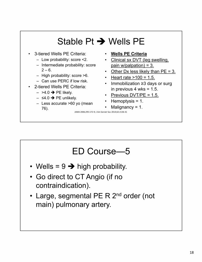
Stable Pt Wells PE• 3-tiered Wells PE Criteria:
– Low probability: score <2.– Intermediate probability: score
2 – 6.– High probability: score >6.– Can use PERC if low risk.
• 2-tiered Wells PE Criteria:– >4.0 PE likely.– ≤4.0 PE unlikely.– Less accurate >60 yo (mean
76).
• Wells PE Criteria• Clinical sx DVT (leg swelling,
pain w/palpation) = 3.• Other Dx less likely than PE = 3.• Heart rate >100 = 1.5.• Immobilization ≥3 days or surg
in previous 4 wks = 1.5.• Previous DVT/PE = 1.5.• Hemoptysis = 1.• Malignancy = 1.
JAMA 2006;295:172‐9; J Am Geriatr Soc 2014;62:2136‐41
ED Course—5
• Wells = 9 high probability.
• Go direct to CT Angio (if no contraindication).
• Large, segmental PE R 2nd order (not main) pulmonary artery.
19
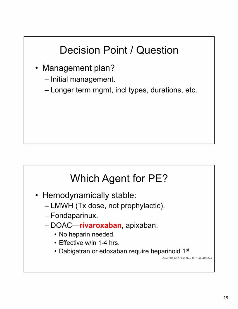
Decision Point / Question
• Management plan?– Initial management.
– Longer term mgmt, incl types, durations, etc.
Which Agent for PE?
• Hemodynamically stable: – LMWH (Tx dose, not prophylactic).– Fondaparinux.– DOAC—rivaroxaban, apixaban.
• No heparin needed.• Effective w/in 1-4 hrs.• Dabigatran or edoxaban require heparinoid 1st.
Chest 2016;149:315‐52; Chest 2012;141:e419S‐94S
20
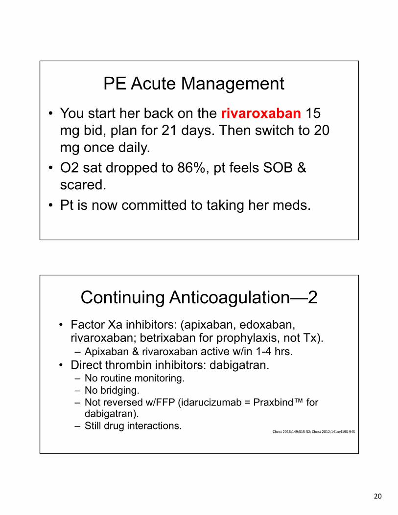
PE Acute Management
• You start her back on the rivaroxaban 15 mg bid, plan for 21 days. Then switch to 20 mg once daily.
• O2 sat dropped to 86%, pt feels SOB & scared.
• Pt is now committed to taking her meds.
Continuing Anticoagulation—2
• Factor Xa inhibitors: (apixaban, edoxaban, rivaroxaban; betrixaban for prophylaxis, not Tx).– Apixaban & rivaroxaban active w/in 1-4 hrs.
• Direct thrombin inhibitors: dabigatran.– No routine monitoring.– No bridging.– Not reversed w/FFP (idarucizumab = Praxbind™ for
dabigatran).– Still drug interactions.
Chest 2016;149:315‐52; Chest 2012;141:e419S‐94S
21
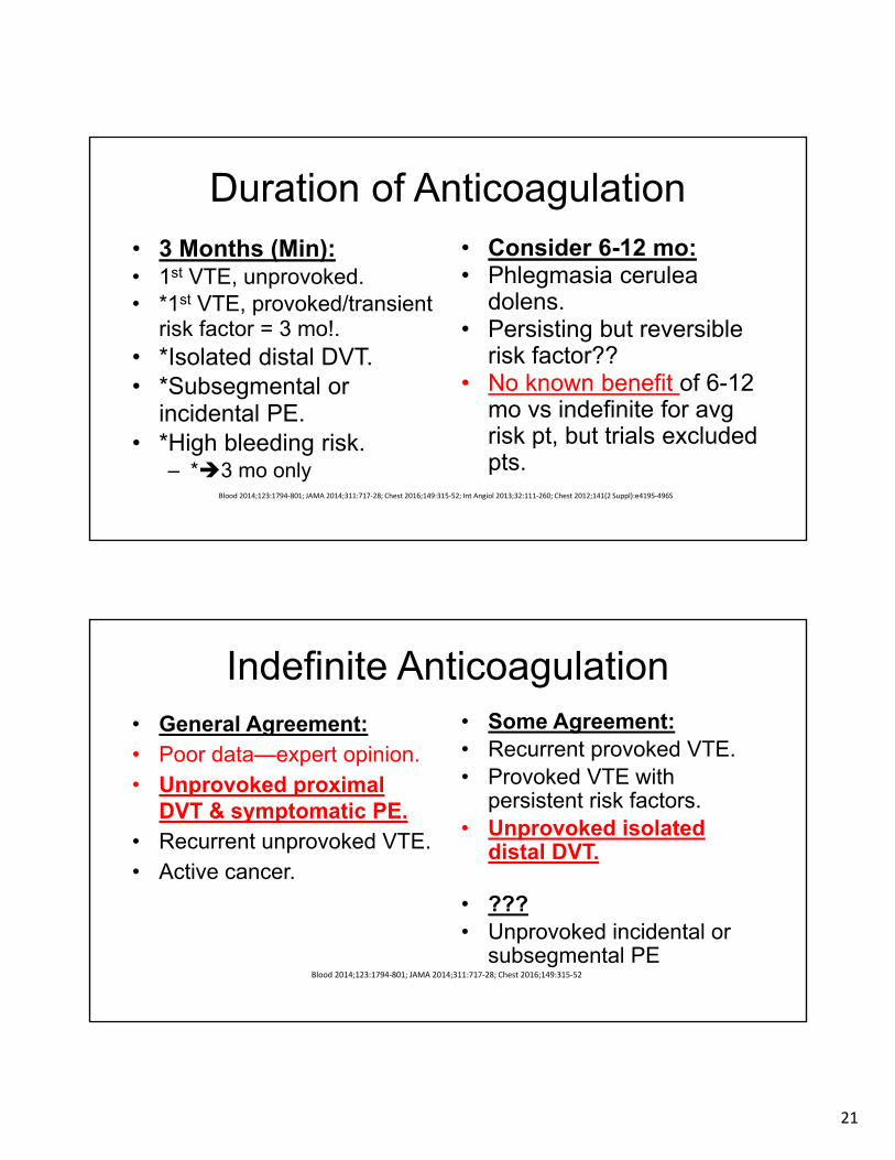
Duration of Anticoagulation• 3 Months (Min):• 1st VTE, unprovoked.• *1st VTE, provoked/transient
risk factor = 3 mo!.• *Isolated distal DVT.• *Subsegmental or
incidental PE.• *High bleeding risk.
– *3 mo only
• Consider 6-12 mo:• Phlegmasia cerulea
dolens.• Persisting but reversible
risk factor??• No known benefit of 6-12
mo vs indefinite for avg risk pt, but trials excluded pts.
Blood 2014;123:1794‐801; JAMA 2014;311:717‐28; Chest 2016;149:315‐52; Int Angiol 2013;32:111‐260; Chest 2012;141(2 Suppl):e419S‐496S
Indefinite Anticoagulation• General Agreement:• Poor data—expert opinion.• Unprovoked proximal
DVT & symptomatic PE.• Recurrent unprovoked VTE.• Active cancer.
• Some Agreement:• Recurrent provoked VTE.• Provoked VTE with
persistent risk factors.• Unprovoked isolated
distal DVT.
• ???• Unprovoked incidental or
subsegmental PEBlood 2014;123:1794‐801; JAMA 2014;311:717‐28; Chest 2016;149:315‐52
22
Hospital Course
• She remains in hospital for 4 days, O2 sats increase to 90% on RA.
• Discharged home on no O2 and rivaroxaban, w/explicit instructions.
• Pt verbalizes understanding & summarizes plan & reasoning back to you.
Follow-Up
• 6 months later she is back to full activity.
• No SOB or chest symptoms.
• Reports chronic, intermittent LLE pain, frequent edema, hyperpigmentation, and occasional open sores which heal relatively well w/OTC Abx ointment + bandage.

23
Decision Point / Question
• Diagnosis?
• Management?
Post-Thrombosis Syndrome
• Signs of chronic venous insufficiency after DVT.
• Common—up to 50% w/in 1st yr after DVT.• Rx:
– Exercise, compression, horse chestnut (escin).– Invasive treatment (IR, cardiology) or surgery if
refractory.