Embed Size (px)
Citation preview
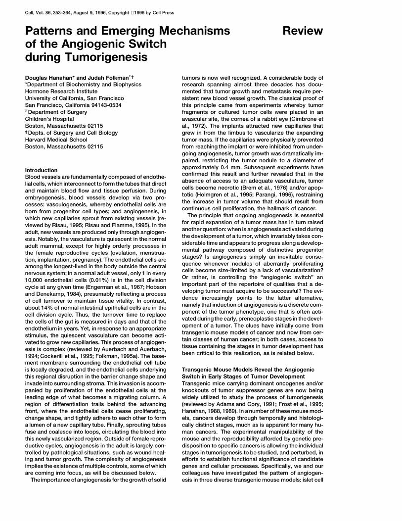
Cell, Vol. 86, 353–364, August 9, 1996, Copyright 1996 by Cell Press
Patterns and Emerging Mechanisms Reviewof the Angiogenic Switchduring Tumorigenesis
Douglas Hanahan* and Judah Folkman†‡ tumors is now well recognized. A considerable body ofresearch spanning almost three decades has docu-*Department of Biochemistry and Biophysics
Hormone Research Institute mented that tumor growth and metastasis require per-sistent new blood vessel growth. The classical proof ofUniversity of California, San Francisco
San Francisco, California 94143-0534 this principle came from experiments whereby tumorfragments or cultured tumor cells were placed in an†Department of Surgery
Children’s Hospital avascular site, the cornea of a rabbit eye (Gimbrone etal., 1972). The implants attracted new capillaries thatBoston, Massachusetts 02115
‡Depts. of Surgery and Cell Biology grew in from the limbus to vascularize the expandingtumor mass. If the capillaries were physically preventedHarvard Medical School
Boston, Massachusetts 02115 from reaching the implant or were inhibited from under-going angiogenesis, tumor growth was dramatically im-paired, restricting the tumor nodule to a diameter ofapproximately 0.4 mm. Subsequent experiments haveIntroductionconfirmed this result and further revealed that in theBlood vessels are fundamentally composed of endothe-absence of access to an adequate vasculature, tumorlial cells, which interconnect to form the tubes that directcells become necrotic (Brem et al., 1976) and/or apop-and maintain blood flow and tissue perfusion. Duringtotic (Holmgren et al., 1995; Parangi, 1996), restrainingembryogenesis, blood vessels develop via two pro-the increase in tumor volume that should result fromcesses: vasculogenesis, whereby endothelial cells arecontinuous cell proliferation, the hallmark of cancer.born from progenitor cell types; and angiogenesis, in
The principle that ongoing angiogenesis is essentialwhich new capillaries sprout from existing vessels (re-for rapid expansion of a tumor mass has in turn raisedviewed by Risau, 1995; Risau and Flamme, 1995). In theanother question: when is angiogenesis activated duringadult, new vessels are produced only through angiogen-the development of a tumor, which invariably takes con-esis. Notably, the vasculature is quiescent in the normalsiderable time and appears to progress along a develop-adult mammal, except for highly orderly processes inmental pathway composed of distinctive progenitorthe female reproductive cycles (ovulation, menstrua-stages? Is angiogenesis simply an inevitable conse-tion, implantation, pregnancy). The endothelial cells arequence whenever nodules of aberrantly proliferatingamong the longest-lived in the body outside the centralcells become size-limited by a lack of vascularization?nervous system; in a normal adult vessel, only 1 in everyOr rather, is controlling the “angiogenic switch” an10,000 endothelial cells (0.01%) is in the cell divisionimportant part of the repertoire of qualities that a de-cycle at any given time (Engerman et al., 1967; Hobsonveloping tumor must acquire to be successful? The evi-and Denekamp, 1984), presumably reflecting a processdence increasingly points to the latter alternative,of cell turnover to maintain tissue vitality. In contrast,namely that induction of angiogenesis is a discrete com-about 14% of normal intestinal epithelial cells are in theponent of the tumor phenotype, one that is often acti-cell division cycle. Thus, the turnover time to replacevated during the early, preneoplastic stages in thedevel-the cells of the gut is measured in days and that of theopment of a tumor. The clues have initially come fromendothelium in years. Yet, in response to an appropriatetransgenic mouse models of cancer and now from cer-stimulus, the quiescent vasculature can become acti-tain classes of human cancer; in both cases, access tovated to grow new capillaries. This process of angiogen-tissue containing the stages in tumor development hasesis is complex (reviewed by Auerbach and Auerbach,been critical to this realization, as is related below.1994; Cockerill et al., 1995; Folkman, 1995a). The base-
ment membrane surrounding the endothelial cell tubeis locally degraded, and the endothelial cells underlying Transgenic Mouse Models Reveal the Angiogenic
Switch in Early Stages of Tumor Developmentthis regional disruption in the barrier change shape andinvade into surrounding stroma. This invasion is accom- Transgenic mice carrying dominant oncogenes and/or
knockouts of tumor suppressor genes are now beingpanied by proliferation of the endothelial cells at theleading edge of what becomes a migrating column. A widely utilized to study the process of tumorigenesis
(reviewed by Adams and Cory, 1991; Frost et al., 1995;region of differentiation trails behind the advancingfront, where the endothelial cells cease proliferating, Hanahan, 1988, 1989). In a number of these mouse mod-
els, cancers develop through temporally and histologi-change shape, and tightly adhere to each other to forma lumen of a new capillary tube. Finally, sprouting tubes cally distinct stages, much as is apparent for many hu-
man cancers. The experimental manipulability of thefuse and coalesce into loops, circulating the blood intothis newly vascularized region. Outside of female repro- mouse and the reproducibility afforded by genetic pre-
disposition to specific cancers is allowing the individualductive cycles, angiogenesis in the adult is largely con-trolled by pathological situations, such as wound heal- stages in tumorigenesis to be studied, and perturbed, in
efforts to establish functional significance of candidateing and tumor growth. The complexity of angiogenesisimplies the existence of multiple controls, some of which genes and cellular processes. Specifically, we and our
colleagues have investigated the pattern of angiogen-are coming into focus, as will be discussed below.Theimportance of angiogenesis for thegrowth of solid esis in three diverse transgenic mouse models: islet cell

Cell354
Figure 1. The Angiogenic Switch Occurs Prior to Tumor Formation in Three Transgenic Mouse Models of Tumorigenesis
(A) Expression of the Tag oncogene in the pancreatic islets elicits four sequential stages in tumor development: normal, oncogene-expressingislets; hyperplastic islets, populated by proliferating cells with the histological hallmarks of CIS; angiogenic islets, in which new blood vesselgrowth has been activated; and solid tumors, which are islet cell carcinomas.(B) In transgenic mice carrying the BPV-1 oncogenes, the normal dermis is initially converted into a state of mild fibromatosis, revealed asfocal accumulation of dermal fibroblasts. Angiogenesis is first evident in the next stage, aggressive fibromatosis, which is also markedby dense arrays of proliferating fibroblastic cells; both hyperproliferation and angiogenesis persist in the subsequent stage, protuberantfibrosarcoma.(C) Targeted expression of the HPV-16 oncogenes to basal cells of the epidermis induces multistage development of squamous cell carcinoma,beginning as hyperplasia of keratinocytes, with a mild increase in vessel density; which progresses to dysplasia, marked by morphologicallyaberrant keratinocytes with a high proliferation index and by abundant neovascularization; finally, two classes of squamous carcinoma arise,both evidencing extensive angiogenesis.
carcinoma, dermal fibrosarcoma, and epidermal squa- 12–16 weeks of age. The distribution of this cell typeinto thesenatural focal nodules hasallowed histological,mous cell carcinoma. In all three models, extensive vas-
cularization and ongoing angiogenesis has been appar- temporal, and statistical analysis of tumorigenesis. Aset of four discrete stages in the pathway to cancer isent in the end-stage tumors. Remarkably, in each case,
an angiogenic switch could also be visualized during apparent by these criteria. Initially, every islet contains b
cells, which express the oncogene but otherwise appearearly stages preceding the appearance of the solid tu-mors, suggesting that activation of angiogenesis is a normal, with a low proliferation index. Then, focal activa-
tion of hyperproliferation occurs in individual isletsdiscrete event in tumor development. These models aresummarized briefly, in turn; the results are schematized (Teitelman et al., 1988). This stage, while historically
referred to as “hyperplastic,” is composed of cells within Figure 1.The so-called “RIP-Tag” transgenic mice express the the histological properties of a carcinoma-in-situ (CIS),
wherein more than 20% of the cells are in S phaseSV40 T antigen (or Tag) oncogene in the insulin-produc-ing b cells (Hanahan, 1985), which are localized in ap- at any given time. Insulin-like growth factor (IGF)-II is
activated at this stage and probably modulatesproximately 400 islet nodules scattered throughout thepancreas. In several independent lines of mice, a few apoptosis that accompanies the aberrant proliferation
(Christofori et al., 1994, 1995a; Naik et al., 1994). Yet,(2–10) of these 400 islets develop into solid tumors by
Review355
these hyperplastic islet nodules, while morphologically important test of the value of mouse models is the ap-plicability of observations made therein to the humantransformed in appearance, are not competent to pro-
ceed directly to a rapidly growing islet cell carcinoma, condition. In two case studies, angiogenesis has beendetected in preneoplastic stages, consistent with identi-since 50% of the islets switch to the hyperproliferative
(CIS) stage, while only 1%–2% progress to solid tumors. fication of theangiogenic switch as a step in thepathwayto human cancer, as the following section elaborates.We have identified a discrete stage, referred to as “angi-
ogenic islet,” that appears to be an intermediate be-tween these two stages, both statistically and tempo-
Angiogenesis in Premalignant Stagesrally. We defined the angiogenic switch using an in vitroof Two Human Cancersbioassay for angiogenic activity, in which capillary endo-The use of immunological markers to visualize the vas-thelial cells were cocultured in a three-dimensional col-culature has become an important new tool in assessinglagen gel with islets isolated at different ages from thethe histopathology of cancerous lesions, as seen forRIP-Tag mice (Folkman et al., 1989). At early ages, noneexample in the murine fibrosarcoma study discussedof the oncogene-expressing islets were angiogenic.above (Kandel et al., 1991). Both von Willebrand’s factorThen, in older mice, individual islets scored as angio-(vWF) and CD31 are expressed widely, albeit variably,genic, attracting a starburst of endothelial cells converg-in the endothelium, which has allowed these and othering on the angiogenic islet. Over time, about 10% of themarkers to be used as tools to assess the density andtotal islets scored as angiogenic. The in vitro bioassaycharacter of capillaries in tissue sections. In human on-was substantiated by histological analysis, which re-cology, an important application of this capability tovealed two hallmarks of angiogenesis, capillary sprout-visualize capillaries has been the development of newing and endothelial cell proliferation, in a subset of theprognostic tests for probability of relapse (or disease-hyperplastic islets and in all solid tumors in thesefree survival) following surgical resection of an invasivetransgenic mice (Folkman et al., 1989). The provocativecancer. Vessel density in invasive cancers has beenresult was that neither oncogene expression nor hyper-demonstrated to be a significant prognostic indicator,proliferation appeared to be sufficient to activate theboth for breast (Weidner et al., 1991) and prostateangiogenic switch, which rather appeared as a discrete,(Weidner et al., 1993) cancers; if the vessel density wastemporally separate step in this multistage pathway (Fig-low, the prognosis was good, whereas the prognosisure 1A).fell with increasing density of blood vessels, the mark ofA second transgenic mouse model of multistage tu-a potent angiogenic response. In addition to evaluatingmorigenesis, in which the bovine papillomavirus onco-malignant carcinomas, immunostaining with endothelialgenes elicit dermal fibrosarcomas (Lacey et al., 1986;cell markers has been used to detect angiogenesis inSippola-Thiele et al., 1988), has also been analyzed forpreneoplastic lesions associated with cancers of thethe appearance of angiogenesis (Kandel et al., 1991).mammary duct and the uterine cervix. The results areDense vascularization and evidence of new blood vesselschematized in Figure 2. Briefly, histopathological evalu-growth first became apparent in a late preneoplastication of biopsies taken from cancerous breasts has re-stage, aggressive fibromatosis; angiogenesis was alsovealed distinctive preneoplastic lesions, including hy-evident in all end-stage fibrosarcomas (Figure 1B). Evenperplastic ducts, dysplastic ducts, and CIS. Using vWFthough the fibrosarcoma model is very different fromimmunostaining, a subset of the CIS lesions in the hu-the islet cell system, the results suggest again that angi-man breast can be seen to be angiogenic (Brown et al.,ogenesis is a rate-limiting step in tumor development.1995; Guidi et al., 1994; Weidner et al., 1992), in looseMoreover, the pattern persisted when a third transgenicanalogy to the subset of hyperproliferative (“CIS-like”)model was investigated, wherein basal keratinocytesislets in the RIP-Tag mice that are angiogenic. In con-were progressively transformed into squamous cell car-trast to the mouse, it has not been possible to performcinomas by the human papillomavirus type 16 onco-comprehensive temporal and histological analyses ongenes (Arbeit et al., 1994, 1996; Arbeit, 1996; Coussensthe early development of these presumptive stages inet al., 1996; Hurlin et al., 1995). Here again, the angio-the human breast, but the patterns are remarkably simi-genic switch was evident prior to the emergence of thelar. Thus, we infer that angiogenic CIS lesions in thesquamous cancers (Figure 1C). In this multistage modelbreast may represent an intermediate stage that sepa-of squamous carcinogenesis, weak angiogenic activityrates preangiogenic CIS and invasive cancer.could be detected in the hyperplastic stage in the form
The human cervix has been extensively characterizedof modestly increased vessel density in the underlyingthrough the routine collection of PAP smears, whichdermis. Angiogenesis became pronounced in the dys-has revealed a series of aberrant proliferative stages,plastic stage, with abundant capillaries juxtaposed toranging from varying grades of dysplasia to CIS (histo-the basement membrane separating stroma from dys-logically graded as CIN I–III) and invasive cancer; aboutplastic cells. Intense vascularization persisted in the in-80% of cervical carcinomas contain fragments of anvasive squamous cancers that arose out of such dys-HPV genome (typically HPV16/18) and express viral on-plastic lesions (Smith-McCune et al., submitted).cogenes. When biopsies of cervical lesions were ana-In summary, all three of the diverse transgenic mouselyzed by immunostaining with vWF to reveal the capillar-models of tumorigenesis analyzed to date have revealedies, an angiogenic switch was readily apparent in thean angiogenic switch that becomes activated duringmid–late dysplasias (CIN II–III), wherein new vessels be-the early stages of tumor development, suggesting thatcame densely apposed along the basement membraneregulation of angiogenesis is a discrete, potentially rate-
limiting step in the pathway to many solid tumors. One underlying the dysplastic epithelium (Guidi et al., 1995;
Cell356
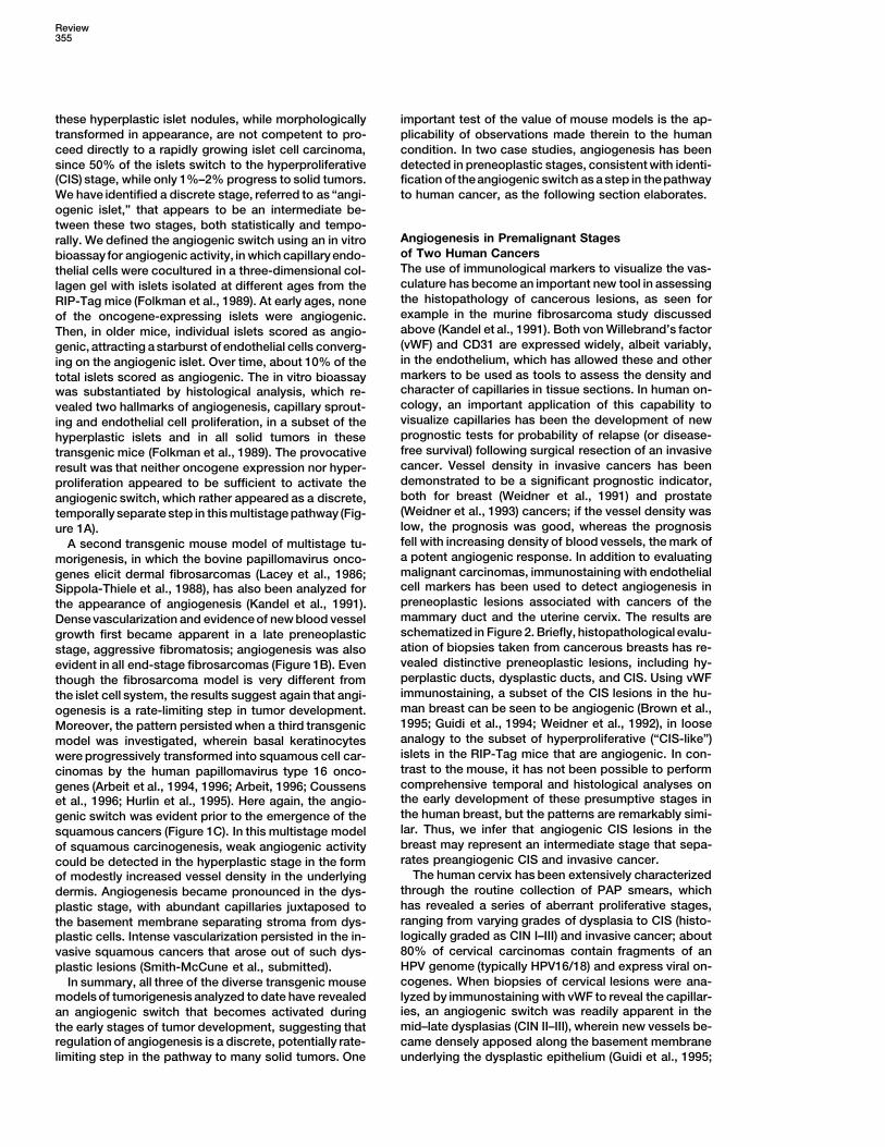
Figure 2. The Angiogenic Switch Can Be Visualized in Neoplastic Lesions Inferred to Be Progenitor Stages to Human Breast and CervicalCancer
(A) Breast cancers arise from the ductal epithelium of the breast; ductal lesions that are presumptive progenitors can be ordered into apathway of increasing aberrancy, beginning with hyperplasia and progressing to dysplasia and CIS. Of these, a subset of CIS lesions haveswitched on angiogenesis, as evidenced by abundant new capillaries, suggesting that angiogenic-CIS is an intermediate stage between CISand invasive cancer.(B) The squamous cervical epithelium evidences premalignant lesions graded as cervical intraepithelial neoplasia I–III, which are inferred torepresent a pathway to cervical cancer. A modest increase in new vessel density is evident in CIN I/II lesions, while CIN III lesions (analogousto advanced dysplasia/CIS) show abundant new vessels, indicative of the angiogenic switch from vascular quiescence to sustained neovascu-larization.
Smith-McCune and Weidner, 1994). An initial mild in- Therefore, having argued that regulation of the switchcrease in vessel density has been detected in the early is important and potentially rate-limiting for initial tumordysplastic (CIN I) stage (Guidi et al., 1995; Smith- development as well as for the expansion and metasta-McCune et al., submitted). This pattern is remarkably sis of an established tumor, we now wish to discuss thesimilar to that seen in the epidermis of K14–HPV16 emerging picture of how the angiogenic switch may betransgenic mice, which presents a similar multistage controlled.pathway in another squamous epithelium (of the skin)under the influence of the HPV16 oncogenes. Thus, we
Emerging Mechanisms of the Angiogenic Switchinfer that the angiogenic switch is a very early event inThe Angiogenic Inducersthe pathways to invasive squamous cell cancers, per-The observation that tumors could be implanted eitherhaps occurring in two regulatory phases, of mild andinto an avascular region, such as the cornea, or on aintense neovascularization. Clues from cell culture bio-characteristically vascularized surface, such as theassays also support this notion that the switch can havechick chorioalantoic membrane, and in each case elicitseveral “on” settings, which progressively ratchet upthe ingrowth of new capillaries, suggested that tumorsthe intensity of neovascularization.released diffusible activators of angiogenesis that couldThe pattern of new blood vessel growth, describedsignal a quiescent vasculature to begin capillary sprout-here in five cancers spanning two species and five celling. This hypothesis motivated a search for so-calledtypes, argues, first, that the angiogenic switch is a dis-tumor angiogenesis factors. Indeed, inducers of angio-crete process, subject to specific regulatory controls;genesis have been identified. A number of in vitro andand second, that the switch is an essential part of thein vivo bioassays have been developed to mimic thephenotypic repertoire that characterizes a successfulcomplex process of angiogenesis (reviewed by Cockerilltumor. Figure 3 presents a histological picture of theet al., 1995). Among these, two assays in particular haveangiogenic switch in these five models. A similar stage-been widely used to screen for angiogenic regulatoryspecificswitch is evident duringdevelopment of cutane-
ous melanoma in humans (reviewed by Rak et al., 1995). factors, each mimicking an aspect of angiogenesis;
Review357
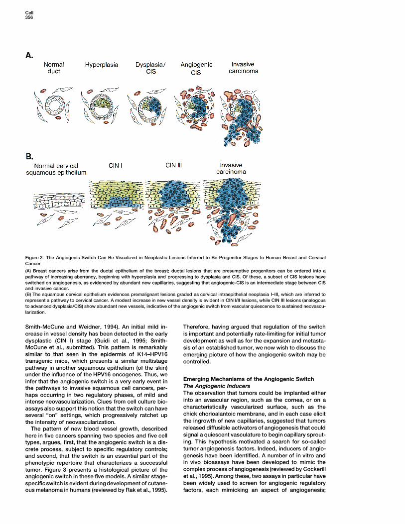
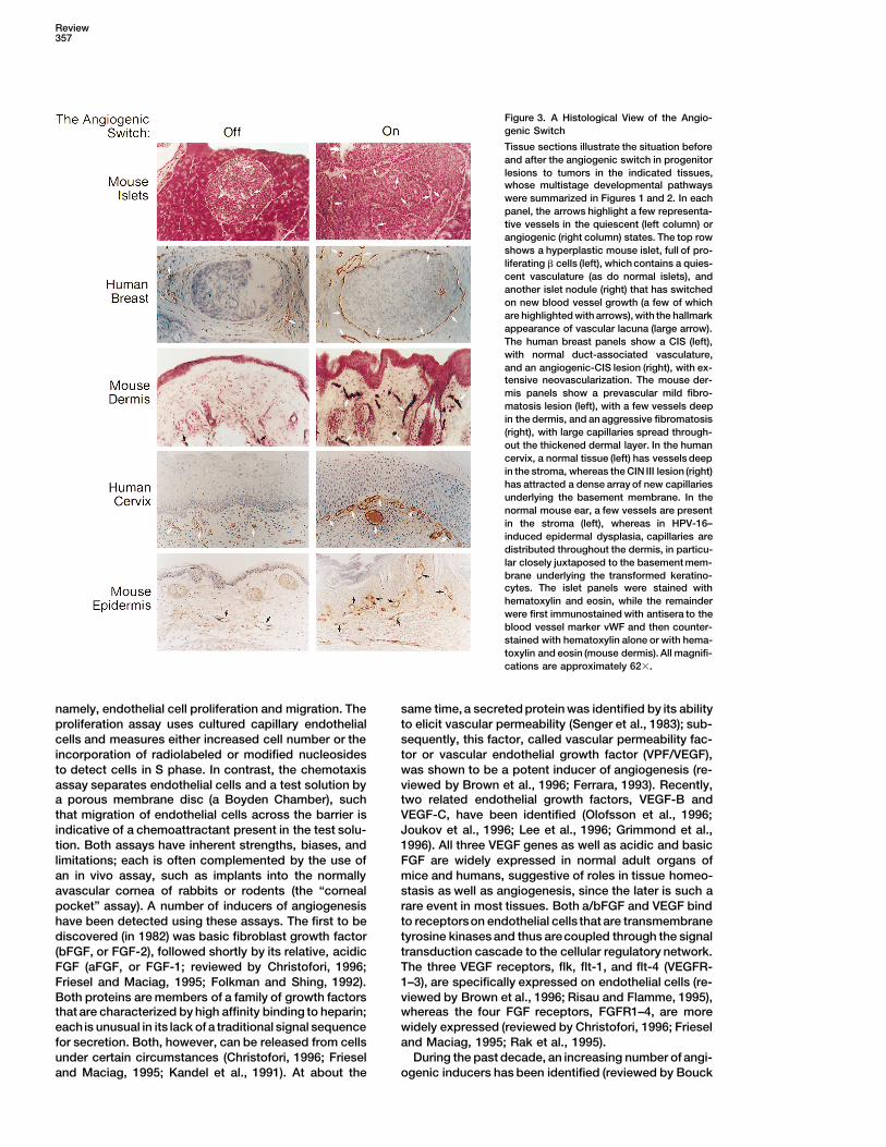
Figure 3. A Histological View of the Angio-genic Switch
Tissue sections illustrate the situation beforeand after the angiogenic switch in progenitorlesions to tumors in the indicated tissues,whose multistage developmental pathwayswere summarized in Figures 1 and 2. In eachpanel, the arrows highlight a few representa-tive vessels in the quiescent (left column) orangiogenic (right column) states. The top rowshows a hyperplastic mouse islet, full of pro-liferating b cells (left), which contains a quies-cent vasculature (as do normal islets), andanother islet nodule (right) that has switchedon new blood vessel growth (a few of whichare highlighted with arrows), with the hallmarkappearance of vascular lacuna (large arrow).The human breast panels show a CIS (left),with normal duct-associated vasculature,and an angiogenic-CIS lesion (right), with ex-tensive neovascularization. The mouse der-mis panels show a prevascular mild fibro-matosis lesion (left), with a few vessels deepin the dermis, and an aggressive fibromatosis(right), with large capillaries spread through-out the thickened dermal layer. In the humancervix, a normal tissue (left) has vessels deepin the stroma, whereas the CIN III lesion (right)has attracted a dense array of new capillariesunderlying the basement membrane. In thenormal mouse ear, a few vessels are presentin the stroma (left), whereas in HPV-16–induced epidermal dysplasia, capillaries aredistributed throughout the dermis, in particu-lar closely juxtaposed to the basement mem-brane underlying the transformed keratino-cytes. The islet panels were stained withhematoxylin and eosin, while the remainderwere first immunostained with antisera to theblood vessel marker vWF and then counter-stained with hematoxylin alone or with hema-toxylin and eosin (mouse dermis). All magnifi-cations are approximately 623.
namely, endothelial cell proliferation and migration. The same time, a secreted protein was identified by its abilityto elicit vascular permeability (Senger et al., 1983); sub-proliferation assay uses cultured capillary endothelial
cells and measures either increased cell number or the sequently, this factor, called vascular permeability fac-tor or vascular endothelial growth factor (VPF/VEGF),incorporation of radiolabeled or modified nucleosides
to detect cells in S phase. In contrast, the chemotaxis was shown to be a potent inducer of angiogenesis (re-viewed by Brown et al., 1996; Ferrara, 1993). Recently,assay separates endothelial cells and a test solution by
a porous membrane disc (a Boyden Chamber), such two related endothelial growth factors, VEGF-B andVEGF-C, have been identified (Olofsson et al., 1996;that migration of endothelial cells across the barrier is
indicative of a chemoattractant present in the test solu- Joukov et al., 1996; Lee et al., 1996; Grimmond et al.,1996). All three VEGF genes as well as acidic and basiction. Both assays have inherent strengths, biases, and
limitations; each is often complemented by the use of FGF are widely expressed in normal adult organs ofmice and humans, suggestive of roles in tissue homeo-an in vivo assay, such as implants into the normally
avascular cornea of rabbits or rodents (the “corneal stasis as well as angiogenesis, since the later is such arare event in most tissues. Both a/bFGF and VEGF bindpocket” assay). A number of inducers of angiogenesis
have been detected using these assays. The first to be to receptorson endothelial cells that are transmembranetyrosine kinases and thus arecoupled through the signaldiscovered (in 1982) was basic fibroblast growth factor
(bFGF, or FGF-2), followed shortly by its relative, acidic transduction cascade to the cellular regulatory network.The three VEGF receptors, flk, flt-1, and flt-4 (VEGFR-FGF (aFGF, or FGF-1; reviewed by Christofori, 1996;
Friesel and Maciag, 1995; Folkman and Shing, 1992). 1–3), are specifically expressed on endothelial cells (re-viewed by Brown et al., 1996; Risau and Flamme, 1995),Both proteins are members of a family of growth factors
that are characterized by high affinity binding to heparin; whereas the four FGF receptors, FGFR1–4, are morewidely expressed (reviewed by Christofori, 1996; Frieseleach is unusual in its lack of a traditional signal sequence
for secretion. Both, however, can be released from cells and Maciag, 1995; Rak et al., 1995).During the past decade, an increasing number of angi-under certain circumstances (Christofori, 1996; Friesel
and Maciag, 1995; Kandel et al., 1991). At about the ogenic inducers has been identified (reviewed by Bouck
Cell358
et al., 1996; Folkman, 1995a, 1995b). Concurrently, how- as astrocytes (Van Meir et al., 1994), to controlangiogen-esis-inhibitory activities distinct from TSP. The bottomever, a pattern has emerged, in that bFGF and VEGF
are commonly expressed in a wide variety of human line of these experiments is, first, that endogenous angi-ogenesis inhibitors can serve to counteract inducer sig-and animal cancers; moreover, bFGF (and now VEGF)
is being detected at elevated levels in the urine and nals to grow new capillaries; and second, that theseangiogenesis inhibitors can be controlled by tumor sup-serum of a significant fraction of tumor-bearing patients
(as discussed in Folkman, 1995a, 1995b). Interestingly, pressor genes, consistent with the functional definitionof such genes as interfering with the tumor phenotype,VEGF and bFGF have been shown to synergize using
in vitro angiogenesis assays (Pepper et al., 1992; Goto et of which angiogenesis is a key component.A New Paradigm of Cryptic Angiogenesisal., 1993), indicating that they can serve complementary
functions, consistent with their common association in Inhibitors within Larger ProteinsThe fact that the endothelium is quiescent for long peri-tumors. Indeed, the tumor development pathways of all
three transgenic mouse models presented in Figure 1 ods and yet can be induced to sprout new capillariesin a matter of hours in response to, for example, aevidence an involvement of VEGF and either aFGF,
bFGF, or both. Thus, these two classes of angiogenic wound, has long suggested that angiogenesis regula-tors might be stored for expedient use. In addition, oneinducer certainly represent a frequent component of the
angiogenic switch. Notwithstanding this consensus, the can expect that regulators will be synthesized for persis-tence of an angiogenic response. It is known that thediversity and number of angiogenic inducers identified
to date argues that there will also prove to be tissue- FGFs and other growth and angiogenic factors can besequestered in the extracellular matrix of many cellspecific and perhaps transformation-specific regulators
associated with particular cancers and with the normal types, including endothelial cells, presumably to be re-leased by proteolytic degradation of the matrix (Bairdphysiological regulation of angiogenesis. Moreover,
convergent on this perspective regarding angiogenic and Ling, 1987; Folkman et al., 1988). It now appearsthat angiogenesis inhibitors are also stored, but in ainducers has been the realization that there is an equally
important component to the switch, one governed by much different fashion, as cryptic parts of larger mole-cules that are not themselves inhibitors of angiogenesis.negative regulatory factors, called angiogenesis inhib-
itors. The prototype came from the discovery that a 29 kDafragment of fibronectin inhibited endothelial cell prolifer-The Angiogenic Inhibitors
The firstclues to the existenceof endogenous angiogen- ation in vitro (Homandberg et al., 1985). Notably, theintact fibronectin molecule, an abundant component ofesis inhibitors came with the observations that a inter-
feron (Brouty-Boye and Zetter, 1980; Sidky and Borden, the circulatory system, was not itself an inhibitor. Subse-quently, a fragment of prolactin, the 16 kDa fragment,1987) and platelet factor-4 (Taylor and Folkman, 1982;
Sato et al., 1990; Sharp et al., 1990) could inhibit endo- was shown to be an inhibitor of endothelial cells,whereas the intact molecule was not (Clapp et al., 1993;thelial cell chemotaxis and proliferation, respectively.
But the significance of negative regulators of angiogen- Ferrera et al., 1991). More recently, a potent inhibitor ofangiogenesis, called angiostatin, has been identified asesis first became apparent through a series of experi-
ments by Bouck and colleagues (Good et al., 1990; Ras- a fragment of plasminogen (O’Reilly et al., 1994). Circu-lating angiostatin is able to maintain the dormancy oftinejad et al., 1989). A nontumorigenic hamster cell line
became tumorigenic concomitant with a mutation that metastases and primary tumors by blocking blood ves-sel growth, resulting in small nests of tumor cells thatinactivated a tumor suppressor gene. Comparison of
cell lines containing or missing the suppressor gene cycle through proliferation and apoptosis, restrained bytheir inability to induce angiogenesis to support theirrevealed that the nontumorigenic line released high lev-
els of a potent angiogenesis inhibitor, bothof endothelial growth (Holmgrenet al., 1995; O’Reilly et al., 1994, 1996).The fact that abundant components of the circulatorychemotaxis in the Boyden Chamber assay and of in vivo
neovascularization using the corneal pocket assay; by system such as fibronectin and plasminogen can beconverted into potent angiogenic factors suggests acomparison, the tumorigenic cell lines had much lower
levels of inhibitor (Rastinejad et al., 1989). The inhibitory new form of regulation, whereby proteases specificallyrelease 29 kDa fibronectinor angiostatin from their intactactivity was purified and shown to be a truncated form
of thrombospondin-1 (TSP-1), a secreted glycoprotein. parental molecules to limit an angiogenic response, forexample in the transient wound healing process. Re-Subsequently, the intact TSP-1 molecule was shown to
be an angiogenesis inhibitor (Good et al., 1990), as were cently, platelet factor-4, itself a weak angiogenesis in-hibitor, has been shown to contain a fragment that issmall peptides contained within it (Tolsma et al., 1993).
Further, TSP-1 has been shown to be expressed at high 50 times more potent at inhibiting angiogenesis (Guptaet al., 1995). The pattern of protease fragments as angio-levels in a number of normal rodent and human cells
and at appreciably lower levels in many tumor cells. genesis inhibitors continues to expand to other mole-cules, including the propeptides of type 1 collagen (Tol-Provocatively, TSP-1 has recently been shown to be
regulated by the wild-type p53 tumor suppressor protein sma et al., 1993) and a peptide fragment of epidermalgrowth factor (Nelson et al., 1995).in fibroblasts (Dameron et al., 1994) and mammary epi-
thelial cells (Volpert et al., 1995), such that upon loss of Thus, a paradigm is emerging which suggests that aclass of angiogenesis inhibitors is contained within otherp53 function in their transformed derivatives, the levels
of this angiogenesis inhibitor dropped precipitously. proteins that are not themselves inhibitors. The capabil-ity to release inhibitor fragments from storage as crypticRestoration of p53 function upregulated TSP-1 and im-
paired the angiogenic capability of the tumor cells. Fur- segments of abundant proteins may contribute to main-taining the normal quiescence of the vasculature andthermore, p53 has been shown in other cell types, such
Review359
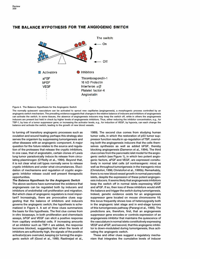
Figure 4. The Balance Hypothesis for the Angiogenic Switch
The normally quiescent vasculature can be activated to sprout new capillaries (angiogenesis), a morphogenic process controlled by anangiogenic switch mechanism. The prevailing evidence suggests that changes in the relative balance of inducers and inhibitors of angiogenesiscan activate the switch. In some tissues, the absence of angiogenesis inducers may keep the switch off, while in others the angiogenesisinducers are present but held in check by higher levels of angiogenesis inhibitors. Thus, either reducing the inhibitor concentration, e.g., forTSP-1, by loss of a tumor suppressor gene; or increasing the activator levels, e.g., for induction of VEGF, by hypoxia, can each change thebalance and activate the switch, leading to the growth of new blood vessels.
to turning off transitory angiogenic processes such as 1989). The second clue comes from studying humantumor cells, in which the restoration of p53 tumor sup-ovulation and wound healing; perhaps this strategy also
serves the organism by suppressing tumorigenesis and pressor function results in up-regulation of TSP, overrul-ing both the angiogenesis inducers that the cells them-other diseases with an angiogenic component. A major
question for the future relates to the source and regula- selves synthesize as well as added bFGF, therebyblocking angiogenesis (Dameron et al., 1994). The thirdtion of the proteases that release the cryptic inhibitors.
In one case, that of angiostatin, certain clones of Lewis clue comes fromthe pancreatic islet model for theangio-genic switch (see Figure 1), in which two potent angio-lung tumor paradoxically induce its release from circu-
lating plasminogen (O’Reilly et al., 1994). Beyond that, genic factors, aFGF and VEGF, are expressed consitu-tively in normal islet cells (of nontransgenic mice) asit is not clear what cell types normally serve to release
cryptic inhibitors and under what circumstances. Eluci- well as throughout tumorigenesis in the transgenic mice(Christofori, 1996; Christofori et al., 1995b). Remarkably,dation of mechanisms and regulation of cryptic angio-
genic inhibitor release could well present therapeutic there is nonew bloodvessel growth innormal pancreaticislets, despite the expression of these potent angiogen-opportunities.
The Balance Hypothesis for the Angiogenic Switch esis inducers. It seems likely that angiogenesis inhibitorskeep the switch off in normal islets expressing VEGFThe above sections have summarized the evidence that
angiogenesis can be regulated both by inducers and and aFGF. If so, then loss of these inhibitors would shiftthe balance and trigger the switch during tumorigenesis.inhibitors of endothelial cell proliferation and migration.
But which class of angiogenic regulators is most impor- Indeed, genetic evidence exists for an angiogenesissuppressor gene located on mouse chromosome 16;tant? Increasingly, there are provocative clues sug-
gesting that the balance of inhibitors and inducers this locus frequently shows loss of heterozygosity bothin the angiogenic islet stage and in end-stage tumorsgoverns the angiogenic switch; the hypothesis is sche-
matized in Figure 4. A set of three clues summarizes of this tumorigenesis pathway (Parangi et al., 1995). Thepredictions are, therefore, first, that an angiogenesisthe basis for this hypothesis. The first clue comes from
in vitro bioassays. In both proliferation and chemotaxis suppressor gene encodes or controls expression of anangiogenesis inhibitor that maintains the quiescence ofassays, bFGF and VEGF can elicit a positive response
from capillary endothelial cells. If increasing amounts the vasculature innormal islets constitutivelyexpressingVEGF and aFGF; and second, that this angiogenic inhibi-of an inhibitor such as TSP-1 are added, the response
becomes blocked, suggesting that when the levels of tor is down-modulated during tumorigenesis, thus acti-vating the angiogenic switch.inhibitors are sufficiently high, the signals of the positive
activator(s) are overruled, keeping (or turning) the angio- These and other clues suggest a regulatory mecha-nism that integrates the cumulative levels of inducergenic switch off (Good et al., 1990; Rastinejad et al.,
Cell360
and inhibitor signals to maintain the endothelial cell in Apoptosis, Clinical Applications, and the Goalto Control the Switchalternative states of quiescence or angiogenesis. Thus,The growing appreciation of the fact that angiogenesischanges in the balance of positive and negative signalsis an integral component of the tumor phenotype hasmediate the angiogenic switch (Figure 4), a point Bouckfueled considerable efforts to identify angiogenesis in-and colleagues have also made recently (Bouck et al.,hibitors that can be used as part of therapeutic strate-1996). A net balance of inhibitors over activators wouldgies to interfere with tumor growth and metastasis. Inmaintain the switch in the off position, whereas a shiftaddition to endogenous inhibitors such as interferon a/bto an excess of activating stimuli would turn on angio-and platelet factor-4, synthetic compounds have beengenesis. There are now ample tools to address this hy-identified using cell culture assays. These include thepothesis, and indeed, new observations seem likely tofungal-derived compound AGM1470 (TNP470), thalido-refine it. For example, aFGF and bFGF are selectivelymide, a number of metalloproteinase inhibitors, and oth-exported by a number of tumor cell lines but not byers (reviewed by Auerbach and Auerbach, 1994; Bouckmany normal cells (Christofori, 1996; Friesel and Maciag,et al., 1996; Folkman, 1995a). Currently, there are nine1995; Kandel et al., 1991), suggesting that another fea-compounds in clinical trials as angiogenesis inhibitors,ture of the switch mechanism may be the capabilitywith perhaps two dozen others in various stages of re-to sequester angiogenesis inducers such that despitesearch and development as potential antiangiogenic
being synthesized they are unavailable to stimulate angi-therapeutics.
ogenesis until the sequestration is relaxed. In regard toLooking beyond this first generation of angiogenesis
endogenous inhibitors, a major objective will be to as-inhibitors identified in cell-based assays, we envision
sess the proteolytic mechanisms and the generality by the capability to control the angiogenic switch throughwhich cells can process abundant circulatory proteins specific knowledge both of the regulatory mechanismsuch as fibrinogen and plasminogen to release cryptic underlying the switch and the cellular details of the pro-angiogenesis inhibitors. Local processing of circulatory cess of new blood vessel morphogenesis. Oneapproachproteins may serve to produce levels of inhibitory activ- will likely involve altering the balance of endogenousity sufficient to maintain the angiogenic switch in the off inhibitors and activators by controlling synthesis or af-position. If the endothelium is normally bathed in locally fecting sequestration and release. The beginnings ofor systemically processed cryptic inhibitors, the regula- this approach are apparent. Monoclonal antibodies thattory balance could be shifted and the angiogenic switch bind and block the actions of VEGF are in development,activated either by reducing the rates of inhibitor pro- as are compounds that interfere with signal transductioncessing or by increasing the levels of the inducers seen from its receptors. Knowledge of the morphogenesis ofat the endothelial cell, for example by up-regulating new vessels is also providing opportunities. For exam-VEGF synthesis or by releasing sequestered FGF. It ple, sprouting capillaries have been shown to expressseems likely that the alternative strategies of increasing a specific type of cell–matrix interaction molecule, theactivator levels or reducing inhibitor levels to activate integrins avb3 or avb5; abrogation of the contacts of thesethe angiogenic switch will each be utilized by different integrins to matrix evokes programmed cell death
(apoptosis) of the new endothelial cells and dramaticallytissues according to their physiological characteristics.impairs neovascularization of tumors (Brooks et al.,Thus, tissues such as the epidermis, which are well1994; Friedlander et al., 1995). Both antiintegrin antibod-separated from the vasculature by a basement mem-ies and interfering RDG peptides are in developmentbrane and a stromal support, may favor mechanismsas candidates for blocking capillary morphogenesis viathat increase inducer levels to elicit angiogenesis. Ininduction of apoptosis.contrast, endocrine organs such as the pancreatic islets
One emerging biological principle is the interrelation-that are intimately vascularized may utilize negative reg-ship between tumor angiogenesis and modulation ofulators to maintain a quiescent endothelium and there-cell death. Classically, it has been apparent in certainfore require down-regulation of inhibitor production totumors that necrotic death accompanies inadequateactivate the angiogenic switch.blood flow; for example, necrosis has been reported inThe realization that increasing numbers of both angio-glioblastomas and attributed to hypoxia resulting fromgenic activators and inhibitors are being discoveredinadequate vascularization. In turn, this clue led to the
(currently, there are at least a dozen of each), mostdiscovery that hypoxia can induce VEGF synthesis
with demonstrable activity directly on endothelial cells,(Shweike et al., 1992; Shweiki et al., 1995; Stein et al.,
raises an interesting question: how are all of these com- 1995). One study in which VEGF receptor function wasplementary and opposing signals integrated by the sig- inhibited in a transplantable glioblastoma with a domi-nal transduction network in endothelial cells, so as to nant-negative VEGF receptor resulted in impaired tumorcontrol the complex dynamics of maintaining quies- growth, which was ascribed to widespread necrotic cellcence, activating angiogenesis, and then completing death (Millauer et al., 1994). In addition, however, todifferentiation to produce functional new capillaries? necrotic cell death, it is now becoming clear that rapidBeyond the manifest goal of further clarifying the regula- expansion of a tumor mass can be significantly affectedtory mechanisms of the angiogenic switch and the com- by the incidence of apoptosis.plex process of angiogenesis, an important opportunity To exemplify the interplay of apoptosis and angiogen-now exists to apply this new knowledge in clinical oncol- esis, consider the transplantable Lewis lung carcinomaogy to restrict angiogenesis by altering the balance of model (Figure 5A). Lewis lung tumors growing subcuta-
neously in mice seed distant metastases in the lung, butangiogenic regulators.
Review361
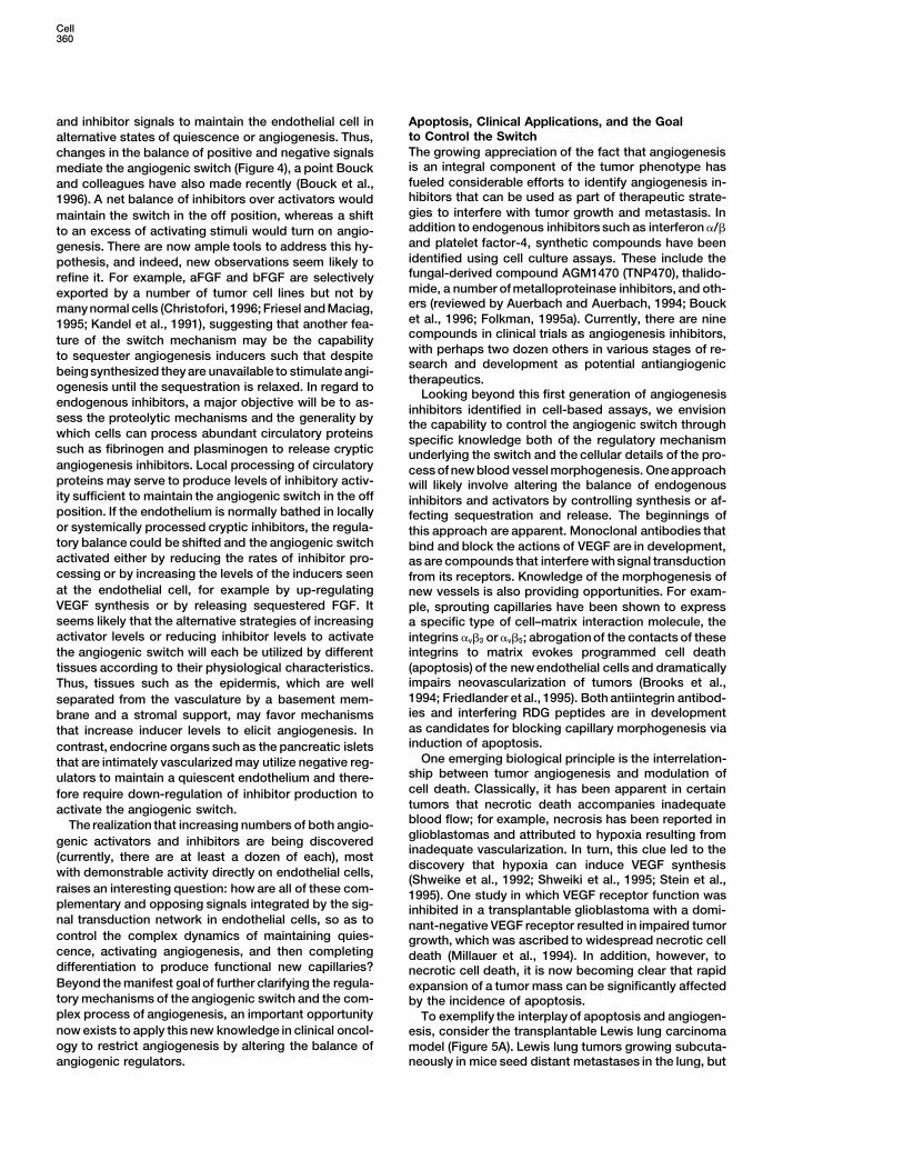
Figure 5. Tumor Growth Can Be Restricted by Apoptosis of Tumor Cells Elicited by Inhibition of Angiogenesis
(A) Liver metastases from a subcutaneous growth of Lewis lung carcinoma will grow explosively in the absence (i) of the primary tumor, whichproduces a circulating endothelial inhibitor (angiostatin). The apoptotic index is low and the proliferation index very high. When the primarytumor is present (ii), resulting in high serum levels of angiostatin, growth of the metastases is impaired, and the apoptotic index is markedlyincreased in the metastatic nodules. If the primary tumor generating angiostatin is absent, but instead the angiogenesis inhibitory drugAGM1470 (TNP470) is inoculated daily (iii), metastatic growth is again restricted, concomitant with increased apoptosis. Neither angiogenesisinhibitor affects the proliferation index of the metastatic tumor cells (shown); in both cases, the density of new capillaries is significantlyreduced (data not shown).(B) Transgenic mice expressing the SV-Tag oncogene in pancreatic b cells develop islet cell carcinomas with a high proliferation index anda low incidence of apoptosis (i). In the absence of the survival factor IGF-II, tumor volume is dramatically impaired, with a concordant 53
increase in apoptotic index (ii), demonstrating that apoptosis can restrain primary tumor growth. When the transgenic mice are treated witha regimen of angiogenesis inhibitors, including AGM1470, tumor growth is again impaired, also with a concordant increase in the incidenceof apoptosis (iii), indicating that the reduced vascularization (data not shown) elicits tumor cell apoptosis. Again, tumor cell proliferation isnot affected by inhibition of angiogenesis.
these remain dormant as small nodules of tumor cells and two additional murine tumors in immunodeficientand syngeneic mice, respectively; tumors in the angios-that do not elicit angiogenesis, as a consequence of the
presence in the blood of high levels of the angiogenesis tatin-treated mice also had comparable proliferation in-dices to saline-treated controls and markedly increasedinhibitor angiostatin, which is released from circulating
plasminogen by the primary tumor (O’Reilly et al., 1994.) frequencies of tumor cell apoptosis (O’Reilly et al., 1996).In a second example, that of islet cell carcinomas(The reason that angiostatin does not inhibit angiogen-
esis in the primary tumor is not clear but is thought to in RIP-Tag transgenic mice (Figure 1A), the end-stagetumors evidence a low incidence of apoptosis, whichreflect a local balance in favor of angiogenesis inducers.)
Examination of the latent metastases revealed a high appears to be maintained both by intrinsic factors andby intense vascularization of tumors (Figure 5B). Whenincidence of S-phase cells (30%) and yet little increase
in metastatic tumor mass. The explanation appears to the embryonic growth/survival factor IGF-II, which isactivated in this tumorigenesis pathway, was abrogatedbe that the latent metastases evidence a high incidence
of apoptosis (8%), balancing rapid cell division with cell using gene-knockout mice, the tumor volume was dra-matically decreased (Christofori et al., 1994, 1995a; Naikdeath (Holmgren et al., 1995). Release of the angiogenic
blockade (by removing the primary tumor) allowed angi- et al., 1994). The S-phase incidence was comparablyhigh (more than 20%) in both normal and IGF-II–deficientogenesis and rapid expansion of lung metastases (Hol-
mgren et al., 1995; O’Reilly et al., 1994). The synthetic tumors (Naik et al., 1994); the difference was in theapop-totic incidence, which rose 5-fold in the IGF-II–null tu-angiogenesis inhibitor AGM1470 could similarly hold la-
tent metastases in place; again, the pattern was one of mors (Christofori et al., 1994). Thus, IGF-II produced bythe tumor cells is implicated in the control of apoptosis.high incidence both of cells in S phase and of cells
undergoing apoptosis (Holmgren et al., 1995). Recently, Yet the low level of apoptosis seen in the islet cell tumorsis also highly dependent on persistent angiogenesis.purified angiostatin has been shown to impair signifi-
cantly the growth of three primary human carcinomas When the RIP-Tag mice were treated with a regimen of
Cell362
Referencesangiogenesis inhibitors (interferon a, minocycline, andAGM1470), tumor growth was dramatically impaired and
Adams, J.M., and Cory,S. (1991). Transgenic models of tumor devel-vessel density significantly reduced (Parangi et al.,opment. Science 254, 1161–1167.
1996). Again, the same pattern was apparent: theArbeit, J.M. (1996). Transgenic models of epidermal neoplasia and
S-phase incidence remained high, while the apoptotic multistage carcinogenesis. Cancer Surv., 26, 7–34.incidence increased significantly in the small tumors
Arbeit, J., Munger, K., Howley, P., and Hanahan, D. (1994). Progres-arising in mice treated with angiogenesis inhibitors. sive squamous epithelial neoplasia in K14–HPV16 transgenic mice.
Together, these two models implicate the vasculature J. Virol. 68, 4358–4368.as a paracrine regulator of apoptosis. Therefore, in addi- Arbeit, J.M., Olson, D., and Hanahan, D. (1996). Upregulation oftion to its long-recognized association with necrotic cell fibroblast growth factors and their receptors during multistage epi-
dermal carcinogenesis in K14–HPV16 transgenic mice. Oncogene,death, inadequate vascularization can also elicit tumorin press.cell apoptosis. And recall that lack of integrin signalingAuerbach, W., and Auerbach, R. (1994). Angiogenesis inhibition: acan produce endothelial cell apoptosis. This convergentreview. Pharmacol. Ther. 63, 265–311.realization, that apoptosis can significantly modulateBaird, A., and Ling, N. (1987). Fibroblast growth factors are presenttumor growth by its occurrence both in tumor cells andin the extracellular matrix produced by endothelilal cells in vitro:in thesupporting endothelium, opens up new opportuni-implications for a role of heparinase-like enzymes in the neovascular
ties for the design of anticancer therapeutics that en- response. Biochem. Biophys. Res. Commun. 142, 428–435.hance apoptosis.
Bouck, N., Stellmach, V., and Hsu, S. (1996). How tumors becomeIn conclusion, the horizons of angiogenesis research angiogenic. Adv. Cancer Res. 69, in press.
include several opportunities in regard to cancer. First, Brem, S., Brem, H., Folkman, J., Finkelstein, D., and Patz, A. (1976).angiogenesis inhibitors seem likely to become an impor- Prolonged tumor dormancy by prevention of neovascularization intant component of therapeutic strategies aimed at inva- the vitreous. Cancer Res. 36, 2807–2812.
sive metastatic tumors. Second, as methods for early Brooks, P.C., Montgomery, A.M.P., Rosenfeld, M., Reisfeld, R.A.,Hu, T., Klier, G., and Cheresh, D.A. (1994). Integrin avb3 antagonistsdetection of certain classes of cancer improve, it maypromote tumor regression by inducing apoptosis of angiogenicbecome possible to interfere with initial tumor develop-blood vessels. Cell 79, 1157–1164.ment by blocking the angiogenic switch that precedesBrouty-Boye, D., and Zetter, B.R. (1980). Inhibition of cell motilitythe progression to invasive cancer, for example at theby interferon. Science 208, 516–518.CIS stage. Third, a major issue in oncology is the devel-Brown, L.F., Berse, B., Jackman, R.W., Tognazzi, K., Guidi, A.J.,opment of effective adjuvant therapies for use followingDvorak, H.F., Senger, D.R., Connolly, J.L., and Schnitt, S.J. (1995).resection of a primary tumor. In many cases, cancerExpression of vascular permeability factor (vascular endothelial
cells, either local or at remote sites, are left behind with a growth factor) and its receptors in breast cancer. Hum. Pathol. 26,potential for progression to produce a recurrent cancer; 86–91.antiangiogenic therapy could restrain such lesions or Brown, L.F., Detmar, M., Claffey, K., Nagy, J.A., Feng, D., Dvorak,even drive them into apoptotic catastrophe. For all of A.M., and Dvorak, H.F. (1996). Vascular permeability factor/vascular
endothelial growth factor: a multifunctional angiogenic cytokine. Inthese applications, more effective angiogenesis inhibi-Control of Angiogenesis, I.D. Goldberg and E. Rosen, eds. (Berlin:tors will likely need to be developed; knowledge of mo-Birkhauser Verlag), in press.lecular and cellular mechanism will certainly facilitateChristofori, G. (1996). The role of fibroblast growth factors in tumourthat process. Moreover, as this review has documented,progression and angiogenesis. In Tumour Angiogenesis, R. Bicknell,
the complementarity of mouse models with human dis- C.E. Lewis, and N. Ferrara, eds. (Oxford: Oxford University Press),eases will continue to afford powerful opportunities. For in press.example, therapeutic trials using mouse models are Christofori, G., Naik, P., and Hanahan, D. (1994). Insulin-like growthamenable to expedient combinatorial testing, as exem- factor II is focally up-regulated and functionally involved as a cofac-
tor in oncogene-induced tumorigenesis. Nature 369, 414–418.plified both by traditional tumor transplantation models(Teicher et al., 1992) and, more recently, by the use of Christofori, G., Naik, P., and Hanahan, D. (1995a). Deregulation of
both imprinted and expressed alleles of the IGF-II gene during b-celltransgenic models of de novo tumorigenesis (Parangitumorigenesis. Nature Genet. 10, 196–201.et al., 1996). There is reason to be optimistic that in theChristofori, G., Naik, P., and Hanahan, D. (1995b). Vascular endothe-foreseeable future, the initial angiogenic switch duringlial growth factor and its receptors, flt-1 and flk-1, are expressed intumorigenesis, as well as ongoing neovascularizationnormal pancreatic islets and throughout islet cell tumorigenesis.
in tumors, can be controlled and perhaps completely Mol. Endocrinol. 9, 1760–1770.curtailed, contributing thereby to more efficacious can-
Clapp, C., Martial, J.A., Guzman, R.C., Rentier-Delrue, F., andcer therapies. Weiner, R.I. (1993). The 16 kDaN-terminal fragment of human prolac-
tin is a potent inhibitor of angiogenesis. Endocrinol. 133, 1292–1299.
Cockerill, G.W., Gamble, J.R., and Vadas, M.A. (1995). Angiogenesis:Acknowledgments models and modulators. Int. Rev. Cytol. 159, 113–160.
Coussens, L.M., Hanahan, D., and Arbeit, J. (1996). Genetic predis-We wish to thank Jeff Arbeit, Karen Smith-McCune, Noel Weidner,position and parameters of malignant progression in K14–HPV16Ella Bossy-Wetzel, and Christine Jolicoeur for providing the tissuetransgenic mice. Am. J. Path., in press.
sections used to prepare Figure 3; Noel Bouck, Karen Smith-Dameron, K.M., Volpert, O.V., Tainsky, M.A., and Bouck, N. (1994).McCune, David Olson, Dowdy Jackson, and Jeff Arbeit for com-Control of angiogenesis in fibroblasts by p53 regulation of throm-ments on the manuscript; and Wendy Gee and Terry Schoop ofbospondin-1. Science 265, 1582–1584.BioMed Arts (San Francisco) for artwork. The work from the authors’
laboratories reviewed herein was supported by grants from the Na- Engerman, R.L., Pfaffenbacvh, D., and Davis, M.D. (1967). Cell turn-over of capillaries. Lab. Invest. 17, 738–743.tional Cancer Institute.
Review363
Ferrara, N. (1993). Vascular endothelial growth factor. Trends micrometastases: balanced proliferation and apoptosis in the pres-ence of angiogenesis suppression. Nature Med. 1, 149–153.Cardiovasc. Med. 3, 244–250.
Homandberg, G.A., Williams, J.E., Grant, D., Schumacher, B., andFerrera, N., Clapp, C., and Weiner, R.I. (1991). The 16 kDa fragmentEisenstein, R. (1985). Heparin-binding fragments of fibronectin areof prolactin specifically inhibits basal or fibroblast growth factor–potent inhibitors of endothelial cell growth. Am. J. Pathol. 120,stimulated growth of capillary endothelial cells. Endocrinol. 129,327–332.896–900.
Hurlin, P.J., Foley, K.P., Aver, D., Eisenman, R.N., Hanahan, D., andFolkman, J. (1995a). Tumor angiogenesis. In The Molecular BasisArbeit, J.M. (1995). Regulation of Myc and Mad during epidermalof Cancer, J. Mendelsohn, P.M. Howley, M.A. Israel, and L.A. Liotta,differentiation and HPV–associated tumorigenesis. Oncogene 11,eds. (Philadelphia: W.B. Saunders Co.), pp. 206–232.2487–2501.Folkman, J. (1995b). Clinical applications of research on angiogen-Joukov, V., Pasujola, K., Kaipainen, A., Chilov, D., Lahtinen, I., Kukk,esis. N. Engl. J. Med. 333, 1757–1763.E., Saksela, O., Kalkkinen, N., and Alitalo, K. (1996). A novel vascularFolkman, J., and Shing, Y. (1992). Angiogenesis. J. Biol. Chem. 267,endothelial growth factor, VEGF-C, is a ligand for the Flt-4 (VEGFR-3)10931–10934.and KDR (VEGFR-2) receptor tyrosinekinases. EMBO J. 15, 290–298.
Folkman, J., Klagsbrun, M., Sasse, J., Wadzinski, M., Ingber, D., andKandel, J., Bossy-Wetzel, E., Radvany, F., Klagsbrun, M., Folkman,Vlodavsky, I. (1988). A heparin-binding angiogenic protein—basicJ., and Hanahan, D. (1991). Neovascularization is associated withfibroblast growth factor—is stored within basement membrane. Am.a switch to the export of bFGF in the multistep development ofJ. Pathol. 130, 393–400.fibrosarcoma. Cell 66, 1095–1104.
Folkman, J., Watson, K., Ingber, D., and Hanahan, D. (1989). Induc-Lacey, M., Alpert, S., and Hanahan, D. (1986). The bovine papillo-tion of angiogenesis during the transition from hyperplasia to neo-mavirus genome elicits skin tumours in transgenic mice. Nature 322,plasia. Nature 339, 58–61.609–612.
Friedlander, M., Brooks, P.C., Shaffer, R.W., Kincaid, C.M., Varner,Lee, J., Gray, A., Yuan, J., Luoh, S.-M., Avraham, H., and Wood,J.A., and Cheresh, D.A. (1995). Definition of two angiogenic path-W.I. (1996). Vascular endothelial growth factor–related protein: aways by distinct av integrins. Science 270, 1500–1502.ligand and specific activator of the tyrosine kinase receptor Flt-4.
Friesel, R.E., and Maciag, T. (1995). Molecular mechanisms of angio- Proc. Natl. Acad. Sci. USA 93, 1988–1992.genesis: fibroblast growth factor signal. FASEB J. 9, 919–925.
Millauer, B., Shawver, L.K., Plate, K.H., Risau, W., and Ullrich, A.Frost, P., Hart, I., and Kerbel, R.S. (1995). Cancer and metastasis (1994). Glioblastoma growth inhibited in vivo by a dominant-negativereviews—transgenic mice. Cancer Metastasis Rev. 14, 77–161. Flk-1 mutant. Nature 367, 576–579.Gimbrone, M.A.J., Leapman, S.B., Cotran, R.S., and Folkman, J. Naik, P., Christofori, G., and Hanahan, D. (1994). Insulin-like growth(1972). Tumor dormancy in vivo by prevention of neovascularization. factor II is focally up-regulated and functionally involved as a secondJ. Exp. Med. 136, 261–276. signal for oncogene-induced tumorigenesis. In The Molecular Ge-Good, D.J., Polverini, P.J., Rastinejad, F., Le Beau, M.M., Lemons, netics of Cancer (Cold Spring Harbor, New York: Cold Spring HarborR.S., Frazier, W.A., and Bouck, N.P. (1990). A tumor suppressor– Symposia on Quantitative Biology), pp. 459–471.dependent inhibitor of angiogenesis is immunologically and func- Nelson, J., Allen, W.E., Scott,W.N., Bailie,J.R., Walker,B., McFerran,tionally indistinguishable from a fragment of thrombospondin. Proc. N.V., and Wilson, D.J. (1995). Murine epidermal growth factor (EGF)Natl. Acad. Sci. USA 87, 6624–6628. fragment (33–42) inhibits both EGF- and laminin-dependent endo-Goto, F., Goto, K., Weindel, K., and Folkman, J. (1993). Synergistic thelial cell motility and angiogenesis. Cancer Res. 55, 3772–3776.effects of vascular endothelial growth factor and basic fibroblast Olofsson, B., Pajusola, K., Kaipainen, A., von Euler, G., Joukov, V.,growth factor on the proliferation and cord formation of bovine Saksela, O., Orpana, A., Pettersson, R.F., Alitalo, K., and Eriksson,capillary endothelial cells within collagen gels. Lab. Invest. 69, U. (1996). Vascular endothelial growth factor B, a novel growth factor508–517. for endothelial cells. Proc. Natl. Acad. Sci. USA 93, 2576–2581.Grimmond, S., Lagercrantz, J., Drinkwater, C., Silins, G., Townson, O’Reilly, M.S., Holmgren, L., Shing, Y., Chen, C., Rosenthal, R.A.,S., Pollock, P., Gotley, D., Carson, E., Rakar, S., Nordenskjold, M., Moses, M., Lane, W.S., Cao, Y., Sage, E.H., and Folkman, J. (1994).Ward, L., Hayward, N., and Weber, G. (1996). Cloning and character- Angiostatin: a novel angiogenesis inhibitor that mediates the sup-ization of a novel human gene related to vascular endothelial growth pression of metastases by a Lewis lung carcinoma. Cell 79, 315–328.factor. Genome Res. 6, 124–131.
O’Reilly, M.S., Holmgren, L., Chen, C., and Folkman, J. (1996). Angi-Guidi, A.J., Fischer, L., Harris, J.R., and Schnitt, S.J. (1994). Mi- ostatin induces and sustains dormancy of human primary tumorscrovessel density and distribution in ductal carcinoma in situ of the in mice. Nature Med. 2, 689–692.breast. J. Natl. Cancer Inst. 86, 614–619.
Parangi, S., Dietrich, W., Christofori, G., Lander, E., and Hanahan,Guidi, A.J., Abu-Jawdeh, G., Berse, B., Jackman, R.W., Tognazzi, D. (1995). Tumor suppressor loci on mouse chromosomes 9 and 16K., Dvorak, H.F., and Brown, L.F. (1995). Vascular permeability factor are lost at distinct stages of tumorigenesis in a transgenic mouse(vascular endothelial growth factor) expression and angiogenesis model of islet cell carcinoma. Cancer Res. 55, 6071–6076.in cervical neoplasia. J. Natl. Cancer Inst. 87, 1237–1245.
Parangi, S., O’Reilly, M.S., Christofori, G., Holmgren, I., Grosfeld,Gupta, S.K., Hassel, T., and Singh, J.P. (1995). A potent inhibitor of J., Folkman, J., and Hanahan, D. (1996). Antiangiogenic therapy ofendothelial cell proliferation is generated by proteolytic cleavage transgenic mice impairs de novo tumor growth. Proc. Natl. Acad.of the chemokine platelet factor-4. Proc. Natl. Acad. Sci. USA 92, Sci. USA 93, 2002–2007.7799–7803.
Pepper, M.S., Ferrara, N., Orci, L., and Montessano, R. (1992). PotentHanahan, D. (1985). Heritable formation of pancreatic b cell tumors synergism between vascular endothelial growth factor and basicin transgenic mice expressing recombinant insulin/simian virus 40 fibroblast growth factor in the induction of angiogenesis in vitro.oncogenes. Nature 315, 115–321. Biochem. Biophys. Res. Commun. 189, 824–831.Hanahan, D. (1988). Dissecting multistep tumorigenesis in trans- Rak, J.W., St. Croix, B.D., and Kerbel, R.S. (1995). Consequencesgenic mice. Annu. Rev. Genet. 22, 479–519. of angiogenesis for tumor progression, metastasis, and cancer ther-Hanahan, D. (1989). Transgenic mice as probes into complex sys- apy. Anticancer Drugs 6, 3–18.tems. Science 246, 1265–1275. Rastinejad, F., Polverini, P.J., and Bouck, N.P. (1989). Regulation ofHobson, B., and Denekamp, J. (1984). Endothelial proliferation in the activity of a new inhibitor of angiogenesis by a cancer suppressortumors and normal tissues: continuous labeling studies. Br. J. Can- gene. Cell 56, 345–355.cer 49, 405–413. Risau, W. (1995). Differentiation of endothelium. FASEB J. 9,
926–933.Holmgren, L., O’Reilly, M.S., and Folkman, J. (1995). Dormancy of
Cell364
Risau, W., and Flamme, I. (1995). Vasculogenesis. Annu. Rev. CellDev. Biol. 11, 73–91.
Sato, Y., Abe, M., and Takaki, R. (1990). Platelet factor-4 blocks thebinding of basic fibroblast growth factor to the receptor and inhibitsthe spontaneous migration of vascular endothelial cells. Biochem.Biophys. Res. Commun. 172, 595–600.
Senger, D.R., Galli, S.J., Dvorak, A.M., Perruzzi, C.A., Harvey, V.S.,and Dvorak, H.F. (1983). Tumor cells secrete a vascular permeabilityfactor that promotes accumulation of ascites fluid. Science 219,983–985.
Sharp, R.J., Byers, H.R., Scott, C.F., Bauer, S.I., and Maione, T.E.(1990). Growth inhibition of murine melanoma and human coloncarcinoma by recombinant human PF-4. J. Natl. Cancer Inst. 82,848–853.
Shweike, D., Itin, A., Soffer, D., and Keshet, E. (1992). Vascularendothelial growth factor induced by hypoxia may mediate hypoxia-initiated angiogenesis. Nature 359, 843–845.
Shweiki, D., Neeman, M., Itin, A., and Keshet, E. (1995). Inductionof vascular endothelial growth factor expression by hypoxia and byglucose deficiency in multicell spheroids: implications for tumorangiogenesis. Proc. Natl. Acad. Sci. USA 92, 768–772.
Sidky, Y.A., and Borden, E.C. (1987). Inhibition of angiogenesis byinterferons: effects on tumor- and lymphocyte-induced vascular re-sponses. Cancer Res. 47, 5155–5161.
Sippola-Thiele, M., Hanahan, D., and Howley, P.M. (1988). Cell heri-table stages of tumor progression in transgenic mice harboring thebovine papillomavirus type 1 genome. Mol. Cell Biol. 9, 925–934.
Smith-McCune, K., andWeidner, N. (1994). Demonstration and char-acterization of the angiogenic properties of cervical dysplasia. Can-cer Res. 54, 800–804.
Stein, I., Neeman, M., Shweike, D., Itin, A., and Keshet, E. (1995).Stabilization of vascular endothelial growth factor mRNA by hypoxiaand hypoglycemia and coregulation with other ischemia-inducedgenes. Mol. Cell Biol. 15, 5363–5368.
Taylor, S., and Folkman, J. (1982). Protamine is an inhibitor of angio-genesis. Nature 297, 307–312.
Teicher, B.A., Sotomayor, E.A., and Huang, Z.D. (1992). Antiangio-genic agents potentiate cytotoxic cancer therapies against primaryand metastatic disease. Cancer Res. 52, 6702–6704.
Teitelman, G., Alpert, S., and Hanahan, D. (1988). Proliferation, se-nescence, and neoplastic progression of b cells in hyperplasticpancreatic islets. Cell 52, 97–105.
Tolsma, S.S., Volpert, O.V., Good, D.J., Frazier, W.A., Polverini, P.J.,and Bouck, N. (1993). Peptides derived from two separate domainsof the matrix protein thrombospondin-1 have antiangiogenic activ-ity. J. Cell Biol. 122, 497–511.
Van Meir, E.G., Polverini, P.J., Chazin, V.R., Su Huang, H.-J., deTribolet, N., and Cavenee, W.K. (1994). Release of an inhibitor ofangiogenesis upon induction of wild-type p53 expression in gli-oblastoma cells. Nature Genet. 8, 171–176.
Volpert, O.V., Stellmach, V., and Bouck, N. (1995). The modulationof thrombospondin and other naturally occurring inhibitors of angio-genesis during tumor progression. Breast Cancer Res. Treat. 36,119–126.
Weidner, N., Semple, J.P., Welch, W.R., and Folkman, J. (1991).Tumor angiogenesis and metastasis correlation in invasive breastcarcinoma. N. Engl. J. Med. 324, 1–8.
Weidner, N., Folkman, J., Pozza, F., Bevilaqua, P., Allred, E.N.,Moore, D.H., Meli, S., and Gasparini, G. (1992). Tumor angiogenesis:a new significant and independent prognostic indicator in early-stage breast carcinoma. J. Natl. Cancer Inst. 84, 1875–1887.
Weidner, N., Carroll, P.R., Flax, J., Blumenfeld, W., and Folkman, J.(1993). Tumor angiogenesis correlates with metastasis in invasiveprostate carcinoma. Am. J. Pathol. 143, 401–409.