Embed Size (px)
Citation preview
SANDIA REPORT
SAND2004-5283 Unlimited Release Printed November 2004 Patterning Quantum Dot Arrays using DNA Replication Principles
George D. Bachand and Kevin K. Crown
Prepared by Sandia National Laboratories Albuquerque, New Mexico 87185 and Livermore, California 94550 Sandia is a multiprogram laboratory operated by Sandia Corporation, a Lockheed Martin Company, for the United States Department of Energy’s National Nuclear Security Administration under Contract DE-AC04-94AL85000. Approved for public release; further dissemination unlimited.
Issued by Sandia National Laboratories, operated for the United States Department of Energy by Sandia Corporation.
NOTICE: This report was prepared as an account of work sponsored by an agency of the United States Government. Neither the United States Government, nor any agency thereof, nor any of their employees, nor any of their contractors, subcontractors, or their employees, make any warranty, express or implied, or assume any legal liability or responsibility for the accuracy, completeness, or usefulness of any information, apparatus, product, or process disclosed, or represent that its use would not infringe privately owned rights. Reference herein to any specific commercial product, process, or service by trade name, trademark, manufacturer, or otherwise, does not necessarily constitute or imply its endorsement, recommendation, or favoring by the United States Government, any agency thereof, or any of their contractors or subcontractors. The views and opinions expressed herein do not necessarily state or reflect those of the United States Government, any agency thereof, or any of their contractors. Printed in the United States of America. This report has been reproduced directly from the best available copy. Available to DOE and DOE contractors from
U.S. Department of Energy Office of Scientific and Technical Information P.O. Box 62 Oak Ridge, TN 37831 Telephone: (865)576-8401 Facsimile: (865)576-5728
E-Mail: [email protected] ordering: http://www.osti.gov/bridge
Available to the public from
U.S. Department of Commerce National Technical Information Service 5285 Port Royal Rd Springfield, VA 22161 Telephone: (800)553-6847 Facsimile: (703)605-6900
E-Mail: [email protected] order: http://www.ntis.gov/help/ordermethods.asp?loc=7-4-0#online
2
SAND2004-5283 Unlimited Release
Printed November 2004
Patterning Quantum Dot Arrays using DNA Replication Principles
George D. Bachand Biomolecular Materials and Interfaces Department
Kevin K. Crown
Biomolecular Materials and Interfaces Department
Sandia National Laboratories P.O. Box 5800, MS1413
Albuquerque, NM 87185-1413
Abstract The convergence of nanoscience and biotechnology has opened the door to the integration of a wide range of biological molecules and processes with synthetic materials and devices. A primary biomolecule of interest has been DNA based upon its role as information storage in living systems, as well as its ability to withstand a wide range of environmental conditions. DNA also offers unique chemistries and interacts with a range of biomolecules, making it an ideal component in biological sensor applications. The primary goal of this project was to develop methods that utilize in vitro DNA synthesis to provide spatial localization of nanocrystal quantum dots (nQDs). To accomplish this goal, three specific technical objectives were addressed: (1) attachment of nQDs to DNA nucleotides, (2) demonstrating the synthesis of nQD-DNA strands in bulk solution, and (3) optimizing the ratio of unlabeled to nQD-labeled nucleotides. DNA nucleotides were successfully attached to nQDs using the biotin-streptavidin linkage. Synthesis of 450-nm long, nQD-coated DNA strands was demonstrated using a DNA template and the polymerase chain reaction (PCR)-based method of DNA amplification. Modifications in the synthesis process and conditions were subsequently used to synthesize 2-µm long linear nQD-DNA assemblies. In the case of the 2-µm structures, both the ratio of streptavidin-coated nQDs to biotinylated dCTP, and streptavidin-coated nQD-dCTPs to unlabeled dCTPs affected the ability to synthesize the nQD-DNA assemblies. Overall, these proof-of-principles experiments demonstrated the successful synthesis of nQD-
3
DNA using DNA templates and in vitro replication technologies. Continued development of this technology may enable rapid, spatial patterning of semiconductor nanoparticles with Ångstrom-level resolution, as well as optically active probes for DNA and other biomolecular analyses.
Acknowledgments
This work was supported by Sandia’s Laboratory Directed Research and Development (LDRD) Project 69165. Sandia is a multiprogram laboratory operated by Sandia Corporation, a Lockheed Martin Company, for the United States Department of Energy under Contract DE-AC04-94AL85000.
4
Table of Contents
1.0 Introduction………………………………………………………………………… 7
2.0 General Strategy…………………………………………………………………… 8
3.0 Materials and Methods…………………………………………………………….. 9
3.1 Materials……………………………………………….……………………….. 9
3.2 nQD-DNA assembly…………..………………………………………………. 10
3.3 nQD-DNA characterization…………………………………………………… 11
4.0 Results……………………………………………………………………………… 11
4.1 Epifluorescence microscopy……………………………………………….….. 11
4.2 Gel electrophoresis……………………………………………………………. 13
5.0 Discussion and Conclusions………………………………………………………. 14
6.0 References…………………………………………………………………………. 16
5

Intentionally Left Blank
6
1.0 Introduction
Nanocrystal quantum dots (nQDs) are crystalline particles composed of semiconductor materials such as cadmium selenide (CdSe) and indium arsenide (InAs), and emit photons of light in which the wavelength is highly size-dependent. nQDs are chemically synthesized in solution by a variety of methods, and subsequently coated with a shell of zinc oxide (ZnO) or zinc sulfide (ZnS) to enhance the photoluminescence and quantum efficiency [1-4]. The utility of these semiconductor nanoparticles is becoming increasingly recognized based upon their exceptional optical properties when compared with standard fluorescent dyes [5-9]. For example, the excitation spectra of nQDs are quite broad, which allows nQDs differing in size and spectral emission to be excited by a single wavelength and source (e.g., laser). In contrast, fluorescent dyes have narrow excitation spectra, and thus require multiple sources to excite multiple dyes. nQDs also do not suffer from photo-bleaching as is common with traditional fluorescent dyes. Because the optical properties of nQDs are based on the specific size of individual particles, the spectral emission may be tuned for specific applications.
Deoxyribose nucleic acid (DNA) represents an excellent nanoscale scaffold for spatially patterning nQDs into 2- and 3-dimensional arrays, based on the composition, structure, and stability of a DNA molecule. The importance of DNA in living organisms has been renowned since its structure was first reported by Watson and Crick in 1953 [10, 11]. The structure of DNA is relatively simple, and composed of two polynucleotide chains that are intertwined to form a double helix. Each DNA chain consists of a sugar-phosphate backbone and four nucleotide subunits: deoxyadenine (dATP), deoxycytidine (dCTP), deoxyguanosine (dGTP), and deoxythymidine (dTTP). Together these subunits encode all the information necessary for living organisms to function and survive. Based on its central role in information storage in living systems, the potential use of DNA in hybrid engineered devices and systems has been increasingly recognized. Further, the structural features (e.g., the highly selective pairing of nucleotides) and ability to engineer DNA sequences have enabled the development of hybrid nanomaterials that utilize DNA as a scaffold for organizing nanoparticles [12-14]. The Watson-Crick base-pair interactions provide a unique way of designing nanoscale scaffolds on which synthetic materials may be formed. Linear assemblies of metal and semiconductor nanocrystals have been formed using bifunctional crosslinking of nQDs to DNA, as well as hybridization of complementary DNA strands [14, 15]. These methods, however, rely on the production of DNA strands, and covalent linking of nanoparticles to the DNA to assemble the hybrid materials. In addition, such assembly methods do not fully utilize the nucleotide sequence of the DNA scaffold to control the relative spacing between nanoparticles.
The ability to synthesize and amplify DNA sequences using the polymerase chain reaction (PCR) was a significant factor leading to the biotechnology revolution. PCR enables the mass production of DNA “copies” using as little as one strand of DNA as the starting template [16]. The PCR process utilizes a thermostable DNA polymerase enzyme to incorporate individual deoxynucleotides into a new strand of DNA based on
7
complementary base pairing with the template strand. The synthesis process involves sequential cycling of the reaction temperature to induce denaturation of parent DNA, oligomer annealing, and DNA synthesis. The number of DNA molecules increases exponentially through repeated cycling of the process. Since its inception, the process of PCR has been adapted for a wide array of applications including site-directed mutagenesis and DNA sequencing. In the case of DNA sequencing, radio- or fluorescently-labeled nucleotides are used in the PCR reaction, and subsequently used to discern the sequence of the target DNA.
From an engineering standpoint, PCR represent a highly efficient process for the mass manufacturing of nanoscale scaffolds with atomic-level precision, and a “bottoms-up” approach to nanotechnology. The overarching goal of the work presented in this report was to explore the use of DNA replication technologies (e.g., PCR) to assemble nQDs into 1-, 2-, and 3-D architectures. It was hypothesized that spatially-resolved patterns of crystalline nanoparticles could be synthesized using DNA scaffolds and in vitro synthesis techniques. To test this hypothesis, it was necessary to address a number of technical issues and questions. For example, can DNA nucleotides be interfaced with nQDs? Will such interfaces be stable to the temperature cycling necessary for DNA replication? Can modified DNA nucleotides be directly incorporated into replicating DNA? The results presented in this report address three specific technical objectives related to these technical issues: (1) attachment of nQDs to DNA nucleotides, (2) demonstrating the synthesis of nQD-DNA strands in bulk solution, and (3) optimizing the ratio of unlabeled to nQD-labeled nucleotides to produce extended nQD-DNA strands. The ability to assemble patterned strands of nQD-DNA using DNA replication techniques offers significant advantages that are lacking in current technologies. For example, while current technologies rely on random crosslinking of nanoparticles to DNA, the proposed method will exploit the intrinsic information defined by the DNA nucleotide sequence to define the specific location and pattern of nQDs with Ångstrom-level precision. Further, nQD-DNA strands may be mass-produced using PCR-based methods that exponentially increase the number of DNA molecules starting from as little as a single template molecule. Lastly, nQD-DNA strands may be used in an array of biomolecular assays, and offer the ability to use a single wavelength laser to excite range of nQDs different emission spectra, thus simplifying detection.
2.0 General Strategy
The overall strategy involved the use of single-stranded (ss-) DNA as a template for the synthesis of new, complementary DNA strands that incorporate nQDs at specific locations dependent upon the position of labeled nucleotides. Figure 1 shows an overview of this strategy. The process requires a known ssDNA template, one or two complementary strand DNA primers, and a thermostable DNA polymerase enzyme such as Taq polymerase. A mixture of DNA nucleotides (i.e., dATP, dGTP, dCTP, and dTTP) is then be added to the solution. One or more of these nucleotides has been modified with biotin, and reacted with a solution of streptavidin-labeled nQDs prior to use. The reaction
8
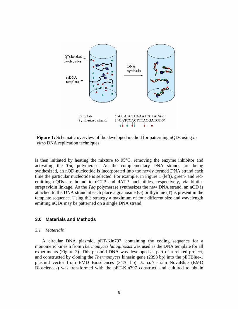
is then initiated by heating the mixture to 95°C, removing the enzyme inhibitor and activating the Taq polymerase. As the complementary DNA strands are being synthesized, an nQD-nucleotide is incorporated into the newly formed DNA strand each time the particular nucleotide is selected. For example, in Figure 1 (left), green- and red-emitting nQDs are bound to dCTP and dATP nucleotides, respectively, via biotin-streptavidin linkage. As the Taq polymerase synthesizes the new DNA strand, an nQD is attached to the DNA strand at each place a guanosine (G) or thymine (T) is present in the template sequence. Using this strategy a maximum of four different size and wavelength emitting nQDs may be patterned on a single DNA strand.
Figure 1: Schematic overview of the developed method for patterning nQDs using in vitro DNA replication techniques.
3.0 Materials and Methods 3.1 Materials
A circular DNA plasmid, pET-Kin797, containing the coding sequence for a monomeric kinesin from Thermomyces lanuginosus was used as the DNA template for all experiments (Figure 2). This plasmid DNA was developed as part of a related project, and constructed by cloning the Thermomyces kinesin gene (2393 bp) into the pETBlue-1 plasmid vector from EMD Biosciences (3476 bp). E. coli strain NovaBlue (EMD Biosciences) was transformed with the pET-Kin797 construct, and cultured to obtain
9
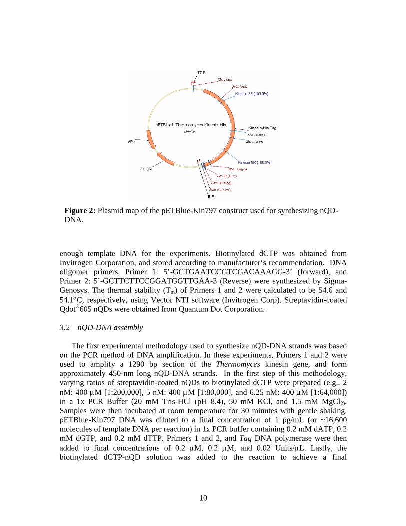
Figure 2: PlasmidDNA.
enough template DInvitrogen Corporatoligomer primers, Primer 2: 5’-GCTTGenosys. The therm54.1°C, respectivelyQdot®605 nQDs we 3.2 nQD-DNA ass
The first experimon the PCR methodused to amplify aapproximately 450-varying ratios of stnM: 400 µM [1:200in a 1x PCR BuffeSamples were then pETBlue-Kin797 Dmolecules of templamM dGTP, and 0.2added to final conbiotinylated dCTP
map of the pETBlue-Kin797 construct used for synthesizing nQD-
NA for the experiments. Biotinylated dCTP was obtained from ion, and stored according to manufacturer’s recommendation. DNA Primer 1: 5’-GCTGAATCCGTCGACAAAGG-3’ (forward), and CTTCCGGATGGTTGAA-3 (Reverse) were synthesized by Sigma-al stability (Tm) of Primers 1 and 2 were calculated to be 54.6 and , using Vector NTI software (Invitrogen Corp). Streptavidin-coated
re obtained from Quantum Dot Corporation.
embly
ental methodology used to synthesize nQD-DNA strands was based of DNA amplification. In these experiments, Primers 1 and 2 were 1290 bp section of the Thermomyces kinesin gene, and form nm long nQD-DNA strands. In the first step of this methodology, reptavidin-coated nQDs to biotinylated dCTP were prepared (e.g., 2 ,000], 5 nM: 400 µM [1:80,000], and 6.25 nM: 400 µM [1:64,000]) r (20 mM Tris-HCl (pH 8.4), 50 mM KCl, and 1.5 mM MgCl2). incubated at room temperature for 30 minutes with gentle shaking. NA was diluted to a final concentration of 1 pg/mL (or ~16,600 te DNA per reaction) in 1x PCR buffer containing 0.2 mM dATP, 0.2 mM dTTP. Primers 1 and 2, and Taq DNA polymerase were then centrations of 0.2 µM, 0.2 µM, and 0.02 Units/µL. Lastly, the
-nQD solution was added to the reaction to achieve a final
10
concentration of 0.2 µM dCTP. The temperature cycling profile for these reactions was: (1) 95°C for 2 min, (2) 95°C for 30 sec, (3) 55°C for 30 sec, (4) 72°C for 3 min, and (5) 4°C hold; steps 2-4 were cycled a total of 35 times.
The second experimental approach used to form nQD-DNA strands was based on methods used to form complementary DNA strand probes used for DNA hybridization assays. Only a single primer (i.e., Primer 1) was used to synthesize a 5869 bp DNA copy of the linearized pETBlue-Kin797 plasmid, which in turn should generate 2-µm long nQD-DNA strands. Solutions of streptavidin-coated nQDs and biotinylated dCTPs were prepared as described above. Because each deoxynucleotide is ~3.4Å in size, varying ratios of nQD-labeled and unlabeled dCTPs were used such that the 15-nm nQDs did not sterically interfere with the synthesis of the nQD-DNA strands. Thus, five ratios of streptavidin-coated nQD-dCTPs to unlabeled dCTPs were evaluated: 1:10, 1:20. 1:30, 1:40, and 1:520. The template DNA consisted of the pETBlue-Kin797 plasmid described above, except the template was digested with the restriction endonuclease EcoRI. Following enzyme digestion, the linearized template DNA was extracted with 25:24:1 phenol:chloroform:isoamyl alcohol to remove the restriction endonuclease, precipitated with sodium acetate and ethanol, and resuspended in deionized water. The concentration of the linearized template was determined by spectrophotometry. nQD-DNA reactions were assembled by diluting the template DNA to a final concentration of 2 µg/mL (~1.7 x 10¹⁰ template molecules per reaction) in 1x PCR buffer containing 0.2 mM dATP, 0.2 mM dGTP, 0.2 mM dTTP, 0.2 µM Primer 1, and 0.02 Units/µL Taq DNA polymerase. Biotinylated dCTP-nQD and unlabeled dCTP were then added to achieve the desired rations described above, as well as a final concentration of 0.2 µM dCTP. The temperature sequence for these reactions consisted of (1) 95°C for 3 min, (2) 55°C for 3 sec, and (3) 72°C for 15 – 30 min. 3.3 nQD-DNA characterization
nQD-DNA reactions were evaluated using two methods: epifluorescence microscopy and agarose gel electrophoresis. Small aliquots (5 – 10 µL) of each reaction were placed on 3 x 5 cm glass microscope slides, and covered with a 15 x 15 mm #1 coverslip. Slides were imaged on an Olympus IX-71 inverted microscope using a 100x oil immersion objective lens, mercury arc lamp for nQD excitation, and a WIG filter cube (520 – 550 nm band pass filter, 565 nm cut-off filter, Olympus Inc). Photomicrographs were captured using a Hamamatsu Orca II-ER CCD camera, and processed using the MicroSuite AnalySIS software package from Olympus.
Agarose gel electrophoresis was used to both separate nQD-DNA strands from unincorporated nQDs, as well as to estimate the size of the nQD-DNA strands that were produced. Aliquots (5 – 20 µL) of the nQD-DNA samples were mixed with DNA loading buffer (8% glycerol, 20 mM Ethylenediaminetetraacetic acid, and 0.05% Bromophenol Blue), and loaded on a 0.7% TBE-agarose gel. Samples were separated by electrophoresis at 100 V for 60 – 90 min, and visualized using a UV transilluminator. For
11
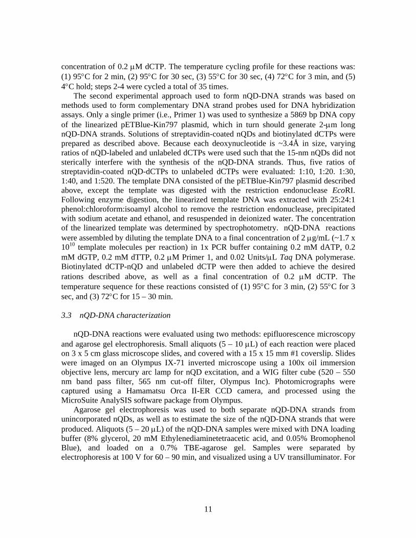
Figure 3: Fluorescence photomicrographs of composite structures formed using a PCR-based approach to synthesizing nQD-DNA strands. Scale bars = 1 µm.
selected samples, nQD-DNA bands were excised from the gel with a razor blade, isolated using a gel extraction kit (Qiagen Inc), and viewed by epifluorescence microscopy. 4.0 Results 4.1 Epifluorescence microscopy
In terms of the first strategy (i.e., PCR-based methodology) used to synthesize nQD-DNA, short (0.5 – 2 µm in length), irregular-shaped arrays of nQDs were observed by epifluorescence microscopy. Figure 3 shows several examples of these nQD-DNA arrays. Similar types of structures were absent from control samples that lacked the DNA template and/or primers, and primarily consisted of randomly disperse nQDs, with some minor aggregation of particles. In general, neither the ratio of streptavidin-coated nQDs to biotinylated dCTP nor the ratio of streptavidin-coated nQD-dCTPs affected the synthesis of these structures. Overall, these experiments provide the simple proof-of-principle that assemblies of nQDs could be synthesized using DNA scaffolds and in vitro replication techniques.
For the second approach (i.e., formation of a single complementary DNA strand), epifluorescence microscopy demonstrated the synthesis of linear nQD-DNA strands of 2 – 5 µm in length, as shown in Figure 4. As with prior experiments, control samples
12
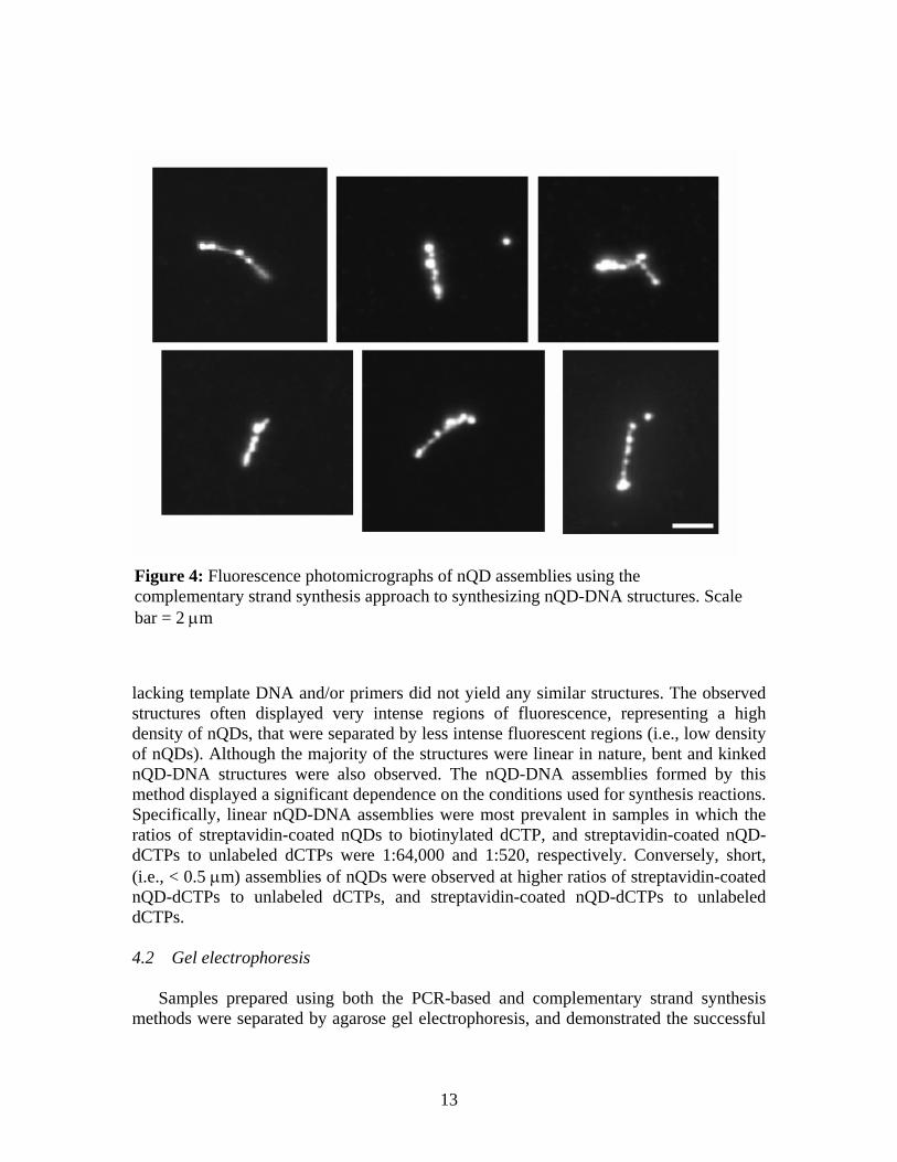
Figure 4: Fluorescence photomicrographs of nQD assemblies using the complementary strand synthesis approach to synthesizing nQD-DNA structures. Scale bar = 2 µm
lacking template DNA and/or primers did not yield any similar structures. The observed structures often displayed very intense regions of fluorescence, representing a high density of nQDs, that were separated by less intense fluorescent regions (i.e., low density of nQDs). Although the majority of the structures were linear in nature, bent and kinked nQD-DNA structures were also observed. The nQD-DNA assemblies formed by this method displayed a significant dependence on the conditions used for synthesis reactions. Specifically, linear nQD-DNA assemblies were most prevalent in samples in which the ratios of streptavidin-coated nQDs to biotinylated dCTP, and streptavidin-coated nQD-dCTPs to unlabeled dCTPs were 1:64,000 and 1:520, respectively. Conversely, short, (i.e., < 0.5 µm) assemblies of nQDs were observed at higher ratios of streptavidin-coated nQD-dCTPs to unlabeled dCTPs, and streptavidin-coated nQD-dCTPs to unlabeled dCTPs. 4.2 Gel electrophoresis
Samples prepared using both the PCR-based and complementary strand synthesis methods were separated by agarose gel electrophoresis, and demonstrated the successful
13
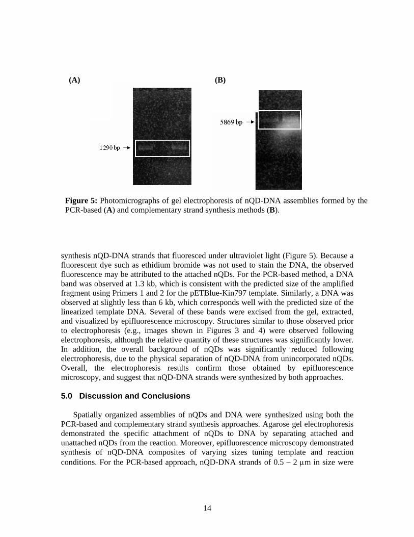
Figure 5: Photomicrographs of gel electrophoresis of nQD-DNA assemblies formed by the PCR-based (A) and complementary strand synthesis methods (B).
(B) (A)
synthesis nQD-DNA strands that fluoresced under ultraviolet light (Figure 5). Because a fluorescent dye such as ethidium bromide was not used to stain the DNA, the observed fluorescence may be attributed to the attached nQDs. For the PCR-based method, a DNA band was observed at 1.3 kb, which is consistent with the predicted size of the amplified fragment using Primers 1 and 2 for the pETBlue-Kin797 template. Similarly, a DNA was observed at slightly less than 6 kb, which corresponds well with the predicted size of the linearized template DNA. Several of these bands were excised from the gel, extracted, and visualized by epifluorescence microscopy. Structures similar to those observed prior to electrophoresis (e.g., images shown in Figures 3 and 4) were observed following electrophoresis, although the relative quantity of these structures was significantly lower. In addition, the overall background of nQDs was significantly reduced following electrophoresis, due to the physical separation of nQD-DNA from unincorporated nQDs. Overall, the electrophoresis results confirm those obtained by epifluorescence microscopy, and suggest that nQD-DNA strands were synthesized by both approaches. 5.0 Discussion and Conclusions
Spatially organized assemblies of nQDs and DNA were synthesized using both the PCR-based and complementary strand synthesis approaches. Agarose gel electrophoresis demonstrated the specific attachment of nQDs to DNA by separating attached and unattached nQDs from the reaction. Moreover, epifluorescence microscopy demonstrated synthesis of nQD-DNA composites of varying sizes tuning template and reaction conditions. For the PCR-based approach, nQD-DNA strands of 0.5 – 2 µm in size were
14
observed, as compared to the predicted size of ~ 0.5 µm based on the DNA sequence. Aggregation of several 0.5 µm nQD-DNA strands into an agglomeration likely explains the presences of the observed larger structures. The ability to discern individual linear nQD-DNA strands from aggregation is difficult for structures of this size using epifluorescence microscopy. A certain degree of nQD-DNA aggregation, however, is expected based on the ability of each streptavidin molecule to bind four biotin molecules. In addition, each nQD is coated with approximately fifteen streptavidin molecules, thus providing a means of aggregating several nQD-DNA into larger agglomerations. nQD-DNA strands larger than the predicted size were also observed using the complementary strand synthesis method (i.e., observed: 2 – 5 µm, expected: 2 µm). Similarly, streptavidin-based aggregation of multiple nQD-DNA likely explains these observations. Potential solutions to eliminate aggregation include pre-conditioning streptavidin-coated nQDs with biotin such that the number of available bindings site would be limited, or synthesizing streptavidin-coated nQDs with only a single streptavidin molecule per nQD.
Formation of the 2-µm nQD-DNA strands using the complementary strand synthesis approach was significantly dependent on both the ratio of streptavidin-coated nQDs to biotinylated dCTP, and streptavidin-coated nQD-dCTPs to unlabeled dCTPs. The dependence of the latter ratio is expected based on a significant difference in the relative size of the nQDs (i.e., ~15 nm) as compared with deoxynucleotides (i.e., 3.4 Å). Approximately 44 deoxynucleotides occupy the same space as a single nQD, which would include presence of about eleven dCTP nucleotides assuming a random distribution. Based on these assumptions, a significant increase in the number of nQD-DNA strands was expected for ratios greater than 1:10. An increase in the production of nQD-DNA, however, was only observed at the 1:520 ratio, suggesting the involvement of other mitigating factors in addition to steric inhibition. Overall, maintaining a proper ratio of unlabeled to streptavidin-coated nQD-dCTP appears to alleviate factors inhibiting the synthesis of nQD-DNA strands. In terms of the streptavidin-coated nQDs to biotinylated dCTP ratio, differences are likely attributable to the large number of streptavidin binding sites on each nQD, as discussed above. Increasing the relative number of biotinylated dCTP nucleotides per nQD should result in an increase in the number of dCTPs per nQD. Thus, incorporation of nQDs into the newly synthesized DNA strand should be more efficient based on an increased number of available dCTPs on a given nQD.
The mechanism by which the nQD-DNA strands are synthesized is not known, but likely occurs by one of two different mechanisms. In the first mechanism, dCTP-conjugated nQDs are used by the polymerase, and directly inserted into the newly synthesized DNA strand. The structure and mechanism by which this enzyme acts must, however, permit the binding and catalytic addition of nucleotide-conjugated nQDs for this mechanism to occur. Alternatively, the DNA polymerase uses only biotinylated dCTP nucleotides to form the new DNA strand, and attachment of a streptavidin-coated nQD occurs immediately following synthesis. Based on the structure and proposed mechanism of DNA synthesis by the Taq polymerase enzyme, the latter mechanism is the more plausible hypothesis. Insertion of nucleotides by DNA polymerase enzymes into a newly synthesized strand is a highly selective process. For example, insertion of ribose-based nucleotides and modified deoxynucleotides by DNA polymerases occurs at a
15
substantially reduced frequency compared with normal, unmodified deoxynucleotides [17, 18]. Such biased incorporation of unmodified deoxynucleotides is attributable to decreased rates of phosphodiester bond formation and the proofreading ability of the enzyme [18, 19]. Thus, incorporation of dCTP-conjugated nQDs directly by the Taq DNA polymerase is unlikely due the considerable modification of the deoxynucleotide, as well as the overall size of the complex compared to the size of the catalytic domain (~2 – 3 nm) of the polymerase enzyme. Therefore, incorporation of biotinylated dCTPs (not attached to nQDs) should happen at a higher frequency based on the ability of the modified nucleotide to fit into the catalytic domain of the enzyme. It is also expected that unmodified dCTPs should be incorporated at a higher frequency than the biotinylated dCTPs, which is beneficial based on the relative size issues discussed above.
Recently, there has been significant interest in using biological molecules as a means for assembling and organizing nanoscale materials [20-24]. In this work, the synthesis of nQD-DNA structures demonstrates the ability to assemble photoactive materials with a range of materials science-, nanoelectronics-, and nanophotonics-based applications. DNA templates possess a number of intrinsic properties that are extremely attractive. For example, DNA molecules are ~2 nm wide and can measure several meters long for uncoiled chromosomal DNA, which translates into an aspect ratio of 109. Genetic design and engineering of DNA sequences enables the ability to “program” nQD-binding sites into a given DNA strand, which in turn may be used to regulate interactions among adjacent nQDs. Information may also be encoded in these strands by controlling the spacing and color of nQDs within a given strand. In terms of manufacturability, the methods of synthesizing nQD-DNA strands developed in this project offer the capability of mass production. Further, the size of each deoxynucleotide translates into Å-level precision during the manufacturing process.
In addition to materials science applications, the ability to synthesize nQD-DNA strands also offers a number of biotechnological applications. Fluorescent organic dyes are widely used for the study of biological systems, and include microarray analysis, DNA sequencing, confocal cell imaging, and protein-protein interactions. nQDs offer superior properties (i.e., high quantum yield, photo-stability), and have begun replacing many of the standard dyes [6, 7]. Thus, the ability to synthesize these photoactive DNA molecules provides an enabling platform for a number of biotechnological and sensor-based applications.
6.0 References
1. D. Gerion, F. Pinaud, S. Williams, W. Parak, D. Zanchet, S. Weiss, and A.
Alivisatos, "Synthesis and properties of biocompatible water-soluble silica-coated CdSe/ZnS semiconductor quantum dots," J. Phys. Chem. B 105, 8861-8871 (2001).
2. X. Peng, T. Wilson, A. Alivisatos, and P. Schultz, "Synthesis and isolation of a homodimer of cadmium selenide nanocrystals," Angew. Chem. 36, 145-147 (1997).
16
3. X. Peng, M. Schlamp, A. Kadavanich, and A. Alivisatos, "Epitaxial growth of highly luminescent CdSe/CdS core/shell nanocrystals with photostability and electronic accessibility," J. Am. Chem. Soc. 119, 7019-7029 (1997).
4. A. Guzelian, U. Banin, A. Kadavanich, X. Peng, and A. Alivisatos, "Colloidal chemical synthesis and characterization of InAs nanocrystal quantum dots," Appl. Phys. Lett. 69, 1432-1434 (1996).
5. W. Chan and S. Nie, "Quantum dot bioconjugates for ultrasensitive nonisotopic detection," Science 281, 2016-2018 (1998).
6. M. Bruchez, M. Moronne, P. Gin, S. Weiss, and A. Alivisatos, "Semiconductor nanocrystals as fluorescent biological labels," Science 281, 2013-2016 (1998).
7. W.C.W. Chan, D.J. Maxwell, X.H. Gao, R.E. Bailey, M.Y. Han, and S.M. Nie, "Luminescent quantum dots for multiplexed biological detection and imaging," Curr. Opin. Biotechnol. 13, 40-46 (2002).
8. X.H. Gao, W.C.W. Chan, and S.M. Nie, "Quantum-dot nanocrystals for ultrasensitive biological labeling and multicolor optical encoding," J. Biomed. Opt. 7, 532-537 (2002).
9. P. Alivisatos, "Colloidal quantum dots. From scaling laws to biological applications," Pure Appl. Chem. 72, 3-9 (2000).
10. J. Watson and F. Crick, "Molecular structure of nucleic acids - A structures for deoxyribose nucleic acid," Nature 171, 737-738 (1953).
11. J. Watson and F. Crick, "Genetical implications of the structure of deoxyribonucleic acid," Nature 171, 964-967 (1953).
12. A. Alivisatos, K. Johnsson, X. Peng, T. Wilson, C. Loweth, M. Bruchez, and P. Schultz, "Organization of 'nanocrystal molecules' using DNA," Nature 382, 609-611 (1996).
13. D. Gerion, W. Parak, S. Williams, D. Zanchet, C. Micheel, and A. Alivisatos, "Sorting fluorescent nanocrystals with DNA," J. Am. Chem. Soc. 124, 7070-7074 (2002).
14. D. Zanchet, C. Micheel, W. Parak, D. Gerion, S. Williams, and A. Alivisatos, "Electrophoretic and structural studies of DNA-directed Au nanoparticle groupings," J. Phys. Chem. B 106, 11758-11763 (2002).
15. W.J. Parak, D. Gerion, D. Zanchet, A.S. Woerz, T. Pellegrino, C. Micheel, S.C. Williams, M. Seitz, R.E. Bruehl, Z. Bryant, C. Bustamante, C.R. Bertozzi, and A.P. Alivisatos, "Conjugation of DNA to silanized colloidal semiconductor nanocrystalline quantum dots.," Chem. Mater. 14, 2113-2119 (2002).
16. R. Saiki, D. Gelfand, S. Stoffel, S. Scharf, R. Higuchi, G. Horn, G. Mullis, and H. Erlich, "Primer-directed enzymatic amplification of DNA with a thermostable DNA polymerase," Science 239, 487-491 (1988).
17
17. J.W. Brandis, "Dye structure affects Taq DNA polymerase terminator selectivity," Nucleic Acids Res. 27, 1912-1918 (1999).
18. S. Doublie and T. Ellenberger, "The mechanism of action of T7 DNA polymerase," Curr. Opin. Struct. Biol. 8, 704-712 (1998).
19. J.W. Brandis, S.G. Edwards, and K.A. Johnson, "Slow rate of phosphodiester bond formation accounts for the strong bias that Taq DNA polymerase shows against 2',3'-dideoxynucleotide terminators," Biochemistry 35, 2189-2200 (1996).
20. N.I. Kovtyukhova and T.E. Mallouk, "Nanowires as building blocks for self-assembling logic and memory circuits," Chem.-Eur. J. 8, 4355-4363 (2002).
21. G.D. Bachand, S.B. Rivera, A.K. Boal, J.P. Bauer, S.J. Koch, R.P. Manginell, J. Liu, and B.C. Bunker. Developing nanoscale materials using biomimetic assembly processes. in Materials Research Society Symposium Proceedings: Micro- and Nanosystems. 2004. Boston, MA: Materials Research Society, Warrendale, PA.
22. G.D. Bachand, S.B. Rivera, A.K. Boal, J. Gaudioso, J. Liu, and B.C. Bunker, "Assembly and transport of nanocrystal CdSe quantum dot nanocomposites using microtubules and kinesin motor proteins," Nano Lett. 4, 817-821 (2004).
23. R. McMillan, "Biomolecular templates - Nanoparticles align," Nat. Mater. 2, 214-215 (2003).
24. R. McMillan, C. Paavola, J. Howard, S. Chan, N. Zaluzec, and J. Trent, "Ordered nanoparticle arrays formed on engineered chaperonin protein templates," Nat. Mater. 1, 247-252 (2002).
18
Distribution 1 MS 0310 M.D. Rintoul, 9212 1 MS 0513 J.P. VanDevender, 1000 1 MS 0603 J.J. Hudgens, 1743 1 MS 0734 J.B. Kelly, 6245 1 MS 0887 M.J. Cieslak, 1800 1 MS 0855 G.S. Heffelfinger, 1802 1 MS 0511 D.B. Dimos, 1803 1 MS 1078 S.J. Martin, 1707 1 MS 1079 M.W. Scott, 1700 1 MS 1411 J. Liu, 1846 1 MS 1413 T.A. Michalske, 1040 1 MS 1413 G.D. Bachand, 1116 1 MS 1413 P.V. Dressendorfer, 1116 1 MS 1413 K.K. Crown, 1116 1 MS 1415 J.C. Barbour, 1110 1 MS 1415 N.D. Shinn, 1114 1 MS 1421 G.A. Samara, 1130 1 MS 1424 S.A. Casalnuovo, 1744 1 MS 1427 J.M. Phillips, 1100 1 MS 9951 L.M. Napolitano, 8140 1 MS 9004 R.H. Stulen, 8100 1 MS 9951 M.M. Young, 8141 1 MS 0123 LDRD Office, 1011 1 MS 0161 Patent & Licensing Office, 11500 1 MS 9018 Central Technical Files 8945-1 2 MS0899 Technical Library, 9616
19


![PhysicaA Stochasticresonanceinperiodicpotentialsdriven ...5284 K.Liu,Y.Jin/PhysicaA392(2013)5283–5288 potentialsystems[19–21].Amongthem,KimandSung[19]foundtheresonancebehaviorinthefrequency](https://img.pdfslide.us/doc/110x75/5ed1eeb4ae118d4f2114c3db/physicaa-stochasticresonanceinperiodicpotentialsdriven-5284-kliuyjinphysicaa39220135283a5288.jpg)