Embed Size (px)
Citation preview
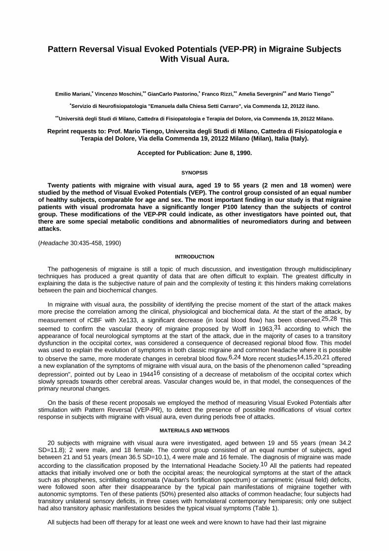
Pattern Reversal Visual Evoked Potentials (VEP-PR) in Migraine SubjectsWith Visual Aura.
Emilio Mariani,* Vincenzo Moschini,** GianCarlo Pastorino,* Franco Rizzi,** Amelia Severgnini** and Mario Tiengo**
*Servizio di Neurofisiopatologia "Emanuela dalla Chiesa Setti Carraro", via Commenda 12, 20122 ilano.
**Università degli Studi di Milano, Cattedra di Fisiopatologia e Terapia del Dolore, via Commenda 19, 20122 Milano.
Reprint requests to: Prof. Mario Tiengo, Universita degli Studi di Milano, Cattedra di Fisiopatologia eTerapia del Dolore, Via della Commenda 19, 20122 Milano (Milan), Italia (Italy).
Accepted for Publication: June 8, 1990.
SYNOPSIS
Twenty patients with migraine with visual aura, aged 19 to 55 years (2 men and 18 women) werestudied by the method of Visual Evoked Potentials (VEP). The control group consisted of an equal numberof healthy subjects, comparable for age and sex. The most important finding in our study is that migrainepatients with visual prodromata have a significantly longer P100 latency than the subjects of controlgroup. These modifications of the VEP-PR could indicate, as other investigators have pointed out, thatthere are some special metabolic conditions and abnormalities of neuromediators during and betweenattacks.
(Headache 30:435-458, 1990)
INTRODUCTION
The pathogenesis of migraine is still a topic of much discussion, and investigation through multidisciplinarytechniques has produced a great quantity of data that are often difficult to explain. The greatest difficulty inexplaining the data is the subjective nature of pain and the complexity of testing it: this hinders making correlationsbetween the pain and biochemical changes.
In migraine with visual aura, the possibility of identifying the precise moment of the start of the attack makesmore precise the correlation among the clinical, physiological and biochemical data. At the start of the attack, bymeasurement of rCBF with Xe133, a significant decrease (in local blood flow) has been observed.25,28 Thisseemed to confirm the vascular theory of migraine proposed by Wolff in 1963,31 according to which theappearance of focal neurological symptoms at the start of the attack, due in the majority of cases to a transitorydysfunction in the occipital cortex, was considered a consequence of decreased regional blood flow. This modelwas used to explain the evolution of symptoms in both classic migraine and common headache where it is possibleto observe the same, more moderate changes in cerebral blood flow.6,24 More recent studies14,15,20,21 offereda new explanation of the symptoms of migraine with visual aura, on the basis of the phenomenon called "spreadingdepression", pointed out by Leao in 194416 consisting of a decrease of metabolism of the occipital cortex whichslowly spreads towards other cerebral areas. Vascular changes would be, in that model, the consequences of theprimary neuronal changes.
On the basis of these recent proposals we employed the method of measuring Visual Evoked Potentials afterstimulation with Pattern Reversal (VEP-PR), to detect the presence of possible modifications of visual cortexresponse in subjects with migraine with visual aura, even during periods free of attacks.
MATERIALS AND METHODS
20 subjects with migraine with visual aura were investigated, aged between 19 and 55 years (mean 34.2SD=11.8); 2 were male, and 18 female. The control group consisted of an equal number of subjects, agedbetween 21 and 51 years (mean 36.5 SD=10.1), 4 were male and 16 female. The diagnosis of migraine was madeaccording to the classification proposed by the International Headache Society.10 All the patients had repeatedattacks that initially involved one or both the occipital areas; the neurological symptoms at the start of the attacksuch as phosphenes, scintillating scotomata (Vauban's fortification spectrum) or campimetric (visual field) deficits,were followed soon after their disappearance by the typical pain manifestations of migraine together withautonomic symptoms. Ten of these patients (50%) presented also attacks of common headache; four subjects hadtransitory unilateral sensory deficits, in three cases with homolateral contemporary hemiparesis; only one subjecthad also transitory aphasic manifestations besides the typical visual symptoms (Table 1).
All subjects had been off therapy for at least one week and were known to have had their last migraine
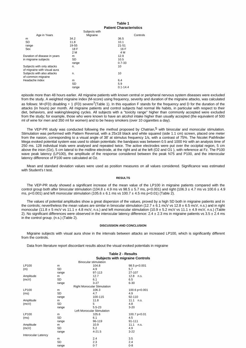
Table 1Patient Characteristics
Subjects withAge in Years Migraine Controls
m 34.2 36.5 SD 11.8 10.1 range 19-55 21-51 Sex 18 F 16 F
2 M 4 MDuration of disease in years m 12.9in migraine subjects SD 10.5
range 0.7-33Subjects with only attacks n. 10of migraine with visual auraSubjects with also attacks n. 10of common migraineHeadache index m 6.4
SD 5.9 range 0.1-14.4
episode more than 48 hours earlier. All migraine patients with known central or peripheral nervous system diseases were excludedfrom the study. A weighted migraine index (M-score) using frequency, severity and duration of the migraine attacks, was calculatedas follows: M=(FD) disabling + 1 (FD) severe7(Table 1). In this equation F stands for the frequency and D for the duration of theattacks (in hours) per month. All migraine patients and control subjects had normal life habits, in particular with respect to theirdiet, behaviors, and waking/sleeping cycles. All subjects with a "toxicity range" higher than commonly accepted were excludedfrom the study; for example, those who were known to have an alcohol intake higher than usually accepted (the equivalent of 500ml of wine for men and 350 ml for women) and to be heavy smokers (over 10 cigarettes a day).
The VEP-PR study was conducted following the method proposed by Chatrian,5 with binocular and monocular stimulation.Stimulation was performed with Pattern Reversal, with a 25x18 black and white squared (side 1.1 cm) screen, placed one meterfrom the nasion, corresponding to a visual angle of 38' at stimulus frequency 1/s, with a contrast of 75%. The Nicolet PathfinderMega evoked potential system was used to obtain potentials: the bandpass was between 0.5 and 1000 Hz with an analysis time of250 ms. 128 individual trials were analysed and repeated twice. The active electrodes were put over the occipital region, 5 cmabove the inion (Oz), 5 cm lateral to the midline electrode, at the right and at the left (O2 and O1 ), with reference at Fz. The P100wave peak latency (LP100), the amplitude of the response considered between the peak N75 and P100, and the interocularlatency difference of P100 were calculated at Oz.
Mean and standard deviation values were used as position measures on all values considered. Significance was estimatedwith Student's t test.
RESULTS
The VEP-PR study showed a significant increase of the mean value of the LP100 in migraine patients compared with thecontrol group both after binocular stimulation (104.8 ± 4.9 ms vs 98.5 ± 5.7 ms, p<0.001) and right (106.3 ± 4.7 ms vs 100.6 ± 4.9ms, p<0.001) and left monocular stimulation (105.6 ± 6.1 ms vs 100.7 ± 4.5 ms p<0.01) (Table 2).
The values of potential amplitudes show a great dispersion of the values, proved by a high SD both in migraine patients and inthe controls; nevertheless the mean values are similar in binocular stimulation (12.7 ± 6.1 mcV vs 12.8 ± 6.5 mcV, n.s.) and in rightmonocular (11.8 ± 5 mcV vs 11.1 ± 4.8 mcV, n.s.) and left monocular stimulation (10.9 ± 5.2 mcV vs 11.1 ± 4.9 mcV, n.s.) (Table2). No significant differences were observed in the interocular latency difference: 2.4 ± 2.3 ms in migraine patients vs 3.5 ± 2.4 msin the control group. (n.s.) (Table 2).
DISCUSSION AND CONCLUSION
Migraine subjects with visual aura show in the intervals between attacks an increased LP100, which is significantly differentfrom the controls.
Data from literature report discordant results about the visual evoked potentials in migraine
Table 2 - ResultsSubjects with migraine Controls
Binocular stimulationLP100 m 104.8 98.5 p<0.001(m) SD 4.9 5.7
range 97-113 27-107Amplitude m 12.7 12.8 n.s.(mcV) SD 6.1 6.5
range 3-27 6-30Right Monocular Stimulation
LP100 m 106.3 100.6 p<0.001(ms) SD 4.7 4.9
range 100-115 92-110Amplitude m 11.8 11.1 n.s.(mcV) SD 5 4.8
range 5.5-23 3-20Left Monocular Stimulation
LP100 m 105.6 100.7 p<0.01(ms) SD 6.1 4.5
range 96-119 91-111Amplitude m 10.9 11.1 n.s.(mcV) SD 5.2 4.9
range 4-21.5 3-22Interocular Latency
m 2.4 3.5SD 2.3 2.4range 0-7 0-8

patients. In a study of migraine subjects with visual aura Kennard (1978)12 found a significant increase in the latency of the P100;Polich (1986)23 showed only a slight increase in the LP100 and P100 amplitude, not statistically significant. Recent studies in subjectswith common migraine2,19 showed no significant differences in the latency of the P100 between subjects with common migraine andthe controls. In another group of migraine patients,8 a significant increase of the amplitude and of the latency of P100 was noted, with asignificant decrease of the amplitude in the same subjects after treatment with beta adrenergic blocking agents.
Our results agree with those of Kennard12 about the modifications of VEP-PR in migraine subjects with visual aura between theattacks: the similarity is probably due to the analogous methodology of stimulation and of obtaining responses; on the contrary, inPolich's study23 there is considerable variability of stimulation and acquisition.
On the basis of these observations, it appears that subjects with migraine with visual prodromata have repeated episodescharacterized by metabolic and vascular modifications, producing functional modifications of the occipital response to standardizedstimulations such as those of Pattern Reversal. Yet it seems very difficult to explain these results. Kennard (1978),12 on the basis ofanatomicopathological observations by Yates (1976),32 postulated that repeated episodes of ischemia and cerebral edema, even thoughslight, could provoke demyelination of visual pathways, and thus increase the LP100. Although this theory is undoubtedly interesting, itseems very unlikely because the decrease in blood flow in the cerebral cortex, besides being sporadic and of short duration, iscompletely resolved during the attack, with consequent total regression of the signs of neurological deficit. There are also many otherstudies which point out that subjects with stabilized sequelae of posterior cerebral artery infarction, documented by clinical examinationand by CAT scan, present usually normal VEP-PR,29 while altered responses could be present after separate stimulation of thehemi-fields4 and only sometimes in case of bilateral occipital lesions1 or lesions of the primary areas.3
The modifications in the VEP-PR observed in migraine with visual aura could reflect specific changes in monoamine neuromediators,which can be shown even between attacks.13,26,22 These specific changes develop acutely in the early phase of migraine attacks, andpresent similarities with those seen in the spreading depression produced in rats under experimental conditions.16,17 This phenomenoncan be provoked by various nociceptive stimuli and consists in a decrease of the metabolism in the cerebral cortex, accompanied by adecrease in blood flow which starts from the posterior areas, propagates toward the front at a speed of 2 mm/m, until it reaches theRolandic fissure where it halts and persists for about an hour. Subsequent studies on animals showed that the phenomenon ofspreading depression is accompanied by an inhibition of cerebral evoked potentials which return to the basal condition ony 10-60minutes after the end of the spreading depression.9,18
On the basis of these observations we consider that the method of VEP-PR is an useful instrument for investigation of migrainepatients with visual prodromata, together with other examinations of the neural function of the central nervous system, such asEEG11,30 and the study of contingent negative variation (CNV),27 which also have revealed particular changes in this complex andinteresting disorder.
REFERENCES
1. Ashworth B, Maloney AFJ, Townsend HRA: Delayed visual evoked potentials with bilateral disease of the posterior vis u a lpathway. J Neurol Neurosurg Psych 41:449-451, 1978.
2. Benna P, Bianco C, Costa P, Piazza D, Bergamasco B: Visual evoked potentials and brainstem auditory evoked potentials inmigraine and transient ischemic attacks. Cephalalgia Supp 5:53-58, 1985.
3. Blumhardt LD, Halliday AM: Cortical abnormalities and the visual evoked response. Doc Ophtalmol Proc Series 27:347-365,1981.
4. Camacho M, Wenzel W, Aschoff J: Clinical applications of the visual evoked potentials for detection of chiasmal andpostchiasmal lesions. Arch Psychiatr Nervenkr 230:243-256, 1981.
5. Chatrian GE: American Electroencephalographic Society-Guidelines for Clinical Evoked Potentials Studies. J Clin Neurophysiol1:3-53, 1984.
6. Dalessio DJ: Wolff's Headache and Other Head Pain. New York, Oxford University Press, 1980.
7. Diamond S, Shenbaum A: Flunarizine a calcium channel blocker in the prophylactic treatment of migraine. Headache 23:39-42,1983.
8. Diener HC, Scholz E, Dichgans J, Gerber WD, Jack A, Bille A, Niederberger U: Central effects of drugs used in migraineprophylaxis evaluated by visual evoked potentials. Ann Neurol 25:125-130, 1989.
9. Freygang WH, Landau WM: Some relations between resistivity and electrical activity in the cerebral cortex of the cat. JNeurophysiol 45:377-392, 1955.
10. Headache Classification Committee of the International Headache Society: Classification and diagnostic criteria for headachedisorders, cranial neuralgias and facial pain. Cephalalgia 8 (supp 7), 1988.
11. Hockaday JM, Witthy CWM: Factors determining the electroencephalogram in migraine: a study of 560 patients according toclinical type of migraine. Brain 92:769-788, 1969.
12. Kennard C, Gawel M, Rudolph N, Rose CF: Visual evoked potentials in migraine subjects, in Friedman AP, Granger ME, CritchlyM (eds): Research and Clinical Studies in Headache. Karger, Basel, 1978, pp 73-80.
13. Lance JW: The pathogenesis of migraine, in Lance JW (ed): Mechanisms and Management of Headache. London, Butterworth,1982, pp 102-120.
14. Lauritzen M, Olesen J: Regional cerebral blood flow during migraine attacks by Xenon-133 inhalation and emission tomography.Brain 107:447-461, 1984.
15. Lauritzen M: On the possible relation of spreading cortical depression to classical migraine. Cephalalgia 5 (supp 2):47-51, 1985.
16. Leao AAP: Spreading depression of activity in cerebral cortex. J Neurophysiol 7:359-390, 1944.
17. Leao AAP, Morrison RS: Propagation of spreading cortical depression. J Neurophysiol 8:33-45, 1945.
18. Marshal WH: The relation of dehydration of the brain to the spreading depression of Leao.Electroencephalogr Clin Neurophysiol 2:177-185, 1950.
19. Mariani E, Moschini V, Pastorino GC, Rizzi F, Severgnini A, Tiengo M: Pattern-reversal visual evokedpotentials and EEG correlation in common migraine patients. Headache 28:269-271, 1988.
20. Olesen J, Larsen B, Skinhoj E: Spontaneous occipitoparietal decrease in rCBF: vasospasm ormetabolic depression?. Acta Neurol Scand 60 (supp 72):28-29, 1979.
21. Olesen J, Larsen B, Lauritzen M: Focal hyperemia followed by spreading oligemia and impairedactivation of the rCBF in classic migraine. Ann Neurol 9:344-352, 1981.
22. Olesen J: The pathophysiology of migraine, in Clifford Rose F (ed): Handbook of Clinical Neurology.Amsterdam, Elsiever, 1986, 59-83.
23. Polich J, Ehlers CL, Dalessio DJ: Pattern-shift visual evoked responses and EEG in migraine.Headache 26:451-456, 1986.
24. Raskin NH, Appenzeller O: Headache: Major Problems in Internal Medicine. WB Saunders,Philadelphia, 1980, pp 111-172.
25. Sakai F, Meyer JS: Regional cerebral hemodynamics during migraine and cluster headachesmeasured by the 133Xe inhalation method. Headache 18:122-132, 1978.
26. Schaffer EWP, McKean CM: Evidence that monoamines influence human evoked potentials. BrainRes 99:49-58, 1975.
27. Schoenen J, Maertens A, Timsit-Berthier M, Timsit M: Contingent negative variation (CNV) as adiagnostic and physiopathological tool in headache patients, in: Clifford Rose (ed): Migraine.Proceedings of the Fifth International Migraine Symposium, London 1984, Basel Karger, 1985: pp17-25.
28. Skinoj E: Hemodynamic studies within the brain during migraine. Arch Neurol 29:95-98, 1973.
29. Streletz LJ, Bae SH, Roeshman AM, Shatz NJ, Savino PJ: Visual evoked potentials in occipital lobelesions. Arch Neurol 38:80-85, 1981.
30. Wessely P, Mayr N, Goldenberg G: EEG-Befunde bei Komplizierter Migrane. EEG-EMG 16:221-226,1985.
31. Wolff AG: Headache and Other Head Pain. Oxford University Press, New York, 1963.
32. Yates PO: Vascular disease of the central nervous system, in Blackwood and Corsellis (eds):Greenfield's Neuropathology. Arnold, London 1976, pp 86-147.