Embed Size (px)
Citation preview
PAIN Pain 123 (2006) 204--209
wwwelseviercomJlocatepain
Clinical note
Patients with Ehlers Danlos syndrome and eRPS A possible flssociation
Joan M Stoler a Anne Louise Oaklander bcd
Genetics and TerCllgy Unit Department of Pediatrics Massaclwsels General Hospital Harvard Medical School BoslOn M A USA b Nerve Injury Unit Deparrment of Anesthesia and Critical Care Massachusels General Hospital Harvard Medical SclIOOI Boston MA USA
C Nerve Injury Unit Deparrment of Neurology Massachusels General Hospital Harvard Medical School Boston MA USA d Nerve Injury Unit Department of Neuropathology Massacwsels General H05pital Harvard Medical School Boston VI A USA
Received I September 2005 received in revised form 6 February 2006 accepted 7 February 2006
Abstract
Rare patients are left with chronic pain vasodysregulation and other symptoms that define complex regional pain syndrome (eRPS) after limb traumas The predisposing factors are unknown Genetic factors undoubtedly contribute but have not yet been identified We report four eRPS patients also diagnosed with the classical or hypermobility forms of Ehlers Danlos syndrome (EDS) inherited disorders of connective tissue These patients had been diagnosed using standard diagnostic criteria for eRPS and for EDS All had sustained joint injury in three this had been surgically treated The association of these two diagnoses leads us to hypothesize that EDS might contribute to the developme~t of eRPS in one or more of the following ways via stretch injury to nerves traversing hypermobile joints increased fragility of nerve connective tissue or nerve trauma from more frequent surgery We review the clinical presentation of the different Ehlers Danlos syndromes and provide clinical criteria that can be used to screen eRPS patients for EDS for clinical or research purposes copy 2006 International Association for the Study of Pain Published by Elsevier BY All rights reserved
Key words Connective tissue Pain Inherited disorder of collagen Genetics Reflex sympathetic dystrophy
1 Introduction
Patients with complex regional pain syndrome (CRPS) also known as reflex sympathetic dystrophy (CRPS-I) or causalgia (CRPS-II) complain of chronic pain vasodysregulation and other symptoms that pershysist after apparent healing of limb trauma Patients with type 2 CRPS are defined as having a known nerve injury and patients with type I CRPS do not (Table S I) although the identification of axonal losses in CRPS-I patients ( un dt r Lmlll e l ltil 1lt)( 9 OClk landcr 1 11 1
20(6) challenges the validity of this dichotomy
Corresponding author Tel + I 617 726 1742 fax + I 617 724 1911
E-mail addressi I(lI ~I ( ptl rtllet org (JM Stoler)
Little is known about why some patients develop CRPS after traumas that usually do not have long-term sequelae Age is important with the average patient being 30-40 years old (VcIJ nlln c t tI 19gt3) It is not known why it is less prevalent among older patients who have increased risk of neuralgic pain in other conshyditions (eg post-herpetic neuralgia trigeminal neuralshygia and small-fiber neuropathies) Spontaneous resolution is more likely in children than adults (Wilder et aI 1992) Genetic factors may influence susceptibility to CRPS The number of patients with more than one episode is greater than that expected by chance alone (Devor 2()()4) Specific HLA types (Kemler c l I I 1999 van de Beek el III 199() ) but no specific genetic disorders have been associated with CRPS
Within the last few years we have identified several patients evaluated at the Pain Center and Genetics Sershy
0304-3959$3200 copy 2006 International Association for the Study of Pain Published by Elsevier BY All rights reserved doi 101 0 16jpain200602022
205 JM SlOler AL Oaklander Pain 123 (2006 ) 204-209
vice of the Massachusetts General Hospital who meet diagnostic criteria for both Ehlers Danlos syndrome (EDS) as well as CRPS (using the current diagnostic crishyteria of the International Association for the Study of Pain (Table S I )) which raises the possibility of an assoshyciation between these conditions
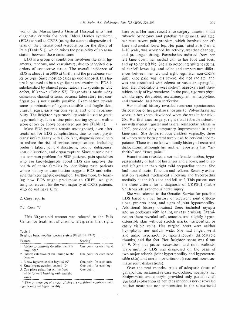
EDS is a group of conditions involving the skin ligshyaments tendons and vasculature due to inherited disshyorders of connective tissue The overall incidence of EDS is about I in 5000 at birth and the prevalence varshyies by type Since most go cases go undiagnosed this figshyure is believed to be a significant underestimate EDS is subclassified by clinical presentation and specific genetic defect if known (Table S2) Diagnosis is made using consensus clinical criteria because laboratory test conshyfirmation is not usually possible Examination reveals some combination of hyperextensible and fragile skin unusual scars early varicose veins and joint hypermoshybility The Beighton hypermobility scale is used to grade hypermobility It is a nine-point scoring system with a score of 59 or above considered positive (T Ibk I)
Most EDS patients remain undiagnosed even after treatment for EDS complications due to most physishycians unfamiliarity with EDS Yet diagnosis could help to reduce the risk of serious complications including preterm labor joint dislocations wound dehiscence aortic dissection and organ rupture Since chronic pain is a common problem for EDS patients pain specialists who are knowledgeable about EDS can improve the health of entire families by identifying pain patients whose history or examination suggests EDS and refershyring them for genetic evaluation Furthermore by learnshying how EDS might predispose to CRPS we gain insights relevant for the vast majority of CRPS patients who do not have EDS
2 Case reports
21 Case 1
This 30-year-old woman was referred to the Pain Center for treatment of chronic left greater than right
Table I Beighton hypermobility scoring system ( Flcighlltlll 1913 )
Fealure Scoring
I Ability 10 passively dorsiflex Ihe fifth One poinl for each hand finger gt900
2 Passive extension of Ihe Ihumb to the One point for each hand forearm
3 Elbow hyperextension beyond 100 One point for each arm 4 Knee hyperextension beyond 100 One point for each leg 5 Can place palms flal on the floor One poinl
while forward bending with slraight knees
Five or more OUI of a total of nine are considered consistent with significant joint hypermobility
knee pain Her most recent knee surgery anterior tibial tubercle osteotomy and patellar realignment initiated her most severe pain problem which involved her left
knee and medial lower leg Her pain rated at 6-7 on a 1-10 scale was worsened by activi ty weather changes and prolonged sitting Paresthesias radiated from her left knee down her medial calf to her foot and toes and up to her left hip She also noted intermittent edema of her left lower leg and color and temperature differshyences between her left and right legs Her non-CRPS right knee pain was less severe did not radiate and was not associated with edema or vascular dysregulashytion Her medications were sodium naproxyn and three tablets daily of hydrocodone In the past rigorous physshyical therapy ibuprofen acetaminophen amitriptyline and tramadol had been ineffective
Her medical history revealed recurrent spontaneous dislocations of her patellae since age 15 Polyarthralgias worse in her knees developed when she was in her midshy20s Her first knee surgery right tibial tubercle osteotoshymy with medial transfer and lateral retinacular release in
1997 provided only temporary improvement in right knee pain She delivered four children vaginally three of whom were born prematurely due to cervical incomshypetence There was no known family history of recurrent dislocations although her mother reportedly had arshythritis and knee pains
Examination revealed a normal female habitus hypeshyrextensibility of both of her knees and elbows and bilatshyeral left grealer than right mild prepatellar edema She had normal motor function and reflexes Sensory examshyination revealed mechanical allodynia and hyperpathia medially at the left knee and left calf This patient met the three criteria for a diagnosis of CRPS-II (Table SI) from left saphenous nerve injury
She was referred to the Genetics Service for possible EDS based on her history of recurrent joint dislocashytions pre term labor and signs of joint hypermobility Additional history obtained there included myopia and no problems wilh healing or easy bruising Examishynation there revealed soft smooth and slightly hypershyextensible skin without stretch marks varicosities or easily visible veins Her surgical scars were neither hypoplastic nor und uly wide She had finger wrist and ankle hypermobility spontaneously dislocatable thumbs and flat feet Her Beighton score was 6 out of 9 She had pectus excavatum and mild scoliosis Hypermobility EDS was diagnosed on the basis of two major criteria (joint hypermobility and hyperextenshysible skin) and one minor criterion (recurrent non-traushymatic joint dislocations)
Over the next months trials of adequate doses of gabapentin sustained-release oxycodone nortriptyline desipramine and doxepin provided only partial relief Surgical exploration of her left saphenous nerve revealed neither neuromas nor compression in the subsartorial
206 JM Sloler AL Oaklander I Pain 123 (2006) 204-209
canal Subsequently she had surgical implantation of a temporary and then a permanent stimulator of her left saphenous nerve which provided initial relief However migration of the paddle electrode eventually caused disshycomfort and she discontinued use of her stimulator and resumed medical management
22 Case 2
This 21-year-old woman was evaluated at the Pain Center for severe right wrist and hand pain that had developed four months earlier when she awoke to find her shoulder out of its socket Although this reduced easily without medical care the pain worsened and spread to her shoulder She complained of numbness weakness and atrophy of her right arm and tingling in her fingers She noted hyperhydrosis and intermittent edema of her right hand Cold exacerbated her sympshytoms Her history revealed multiple prior spontaneous shoulder and patella dislocations Recurrent right ankle sprains had required right ankle ligament reconstrucshytion She reported no difficulty with either healing or abnormal scarring She had a history of a languageshybased learning disability and depression Both her mother and maternal grandmother also had joint hypermobility
On examination at the Pain Center she guarded her right arm and hand and the plantar surface of her wrist was tender to palpation There was atrophy of the first right dorsal interosseous muscle and decreased pin senshysation in the ulnar distribution of the right hand A Tinels sign was elicitable at the right supraclavicular fossa Edema and trophic changes of the skin or nails were absent Generalized joint laxity was noted Electroshydiagnostic study which ~equired intravenous sedation to perform was interpreted as normal She was diagnosed with CRPS-II after presumed stretch injury to the medial cord of her brachial plexus or her ulnar nerve Nortriptyline and a topical lidocaine patch were preshyscribed She found the nortriptyline but not the patch partially efficacious
She was referred to the Genetics Service because of concern about possible EDS based on her history of recurrent spontaneous joint dislocations and signs of joint hypermobility Examination there revealed very soft and smooth skin with well-healed surgical scars Her skin was neither hyperextensible nor translucent She had no varicosities or easily visible veins Striae were prominent across her lower back and trunk She had gingival recession mild scoliosis and widespread joint hypermobility (Beighton score 8 out of 9) with hyperexshytensible fingers wrists and elbows She was diagnosed with hypermobility EDS on the basis of two major criteshyria (abnormally soft smooth skin and joint hypermobilshyity) plus one minor criterion (recurrent non-traumatic dislocations)
23 Case 3
The Genetics Service evaluated this 40-year-old womshyan for a connective tissue disorder She had a history of dislocations and subluxations of many joints these had required eleven surgeries at various hospitals These included reconstructions of both ankles fusions of both hands left and right rotator cuff repairs and reconstrucshytion of her right shoulder to treat multi-directional instability This last surgery was immediately followed by new chronic pain paresthesias and edema of the dorsum of both of her hands She reported intolerance of light touch on her hands or water running over them Her pain was exacerba ted by changes in temperature and with activity but also was present at rest Her medshyical history also revealed gastroesophageal reflux with hiatal hernia treated by fundoplication and cholecystecshytomy She had a history of easy bruising and increased joint mobility She had one term pregnancy that proshyduced a daughter with increased joint flexibility Examshyination of the dorsum of her hands revealed allodynia to touch decreased pin sensation and edema Electromyshyography and nerve conduction studies of both upper extremities had been interpreted as normal Clinical and radiological evaluations of her cervical spine had revealed no abnormalities She had been diagnosed elseshywhere with reflex sympathetic dystrophy (CRPS-I) and treated with methadone and nerve blocks in both of her hands
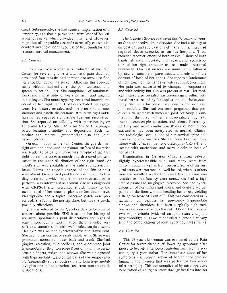
Examination in Genetics Clinic showed velvety slightly hyperextensible skin and many scars from minor trauma as well as from surgery Some of the surshygical scars were narrow and well healed whereas others were abnormally atrophic and broad No cutaneous varshyicosities or translucency were noted She had a high arched palate and no gingival recession She had hypershyextension of her fingers and knees and could place her palms on the floor without bending her knees yielding a Beighton score of 5 out of 9 This was considered artishyfactually low because her previously hypermobile elbows and shoulders had been surgically tightened She was diagnosed with classical EDS on the basis of two major criteria (widened atrophic scars and joint hypermobility) plus two minor criteria (smooth velvety skin and complications of joint hypermobility) (Fig I)
24 Case 4
This 33-year-old woman was evaluated at the Pain Center for severe chronic left lower leg symptoms after injury to her left anterior-cruciate-Iigament from a socshycer injury a year earlier The immediate cause of her symptoms was surgical repair of her anterior cruciate ligament and menisci that was performed two weeks after her injury This was complicated by intra-operative penetration of a surgical screw through her tibia into her
207 1 M Slole AL Oaklander I Pain 123 (2006 ) 204-209
Fig I This patient with classical EDS can passively ex end her thumb to tOllch her forearm
popliteal artery which caused intra-arterial thrombus formation This was identified and treated with thromshybolytic and anticoagulant therapy which lysed the clot but initiated arterial bleeding into her leg This produced severe lower leg pain numbness and increased limb girth Compartment syndrome with critical ischemia and elevated pressures was documented and treated the next day by fasciotomy and repair of her left poplishyteal artery using a right saphenous vein graft In the immediate postoperative period she complained of severe lancinating pain radiating down her left lower leg to her toes numbness and stimulus-independent pain at the dorsum of her left foot lower-left-limb edema and hyperhydrosis She also noted tactile hallucinations of bugs crawling on top of her left foot She denied mechanical aJlodynia Despite good recovery from surshygery these symptoms persisted and a few months postshyoperatively similar though less-severe symptoms developed at the mirror-image location on her right low- er leg Autonomic testing had documented left-right asymmetries in lower-limb skin temperature and sweatshying Electrodiagnostic study had revealed reduced amplitude of compound motor action potentials in her left peroneal and tibial nerves and spontaneous activity in the left gastrocnemius but not tibialis anterior musshycles Triple-phase bone scan identified abnormally increased uptake in both calves and at the left proximal tibia Treatment with gabapentin amitriptyline and occasional oxycodone had been partly efficacious The bottom of her left foot had improved more than the top of her foot A consultant identified no rheumatologshyical cause for her chronic pain
Her pertinent medical history included one earlier traumatic shoulder dislocation and sleep apnea treated
with nocturnal continuous positive airway pressure A heart murmur had prompted echocardiography which was said to show multi valvular leakage Family history revealed that her mother had mitral valve prolapse
and an abdominal aortic aneurysm Her father had unspecified knee problems and several family members were said to have increased joint mobility
Examination at the Pain Center revealed normal athshyletic female habitus Spontaneously dislocatable thumbs and elbow and knee hyperextension suggested EDS There was color asymmetry between her two feet but no overt edema nor changes in skin and nails Motor function was intact Sensory examination demonstrated hypesthesia to pinprick light touch and cold on the dorsal left foot and calf (medial worse than lateral) Her left ankle reflex was diminished She met all criteria for and was diagnosed with CRPS-II caused by injury to the left common peroneal and tibial nerves The proxishymate cause of nerve injury was felt to be ischemia from her documented compartment syndrome The recomshymendations were to switch from amitriptyline to norshytriptyline to try to reduce amitriptyline-related orthostatic hypotension and to consider evaluation for possible EDS
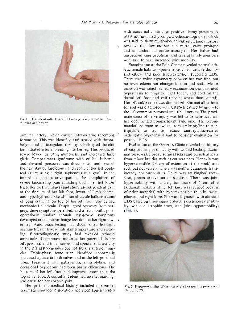
Evaluation at the Genetics Clinic revealed no history of easy bruising or difficulty with wound healing Examshyination revealed broad surgical scars and persistent scars from minor injuries such as cat scratches Her skin was hyperextensible (gt4 cm of extension at the neck) and soft but not velvety There was neither cutaneous transshylucency nor varicosities There was no gingival recesshysion pectus excavatum or scoliosis There was joint hypermobility with a Beighton score of 6 out of 9 (although mobility of her left knee was reduced because of prior surgeries) with hyperextensible thumbs wrist elbows and right knee She was diagnosed with classical EDS based on three major criteria (skin hyperextensibilshyity widened atrophic scars and joint hypermobility) ( ig 2)
Fig 2 Hyperextensibility of the skin of the forearm in a patient with classical EDS
208 1M SIOer AL Oakander I Pain 23 ( 2006) 204-209
3 Discussion
EDS is divided into several different types based on clinical and molecular criteria (Table S I) Reviews of the different types of EDS can be found at li ll p ~ wwld ni org h tl p wwweilltI- ( lI1 lo$org and http wwV cgtn c test~o rg Classical EDS comprises 90 of cases (Steinma nn I HI Z002) The diagnosis is based on skin hyperextensibility (gt4 cm of skin extenshysion at the neck) broad atrophic scars and joint hypershymobility ( l3eighton 199 Heighton et 11 11)1)R) Patients skin is typically smooth and velvety Complicashytions include hernias pelvic organ prolapse premature arthritis joint pain and cervical insufficiency The sever- ity is variable type II EDS designates patients with milder manifestations particularly those lacking widshyened atrophic scars (Wclsl rup 1 [ a l 1(96) Classical EDS is autosomal dominant and mutations in the COLSAI COLSA2 genes (responsible for type V collashygen synthesis) or in the COLIAI gene (encoding one of the subunits of type I collagen) (Wenslnljl ct ti L 2()UO) have been identified in about 30 of cases
Hypermobility EDS (formerly type III) is less comshymon and involves fewer skin abnormalities Diagnosis is based on joint hypermobility frequent dislocations and hyperextensible skin that heals normally ( Bcightc 11
1993 Bcigh lL111 c t aI 191) ) Chronic joint pain is comshymon Mutations in two genes have been found in a few cases COL3A I (responsible for type III collagen) (N It isi cl li l 1(N4) and Tenascin-XB (Zwecrs I lt11 200~ ) Most cases are autosomal dominant although Tenascin-XB mutations are autosomal recessive
Vascular EDS (EDS IV) the most dangerous form accounts for only 4 of cases and has a prevalence of less than I in 100000 (Byers 1995 ) Affected individuals have thin translucent skin easy bruising tissue fragility and a characteristic thin face with large eyes There may also be hypermobility of small joints acrogeria and gingival recession Potentially lethal complications include develshyopment of aneurysms or pneumothoraces and rupture of arteries or hollow organs including the gravid uterus It is autosomal dominant and caused by COL3A I mutations
Extremely rare forms of EDS include Kyphoscoliosis (EDS VI) Dermatosparaxis (EDS VIlC) and Arthroshychalasia EDS Kyphoscoliosis EDS is autosomal recesshysive caused by mutations in the Iysl hydroxylase gene and consists of generalized joint laxity keratoconus scoliosis and scleral and vascular fragility Dermatoshysparaxis is autosomal recessive due to a deficiency of procollagen I N-terminal peptidase and consists of lax redundant fragile skin and large hernias Arthrochalasia EDS is autosomal dominant due to specific mutations in the COLlA I and COLI A2 genes resulting in abnorshymal cleavage of the precursor type I procollagen to colshylagen Affected individuals have congenital hip
dislocation joint hypermobility recurrent dislocations scoliosis and short stature There are also other rare forms of EDS that are not well characterized ( te inshy111 111 1l c-t al 2(02) Confirmatory testing is available for the Vascular Kyphoscoliotic Arthrocholasia and Dermatosparaxis types of EDS Genetic testing is unavailable for patients with hypermobility EDS and most patients Nith classical EDS
These four patients were diagnosed with CRPS-I or CRPS-II according to the criteria of the International Association for the Study of Pain (Table S I) (Mers)ey Hnd B J u) I))-I ) A clinical geneticist also diagnosed each with EDS Two patients were diagnosed with classhysical EDS and two with hypermobility EDS Chronic pain is common in EDS (ilch ti d al 19(17 ) particularshyly in the hypermobility and classical forms Usually the cause is premature joint or tendon wear or injuries however some chronic pain in EDS is due to post-traushymatic neuralgia There are a few previous reports of patients with EDS-associated nerve injury (Kn lt1 ilnu I flSS 1gt79 Hell lt11 1(1 C I 1tl I lller~ 1(1)1 Chntl(lpndhHY ct iI IQ 5 Galan (11( KlHlssdT 191)-) including one vascular EDS patient six hypermobility EDS patients and two classical EDS patients The nerves involved were the brachial plexus lumbosacral plexus sciatic nerve and common peroneal These had been injured by fracture traction sling or normal activities of daily living The outcome varied but incomplete resolution was common Tomaculous neuropathy with pressure related nerve injuries has also been associated with EDS ( chncly and Ch~ll i 19R4 )
OUf cases demonstrate the development of CRPS in patients with EDS by two distinct ways specifically stretch injury to nerves by joint dislocation or hyperexshytension and increased exposure to medical procedures such as surgery Medical procedures have been linked to the onset of CRPS in a number of studies Vtldmllll I t li t (1 993 ) showed that 23 of 829 CRPS patients underwent an invasive procedure (surgery injection or intravenous infusion) just before the development of their symptoms Medical procedures (surgery casting or phlebotomy) immediately preceded onset of CRPS-I in 1618 subjects in a recent study (Ou)IHntier lid HroII 200-l-) Of our four cases it is unclear whether the first patient sustained her presumed saphenous nerve injury from patellar dislocation or subsequent knee surgery Case 2 had sustained joint dislocation only without surgical procedures Case 3 attributed her CRPS to a surshygery that immediately preceded onset of symptoms and in Case 4 CRPS was clearly a complication of surgery
We suggest a third possible link between EDS and CRPS for which there is no direct evidence It is possible that the nerve connective tissue is fragile in EDS patients and less able to protect the axons within from trauma Type V collagen one of the components of nerve tissue (Rn ll1h lu1l1 et nl ~004) is abnormal in some cases of
209 J M Sroler A L Oaklander I Pain 123 (2006) 204-209
classical EDS While we are unaware of testing of the strength of nerve connective tissue in EDS patients other tissues that con tain type V collagen such as skin tendons and ligaments are more fragile in classical EDS patients More work is needed to define the relative contributions of these factors An existing rabbit model of EDS C inkc l d I( 97) is a potential tool for such studies
In conclusion it is important for physicians to conshysider EDS when evaluating a patient with multiple joint problems We recommend that physicians caring for eRPS patients consider the possibility of an underlying connective tissue disorder in patients with histories or examinations revealing joint laxity
Acknowledgements
This work was support contributed by the Mayday Fund NINDS ROINS42866 and a Paul Beeson Faculty Physician Scholarship from the American Federation for Aging Research
Appendix A Supplementary data
Supplemen tary data associated with this article can be found in the online version at lit i 10 I (J I(j j pa in 1U0601 022
References
Beighton P The Ehlers Danlos syndromes In Beighton P editor Heritable disorders of connective tissue Mosby Sl Louis 1993 p189-251
Beighton P DePaepe A Steinmann B Tsipouras P Wenstrup Rl Ehlers-Danlos syndrome revised nosology Villefranche 1997 Am 1 Med Genet 19987731-7
Bell KM Chalmers 1 Recurrent common peroneal palsy in associashytion with the Ehlers Danlos syndrome Acta Orthop Scand 1991 62612-3
Byers PH Ehlers-Danlos syndrome type IV a genetic disorder in many guises 1 Invest Derm 1995105311-3
Chattopadhyay AK Kandler RH Sharrack B The association of hereditary neuropathies and heritable skeletal disorders Postgrad Med 1 199571 245-6
Devor M Evidence for heritability of pain in patients with traumatic neuropathy Pain 2004 108200-1
Galan E Kousseff B Peripheral neuropathy in Ehlers-Danlos syndrome Pediatr Neurol 1995 I 2242- 345
Kayed K Kass B Acute multiple brachial neuropathy and EhlersshyDanlos syndrome Neurology 197929 1620-1
Kemler MA van de Vusse AC van den Berg-Loonen EM Barendse GAM van Kleef M Weber WE1 HLA-DQI associated with reflex sympathetic dystrophy Neurology 199953 1350-1
Merskey H Bogduk N editors Classification of chronic pain descriptions of chronic pain syndromes and definitions of pain terms Seattle [ASP Press 1994
Narcisi PA Richards 1 Ferguson SO Pope FM A family with EhlersshyDanlos syndrome type Illarticular hypermobility syndrome has a glycine 637 to serine substitution type III collagen Hum Mol Genet 19943 1617-20
Oaklander AL Rissmiller lG Gelman LB Zheng L Chang Y Gott R Evidence of focal small-fiber axonal degeneration in complex regional pain syndrome-[ (reflex sympathetic dystrophy) Pain 2006 120235-43
Oaklander AL Brown 1M Unilateral nerve injury produces bilateral loss of distal innervation Ann Neurol 200455639-44
Rothblum K Stahl RC Carey 01 Constitutive release of a4 Type V collagen N-terminal domain by Schwann cells and binding to cell surface and extracellular matrix heparan sulfate proteoglycans 1 Bioi Chem 200427951282-8
Sacheti A Szemere 1 Bernstein B Tafas T Schechter N Tsipomas P Chronic pain is a manifestation of the Ehlers-Danlos syndrome 1 Pain Symptom Manage 19971488- 93
Schady W Ochoa 1 Ehlers-Danlos in association with tomaculous nemopathy Neurology 198434 1270
Sinke 10 van Dijk JE Willemse T A case of Ehlers-Danlos-like syndrome in a rabbit with a review of the disease in other species Vet Quart 199719182-5
Steinmann B Royce P Supeni-Furga A The Ehlers Danlos synshydromes In Royce P Steinmann B editors Connective tissue and its heritable disorders molecular genetic and medical aspects shyNew York Wiley-Liss 2002 p 431-523
van de Beek WJ van Hilten JJ Roep BO HLA-DQI associated with reflex sympathetic dystrophy (comment) Neurology 199955457-8
van der Laan L van Spaendonck K Horstink MW Goris RJ The symptom checklist-90 revised questionnaire no psychological profiles in complex regional pain syndrome-dystonia J Pain Symptom Manage 1999 17357-62
Veldman PH Reynen HM Arntz IE Goris RJ Signs and symptoms of reflex sympathetic dystrophy prospective study of 829 patients Lancet 19938878 1012-6
Wenstrup RJ Langland GT Willing MC DSouza VN Cole WG A splice-junction mutation in the region of COL5A I that codes the carboxyl propeptide of pro a I(V) chains results in the gravis form of the Ehlers-Danlos syndrome (type I) Hum Mol Genet 19965 1773-6
Wenstrup RJ Florer lB Willing MC Giunta C Steinmann B Young F et al COL5A I haploinsufficiency is a common molecular mechanism underlying the classical form of EDS Am J Hum Genet 200066 1766-76
Wilder RT Berde CB Wolohan M Vicyra MA Masek BJ Micheli LJ Reflex sympathetic dystrophy in children Clinical charactershyistics and follow-up of seventy patients J Bone Joint Surg Am 199274910-9
Zweers C Bristow J Steijlen PM Dean WB Hamel BC Otero M et al Haploinsufficiency ofTNXB is associated with hypermobility lype of Ehlers-Danlos syndrome Am J Hum Genet 200373214--7
205 JM SlOler AL Oaklander Pain 123 (2006 ) 204-209
vice of the Massachusetts General Hospital who meet diagnostic criteria for both Ehlers Danlos syndrome (EDS) as well as CRPS (using the current diagnostic crishyteria of the International Association for the Study of Pain (Table S I )) which raises the possibility of an assoshyciation between these conditions
EDS is a group of conditions involving the skin ligshyaments tendons and vasculature due to inherited disshyorders of connective tissue The overall incidence of EDS is about I in 5000 at birth and the prevalence varshyies by type Since most go cases go undiagnosed this figshyure is believed to be a significant underestimate EDS is subclassified by clinical presentation and specific genetic defect if known (Table S2) Diagnosis is made using consensus clinical criteria because laboratory test conshyfirmation is not usually possible Examination reveals some combination of hyperextensible and fragile skin unusual scars early varicose veins and joint hypermoshybility The Beighton hypermobility scale is used to grade hypermobility It is a nine-point scoring system with a score of 59 or above considered positive (T Ibk I)
Most EDS patients remain undiagnosed even after treatment for EDS complications due to most physishycians unfamiliarity with EDS Yet diagnosis could help to reduce the risk of serious complications including preterm labor joint dislocations wound dehiscence aortic dissection and organ rupture Since chronic pain is a common problem for EDS patients pain specialists who are knowledgeable about EDS can improve the health of entire families by identifying pain patients whose history or examination suggests EDS and refershyring them for genetic evaluation Furthermore by learnshying how EDS might predispose to CRPS we gain insights relevant for the vast majority of CRPS patients who do not have EDS
2 Case reports
21 Case 1
This 30-year-old woman was referred to the Pain Center for treatment of chronic left greater than right
Table I Beighton hypermobility scoring system ( Flcighlltlll 1913 )
Fealure Scoring
I Ability 10 passively dorsiflex Ihe fifth One poinl for each hand finger gt900
2 Passive extension of Ihe Ihumb to the One point for each hand forearm
3 Elbow hyperextension beyond 100 One point for each arm 4 Knee hyperextension beyond 100 One point for each leg 5 Can place palms flal on the floor One poinl
while forward bending with slraight knees
Five or more OUI of a total of nine are considered consistent with significant joint hypermobility
knee pain Her most recent knee surgery anterior tibial tubercle osteotomy and patellar realignment initiated her most severe pain problem which involved her left
knee and medial lower leg Her pain rated at 6-7 on a 1-10 scale was worsened by activi ty weather changes and prolonged sitting Paresthesias radiated from her left knee down her medial calf to her foot and toes and up to her left hip She also noted intermittent edema of her left lower leg and color and temperature differshyences between her left and right legs Her non-CRPS right knee pain was less severe did not radiate and was not associated with edema or vascular dysregulashytion Her medications were sodium naproxyn and three tablets daily of hydrocodone In the past rigorous physshyical therapy ibuprofen acetaminophen amitriptyline and tramadol had been ineffective
Her medical history revealed recurrent spontaneous dislocations of her patellae since age 15 Polyarthralgias worse in her knees developed when she was in her midshy20s Her first knee surgery right tibial tubercle osteotoshymy with medial transfer and lateral retinacular release in
1997 provided only temporary improvement in right knee pain She delivered four children vaginally three of whom were born prematurely due to cervical incomshypetence There was no known family history of recurrent dislocations although her mother reportedly had arshythritis and knee pains
Examination revealed a normal female habitus hypeshyrextensibility of both of her knees and elbows and bilatshyeral left grealer than right mild prepatellar edema She had normal motor function and reflexes Sensory examshyination revealed mechanical allodynia and hyperpathia medially at the left knee and left calf This patient met the three criteria for a diagnosis of CRPS-II (Table SI) from left saphenous nerve injury
She was referred to the Genetics Service for possible EDS based on her history of recurrent joint dislocashytions pre term labor and signs of joint hypermobility Additional history obtained there included myopia and no problems wilh healing or easy bruising Examishynation there revealed soft smooth and slightly hypershyextensible skin without stretch marks varicosities or easily visible veins Her surgical scars were neither hypoplastic nor und uly wide She had finger wrist and ankle hypermobility spontaneously dislocatable thumbs and flat feet Her Beighton score was 6 out of 9 She had pectus excavatum and mild scoliosis Hypermobility EDS was diagnosed on the basis of two major criteria (joint hypermobility and hyperextenshysible skin) and one minor criterion (recurrent non-traushymatic joint dislocations)
Over the next months trials of adequate doses of gabapentin sustained-release oxycodone nortriptyline desipramine and doxepin provided only partial relief Surgical exploration of her left saphenous nerve revealed neither neuromas nor compression in the subsartorial
206 JM Sloler AL Oaklander I Pain 123 (2006) 204-209
canal Subsequently she had surgical implantation of a temporary and then a permanent stimulator of her left saphenous nerve which provided initial relief However migration of the paddle electrode eventually caused disshycomfort and she discontinued use of her stimulator and resumed medical management
22 Case 2
This 21-year-old woman was evaluated at the Pain Center for severe right wrist and hand pain that had developed four months earlier when she awoke to find her shoulder out of its socket Although this reduced easily without medical care the pain worsened and spread to her shoulder She complained of numbness weakness and atrophy of her right arm and tingling in her fingers She noted hyperhydrosis and intermittent edema of her right hand Cold exacerbated her sympshytoms Her history revealed multiple prior spontaneous shoulder and patella dislocations Recurrent right ankle sprains had required right ankle ligament reconstrucshytion She reported no difficulty with either healing or abnormal scarring She had a history of a languageshybased learning disability and depression Both her mother and maternal grandmother also had joint hypermobility
On examination at the Pain Center she guarded her right arm and hand and the plantar surface of her wrist was tender to palpation There was atrophy of the first right dorsal interosseous muscle and decreased pin senshysation in the ulnar distribution of the right hand A Tinels sign was elicitable at the right supraclavicular fossa Edema and trophic changes of the skin or nails were absent Generalized joint laxity was noted Electroshydiagnostic study which ~equired intravenous sedation to perform was interpreted as normal She was diagnosed with CRPS-II after presumed stretch injury to the medial cord of her brachial plexus or her ulnar nerve Nortriptyline and a topical lidocaine patch were preshyscribed She found the nortriptyline but not the patch partially efficacious
She was referred to the Genetics Service because of concern about possible EDS based on her history of recurrent spontaneous joint dislocations and signs of joint hypermobility Examination there revealed very soft and smooth skin with well-healed surgical scars Her skin was neither hyperextensible nor translucent She had no varicosities or easily visible veins Striae were prominent across her lower back and trunk She had gingival recession mild scoliosis and widespread joint hypermobility (Beighton score 8 out of 9) with hyperexshytensible fingers wrists and elbows She was diagnosed with hypermobility EDS on the basis of two major criteshyria (abnormally soft smooth skin and joint hypermobilshyity) plus one minor criterion (recurrent non-traumatic dislocations)
23 Case 3
The Genetics Service evaluated this 40-year-old womshyan for a connective tissue disorder She had a history of dislocations and subluxations of many joints these had required eleven surgeries at various hospitals These included reconstructions of both ankles fusions of both hands left and right rotator cuff repairs and reconstrucshytion of her right shoulder to treat multi-directional instability This last surgery was immediately followed by new chronic pain paresthesias and edema of the dorsum of both of her hands She reported intolerance of light touch on her hands or water running over them Her pain was exacerba ted by changes in temperature and with activity but also was present at rest Her medshyical history also revealed gastroesophageal reflux with hiatal hernia treated by fundoplication and cholecystecshytomy She had a history of easy bruising and increased joint mobility She had one term pregnancy that proshyduced a daughter with increased joint flexibility Examshyination of the dorsum of her hands revealed allodynia to touch decreased pin sensation and edema Electromyshyography and nerve conduction studies of both upper extremities had been interpreted as normal Clinical and radiological evaluations of her cervical spine had revealed no abnormalities She had been diagnosed elseshywhere with reflex sympathetic dystrophy (CRPS-I) and treated with methadone and nerve blocks in both of her hands
Examination in Genetics Clinic showed velvety slightly hyperextensible skin and many scars from minor trauma as well as from surgery Some of the surshygical scars were narrow and well healed whereas others were abnormally atrophic and broad No cutaneous varshyicosities or translucency were noted She had a high arched palate and no gingival recession She had hypershyextension of her fingers and knees and could place her palms on the floor without bending her knees yielding a Beighton score of 5 out of 9 This was considered artishyfactually low because her previously hypermobile elbows and shoulders had been surgically tightened She was diagnosed with classical EDS on the basis of two major criteria (widened atrophic scars and joint hypermobility) plus two minor criteria (smooth velvety skin and complications of joint hypermobility) (Fig I)
24 Case 4
This 33-year-old woman was evaluated at the Pain Center for severe chronic left lower leg symptoms after injury to her left anterior-cruciate-Iigament from a socshycer injury a year earlier The immediate cause of her symptoms was surgical repair of her anterior cruciate ligament and menisci that was performed two weeks after her injury This was complicated by intra-operative penetration of a surgical screw through her tibia into her
207 1 M Slole AL Oaklander I Pain 123 (2006 ) 204-209
Fig I This patient with classical EDS can passively ex end her thumb to tOllch her forearm
popliteal artery which caused intra-arterial thrombus formation This was identified and treated with thromshybolytic and anticoagulant therapy which lysed the clot but initiated arterial bleeding into her leg This produced severe lower leg pain numbness and increased limb girth Compartment syndrome with critical ischemia and elevated pressures was documented and treated the next day by fasciotomy and repair of her left poplishyteal artery using a right saphenous vein graft In the immediate postoperative period she complained of severe lancinating pain radiating down her left lower leg to her toes numbness and stimulus-independent pain at the dorsum of her left foot lower-left-limb edema and hyperhydrosis She also noted tactile hallucinations of bugs crawling on top of her left foot She denied mechanical aJlodynia Despite good recovery from surshygery these symptoms persisted and a few months postshyoperatively similar though less-severe symptoms developed at the mirror-image location on her right low- er leg Autonomic testing had documented left-right asymmetries in lower-limb skin temperature and sweatshying Electrodiagnostic study had revealed reduced amplitude of compound motor action potentials in her left peroneal and tibial nerves and spontaneous activity in the left gastrocnemius but not tibialis anterior musshycles Triple-phase bone scan identified abnormally increased uptake in both calves and at the left proximal tibia Treatment with gabapentin amitriptyline and occasional oxycodone had been partly efficacious The bottom of her left foot had improved more than the top of her foot A consultant identified no rheumatologshyical cause for her chronic pain
Her pertinent medical history included one earlier traumatic shoulder dislocation and sleep apnea treated
with nocturnal continuous positive airway pressure A heart murmur had prompted echocardiography which was said to show multi valvular leakage Family history revealed that her mother had mitral valve prolapse
and an abdominal aortic aneurysm Her father had unspecified knee problems and several family members were said to have increased joint mobility
Examination at the Pain Center revealed normal athshyletic female habitus Spontaneously dislocatable thumbs and elbow and knee hyperextension suggested EDS There was color asymmetry between her two feet but no overt edema nor changes in skin and nails Motor function was intact Sensory examination demonstrated hypesthesia to pinprick light touch and cold on the dorsal left foot and calf (medial worse than lateral) Her left ankle reflex was diminished She met all criteria for and was diagnosed with CRPS-II caused by injury to the left common peroneal and tibial nerves The proxishymate cause of nerve injury was felt to be ischemia from her documented compartment syndrome The recomshymendations were to switch from amitriptyline to norshytriptyline to try to reduce amitriptyline-related orthostatic hypotension and to consider evaluation for possible EDS
Evaluation at the Genetics Clinic revealed no history of easy bruising or difficulty with wound healing Examshyination revealed broad surgical scars and persistent scars from minor injuries such as cat scratches Her skin was hyperextensible (gt4 cm of extension at the neck) and soft but not velvety There was neither cutaneous transshylucency nor varicosities There was no gingival recesshysion pectus excavatum or scoliosis There was joint hypermobility with a Beighton score of 6 out of 9 (although mobility of her left knee was reduced because of prior surgeries) with hyperextensible thumbs wrist elbows and right knee She was diagnosed with classical EDS based on three major criteria (skin hyperextensibilshyity widened atrophic scars and joint hypermobility) ( ig 2)
Fig 2 Hyperextensibility of the skin of the forearm in a patient with classical EDS
208 1M SIOer AL Oakander I Pain 23 ( 2006) 204-209
3 Discussion
EDS is divided into several different types based on clinical and molecular criteria (Table S I) Reviews of the different types of EDS can be found at li ll p ~ wwld ni org h tl p wwweilltI- ( lI1 lo$org and http wwV cgtn c test~o rg Classical EDS comprises 90 of cases (Steinma nn I HI Z002) The diagnosis is based on skin hyperextensibility (gt4 cm of skin extenshysion at the neck) broad atrophic scars and joint hypershymobility ( l3eighton 199 Heighton et 11 11)1)R) Patients skin is typically smooth and velvety Complicashytions include hernias pelvic organ prolapse premature arthritis joint pain and cervical insufficiency The sever- ity is variable type II EDS designates patients with milder manifestations particularly those lacking widshyened atrophic scars (Wclsl rup 1 [ a l 1(96) Classical EDS is autosomal dominant and mutations in the COLSAI COLSA2 genes (responsible for type V collashygen synthesis) or in the COLIAI gene (encoding one of the subunits of type I collagen) (Wenslnljl ct ti L 2()UO) have been identified in about 30 of cases
Hypermobility EDS (formerly type III) is less comshymon and involves fewer skin abnormalities Diagnosis is based on joint hypermobility frequent dislocations and hyperextensible skin that heals normally ( Bcightc 11
1993 Bcigh lL111 c t aI 191) ) Chronic joint pain is comshymon Mutations in two genes have been found in a few cases COL3A I (responsible for type III collagen) (N It isi cl li l 1(N4) and Tenascin-XB (Zwecrs I lt11 200~ ) Most cases are autosomal dominant although Tenascin-XB mutations are autosomal recessive
Vascular EDS (EDS IV) the most dangerous form accounts for only 4 of cases and has a prevalence of less than I in 100000 (Byers 1995 ) Affected individuals have thin translucent skin easy bruising tissue fragility and a characteristic thin face with large eyes There may also be hypermobility of small joints acrogeria and gingival recession Potentially lethal complications include develshyopment of aneurysms or pneumothoraces and rupture of arteries or hollow organs including the gravid uterus It is autosomal dominant and caused by COL3A I mutations
Extremely rare forms of EDS include Kyphoscoliosis (EDS VI) Dermatosparaxis (EDS VIlC) and Arthroshychalasia EDS Kyphoscoliosis EDS is autosomal recesshysive caused by mutations in the Iysl hydroxylase gene and consists of generalized joint laxity keratoconus scoliosis and scleral and vascular fragility Dermatoshysparaxis is autosomal recessive due to a deficiency of procollagen I N-terminal peptidase and consists of lax redundant fragile skin and large hernias Arthrochalasia EDS is autosomal dominant due to specific mutations in the COLlA I and COLI A2 genes resulting in abnorshymal cleavage of the precursor type I procollagen to colshylagen Affected individuals have congenital hip
dislocation joint hypermobility recurrent dislocations scoliosis and short stature There are also other rare forms of EDS that are not well characterized ( te inshy111 111 1l c-t al 2(02) Confirmatory testing is available for the Vascular Kyphoscoliotic Arthrocholasia and Dermatosparaxis types of EDS Genetic testing is unavailable for patients with hypermobility EDS and most patients Nith classical EDS
These four patients were diagnosed with CRPS-I or CRPS-II according to the criteria of the International Association for the Study of Pain (Table S I) (Mers)ey Hnd B J u) I))-I ) A clinical geneticist also diagnosed each with EDS Two patients were diagnosed with classhysical EDS and two with hypermobility EDS Chronic pain is common in EDS (ilch ti d al 19(17 ) particularshyly in the hypermobility and classical forms Usually the cause is premature joint or tendon wear or injuries however some chronic pain in EDS is due to post-traushymatic neuralgia There are a few previous reports of patients with EDS-associated nerve injury (Kn lt1 ilnu I flSS 1gt79 Hell lt11 1(1 C I 1tl I lller~ 1(1)1 Chntl(lpndhHY ct iI IQ 5 Galan (11( KlHlssdT 191)-) including one vascular EDS patient six hypermobility EDS patients and two classical EDS patients The nerves involved were the brachial plexus lumbosacral plexus sciatic nerve and common peroneal These had been injured by fracture traction sling or normal activities of daily living The outcome varied but incomplete resolution was common Tomaculous neuropathy with pressure related nerve injuries has also been associated with EDS ( chncly and Ch~ll i 19R4 )
OUf cases demonstrate the development of CRPS in patients with EDS by two distinct ways specifically stretch injury to nerves by joint dislocation or hyperexshytension and increased exposure to medical procedures such as surgery Medical procedures have been linked to the onset of CRPS in a number of studies Vtldmllll I t li t (1 993 ) showed that 23 of 829 CRPS patients underwent an invasive procedure (surgery injection or intravenous infusion) just before the development of their symptoms Medical procedures (surgery casting or phlebotomy) immediately preceded onset of CRPS-I in 1618 subjects in a recent study (Ou)IHntier lid HroII 200-l-) Of our four cases it is unclear whether the first patient sustained her presumed saphenous nerve injury from patellar dislocation or subsequent knee surgery Case 2 had sustained joint dislocation only without surgical procedures Case 3 attributed her CRPS to a surshygery that immediately preceded onset of symptoms and in Case 4 CRPS was clearly a complication of surgery
We suggest a third possible link between EDS and CRPS for which there is no direct evidence It is possible that the nerve connective tissue is fragile in EDS patients and less able to protect the axons within from trauma Type V collagen one of the components of nerve tissue (Rn ll1h lu1l1 et nl ~004) is abnormal in some cases of
209 J M Sroler A L Oaklander I Pain 123 (2006) 204-209
classical EDS While we are unaware of testing of the strength of nerve connective tissue in EDS patients other tissues that con tain type V collagen such as skin tendons and ligaments are more fragile in classical EDS patients More work is needed to define the relative contributions of these factors An existing rabbit model of EDS C inkc l d I( 97) is a potential tool for such studies
In conclusion it is important for physicians to conshysider EDS when evaluating a patient with multiple joint problems We recommend that physicians caring for eRPS patients consider the possibility of an underlying connective tissue disorder in patients with histories or examinations revealing joint laxity
Acknowledgements
This work was support contributed by the Mayday Fund NINDS ROINS42866 and a Paul Beeson Faculty Physician Scholarship from the American Federation for Aging Research
Appendix A Supplementary data
Supplemen tary data associated with this article can be found in the online version at lit i 10 I (J I(j j pa in 1U0601 022
References
Beighton P The Ehlers Danlos syndromes In Beighton P editor Heritable disorders of connective tissue Mosby Sl Louis 1993 p189-251
Beighton P DePaepe A Steinmann B Tsipouras P Wenstrup Rl Ehlers-Danlos syndrome revised nosology Villefranche 1997 Am 1 Med Genet 19987731-7
Bell KM Chalmers 1 Recurrent common peroneal palsy in associashytion with the Ehlers Danlos syndrome Acta Orthop Scand 1991 62612-3
Byers PH Ehlers-Danlos syndrome type IV a genetic disorder in many guises 1 Invest Derm 1995105311-3
Chattopadhyay AK Kandler RH Sharrack B The association of hereditary neuropathies and heritable skeletal disorders Postgrad Med 1 199571 245-6
Devor M Evidence for heritability of pain in patients with traumatic neuropathy Pain 2004 108200-1
Galan E Kousseff B Peripheral neuropathy in Ehlers-Danlos syndrome Pediatr Neurol 1995 I 2242- 345
Kayed K Kass B Acute multiple brachial neuropathy and EhlersshyDanlos syndrome Neurology 197929 1620-1
Kemler MA van de Vusse AC van den Berg-Loonen EM Barendse GAM van Kleef M Weber WE1 HLA-DQI associated with reflex sympathetic dystrophy Neurology 199953 1350-1
Merskey H Bogduk N editors Classification of chronic pain descriptions of chronic pain syndromes and definitions of pain terms Seattle [ASP Press 1994
Narcisi PA Richards 1 Ferguson SO Pope FM A family with EhlersshyDanlos syndrome type Illarticular hypermobility syndrome has a glycine 637 to serine substitution type III collagen Hum Mol Genet 19943 1617-20
Oaklander AL Rissmiller lG Gelman LB Zheng L Chang Y Gott R Evidence of focal small-fiber axonal degeneration in complex regional pain syndrome-[ (reflex sympathetic dystrophy) Pain 2006 120235-43
Oaklander AL Brown 1M Unilateral nerve injury produces bilateral loss of distal innervation Ann Neurol 200455639-44
Rothblum K Stahl RC Carey 01 Constitutive release of a4 Type V collagen N-terminal domain by Schwann cells and binding to cell surface and extracellular matrix heparan sulfate proteoglycans 1 Bioi Chem 200427951282-8
Sacheti A Szemere 1 Bernstein B Tafas T Schechter N Tsipomas P Chronic pain is a manifestation of the Ehlers-Danlos syndrome 1 Pain Symptom Manage 19971488- 93
Schady W Ochoa 1 Ehlers-Danlos in association with tomaculous nemopathy Neurology 198434 1270
Sinke 10 van Dijk JE Willemse T A case of Ehlers-Danlos-like syndrome in a rabbit with a review of the disease in other species Vet Quart 199719182-5
Steinmann B Royce P Supeni-Furga A The Ehlers Danlos synshydromes In Royce P Steinmann B editors Connective tissue and its heritable disorders molecular genetic and medical aspects shyNew York Wiley-Liss 2002 p 431-523
van de Beek WJ van Hilten JJ Roep BO HLA-DQI associated with reflex sympathetic dystrophy (comment) Neurology 199955457-8
van der Laan L van Spaendonck K Horstink MW Goris RJ The symptom checklist-90 revised questionnaire no psychological profiles in complex regional pain syndrome-dystonia J Pain Symptom Manage 1999 17357-62
Veldman PH Reynen HM Arntz IE Goris RJ Signs and symptoms of reflex sympathetic dystrophy prospective study of 829 patients Lancet 19938878 1012-6
Wenstrup RJ Langland GT Willing MC DSouza VN Cole WG A splice-junction mutation in the region of COL5A I that codes the carboxyl propeptide of pro a I(V) chains results in the gravis form of the Ehlers-Danlos syndrome (type I) Hum Mol Genet 19965 1773-6
Wenstrup RJ Florer lB Willing MC Giunta C Steinmann B Young F et al COL5A I haploinsufficiency is a common molecular mechanism underlying the classical form of EDS Am J Hum Genet 200066 1766-76
Wilder RT Berde CB Wolohan M Vicyra MA Masek BJ Micheli LJ Reflex sympathetic dystrophy in children Clinical charactershyistics and follow-up of seventy patients J Bone Joint Surg Am 199274910-9
Zweers C Bristow J Steijlen PM Dean WB Hamel BC Otero M et al Haploinsufficiency ofTNXB is associated with hypermobility lype of Ehlers-Danlos syndrome Am J Hum Genet 200373214--7