Embed Size (px)
Citation preview
Patient Safety Advisory Vol. 1, No. 4—Dec. 2004 Produced by ECRI & ISMP under contract to the Patient Safety Authority
The Role of Empowerment in Patient Safety
• A surgeon left rings on while scrubbing for a procedure in the operating room.
• A nurse witnessed a physician insert a central venous catheter without using maximum sterile barriers.
• A physician did not wash his or her hands after examining a patient with a MRSA infection be-
C an any member of your healthcare team stop the delivery of healthcare because of concerns
for patient safety?
One hospital reported to PA-PSRS that a patient’s pre-operative EKG was read by a cardiologist as indicating possible myocardial injury. The patient was nevertheless cleared for surgery by a physi-cian. A nurse brought the EKG reading to the atten-tion of a senior anesthesiologist, who responded to the finding with a workup before clearing the patient for surgery.
This is an example of what safety experts call a high reliability team.1 One team mem-ber had a concern that another member may have made an error and felt confident in questioning the decision. The response was to focus on the core issue of patient safety rather than the peripheral issue of hierarchy.
In contrast, other reports submitted to PA-PSRS suggest that members of some healthcare teams are reluctant to speak up:
Page 1 ©2004 Patient Safety Authority
Patient Safety Authority Update
I t has been five years since the release of the Insti-tute of Medicine’s seminal report To Err Is Human,
and there has been considerable discussion among both health policy makers and the media on the re-port’s impact. In response to the question, “Is healthcare any safer today than it was five years ago?”, an honest answer would be, “Yes, but there is a lot more to do.” Certainly, the development and implementation of the PA-PSRS system is, in great measure, a direct outcome of the groundbreaking IOM report. In the six months since the start of statewide mandatory reporting, we have received more than 60,000 re-ports of Serious Events and Incidents. This is a sig-nificant database that allows individual facilities and PA-PSRS analysts to assess the types of adverse events and near misses that are occurring, identify why they occurred, and suggest steps they can take to prevent reoccurrence. A distinguishing characteristic of the PA-PSRS sys-tem, one that sets it apart from other adverse event
reporting systems around the country, is that PA-PSRS contains integral analytical components that provide immediate feedback to facilities. These analytical tools, as well as Patient Safety Advisories based on specific reports submitted through PA-PSRS, will be a measure of the system’s success as we move forward. Correspondence from facilities indicates considerable use of the analytical tools. We are told that Advisory articles are widely distrib-uted to clinical and program staff. And we are en-couraged to hear “success” stories detailing changes made by individual facilities as a result of the lessons learned through the PA-PSRS system. As PA-PSRS staff have repeatedly counseled, the success of this program is not in the number of re-ports the system collects, but in what individual fa-cilities do to enhance their internal quality improve-ment and patient safety efforts. So, five years after the IOM report and six months after the start of PA-PSRS, how is your facility responding to the issue of patient safety?
Vol. 1, No. 4—Dec. 2004
Patient Safety Authority Update ....................................................................... Page 1 The Role of Empowerment in Patient Safety .................................................. Page 1 Risk of Unnecessary Gall Bladder Surgery ...................................................... Page 3 Snip-It-Safety ...................................................................................................... Page 4 A Different Look at Scissors Safety: Infection Control………………………………..Page 5 Follow-up on Previous Advisory Articles ........................................................... Page 6 Medications Contributing to Fall Risk .............................................................. Page 6 Medication Errors Linked to Drug Name Confusion........................................ Page 7 Fetal Lacerations Associated with Cesarean Section ..................................... Page 9 Early Discharge from the ED ............................................................................. Page 10 A Rare but Potentially Fatal Complication of Colonoscopy ............................ Page 11 Venous Air Emboli and Automatic Contrast Media Injectors ......................... Page 13 A Word About Air Detection Devices ................................................................ Page 16 Drug Name Suffix Confusion is a Common Source of Errors.......................... Page 17 Pennsylvania Facilities Recognized for Patient Safety.................................... Page 18 Understanding the Benchmarking Process .................................................... Page 19
In This Issue

fore proceeding to examine another patient.
• A physician started a trans-esophageal echocardiogram before appropri-ate monitoring devices and suction were connected.
In other reports, team members who did act on concerns did not receive re-sponses that indicated respect for the underlying patient safety issues: • A surgical skin preparation tray became accidentally contaminated when
a non-sterile towel touched it. The surgeon was informed of the contact, but proceeded to use the tray to decontaminate the patent’s skin anyway.
• A physician wrote an order for the patient to continue medications from
home per a list that would be provided by the patient’s spouse. The medication list was obtained, but no medications were given before the physician’s next visit, in the absence of specific medication orders from the physician. The physician was upset that the patient had not yet re-ceived any medications per the original surrogate order.
High reliability organizations are characterized by a total commitment to safety, an understanding of safe and unsafe practices by everyone involved, attention to safety by all members of the team, and both a responsibility and respect for acting on potential unsafe practices.1 On November 10, 2004, the Agency for Healthcare Research and Quality released a survey instrument that will allow healthcare facilities to assess the “culture of safety” in their institutions.2 Among the questions for healthcare workers are: • Will staff speak up if they see something that will negatively affect patient
care? • Do staff feel free to question the decisions or actions of those with more
authority? • Are staff afraid to ask questions when something does not seem right? No set of procedures will improve patient safety if team members do not feel empowered to act when they believe the procedures are not being followed. This sense of empowerment is not an accident of hiring individuals who al-ready have that attitude. It is the result of an effort by the organization and a demonstrated commitment from senior leadership that creates this attitude among staff. Notes 1. Roberts KH, Yu K, Van Stralen D. Patient safety as an organizational systems issue: Lessons from a variety of industries. In Youngberg BJ, Hatlie MJ, eds. The patient safety handbook. Sud-bury (MA): Jones & Bartlett; 2004:169-86. 2. Sorra JS, Nieva VF. Hospital survey on patient safety culture [online]. AHRQ Publication No.
Vol. 1, No. 4—Dec. 2004 Page 2 ©2004 Patient Safety Authority
Patient Safety Authority Board of Directors
Robert S. Muscalus, DO (Chair) Hon. Mary Ann Dailey, RN, DNSc
Anita Fuhrman, RN, BS Joan M. Garzarelli, RN, MSN
William F. Goodrich, Esq. Roosevelt Hairston, Esq. Lorina L. Marshall-Blake
Gary A. Merica, RPh Danae Powers, MD Cliff Rieders, Esq.
Nathan J. Zuckerman, MD Patient Safety Authority Staff
Alan B.K. Rabinowitz Administrator
Laurene Baker Communications Director
Michael Doering Project Manager Sharon Hutton
Administrative Assistant PA-PSRS Patient Safety Advisory Staff
John R. Clarke, MD Editor
Contributing Editors Arthur J. Augustine, BS
Monica Davis, RN, MSN, MBA Jonathan Gaev, MSE, HEM Michael J. Gaunt, PharmD Matthew Grissinger, RPh
Hedy Cohen, RN, BSN, MS Janet Johnston, RN, JD William M. Marella, MBA
Advisors Michael Cohen, RPh, MS, ScD
Ronni Solomon, JD Allen Vaida, PharmD
Production Assistance Courtney Bowman
John Hall Miranda Minetti
Editorial Policy
PA-PSRS Patient Safety Advisory (ISSN 1552-8596) is published quarterly, with periodic supplements, by the Pennsylvania Patient Safety Authority. This publication is produced by ECRI & ISMP under contract to the Authority as part of the Pennsylvania Patient Safety Reporting System (PA-PSRS). Copyright 2004 by the Patient Safety Author-ity. This publication may be reprinted and distributed without restriction, provided it is printed or distributed in its entirety and without alteration. Individual articles may be reprinted in their entirety and without altera-tion provided the source is clearly attributed. Office Location: 539 Forum Building Harrisburg, PA 17120 Mailing Address: P.O. Box 8410 Harrisburg, PA 17105-8410 Telephone: 717-346-0469 Facsimile: 717-346-1090 Web site: www.psa.state.pa.us E-mail: [email protected]
The Role of Empowerment in Patient Safety (Continued)
PA-PSRS Patient Safety Advisory
Acknowledgements The staff of PA-PSRS would like to thank the following individuals, who graciously agreed to review selected articles prior to publication:
Christine M. Callahan, RN, MBA (CPMA, Ltd.) Bruce Hansel, Ph.D. (ECRI) Richard J. Hamilton, MD (Drexel University College of Medicine) Chris Lavanchy (ECRI)

Page 3 ©2004 Patient
Risk of Unnecessary Gall Bladder Surgery
P A-PSRS has received three reports of at-tempted cholecystectomy in patients who had
previously had their gall bladders removed. In each case, the patient was misdiagnosed with cholelithi-asis following symptomotology consistent with this diagnosis as well as an ultrasound read as positive for gallstones. All three patients’ previous cholecys-tectomies did not become known until their surger-ies were performed. These cases share several characteristics that sug-gest potential risk factors for this type of problem: • All three patients were of advanced age, with
the youngest being over age 80. • All three patients were poor historians and
could not inform their clinicians definitively that they had previously undergone cholecystec-tomy. In one report, the patient suffered from Alzheimers-related dementia, and the other two reports indicate that family members were in-volved in providing the patient history.
• In each case, either the patient or a family
member expressed uncertainty about a prior cholecystectomy.
• All three reports cite an ultrasonogram that
was read as positive for cholelithiasis. • In one case, the patient had a history of
unrelated prior abdominal surgery that could have explained a visible surgical scar without necessarily alerting the surgeon to a likely prior cholecystectomy.
An extensive search of the clinical literature failed to identify any published reports of attempted chole-cystectomy in patients with prior gall bladder re-moval. However, the literature contains a number of case reports of misdiagnosed cholelithiasis or chole-cystitis and attempted cholecystectomy in patients with a rare congenital anomaly in which the gall bladder fails to develop (known as agenesis).1-5 While these cases are fundamentally different from those reported to PA-PSRS, they document the po-tential for false-positive results not only from ultra-sound but also from cholescintigraphy. A small, con-tracted gall bladder—which often accompanies gall-stones—can be difficult to visualize on ultrasound.6-7 Similarly, complete inability to visualize the gall bladder in cholescintigraphy usually indicates acute cholecystitis, often secondary to gallstones.8-9
In followup, the Patient Safety Officer (PSO) from one facility informed us that in retrospect, their clini-
cians may have mistaken a loop of bowel for the gall bladder, while another facility’s PSO believes the hyperechoic surgical clips from the patient’s previ-ous surgery were mistakenly interpreted as gall-stones. A possible contributing factor to the misdiag-nosis was that the radiologist was not informed of the uncertainty about the presence or prior surgical removal of the gall bladder. In one case, the patient had recently had a chest CT performed for a comorbid condition, which showed surgical clips in the upper right quadrant. A history of prior gall bladder removal noted on a consultant’s report as part of a comprehensive workup was not reconciled with the clinical diagnosis. We contacted the PSO from each facility, and they could not confirm in any of the three cases whether the ultrasound technician or the radiologist were aware of any uncertainty about prior cholecystecto-mies. We cannot know, of course, whether these patients’ sonograms might have been interpreted differently had this information been available. Suggestions that may help to avoid similar problems in the future include: • Understanding the risk factors outlined above. • Pursuing uncertainty about possible prior re-
moval of a potentially diseased organ.
• Ensuring that radiologists and technicians are apprised of any uncertainty about prior organ removal.
PA-PSRS has also received two additional reports of patients with prior gall bladder removal whose imaging studies were read as positive for cholelithi-asis. However, these patients helped to avert un-necessary surgery by speaking up and correcting the misdiagnosis. The patients in these cases were markedly younger than those in the cases described above and were not poor historians. Notes 1. Praseedom RK, Mohammed R. Two cases of gall bladder agenesis and review of the literature. Hepatogastroenterology. 1998 Jul-Aug;45(22):954-5. 2. Vijay KT, Kocher HH, Koti RS, et al. Agenesis of the gall blad-der—a diagnostic dilemma. J Postgrad Med. 1996 Jul-Sep;42(3):80-2. 3. Watemberg S, Rahmani H, Avrahami R, et al. Agenesis of the gall bladder found at laparoscopy for cholecystectomy: an un-pleasant surprise. Am J Gastroenterol. 1995 Jun;90(6):1020-1. 4. Singh B, Moodley J, Haffejee AA, et al. Laparoscopic diagno-sis of gallbladder agenesis. Surg Laparosc Endosc. 1997 Apr;7(2):129-32.
Vol. 1, No. 4—Dec. 2004
PA-PSRS Patient Safety Advisory

5. Chopra PJ, Hussein SS. Isolated agenesis of the gallbladder. Saudi Med J. 2003 Apr;24(4):409-10. 6. Chung JB, Yim DS, Chon CY, et al. Analysis of cases of non-visualized gallbladder by ultrasonography. Korean J Intern Med. 1987 Jan;2(1):84-9. 7. Serour F, Klin B, Strauss S, et al. False-positive ultrasonogra-phy in agenesis of the gallbladder: a pitfall in the laparoscopic
cholecystectomy approach. Surg Laparosc Endosc. 1993 Apr;3(2):144-6. 8. Giuliano V, Dadparvar S, Savit R, et al. Contracted gallblad-der: a cause of false-positive hepatobiliary scan in patients with cystic fibrosis. Eur J Nucl Med. 1996 May;23(5):595-7. 9. Arose B, Shreeve WW, Baim RS, et al. Phantom gallbladder. A variant of the rim sign. Clin Nucl Med. 1987 Jun;12(6):457-60.
Risk of Unnecessary Gall Bladder Surgery (Continued)
Page 4 ©2004 Patient Safety Authority
PA-PSRS Patient Safety Advisory
Vol. 1, No. 4—Dec. 2004
Snip-It Safety
P ennsylvania healthcare facilities have submitted several reports to PA-PSRS involving patients
injured by scissors during the provision of care. Scissors-related injuries reported to PA-PSRS range from superficial nicks to lacerations requiring closure with adhesive strips or sutures, and even amputation of a finger tip. The clinical literature on scissors injuries focuses on: 1) occupational injuries, 2) first aid equipment, 3) patient self-injury, and 4) child safety. Very little information is available concerning scissors-related injuries to patients during care. One article de-scribes reconstructive surgery related to a subtotal finger amputation of a neonate while the umbilical cord was being cut upon delivery.1 A search of the clinical-legal literature revealed a case in which a physician cut a neonate’s finger tip off with bandage scissors during uterine entry for a Cesarean section delivery.2 Information from PA-PSRS reports is presented be-low in order to reveal possible causative factors and risk reduction strategies. Age The reports reveal a dichotomy concerning the ages of patients sustaining scissors injuries. Forty-six percent (46%) of these injuries occurred in children ranging from 2 days old to 17 months. The remain-ing 54% of the patients were aged 59 to 76 years old. Therefore, patients at the extremes of age were involved in scissors injuries caused by healthcare workers. This may be associated with the patient’s inability to control the body part involved in the injury or to provide meaningful sensory feedback. Circumstances An analysis of the circumstances involved in these reports indicates the following patterns. Difficulty removing adhesive tape (during IV or dressing changes) was documented in 38% of the reports, while removing patient identification bands was in-volved in 31% of the reports. Other factors cited in these reports included: bandage removal; ob-structed view of the area in which scissors were
used; and use of scissors when other equipment may have been safer (such as using scissors to re-move excessive hair from an area). Type of Scissors Bandage scissors were documented in only 15% of the reports. Suture scissors were used to cut stocki-nette from a forearm in one case. The types of scis-sors were unspecified in the other reports. Location of Injury Fingers, arms, and hands were injured in 54% of the cases. Legs, calves, and feet were injured in 23% of the reports. The remaining reports did not specify the location of the injury. Risk Reduction Tips What can be learned from these occurrences? The following tips may be helpful in reducing scissors injuries to patients. Avoidance • Not using scissors to remove dressing material,
tape, or securement devices at or near an infu-sion insertion site: a suggestion of the Infusion Nurses Society.3
• Not using scissors when another, non-sharp
approach can be used. • Minimizing the use of scissors in the very young
and very old. • Using a shaver/clipper to remove body hair,
rather than scissors. • Using tape that secures, yet is readily remov-
able, thus reducing the need to use scissors.
• Considering the use of Montomery straps when bulky, frequently changed dressings are re-quired.
Assessment • Confirming the location of the body part and all
digits/appendages in the area prior to cutting.

Page 5 ©2004 Patient Safety Authority
PA-PSRS Patient Safety Advisory
Vol. 1, No. 4—Dec. 2004
For decades, personal bandage scissors have been part of the basic and necessary equipment carried by many healthcare providers. From patient to patient, these scissors have tradition-ally been used in the routine provision of care, such as during dressing changes. In this age of increasing bacterial resistance, however, healthcare workers may wish to re-examine the use of such equipment in light of studies that indicate a risk of infec-tion when using their scissors for a multitude of purposes. One study involving 100 healthcare workers’ scissors revealed that bacteria were cultured from 59% of the scissors swabbed.1 In addition, 16% of the scissors continued to be contaminated even after cleaning. Another study involved a sample of 232 healthcare workers’ scissors.2 Bacteria were colonized on over 78% of these scissors. The study also revealed that cleaning of scissors did not occur frequently. Adequate disinfection of 89% of the scissors was accomplished, however, by wiping them with an alcohol swab. A third study, however, indicated that wiping each scissor blade 20 times and the hinge 10 times with a 70% isopropyl alco-hol swab effectively disinfected only 80% of the scissors cul-tured.3 Even with careful disinfection practices, some personal bandage scissors may continue to harbor microorganisms. One hospital is attempting to reduce the transmision of microor-ganisms from non-dedicated healthcare equipment by imple-
menting a pilot project on one nursing unit.4 Each patient, re-gardless of whether he/she is infected with a resistant organ-ism, is provided his/her own equipment such as blood pressure cuff, bandage scissors and digital thermometer. The patient’s bedside supply box includes everything needed to care for the patient, and that equipment is used only for that patient. Infec-tions will be monitored to determine whether there is a de-crease in the transmission of microorganisms from one patient to another. Using sterile-packaged bandage scissors or dispos-able, one-time use scissors may also help control infection. Healthcare workers’ scissors may potentially transmit microor-ganisms, including antibiotic-resistant bacteria, from one patient to another. Rethinking how equipment is used in providing pa-tient care may help to reduce infection transmission. Notes 1. Kelly J, Trundle C. Scissors: Are they an infection control risk? Prof Nurse. 2000 Nov; 16(2):876. 2. Embil JM, Zhanel GG, Plourde PJ, et al. Scissors: A potential source of nosocomial infection. Infect Control Hosp Epidemiol. 2002 Mar; 23(3):147-51. 3. Oldman P. An unkind cut? Nursing Times. 1987; 83(48);71-4. 4. Ringler RD. Infection control nurses must think outside the box. Nursing Spectrum. 2004 Mar 8 [online]. [cited 2004 Oct 20] Available from Internet: http://www.nursingspectrum.com/MagazineArticles/article.cfm?AID-11542.
A Different Look at Scissors Safety : Infection Control
The Blunt Approach • Using blunt-edged scissors. • Applying acetone-free adhesive tape remover or
baby oil4 to address difficulties in removing tape, rather than cutting the tape.
Visibility • Keeping one’s eyes on the task at hand; avoid-
ing distractions that take eyes away during scis-sors positioning and the cutting process.
• Cutting only what can be seen. If a bandage is thick, cutting one layer at a time, after removal of tape.
• Using clear/transparent dressings so that the
area can be fully visualized. • Using a minimum amount of tape necessary to
secure a dressing or arm board.
Control • Having a second person secure the body part
and digits prior to cutting when the patient is unable to keep still.
Positioning • Placing scissors blades so as to cut away from
the body/extremity surface. • With the blunt side of lower scissors blade
touching the body surface, positioning the cut-ting surfaces of the blades at 90o to the body surface, rather than parallel.
• Cutting from the larger, proximal portion of an
extremity toward the direction of the distal, nar-rower portion.
• Lifting the ID band away from the extremity be-
fore using the scissors to cut the band. • With thick bandages, using a blunt instrument to
lift a portion of the bandage/tape away from the body before introducing bandage scissors.
Notes 1. Lees VC. Successful revascularization of subtotal amputation of a digit in a neonate. J Hand Surg [Am] 1999 Jul;24A(4):812-5. 2. Hurst and Hurst v. Dougherty, 800 S.W.2d 183 (Ct. App. Tenn. 1990). 3. Infusion Nurses Society. 2000 infusion nursing standards of practice. J Intraven Nurs. 2000 Nov/Dec;23(6S):S1-88. 4. Goldberg K, ed. Nursing procedures. 2nd edition. Springhouse (PA): Springhouse Corporation;1996:194.

Page 6 ©2004 Patient Safety Authority
PA-PSRS Patient Safety Advisory
Vol. 1, No. 4—Dec. 2004
Insulin and Tuberculin Syringe Confusion In an October 28, 2004, Supplementary Advisory (Vol. 1, Sup. 1), PA-PSRS informed Pennsylvania healthcare facilities about the risk of insulin over-dose from confusion between insulin and tuberculin (TB) syringes. A hospital-based Patient Safety Offi-cer contacted us to share his facility’s three strate-gies for dealing with this hazard: 1. They order only orange insulin syringes and
green TB syringes. 2. They print their insulin order set in orange to
reinforce with staff the association between orange color-coding and insulin products.
3. They physically separate insulin and TB sy-
ringes in all supply rooms and enhance the markings on each storage bin.
“Time Out” The September 2004 Advisory (Vol. 1, No. 3) discussed the topic of pausing for verification of a patient’s identity, procedure, operative site, position, and special needs. Two Patient Safety Officers have given PSRS feedback about improvements they have made in their “time out” process that may be of interest to other facilities.
Follow-up on Previous Advisory Articles One hospital recognized that some procedures, such as anesthetic blocks for relief of pain, are done by a single provider. Their “time out” policy includes a requirement that an individual doing a procedure alone get another provider to participate in the time out, just as a nurse transfusing a unit of blood would get another provider to verify the correct match of patient and blood before starting a transfusion. Another hospital is doing an “extended” time out, which includes not only the standard identification of the patient, procedure, site, and position, but also a review of the patient’s allergies and comorbid condi-tions and a check to ensure that all equipment to be used in the procedure is functioning properly.
MEDICATIONS CONTRIBUTING TO
FALL RISK
Approximately 21% of all reports of patient falls submitted to PA-PSRS indicate that the patient was receiving one or more drugs that can contribute to the risk of falling. The accompanying chart shows the percentage of these reports citing potentially contributing drug classes. Forty-three per-cent (43%) of these reports in-volved patients who were receiv-ing drugs in more than one class.
45%
34%
31%
20%
18%
12%
11%
0% 10% 20% 30% 40% 50%
Cardiac/hypertensivemedications
Pain medications/Opiates
Benzodiazepines
Diuretics
Anticoagulants
Anti-seizure medications
Laxatives
Contact Us You may have other strategies for dealing with the hazards identified in the PA-PSRS Patient Safety Advisory. You can e-mail us directly at [email protected] or call the Help Desk at 866-316-1070. We welcome your feedback and look forward to hearing from you. Your feedback may be shared with others in a future Patient Safety Advi-sory.

Page 7 ©2004 Patient Safety Authority Page 7 ©2004 Patient Safety Authority
PA-PSRS Patient Safety Advisory
Medication Errors Linked to Drug Name Confusion
A pproximately 25% of medication errors reported to national medication error reporting programs
result from confusion with drug names that look or sound alike.1 A list of easily confused drug name pairs reported over the years to the Institute for Safe Medication Practices (ISMP) and U.S. Pharma-copeia (USP) is available online.2 A similarity of characters in brand drug names, ge-neric names, and brand-to-generic names can lead to confusion. Similar-sounding drug names present additional problems. These similarities are com-pounded by practitioners attempting to keep up with the vast array of new products introduced to the marketplace, illegible handwriting, orally communi-cated prescriptions, similar labeling or packaging of medications, and incorrect selection of a drug names that may appear in close proximity (e.g., ZYPREXA/ZYRTEC) when entering orders into electronic order entry systems. For example, ISMP recently wrote about a handwrit-ten order for the bronchodilator FORADIL (formoterol) that was misinterpreted as TORADOL (ketorolac). In another report, a hospitalized patient reported taking “Plaxil” at home, but she was actu-ally taking PLAVIX (clopidogrel). The admitting phy-sician misinterpreted “Plaxil” as PAXIL (paroxetine) and prescribed this medication for the patient, which caused several days of severe disorientation.3 Eleven percent (11%) of the medication error re-ports submitted to PA-PSRS were classified as wrong drug errors, where one drug was prescribed, dispensed, or administered in place of another drug. Of those reports, 34% were due to confusion be-tween similar medication names. The most serious errors reported due to similar names involve high alert medications. Insulin prod-ucts were involved in 9% of the reports, and 21% involved opiate narcotics. Errors involving opiate narcotics include name confusion between mor-phine and meperidine (DEMEROL) as well as name confusion between immediate release and sus-tained released opiate products such as morphine immediate release products and morphine sus-tained release products (MS CONTIN); and oxy-codone and sustained release oxycodone (OXYCONTIN). One of the most commonly confused name pairs reported to PA-PSRS has been morphine and hy-dromorphone. Thirty-two percent (32%) of the opi-ate/narcotic look-alike name reports include these two drugs. A number of events reported to national
systems involving this combination have been fatal. In fact, mix-ups between these drugs are among the most common and most serious errors that occur involving two high-alert drugs, based on reports to national reporting programs. Contributing factors include the mistaken belief that hydromorphone is the generic name for morphine, as well as both drugs being available in 1 mg/mL, 2 mg/mL and 4 mg/mL prefilled syringes.4 We have also received reports involving mix-ups between the pegylated liposomal form of doxorubicin (DOXIL), instead of the conventional form, doxorubicin hydrochloride, as well as confusion between cephalosporin antibiotics. Examples of error reports submitted to PA-PSRS include: • Six percent (6%) of all reports of name confu-
sion occurred between alprazolam (XANAX) and lorazepam (ATIVAN).
• Mix-ups between similar names of insulin prod-
ucts such as:
HUMALOG and HUMALOG 75/25 HUMALOG and HUMULIN R HUMULIN N and HUMULIN R HUMALOG 75/25 and HUMULIN 70/30 NOVOLOG and HUMALOG NOVOLOG and NOVOLIN R NOVOLOG 70/30 and NOVOLIN 70/30
• Mix-ups between AVANDIA and COUMADIN, including one report where AVANDIA 4 mg was ordered but COUMADIN 4 mg was removed from floor stock and reports where COUMADIN was ordered but dispensed as AVANDIA. Simi-lar errors have been reported outside of PA-PSRS, some with serious consequences.5
• A prescriber incorrectly choosing nitroprusside
sodium injection from an electronic order entry system, instead of nitroglycerin injection.
• Reports of mix-ups between DEPO-PROVERA
and DEPO-MEDROL. The issue of confusing drug names has become a concern with the Joint Commission on Accreditation of Healthcare Organizations (JCAHO) as well. A new national patient safety goal for 2005 states that organizations, in order to improve the safety of us-ing medications, “[i]dentify and, at a minimum, annu-ally review a list of look-alike/sound-alike drugs used in the organization, and take action to prevent
Vol. 1, No. 4—Dec. 2004

Page 8 ©2004 Patient Safety Authority
PA-PSRS Patient Safety Advisory
errors involving the interchange of these drugs.”6
JCAHO expects facilities to develop a list of look-alike/sound-alike drugs that contains a minimum of 10 drug combinations from a JCAHO-provided list.7
Following is a list of JCAHO-identified name pairs that have been reported to PA-PSRS: • Hydromorphone and morphine • Insulin products • Lipid-based doxorubicin (DOXIL) and conven-
tional doxorubicin (ADRIAMYCIN) • TAXOL (paclitaxel) and TAXOTERE (docetaxel) • AMARYL (glimepiride) and REMINYL
(galantamine) • AVANDIA (rosiglitazone) and COUMADIN
(warfarin) • KLONOPIN (clonazepam) and clonidine
(CATAPRES) • LAMISIL (terbinafine) and LAMICTAL
(lamotrigine) • HESPAN (hetastarch) and heparin There are many strategies organizations can imple-ment that may help prevent medication errors due to confusion between drug names. Identifying look-alike and sound-alike drug pairs used in your facility that are most often involved in errors can be a help-ful first step. Then, consider incorporating the follow-ing strategies to reduce the risk of errors with those medications: • Separating products with look-alike names on
storage shelves, computer screens, and on any printed prescriber or stock order forms.
• Building computer alerts notifying the pre-
scriber, pharmacy, and nursing and affixing warning labels to products or storage areas as appropriate.
• Advising staff and patients about the potential
for confusion. • Using bold print to clearly distinguish letters
which differ on product and storage bins labels with look-alike drug names. This strategy is commonly referred to as “tall man lettering,” (e.g., chlorPROMAZINE and chlorPROPA-MIDE).8
• Whenever possible, having prescribers indicate the purpose of the medication on the order form or electronic transmission. Pharmacy and nurs-ing could determine the indication or purpose of the medication if not noted by the prescriber prior to dispensing or drug administration. Most products with look-alike/sound-alike names do not have similar indications for use.
• Considering the possibility of name confusion
and instituting safeguards to avoid confusion when adding a new product to your organiza-tion’s formulary.
• Encouraging the reporting of errors and poten-
tially hazardous conditions within your organiza-tion to help focus your error prevention activities on those drug names that are commonly in-volved in errors.
PA-PSRS users can track medication errors associ-ated with look-alike/sound-alike names. When en-tering medication error reports, Question 22, “System Factors Contributing to Medication Errors” allows you to indicate if drug name confusion played a role in medication errors during prescribing, preparation/dispensing, or administration. Notes 1. ISMP Medication Safety Alert! 19 Apr 2000;(5)8. 2. United States Pharmacopeia. USP Quality Review [online]. [cited 4 Nov 2004]. Available from Internet: http://www.usp.org/pdf/patientSafety/qr792004-04-01.pdf. 3. ISMP Medication Safety Alert! [online] 12 Jun 2002;(7)12. Available from Internet: http://www.ismp.org/MSAarticles/ name.htm. 4. ISMP Medication Safety Alert! [online] 1 Jul 2004;(9)12. Avail-able from Internet: http://www.ismp.org/MSAarticles/ morphine.htm. 5. ISMP Medication Safety Alert! 26 Jul 2000;(5)15. 6. Joint Commission on Accreditation of Healthcare Organiza-tions. Facts about the 2005 national patient safety goals [online]. [cited 2004 Nov 1] Available from Internet: http://www.jcaho.org/ accredited+organizations/patient+safety/05+npsg/ npsg_facts.htm. 7. Joint Commission on Accreditation of Healthcare Organiza-tions. 2005 national patient safety FAQs [online]. [cited 2004 Nov 5.] Available from Internet: http://www.jcaho.org/ accred-ited+organizations/patient+safety/05+npsg/lasa.pdf. 8. U.S. Food and Drug Administration, Center for Drug Evalua-tion and Research. Name Differentiation Project [online]. [cited 2004 Nov 5] Available from Internet: http://www.fda.gov/cder/ drug/mederrors/namediff.htm.
Vol. 1, No. 4—Dec. 2004
Medication Errors Linked to Drug Name Confusion (Continued)

Page 9 ©2004 Patient Safety Authority
Fetal Lacerations Associated with Cesarean Section
P A-PSRS has received a number of reports of a complication during delivery that may be pre-
vented or substantially reduced: fetal lacerations associated with Cesarean section (C-section). While approximately half of the reports of this type of oc-currence are reported under Event Type F.5.d. (neonatal complication, birth injury or trauma), the remainder are classified under other Event Types. Incidence in Reporting While most of the lacerations reported to date have been superficial, some have required suturing and/or plastic surgery intervention. This occurrence has been reported by at least 20 facilities, ranging from university medical centers to small community hospitals. Consistent with the clinical literature, ap-proximately 70% of the lacerations occurred on the face, head, and ear. Approximately 20% of the lac-erations occurred below the waist (buttocks, leg, ankle), while 10% were on the back. Emergency C-sections were documented in 20% of the reports. Background A range of incidence rates for this complication ex-ists in the clinical literature. In studies involving a review of nearly 900 C-sections, the rate of fetal laceration injury ranged from 1.5% to 1.9%.1,2 How-ever, in one larger study involving over 2,000 C-sections, the incidence of fetal laceration was 0.74%.3 One study indicated that a non-vertex pres-entation was associated with a 6% fetal laceration rate.1 However, this factor was not found to be sta-tistically significant in other studies.2-4 Risk factors associated with increased risk for neo-natal laceration identified in the literature include:3-5
• Ruptured membranes prior to C-section • Low transverse uterine incision • Active labor • Emergent/urgent C-section • Inexperience of the surgeon or resident Prevention Strategies Several interventions may reduce the risk of this injury, including use of blunt instrumentation, mov-ing the uterine wall away from the fetus prior to inci-sion, and removing abdominal wall retractors prior to delivery. A common prevention strategy involves blunt entry into the uterine cavity. Blunt entry can be done us-ing fingers or blunt-ended or bandage scissors. For example, scoring the uterus with a scalpel along the length of the proposed incision, the uterine cavity is then entered bluntly by inserting fingers into the
central portion of the incision and moving the fingers in both directions laterally.5
The use of blunt-ended or bandage scissors is a generally recognized good practice.1,4 Other forms of instrumentation require greater evaluation before they can be suggested for widespread use. Ishii and Endo6 describe a serrated, blunt-edged scalpel that splits uterine muscle fibers to open the uterus but does not penetrate the uterine wall. Hulbert8 claims to have prevented neonatal lacerations by scoring with a scalpel to begin the incision and using a pean clamp to enter the uterus. The incision is continued either bluntly or with blunt-edged scissors. Another method involves moving the uterine wall away from the fetus prior to incision. For example, forceps or Allis clamps are used to grasp the lateral edges of the uterine incision, to elevate the incision from the fetal presenting part. Then bandage scis-sors can be used to complete uterine entry.1,6 In addition, if the direction of the cutting action occurs from within the uterine cavity outward, the fetus may be less likely to be cut.7 Removing abdominal wall retractors prior to delivery of the fetus may also reduce laceration risk.1,5 Fre-quent, thorough suctioning at the time of entering the uterus increases visibility.2 Seeing the present-ing fetal tissue, such as hair in non-vertex presenta-tions,1 may help the surgeon to avoid that area when using sharp instrumentation. Additional Considerations Timely reporting and documentation of this occur-rence may facilitate quality improvement. Several studies reveal an incidental finding during reviews of C-section records: documentation of fetal laceration injuries was poor.1-3 A minority of obstetric records contained documentation when such lacerations occurred. The exact location and dimension of the injury often was not specified. Treatment was rarely described, and documentation was lacking concern-ing notification of/discussion with the parents con-cerning the injury. It is possible that such injuries may not be identified at the time of delivery and, therefore, may not be recognized by obstetricians. Heightening awareness of staff to this complication may reduce the risk of injury. Interventions such as including prevention strategies in emergency C-section drills and providing blunt instrumentation in all C-section kits may help to mitigate risk. Docu-menting review of this occurrence prior to leaving the delivery room might be incorporated into an ex-isting standardized checklist. Routine disclosure of
PA-PSRS Patient Safety Advisory
Vol. 1, No. 4—Dec. 2004

this complication can be incorporated into the in-formed consent process for patients that are about to undergo C-section.1-3,5 Knowledge of this risk, particularly in situations where risk factors are pre-sent or elective C-section is being considered may help patients make more informed decisions con-cerning the delivery and the well being of the infant. Notes 1. Smith JF, Hernandez C, Wax JR. Fetal laceration injury at Cesar-ean delivery. Obstetrics and Gynecology 1997 Sep;90(3):344-6.
2. Wiener JJ, Westwood J. Fetal lacerations at Caesarean section. J Obstetrics and Gynecology 2002;22(1):23-4. 3. Haas DM, Ayres AW. Laceration injury at Cesarean section. J Matern Fetal Neonatal Med 2002;11(3):196-8. 4. Puza S, Roth N, Macones GA, Mennuti MT, Morgan MA. Does Cesarean section decrease the incidence of major birth trauma? J Perinatol 1998;18:9-12. 5. Gerber, AH. Accidental incision of the fetus during Cesarean sec-tion delivery. Int J Gynaecol Obstet 1974;12:46-8. 6. Ishii S, Masahiro E. Blunt-edged, notched scalpel for Cesarean incision. Obstetrics & Gynecology (1999) Sep;94(3):469-70. 7. Hulbert L. Fetal laceration at Cesarean delivery. ACOG Clinical Review (1998) May-Jun;3(3):15-15.
Page 10 ©2004 Patient Safety Authority
Fetal Lacerations Associated with Cesarean Section (Continued)
PA-PSRS Patient Safety Advisory
Vol. 1, No. 4—Dec. 2004
Early Discharge from the ED
P A-PSRS has received several reports in which patients were discharged too soon after presenting
to the Emergency Department (ED) with poisoning. Case #1: A four year-old patient presents to the ED with the mother after having ingested a large number of anticonvulsant pills prescribed for an adult; the patient appears asymptomatic at presentation. The ED physi-cian orders a test which measures the level of the drug in the patient’s blood; this test comes back in the nor-mal therapeutic range. The patient is then discharged but returns to the ED unresponsive several hours later. The patient ultimately recovered without sequelae. Case #2: A patient presents to the ED following a sui-cide attempt in which he ingested ethylene glycol (i.e., antifreeze) along with several alcoholic beverages. He is asymptomatic on presentation, and initial laboratory tests are negative. The patient is transferred to a psy-chiatric facility for evaluation before the physician re-ceives the result of another diagnostic test showing a very high ethylene glycol level. The patient required urgent dialysis. In both cases, the patients presented as asymptomatic and presumably remained asymptomatic at discharge. Also, both involve scenarios in which the pharmacoki-netics of the ingested substances result in a delay in presentation of the signs and symptoms of toxicity. In Case #1, the drug the patient ingested has a variable rate of metabolism and a long half-life. The Patient Safety Officer at this facility indicates that the pharma-cokinetics of the particular drug ingested were not taken into account. Without holding the child for obser-vation and taking at least a second blood level, there was no way to know whether the drug level was in-creasing or decreasing at the time of examination. In Case #2, the patient likely failed to develop classic signs of toxicity on some standard lab tests because he
ingested alcoholic beverages along with the ethylene glycol. Ethanol (the alcohol found in alcoholic bever-ages) is one treatment for ethylene glycol poisoning. The ethanol in the alcoholic beverages likely sup-pressed metabolism of the ethylene glycol enough for the patient to test negative for acidosis and fail to de-velop an anion gap characteristic of ethylene glycol poisoning.1,2 The physician may not have considered that the clinical signs and symptoms usually associated with ethylene glycol poisoning would be tempered by the concomitant alcohol ingestion. All diagnostic tests, including the physical exam, repre-sent a “snapshot” of the patient’s condition at a single moment in time. That condition may change quickly and dramatically. While clinicians may understand this at an intellectual level, they may, like any person, be victims of well-known cognitive biases. One form of cognitive bias described by Cook and Woods that may have played a role in both of these cases is treating a dynamic situation as static.3 Other forms of cognitive bias may also have played a role. For example, in the first case, the physician may have developed a false sense of security from the blood test result’s being “in the normal therapeutic range.” While the test suggested that at that time the patient did not have toxic levels of the drug circulating in the blood stream, the concept of the therapeutic range was irrelevant for this patient, who was not pre-scribed this drug and was not taking a stable daily dose. The physician surely reviews dozens of test re-sults every day, which are typically presented with ref-erence values that constitute the normal range for each parameter tested. That the lab test was interpreted as “normal” may have framed the clinical situation in a way that downplayed its critical nature. This is consis-tent with the “framing effect,” in which one’s interpreta-tion of information is influenced by the form in which it is presented.4

Page 11 ©2004 Patient Safety Authority
PA-PSRS Patient Safety Advisory
Early Discharge from the ED (Continued) The ethylene glycol case is suggestive of confirmation bias—a tendency to selectively perceive information consistent with our prior beliefs and to discount or avoid information that is inconsistent with those beliefs.5 The physician seems to have ordered a range of tests dur-ing the initial evaluation. Presumably, he or she be-lieved those tests to be necessary for an adequate di-agnosis at that time. That the patient appeared asymp-tomatic was surely considered as part of the diagnosis, and when initial lab tests returned negative they may have “confirmed” a prior belief based on the lack of expected symptomatology. Not waiting for the ethylene glycol level suggests a discounting of potentially rele-vant and disconfirming information. Cognitive biases are errors in human decision-making resulting from the heuristics (or “rules of thumb”) we use in everyday life to process information. These er-rors have been shown in individuals at all levels of edu-cation.6 When performing a root cause analysis of your facility’s Serious Events or Incidents, consider whether cognitive biases may have played a role and whether
any systems changes may help to mitigate the risk these biases pose. Protocols or algorithms based on evidence-based rational decision-making processes can provide structure that may minimize these biases. In poisoning cases, poison control centers are at the core of these protocols and algorithms. Notes 1. Ammar KA, Heckerling PS. Ethylene glycol poisoning with a normal anion gap caused by concurrent alcohol ingestion: Importance of the osmolal gap. Am J Kidney Dis. 1996 Jan;27(1):130-3. 2. Eder AF, McGrath CM, Dowdy YG, et al. Ethylene glycol poisoning: Toxicokinetic and analytical factors affecting laboratory diagnosis. Clin Chem. 1998 Jan;44(1):168-77. 3. Cook RI, Woods DD. Operating at the sharp end: The complexity of human error, 13. In: Bogner, MS, Ed. Human Error in Medicine. Hills-dale (NJ): Lawrence Erlbaum Associates; 1994. 4. Tversky A, Kahneman D. Judgment under uncertainty: Heuristics and biases. Science. 1974;185:1124-31. 5. Nickerson RS. Confirmation bias: a ubiquitous phenomenon in many guises. Rev Gen Psychol 1998;2:175-220. 6. Clarke JR. Clinical Surgical Decision Making. In: Rutkow, I.M., ed. Socioeconomics of Surgery. St. Louis: Mosby, 1989:315-331.
Vol. 1, No. 4—Dec. 2004
A Rare but Potentially Fatal Complication of Colonoscopy
R eports have been submitted to PA-PSRS of a rare but potentially fatal complication of colono-
scopy requiring immediate intervention. The report involves a splenic capsule avulsion in which the pa-tient complained of gas pain after colonoscopy. Ap-proximately three hours later the patient had a syn-copal episode. The patient was placed in a moni-tored bed and continued to complain of recurrent, crampy abdominal pain. Abdominal x-rays indicated no free air. Repeat testing indicated a reduction in hemoglobin and hematocrit. A CT of the abdomen revealed hemoperitoneum. The patient was taken to the operating room for an exploratory laparotomy and a splenorrhaphy to repair an inch-long avulsion of the splenic lateral capsule. The patient pro-gressed well thereafter and was later discharged. This report indicates a success story. This rare com-plication was identified and resolved in a timely manner, resulting in a good patient outcome. Splenic injury associated with colonoscopy is ex-tremely rare but can be fatal, especially in patients with late onset of symptoms and treatment.1-4 Only 33 such events have been reported in the clinical literature since the complication was first reported in 1974.5-6 As the population ages, the use of colono-scopy for diagnosis and screening is increasing. In addition, as individuals live longer, they are likely to have multiple colonoscopies in their lifetimes. Both
factors may result in splenic injury complications becoming more frequent. Causes of splenic injury include: 1) tugging of adhe-sions between the spleen and splenic flexure of the colon; 2) excessive traction upon the splenocolic ligament; 3) extensive movement of the colon during difficult pass through of the colonoscope through the splenic flexure.7 As the report submitted to PA-PSRS indicates, this complication can occur without colon perforation. Splenic injuries after colonoscopy may include not only an avulsion of the splenic capsule, as de-scribed in the PA-PSRS report and in the litera-ture.3,4 Splenic injuries such as laceration or hema-toma have also been reported. More extensive inju-ries have also occurred, such as splenic rupture, perisplenic clots, and hemoperitoneum.4 Certain patients are at increased risk for this compli-cation. Those with a history of prior abdominal sur-gery or of difficult colonoscopic or therapeutic proce-dures are more likely to develop splenic injury.3,5
Repeated traction during multiple prior colono-scopies may be associated with the formation of adhesions in the area of the splenocolic ligament.5 Certain medical diagnoses may also be associated with increased risk.
Page 12 ©2004 Patient Safety Authority
PA-PSRS Patient Safety Advisory
A Rare but Potentially Fatal Complication of Colonoscopy (Continued) Previous trauma/injury to the spleen, splenomegaly, left colonic inflammatory bowel disease, and pan-creatitis all may promote adhesions between the splenic flexure of the colon and the spleen.2-5,7 Patients with portal hypertension or those receiving anticoagulation therapy are also at risk.3,5 There-fore, a careful patient history prior to colonoscopy is an important method of identifying which patients are at risk for splenic injury.4 Females are slightly more likely to incur this complication than males. Polypectomy does not appear to increase the risk of splenic injury.7 Symptoms of this complication include acute ab-dominal pain after colonoscopy particularly in the upper quadrants or upper left quadrant of the abdo-men.2-4 Sometimes, the acute abdominal pain radi-ates to the left shoulder (Kehr sign).5,7 Other symp-toms include nausea, vomiting, and weakness.7 As the PA-PSRS report describes, syncope may occur. Hypotension, shock, decreased hemoglobin, and leukocytosis also may occur.2,3,5 In most cases, the onset of these symptoms occurs within 24 hours of the procedure.5,7 However, some patients remain asymptomatic for 36 to 60 hours following the colonoscopy.7 Definitive diagnosis can be delayed by a few hours to as long as 10 days.2,7
Diagnosing this complication can be a challenge. Because splenic injury is a rare event, and some patients present with mild or late symptoms, pain may initially be attributed to the gaseous distension of the colon.3,4 Sedation routinely used during colonoscopy may hamper accurate interpretation of the patient’s complaints.7 Most cases are relatively asymptomatic immediately following the procedure.3 As a result, the knowledge of splenic complication is the best aid to early diagnosis of this condition.4,8 Diagnosing splenic injury involves first ruling out colon perforation and mucosal hemorrhage.7 Clinical examination can indicate hemodynamic instability. X-rays of the abdomen can reveal the presence of a large hematoma (Ballance sign) and rule out perfo-ration.4 Laboratory testing can document anemia over time and rule out mucosal hemorrhage.7 Ab-dominal ultrasonography, angiography, paracente-sis, and laparotomy have been utilized to document intra-abdominal bleeding.3-5 CT examination of the abdomen, however, determines whether conserva-tive treatment (for well-contained splenic lesions)4,5 or surgical treatment (for extravsating hemor-rhage)2,4 is indicated. Treatment depends upon the hemodynamic status of the patient and the nature of and size of the in-
jury, as with splenic injuries from more conventional causes.5 Conservative treatments include hemody-namic correction, monitoring, and bedrest.4 Surgical treatments include splenectomy, splenorrhaphy, or splenic artery embolization.5 Prevention of a lethal outcome involves early identi-fication of splenic injury. Early recognition of symp-toms and prompt/proper management of this com-plication are more likely to result in a favorable pa-tient outcome.3,4 The clinical literature indicates the following measures: • Identification and close monitoring of high-risk
patients after colonoscopy.3 • Considering hospital admission/extended obser-
vation of patients at high risk or who have gone through a difficult colonoscopic procedure, for closer observation.3
• Placing the high-risk patient in the left lateral
position (rather than in the supine position), which reduces the risk of splenic injury.3,4,9
• Educating patients to return/contact the physi-
cian if abdominal discomfort or fever develops within a few days of the colonoscopy.3
If the healthcare team has a high index of suspicion of splenic injury in patients who develop abdominal symptoms after colonoscopy, successful outcomes from this rare complication are more likely to occur, as in the report submitted to PA-PSRS.3,5 Notes 1. Ahmed A, Eller PM, Schiffman FJ. Splenic rupture: an unusual complication of colonoscopy. Am J Gastroenterol 1997;92:1201-4. 2. Boghossian T, Carter JW. Early presentation of splenic injury after colonoscopy. Can J Surg 2004 Apr;47(2):148. 3. Tse CCW, Chung KM, Hwang JST. Splenic injury following colono-scopy. Hong Kong Med J 1999 Jun; 5(2):202-3. 4. Rinzivillo C, Minutolo V, Gagliano G, et al. Splenic trauma following colonoscopy. G Chir 2003 Aug-Sep 24 (8-9):309-11. 5. Stein DF, Myaing M, Guillaume C. Splenic rupture after colono-scopy treated by splenic artery embolization. Gastrointest Endosc 2002 Jun;55(7):946-8. 6. Wherry DC, Sehner H Jr. Colonoscopy – fiberoptic endoscopic approach to the colon and polypectomy. Med Ann Dist Columbia 1974;43(4):189-92. 7. Goitein D, Goitein O, Pitarski A. Splenic rupture after colonoscopy. Isr Med Assoc J 2004 Jan;6(1):61-2. 8. Coughlin F, Aanning HL. Delayed presentation of splenic trauma following colonoscopy. SDJ Med 1997;50(9):325. 9. Tse CC, Chung KM, Hwang JS. Prevention of splenic injury during colonoscopy by positioning of the patient. Endoscopy 1998;30(6):574.
Vol. 1, No. 4—Dec. 2004

Page 13 ©2004 Patient Safety Authority
PA-PSRS Patient Safety Advisory
Vol. 1, No. 4—Dec. 2004
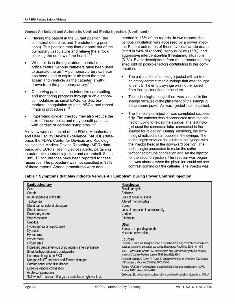
Venous Air Emboli and Automatic Contrast Media Injectors Several reports have been submitted to PA-PSRS in which intravascular air emboli occurred with the use of automatic contrast media injectors during CT scans. One report indicates that the patient became diaphoretic and was transferred to an intensive care environment after undergoing a CT scan with con-trast. In this case, the patient was prepared for the CT scan, but the empty contrast syringe from a pre-vious case had not been removed from the injector. The automatic injector injected 25cc of air into the patient. Small and moderate-sized air emboli are estimated to occur in 12% to 23% of patients undergoing con-trast-enhanced CT examination.1,2 Most of these air emboli are undetected because patients are asymp-tomatic,2 and the air is absorbed without difficulty. However, larger air emboli have been reported as a complication of pressure injection of contrast mate-rial during CT scans.2 In contrast-enhanced imag-ing, venous air emboli are more common than those in the arterial vasculature.2 Though increased mor-bidity and mortality is more likely to be associated with arterial air emboli, significant consequences can result from venous air injection, such as cardiac and/or respiratory arrest, seizures, and neurological deficits.1 Accidental venous injection of air may pro-duce a fatal air embolism.3
Three elements must be present in order for air to be admitted into the vascular system: 1) a source of air (the atmosphere); 2) a connection between the vascular system and the air source; and 3) a pres-sure gradient that favors air entry.4 Air can enter an open blood vessel when either of the following con-ditions exist: 1) a negative intravascular pressure relative to air pressure; or 2) the air is under pres-sure and is pushed into vessels with or without a negative intravascular pressure.4-6 The use of auto-matic contrast injectors meets this second condition. Air can also be introduced into the vascular system during contrast administration during cannula inser-tion, when connecting the cannula to the injector tube, and through microbubbles in the contrast.7 Once air enters the venous circulation, it moves to-ward the right atrium and then to the right ventricle. From there, emboli usually travel via the pulmonary artery to the lungs. Small emboli are usually ab-sorbed in the blood or alveoli of the lungs. Larger emboli may obstruct the outflow from the right ven-tricle or block pulmonary arterioles.2,8 The severity of symptoms resulting from air embo-lism is dependent upon such factors as the volume
of air injected and the speed of the injection.1,2 The position of the body at the time of air entry and the patient’s state of health also affect the outcome.1 Certain medical conditions may allow a venous air embolism to enter the arterial circulation, increasing the risk.1,8 Such conditions may include atrial or ventricular septal defects and those with arterioven-ous malformation.1,2 Approximately 25% to 35% of the general population with otherwise normal hearts retain a patent foramen ovale, which could allow a venous air embolism that reaches the heart to cross over into the arterial circulation. This is significant because an air embolus as small as 1 ml in the arte-rial circulation may travel to the brain or coronary arteries, causing significant blockage.5 While most air emboli associated with the use of intravenous contrast media are asymptomatic, the clinical literature reports numerous symptoms that can indicate this complication (see Table 1). Most significantly, a patient may have a reflexive gasp following an infusion of air into the pulmonary circu-lation. The gasp causes decreased intrathoracic and central venous pressure, which allows a larger vol-ume of air to enter any patent opening into a vein, potentially contributing to sufficient volume to cause cardiopulmonary collapse.4 Cardiovascular changes are generally associated with the size of the air infu-sion. Small air infusions are associated with a mod-erate decrease in blood pressure, while larger air infusions may result in a further decrease in blood pressure due to decreased cardiac output. A large air infusion may create an air lock in the pulmonary artery causing the blood pressure to drop abruptly, followed by cardiovascular collapse. The outcome may be permanent neurological damage or death.5 The most specific and sensitive methodologies used to diagnose this complication are transesophageal echocardiography and Doppler ultrasonography.1,2,9 Contrast-enhanced CT of the chest can also identify intravascular air emboli. Plain chest radiographs have occasionally identified air emboli but are less sensitive.4 In mechanically ventilated patients, this complication is associated with a reduction in the monitored end-tidal carbon dioxide level.2
If symptomatic venous air embolism occurs, the fol-lowing interventions may help to minimize harm: • Identifying the source of air entry and prevent-
ing further air entry into the venous circula-tion.4,8
Page 14 ©2004 Patient Safety Authority
PA-PSRS Patient Safety Advisory
Vol. 1, No. 4—Dec. 2004
• Placing the patient in the Durant position (the left lateral decubitus and Trendelenburg posi-tions). This position may float air back out of the pulmonary vasculature and relieve the airlock blocking the outflow of the heart.1,2,9
• When air is in the right atrium, central multi-orifice central venous catheters have been used to aspirate the air.4 A pulmonary artery catheter has been used to aspirate air from the right atrium and ventricle as the catheter is with-drawn from the pulmonary artery.2,9
• Observing patients in an intensive care setting and monitoring progress through such diagnos-tic modalities as serial EKGs, cardiac bio-markers, coagulation studies, ABGs, and repeat imaging procedures.1,9
• Hyperbaric oxygen therapy may also reduce the size of the embolus and may benefit patients with cardiac or cerebral symptoms.1,2,9
A review was conducted of the FDA’s Manufacturer and User Facility Device Experience (MAUDE) data-base, the FDA’s Center for Devices and Radiologi-cal Health’s Medical Device Reporting (MDR) data-base, and ECRI’s Health Devices Alerts, pertaining to automatic contrast injectors and air emboli. Since 1985, 13 occurrences have been reported to these resources. The procedure was not specified in 38% of these reports. Arterial procedures were docu-
mented in 46% of the reports. In two reports, the venous circulation was accessed by a power injec-tor. Patient outcomes of these events include death (cited in 54% of reports), serious injury (15%), and aggressive intervention/life threatening situations (31%). Event descriptions from these resources may shed light on possible factors contributing to this com-plication: • The patient died after being injected with air from
an empty contrast media syringe that was thought to be full. The empty syringe was not removed from the injector after a procedure.
• The technologist thought there was contrast in the
syringe because of the placement of the syringe in the pressure jacket. Air was injected into the patient.
• The first contrast injection was completed success-
fully. The catheter was disconnected from the con-nector tubing to reload the syringe. The technolo-gist used the connector tube connected to the syringe for reloading. During reloading, the tech-nologist noticed an air bubble in the syringe. The technologist expelled the air from the syringe with the injector head in the downward position. The technologist proceeded to make the cathe-ter/connector tube connection and set the injector for the second injection. The injection was begun but was aborted when the physician could not see contrast coming out the catheter. The injector was
Venous Air Emboli and Automatic Contrast Media Injectors (Continued)
Table 1 Symptoms that May Indicate Venous Air Embolism During Power Contrast Injection
Cardiopulmonary Gasp Cough Acute shortness of breath Tachypnea Chest pain/unilateral chest pain Chest pressure Pulmonary edema Bronchospasm Crepitus Hypercapnea or hypocapnea Cyanosis Hypoxemia Hypotension Hypercarbia Increased central venous or pulmonary artery pressure Sinus tachycardia/sinus bradycardia Ischemic changes on EKG Nonspecific ST segment and T wave changes Cardiac conduction disturbance Extreme venous congestion Acute cor pulmonale “Mill-wheel” murmer – if large air embolus in right ventricle
Neurological Focal paralysis Seizures Loss of consciousness Altered mental status Coma Loss of sensation in an extremity Vertigo Blindness Other Sense of impending death Nausea and vomiting Sources Pham KL, Cohen AJ. Iatrogenic venous air embolism during contrast enhanced com-puted tomography: a report of two cases. Emergency Radiology 2003; 10:147-51. Ie SR, Rozans MH, Szerlip HM. Air embolism after intravenous injection of contrast material. Southern Medical Journal 1999 Sep;92(9):930-3. Aurora R, Ward KR, Garza R, Rivers E. Iatrogenic venous air embolism. The Journal of Emergency Medicine 2000 Feb;18(2):255-6. Temple AP, Katz J. Air embolism: a potentially lethal surgical complication. AORN Journal 1987 Feb;45(2):387-400. Orebaugh SL. Venous air embolism: clinical and experimental considerations. Critical

Page 15
PA-PSRS Patient Safety Advisory
Vol. 1, No. 4—Dec. 2004
Venous Air Emboli and Automatic Contrast Media Injectors (Continued)
©2004 Patient Safety Authority
reset and injection was completed. Expelling air from the syringe with the injector head in the down-ward position is not consistent with the recom-mended procedure in the operator’s manual.
Where documented in the above reports, as well as in reports to PA-PSRS, the automatic injectors were found to have functioned properly. However, there are several things that can be done to address the risk of this complication, which may help reduce the potential for user error: Education • Limiting use of contrast media injectors to those
with adequate training and those familiar with cur-rent operating procedures as well as risks associ-ated with injection of air.1,3
Competency • Periodically verifying radiologists’ and technolo-
gists’ performance compared to current protocols.1 Written Procedures/Instructions • Having contrast injector procedures readily avail-
able to the healthcare workers.10,11
• Reviewing procedures and operator’s instructions before using any invasive diagnostic equip-ment.3,10,11
• Following the manufacturer’s instructions and op-erating manuals concerning all aspects of contrast injection, including the prescribed loading se-quence before arming the injector or preparing the contrast for injection.12
• Using a double check system to help ensure that the syringe is removed from its jacket and filled with contrast media, the system is purged of air, and the syringe is loaded—all before attaching the injector syringe and tubing to the IV cannula.10,11,13
• Verifying that empty syringes are not left in injec-tors at the end of the procedure.3
• Inspecting the cannula and the connection be-tween the cannula and power injector system to verify that no air is introduced into the system, both prior to initial injection and between multiple injec-tions of contrast.1,3,13
• Aborting the procedure if air is noticed in the con-trast injection system/tubing or when contrast is not seen coming out of the catheter.3,10,11
Protocols Developing protocols, conducting drills, and promoting compliance to clarify:
• How contrast injection responsibilities will be han-dled and transitioned during work shift changes.
• How specific tasks will be accomplished, according to the type and number of staff involved.1 For ex-ample, while a radiologist is involved in contrast-enhanced imaging, it is possible that one or two technologists may also be involved. Defining tasks for each healthcare worker in these different situa-tions may help to prevent duplication or perform-ance gaps.
Equipment • Air detection devices may reduce the risk of air
embolism associated with contrast media injectors, but only if used in conjunction with other risk reduc-tion measures designed to address user error.
• Using tightly sealed, locking connections to the
venous line may reduce the risk of air entry from a source outside of the contrast media injector.
Reporting • Notifying the person at your facility responsible for
reporting air emboli associated with contrast injec-tors.
Notes 1. Pham KL, Cohen AJ. Iatrogenic venous air embolism during con-trast enhanced computed tomography: a report of two cases. Emerg Radiol 2003;10:147-51. 2. Ie SR, Rozans MH, Szerlip HM. Air embolism after intravenous injection of contrast material. South Med J 1999 Sep;92(9):930-3. 3. Gallauresi BA. Safeguarding contrast media injections. Nursing 2001 Jan;31(1):24. 4. Orebaugh SL. Venous air embolism: clinical and experimental considerations. Crit Care Med 1992 Aug;20(8):1169-77. 5. Petts JS, Presson Jr. RG. A review of the pathophysiology of ve-nous air embolism. Anesthesiol Rev 1991 Sep/Oct:18(5):29-37. 6. Lambert MJ. Air embolism in central venous catheterization. South Med J 1982 Oct;75(10):1189-91. 7. Groell R, Schaffler GJ, Reinmueller R, Kern R. Vascular air embo-lism: location, frequency, and cause on electron-beam CT studies of the chest. Radiol 1997 Feb;202:459-62. 8. Temple AP, Katz J. Air embolism: a potentially lethal surgical com-plication. AORN J 1987 Feb;45(2):387-400. 9. Aurora T, Ward KR, Garza R, Rivers E. Iatrogenic venous air em-bolism. J Emerg Med 2000 Feb;18(2):255-6. 10. Health Devices Alerts Accession Number 30082 [database online]. Plymouth Meeting (PA): ECRI; 1997 Jan 17. 11. Health Devices Alerts Accession Number 38144 [database online]. Plymouth Meeting (PA): ECRI; 2001 Mar 16. 12. Medical Device Reporting (MDR) Database Access Number M102517 [database online] Washington (DC): FDA Center for De-vices and Radiological Health; 1985 Mar 11. [cited 2004 Sep 17]. Available from Internet: http://www.accessdata.fda.gov/ scripts/cdrh/cfdocs/cfmdr/Detail.CFM?ID=617011 13. ECRI. Healthcare Product Comparison System. Technology over-view: injectors, contrast media; angiography; computed tomography; magnetic resonance imaging. 2002 Oct.

Page 16 ©2004 Patient Safety Authority
PA-PSRS Patient Safety Advisory
Vol. 1, No. 4—Dec. 2004
A Word About Air Detection Devices
A ir detection devices, used in conjunction with other risk reduction measures, may reduce the
risk of air embolism associated with contrast media injectors. However, these systems are not fool-proof,1 and they cannot be relied upon solely to pre-vent this complication.2 Such devices include: • An oval marker that appears to be a circular dot
when observed through fluid present in the syringe. This may allow the technologist to see more readily whether the syringe contains such liquids as contrast or saline.3,4
• An optional air detection device that identifies
empty syringes and an air bolus.5,6 • A tilt sensor lockout mechanism that prevents
injection unless the injector head is tilted downward. This reduces the risk of air bubbles reaching the syringe tip.5,6
• Transparent syringes that allow air bubbles to
be more easily seen, so that air can be purged before injecton.7
• Prefilled syringes that are air-free, thus avoiding
the risk of introducing air into syringes filled on-site.6
• Ultrasound technology that stops the injection if
air is detected in the system.6 The following problems, however, can prevent such systems from working effectively:2 • If the liquid in the syringe becomes opacified
(such as when intermingled with blood), the technologist will be unable to see a liquid-detecting dot.
• Contrast procedures are performed in the dark or with reduced lighting. If a light source (such as a flashlight) is not available, the technologist may not be able to see bubbles in a transparent syringe or tubing.
• The other mechanisms address air detection in
the syringe but may not identify air entering the vascular system in other ways during contrast injection. Multiple port lines, stopcocks, trans-ducer lines, contrast conservation systems, and flush lines also may be portals of air entry.
Reduction of air emboli associated with contrast injection involves implementation of a multi-pronged approach involving not only equipment (such as air detection devices), but also training, competency assessment, procedures, manufacturer operating instructions, protocols, drills, and reporting. Notes 1. Gallauresi BA. Safeguarding contrast media injections. Nursing 2001 Jan; 31(1):24. 2. Hansel, B (ECRI). Conversation with: J. Johnston. 2004 Oct 1. 3. Pham KL, Cohen AJ. Iatrogenic venous air embolism during con-trast enhanced computed tomography: a report to two cases. Emerg Radiol (2003) 10:147-151. 4. Medrad Special Features Fluid Dots. 2003 [cited 22004 Sep 29] Available from Internet: http://www.medrad.com/systems-and-products/syringes-and-disposables/special_features.html. 5. Massat MB, ed. Technology overview: contrast media injectors-growth of MRI and CT procedure drives market demand for power injectors. Reilly Communications Group. 2003 March/April. [cited 2004 Sep 29]. Available from Internet: http://www.reillycomm.com/ it_archive/it_to0303_2.htm. 6. ECRI. Healthcare Product Comparison System. Technology over-view: injectors, contrast media, angiography; computed tomography; magnetic resonance imaging. 2002 Oct. 7. ECRI. Healthcare Product Comparison System. Comparison chart. Products for injectors, contrast media, angiography; computed tomo-graphy; magnetic resonance imaging. 2002.

Page 17 ©2004 Patient Safety Authority
PA-PSRS Patient Safety Advisory
Vol. 1, No. 4—Dec. 2004
Drug Name Suffix Confusion is a Common Source of Errors
M edications with delayed- or extended-release formulations can play a vital role in improving
adherence to drug therapy. These unique dosage formulations avoid the need for multiple daily doses of a medication due to their delayed or sustained delivery of a total daily dose steadily throughout the day. This is convenient for patients, may reduce certain side effects, and, on occasion, even allows use for different indications. However, the nomen-clature used for long-acting dosage forms is often confusing, and errors may occur when the same drug has several oral dosage forms with different release rates.
The practice of adding “suffixes” or “modifiers” (e.g., Depakote ER or Cardizem CD) to medica-tion names is used by manufacturers to main-tain brand awareness while signifying that the formulation is different from the immediate-release version of the
product. However, there is no standardization of the terms for the many different kinds of long-acting formulations. As a result, there are many inconsis-tencies, allowing different suffixes to be used for an identical formulation by two different manufacturers or even similar suffixes for dissimilar formulations. In short, the nomenclature used for these formulations often fails to provide appropriate “cues” regarding proper use of a dosage form.1
In addition to lack of standards, another problem is that health professionals have been known to com-municate drug names that have suffixes, but omit the suffix. This occasionally results in patients get-ting the immediate-release version and thus, an en-tire day’s dose at one time, sometimes with adverse effects. Practitioners have also been known to in-clude suffixes that do not exist for the specified product.2 Additional contributing factors reported to PA-PSRS include similar packaging, overlapping dosages, and storage of the products next to each other. These factors combine to allow confusion, inefficiencies, and medication errors at various stages in the medication use system.
In an analysis of 402 prescribing errors, Lesar3
found that the most common type of error was fail-ure to specify the controlled-release formulation (280 cases, 69.7%). The Institute for Safe Medica-
tion Practices (ISMP) has received reports of confu-sion between Abbott’s DEPAKOTE ER (divalproex sodium extended release) and DEPAKOTE (divalproex sodium delayed release).4 Additional examples include GLUCOTROL and GLUCOTROL XL as well as GLUCOPHAGE and GLUCOPHAGE XR.
The most common examples of this type of error reported to PA-PSRS include mix-ups between products such as:
• ADDERALL and ADDERALL XR • EFFEXOR and EFFEXOR XR • VICODIN and VICODIN ES Additional examples include:
• AUGMENTIN and AUGMENTIN XR • CARDIZEM and CARDIZEM CD • CIPRO and CIPRO XR • DEPAKOTE and DEPAKOTE ER • DETROL and DETROL LA • LOPRESSOR and LOPRESSOR XL (the XL
formulation is Toprol XL) • RYTHMOL and RYTHMOL SR • SENOKOT and SENOKOT S • SINEMENT and SINEMENT CR • verapamil and verapamil SR
The confusion multiplies when there are two or more “extended” release formulations for the same products, which are not therapeutically equivalent or “substitutable.” Some products have numerous suf-fixes to differentiate formulations of the same drug. For example, suffixes for various diltiazem products include SR, CD, XR, XT, and LA. ISMP also has received reports where pharmacists dispensed METADATE ER instead of METADATE CD. Simi-larly, ISMP has received a report where a prescrip-tion for METADATE CD 20 mg was dispensed as METADATE ER 20 mg. The pharmacists involved in these errors weren’t aware that the METADATE CD product existed.5 PA-PSRS has received reports noting confusion between medications such as the once-a-day formulation WELLBUTRIN XL (bupropion extended-release) and WELLBUTRIN SR (buproprion sustained-release), which is indi-cated for twice-daily dosing. Wellbutrin mix-ups are especially likely since both the SR and XL formula-tions are available in 150 mg tablet strengths, and it’s not unusual for the SR formulation to be pre-scribed once daily.
The nomenclature used to distinguish different drug for-
mulations often fails to provide “cues”
regarding proper use of a dosage form.

Page 18 ©2004 Patient Safety Authority Vol. 1, No. 4—Dec. 2004
PA-PSRS Patient Safety Advisory
Drug name suffixes are confusing enough without coining them on our own. One report to ISMP in-volved a physician assistant that wrote a prescrip-tion for a patient, which was misread by both the pharmacist and technician as VICODIN ES (hydrocodone 7.5 mg/ acetaminophen 750 mg). However, on closer inspection, the suffix looked more like RS. The pharmacist called the physician assistant and learned that he had used “RS” to mean “regular strength.” VICODIN (hydrocodone 5 mg/acetaminophen 500 mg) was then dispensed.6
In order to improve the landscape when suffixes are used and reduce the risk of errors, nomenclature standards would help to diminish confusion between various formulations of the same drug.7 Standard suffixes or descriptive phrases might be incorpo-rated directly into the drug name, or a unique brand name might be needed to designate a different for-mulation property, as was done with NEORAL (cyclosporine modified) and SANDIMMUNE (cyclosporine). FDA is aware of these problems and will be examining ways to improve trademark no-menclature.
Being mindful of the potential for this type of confu-sion when prescribing, storing, dispensing, and ad-ministrating such medications is just the first step in prevention. Other preventive strategies to consider include:
• Selectively building flags into computer systems and marking drug containers to warn staff about the differences where a high risk of error exists or when a mix-up might be very serious.
• Designing computer mnemonics to separate the
different formulations on computer screens used during order entry.
Drug Name Suffix Confusion is a Common Source of Errors (Continued) • Storing similarly named drugs separately and
using auxiliary labels to differentiate the prod-ucts in medication storage areas.
• Verifying new prescriptions for any of these
medications where prescriber confusion among suffixes has been reported.
• When communicating orders orally, using the
full words “extended release” or “sustained re-lease,” not abbreviations, especially for those medications that are available as an immediate release formulation.
• Involving patients also may help. When pre-
scribing and dispensing one of these medica-tions, practitioners may want to inform patients of the potential for confusion between the vari-ous formulations and suffixes.
Notes 1. Cohen MR, ed. Medication Errors. Causes, Preventions and Risk Management. Sudbury (MA): Jones and Bartlett Publishers; 2000. 2. ISMP Medication Safety Alert! Community Phar-macy/Ambulatory Edition. 15 Jul 2004;(9)14. 3. Lesar TS. Medication errors related to dosage formulation issues. Medscape Pharmacists 2001. Available on Internet: http://www.medscape.com/viewarticle/408579_print 4. ISMP Medication Safety Alert! 7 Feb 2001;(6)3. 5. ISMP Medication Safety Alert!. 28 Nov 2001;(6)24. 6. ISMP Medication Safety Alert! [online]. 15 Nov 2004;(9)14. 7. Cohen MR. Medication error reports. Hosp Pharm. 1990;25:747-8.
The Institute for Safe Medication Practices (ISMP) recently presented its 2004 Cheers awards to organizations and individuals who have set a standard of excellence this year in the prevention of medication errors.
The following Pennsylvania organizations were among the recipients for innovative technology in the prevention of medi-cation errors: Abington Physician Network of Abington Memorial Hospital for its creation of webINR, a virtual antico-agulation clinic that was successfully implemented in more
than two dozen primary care offices, and Reading Hospital and Medical Center for its web-based RxToolkit, a point-of-care program for pharmacists and nurses.
The 2004 ISMP Lifetime Achievement Award was presented to Neil M. Davis, PharmD, MS, FASHP (Professor Emeritus, Temple University School of Pharmacy, and Editor Emeritus, Hospital Pharmacy) for his pioneering efforts envisioning a nationwide medication safety initiative as far back as 1974.
Pennsylvania Facilities Recognized for Patient Safety

Page 19 ©2004 Patient Safety Authority Vol. 1, No. 4—Dec. 2004
PA-PSRS Patient Safety Advisory
Understanding the Benchmarking Process
B enchmarking is an ongoing process that deter-mines how other organizations have achieved
optimal performance. Through the process of benchmarking, ways are suggested for adapting the best practices that result in exception performance. Although measurement is one of its components, effective benchmarking is a dual process that re-quires two products: performance measurement and enablers. Benchmarks are measures of comparative perform-ance that answer the question: What is your level of performance? By itself, this information has little use in improving performance. To be effective, bench-marking must also provide a systematic method of understanding the underlying process that deter-mines an organization’s performance. To that end, enablers must be identified. Enablers are the spe-cific practices that lead to exemplary performance; they answer the question: How do you do it? Over-looking either one of these components in the benchmarking process renders it useless, even dan-gerous. Although medication error rates, for example, may seem ideal for benchmarking, we must question the wisdom of applying the benchmarking concept to the medication use process when the focus is on error rates. Certainly, the confusion surrounding the term “benchmarking” perpetuates the myth that one can gauge the quality and safety of the medication use process simply by comparing error rates, both within an organization and externally. The true inci-dence of medication errors varies, however, de-pending heavily on the rigor with which the events are clearly identified and reported. Because many medication errors cause no harm to patients, they remain undetected or unreported. Still, organizations often depend only on spontane-ous, voluntary reporting of errors to determine the rate at which errors occur. The inherent variability of determining an error rate in this way invalidates the measurement, or benchmark. A high error rate may suggest either unsafe medication practices or an organizational culture that promotes error reporting. Conversely, a low error rate may suggest either suc-cessful error prevention strategies or a punitive cul-ture that inhibits error reporting. Moreover, the defi-
nition of a medication error may not be consistent among organizations or even between individual practitioners in the same organization. Thus, spon-taneous error reporting is a poor method of gather-ing benchmarks; it is not designed to measure medi-cation error rates. Of equal concern is the mistaken belief that bench-marking is simply a process of comparing numbers. Although this activity produces no meaningful infor-mation, healthcare organizations have embraced the practice of comparing error rates. Yet, there has been little effective effort directed at identifying en-ablers for safe medication use to accompany this attempt at benchmarking. As a result, organizations focus undue attention on maintaining a low error rate, giving the errors themselves, rather than their correction, disproportionate importance. This pro-motes an unproductive cycle of underreporting er-rors, which results in unrecognized weaknesses in the medication use system. Thus, low error rates can result in a false sense of security and a tacit acceptance of preventable errors. Benchmarking for the medication use process can be effective only if a system of objective measure-ment, which is more reliable than spontaneous error reporting alone, is used to identify best practices (such as observational methods or systematic evaluation of errors). In addition, the benchmarking process must include a method for accurately determining the specific processes that enable the organization to achieve an environment in which medications are safely used. Success is more likely with benchmarking projects that are focused on specific areas of drug therapy (such as insulin or anticoagulant therapy) so that accurate benchmarks (performance measures) and enablers (practices that lead to exemplary per-formance) can be more easily identified and imple-mented. Benchmarking projects should be carefully selected. Organizations are urged to place less emphasis on error rates that are based solely on spontaneous, voluntary reporting programs. Instead, error report-ing should be encouraged in order to identify and remedy problems, rather than to provide statistics for comparison. Source Cohen MR, ed. Medication Errors. Washington, DC: American Pharmacists Association (APhA); 1999. Copyright APhA. Re-printed with permission.
This article is excerpted from the book Medication Errors by Michael Cohen, RPh, MS, ScD. Dr. Cohen is President of the Institute for Safe Medication Practices (ISMP) and serves as an expert advisor to PA-PSRS in the area of medication safety.

Page 20 ©2004 Patient Safety Authority Vol. 1, No. 4—Dec. 2004
PA-PSRS Patient Safety Advisory
ECRI is an independent, nonprofit health services research agency dedicated to improving the safety, efficacy and cost-effectiveness of healthcare. ECRI’s focus is healthcare technology, healthcare risk and quality management and healthcare environmental management. ECRI provides information services and technical assistance to more than 5,000 hospitals, healthcare organizations, ministries of health, government and planning agencies, and other organizations worldwide.
The Patient Safety Authority is an independent state agency created by Act 13 of 2002, the Medical Care Availability and Reduction of Error (“Mcare”) Act. Consistent with Act 13, ECRI, as contractor for the PA-PSRS program, is issuing this newsletter to advise medical facilities of immediate changes that can be instituted to reduce serious events and incidents. For more information about the PA-PSRS program or the Patient Safety Authority, see the Authority’s website at www.psa.state.pa.us.
The Institute for Safe Medication Practices (ISMP) is an independent, nonprofit organization dedicated solely to medication error prevention and safe medication use. ISMP provides recommendations for the safe use of medications to the healthcare community including healthcare professionals, government agencies, accrediting organizations, and consumers. ISMP's efforts are built on a non-punitive approach and systems-based solutions.
An Independent Agency of the Commonwealth of Pennsylvania