Embed Size (px)
Citation preview

PATIENT INFORMATION Spiration® Valve System
Humanitarian Device
2 Patient Information for the Spiration® Valve System
GLOSSARYAir leak: Air leaking from lung tissue into the chest cavity.
Air leak localization: Process by which an air leak in the lung is found.
Airway: The tubes in the lungs that pass air to and from the lung tissue.
Anesthesia: A medical procedure that makes the patient unconscious and makes the body insensitive to pain.
Asthma: Swelling of the airways in the lung, making breathing difficult.
Atelectasis: Collapse of all or part of the lung.
Balloon Catheter: A narrow, flexible tube with a small balloon at the end.
Bronchiectasis: A lung condition in which the airways of the lungs get bigger and often get infected.
Bronchitis: An infection that causes inflammation of lung tissue.
Bronchoscope: A narrow, flexible tube, with a camera on the end that is used to see the lung airways.
CT Scan: An image taken of a body part.
Catheter: A narrow, flexible tube used to deliver medicine or medical devices inside the body.
Chest drainage system: A device that allows medical staff to measure and monitor an air leak.
Chest tube: A plastic tube that is inserted through the skin and rib cage and into the chest cavity to allow air and fluids leaking from the lung to exit the body.
Chest X-ray: a picture of the chest that shows your heart, lungs, airway, blood vessels, and lymph nodes.
Contraindication: Reason not to use the device/therapy.
Expiration: When a person breaths out.
Forceps: A common tool used in medical procedures to help doctors grab onto tissue and other objects in the body.
Intubation: Placement of a plastic tube into the mouth and down into the airway so that a person can be put on a breathing machine or so that a procedure can be done.
Lobectomy: Surgery to remove a lobe of the lung.
Lung Volume Reduction Surgery (LVRS): A major surgery in which more than one portion of the lung is removed.
MRI: Magnetic Resonance Imaging. A method for taking pictures of your internal organs.
Nitinol: A metal made of nickel and titanium. This is a common material used in medical devices.
Pleurodesis: A procedure that causes the outside of the lung to stick to the inside of the chest cavity to prevent the lung from collapsing.
Pneumonia: A lung infection with symptoms like a cough, fever, and difficulty breathing.
Polymer membrane: A thin sheet of flexible plastic. This is a common material used in medical devices.
Prolonged air leak: An air leak that lasts for 7 or more days.
Re-operation: The chest is surgically re-opened and a new attempt is made to close the leaking lung tissue.
Respiratory compromise: Breathing difficulty — causes less oxygen to be available to the body and negatively affects bodily functions.
Sedation: A medical procedure in which medicine is given to the patient to make him or her comfortable and unaware of pain during surgery. It relaxes the central nervous system and can cause sleepiness.
Segmentectomy: Surgery to remove a segment (small portion) of the lung.
Significant air leak: An air leak that is severe and/or produces other health problems at the same time.
Subcutaneous emphysema: Occurs when air gets into tissues under the skin.
Ventilator: A machine that moves air into and out of the lungs for people who have a hard time breathing on their own.
3Humanitarian Device
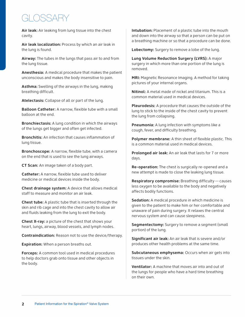
Glossary .............................................................................................................................................................. page 2
Device Description ........................................................................................................................................ page 4
Spiration Valve System Intended Use ................................................................................................ page 5
When the Spiration Valve System Should Not Be Used (Contraindications) ................................................................................................................................. page 5
Potential Risks and Benefits of the Spiration Valve System for Treatment of Air Leaks ...................................................................................................................page 6
The Procedure .................................................................................................................................................page 8
What Happens if the Spiration Valve Does Not Heal the Air Leak? ..................................page 10
Other Treatments for Patients with a Prolonged Air Leak .....................................................page 10
Summary of Clinical Data ......................................................................................................................... page 11
Figure 1. The Spiration Valve
TABLE OF CONTENTS
4 Patient Information for the Spiration® Valve System
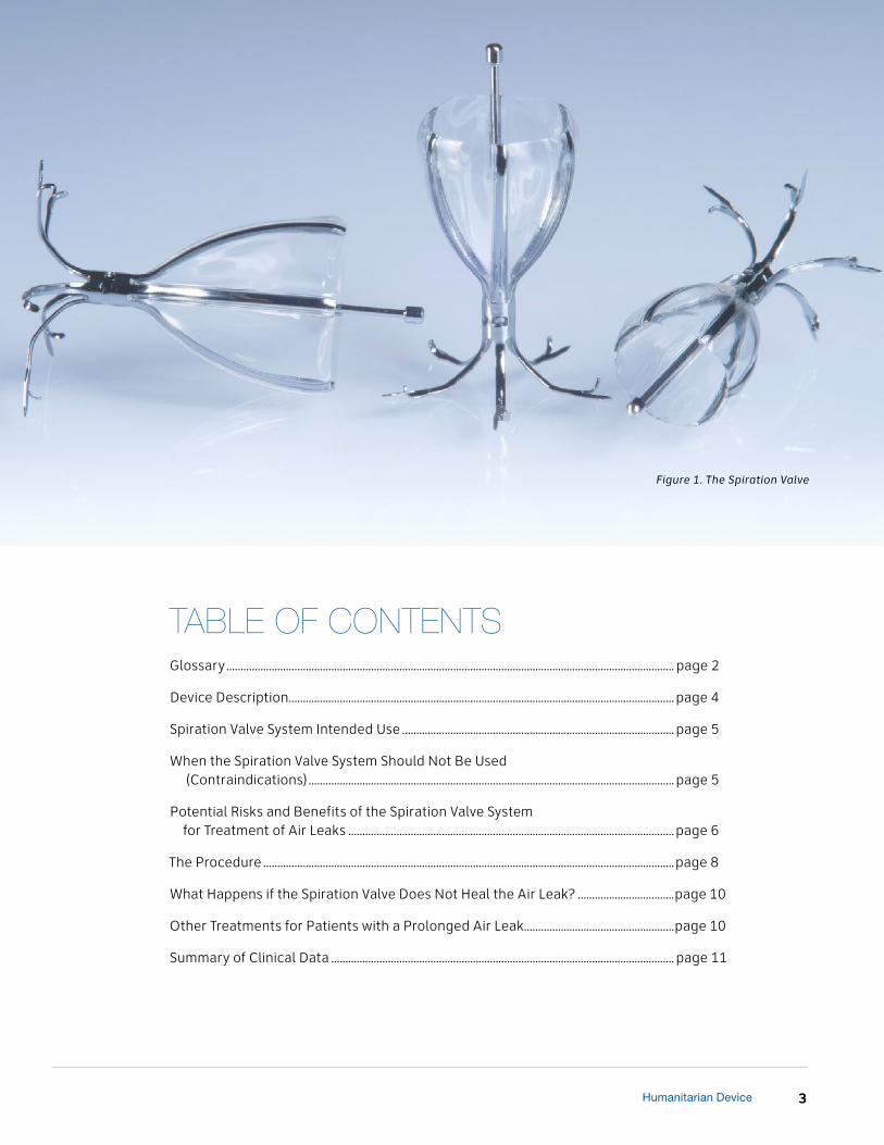
The Spiration Valve System consists of three main parts.
The ValveThe umbrella shaped valve is made of a flexible metal frame (nitinol – composed of titanium and nickel) covered by a thin sheet of flexible plastic, a polymer membrane (see Figure 1). The valve is designed to fit the shape and size of airways of the lung. The valve is a one-way device, which means that the plastic umbrella blocks air from moving further into your lungs toward the air leak, but allows trapped air to move out of your lungs as you breath out (see Figures 2 a and b). Blocking air from reaching the air leak is what may help the air leak heal. The valve has five flexible anchors that settle into the airway wall helping the valve stay in place. The valves are available in a few different sizes to fit various airways. The valve is designed to be removed once the air leak has healed.
The Deployment Catheter and Loader The loader (see Figure 3 a) is used during the procedure to load the valve into a narrow tube called the deployment catheter. The deployment catheter (see Figure 3 b) delivers the valve to your airway. The parts of this catheter that touch the patient are made of stainless steel, plastic, nylon, and non-toxic yellow dye.
The Airway Sizing Kit The airway sizing kit is used to prepare a narrow, flexible tube with a small balloon at the end (balloon catheter) for use during the procedure. None of the contents of this kit are used as part of the valve placement procedure.
Other Required ItemsThere are a few things required for the procedure that are not included in the Spiration Valve System. These include a balloon catheter and a bronchoscope.
The Balloon CatheterThe balloon catheter is a narrow, flexible tube with a small balloon at the end. It is used to find the air leak and to measure airways inside of your lungs (see Figure 4).
BronchoscopeA bronchoscope is a narrow, flexible tube that goes into your lungs. There is a camera on the end so that doctors can see internal organs. In this procedure the camera will allow your doctor to see the airways of your lung. The deployment catheter and balloon catheter go through the bronchoscope to reach the airways (see Figure 5).
Figure 4. A Balloon Catheter
Figure 5. Bronchoscope with a Spiration Valve Catheter
Figure 2 a and b. Illustration of the Spiration Valve blocking new air while allowing trapped air and fluids to escape.
Figure 3 a and b. The Loader and Deployment Catheter
a)
b)
DEVICE DESCRIPTION
5Humanitarian Device
WHEN THE SPIRATION VALVE SYSTEM SHOULD NOT BE USED (CONTRAINDICATIONS)Your doctor will not place a Spiration Valve in your lungs if you are unable to have a narrow, flexible tube (bronchoscope) placed through your nose or mouth and into your lungs.
Intended UseThe Spiration Valve System is intended to control prolonged air leaks (see definition below) of the lung, or significant air leaks that are likely to become prolonged air leaks, following:
• surgery to remove a lobe of the lung (a lobectomy),
• surgery to remove a segment, or small portion, of the lung (a segmentectomy) or
• a major surgery in which multiple portions of the lung are removed (Lung Volume Reduction Surgery or LVRS).
If you still have an air leak 7 days after your surgery, you have a prolonged air leak, unless the leak is there only when you:
• forcefully blow out air (forced exhale — this is done to see if you have an air leak) or
• cough.
You can be considered for a Spiration Valve placement if you have an air leak 5 days after surgery and if the air leak is:
• continuous,
• present when you breath in normally (not forced), or
• present when you breath out normally (not forced) and you have either trapped air under the skin (subcutaneous emphysema) or difficulty breathing (respiratory compromise).
Use of the Spiration Valve System is limited to 6 weeks for each prolonged air leak.
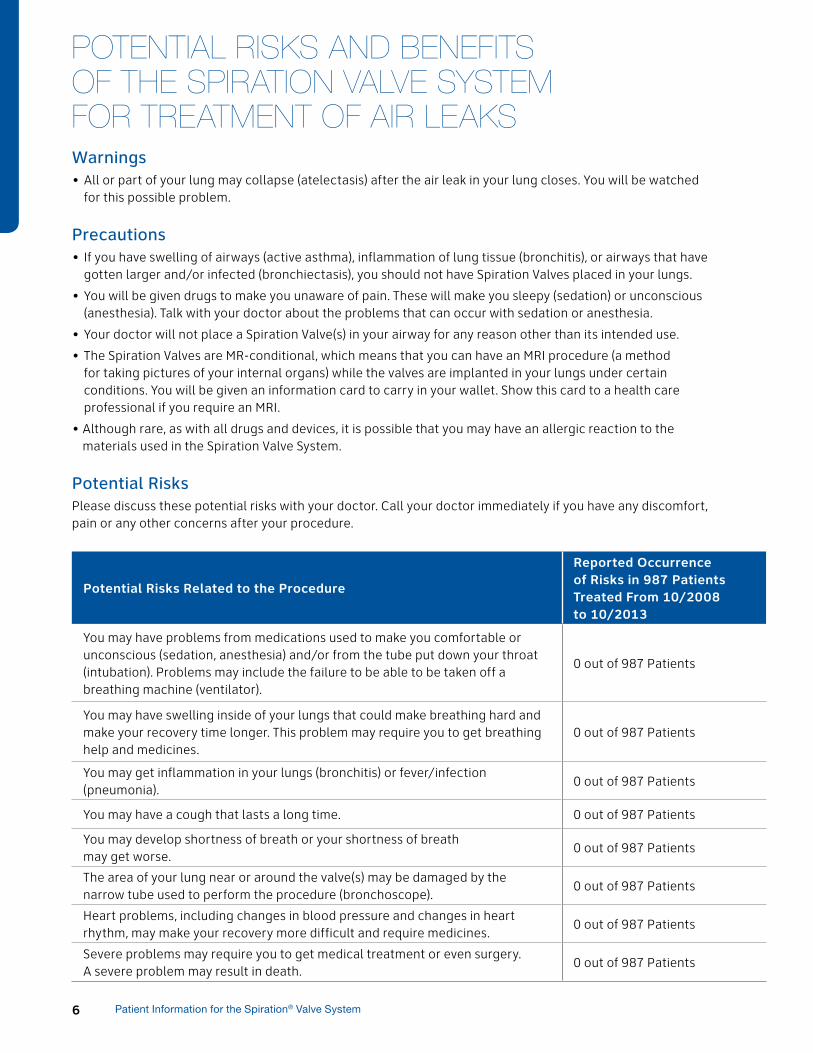
What is a prolonged air leak?In surgeries where part of the lung has been removed, air leaks are common problems. Air leaks are caused because the lung tissue does not always close completely after surgery. Air that continues to leak from the lung(s) can build up in the patient’s chest and can make breathing difficult. Air leaks are usually treated with a temporary tube inserted into the chest cavity (chest tube) that removes the air from between the lung and the ribcage (see Figure 6 a and b). In most cases air leaks will close after a few days. However, some patients will have a leak that does not close. These are referred to as “prolonged” air leaks. If you have more questions about air leaks please discuss them with your doctor.
Figure 6 a and b. Chest tube inserted to remove air between lung and ribcage due to lung air leak.
Figure 3 a and b. The Loader and Deployment Catheter
SPIRATION VALVE SYSTEM INTENDED USE
6 Patient Information for the Spiration® Valve System
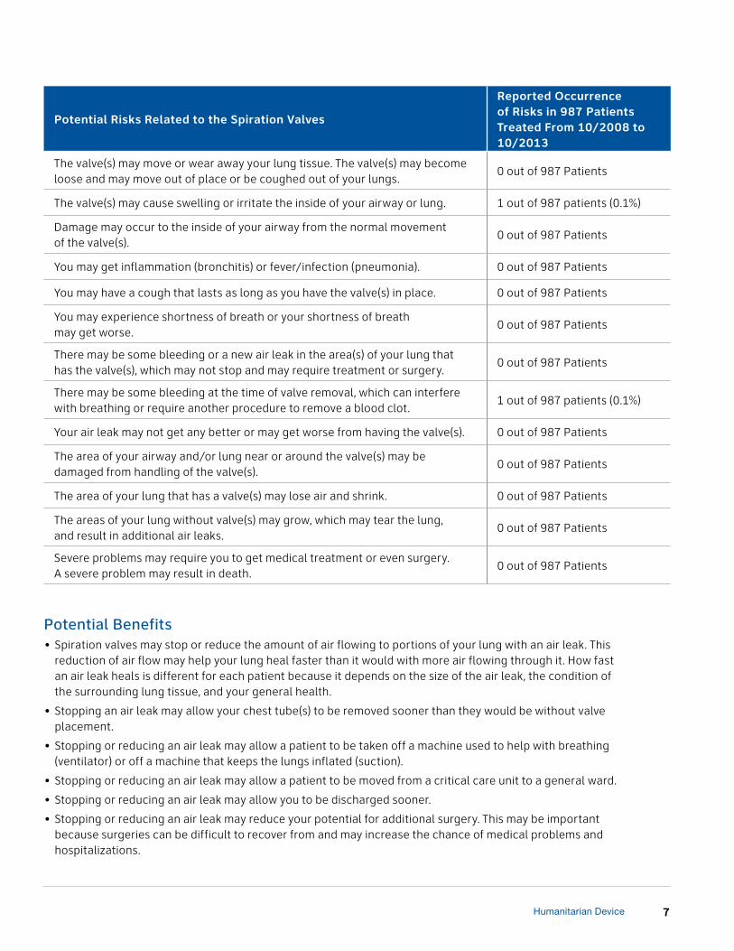
Potential Risks Related to the Procedure
Reported Occurrence of Risks in 987 Patients Treated From 10/2008 to 10/2013
You may have problems from medications used to make you comfortable or unconscious (sedation, anesthesia) and/or from the tube put down your throat (intubation). Problems may include the failure to be able to be taken off a breathing machine (ventilator).
0 out of 987 Patients
You may have swelling inside of your lungs that could make breathing hard and make your recovery time longer. This problem may require you to get breathing help and medicines.
0 out of 987 Patients
You may get inflammation in your lungs (bronchitis) or fever/infection (pneumonia).
0 out of 987 Patients
You may have a cough that lasts a long time. 0 out of 987 Patients
You may develop shortness of breath or your shortness of breath may get worse.
0 out of 987 Patients
The area of your lung near or around the valve(s) may be damaged by the narrow tube used to perform the procedure (bronchoscope).
0 out of 987 Patients
Heart problems, including changes in blood pressure and changes in heart rhythm, may make your recovery more difficult and require medicines.
0 out of 987 Patients
Severe problems may require you to get medical treatment or even surgery. A severe problem may result in death.
0 out of 987 Patients
Warnings• All or part of your lung may collapse (atelectasis) after the air leak in your lung closes. You will be watched
for this possible problem.
Precautions• If you have swelling of airways (active asthma), inflammation of lung tissue (bronchitis), or airways that have
gotten larger and/or infected (bronchiectasis), you should not have Spiration Valves placed in your lungs.
• You will be given drugs to make you unaware of pain. These will make you sleepy (sedation) or unconscious (anesthesia). Talk with your doctor about the problems that can occur with sedation or anesthesia.
• Your doctor will not place a Spiration Valve(s) in your airway for any reason other than its intended use.
• The Spiration Valves are MR-conditional, which means that you can have an MRI procedure (a method for taking pictures of your internal organs) while the valves are implanted in your lungs under certain conditions. You will be given an information card to carry in your wallet. Show this card to a health care professional if you require an MRI.
• Although rare, as with all drugs and devices, it is possible that you may have an allergic reaction to the materials used in the Spiration Valve System.
Potential RisksPlease discuss these potential risks with your doctor. Call your doctor immediately if you have any discomfort, pain or any other concerns after your procedure.
POTENTIAL RISKS AND BENEFITS OF THE SPIRATION VALVE SYSTEM FOR TREATMENT OF AIR LEAKS
7Humanitarian Device
Potential Risks Related to the Spiration Valves
Reported Occurrence of Risks in 987 Patients Treated From 10/2008 to 10/2013
The valve(s) may move or wear away your lung tissue. The valve(s) may become loose and may move out of place or be coughed out of your lungs.
0 out of 987 Patients
The valve(s) may cause swelling or irritate the inside of your airway or lung. 1 out of 987 patients (0.1%)
Damage may occur to the inside of your airway from the normal movement of the valve(s).
0 out of 987 Patients
You may get inflammation (bronchitis) or fever/infection (pneumonia). 0 out of 987 Patients
You may have a cough that lasts as long as you have the valve(s) in place. 0 out of 987 Patients
You may experience shortness of breath or your shortness of breath may get worse.
0 out of 987 Patients
There may be some bleeding or a new air leak in the area(s) of your lung that has the valve(s), which may not stop and may require treatment or surgery.
0 out of 987 Patients
There may be some bleeding at the time of valve removal, which can interfere with breathing or require another procedure to remove a blood clot.
1 out of 987 patients (0.1%)
Your air leak may not get any better or may get worse from having the valve(s). 0 out of 987 Patients
The area of your airway and/or lung near or around the valve(s) may be damaged from handling of the valve(s).
0 out of 987 Patients
The area of your lung that has a valve(s) may lose air and shrink. 0 out of 987 Patients
The areas of your lung without valve(s) may grow, which may tear the lung, and result in additional air leaks.
0 out of 987 Patients
Severe problems may require you to get medical treatment or even surgery. A severe problem may result in death.
0 out of 987 Patients
Potential Benefits• Spiration valves may stop or reduce the amount of air flowing to portions of your lung with an air leak. This
reduction of air flow may help your lung heal faster than it would with more air flowing through it. How fast an air leak heals is different for each patient because it depends on the size of the air leak, the condition of the surrounding lung tissue, and your general health.
• Stopping an air leak may allow your chest tube(s) to be removed sooner than they would be without valve placement.
• Stopping or reducing an air leak may allow a patient to be taken off a machine used to help with breathing (ventilator) or off a machine that keeps the lungs inflated (suction).
• Stopping or reducing an air leak may allow a patient to be moved from a critical care unit to a general ward.
• Stopping or reducing an air leak may allow you to be discharged sooner.
• Stopping or reducing an air leak may reduce your potential for additional surgery. This may be important because surgeries can be difficult to recover from and may increase the chance of medical problems and hospitalizations.
8 Patient Information for the Spiration® Valve System
How to Prepare for the ProcedureBefore the procedure begins your doctor will give you medicine that will make you unaware of pain and will make you sleepy (sedation) or unconscious (anesthesia). If you know you are allergic to the drugs used to make you sleepy (sedatives) or unconscious (anesthesia), let your doctor know. If you have any concerns about sedation or anesthesia, talk with your doctor before the procedure. Your doctor will talk with you about the best type of sedation or anesthesia for you.
The narrow tube with a balloon at the end (balloon catheter) used to find the air leak and to measure the airways may be made of latex. If you are allergic to latex, let your doctor know and a non-latex balloon catheter will be used. A person who is highly allergic to latex can have an anaphylactic shock, a life threatening allergic reaction that may cause death.
What to Expect During the Placement ProcedureThe amount of time needed for the procedure will vary depending on the number and location of the air leaks. Based on use of the Spiration Valve System since June 2008, the average number of valves placed per patient is 3.
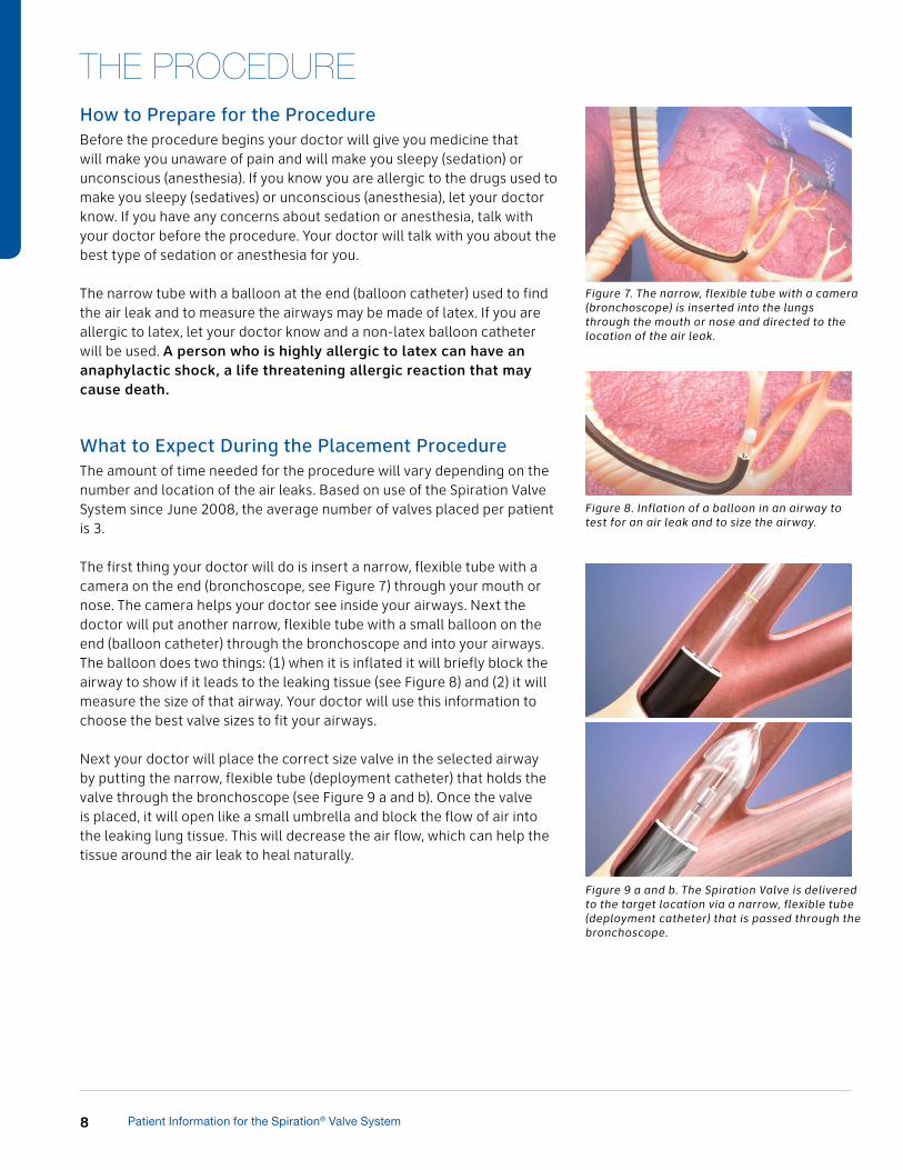
The first thing your doctor will do is insert a narrow, flexible tube with a camera on the end (bronchoscope, see Figure 7) through your mouth or nose. The camera helps your doctor see inside your airways. Next the doctor will put another narrow, flexible tube with a small balloon on the end (balloon catheter) through the bronchoscope and into your airways. The balloon does two things: (1) when it is inflated it will briefly block the airway to show if it leads to the leaking tissue (see Figure 8) and (2) it will measure the size of that airway. Your doctor will use this information to choose the best valve sizes to fit your airways.
Next your doctor will place the correct size valve in the selected airway by putting the narrow, flexible tube (deployment catheter) that holds the valve through the bronchoscope (see Figure 9 a and b). Once the valve is placed, it will open like a small umbrella and block the flow of air into the leaking lung tissue. This will decrease the air flow, which can help the tissue around the air leak to heal naturally.
Figure 7. The narrow, flexible tube with a camera (bronchoscope) is inserted into the lungs through the mouth or nose and directed to the location of the air leak.
Figure 8. Inflation of a balloon in an airway to test for an air leak and to size the airway.
Figure 9 a and b. The Spiration Valve is delivered to the target location via a narrow, flexible tube (deployment catheter) that is passed through the bronchoscope.
THE PROCEDURE
9Humanitarian Device
What to Expect After the Placement ProcedureFollowing the procedure you will likely still have your chest tube(s) in place. The reason for this is to let air in your chest escape and allow your doctor to measure the air leak. Depending on your overall health and how well your air leak is healing, your doctor will decide if you should stay in the hospital and how long your chest tube(s) should stay in place. Every air leak is different and the time it takes to heal can be very different from person to person. Therefore, it is important for you to talk to your doctor about your condition and the post-procedure plan.
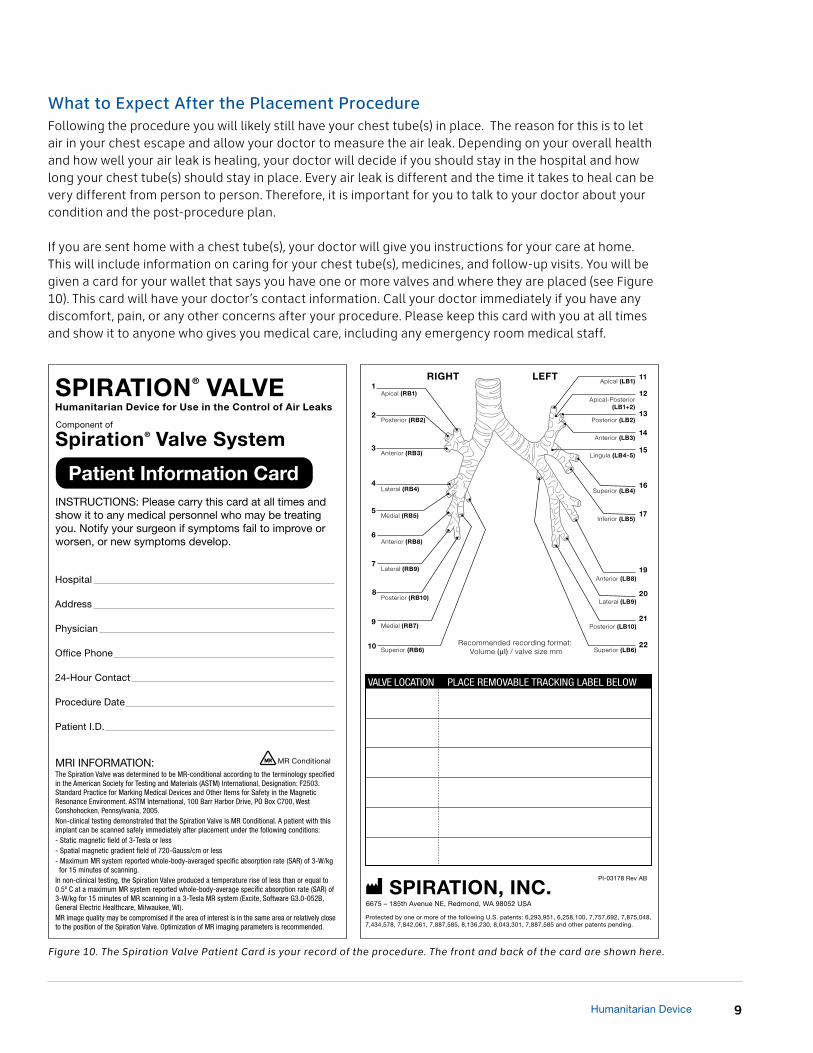
If you are sent home with a chest tube(s), your doctor will give you instructions for your care at home. This will include information on caring for your chest tube(s), medicines, and follow-up visits. You will be given a card for your wallet that says you have one or more valves and where they are placed (see Figure 10). This card will have your doctor’s contact information. Call your doctor immediately if you have any discomfort, pain, or any other concerns after your procedure. Please keep this card with you at all times and show it to anyone who gives you medical care, including any emergency room medical staff.
Figure 10. The Spiration Valve Patient Card is your record of the procedure. The front and back of the card are shown here.
10 Patient Information for the Spiration® Valve System
What to Expect When the Spiration Valve is RemovedThe valve will be removed when your doctor is confident that the air leak has stopped and there has been sufficient time for the tissue to heal. This should be about 2-6 weeks. Your doctor will check to see if the air leak has stopped by taking a picture of your lungs (using a chest x-ray or CT scans) or by bending/kinking your chest tube while it is attached to a device that measures and monitors air leaks (chest drainage system).
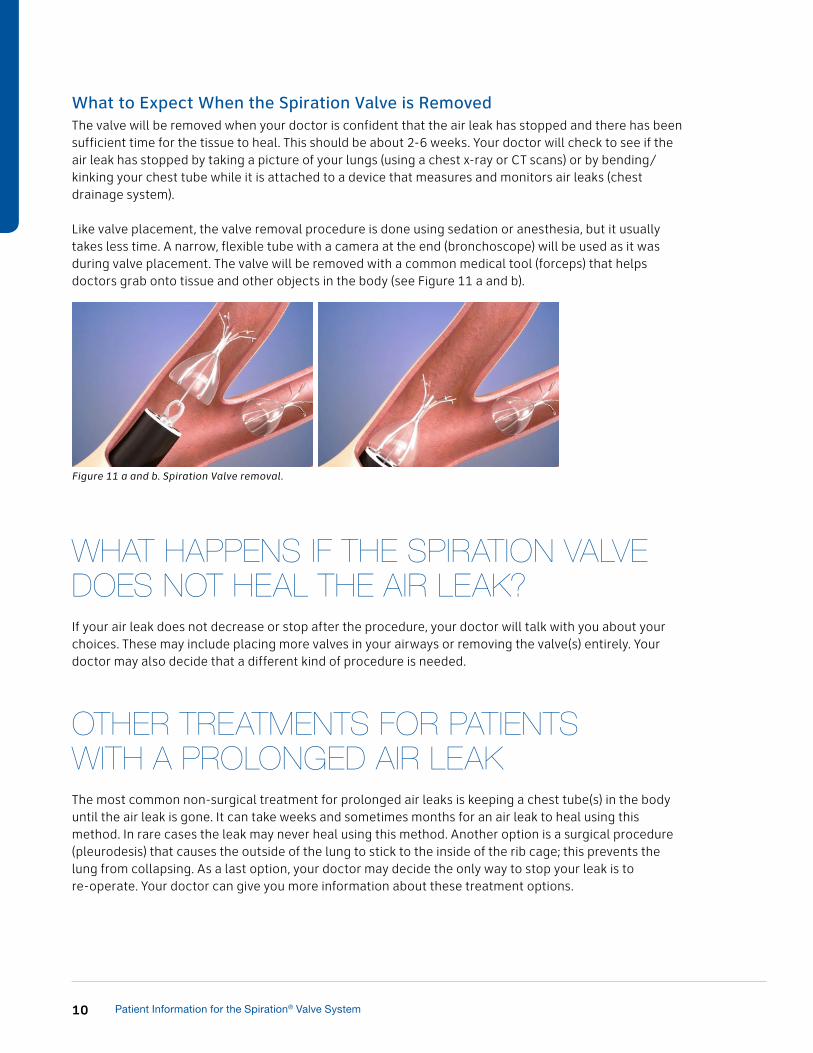
Like valve placement, the valve removal procedure is done using sedation or anesthesia, but it usually takes less time. A narrow, flexible tube with a camera at the end (bronchoscope) will be used as it was during valve placement. The valve will be removed with a common medical tool (forceps) that helps doctors grab onto tissue and other objects in the body (see Figure 11 a and b).
WHAT HAPPENS IF THE SPIRATION VALVE DOES NOT HEAL THE AIR LEAK?If your air leak does not decrease or stop after the procedure, your doctor will talk with you about your choices. These may include placing more valves in your airways or removing the valve(s) entirely. Your doctor may also decide that a different kind of procedure is needed.
OTHER TREATMENTS FOR PATIENTS WITH A PROLONGED AIR LEAKThe most common non-surgical treatment for prolonged air leaks is keeping a chest tube(s) in the body until the air leak is gone. It can take weeks and sometimes months for an air leak to heal using this method. In rare cases the leak may never heal using this method. Another option is a surgical procedure (pleurodesis) that causes the outside of the lung to stick to the inside of the rib cage; this prevents the lung from collapsing. As a last option, your doctor may decide the only way to stop your leak is to re-operate. Your doctor can give you more information about these treatment options.
Figure 11 a and b. Spiration Valve removal.
11Humanitarian Device
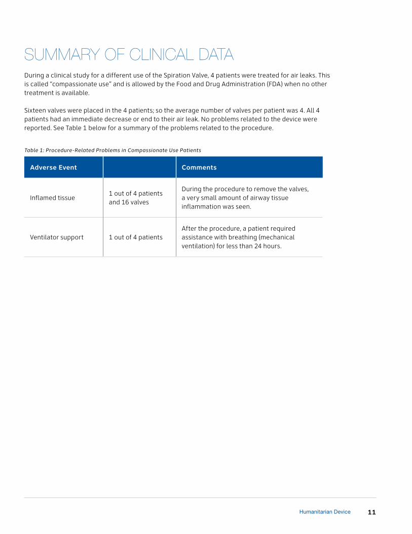
SUMMARY OF CLINICAL DATADuring a clinical study for a different use of the Spiration Valve, 4 patients were treated for air leaks. This is called “compassionate use” and is allowed by the Food and Drug Administration (FDA) when no other treatment is available.
Sixteen valves were placed in the 4 patients; so the average number of valves per patient was 4. All 4 patients had an immediate decrease or end to their air leak. No problems related to the device were reported. See Table 1 below for a summary of the problems related to the procedure.
Adverse Event Comments
Inflamed tissue1 out of 4 patients and 16 valves
During the procedure to remove the valves, a very small amount of airway tissue inflammation was seen.
Ventilator support 1 out of 4 patientsAfter the procedure, a patient required assistance with breathing (mechanical ventilation) for less than 24 hours.
Table 1: Procedure-Related Problems in Compassionate Use Patients
For full prescribing information, go to: www.spiration.com/SVS-IFUSPIRATION, INC.Redmond, Washington 98052 USA
PI-
037
21
Rev
AA
Copyright © 2014 Spiration, Inc. All rights reserved.
CAUTION: Humanitarian Device. Authorized by Federal law for use in the control of prolonged air leaks of the lung, or significant air leaks that are likely to become prolonged air leaks, following lobectomy, segmentectomy, or Lung Volume Reduction Surgery (LVRS). The effectiveness of this device for this use has not been demonstrated. Federal law restricts this device to sale by or on the order of a physician.


![Rheumatic heart disease in a patient with Marfan’s syndrome Journal of... · scoliosis [17]. Surgical therapy options available for this patient include mitral valve repair and](https://img.pdfslide.us/doc/110x75/5e160ce417fa1e589e215d2b/rheumatic-heart-disease-in-a-patient-with-marfanas-syndrome-journal-of-scoliosis.jpg)