Embed Size (px)
Citation preview
Patient derived models for Breast Cancer
Tamara Tanos and Adam Nopora
Pharma Research and Early Development (pRED) Roche Diagnostics GmbH
Penzberg / Germany
Presentation Overview
Introduction to the limitations of current models of cancer
In house data supporting the 3 D spheroid culture as a model for human
breast cancer
New orthtopic in vivo models for breast cancer
Conclusion and outlook
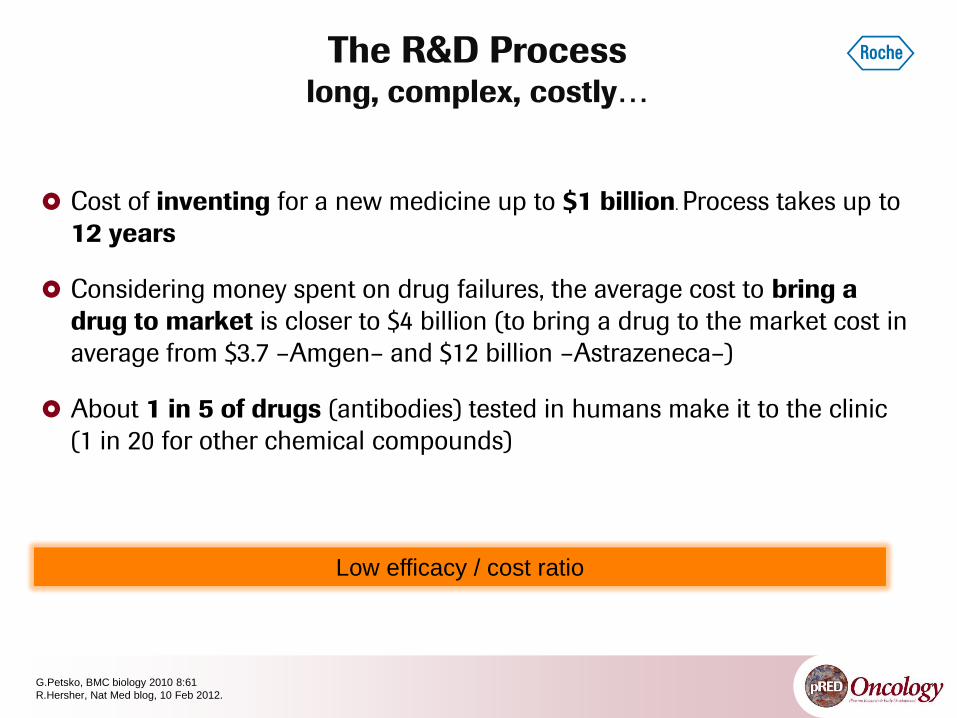
The R&D Process long, complex, costly…
Cost of inventing for a new medicine up to $1 billion. Process takes up to
12 years
Considering money spent on drug failures, the average cost to bring a
drug to market is closer to $4 billion (to bring a drug to the market cost in
average from $3.7 –Amgen– and $12 billion –Astrazeneca–)
About 1 in 5 of drugs (antibodies) tested in humans make it to the clinic
(1 in 20 for other chemical compounds)
G.Petsko, BMC biology 2010 8:61
R.Hersher, Nat Med blog, 10 Feb 2012.
Low efficacy / cost ratio
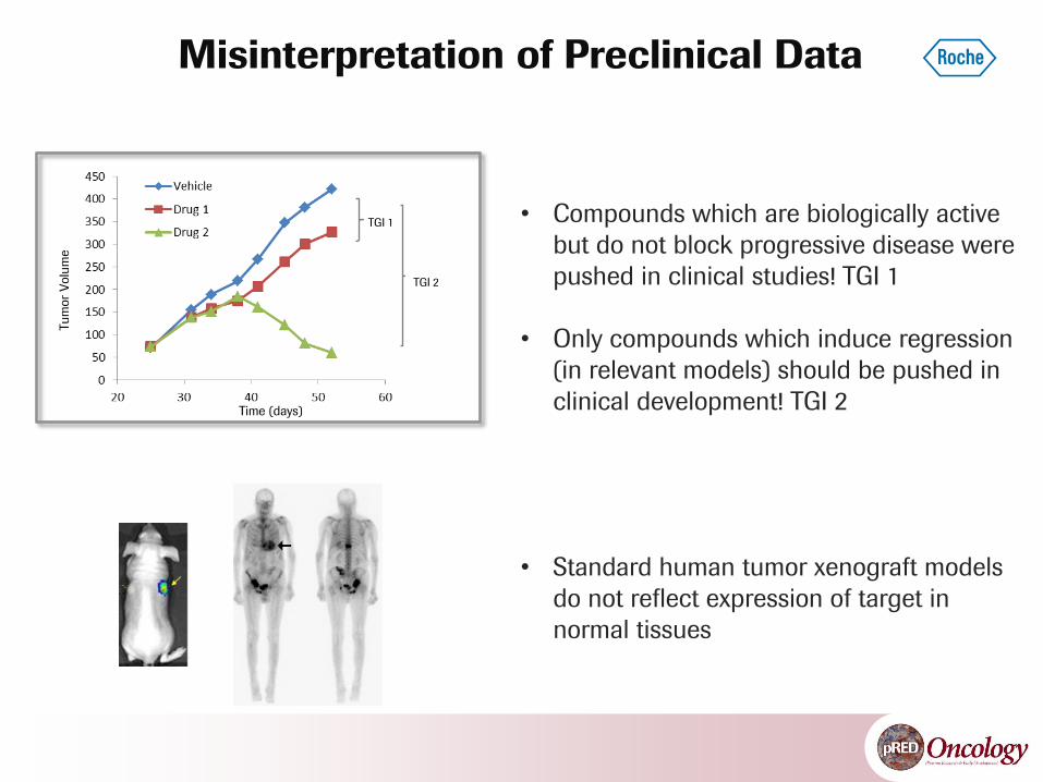
• Compounds which are biologically active
but do not block progressive disease were
pushed in clinical studies! TGI 1
• Only compounds which induce regression
(in relevant models) should be pushed in
clinical development! TGI 2
Tu
mor
Volu
me
Time (days)
TGI 1
TGI 2
Misinterpretation of Preclinical Data
• Standard human tumor xenograft models
do not reflect expression of target in
normal tissues

A Disease: Model vs Reality
Large number of cells
Homogeneous
Single site
Rapid growth
Restricted
Weeks
Before metastasis
Clonal form a single cells
Heterogeneous
Multiple sites
Slow growth
Diverse
Months
After metastasis
Tumor initiation
Cellular composition
Primary neoplasms
Growth rate
Genetic background
Therapy duration
Initiation of therapy
Requirements for a tumor model:
Similarity to human biology to ensure predictability for clinical trials
Major differences between standard mouse tumors and cancers in patients
Topics

6

Breast Cancer
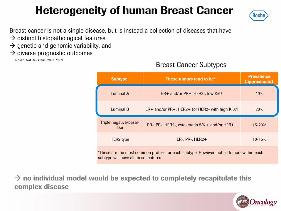
Heterogeneity of human Breast Cancer
Breast cancer is not a single disease, but is instead a collection of diseases that have
distinct histopathological features,
genetic and genomic variability, and
diverse prognostic outcomes
J.Rosen, Nat Rev Canc 2007 7:659
no individual model would be expected to completely recapitulate this
complex disease
Breast Cancer Subtypes
Subtype These tumors tend to be* Prevalence
(approximate)
Luminal A ER+ and/or PR+, HER2-, low Ki67 40%
Luminal B ER+ and/or PR+, HER2+ (or HER2- with high Ki67) 20%
Triple negative/basal-
like ER-, PR-, HER2-, cytokeratin 5/6 + and/or HER1+ 15-20%
HER2 type ER-, PR-, HER2+ 10-15%
*These are the most common profiles for each subtype. However, not all tumors within each
subtype will have all these features.
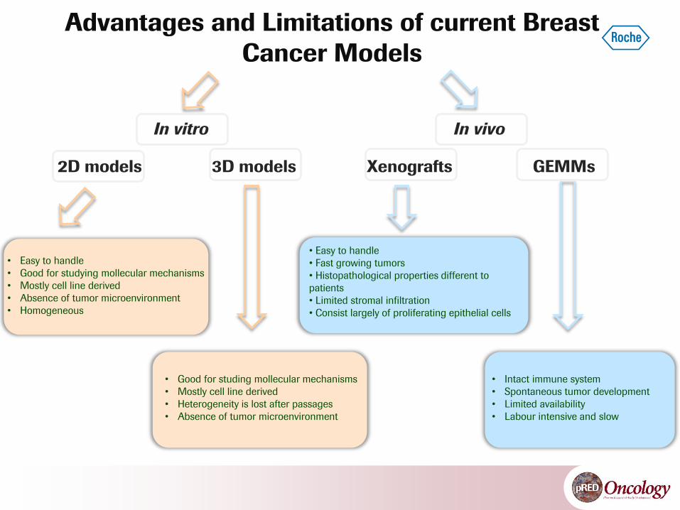
Advantages and Limitations of current Breast
Cancer Models
In vitro In vivo
2D models 3D models Xenografts GEMMs
• Easy to handle
• Good for studying mollecular mechanisms
• Mostly cell line derived
• Absence of tumor microenvironment
• Homogeneous
• Good for studing mollecular mechanisms
• Mostly cell line derived
• Heterogeneity is lost after passages
• Absence of tumor microenvironment
• Easy to handle
• Fast growing tumors
• Histopathological properties different to
patients
• Limited stromal infiltration
• Consist largely of proliferating epithelial cells
• Intact immune system
• Spontaneous tumor development
• Limited availability
• Labour intensive and slow
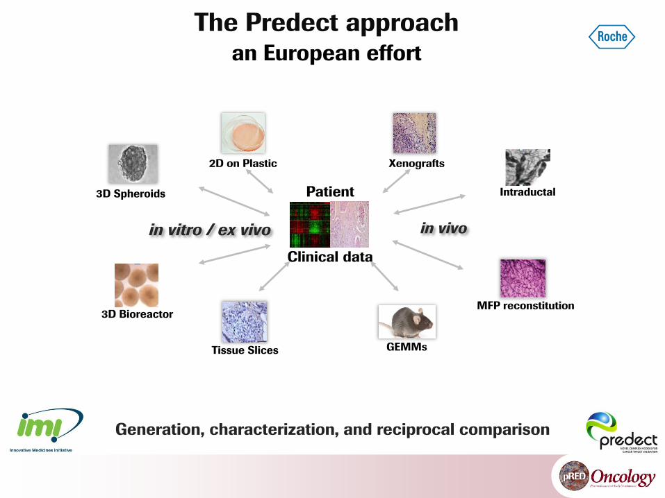
Generation, characterization, and reciprocal comparison
3D Spheroids
3D Bioreactor
Tissue Slices
2D on Plastic
Clinical data
in vitro / ex vivo in vivo
Xenografts
Intraductal
GEMMs
MFP reconstitution
Patient
The Predect approach an European effort
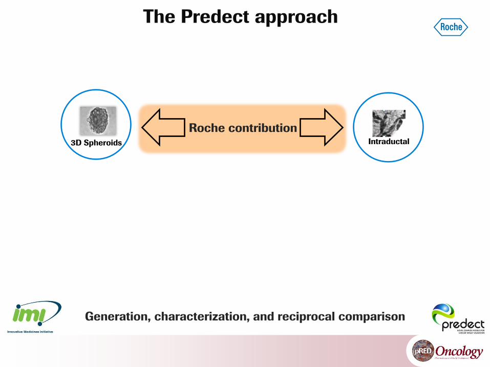
Generation, characterization, and reciprocal comparison
3D Spheroids Intraductal
The Predect approach
Roche contribution
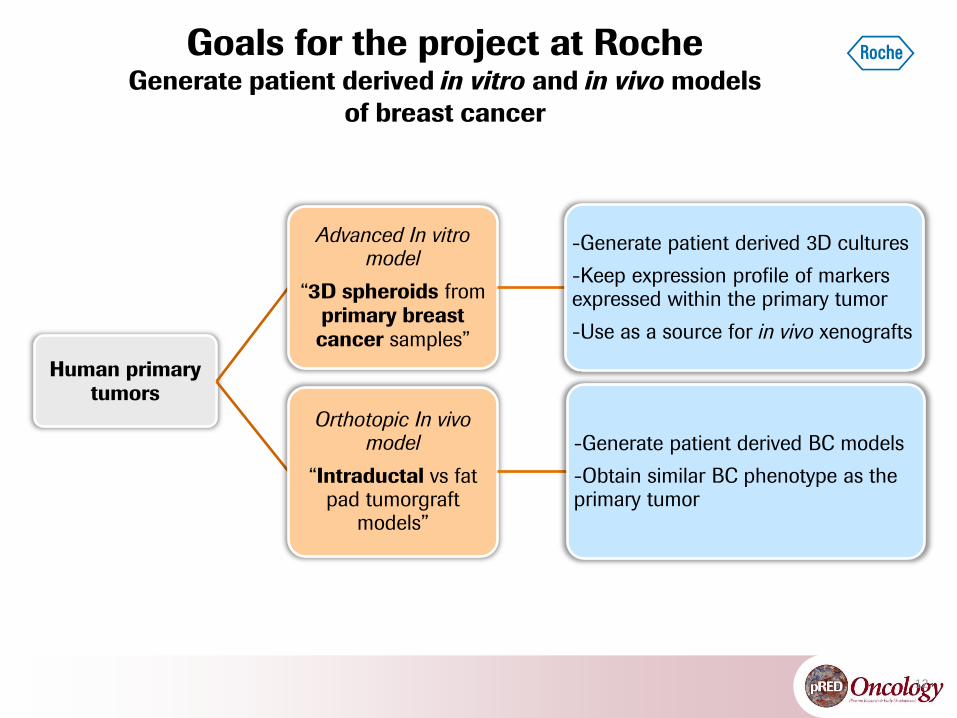
Goals for the project at Roche Generate patient derived in vitro and in vivo models
of breast cancer
12
Human primary tumors
Advanced In vitro model
“3D spheroids from primary breast cancer samples”
-Generate patient derived 3D cultures
-Keep expression profile of markers expressed within the primary tumor
-Use as a source for in vivo xenografts
Orthotopic In vivo model
“Intraductal vs fat pad tumorgraft
models”
-Generate patient derived BC models
-Obtain similar BC phenotype as the primary tumor
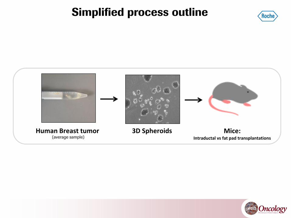
Simplified process outline
(average sample)
Human Breast tumor 3D Spheroids Mice: Intraductal vs fat pad transplantations
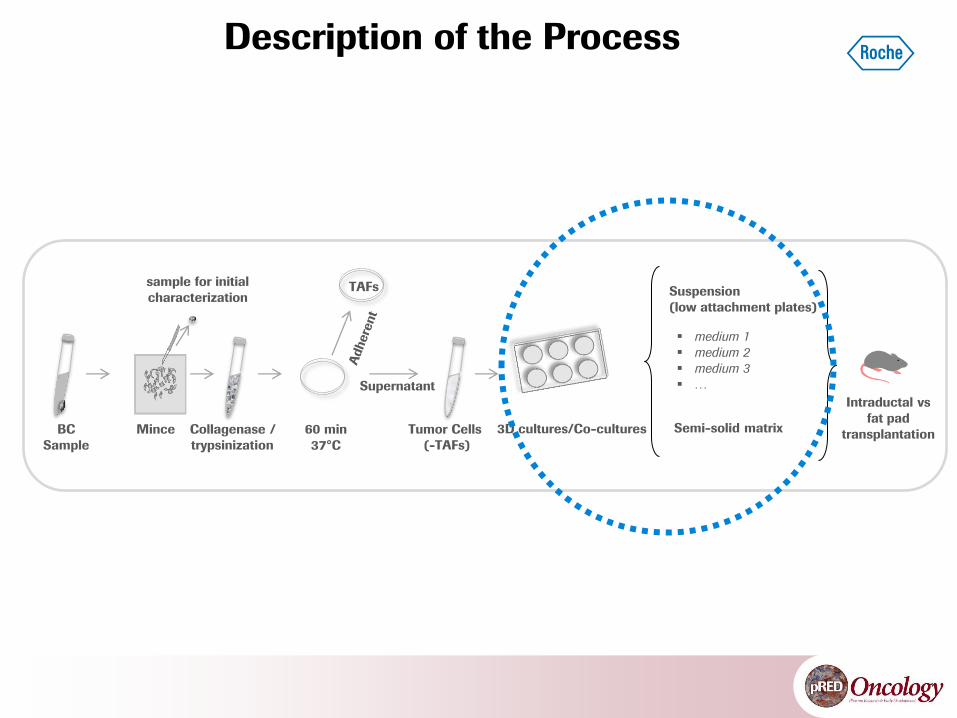
TAFs
Collagenase /
trypsinization
60 min
37°C
Mince Tumor Cells
(-TAFs)
sample for initial
characterization
BC
Sample
3D cultures/Co-cultures
Supernatant
Suspension
(low attachment plates)
medium 1
medium 2
medium 3
…
Semi-solid matrix
Description of the Process
Intraductal vs
fat pad
transplantation
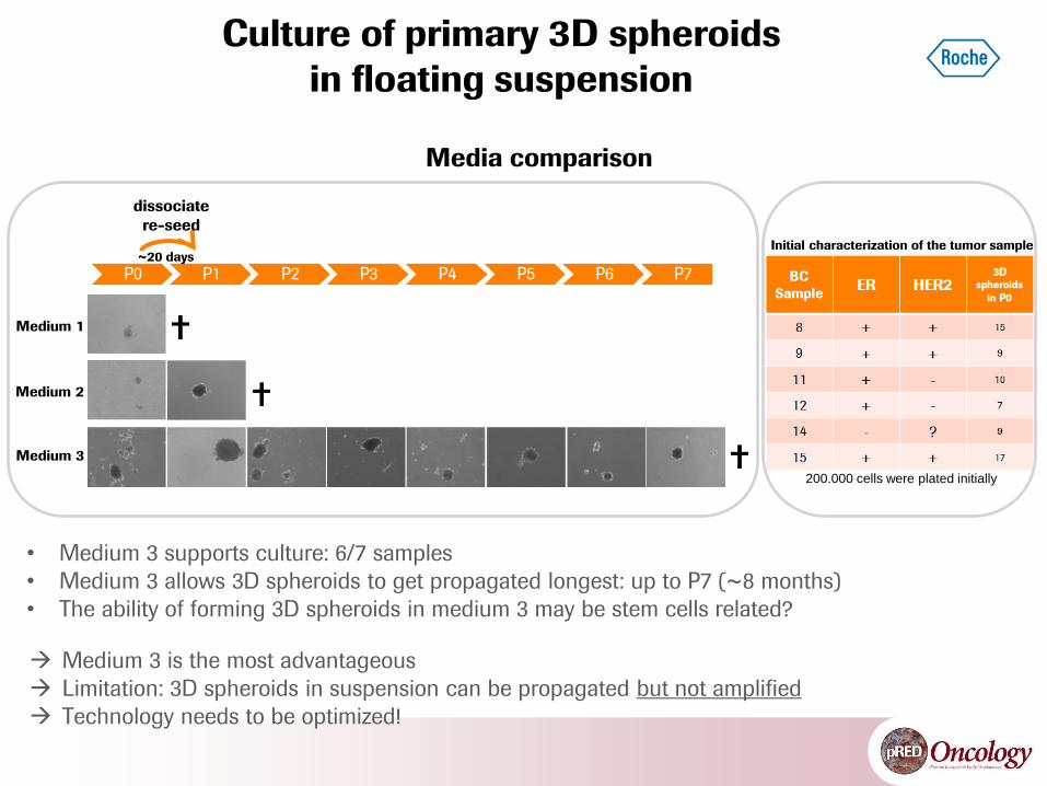
Culture of primary 3D spheroids
in floating suspension
• Medium 3 supports culture: 6/7 samples
• Medium 3 allows 3D spheroids to get propagated longest: up to P7 (~8 months)
• The ability of forming 3D spheroids in medium 3 may be stem cells related?
Medium 1
Medium 2
Medium 3
P0 P1 P2 P3 P4 P5 P6 P7
dissociate
re-seed
Initial characterization of the tumor sample
200.000 cells were plated initially
~20 days
Medium 3 is the most advantageous
Limitation: 3D spheroids in suspension can be propagated but not amplified
Technology needs to be optimized!
Media comparison
Matrix 2
Matrix 1
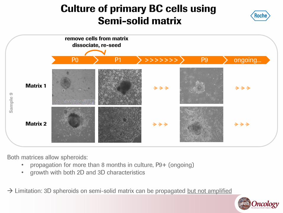
Culture of primary BC cells using
Semi-solid matrix S
am
ple
9
P0 P1 >>>>>>> P9 ongoing...
remove cells from matrix
dissociate, re-seed
Both matrices allow spheroids:
• propagation for more than 8 months in culture, P9+ (ongoing)
• growth with both 2D and 3D characteristics
Limitation: 3D spheroids on semi-solid matrix can be propagated but not amplified
17
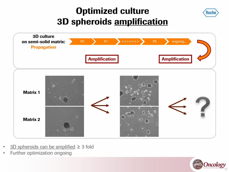
• 3D spheroids can be amplified ≥ 3 fold
• Further optimization ongoing
Optimized culture
3D spheroids amplification
3D culture
on semi-solid matrix:
Propagation
Amplification
Matrix 1
Matrix 2
P0 P1 >>>>>>> P9 ongoing...
Amplification
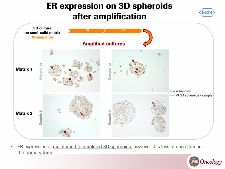
ER expression on 3D spheroids
after amplification
• ER expression is maintained in amplified 3D spheroids, however it is less intense than in
the primary tumor
Amplified cultures
Matrix 1
Matrix 2
3D culture
on semi-solid matrix
Propagation
P6 P7
Sa
mp
le 1
4
n = 4 samples
n=1-9 3D spheroids / sample
Sa
mp
le 1
2
Sa
mp
le 8
Sa
mp
le 9
19
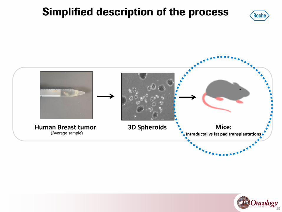
Simplified description of the process
(Average sample)
Human Breast tumor 3D Spheroids Mice: Intraductal vs fat pad transplantations
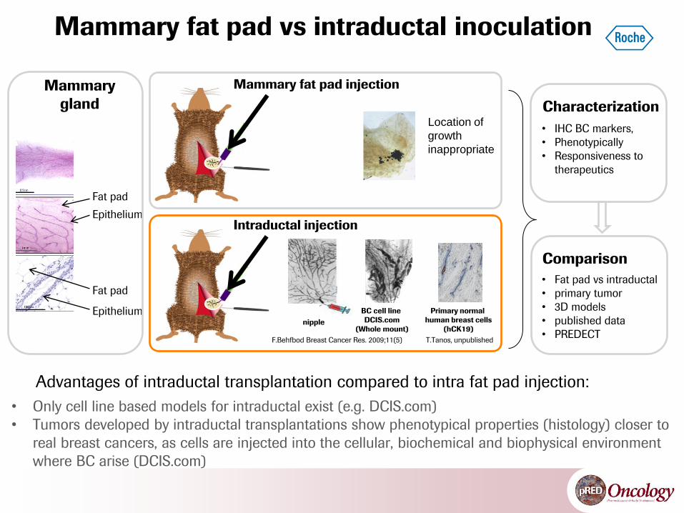
Mammary
gland
Fat pad
Epithelium
Fat pad
Epithelium
Mammary fat pad injection
Intraductal injection
Characterization
Primary normal
human breast cells
(hCK19)
BC cell line
DCIS.com
(Whole mount)
• IHC BC markers,
• Phenotypically
• Responsiveness to
therapeutics
F.Behfbod Breast Cancer Res. 2009;11(5) T.Tanos, unpublished
• Only cell line based models for intraductal exist (e.g. DCIS.com)
• Tumors developed by intraductal transplantations show phenotypical properties (histology) closer to
real breast cancers, as cells are injected into the cellular, biochemical and biophysical environment
where BC arise (DCIS.com)
Mammary fat pad vs intraductal inoculation
• Fat pad vs intraductal
• primary tumor
• 3D models
• published data
• PREDECT
Comparison
Via cleaved
nipple
Advantages of intraductal transplantation compared to intra fat pad injection:
Location of
growth
inappropriate
21
• Patient derived 3D spheroids can be kept in culture for several months
In both: suspension and semi-solid matrix
In semi-solid matrix they are maintained for 8+ months (ongoing)
• 3D spheroids can be amplified
Currently limited (~3 fold), optimization ongoing
• Maintenance of ER expression
Lost in classical culture systems
Retained in our culture system
Conclusions
22
• Optimize amplification for model use in vitro and in vivo
• Extend IHC charaterization, include: HER2, PR and ki67
• Assess the functionality of ER
• Analyze cellular composition, stem cells markers
• Study more samples, different passages
• Reciprocal comparison of model and tumor characteristics
Outlook
Wolfgang Eiermann
and Stefan Paepke
Friedrich Feuerhake
Elena Panzilius
Joachim Müller
John Hickman
The research leading to these results has received support from the Innovative Medicines Initiative Joint Undertaking under
grant agreement n° 115188, resources of which are composed of financial contribution from the European Union's Seventh
Framework Program (FP7/2007-2013) and EFPIA companies’ in kind contribution.
www.imi.europa.eu/
Tamara Tanos has been sponsored by the RPF (Roche Postdoc Fellowship) program

24