Embed Size (px)
Citation preview

Pathophysiology of asthmaPathophysiology of asthmaand and
chronic obstructive pulmonary chronic obstructive pulmonary diseasedisease
M. TatárM. Tatár

Thyroid cartilage
Cricothyroid ligament
Cricoid cartilage
Connective tissuesheath (cut away)
Intercartilaginousligaments
Mucosa showinglongitudual folds formedby dense collectionsof elastic fibres
Tracheal cartilages
Toupperlobe
Eparterialbronchus
Tomiddlelobe
Tolowerlobe
R. mainbronchus
L. mainbronchus
Intrapulmonary Extrapulmonary Intrapulmonary
Tolowerlobe
Tolingula
Toupperlobe
Trachealis muscle
Oesophageal muscle
Epithelium
Lymph vesselsElastic fibres
Gland
Small arteries
Nerve
Posterior wall
Cross sectionthrough trachea
Anterior wall
Epithelium
Nerve
Lymph vessels
Small artery
Gland
Elastic fibres
Cartilage
Connective tissue sheath
Structure of trachea and major bronchi
© Novartis
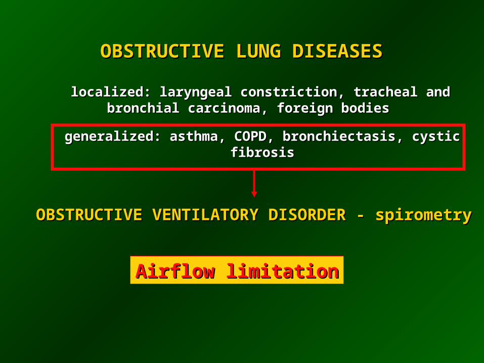
OBSTRUCTIVE LUNG DISEASES OBSTRUCTIVE LUNG DISEASES
localized: laryngeal constriction, tracheal and localized: laryngeal constriction, tracheal and bronchial carcinoma, foreign bodiesbronchial carcinoma, foreign bodies
generalized: asthma, COPD, bronchiectasis, cystic generalized: asthma, COPD, bronchiectasis, cystic
fibrosisfibrosis
OBSTRUCTIVE VENTILATORY DISORDER - spirometryOBSTRUCTIVE VENTILATORY DISORDER - spirometry
Airflow limitationAirflow limitation

Cartilage
Segmentalbronchus
Largesubsegmentalbronchi(about 5generations)
Small bronchi(about 15generations)
Terminalbronchioles
Respiratorybronchioles(3 orders)
Alveolarducts andalveolar sacs
Lobule
Acinus
Acinus
Poresof Kohn
Alveolar sacsand alveoli
Alveolar ducts
3rd order
2nd order
1st order
Respiratorybronchioles
Alveolus
Elastic fibres
Smooth muscle
Terminal bronchiole
Subdivisions and structure ofintrapulmonary airways
© Novartis

0.5
End of quiet expirationEnd of quiet expiration
- 0.5- 0.5
00 0 0 0 0
InspirationInspiration
- 2.5- 2.5

0.5
- 2.5- 2.5
0- 1.0 - 0.5- 1.5- 2.0- 2.0
InspirationInspiration
ForcedForcedexpirationexpiration
+ 2.0+ 2.0

0.5
Forced expirationForced expiration
+ 2.0+ 2.0
+ 2.5+ 2.5 0+ 2.0 + 1.5 + 1.0
EPPEPP

ASTHMA - definitionASTHMA - definition
Chronic inflammatory disorder of the airwaysChronic inflammatory disorder of the airways
Mast cells, eosinophils, T-lymphocytes
Recurrent episodes of wheezing, dyspnoea, Recurrent episodes of wheezing, dyspnoea, and cough particularly at night and early and cough particularly at night and early
morningmorning
Symptoms are associated with airflow Symptoms are associated with airflow limitation that is partly reversible either limitation that is partly reversible either
spontaneously or with therapyspontaneously or with therapy
Bronchial hyperresponsiveness is present very often
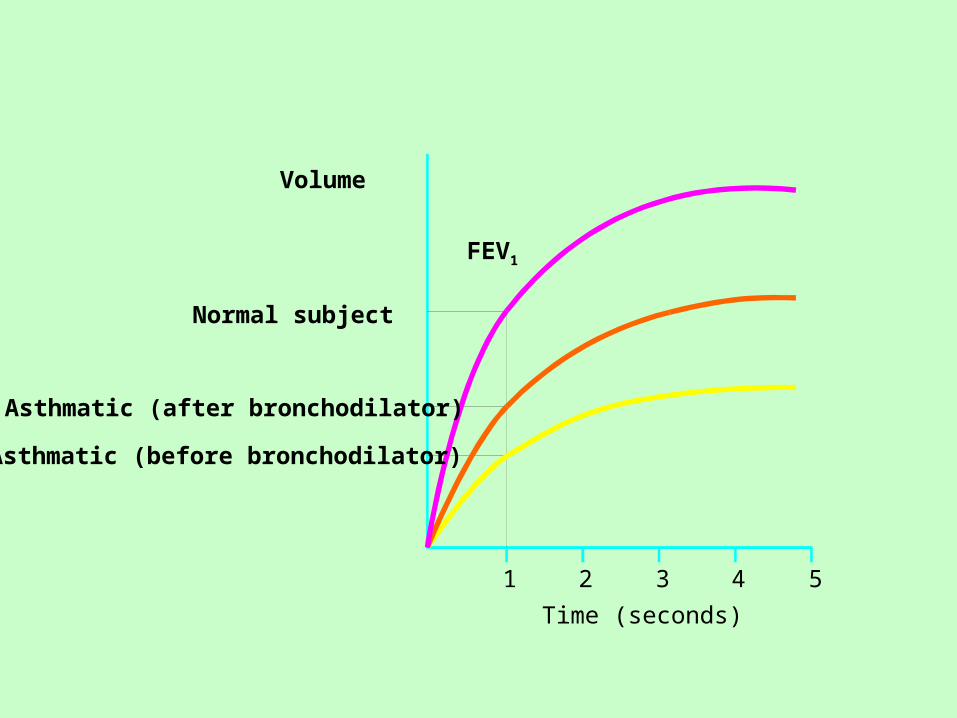
1 2 3 4 5
Time (seconds)
FEV1
Volume
Normal subject
Asthmatic (after bronchodilator)
Asthmatic (before bronchodilator)
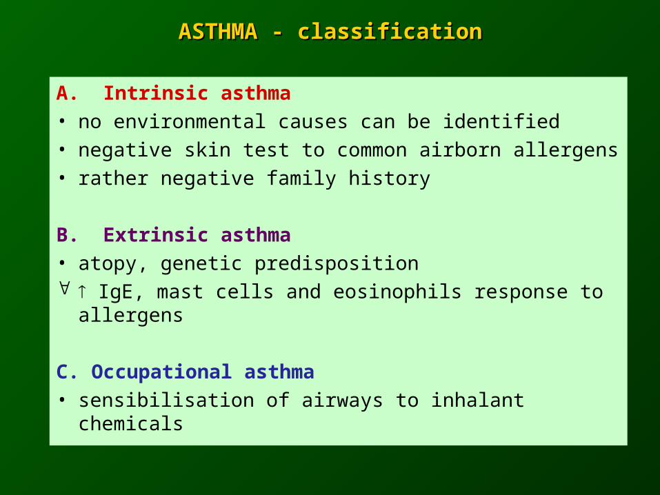
ASTHMA - classificationASTHMA - classification
A. Intrinsic asthma• no environmental causes can be identified• negative skin test to common airborn allergens• rather negative family history
B. Extrinsic asthma• atopy, genetic predisposition IgE, mast cells and eosinophils response to
allergens
C. Occupational asthma• sensibilisation of airways to inhalant chemicals
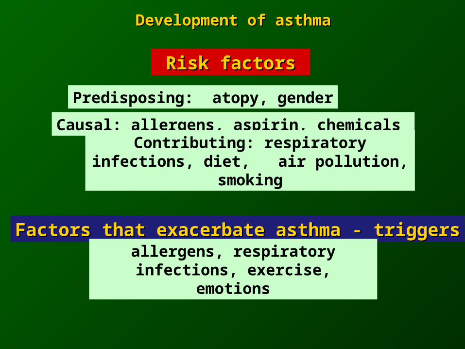
Development of asthmaDevelopment of asthma
Risk factorsRisk factors
Predisposing: atopy, gender
Causal: allergens, aspirin, chemicals
Contributing: respiratory infections, diet, air pollution, smoking
Factors that exacerbate asthma - triggersFactors that exacerbate asthma - triggers
allergens, respiratory infections, exercise, emotions
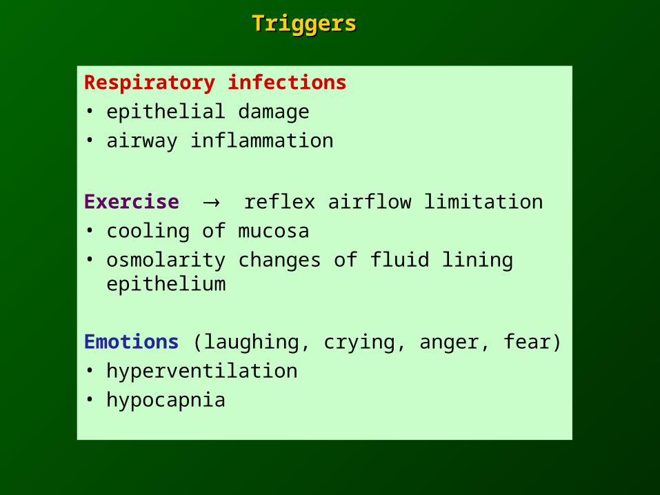
Respiratory infections• epithelial damage• airway inflammation
Exercise reflex airflow limitation• cooling of mucosa• osmolarity changes of fluid lining
epithelium
Emotions (laughing, crying, anger, fear)• hyperventilation• hypocapnia
Triggers Triggers

Asthma - bronchial hyperresponsivenessAsthma - bronchial hyperresponsiveness
Instability of the airways =Instability of the airways =
exaggerated bronchoconstrictor exaggerated bronchoconstrictor response to a wide variety of response to a wide variety of
stimulistimuli
Key factor - airway inflammation
Mechanisms: direct and indirect

Direct agonistsDirect agonists e.g. methacholine
Airway with limited airflow
Mediators
Nerve
SO2, bradykinin
Indirect agonistsIndirect agonists e.g. exercise, adenosine, hypotonic
or hypertonic aerosols
Mast cell
Airway hyperresponsivenessAirway hyperresponsiveness

balancebalance
antihyperreactiv factorsantihyperreactiv factors prohyperreactiv factorsprohyperreactiv factors
Normal airway reactivityNormal airway reactivity
2-adrenergic
VIP/PHM
anticholinergic
NEP
antioxidants
corticoids
-adrenergic
cholinergic
SP/NK
oxygen-free radicals
peptidases
Airway hyperresponsivenessAirway hyperresponsiveness
imim
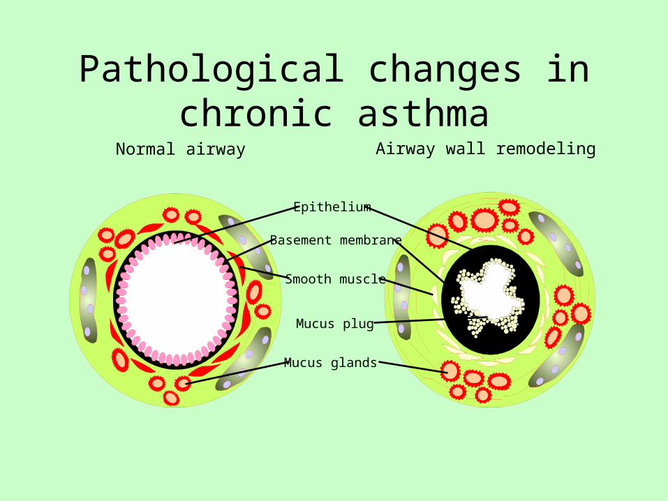
Pathological changes in chronic asthma
Normal airway Airway wall remodeling
Epithelium
Basement membrane
Smooth muscle
Mucus plug
Mucus glands

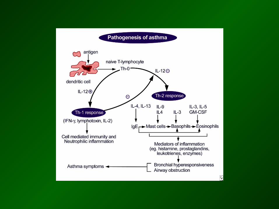
Mechanisms of asthmaMechanisms of asthma
1. Airway inflammation1. Airway inflammation
- recruitments of inflammatory cells from circulation
- endothelial adhesion molecules
- activation of T lymphocytes (Th2 clone)
- production of IgE, leukotriens, prostanoids
- cytokines (CD4+ Th subtype)
2. Neural control of airways
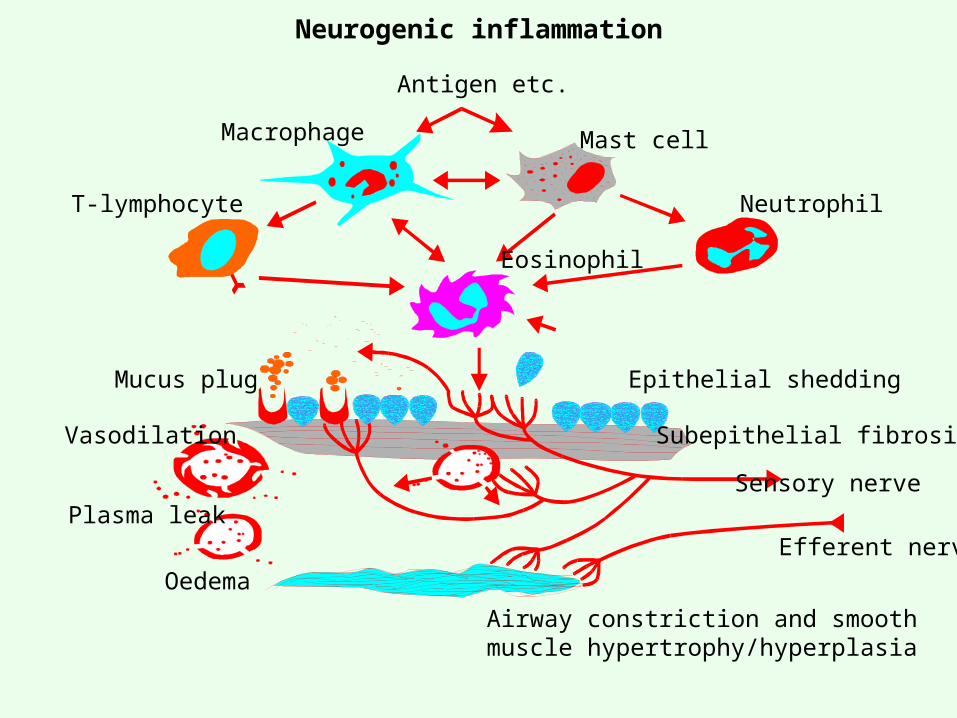
Antigen etc.
Macrophage
T-lymphocyte Neutrophil
Mast cell
Eosinophil
Mucus plug
Vasodilation
Plasma leak
Oedema
Epithelial shedding
Subepithelial fibrosis
Sensory nerve
Efferent nerve
Airway constriction and smooth muscle hypertrophy/hyperplasia
Neurogenic inflammation
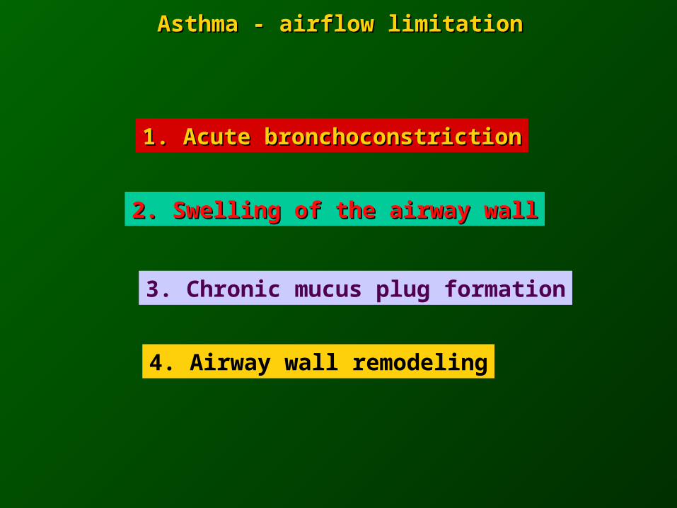
Asthma - airflow limitationAsthma - airflow limitation
1. Acute bronchoconstriction1. Acute bronchoconstriction
2. Swelling of the airway wall2. Swelling of the airway wall
3. Chronic mucus plug formation
4. Airway wall remodeling

Relaxation Constriction
Normal
Asthma
Airway narrowing
Exaggeratedairway
narrowing
R = 1R = 1 R = 10R = 10
R = 2R = 2 R = 40R = 40
muscle constriction
35 %
muscle constriction
35 %

INFLAMMATIONINFLAMMATION
Risk factors(for development of asthma)
Airwayhyperresponsiveness Airflow limitation
Risk factors(for exacerbations)
Symptoms

Asthma is a highly variable diseaseAsthma is a highly variable disease
Asthma is a chronic inflammatory disease of variable Asthma is a chronic inflammatory disease of variable severity. Worsening and exacerbations of asthma are severity. Worsening and exacerbations of asthma are associated with episodes of acute inflammation, associated with episodes of acute inflammation, which develop on top of persistent underlying which develop on top of persistent underlying chronic inflammation.chronic inflammation.
This acute inflammation causes an increase in This acute inflammation causes an increase in symptoms and may also lead to an increased symptoms and may also lead to an increased sensitivity to triggers and a worsening in airway sensitivity to triggers and a worsening in airway hyperresponsiveness.hyperresponsiveness.
The variability and severity of „real life“ asthma is The variability and severity of „real life“ asthma is dependent on a number of factors, including a dependent on a number of factors, including a patient´s adherence to the prescribed treatment.patient´s adherence to the prescribed treatment.


COPD - definitionCOPD - definition
Chronic airflow limitation Chronic airflow limitation ( ( maximum expiratory flow, slow maximum expiratory flow, slow
forcedforcedemptying of the lungs) emptying of the lungs)
Airflow limitation is slowly progressive
and irreversible
Due to varying combinations of:• airway disease• emphysema

COPDCOPD
Chronic bronchitisChronic bronchitis
• defined in clinical terms
• chronic cough with sputum production
- (3 months a year, 2 successive years)
- excluded cardiac or other pulmonary causes
EmphysemaEmphysema
• defined anatomically• permanent,
destructive enlagrement of airspaces distal to the terminal bronchioles without obvious fibrosis

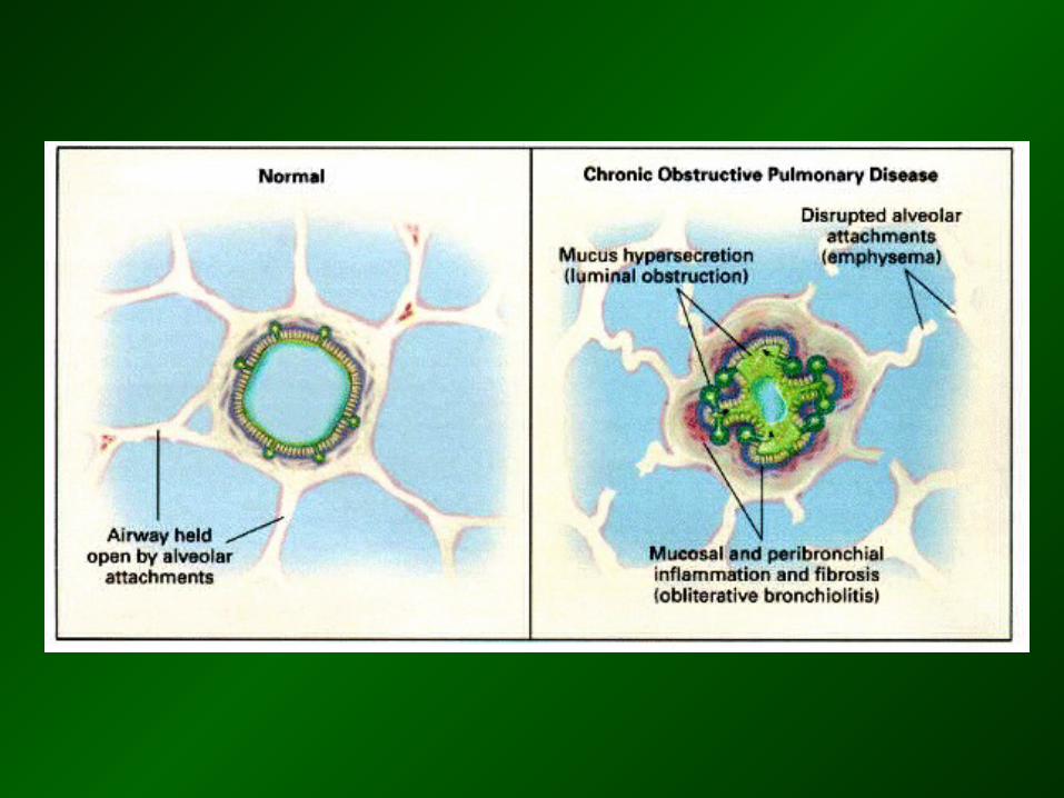
Chronic obstructive pulmonary disease
Interrelationship of chronic bronchitis and emphysema
Normal
Chronicbronchitis
Mixed(in variable
degree)
Emphysema
© Novartis
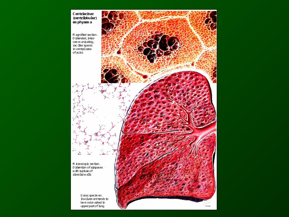
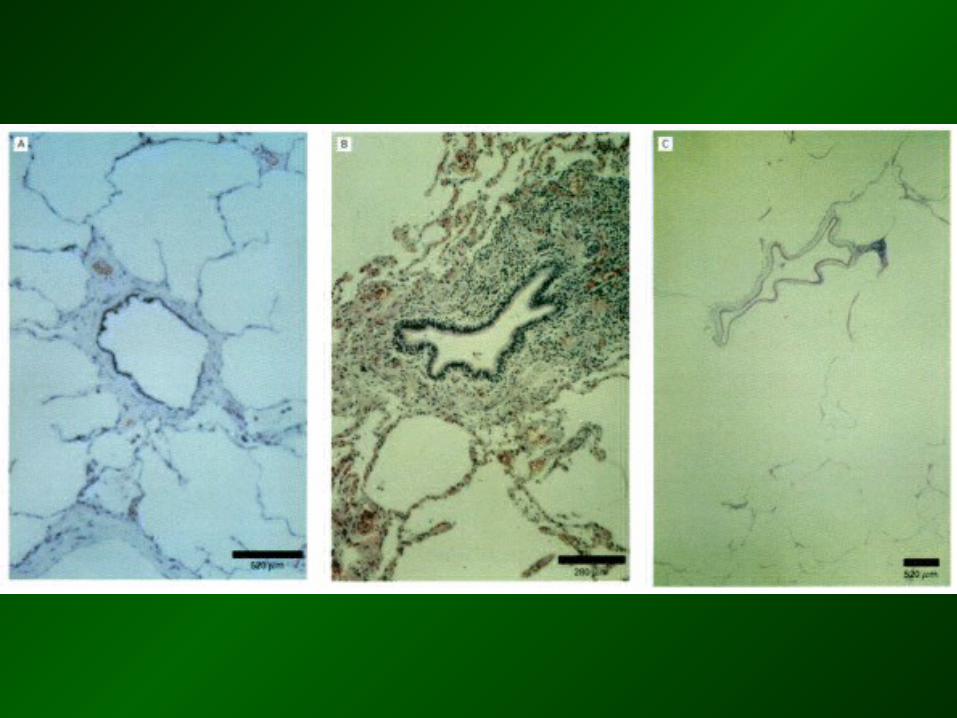
Centriacinar(centrilobular)emphysema
Magnified section.Distended, inter-communicating,sac-like spacesin central areaof acini
Microscopic section.Distention of airspaceswith rupture ofalveolar walls
Gross specimen.Involvement tends tobe most marked inupper part of lung © Novartis
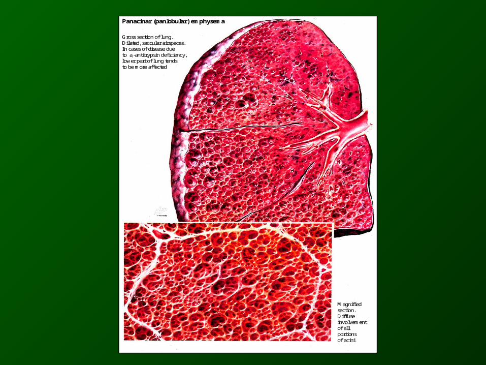
Panacinar (panlobular) emphysema
Gross section of lung.Dilated, saccular airspaces.In cases of disease dueto a1-antitrypsin deficiency,lower part of lung tendsto be more affected
Magnifiedsection.Diffuseinvolvementof allportionsof acini
© Novartis

COPD - risk factorsCOPD - risk factors
Cigarette Cigarette smoking smoking
11 - antitrypsin deficiency - antitrypsin deficiency
Solid fuel used for indoor heating or cooking without adequate
ventilation
Heavily polluted environments
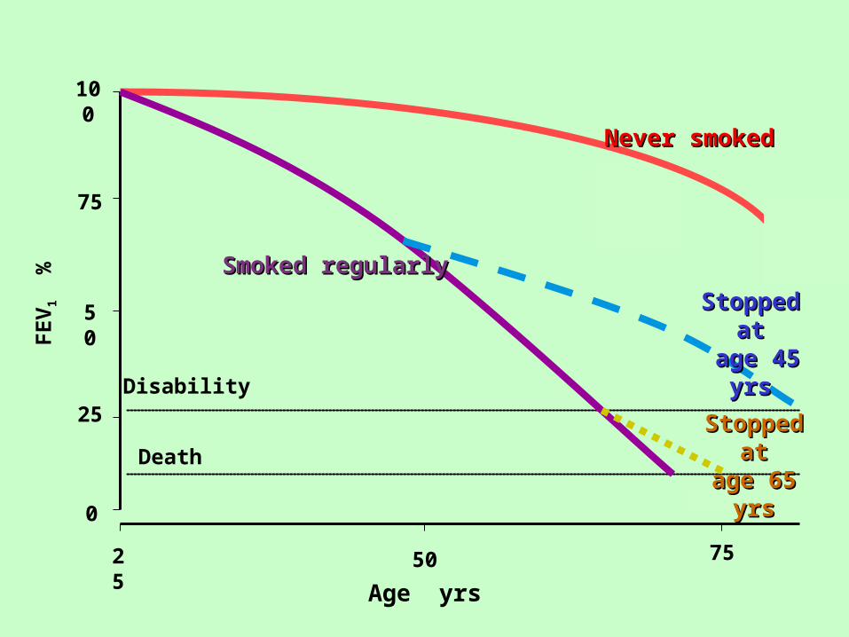
100
75
50
25
0
25 50 75
Age yrs
FE
V1
%
Disability
Death
Never smokedNever smoked
Stopped atStopped at age 45 yrsage 45 yrs
Stopped atStopped atage 65 yrsage 65 yrs
Smoked regularlySmoked regularly

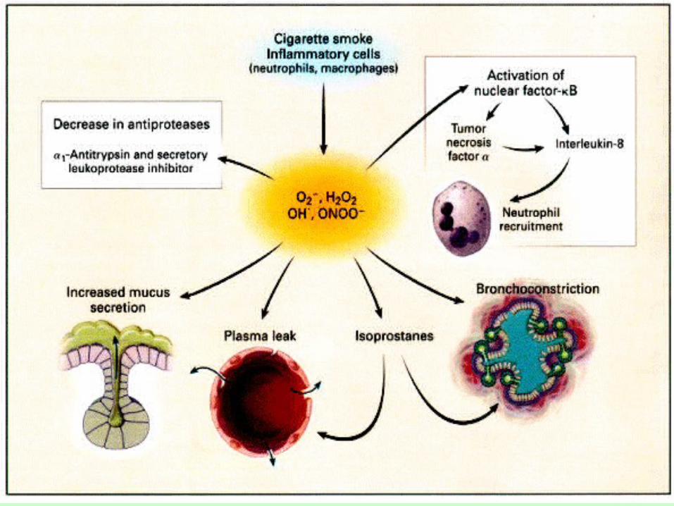
COPD - cellular and biochemical mechanismsCOPD - cellular and biochemical mechanisms
Inflammation: alveolar macrophages, neutrophilsInflammation: alveolar macrophages, neutrophils
Neutrophil and macrophage enzymes and oxidants
destroy components of extracellular matrix (collagen, elastin, fibronectine, proteoglycans)
Loss of cellular components of lung parenchyma:- elastase can induce apoptosis
- cells exposed to oxidants may undergo apoptosis or necrosis
oxidative stress in smokers and in COPD patients
production of elastase, cathepsine G, collagenase
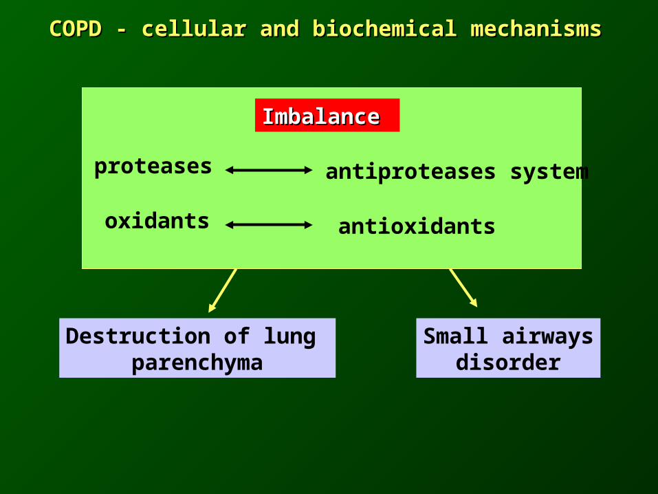
COPD - cellular and biochemical mechanismsCOPD - cellular and biochemical mechanisms
Destruction of lung parenchyma
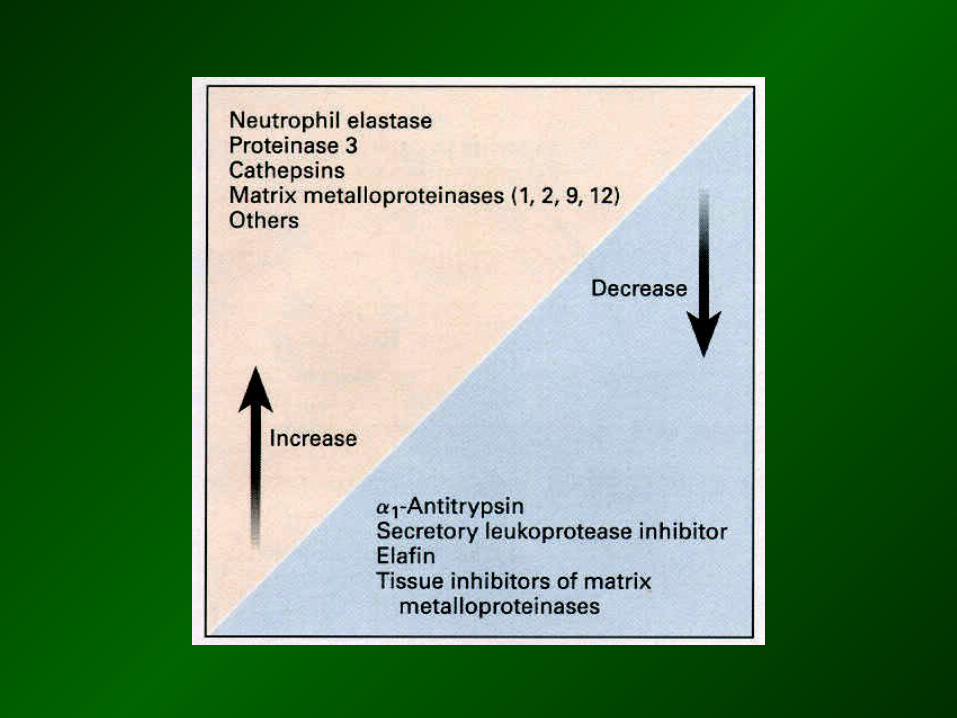
Imbalance Imbalance
proteases antiproteases system
oxidants antioxidants
Small airwaysdisorder

COPD - pathology of peripheral COPD - pathology of peripheral airwaysairways
• mucus plugging• goblet cell metaplasia• fibrosis• smooth muscle hypertrophy
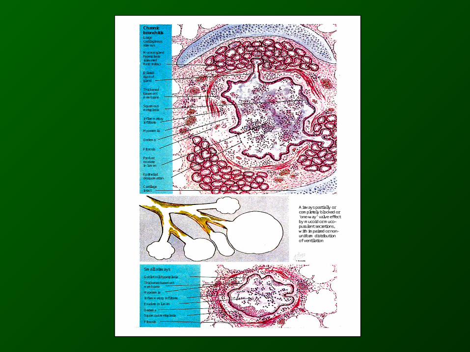
ChronicbronchitisLargecartilaginousairways
Mucous glandhyperplasia(elevatedReid index)
Dilatedduct ofgland
Thickenedbasementmembrane
Squamousmetaplasia
Inflammatoryinfiltrate
Hyperemia
Oedema
Fibrosis
Profuseexudatein lumen
Epithelialdesquamation
Cartilageintact
Small airways
Goblet cell hyperplasia
Thickened basementmembrane
Hyperemia
Inflammatory infiltrate
Exudate in lumen
Oedema
Squamous metaplasia
Fibrosis
Airways partially orcompletely blocked or‘one-way’ valve effectby mucoid or muco-purulent secretions,with impaired or non-uniform distributionof ventilation
© Novartis
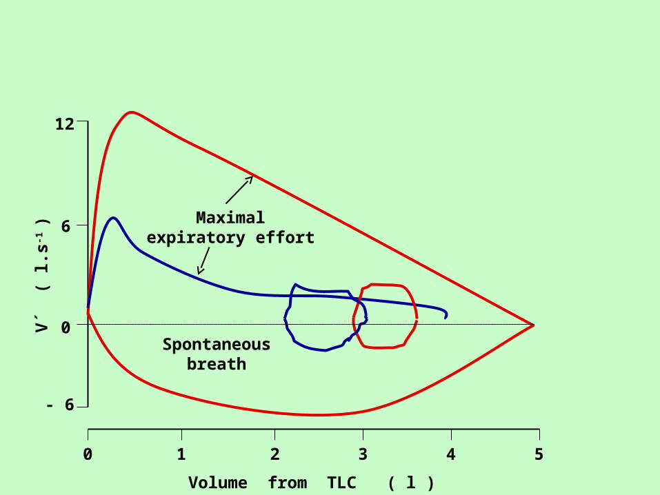
10 2 3 4 5
Volume from TLC ( l )
- 6
0
6
12
V´
( l.
s-1 )
Maximalexpiratory effort
Spontaneousbreath

0 20 40
40
60
80
100
Lu
ng v
olu
me
(%
TL
C)
Oxygen consumption (ml.min-1.kg-1)
Normals
VT
IRV
VT
IRV
Airflow limit

Relatively normal lung region, normal PAO2
Emphysema
Pulm. vein
Pulm. artery Normal CaO2
´
CaO2
Airway narrowing
Emphysematous region PAO2
Destruction of capillary
V´ V´
Q´ Q´
Relatively normal CaO2

Relatively normal lung region, normal PAO2
normalCaO2
Airway narrowing
Pulm. v.Pulm. a.
Bronchitis
PAO2
CaO2
V´norm
V´
norm Q´ norm Q´
CaO2

04
6
10
0,5 1,0 1,5 2,0 2,5 3,0 FEV1 ( l )
PaC
O2 (
kP
a )
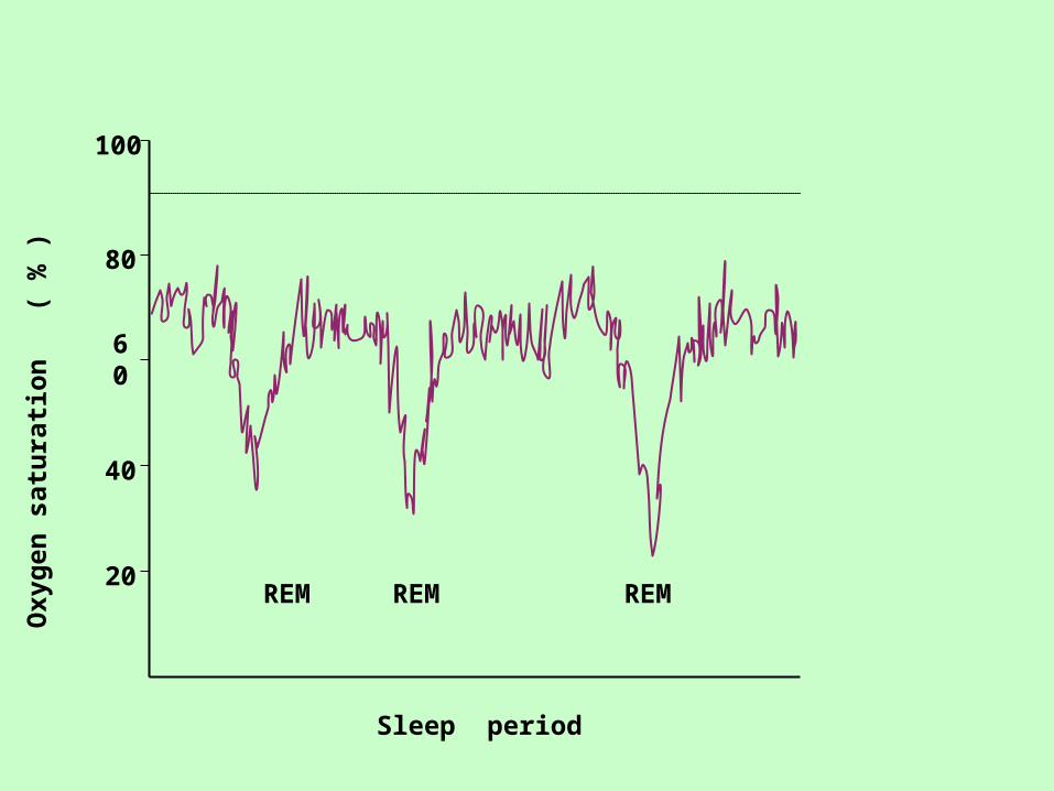
Sleep period
20
40
60
80
100
Oxy
gen
sat
ura
tion
(
% )
REM REM REM

Components of chronic obstructive pulmonary disease
Airflow limitationby spirometry
Chronic bronchitisEmphysema
Asthma
Simple bronchitis
Asthma with no airflow limitation
Emphysema but no COPD